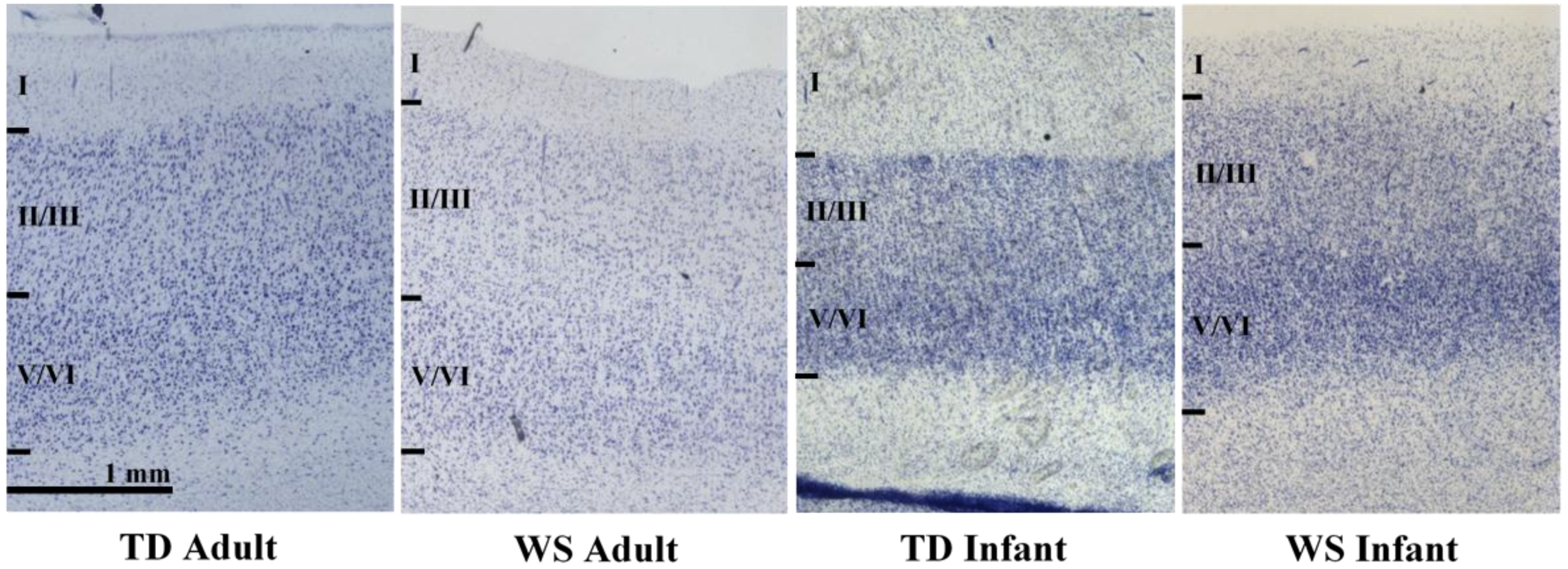
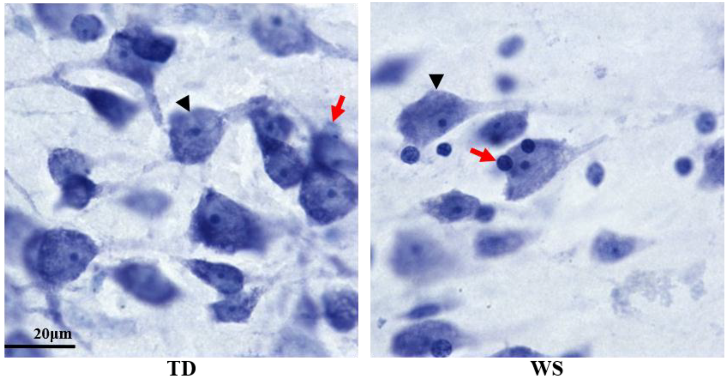
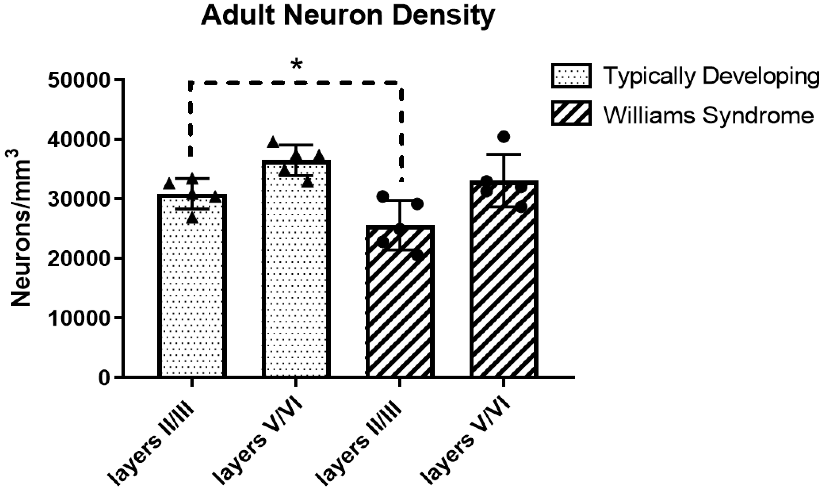
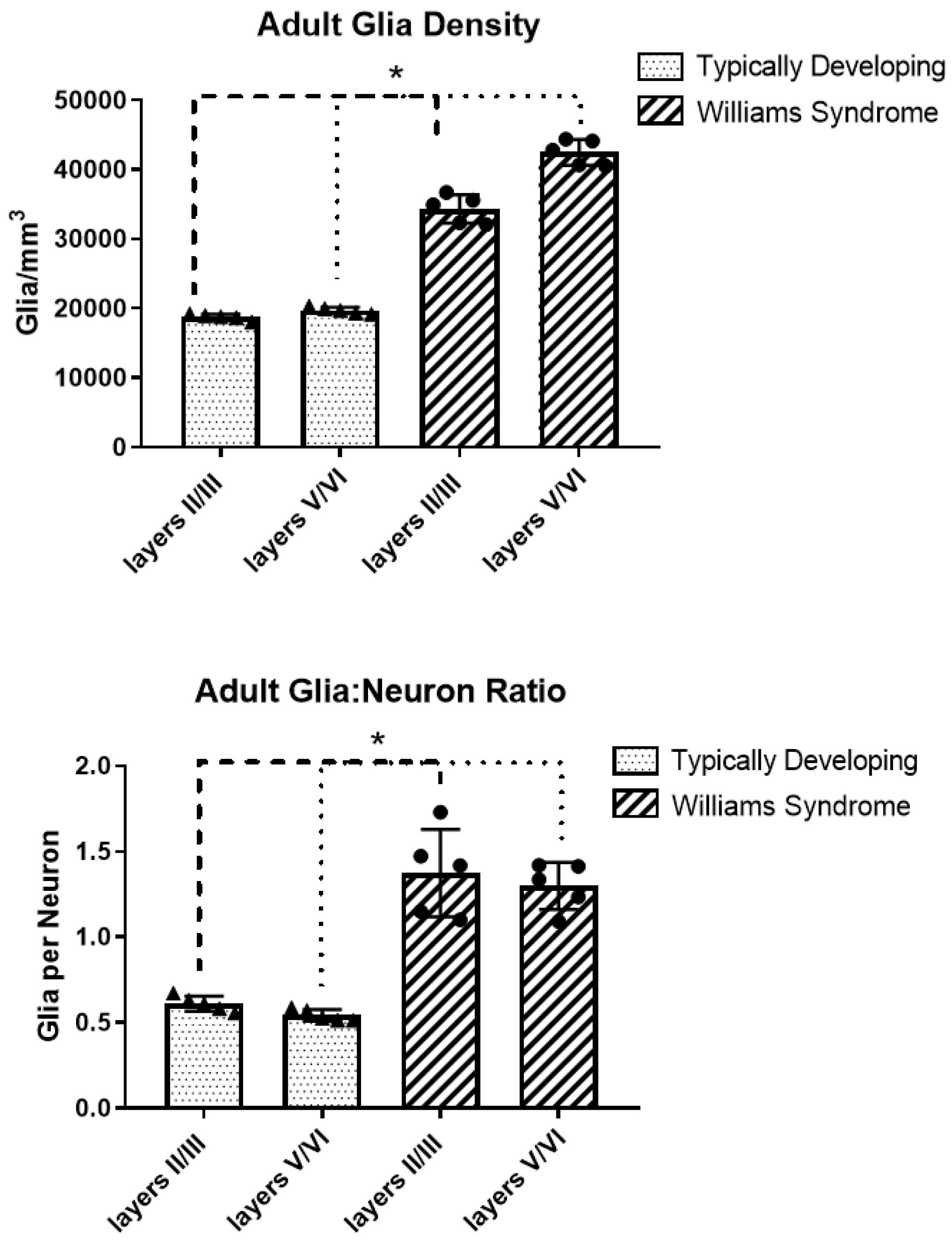
Decreased Neuron Density and Increased Glia Density in the Ventromedial Prefrontal Cortex (Brodmann Area 25) in Williams Syndrome

Abstract
Share and Cite
Wilder, L.; Hanson, K.L.; Lew, C.H.; Bellugi, U.; Semendeferi, K. Decreased Neuron Density and Increased Glia Density in the Ventromedial Prefrontal Cortex (Brodmann Area 25) in Williams Syndrome. Brain Sci. 2018, 8, 209. https://doi.org/10.3390/brainsci8120209
Wilder L, Hanson KL, Lew CH, Bellugi U, Semendeferi K. Decreased Neuron Density and Increased Glia Density in the Ventromedial Prefrontal Cortex (Brodmann Area 25) in Williams Syndrome. Brain Sciences. 2018; 8(12):209. https://doi.org/10.3390/brainsci8120209
Chicago/Turabian StyleWilder, Linnea, Kari L. Hanson, Caroline H. Lew, Ursula Bellugi, and Katerina Semendeferi. 2018. "Decreased Neuron Density and Increased Glia Density in the Ventromedial Prefrontal Cortex (Brodmann Area 25) in Williams Syndrome" Brain Sciences 8, no. 12: 209. https://doi.org/10.3390/brainsci8120209
APA StyleWilder, L., Hanson, K. L., Lew, C. H., Bellugi, U., & Semendeferi, K. (2018). Decreased Neuron Density and Increased Glia Density in the Ventromedial Prefrontal Cortex (Brodmann Area 25) in Williams Syndrome. Brain Sciences, 8(12), 209. https://doi.org/10.3390/brainsci8120209