Mechanisms and Therapeutic Targets of Ischemia—Reperfusion Injury in Stroke: A Narrative Review Focusing on Blood—Brain Barrier Dysfunction

Highlights
- BBB disruption in stroke I/R injury follows a distinct spatiotemporal hierarchy.
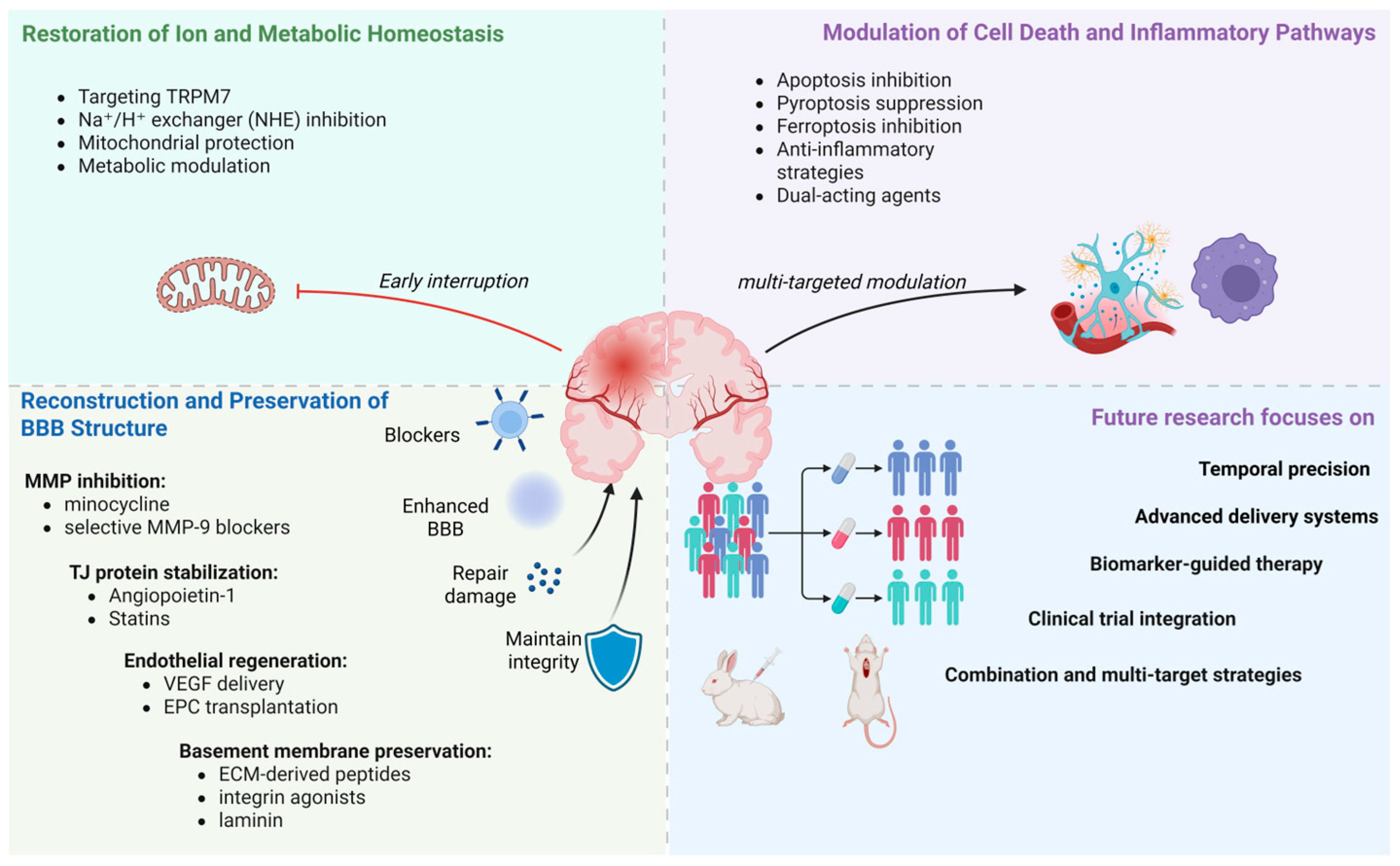
- A comprehensive treatment framework covering three major strategies has been proposed.
- Therapeutic strategies must be phase-specific and time-limited rather than continuously blocking a single pathway.
- This explains why many intervention measures that have shown efficacy in animal models have failed in clinical trials.
Abstract
1. Introduction
1.1. Background
1.2. Rationale for BBB-Focused Intervention
1.3. Literature Search Strategy
1.4. What Is New in This Review?
2. Structure and Function of the BBB
3. The Mechanism of I/R on BBB
3.1. Early Injury Stage
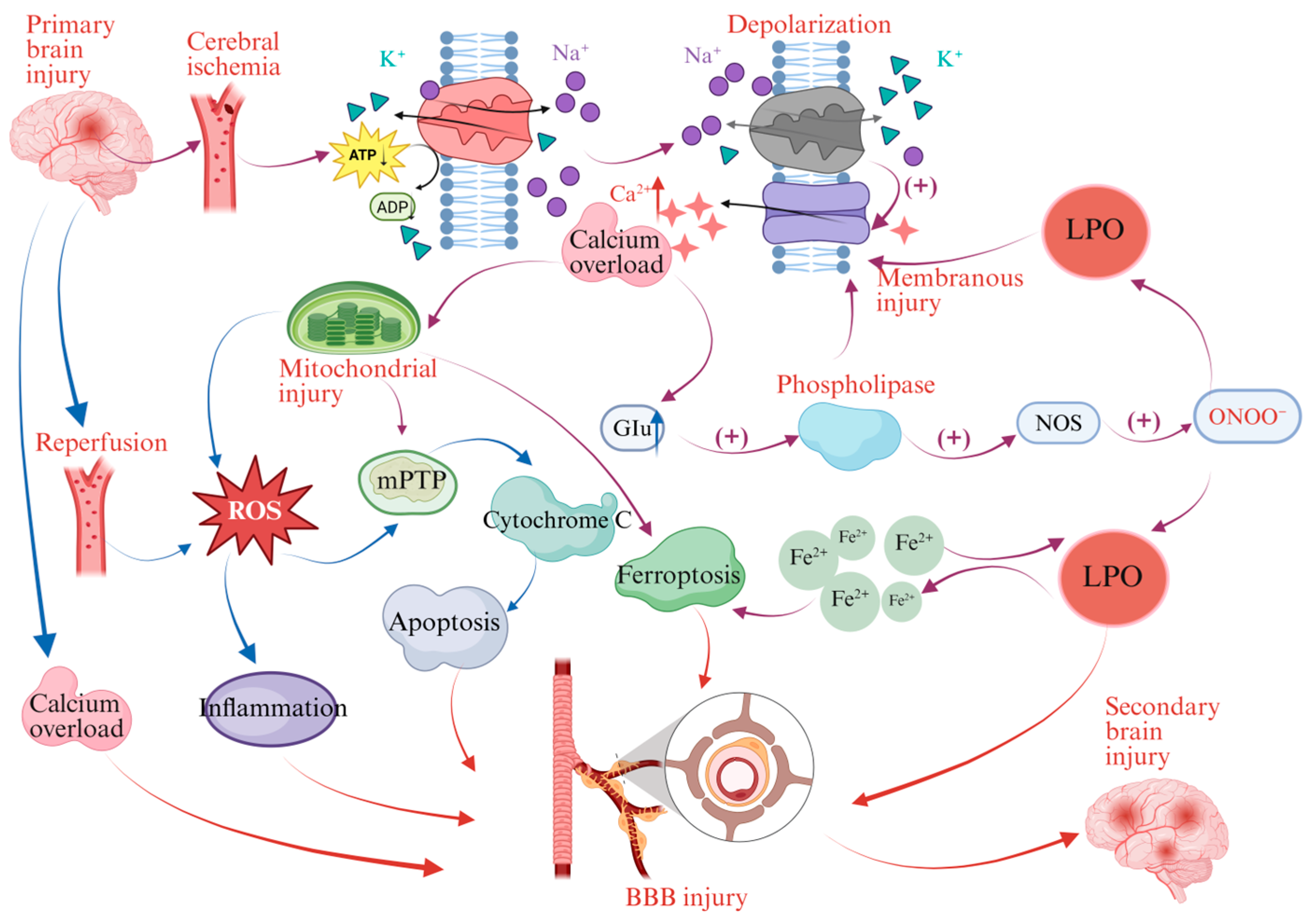
3.1.1. Energy Depletion, Calcium Overload and Initial Injury
- Activation of Ca2+-dependent enzymes (phospholipases, proteases, nitric oxide synthase);
- Calpain-mediated degradation of cytoskeletal and pro-apoptotic proteins;
- Mitochondrial permeability transition pore (mPTP) opening;
- Cytochrome C release and ATP depletion;
- Excess ROS production and nuclear factor kappaB (NF-κB)-driven neuroinflammation.
3.1.2. Oxidative Stress Burst and ROS-Mediated Injury
3.1.3. Inflammatory Response and Immune Amplification
3.2. Matrix Degradation Stage
3.3. Programmed Cell Death Stage
3.3.1. Apoptosis
3.3.2. Pyroptosis
3.3.3. Ferroptosis
4. Therapeutic Strategies for Protecting the BBB in CIRI
4.1. Restoration of Ion and Metabolic Homeostasis
4.1.1. Regulation of Ion Channels/Exchangers
4.1.2. Mitochondrial Protection
4.1.3. Metabolic Regulation
4.2. Immune Regulation and Oxidative Stress Control
4.2.1. Dual-Acting Agents
4.2.2. Anti-Inflammatory Strategies
4.3. Inhibition of Excessive Proteolysis and Matrix Remodeling
4.3.1. MMP Inhibition
4.3.2. Endothelial Cell Regeneration and Basement Membrane Protection
4.4. Inhibition of the Programmed Cell Death Pathway
4.5. Non-Pharmacological Interventions
4.6. Summary of Intervention Measures for CIRI-Related BBB Protection in Clinical Trials
5. Conclusions and Future Directions
Author Contributions
Funding
Data Availability Statement
Conflicts of Interest
Abbreviations
| CIRI | Cerebral Ischemia–Reperfusion Injury |
| MCAO | Middle Cerebral Artery Occlusion |
| BMEC | Brain Microvascular Endothelial Cells |
| MPTP | Mitochondrial Permeability Transition Pore |
| BBB | Blood–Brain Barrier |
| ACSL4 | Acyl-CoA Synthetase Long-chain Family Member-4 |
| LOX15 | Arachidonate 15-Lipoxygenase |
| NLRP3 | NOD-like Receptor Thermal Protein Domain Associated Protein-3 |
| TJ | Tight Junction |
| CNS | Central Nervous System |
| ANLS | Astrocyte Neuron Lactate Shuttle |
| MMP-9 | Matrix Metalloproteinases-9 |
| GSDMD | Gasdermin D |
| DAMPs | Damage-Associated Molecular Patterns |
| PRRs | Pattern Recognition Receptors |
References
- Martin, S.S.; Aday, A.W.; Allen, N.B.; Almarzooq, Z.I.; Anderson, C.A.; Arora, P.; Avery, C.L.; Baker-Smith, C.M.; Bansal, N.; Beaton, A.Z.; et al. 2025 Heart Disease and Stroke Statistics: A Report of US and Global Data from the American Heart Association. Circulation 2025, 151, e41–e660. [Google Scholar] [CrossRef] [Scilit] [PubMed]
- Majumder, D. Ischemic Stroke: Pathophysiology and Evolving Treatment Approaches. Neurosci. Insights 2024, 19, 26331055241292600. [Google Scholar] [CrossRef] [Scilit]
- Weiss, A.; Ding, Y. Beyond Reperfusion: Adjunctive Therapies Targeting Inflammation, Edema, and Blood-Brain Barrier Dysfunction in Ischemic Stroke. Cerebrovasc. Dis. 2025, 55, 292–301. [Google Scholar] [CrossRef] [Scilit] [PubMed]
- Zhang, M.; Liu, Q.; Meng, H.; Duan, H.; Liu, X.; Wu, J.; Gao, F.; Wang, S.; Tan, R.; Yuan, J. Ischemia-Reperfusion Injury: Molecular Mechanisms and Therapeutic Targets. Signal Transduct. Target. Ther. 2024, 9, 12. [Google Scholar] [CrossRef] [Scilit]
- Alhadidi, Q.; Bin Sayeed, M.S.; Shah, Z.A. Cofilin as a Promising Therapeutic Target for Ischemic and Hemorrhagic Stroke. Transl. Stroke Res. 2016, 7, 33–41. [Google Scholar] [CrossRef] [Scilit] [PubMed]
- Gong, S.; Ma, H.; Zheng, F.; Huang, J.; Zhang, Y.; Yu, B.; Li, F.; Kou, J. Inhibiting YAP in Endothelial Cells From Entering the Nucleus Attenuates Blood-Brain Barrier Damage During Ischemia-Reperfusion Injury. Front. Pharmacol. 2021, 12, 777680. [Google Scholar] [CrossRef] [Scilit] [PubMed]
- Olivares-Hernández, J.D.; Balderas-Márquez, J.E.; Carranza, M.; Luna, M.; Martínez-Moreno, C.G.; Arámburo, C. Growth Hormone (GH) Enhances Endogenous Mechanisms of Neuroprotection and Neuroplasticity after Oxygen and Glucose Deprivation Injury (OGD) and Reoxygenation (OGD/R) in Chicken Hippocampal Cell Cultures. Neural Plast. 2021, 2021, 9990166. [Google Scholar] [CrossRef] [Scilit]
- Li, S.; Fisher, M. Improving Large Animal Ischemic Stroke Models for Translational Studies in the Era of Recanalization. Stroke 2023, 54, e16–e19. [Google Scholar] [CrossRef] [Scilit]
- Tang, Z.; Li, R.; Guo, X.; Wang, Z.; Wu, J. Regulation of Blood-Brain Barrier Integrity by Brain Microvascular Endothelial Cells in Ischemic Stroke: A Therapeutic Opportunity. Eur. J. Pharmacol. 2025, 996, 177553. [Google Scholar] [CrossRef] [Scilit] [PubMed]
- Jiang, X.; Andjelkovic, A.V.; Zhu, L.; Yang, T.; Bennett, M.V.L.; Chen, J.; Keep, R.F.; Shi, Y. Blood-Brain Barrier Dysfunction and Recovery after Ischemic Stroke. Prog. Neurobiol. 2018, 163–164, 144–171. [Google Scholar] [CrossRef] [Scilit]
- Jin, X.; Liu, J.; Liu, W. Early Ischemic Blood Brain Barrier Damage: A Potential Indicator for Hemorrhagic Transformation Following Tissue Plasminogen Activator (tPA) Thrombolysis? Curr. Neurovascular Res. 2014, 11, 254–262. [Google Scholar] [CrossRef] [Scilit]
- Teske, N.C.; Dyckhoff-Shen, S.; Beckenbauer, P.; Bewersdorf, J.P.; Engelen-Lee, J.-Y.; Hammerschmidt, S.; Kälin, R.E.; Pfister, H.-W.; Brouwer, M.C.; Klein, M.; et al. Pericytes Are Protective in Experimental Pneumococcal Meningitis through Regulating Leukocyte Infiltration and Blood-Brain Barrier Function. J. Neuroinflammation 2023, 20, 267. [Google Scholar] [CrossRef] [Scilit]
- Schofield, C.L.; Rodrigo-Navarro, A.; Dalby, M.J.; Van Agtmael, T.; Salmeron-Sanchez, M. Biochemical- and Biophysical-Induced Barriergenesis in the Blood–Brain Barrier: A Review of Barriergenic Factors for Use in In Vitro Models. Adv. NanoBiomed Res. 2021, 1, 2000068. [Google Scholar] [CrossRef] [Scilit]
- Chen, Z.; Yuan, Z.; Yang, S.; Zhu, Y.; Xue, M.; Zhang, J.; Leng, L. Brain Energy Metabolism: Astrocytes in Neurodegenerative Diseases. CNS Neurosci. Ther. 2023, 29, 24–36. [Google Scholar] [CrossRef] [Scilit] [PubMed]
- Liu, M.; Xu, Z.; Wang, L.; Zhang, L.; Liu, Y.; Cao, J.; Fu, Q.; Liu, Y.; Li, H.; Lou, J.; et al. Cottonseed Oil Alleviates Ischemic Stroke Injury by Inhibiting the Inflammatory Activation of Microglia and Astrocyte. J. Neuroinflammation 2020, 17, 270. [Google Scholar] [CrossRef] [Scilit] [PubMed]
- Liu, J.; Liu, L.; Wang, X.; Jiang, R.; Bai, Q.; Wang, G. Microglia: A Double-Edged Sword in Intracerebral Hemorrhage from Basic Mechanisms to Clinical Research. Front. Immunol. 2021, 12, 675660. [Google Scholar] [CrossRef] [Scilit]
- Bai, M.; Cui, N.; Liao, Y.; Guo, C.; Li, L.; Yin, Y.; Wen, A.; Wang, J.; Ye, W.; Ding, Y. Astrocytes and Microglia-Targeted Danshensu Liposomes Enhance the Therapeutic Effects on Cerebral Ischemia-Reperfusion Injury. J. Control Release 2023, 364, 473–489. [Google Scholar] [CrossRef] [Scilit]
- Ye, Z.; Jin, Y.; Xu, H.; Wang, X.; Lin, A.; Miao, Q.; Luo, W.; Jin, Y. From Tim4 to Ischemic Stroke: A Mitochondrial Pathway Driving Microglial M1 Polarization. Brain Res. 2026, 1874, 150140. [Google Scholar] [CrossRef] [Scilit] [PubMed]
- Liu, S.; Xiong, L.; Yu, Z.; Zhang, L.; Liu, G.; Su, G.; Huang, C.; Huang, Z.; Li, L. Icaritin Alleviates Cerebral Ischemia–reperfusion Injury by Regulating NMDA Receptors through ERK Signaling. Eur. J. Pharmacol. 2023, 941, 175492. [Google Scholar] [CrossRef] [Scilit]
- Gotru, S.K.; Chen, W.; Kraft, P.; Becker, I.C.; Wolf, K.; Stritt, S.; Zierler, S.; Hermanns, H.M.; Rao, D.; Perraud, A.-L.; et al. TRPM7 Kinase Controls Calcium Responses in Arterial Thrombosis and Stroke in Mice. Arterioscler. Thromb. Vasc. Biol. 2018, 38, 344–352. [Google Scholar] [CrossRef] [Scilit]
- Zong, P.; Feng, J.; Li, C.X.; Jellison, E.R.; Yue, Z.; Miller, B.; Yue, L. Activation of Endothelial TRPM2 Exacerbates Blood–Brain Barrier Degradation in Ischemic Stroke. Cardiovasc. Res. 2023, 120, 188–202. [Google Scholar] [CrossRef] [Scilit]
- Zhang, Y.; Ye, P.; Zhu, H.; Gu, L.; Li, Y.; Feng, S.; Zeng, Z.; Chen, Q.; Zhou, B.; Xiong, X. Neutral Polysaccharide from Gastrodia Elata Alleviates Cerebral Ischemia-Reperfusion Injury by Inhibiting Ferroptosis-Mediated Neuroinflammation via the NRF2/HO-1 Signaling Pathway. CNS Neurosci. Ther. 2024, 30, e14456. [Google Scholar] [CrossRef] [Scilit]
- Duan, W.-L.; Wang, X.-J.; Ma, Y.-P.; Sheng, Z.-M.; Dong, H.; Zhang, L.-Y.; Zhang, B.-G.; He, M.-T. Therapeutic Strategies Targeting the NLRP3-mediated Inflammatory Response and Pyroptosis in Cerebral Ischemia/Reperfusion Injury (Review). Mol. Med. Rep. 2024, 29, 46. [Google Scholar] [CrossRef] [Scilit] [PubMed]
- Fan, S.; Du, H.; Yao, Y.; Wang, H.; Zhang, M.; Shi, X.; Yan, J.; Peng, L.; Xiao, G.; He, S.; et al. Protection against Stroke-Induced Blood-Brain Barrier Disruption by Guanxinning Injection and Its Active-Component Combination via TLR4/NF-κB/MMP9-Mediated Neuroinflammation. Phytomedicine 2025, 147, 157162. [Google Scholar] [CrossRef] [Scilit]
- Wang, K.; Zhou, W.; Wen, L.; Jin, X.; Meng, T.; Li, S.; Hong, Y.; Xu, Y.; Yuan, H.; Hu, F. The Protective Effects of Axitinib on Blood-Brain Barrier Dysfunction and Ischemia-Reperfusion Injury in Acute Ischemic Stroke. Exp. Neurol. 2024, 379, 114870. [Google Scholar] [CrossRef] [Scilit]
- Chen, J.; Wu, L.; Xie, X.; Peng, C. Carthamus Tinctorius L. Protects Cerebral Ischemia/Reperfusion Injury via Arachidonic Acid/P53-Mediated Apoptosis Axis. Front. Pharmacol. 2024, 15, 1504109. [Google Scholar] [CrossRef] [Scilit]
- Liang, Y.; Song, P.; Chen, W.; Xie, X.; Luo, R.; Su, J.; Zhu, Y.; Xu, J.; Liu, R.; Zhu, P.; et al. Inhibition of Caspase-1 Ameliorates Ischemia-Associated Blood-Brain Barrier Dysfunction and Integrity by Suppressing Pyroptosis Activation. Front. Cell. Neurosci. 2021, 14, 540669. [Google Scholar] [CrossRef] [Scilit]
- Huang, Y.; Yang, J.; Lu, T.; Shao, C.; Wan, H. Puerarin Alleviates Cerebral Ischemia-Reperfusion Injury by Inhibiting Ferroptosis Through SLC7A11/GPX4/ACSL4 Axis and Alleviate Pyroptosis Through Caspase-1/GSDMD Axis. Mol. Neurobiol. 2025, 62, 8931–8948. [Google Scholar] [CrossRef] [Scilit] [PubMed]
- Lan, X.; Wang, Q.; Liu, Y.; You, Q.; Wei, W.; Zhu, C.; Hai, D.; Cai, Z.; Yu, J.; Zhang, J.; et al. Isoliquiritigenin Alleviates Cerebral Ischemia-Reperfusion Injury by Reducing Oxidative Stress and Ameliorating Mitochondrial Dysfunction via Activating the Nrf2 Pathway. Redox Biol. 2024, 77, 103406. [Google Scholar] [CrossRef] [Scilit]
- Meng, Y.; Li, X.; Bi, Y.; Duan, P.; Jin, Z.; Luo, L.; Feng, W.; Li, H.; Zhao, X.; Zuo, K.; et al. Tregs Promote Astrocyte-Neuron Lactate Shuttle via Inhibiting STING Pathway to Improve Neurological Recovery After Ischemic Stroke. CNS Neurosci. Ther. 2026, 32, e70753. [Google Scholar] [CrossRef] [Scilit] [PubMed]
- Andrew, R.D.; Farkas, E.; Hartings, J.A.; Brennan, K.C.; Herreras, O.; Müller, M.; Kirov, S.A.; Ayata, C.; Ollen-Bittle, N.; Reiffurth, C.; et al. Questioning Glutamate Excitotoxicity in Acute Brain Damage: The Importance of Spreading Depolarization. Neurocrit Care 2022, 37, 11–30. [Google Scholar] [CrossRef] [Scilit] [PubMed]
- Zhou, X.; Chen, H.; Wang, L.; Lenahan, C.; Lian, L.; Ou, Y.; He, Y. Mitochondrial Dynamics: A Potential Therapeutic Target for Ischemic Stroke. Front. Aging Neurosci. 2021, 13, 721428. [Google Scholar] [CrossRef] [Scilit] [PubMed]
- Mendoza, A.; Patel, P.; Robichaux, D.; Ramirez, D.; Karch, J. Inhibition of the mPTP and Lipid Peroxidation Is Additively Protective Against I/R Injury. Circ. Res. 2024, 134, 1292–1305. [Google Scholar] [CrossRef] [Scilit]
- Okahara, A.; Koga, J.-I.; Matoba, T.; Fujiwara, M.; Tokutome, M.; Ikeda, G.; Nakano, K.; Tachibana, M.; Ago, T.; Kitazono, T.; et al. Simultaneous Targeting of Mitochondria and Monocytes Enhances Neuroprotection against Ischemia-Reperfusion Injury. Sci. Rep. 2020, 10, 14435. [Google Scholar] [CrossRef] [Scilit]
- Allen, C.L.; Bayraktutan, U. Oxidative Stress and Its Role in the Pathogenesis of Ischaemic Stroke. Int. J. Stroke 2009, 4, 461–470. [Google Scholar] [CrossRef] [Scilit] [PubMed]
- Nathan, C.; Ding, A. SnapShot: Reactive Oxygen Intermediates (ROI). Cell 2010, 140, 951–951.e2. [Google Scholar] [CrossRef] [Scilit]
- Huang, L.; Liu, M.; Jiang, W.; Ding, H.; Han, Y.; Wen, M.; Li, Y.; Liu, X.; Zeng, H. Bradykinin/Bradykinin 1 Receptor Promotes Brain Microvascular Endothelial Cell Permeability and Proinflammatory Cytokine Release by Downregulating Wnt3a. J. Biochem. Mol. Toxicol. 2022, 36, e23213. [Google Scholar] [CrossRef] [Scilit]
- Lochhead, J.J.; Ronaldson, P.T.; Davis, T.P. The Role of Oxidative Stress in Blood–Brain Barrier Disruption during Ischemic Stroke: Antioxidants in Clinical Trials. Biochem. Pharmacol. 2024, 228, 116186. [Google Scholar] [CrossRef] [Scilit]
- Yingze, Y.; Zhihong, J.; Tong, J.; Yina, L.; Zhi, Z.; Xu, Z.; Xiaoxing, X.; Lijuan, G. NOX2-Mediated Reactive Oxygen Species Are Double-Edged Swords in Focal Cerebral Ischemia in Mice. J. Neuroinflammation 2022, 19, 184. [Google Scholar] [CrossRef] [Scilit]
- Yang, C.; Hawkins, K.E.; Doré, S.; Candelario-Jalil, E. Neuroinflammatory Mechanisms of Blood-Brain Barrier Damage in Ischemic Stroke. Am. J. Physiol. Cell Physiol. 2018, 316, C135. [Google Scholar] [CrossRef] [Scilit]
- Jayaraj, R.L.; Azimullah, S.; Beiram, R.; Jalal, F.Y.; Rosenberg, G.A. Neuroinflammation: Friend and Foe for Ischemic Stroke. J. Neuroinflammation 2019, 16, 142. [Google Scholar] [CrossRef] [Scilit]
- Pan, J.; Wang, Z.; Huang, X.; Xue, J.; Zhang, S.; Guo, X.; Zhou, S. Bacteria-Derived Outer-Membrane Vesicles Hitchhike Neutrophils to Enhance Ischemic Stroke Therapy. Adv. Mater. 2023, 35, e2301779. [Google Scholar] [CrossRef] [Scilit]
- Khoyetsyan, A.; Kacimi, R.; Tsakanova, G.; Boyajyan, A.; Arakelyan, A.; Yenari, M.A. Activated Complement Protein C5a Does Not Affect Brain-Derived Endothelial Cell Viability and Zonula Occludens-1 Levels Following Oxygen-Glucose Deprivation. Brain Circ. 2017, 3, 14–20. [Google Scholar] [CrossRef] [Scilit] [PubMed]
- Fischer, R.; Maier, O. Interrelation of Oxidative Stress and Inflammation in Neurodegenerative Disease: Role of TNF. Oxid. Med. Cell. Longev. 2015, 2015, 610813. [Google Scholar] [CrossRef] [Scilit] [PubMed]
- Sun, M.; Ma, J.; Zhang, G.; Song, M.; Lv, R.; Liang, J.; Shi, Y.; Zhao, L. Brain Targeting Bacterial Extracellular Vesicles Enhance Ischemic Stroke Therapy via Efficient ROS Elimination and Suppression of Immune Infiltration. ACS Nano 2025, 19, 15491–15508. [Google Scholar] [CrossRef] [Scilit] [PubMed]
- Yang, D.; Elner, S.G.; Bian, Z.-M.; Till, G.O.; Petty, H.R.; Elner, V.M. Pro-Inflammatory Cytokines Increase Reactive Oxygen Species through Mitochondria and NADPH Oxidase in Cultured RPE Cells. Exp. Eye Res. 2007, 85, 462–472. [Google Scholar] [CrossRef] [Scilit]
- Zhang, X.; Zhao, H.-H.; Li, D.; Li, H.-P. Neuroprotective Effects of Matrix Metalloproteinases in Cerebral Ischemic Rats by Promoting Activation and Migration of Astrocytes and Microglia. Brain Res. Bull. 2019, 146, 136–142. [Google Scholar] [CrossRef] [Scilit]
- Chen, H.; Luo, Y.; Tsoi, B.; Gu, B.; Qi, S.; Shen, J. Angong Niuhuang Wan Reduces Hemorrhagic Transformation and Mortality in Ischemic Stroke Rats with Delayed Thrombolysis: Involvement of Peroxynitrite-Mediated MMP-9 Activation. Chin. Med. 2022, 17, 51. [Google Scholar] [CrossRef] [Scilit]
- Nadareishvili, Z.; Luby, M.; Leigh, R.; Shah, J.; Lynch, J.K.; Hsia, A.W.; Benson, R.T.; Latour, L.L. An MRI Hyperintense Acute Reperfusion Marker Is Related to Elevated Peripheral Monocyte Count in Acute Ischemic Stroke. J. Neuroimaging 2018, 28, 57–60. [Google Scholar] [CrossRef] [Scilit]
- Lu, W.; Wen, J. The Relationship among H2S, Neuroinflammation and MMP-9 in BBB Injury Following Ischemic Stroke. Int. Immunopharmacol. 2025, 146, 113902. [Google Scholar] [CrossRef] [Scilit]
- Rosell, A.; Cuadrado, E.; Ortega-Aznar, A.; Hernández-Guillamon, M.; Lo, E.H.; Montaner, J. MMP-9-Positive Neutrophil Infiltration Is Associated to Blood-Brain Barrier Breakdown and Basal Lamina Type IV Collagen Degradation during Hemorrhagic Transformation after Human Ischemic Stroke. Stroke 2008, 39, 1121–1126. [Google Scholar] [CrossRef] [Scilit]
- Kawa, H.; Ahmed, Z.; Majid, A.; Chen, R. Inhibition of Matrix Metalloproteinases to Reduce Blood Brain Barrier Disruption and Haemorrhagic Transformation in Ischaemic Stroke: Go Broad or Go Narrow? Neuropharmacology 2025, 262, 110192. [Google Scholar] [CrossRef] [Scilit] [PubMed]
- Kerr, J.F.; Wyllie, A.H.; Currie, A.R. Apoptosis: A Basic Biological Phenomenon with Wide-Ranging Implications in Tissue Kinetics. Br. J. Cancer 1972, 26, 239–257. [Google Scholar] [CrossRef] [Scilit]
- Duan, W.-L.; Gu, L.-H.; Guo, A.; Wang, X.-J.; Ding, Y.-Y.; Zhang, P.; Zhang, B.-G.; Li, Q.; Yang, L.-X. Molecular Mechanisms of Programmed Cell Death and Potential Targeted Pharmacotherapy in Ischemic Stroke (Review). Int. J. Mol. Med. 2025, 56, 103. [Google Scholar] [CrossRef] [Scilit] [PubMed]
- Liu, C.-J.; Sang, P. Progress of Researches on Mechanisms of Acupuncture Underlying Improvement of Cerebral Ischemia-Reperfusion Injury by Regulating Calcium Overload. Zhen Ci Yan Jiu 2024, 49, 302–306. [Google Scholar] [CrossRef] [Scilit]
- Broughton, B.R.S.; Reutens, D.C.; Sobey, C.G. Apoptotic Mechanisms after Cerebral Ischemia. Stroke 2009, 40, 788–794. [Google Scholar] [CrossRef] [Scilit]
- Wang, J.; Zhang, L.; Li, S.; Tong, T.; Li, C.; Ji, H.; Zhang, J.; Li, K.; Song, X.; Han, W.; et al. Electroacupuncture Pretreatment Ameliorates Cerebral Ischemia/Reperfusion Injury by Inhibiting the miR-124/NF-κB/Fas Signaling Pathway. Neuroreport 2025, 36, 916–926. [Google Scholar] [CrossRef] [Scilit] [PubMed]
- Chen, Z.; Wang, S.; Shu, T.; Xia, S.; He, Y.; Yang, Y. Progress in Research on Regulated Cell Death in Cerebral Ischaemic Injury After Cardiac Arrest. J. Cell. Mol. Med. 2025, 29, e70404. [Google Scholar] [CrossRef] [Scilit]
- Mohtasham, N.; Zare Mahmoudabadi, R.; Mohajertehran, F. Extrinsic and Intrinsic Pathways of Apoptosis and Related Molecules in Ischemic Stroke. Cent. Asian J. Med. Pharm. Sci. Innov. 2021, 1, 194–204. [Google Scholar] [CrossRef]
- Zhang, Y.-M.; Xu, H.; Chen, S.-H.; Sun, H. Electroacupuncture Regulates Endoplasmic Reticulum Stress and Ameliorates Neuronal Injury in Rats with Acute Ischemic Stroke. Evid. Based Complement. Altern. Med. 2021, 2021, 9912325. [Google Scholar] [CrossRef] [Scilit]
- Li, W.; Shen, N.; Kong, L.; Huang, H.; Wang, X.; Zhang, Y.; Wang, G.; Xu, P.; Hu, W. STING Mediates Microglial Pyroptosis via Interaction with NLRP3 in Cerebral Ischaemic Stroke. Stroke Vasc. Neurol. 2024, 9, 153–164. [Google Scholar] [CrossRef] [Scilit]
- He, Y.; Wang, J.; Ying, C.; Xu, K.L.; Luo, J.; Wang, B.; Gao, J.; Yin, Z.; Zhang, Y. The Interplay between Ferroptosis and Inflammation: Therapeutic Implications for Cerebral Ischemia-Reperfusion. Front. Immunol. 2024, 15, 1482386. [Google Scholar] [CrossRef] [Scilit]
- Thapa, A.; Adamiak, M.; Bujko, K.; Ratajczak, J.; Abdel-Latif, A.K.; Kucia, M.; Ratajczak, M.Z. Danger-Associated Molecular Pattern Molecules Take Unexpectedly a Central Stage in Nlrp3 Inflammasome-Caspase-1-Mediated Trafficking of Hematopoietic Stem/Progenitor Cells. Leukemia 2021, 35, 2658–2671. [Google Scholar] [CrossRef] [Scilit]
- Ran, Y.; Su, W.; Gao, F.; Ding, Z.; Yang, S.; Ye, L.; Chen, X.; Tian, G.; Xi, J.; Liu, Z. Curcumin Ameliorates White Matter Injury after Ischemic Stroke by Inhibiting Microglia/Macrophage Pyroptosis through NF-κB Suppression and NLRP3 Inflammasome Inhibition. Oxidative Med. Cell. Longev. 2021, 2021, 1552127. [Google Scholar] [CrossRef] [Scilit]
- Liu, H.; Zhang, T.-A.; Zhang, W.-Y.; Huang, S.-R.; Hu, Y.; Sun, J. Rhein Attenuates Cerebral Ischemia-Reperfusion Injury via Inhibition of Ferroptosis through NRF2/SLC7A11/GPX4 Pathway. Exp. Neurol. 2023, 369, 114541. [Google Scholar] [CrossRef] [Scilit] [PubMed]
- Sun, D.; Wang, L.; Wu, Y.; Yu, Y.; Yao, Y.; Yang, H.; Hao, C. Lipid Metabolism in Ferroptosis: Mechanistic Insights and Therapeutic Potential. Front. Immunol. 2025, 16, 1545339. [Google Scholar] [CrossRef] [Scilit]
- Ding, H.; Yan, C.-Z.; Shi, H.; Zhao, Y.-S.; Chang, S.-Y.; Yu, P.; Wu, W.-S.; Zhao, C.-Y.; Chang, Y.-Z.; Duan, X.-L. Hepcidin Is Involved in Iron Regulation in the Ischemic Brain. PLoS ONE 2011, 6, e25324. [Google Scholar] [CrossRef] [Scilit]
- Tuo, Q.-Z.; Lei, P. Ferroptosis in Ischemic Stroke: Animal Models and Mechanisms. Zool. Res. 2024, 45, 1235–1248. [Google Scholar] [CrossRef] [Scilit]
- Sharma, M.; Molina, C.A.; Toyoda, K.; Bereczki, D.; Bangdiwala, S.I.; Kasner, S.E.; Lutsep, H.L.; Tsivgoulis, G.; Ntaios, G.; Czlonkowska, A.; et al. Safety and Efficacy of Factor XIa Inhibition with Milvexian for Secondary Stroke Prevention (AXIOMATIC-SSP): A Phase 2, International, Randomised, Double-Blind, Placebo-Controlled, Dose-Finding Trial. Lancet Neurol. 2024, 23, 46–59. [Google Scholar] [CrossRef] [Scilit] [PubMed]
- Nong, J.; Glassman, P.M.; Shuvaev, V.V.; Reyes-Esteves, S.; Descamps, H.C.; Kiseleva, R.Y.; Papp, T.E.; Alameh, M.-G.; Tam, Y.K.; Mui, B.L.; et al. Targeting Lipid Nanoparticles to the Blood-Brain Barrier to Ameliorate Acute Ischemic Stroke. Mol. Ther. 2024, 32, 1344–1358. [Google Scholar] [CrossRef] [Scilit] [PubMed]
- Xiao, R.; Pan, J.; Yang, M.; Liu, H.; Zhang, A.; Guo, X.; Zhou, S. Regulating Astrocyte Phenotype by Lcn2 Inhibition toward Ischemic Stroke Therapy. Biomaterials 2025, 317, 123102. [Google Scholar] [CrossRef] [Scilit]
- Khojah, A.M.; Al Dera, H. Neuroprotective Role of Carvacrol in Ischemic Brain Injury: A Systematic Review of Preclinical Evidence and Proposed TRPM7 Involvement. Front. Pharmacol. 2025, 16, 1687119. [Google Scholar] [CrossRef] [Scilit]
- Sun, H.-S.; Horgen, F.D.; Romo, D.; Hull, K.G.; Kiledal, S.A.; Fleig, A.; Feng, Z.-P. Waixenicin A, a Marine-Derived TRPM7 Inhibitor: A Promising CNS Drug Lead. Acta Pharmacol. Sin. 2020, 41, 1519–1524. [Google Scholar] [CrossRef] [Scilit]
- Jia, M.; Zhang, Q.; Guo, X.; Liu, R.; Liu, S.; Chen, N.; Wang, Y.; Wang, Q.; Wu, J.; Campbell, S.L. Na+/HCO3− Co-Transporters Inhibitor S0859 Attenuates Global Cerebral Ischemia-Reperfusion Injury of the CA1 Neurons in the Gerbil’s Hippocampus. CNS Neurol. Disord. Drug Targets 2023, 22, 1109–1119. [Google Scholar] [CrossRef] [Scilit] [PubMed]
- Metwally, S.A.H.; Paruchuri, S.S.; Yu, L.; Capuk, O.; Pennock, N.; Sun, D.; Song, S. Pharmacological Inhibition of NHE1 Protein Increases White Matter Resilience and Neurofunctional Recovery after Ischemic Stroke. Int. J. Mol. Sci. 2023, 24, 13289. [Google Scholar] [CrossRef] [Scilit]
- Liu, D.; Ji, Q.; Cheng, Y.; Liu, M.; Zhang, B.; Mei, Q.; Huan, M.; Zhou, S. Cyclosporine A Loaded Brain Targeting Nanoparticle to Treat Cerebral Ischemia/Reperfusion Injury in Mice. J. Nanobiotechnology 2022, 20, 256. [Google Scholar] [CrossRef] [Scilit] [PubMed]
- Williamson, J.; Hughes, C.M.; Cobley, J.N.; Davison, G.W. The Mitochondria-Targeted Antioxidant MitoQ, Attenuates Exercise-Induced Mitochondrial DNA Damage. Redox Biol. 2020, 36, 101673. [Google Scholar] [CrossRef] [Scilit] [PubMed]
- Craik, D.J.; Fairlie, D.P.; Liras, S.; Price, D. The Future of Peptide-Based Drugs. Chem. Biol. Drug Des. 2013, 81, 136–147. [Google Scholar] [CrossRef] [Scilit]
- Du, X.; Zeng, Q.; Luo, Y.; He, L.; Zhao, Y.; Li, N.; Han, C.; Zhang, G.; Liu, W. Application Research of Novel Peptide Mitochondrial-Targeted Antioxidant SS-31 in Mitigating Mitochondrial Dysfunction. Mitochondrion 2024, 75, 101846. [Google Scholar] [CrossRef] [Scilit]
- Zhao, X.; Li, S.; Mo, Y.; Li, R.; Huang, S.; Zhang, A.; Ni, X.; Dai, Q.; Wang, J. DCA Protects against Oxidation Injury Attributed to Cerebral Ischemia-Reperfusion by Regulating Glycolysis through PDK2-PDH-Nrf2 Axis. Oxid. Med. Cell. Longev. 2021, 2021, 5173035. [Google Scholar] [CrossRef] [Scilit]
- Wang, X.; Rong, C.; Leng, W.; Niu, P.; He, Z.; Wang, G.; Qi, X.; Zhao, D.; Li, J. Effect and Mechanism of Dichloroacetate in the Treatment of Stroke and the Resolution Strategy for Side Effect. Eur. J. Med. Res. 2025, 30, 148. [Google Scholar] [CrossRef] [Scilit]
- Qu, Z.-Y.; Zhang, C.-J.; Hou, Y.-L.; Li, H.-L.; Lin, L.; Teng, A.; Shi, C.-R.; Lu, W.-S.; Zhang, X.-W.; Li, F.; et al. Environmental Enrichment Promotes Functional Recovery from Stroke via Enhancing Neuroplasticity through the Action of β-HB. J. Cereb. Blood Flow Metab. 2025, 45, 1918–1931. [Google Scholar] [CrossRef] [Scilit] [PubMed]
- Sander, N.H.; Soni, S.; Wilkinson, C.M.; Khiabani, E.; Dyck, J.R.B.; Colbourne, F. Exogenous Ketone Therapy Does Not Protect Brain Tissue after Moderate-Sized Intracerebral Hemorrhage despite Signs of Early Neurological Benefit. PLoS ONE 2024, 19, e0311778. [Google Scholar] [CrossRef] [Scilit]
- Ni, H.; Li, J.; Zheng, J.; Zhou, B. Cardamonin Attenuates Cerebral Ischemia/Reperfusion Injury by Activating the HIF-1α/VEGFA Pathway. Phytother. Res. 2022, 36, 1736–1747. [Google Scholar] [CrossRef] [Scilit]
- Tsao, C.-C.; Baumann, J.; Huang, S.-F.; Kindler, D.; Schroeter, A.; Kachappilly, N.; Gassmann, M.; Rudin, M.; Ogunshola, O.O. Pericyte Hypoxia-Inducible Factor-1 (HIF-1) Drives Blood-Brain Barrier Disruption and Impacts Acute Ischemic Stroke Outcome. Angiogenesis 2021, 24, 823–842. [Google Scholar] [CrossRef] [Scilit]
- Choi, Y.K. Detrimental Roles of Hypoxia-Inducible Factor-1α in Severe Hypoxic Brain Diseases. Int. J. Mol. Sci. 2024, 25, 4465. [Google Scholar] [CrossRef] [Scilit]
- Li, D.; He, T.; Zhang, Y.; Liu, J.; Zhao, H.; Wang, D.; Wang, Q.; Yuan, Y.; Zhang, S. Melatonin Regulates Microglial Polarization and Protects against Ischemic Stroke-Induced Brain Injury in Mice. Exp. Neurol. 2023, 367, 114464. [Google Scholar] [CrossRef] [Scilit] [PubMed]
- Jiang, Z.; Chen, Q.; Yang, H. Drug Delivery Strategies for Neuroprotective Therapy in Ischemic Stroke: Application of Nanotechnology. Neural Regen. Res. 2026, 21, 1793–1808. [Google Scholar] [CrossRef] [Scilit]
- Matsumoto, S.; Murozono, M.; Kanazawa, M.; Nara, T.; Ozawa, T.; Watanabe, Y. Edaravone and Cyclosporine A as Neuroprotective Agents for Acute Ischemic Stroke. Acute Med. Surg. 2018, 5, 213–221. [Google Scholar] [CrossRef] [Scilit]
- Wu, M.-H.; Huang, C.-C.; Chio, C.-C.; Tsai, K.-J.; Chang, C.-P.; Lin, N.-K.; Lin, M.-T. Inhibition of Peripheral TNF-α and Downregulation of Microglial Activation by Alpha-Lipoic Acid and Etanercept Protect Rat Brain Against Ischemic Stroke. Mol. Neurobiol. 2016, 53, 4961–4971. [Google Scholar] [CrossRef] [Scilit] [PubMed]
- Chaparro-Cabanillas, N.; Aliena-Valero, A.; Pedragosa, J.; Pedrosa, L.; Honores, M.; Rossinyol-Boladeres, M.; Salom, J.B.; Chamorro, Á.; Arostegui, J.I.; Urra, X.; et al. Protective Effects of Interleukin-1 Inhibition with Anakinra in Mouse Models of Ischemic Stroke with and Without Reperfusion. J. Am. Heart Assoc. 2025, 14, e040474. [Google Scholar] [CrossRef] [Scilit] [PubMed]
- Oztanir, M.N.; Dogan, M.F.; Turkmen, N.B.; Taslidere, A.; Sahin, Y.; Ciftci, O. Secukinumab Ameliorates Oxidative Damage Induced by Cerebral Ischemia-Reperfusion in Rats. Turk. Neurosurg. 2022, 32, 732–739. [Google Scholar] [CrossRef] [Scilit] [PubMed]
- Enlimomab Acute Stroke Trial Investigators Use of Anti-ICAM-1 Therapy in Ischemic Stroke: Results of the Enlimomab Acute Stroke Trial. Neurology 2001, 57, 1428–1434. [CrossRef] [Scilit]
- He, S.; Liu, R.; Li, B.; Huang, L.; Fan, W.; Tembachako, C.R.; Zheng, X.; Xiong, X.; Miyata, M.; Xu, B.; et al. Propagermanium, a CCR2 Inhibitor, Attenuates Cerebral Ischemia/Reperfusion Injury through Inhibiting Inflammatory Response Induced by Microglia. Neurochem. Int. 2019, 125, 99–110. [Google Scholar] [CrossRef] [Scilit]
- Lattanzi, S.; Di Napoli, M.; Ricci, S.; Divani, A.A. Matrix Metalloproteinases in Acute Intracerebral Hemorrhage. Neurotherapeutics 2020, 17, 484–496. [Google Scholar] [CrossRef] [Scilit]
- Huang, L.; Chen, Y.; Liu, R.; Li, B.; Fei, X.; Li, X.; Liu, G.; Li, Y.; Xu, B.; Fang, W. P-Glycoprotein Aggravates Blood Brain Barrier Dysfunction in Experimental Ischemic Stroke by Inhibiting Endothelial Autophagy. Aging Dis. 2022, 13, 1546–1561. [Google Scholar] [CrossRef] [Scilit]
- Nagel, S.; Heinemann, P.V.; Heiland, S.; Koziol, J.; Gardner, H.; Wagner, S. Selective MMP-Inhibition with Ro 28-2653 in Acute Experimental Stroke—A Magnetic Resonance Imaging Efficacy Study. Brain Res. 2011, 1368, 264–270. [Google Scholar] [CrossRef] [Scilit] [PubMed]
- Murata, Y.; Fujiwara, N.; Seo, J.H.; Yan, F.; Liu, X.; Terasaki, Y.; Luo, Y.; Arai, K.; Ji, X.; Lo, E.H. Delayed Inhibition of C-Jun N-Terminal Kinase Worsens Outcomes after Focal Cerebral Ischemia. J. Neurosci. 2012, 32, 8112–8115. [Google Scholar] [CrossRef] [Scilit]
- Chen, X.-Y.; Wan, S.-F.; Yao, N.-N.; Lin, Z.-J.; Mao, Y.-G.; Yu, X.-H.; Wang, Y.-Z. Inhibition of the Immunoproteasome LMP2 Ameliorates Ischemia/Hypoxia-Induced Blood-Brain Barrier Injury through the Wnt/β-Catenin Signalling Pathway. Mil. Med. Res. 2021, 8, 62. [Google Scholar] [CrossRef] [Scilit]
- He, F.; Ma, C.; Feng, J.; Li, X.; Xia, S.; Lin, Q.; Dai, R. Angiogenesis Effects of 4-Methoxy Benzyl Alcohol on Cerebral Ischemia-Reperfusion Injury via Regulation of VEGF-Ang/Tie2 Balance. Can. J. Physiol. Pharmacol. 2021, 99, 1253–1263. [Google Scholar] [CrossRef] [Scilit]
- Liu, J.-C.; Lei, S.-Y.; Zhang, D.-H.; He, Q.-Y.; Sun, Y.-Y.; Zhu, H.-J.; Qu, Y.; Zhou, S.-Y.; Yang, Y.; Li, C.; et al. The Pleiotropic Effects of Statins: A Comprehensive Exploration of Neurovascular Unit Modulation and Blood-Brain Barrier Protection. Mol. Med. 2024, 30, 256. [Google Scholar] [CrossRef] [Scilit]
- Yoo, K.-Y.; Won, M.-H.; Ahn, J.H.; Park, J.H. Therapeutic Potential of Natural Compounds for Brain Ischemia-Reperfusion Injury. Biology 2025, 14, 1153. [Google Scholar] [CrossRef] [Scilit] [PubMed]
- Babkina, A.S.; Yadgarov, M.Y.; Ostrova, I.V.; Zakharchenko, V.E.; Kuzovlev, A.N.; Grechko, A.V.; Lyubomudrov, M.A.; Golubev, A.M. Serum Levels of VEGF-A and Its Receptors in Patients in Different Phases of Hemorrhagic and Ischemic Strokes. Curr. Issues Mol. Biol. 2022, 44, 4888–4901. [Google Scholar] [CrossRef] [Scilit]
- Sargento-Freitas, J.; Aday, S.; Nunes, C.; Cordeiro, M.; Gouveia, A.; Silva, F.; Machado, C.; Rodrigues, B.; Santo, G.C.; Ferreira, C.; et al. Endothelial Progenitor Cells Enhance Blood-Brain Barrier Permeability in Subacute Stroke. Neurology 2018, 90, e127–e134. [Google Scholar] [CrossRef] [Scilit] [PubMed]
- Edwards, D.N.; Salmeron, K.; Lukins, D.E.; Trout, A.L.; Fraser, J.F.; Bix, G.J. Integrin A5β1 Inhibition by ATN-161 Reduces Neuroinflammation and Is Neuroprotective in Ischemic Stroke. J. Cereb. Blood Flow Metab. 2020, 40, 1695–1708. [Google Scholar] [CrossRef] [Scilit]
- Shibahara, T.; Nakamura, K.; Wakisaka, Y.; Shijo, M.; Yamanaka, K.; Takashima, M.; Takaki, H.; Hidaka, M.; Kitazono, T.; Ago, T. PDGFRβ-Positive Cell-Mediated Post-Stroke Remodeling of Fibronectin and Laminin A2 for Tissue Repair and Functional Recovery. J. Cereb. Blood Flow Metab. 2023, 43, 518–530. [Google Scholar] [CrossRef] [Scilit]
- Liu, W.; Chen, X.; Zhang, Y. Effects of microRNA-21 and microRNA-24 Inhibitors on Neuronal Apoptosis in Ischemic Stroke. Am. J. Transl. Res. 2016, 8, 3179–3187. [Google Scholar]
- Glab, J.A.; Puthalakath, H.; Zhang, S.R.; Vinh, A.; Drummond, G.R.; Sobey, C.G.; De Silva, T.M.; Kim, H.A. Bim Deletion Reduces Functional Deficits Following Ischemic Stroke in Association with Modulation of Apoptosis and Inflammation. Neuromolecular Med. 2022, 24, 405–414. [Google Scholar] [CrossRef] [Scilit] [PubMed]
- Wang, S.-N.; Wang, Z.; Wang, X.-Y.; Zhang, X.-P.; Xu, T.-Y.; Miao, C.-Y. Humanized Cerebral Organoids-Based Ischemic Stroke Model for Discovering of Potential Anti-Stroke Agents. Acta Pharmacol. Sin. 2023, 44, 513–523. [Google Scholar] [CrossRef] [Scilit]
- Hara, H.; Friedlander, R.M.; Gagliardini, V.; Ayata, C.; Fink, K.; Huang, Z.; Shimizu-Sasamata, M.; Yuan, J.; Moskowitz, M.A. Inhibition of Interleukin 1beta Converting Enzyme Family Proteases Reduces Ischemic and Excitotoxic Neuronal Damage. Proc. Natl. Acad. Sci. USA 1997, 94, 2007–2012. [Google Scholar] [CrossRef] [Scilit]
- Chung, K.; Ullah, I.; Yi, Y.; Kang, E.; Yun, G.; Heo, S.; Kim, M.; Chung, S.-E.; Park, S.; Lim, J.; et al. Intranasal Delivery of Anti-Apoptotic siRNA Complexed with Fas-Signaling Blocking Peptides Attenuates Cellular Apoptosis in Brain Ischemia. Pharmaceutics 2024, 16, 290. [Google Scholar] [CrossRef] [Scilit]
- Ward, R.; Li, W.; Abdul, Y.; Jackson, L.; Dong, G.; Jamil, S.; Filosa, J.; Fagan, S.C.; Ergul, A. NLRP3 Inflammasome Inhibition with MCC950 Improves Diabetes-Mediated Cognitive Impairment and Vasoneuronal Remodeling after Ischemia. Pharmacol. Res. 2019, 142, 237–250. [Google Scholar] [CrossRef] [Scilit]
- Sun, Z.; Gu, L.; Wu, K.; Wang, K.; Ru, J.; Yang, S.; Wang, Z.; Zhuge, Q.; Huang, L.; Huang, S. VX-765 Enhances Autophagy of Human Umbilical Cord Mesenchymal Stem Cells against Stroke-Induced Apoptosis and Inflammatory Responses via AMPK/mTOR Signaling Pathway. CNS Neurosci. Ther. 2020, 26, 952–961. [Google Scholar] [CrossRef] [Scilit] [PubMed]
- Yang, S.; Li, X.; Yan, J.; Jiang, F.; Fan, X.; Jin, J.; Zhang, W.; Zhong, D.; Li, G. Disulfiram Downregulates Ferredoxin 1 to Maintain Copper Homeostasis and Inhibit Inflammation in Cerebral Ischemia/Reperfusion Injury. Sci. Rep. 2024, 14, 15175. [Google Scholar] [CrossRef] [Scilit]
- Vertex Announces Completion of Phase 2 Study of VX-765 in People with Epilepsy Who Did Not Respond to Previous Treatment | Fierce Biotech. Available online: https://www.fiercebiotech.com/biotech/vertex-announces-completion-of-phase-2-study-of-vx-765-people-epilepsy-who-did-not-respond (accessed on 1 April 2026).
- Hu, J.J.; Liu, X.; Xia, S.; Zhang, Z.; Zhang, Y.; Zhao, J.; Ruan, J.; Luo, X.; Lou, X.; Bai, Y.; et al. FDA-Approved Disulfiram Inhibits Pyroptosis by Blocking Gasdermin D Pore Formation. Nat. Immunol. 2020, 21, 736–745. [Google Scholar] [CrossRef] [Scilit] [PubMed]
- Zhou, C.; Liang, T.-S.; Zhan, Y.-B.; Fu, S.-S.; Xu, J.-P.; Zhao, Y.-Q.; Zheng, Z.-Q. Giving Old Drugs New Life: Disulfiram Alleviates Microglial Pyroptosis and Disulfidptosis-like Cytoskeletal/Mitochondrial Alterations in Cerebral Ischemia-Reperfusion Injury. Brain Res. Bull. 2026, 238, 111845. [Google Scholar] [CrossRef] [Scilit]
- Cheng, R.; Kim, J. Intranasal Delivery of Iron Chelators and Management of Central Nervous System Disease. Front. Pharmacol. 2025, 16, 1709259. [Google Scholar] [CrossRef] [Scilit]
- Yang, K.; Zeng, L.; Yuan, X.; Wang, S.; Ge, A.; Xu, H.; Zeng, J.; Ge, J. The Mechanism of Ferroptosis Regulating Oxidative Stress in Ischemic Stroke and the Regulation Mechanism of Natural Pharmacological Active Components. Biomed. Pharmacother. 2022, 154, 113611. [Google Scholar] [CrossRef] [Scilit]
- Devos, D.; Rascol, O.; Meissner, W.G.; Foubert-Samier, A.; Lewis, S.; Tranchant, C.; Anheim, M.; Maltête, D.; Remy, P.; Eggert, K.; et al. Therapeutic Modalities of Deferiprone in Parkinson’s Disease: SKY and EMBARK Studies. J. Park. Dis. 2025, 15, 72–86. [Google Scholar] [CrossRef] [Scilit]
- Ma, Q.; Luo, L.; Liao, Y.; Yang, R.; Ouyang, X.; Zhang, Y.; Yan, S.; Gong, C.; Liu, L.; Zhou, D. Protecting the Brain from Stroke: Huoxue Rongluo Formula (HXRLF) Targets Ferroptosis for Neuroprotection. J. Ethnopharmacol. 2025, 353, 120329. [Google Scholar] [CrossRef] [Scilit] [PubMed]
- Kentar, M.; Ramirez-Cuapio, F.L.; Gutiérrez-Herrera, M.A.; Sanchez-Porras, R.; Díaz-Peregrino, R.; Holzwarth, N.; Maier-Hein, L.; Woitzik, J.; Santos, E. Mild Hypothermia Reduces Spreading Depolarizations and Infarct Size in a Swine Model. J. Cereb. Blood Flow Metab. 2023, 43, 999–1009. [Google Scholar] [CrossRef] [Scilit]
- Liu, X.; Wen, S.; Zhao, S.; Yan, F.; Zhao, S.; Wu, D.; Ji, X. Mild Therapeutic Hypothermia Protects the Brain from Ischemia/Reperfusion Injury through Upregulation of iASPP. Aging Dis. 2018, 9, 401–411. [Google Scholar] [CrossRef] [Scilit]
- Labusek, N.; Ghari, P.; Mouloud, Y.; Köster, C.; Diesterbeck, E.; Hadamitzky, M.; Felderhoff-Müser, U.; Bendix, I.; Giebel, B.; Herz, J. Hypothermia Combined with Extracellular Vesicles from Clonally Expanded Immortalized Mesenchymal Stromal Cells Improves Neurodevelopmental Impairment in Neonatal Hypoxic-Ischemic Brain Injury. J. Neuroinflammation 2023, 20, 280. [Google Scholar] [CrossRef] [Scilit]
- van der Worp, H.B.; Macleod, M.R.; Bath, P.M.W.; Demotes, J.; Durand-Zaleski, I.; Gebhardt, B.; Gluud, C.; Kollmar, R.; Krieger, D.W.; Lees, K.R.; et al. EuroHYP-1: European Multicenter, Randomized, Phase III Clinical Trial of Therapeutic Hypothermia plus Best Medical Treatment vs. Best Medical Treatment Alone for Acute Ischemic Stroke. Int. J. Stroke 2014, 9, 642–645. [Google Scholar] [CrossRef] [Scilit]
- Li, Y.; Huang, J.; Wang, J.; Xia, S.; Ran, H.; Gao, L.; Feng, C.; Gui, L.; Zhou, Z.; Yuan, J. Human Umbilical Cord-Derived Mesenchymal Stem Cell Transplantation Supplemented with Curcumin Improves the Outcomes of Ischemic Stroke via AKT/GSK-3β/β-TrCP/Nrf2 Axis. J. Neuroinflammation 2023, 20, 49. [Google Scholar] [CrossRef] [Scilit]
- Chung, J.-W.; Chang, W.H.; Bang, O.Y.; Moon, G.J.; Kim, S.J.; Kim, S.-K.; Lee, J.S.; Sohn, S.-I.; Kim, Y.-H. STARTING-2 Collaborators Efficacy and Safety of Intravenous Mesenchymal Stem Cells for Ischemic Stroke. Neurology 2021, 96, e1012–e1023. [Google Scholar] [CrossRef] [Scilit]
- Bang, O.Y.; Lee, J.S.; Lee, P.H.; Lee, G. Autologous Mesenchymal Stem Cell Transplantation in Stroke Patients. Ann. Neurol. 2005, 57, 874–882. [Google Scholar] [CrossRef] [Scilit] [PubMed]
- Li, J.; Hu, X.-S.; Zhou, F.-F.; Li, S.; Lin, Y.-S.; Qi, W.-Q.; Qi, C.-F.; Zhang, X. Limb Remote Ischemic Postconditioning Protects Integrity of the Blood-Brain Barrier after Stroke. Neural Regen. Res. 2018, 13, 1585. [Google Scholar] [CrossRef] [Scilit] [PubMed]
- Zhao, Y.; Xue, Y.; Liu, Z.; Ren, S.; Guan, X.; Li, M.; Zhao, X.; Song, Y.; Ren, X. Role of the Janus Kinase 2/Signal Transducers and Activators of Transcription 3 Pathway in the Protective Effect of Remote Ischemia Preconditioning against Cerebral Ischemia-Reperfusion Injury in Rats. Neuroreport 2019, 30, 664–670. [Google Scholar] [CrossRef] [Scilit]
- Nighoghossian, N.; Berthezène, Y.; Mechtouff, L.; Derex, L.; Cho, T.H.; Ritzenthaler, T.; Rheims, S.; Chauveau, F.; Béjot, Y.; Jacquin, A.; et al. Cyclosporine in Acute Ischemic Stroke. Neurology 2015, 84, 2216–2223. [Google Scholar] [CrossRef] [Scilit] [PubMed]
- Ong, E.; Mewton, N.; Bouvier, J.; Chauveau, F.; Ritzenthaler, T.; Mechtouff, L.; Derex, L.; Buisson, M.; Berthezène, Y.; Ovize, M.; et al. Effect of Cyclosporine on Lesion Growth and Infarct Size within the White and Gray Matter. Front. Neurol. 2017, 8, 151. [Google Scholar] [CrossRef] [Scilit]
- Hill, M.D.; Goyal, M.; Demchuk, A.M.; Menon, B.K.; Field, T.S.; Guest, W.C.; Berrouschot, J.; Bormann, A.; Pham, M.; Haeusler, K.G.; et al. Efficacy and Safety of Nerinetide in Acute Ischaemic Stroke in Patients Undergoing Endovascular Thrombectomy without Previous Thrombolysis (ESCAPE-NEXT): A Multicentre, Double-Blind, Randomised Controlled Trial. Lancet 2025, 405, 560–570. [Google Scholar] [CrossRef] [Scilit]
- Saver, J.L.; Starkman, S.; Eckstein, M.; Stratton, S.J.; Pratt, F.D.; Hamilton, S.; Conwit, R.; Liebeskind, D.S.; Sung, G.; Kramer, I.; et al. Prehospital Use of Magnesium Sulfate as Neuroprotection in Acute Stroke. N. Engl. J. Med. 2015, 372, 528–536. [Google Scholar] [CrossRef] [Scilit]
- Mousavi, S.A.; Saadatnia, M.; Khorvash, F.; Hoseini, T.; Sariaslani, P. Evaluation of the Neuroprotective Effect of Dextromethorphan in the Acute Phase of Ischaemic Stroke. Arch. Med. Sci. 2011, 3, 465–469. [Google Scholar] [CrossRef] [Scilit]
- Albers, G.W.; Goldstein, L.B.; Hall, D.; Lesko, L.M. Aptiganel Hydrochloride in Acute Ischemic Stroke: A Randomized Controlled Trial. JAMA 2001, 286, 2673–2682. [Google Scholar] [CrossRef] [Scilit]
- Davis, S.M.; Lees, K.R.; Albers, G.W.; Diener, H.C.; Markabi, S.; Karlsson, G.; Norris, J. Selfotel in Acute Ischemic Stroke: Possible Neurotoxic Effects of an NMDA Antagonist. Stroke 2000, 31, 347–354. [Google Scholar] [CrossRef] [Scilit]
- Wang, C.; Gu, H.; Huo, X.; Yuan, B.; Li, S.; Xu, J.; Jiang, Y.; Jing, J.; Yao, X.; Li, Z.; et al. Edaravone Dexborneol versus Placebo on Functional Outcomes in Patients with Acute Ischaemic Stroke Undergoing Endovascular Thrombectomy (TASTE-2): Randomised Controlled Trial. BMJ 2026, 392, e086850. [Google Scholar] [CrossRef] [Scilit]
- Xu, J.; Wang, A.; Meng, X.; Yalkun, G.; Xu, A.; Gao, Z.; Chen, H.; Ji, Y.; Xu, J.; Geng, D.; et al. Edaravone Dexborneol Versus Edaravone Alone for the Treatment of Acute Ischemic Stroke: A Phase III, Randomized, Double-Blind, Comparative Trial. Stroke 2021, 52, 772–780. [Google Scholar] [CrossRef] [Scilit] [PubMed]
- Diener, H.-C.; Lees, K.R.; Lyden, P.; Grotta, J.; Davalos, A.; Davis, S.M.; Shuaib, A.; Ashwood, T.; Wasiewski, W.; Alderfer, V.; et al. NXY-059 for the Treatment of Acute Stroke: Pooled Analysis of the SAINT I and II Trials. Stroke 2008, 39, 1751–1758. [Google Scholar] [CrossRef] [Scilit] [PubMed]
- Mohammadi, V.; Aghababaee, S.K.; Khorvash, F.; Dehghani, S.; Askari, G. The Effect of Alpha-Lipoic Acid Supplementation on Vascular Function and Inflammation in Patients Newly Experienced Stroke. J. Res. Med. Sci. 2025, 30, 61. [Google Scholar] [CrossRef] [Scilit] [PubMed]
- Lu, Y.; Guan, L.; Wu, J.; Yang, Q.; Zhang, M.; Zhou, D.; Yang, H.; Pan, Y.; Wang, L.; Qiu, B.; et al. Efficacy and Safety of Minocycline in Patients with Acute Ischaemic Stroke (EMPHASIS): A Multicentre, Double-Blind, Randomised Controlled Trial. Lancet 2026, 407, 679–688. [Google Scholar] [CrossRef] [Scilit]
- Sheth, K.N.; Albers, G.W.; Saver, J.L.; Campbell, B.C.V.; Molyneaux, B.J.; Hinson, H.E.; Cordonnier, C.; Steiner, T.; Toyoda, K.; Wintermark, M.; et al. Intravenous Glibenclamide for Cerebral Oedema after Large Hemispheric Stroke (CHARM): A Phase 3, Double-Blind, Placebo-Controlled, Randomised Trial. Lancet Neurol. 2024, 23, 1205–1213. [Google Scholar] [CrossRef] [Scilit]
- Boisserand, L.S.B.; Herman, A.L.; Sanganahalli, B.G.; Mihailovic, J.; Beatty, H.E.; Johnson, C.W.; Diaz, S.; DeLong, J.H.; Velazquez, S.; Grutzendler, J.; et al. Fingolimod as a Potential Cerebroprotectant Results From the Stroke Preclinical Assessment Network. Stroke 2025, 56, 3280–3293. [Google Scholar] [CrossRef] [Scilit] [PubMed]
- Fu, Y.; Zhang, N.; Ren, L.; Yan, Y.; Sun, N.; Li, Y.-J.; Han, W.; Xue, R.; Liu, Q.; Hao, J.; et al. Impact of an Immune Modulator Fingolimod on Acute Ischemic Stroke. Proc. Natl. Acad. Sci. USA 2014, 111, 18315–18320. [Google Scholar] [CrossRef] [Scilit] [PubMed]
- Elkind, M.S.V.; Veltkamp, R.; Montaner, J.; Johnston, S.C.; Singhal, A.B.; Becker, K.; Lansberg, M.G.; Tang, W.; Kasliwal, R.; Elkins, J. Natalizumab in Acute Ischemic Stroke (ACTION II): A Randomized, Placebo-Controlled Trial. Neurology 2020, 95, e1091–e1104. [Google Scholar] [CrossRef] [Scilit]
- Smith, C.J.; Hulme, S.; Vail, A.; Heal, C.; Parry-Jones, A.R.; Scarth, S.; Hopkins, K.; Hoadley, M.; Allan, S.M.; Rothwell, N.J.; et al. SCIL-STROKE (Subcutaneous Interleukin-1 Receptor Antagonist in Ischemic Stroke): A Randomized Controlled Phase 2 Trial. Stroke 2018, 49, 1210–1216. [Google Scholar] [CrossRef] [Scilit]
- Wang, A.; Jia, B.; Zhang, X.; Huo, X.; Chen, J.; Gui, L.; Cai, Y.; Guo, Z.; Han, Y.; Peng, Z.; et al. Efficacy and Safety of Butylphthalide in Patients with Acute Ischemic Stroke: A Randomized Clinical Trial. JAMA Neurol. 2023, 80, 851–859. [Google Scholar] [CrossRef] [Scilit]
- Xu, Z.-Q.; Zhou, Y.; Shao, B.-Z.; Zhang, J.-J.; Liu, C. A Systematic Review of Neuroprotective Efficacy and Safety of DL-3-N-Butylphthalide in Ischemic Stroke. Am. J. Chin. Med. 2019, 47, 507–525. [Google Scholar] [CrossRef] [Scilit]
- Torne, M.M. Double-Blind, Randomized, Placebo Controlled, Dose-Finding Phase 2 Clinical Trial of Intravenous Deferoxamine in Patients with Acute Ischemic Stroke Treated with Tissue Plasminogen Activator; ClinicalTrials.gov: Bethesda, MD, USA, 2022. [Google Scholar]
- Kim, J.S.; Lee, K.B.; Park, J.-H.; Sung, S.M.; Oh, K.; Kim, E.-G.; Chang, D.-I.; Hwang, Y.H.; Lee, E.-J.; Kim, W.-K.; et al. Safety and Efficacy of Otaplimastat in Patients with Acute Ischemic Stroke Requiring tPA (SAFE-TPA): A Multicenter, Randomized, Double-Blind, Placebo-Controlled Phase 2 Study. Ann. Neurol. 2020, 87, 233–245. [Google Scholar] [CrossRef] [Scilit]
- Huang, D.; Lu, Y.; Sun, W.; Sun, W.; Sun, Y.; Huang, Y.; Song, Y.; Tai, L.; Li, G.; Chen, H.; et al. Functional Outcomes of Human Urinary Kallindinogenases in Treatment of Acute Ischemic Stroke. Stroke 2026, 57, 63–76. [Google Scholar] [CrossRef] [Scilit] [PubMed]
- Toyoda, K.; Uchiyama, S.; Yamaguchi, T.; Easton, J.D.; Kimura, K.; Hoshino, H.; Sakai, N.; Okada, Y.; Tanaka, K.; Origasa, H.; et al. Dual Antiplatelet Therapy Using Cilostazol for Secondary Prevention in Patients with High-Risk Ischaemic Stroke in Japan: A Multicentre, Open-Label, Randomised Controlled Trial. Lancet Neurol. 2019, 18, 539–548. [Google Scholar] [CrossRef] [Scilit]
- Muir, K.W.; Ford, G.A.; Ford, I.; Wardlaw, J.M.; McConnachie, A.; Greenlaw, N.; Mair, G.; Sprigg, N.; Price, C.I.; MacLeod, M.J.; et al. Tenecteplase versus Alteplase for Acute Stroke within 4·5 h of Onset (ATTEST-2): A Randomised, Parallel Group, Open-Label Trial. Lancet Neurol. 2024, 23, 1087–1096. [Google Scholar] [CrossRef] [Scilit]
- Walie, B.E.; Hagemeyer, C.E.; Xu, R.; Niego, B. Thrombolytic Drugs for Ischemic Stroke: Historical Perspective, State of Play, and Future Developments. J. Thromb. Haemost. 2026, 24, 26–46. [Google Scholar] [CrossRef] [Scilit] [PubMed]
- Coutts, S.B.; Ankolekar, S.; Appireddy, R.; Arenillas, J.F.; Assis, Z.; Bailey, P.; Barber, P.A.; Bazan, R.; Buck, B.H.; Butcher, K.S.; et al. Tenecteplase versus Standard of Care for Minor Ischaemic Stroke with Proven Occlusion (TEMPO-2): A Randomised, Open Label, Phase 3 Superiority Trial. Lancet 2024, 403, 2597–2605. [Google Scholar] [CrossRef] [Scilit]
- Tao, C.; Liu, T.; Cui, T.; Liu, J.; Li, Z.; Ren, Y.; Zhao, X.; Xie, F.; Li, J.; Wang, H.; et al. Early Tirofiban Infusion after Intravenous Thrombolysis for Stroke. N. Engl. J. Med. 2025, 393, 1191–1201. [Google Scholar] [CrossRef] [Scilit]
- Adeoye, O.; Broderick, J.; Derdeyn, C.P.; Grotta, J.C.; Barsan, W.; Bentho, O.; Berry, S.; Concha, M.; Davis, I.; Demel, S.; et al. Adjunctive Intravenous Argatroban or Eptifibatide for Ischemic Stroke. N. Engl. J. Med. 2024, 391, 810–820. [Google Scholar] [CrossRef] [Scilit]
- Zhao, T.; Li, M.; Yan, Q.; Gu, J.; Liu, L. Effect of Remote Ischemic Preconditioning Intervention on Serum Levels of microRNA-582-5p/HMGB1 in Patients with Acute Cerebral Infarction. Clin. Neurol. Neurosurg. 2024, 241, 108291. [Google Scholar] [CrossRef] [Scilit] [PubMed]
- An, J.; Wei, M.; Wang, D.; Zhao, C.; Yuan, X.; Kimberly, W.T.; Luo, G.; Li, G. Long-Term Efficacy of Remote Ischaemic Postconditioning after Rt-PA Intravenous Thrombolysis in Patients with Acute Ischaemic Stroke. BMJ Open 2025, 15, e094001. [Google Scholar] [CrossRef] [Scilit] [PubMed]
- Li, W.; Lan, J.; Wei, M.; Liu, L.; Hou, C.; Qi, Z.; Li, C.; Jiao, L.; Yang, Q.; Chen, W.; et al. Normobaric Hyperoxia Combined with Endovascular Treatment for Acute Ischaemic Stroke in China (OPENS-2 Trial): A Multicentre, Randomised, Single-Blind, Sham-Controlled Trial. Lancet 2025, 405, 486–497. [Google Scholar] [CrossRef] [Scilit]
- Lin, H.-S.; Sung, P.-H.; Huang, S.-H.; Lin, W.-C.; Chiang, J.Y.; Ma, M.-C.; Chen, Y.-L.; Chen, K.-H.; Lee, F.-Y.; Ko, S.-F.; et al. Long Term Outcomes of Intracarotid Arterial Transfusion of Circulatory-Derived Autologous CD34 + Cells for Acute Ischemic Stroke Patients—A Randomized, Open-Label, Controlled Phase II Clinical Trial. Stem Cell Res. Ther. 2024, 15, 443. [Google Scholar] [CrossRef] [Scilit]



| Molecular Type | Representative Molecules | Expression Changes | Functions and Mechanisms | Ref. |
|---|---|---|---|---|
| Excitotoxic molecules | Glu, NMDAR | Accumulation of Glu in the synaptic cleft → Overactivation of NMDA receptors | Neuronal overexcitation → Increased Ca2+ influx → Endothelial cell injury. | [19] |
| Calcium ion-regulated protein | Calpain, TRPM7 | Intracellular Ca2+ ↑, Calpain activation | Calcium overload leads to mitochondrial dysfunction and cytoskeletal degradation. | [20,21] |
| Oxidative stress markers | MDA, GSH-Px, SOD | MDA ↑, GSH-Px, SOD ↓ | Accumulation of ROS leads to lipid peroxidation. | [22] |
| Inflammatory factors | IL-1β, NLRP3, TNF-α, cleaved caspase-1 | Activation of the NLRP3 inflammasome leads to increased IL-1β release | Activation of pyroptosis-related inflammatory pathways. | [23] |
| Basal membrane components | Collagen IV, Laminin | Degradation or fracture | MMP9-mediated degradation promotes BBB disruption. | [24] |
| TJ proteins | ZO-1, Occludin, Claudin-5 | Reduced expression, disordered positioning | Maintains TJs between endothelial cells; their degradation increases BBB permeability. | [25] |
| Cell apoptosis markers | Cleaved Caspase-3, BAX, Bcl-2 | Caspase-3 activation, increased BAX/Bcl-2 ratio | Mitochondrial pathway apoptosis (BAX translocation to mitochondria → Cytochrome C release → Caspase cascade reaction). | [26] |
| Pyroptosis-related molecules | GSDMD, Caspase-1 | The formation of GSDMD cleavage products leads to cell membrane perforation | The executioner protein of pyroptosis (GSDMD) mediates cell lysis and exacerbates BBB permeability. | [27] |
| Ferroptosis markers | GPX4, ACSL4, PTGS2 | GPX4 ↓, ACSL4 ↑, lipid ROS ↑ | Iron-dependent lipid peroxidation leads to endothelial cell death. | [28] |
| Category | Drug | Conclusion | Critical Analysis & Lessons for Future Research | Clinic Trail | Ref. |
|---|---|---|---|---|---|
| Restoration of ion and metabolic homeostasis | Cyclosporine A | It failed to reduce infarction size effectively and studies have shown no benefit. | Failure attributed to poor BBB penetration and a mismatch between preclinical (young animals) and clinical (aged/comorbid patients) models. Future mitochondrial-targeted agents must prioritize brain delivery and validate in clinically relevant models. | Phase II clinical trial | [131,132] |
| Nerinetide | It could slightly reduce the mortality rate of AIS, but functional outcomes were not significantly improved. | A modest benefit reflects a narrow therapeutic window and incomplete targeting of excitotoxicity. Future excitotoxicity inhibitors should combine with other pathways (e.g., anti-inflammation) to enhance efficacy. | Phase III clinical trial | [133] | |
| Magnesium sulfate | Ultra-early administration was safe but did not have a significant neuroprotective effect. | Failure due to delayed administration in most patients (beyond the ultra-early window) and inadequate consideration of comorbidities. Highlights the need for extended therapeutic windows or patient stratification by treatment timing. | Phase III clinical trial | [134] | |
| Dextromethorphan | Safe, but with no significant neuroprotective benefits; it was not advanced to Phase III and has limited clinical application. | Lack of efficacy reflects weak preclinical evidence (overreliance on acute models) and poor BBB penetration. Future agents must validate efficacy in chronic models and optimize delivery. | Phase II clinical trial | [135] | |
| Aptiganel/ Cerestat | The high-dose group had a higher mortality rate, obvious central adverse reactions and no neuroprotective benefits. | Failure due to poor dose optimization and off-target effects. Emphasizes the need for preclinical dose–response studies and rigorous safety testing before Phase III. | Phase III clinical trial | [136] | |
| Selfotel | The therapeutic effect is not definite, and it may also have neurotoxic effects in cerebral ischemia. | Neurotoxicity is likely due to non-selective glutamate receptor targeting. Future excitotoxicity inhibitors must be subtype-specific to avoid off-target harm. | Phase III clinical trial | [137] | |
| Modulation of cell death and inflammatory pathways | Edaravone dexborneol | Its therapeutic effect was superior to that of edaravone, but its safety issues were a concern. | Relative success due to multi-target (antioxidant, anti-inflammatory) mechanism. Safety concerns highlight the need for long-term follow-up in diverse patient populations. | Phase III/IV clinical trials | [138,139] |
| Cerovive | Failure was declared, as no positive results could be replicated. | Failure due to inconsistent preclinical reproducibility and poor trial design. Highlights the need for rigorous preclinical validation before clinical testing. | Phase III clinical trial | [140] | |
| Alpha-lipoic acid | It was in the exploration stage, and no clear evidence of clinical benefits has emerged yet. | Limited evidence due to small sample size and lack of BBB penetration data. Future trials should measure cerebral drug concentrations and use larger cohorts. | Phase II exploratory clinical trial | [141] | |
| Minocycline | Effectiveness in improving patients’ functional prognosis and ensuring good safety. | Success attributed to multi-target effects (MMP inhibition, anti-inflammation) and good BBB penetration. Serves as a model for future multi-target agents. | Phase III clinical trial | [142] | |
| glibenclamide | The primary endpoint was not reached, and there was no significant improvement in functional outcomes. | Failure due to oversimplification of pathophysiology and delayed administration. Future strategies should combine with other pathways. | Phase III clinical trial | [143] | |
| Fingolimod | It has good safety and can improve early neurological deficits. | Promising early results, but limited by a small sample size. Future trials should focus on patient stratification (e.g., by stroke severity) to identify responders. | Phase II exploratory clinical trial | [144,145] | |
| Natalizumab | The primary endpoint was not reached, and there was no significant improvement in functional outcomes. | Failure due to non-selective inhibition of leukocyte infiltration, which disrupts reparative processes. Highlights the need for context-specific anti-inflammatory targeting (acute vs. subacute). | Phase II clinical trial | [146] | |
| Anakinra | Preliminary exploration of anti-inflammatory effects has been conducted, but no clear evidence of reperfusion injury protection has been formed yet. | Limited benefit due to targeting a single cytokine (IL-1β) in a redundant inflammatory network. Future anti-inflammatory strategies should target multiple mediators or upstream pathways. | Phase II clinical trial | [147] | |
| DL-3-n-butylphthalide | It significantly improved neurological deficits and has good safety, making it a first-line drug for acute ischemic stroke in China. | Success due to multi-target mechanisms (antioxidation, anti-inflammation, BBB protection) and optimization for Chinese patient populations. Demonstrates the value of population-specific drug development. | Phase III/IV clinical trials | [148,149] | |
| deferoxamine | Dose exploration and safety trials were lacking in confirmatory efficacy studies. | Limited evidence due to inadequate dose optimization. Future ferroptosis inhibitors must prioritize dose–response studies to balance efficacy and safety. | Phase II clinical trial | [150] | |
| Otaplimastat | Intravenous administration of oplimaristat as an adjuvant treatment for patients receiving rt-PA was feasible and generally safe. | Safety established, but efficacy unclear due to small sample size. Future trials should evaluate efficacy in combination with rt-PA, focusing on BBB leakage endpoints. | Phase II clinical trial | [151] | |
| Human Urinary kallidinogenase | It effectively improved perfusion in ischemic areas and neurological function and was used in China for reperfusion injury in AIS. | Success is attributed to the targeted improvement of cerebral microcirculation. Highlights the value of strategies that address BBB dysfunction indirectly via microvascular repair. | Phase IV clinical trial | [152] | |
| Regulation of microcirculation, anticoagulation and thrombolysis | Cilostazol | For secondary prevention of non-cardiogenic ischemic stroke. | Success in secondary prevention, but no role in acute BBB protection. Emphasizes the need to distinguish between acute and chronic therapeutic goals. | Phase IV clinical trial | [153] |
| rt-PA | Significantly improved prognosis. | Gold standard for thrombolysis, but may exacerbate BBB leakage in some patients. Future research should combine rt-PA with BBB-stabilizing agents to mitigate risk. | Phase III clinical trial | [154,155] | |
| Tenecteplas | The recanalization rate was higher than that of alteplase, and the safety was comparable. | Superior recanalization, but similar BBB leakage risk to rt-PA. Combination with TJ stabilizers may further improve outcomes. | Phase III clinical trial | [155,156] | |
| Tirofiban | Early use was associated with good functional outcomes, but the risk of bleeding increased. | Balancing efficacy (functional improvement) and safety (bleeding risk) is critical. Future trials should stratify patients by bleeding risk to optimize use. | Phase III clinical trial | [157] | |
| Eptifibatide and Argatroban | Negative outcome; failed to demonstrate functional benefit | Failure due to inadequate targeting of microcirculatory dysfunction and increased bleeding risk. Highlights the need for more precise anticoagulant/antiplatelet strategies in acute stroke. | Phase III clinical trial | [158] | |
| Physical intervention and stem cell regeneration | Remote ischemic preconditioning | Exploratory application, no standardized protocol has been formed, and clinical evidence is limited. | Limited progress due to lack of standardized protocols (e.g., ischemia duration, number of cycles). Future research must establish consensus on treatment parameters before large-scale trials. | Clinical exploratory research | [159,160] |
| Therapeutic hypothermia | Only a portion of the patients reached the target temperature, and there was no difference in the efficacy analysis. Additionally, the trial was terminated due to slow enrollment and financial issues. | Failure due to logistical challenges (target temperature maintenance) and poor trial design. Future hypothermia studies should optimize delivery (e.g., endovascular cooling) and improve enrollment strategies. | Phase III clinical trial | [125] | |
| Hyperbaric oxygen therapy | It was used as an adjuvant treatment for reperfusion injury, and its therapeutic effect shows individual differences, warranting further experimental research. | Variable efficacy due to patient heterogeneity (e.g., stroke severity, treatment timing). Future trials should stratify patients to identify responders. | Phase II clinical trial | [161] | |
| Mesenchymal stromal cell | It may improve functional recovery, but its therapeutic effect has not been fully confirmed. | Limited evidence due to small cohorts and unclear mechanisms. Future studies should focus on cell source, dose and administration route, with RCTs to validate efficacy. | Phase II clinical trial | [128] | |
| Endothelial progenitor cell | Intracarotid infusion of autologous CD34+ cells was safe and may improve the long-term prognosis of patients with acute ischemic stroke. | Promising safety data, but efficacy requires larger RCTs with long-term follow-up. Highlights the potential of cell-based therapies for chronic BBB repair. | Phase II clinical trial | [162] |
Disclaimer/Publisher’s Note: The statements, opinions and data contained in all publications are solely those of the individual author(s) and contributor(s) and not of MDPI and/or the editor(s). MDPI and/or the editor(s) disclaim responsibility for any injury to people or property resulting from any ideas, methods, instructions or products referred to in the content. |
© 2026 by the authors. Licensee MDPI, Basel, Switzerland. This article is an open access article distributed under the terms and conditions of the Creative Commons Attribution (CC BY) license.
Share and Cite
Guo, S.; Liu, R.; Cheng, S.; Liu, X.; Wu, J. Mechanisms and Therapeutic Targets of Ischemia—Reperfusion Injury in Stroke: A Narrative Review Focusing on Blood—Brain Barrier Dysfunction. Brain Sci. 2026, 16, 469. https://doi.org/10.3390/brainsci16050469
Guo S, Liu R, Cheng S, Liu X, Wu J. Mechanisms and Therapeutic Targets of Ischemia—Reperfusion Injury in Stroke: A Narrative Review Focusing on Blood—Brain Barrier Dysfunction. Brain Sciences. 2026; 16(5):469. https://doi.org/10.3390/brainsci16050469
Chicago/Turabian StyleGuo, Suqin, Rui Liu, Si Cheng, Xia Liu, and Jianping Wu. 2026. "Mechanisms and Therapeutic Targets of Ischemia—Reperfusion Injury in Stroke: A Narrative Review Focusing on Blood—Brain Barrier Dysfunction" Brain Sciences 16, no. 5: 469. https://doi.org/10.3390/brainsci16050469
APA StyleGuo, S., Liu, R., Cheng, S., Liu, X., & Wu, J. (2026). Mechanisms and Therapeutic Targets of Ischemia—Reperfusion Injury in Stroke: A Narrative Review Focusing on Blood—Brain Barrier Dysfunction. Brain Sciences, 16(5), 469. https://doi.org/10.3390/brainsci16050469