Investigating Age-Related Neural Compensation During Emotion Perception Using Electroencephalography

Abstract
1. Introduction
Age-Related Neural Compensation in Emotion Perception
2. Materials and Methods
2.1. Participants
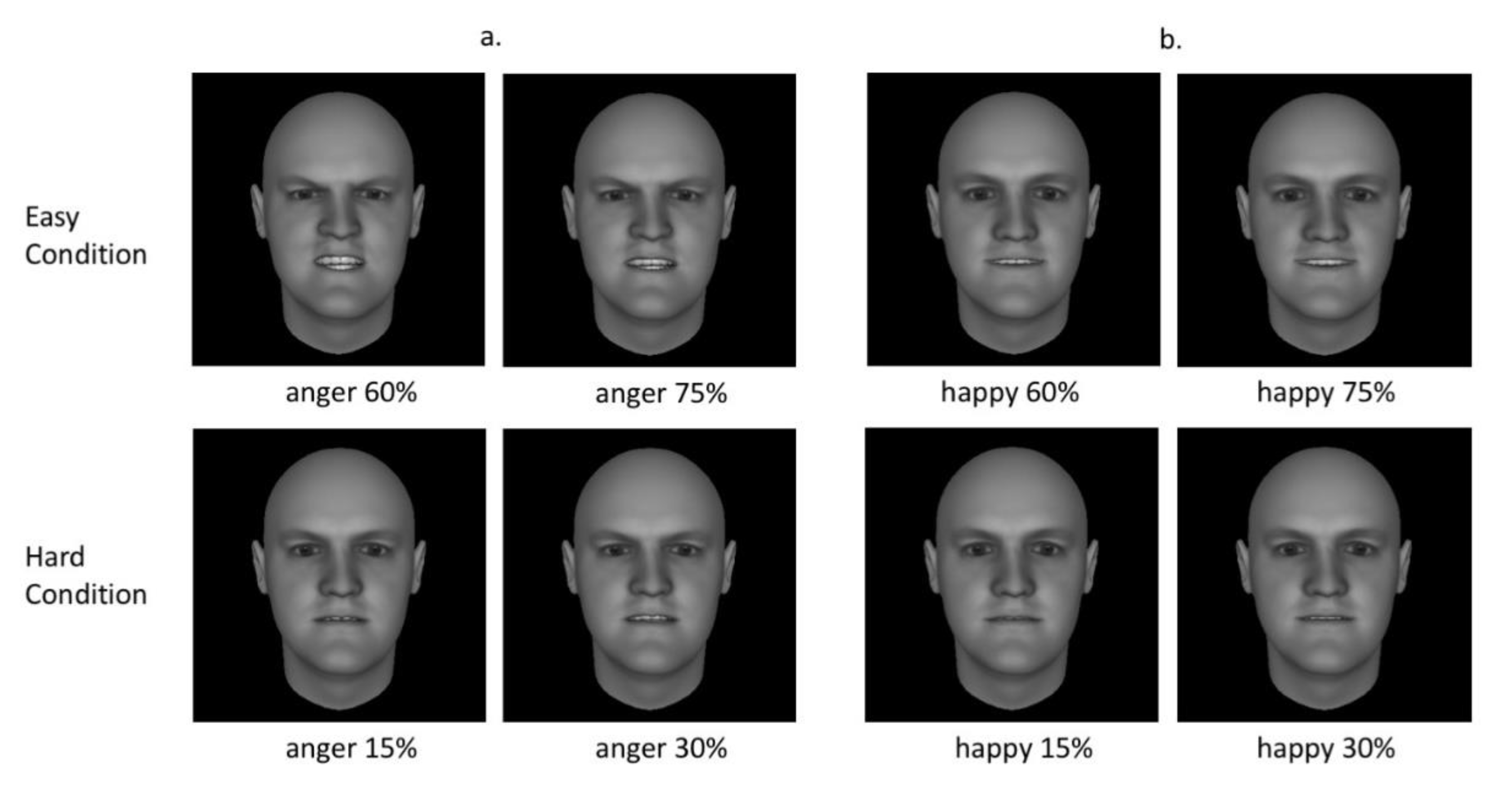
2.2. Stimuli
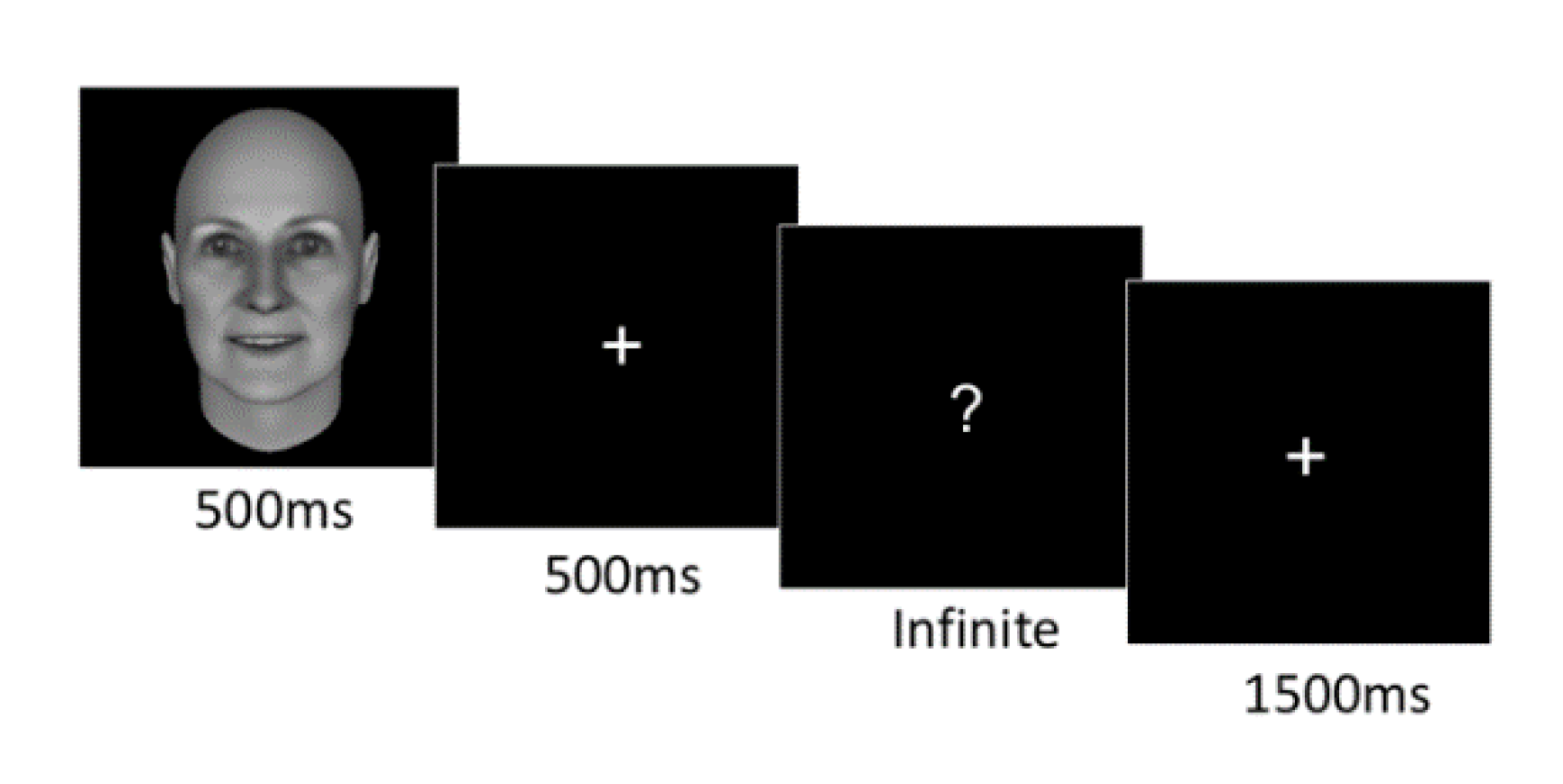
2.3. Experimental Procedure
2.4. EEG Recording and Analysis
2.5. Preprocessing
2.6. ERP Analysis
3. Results
3.1. Participants’ Characteristics
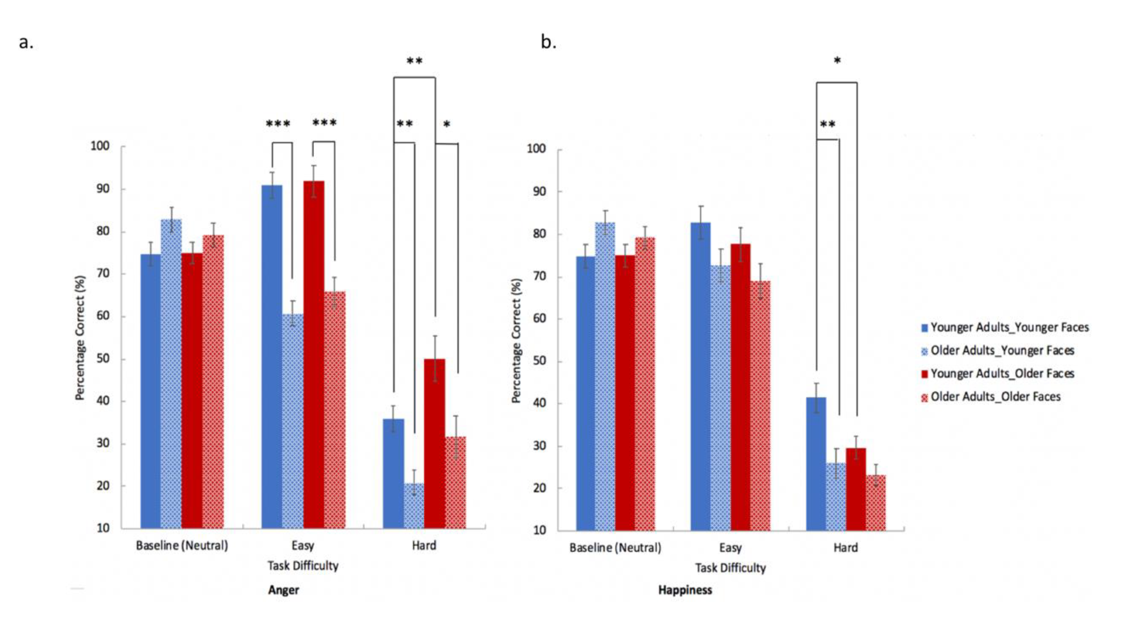
3.2. Behavioral Results
3.2.1. Perception of Neutral Facial Expression
3.2.2. Perception of Anger and Happiness Facial Expressions
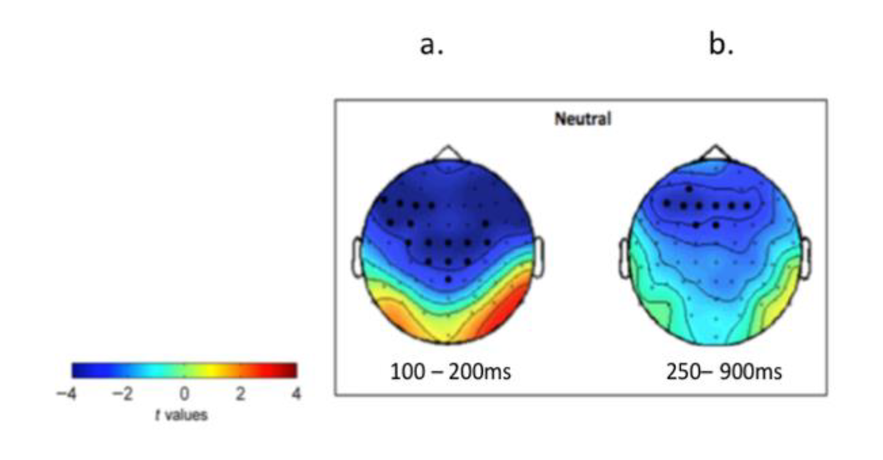
3.3. ERP Analysis
3.3.1. Selections of ERP Clusters that Showed Difference between Old and Young Group
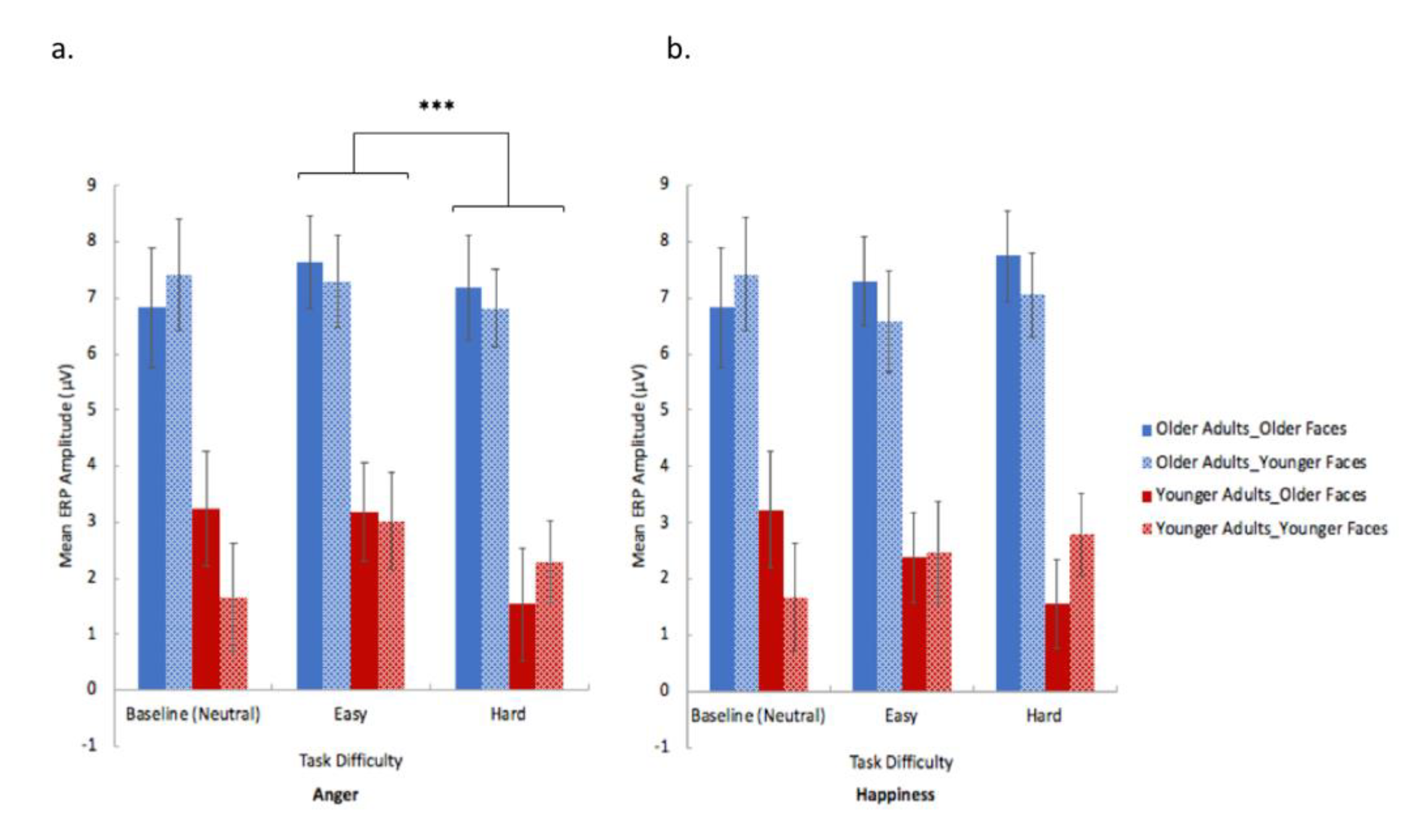
3.3.2. The Effect of Emotion, Face-age and Task Difficulty in Modulating ERPs
4. Discussion
4.1. Age-Related Behavioural Perceptual Performance Differences
4.2. Effect of Face-Age on Behavioural Perceptual Performance
4.3. Facial Emotion Perception and Neural Compensation in Older People Revealed by ERPs
4.4. Effects of “Emotion Type”, “Task Difficulty” and “Face-Age” on ERPs
4.5. Limitations and Future Avenues
Supplementary Materials
Author Contributions
Funding
Acknowledgments
Conflicts of Interest
References
- Ruffman, T.; Henry, J.D.; Livingstone, V.; Phillips, L.H. A meta-analytic review of emotion recognition and aging: Implications for neuropsychological models of aging. Neurosci. Biobehav. Rev. 2008, 32, 863–881. [Google Scholar] [CrossRef]
- Ryan, M.; Murray, J.; Ruffman, T. Aging and the perception of emotion: Processing vocal expressions alone and with faces. Exp. Aging Res. 2009, 36, 1–22. [Google Scholar] [CrossRef]
- Spell, L.A.; Frank, E. Recognition of nonverbal communication of affect following traumatic brain injury. J. Nonverbal Behav. 2000, 24, 285–300. [Google Scholar] [CrossRef]
- Kanai, R.; Bahrami, B.; Duchaine, B.; Janik, A.; Banissy, M.J.; Rees, G. Brain structure links loneliness to social perception. Curr. Biol. 2012, 22, 1975–1979. [Google Scholar] [CrossRef] [PubMed]
- Holt-Lunstad, J.; Smith, T.B.; Baker, M.; Harris, T.; Stephenson, D. Loneliness and social isolation as risk factors for mortality: A meta-analytic review. Perspect. Psychol. Sci. 2015, 10, 227–237. [Google Scholar] [CrossRef] [PubMed]
- McDowell, C.L.; Harrison, D.W.; Demaree, H.A. Is right hemisphere decline in the perception of emotion a function of aging? Int. J. Neurosci. 1994, 79, 1–11. [Google Scholar] [CrossRef]
- Phillips, L.H.; MacLean, R.D.; Allen, R. Age and the understanding of emotions neuropsychological and sociocognitive perspectives. J. Gerontol. Ser. B Psychol. Sci. Soc. Sci. 2002, 57, 526–530. [Google Scholar] [CrossRef]
- Calder, A.J.; Keane, J.; Manly, T.; Sprengelmeyer, R.; Scott, S.; Nimmo-Smith, I.; Young, A.W. Facial expression recognition across the adult life span. Neuropsychologia 2003, 41, 195–202. [Google Scholar] [CrossRef]
- Sullivan, S.; Ruffman, T. Emotion recognition deficits in the elderly. Int. J. Neurosci. 2004, 114, 403–432. [Google Scholar] [CrossRef]
- Mill, A.; Allik, J.; Realo, A.; Valk, R. Age-related differences in emotion recognition ability: A cross-sectional study. Emotion 2009, 9, 619. [Google Scholar] [CrossRef]
- Moreno, C.; Borod, J.C.; Welkowitz, J.; Alpert, M. The perception of facial emotion across the adult life span. Dev. Neuropsychol. 1993, 9, 305–314. [Google Scholar] [CrossRef]
- Orgeta, V.; Phillips, L.H. Effects of age and emotional intensity on the recognition of facial emotion. Exp. Aging Res. 2007, 34, 63–79. [Google Scholar] [CrossRef] [PubMed]
- Yang, T.; Penton, T.; Köybaşı, Ş.L.; Banissy, M.J. Social perception and aging: The relationship between aging and the perception of subtle changes in facial happiness and identity. Acta Psychol. 2017, 179, 23–29. [Google Scholar] [CrossRef] [PubMed]
- Anastasi, J.S.; Rhodes, M.G. An own-age bias in face recognition for children and older adults. Psychon. Bull. Rev. 2005, 12, 1043–1047. [Google Scholar] [CrossRef] [PubMed]
- Ebner, N.C.; He, Y.I.; Johnson, M.K. Age and emotion affect how we look at a face: Visual scan patterns differ for own-age versus other-age emotional faces. Cogn. Emot. 2011, 25, 983–997. [Google Scholar] [CrossRef] [PubMed]
- Ebner, N.C.; Johnson, M.K. Young and older emotional faces: Are there age group differences in expression identification and memory? Emotion 2009, 9, 329. [Google Scholar] [CrossRef]
- Wright, D.B.; Stroud, J.N. Age differences in lineup identification accuracy: People are better with their own age. Law Hum. Behav. 2002, 26, 641. [Google Scholar] [CrossRef]
- Hess, U.; Adams, R.B., Jr.; Simard, A.; Stevenson, M.T.; Kleck, R.E. Smiling and sad wrinkles: Age-related changes in the face and the perception of emotions and intentions. J. Exp. Soc. Psychol. 2012, 48, 1377–1380. [Google Scholar] [CrossRef]
- Courgeon, M.; Buisine, S.; Martin, J.C. Impact of expressive wrinkles on perception of a virtual character’s facial expressions of emotions. In International Workshop on Intelligent Virtual Agents; Springer: Berlin/Heidelberg, Germany, 2009; pp. 201–214. [Google Scholar]
- Richter, D.; Dietzel, C.; Kunzmann, U. Age differences in emotion recognition: The task matters. J. Gerontol. Ser. B Psychol. Sci. Soc. Sci. 2010, 66, 48–55. [Google Scholar] [CrossRef]
- Murphy, N.A.; Isaacowitz, D.M. Age effects and gaze patterns in recognising emotional expressions: An in-depth look at gaze measures and covariates. Cogn. Emot. 2010, 24, 436–452. [Google Scholar] [CrossRef]
- Hühnel, I.; Fölster, M.; Werheid, K.; Hess, U. Empathic reactions of younger and older adults: No age related decline in affective responding. J. Exp. Soc. Psychol. 2014, 50, 136–143. [Google Scholar] [CrossRef]
- Gunning-Dixon, F.M.; Gur, R.C.; Perkins, A.C.; Schroeder, L.; Turner, T.; Turetsky, B.I.; Chan, R.M.; Loughead, J.W.; Alsop, D.C.; Maldjian, J.; et al. Age-related differences in brain activation during emotional face processing. Neurobiol. Aging 2003, 24, 285–295. [Google Scholar] [CrossRef]
- Tessitore, A.; Hariri, A.R.; Fera, F.; Smith, W.G.; Das, S.; Weinberger, D.R.; Mattay, V.S. Functional changes in the activity of brain regions underlying emotion processing in the elderly. Psychiatry Res. Neuroimaging 2005, 139, 9–18. [Google Scholar] [CrossRef] [PubMed]
- Fischer, H.; Nyberg, L.; Bäckman, L. Age-related differences in brain regions supporting successful encoding of emotional faces. Cortex 2010, 46, 490–497. [Google Scholar] [CrossRef]
- Fischer, H.; Sandblom, J.; Gavazzeni, J.; Fransson, P.; Wright, C.I.; Bäckman, L. Age-differential patterns of brain activation during perception of angry faces. Neurosci. Lett. 2005, 386, 99–104. [Google Scholar] [CrossRef] [PubMed]
- West, J.T.; Horning, S.M.; Klebe, K.J.; Foster, S.M.; Cornwell, R.E.; Perrett, D.; Burt, D.M.; Davis, H.P. Age effects on emotion recognition in facial displays: From 20 to 89 years of age. Exp. Aging Res. 2012, 38, 146–168. [Google Scholar] [CrossRef] [PubMed]
- Mienaltowski, A.; Groh, B.; Secula, D.; Rinne, A.; Rogers, C. Impact of expressive intensity on age differences in fear and anger detection in the periphery. J. Vis. 2018, 18, 568. [Google Scholar] [CrossRef]
- Eimer, M.; Holmes, A. Event-related brain potential correlates of emotional face processing. Neuropsychologia 2007, 45, 15–31. [Google Scholar] [CrossRef]
- Eimer, M.; Holmes, A.; McGlone, F.P. The role of spatial attention in the processing of facial expression: An ERP study of rapid brain responses to six basic emotions. Cogn. Affect. Behav. Neurosci. 2003, 3, 97–110. [Google Scholar] [CrossRef]
- Kisley, M.A.; Wood, S.; Burrows, C.L. Looking at the sunny side of life: Age-related change in an event-related potential measure of the negativity bias. Psychol. Sci. 2007, 18, 838–843. [Google Scholar] [CrossRef]
- Wieser, M.J.; Mühlberger, A.; Kenntner-Mabiala, R.; Pauli, P. Is emotion processing affected by advancing age? An event-related brain potential study. Brain Res. 2006, 1096, 138–147. [Google Scholar] [CrossRef] [PubMed]
- Tsolaki, A.C.; Kosmidou, V.E.; Kompatsiaris, I.Y.; Papadaniil, C.; Hadjileontiadis, L.; Tsolaki, M. Age-induced differences in brain neural activation elicited by visual emotional stimuli: A high-density EEG study. Neuroscience 2017, 340, 268–278. [Google Scholar] [CrossRef] [PubMed]
- Nelson, D.B. Conditional heteroskedasticity in asset returns: A new approach. Econom. J. Econom. Soc. 1991, 59, 347–370. [Google Scholar] [CrossRef]
- Turner, M.; Ridsdale, J. The digit memory test. Dyslexia Int. 2004. [Google Scholar]
- Bagby, R.M.; Parker, J.D.; Taylor, G.J. The twenty-item Toronto Alexithymia Scale—I. Item selection and cross-validation of the factor structure. J. Psychosom. Res. 1994, 38, 23–32. [Google Scholar] [CrossRef]
- Reuter-Lorenz, P.A.; Cappell, K.A. Neurocognitive aging and the compensation hypothesis. Curr. Dir. Psychol. Sci. 2008, 17, 177–182. [Google Scholar] [CrossRef]
- Folstein, M.F.; Folstein, S.E.; McHugh, P.R. “Mini-mental state”: A practical method for grading the cognitive state of patients for the clinician. J. Psychiatr. Res. 1975, 12, 189–198. [Google Scholar] [CrossRef]
- Chayer, C. The neurologic examination: Brief mental status. Can. J. Geriatr. Care 2002, 1, 265–267. [Google Scholar]
- Roesch, E.B.; Tamarit, L.; Reveret, L.; Grandjean, D.; Sander, D.; Scherer, K.R. FACSGen: A tool to synthesize emotional facial expressions through systematic manipulation of facial action units. J. Nonverbal Behav. 2011, 35, 1–16. [Google Scholar] [CrossRef]
- Oosterhof, N.N.; Todorov, A. Shared perceptual basis of emotional expression and trustworthiness impression from faces. Emotion 2009, 9, 128–133. [Google Scholar] [CrossRef]
- Penton, T.; Bate, S.; Dalrymple, K.; Reed, T.; Kelly, M.; Godovich, S.; Tamm, M.; Duchaine, B.; Banissy, M.J. Using high frequency transcranial random noise stimulation to modulate face memory performance in younger and older adults: Lessons learnt from mixed findings. Front. Neurosci. 2018, 12, 863. [Google Scholar] [CrossRef] [PubMed]
- Delorme, A.; Makeig, S. EEGLAB: An open source toolbox for analysis of single-trial EEG dynamics including independent component analysis. J. Neurosci. Methods 2004, 134, 9–21. [Google Scholar] [CrossRef] [PubMed]
- Oostenveld, R.; Fries, P.; Maris, E.; Schoffelen, J.M. FieldTrip: Open source software for advanced analysis of MEG, EEG, and invasive electrophysiological data. Comput. Intell. Neurosci. 2011, 1, 1–9. [Google Scholar] [CrossRef] [PubMed]
- Picton, T.W.; van Roon, P.; Armilio, M.L.; Berg, P.; Ille, N.; Scherg, M. Blinks, saccades, extraocular muscles and visual evoked potentials (Reply to Verleger). J. Psychophysiol. 2000, 14, 210–217. [Google Scholar] [CrossRef]
- Pfurtscheller, G.; Da Silva, F.L. Event-related EEG/MEG synchronization and desynchronization: Basic principles. Clin. Neurophysiol. 1999, 110, 1842–1857. [Google Scholar] [CrossRef]
- Maris, E.; Oostenveld, R. Nonparametric statistical testing of EEG-and MEG-data. J. Neurosci. Methods 2007, 164, 177–190. [Google Scholar] [CrossRef]
- Lindsen, J.P.; Jones, R.; Shimojo, S.; Bhattacharya, J. Neural components underlying subjective preferential decision making. Neuroimage 2010, 50, 1626–1632. [Google Scholar] [CrossRef]
- Luft, C.D.B.; Takase, E.; Bhattacharya, J. Processing graded feedback: Electrophysiological correlates of learning from small and large errors. J. Cogn. Neurosci. 2014, 26, 1180–1193. [Google Scholar] [CrossRef]
- Sandkühler, S.; Bhattacharya, J. Deconstructing insight: EEG correlates of insightful problem solving. PLoS ONE 2008, 3, 1459. [Google Scholar] [CrossRef]
- MacPherson, S.E.; Phillips, L.H.; Sala, S.D. Age-related differences in the ability to perceive sad facial expressions. Aging Clin. Exp. Res. 2006, 18, 418–424. [Google Scholar] [CrossRef]
- Gratton, G.; Coles, M.G.; Donchin, E. Optimizing the use of information: Strategic control of activation of responses. J. Exp. Psychol. Gen. 1992, 121, 480. [Google Scholar] [CrossRef] [PubMed]
- Schmidt, J.R. Questioning conflict adaptation: Proportion congruent and Gratton effects reconsidered. Psychon. Bull. Rev. 2013, 20, 615–630. [Google Scholar] [CrossRef] [PubMed]
- Aschenbrenner, A.J.; Balota, D.A. Dynamic adjustments of attentional control in healthy aging. Psychol. Aging 2017, 32, 1–15. [Google Scholar] [CrossRef] [PubMed]
- Lamont, A.C.; Stewart-Williams, S.; Podd, J. Face recognition and aging: Effects of target age and memory load. Mem. Cogn. 2005, 33, 1017–1024. [Google Scholar] [CrossRef]
- Eimer, M.; Holmes, A. An ERP study on the time course of emotional face processing. Neuroreport 2002, 13, 427–431. [Google Scholar] [CrossRef]
- Balconi, M.; Pozzoli, U. Face-selective processing and the effect of pleasant and unpleasant emotional expressions on ERP correlates. Int. J. Psychophysiol. 2003, 49, 67–74. [Google Scholar] [CrossRef]
- Raz, N. Aging of the brain and its impact on cognitive performance: Integration of structural and functional findings. In The Handbook of Aging and Cognition; Craik, F.I.M., Salthouse, T.A., Eds.; Lawrence Erlbaum Associates Publishers: Mahwah, NJ, USA, 2000; pp. 1–90. [Google Scholar]
- Grady, C.L. Functional brain imaging and age-related changes in cognition. Biol. Psychol. 2000, 54, 259–281. [Google Scholar] [CrossRef]
- Halpern, A.R.; Zioga, I.; Shankleman, M.; Lindsen, J.; Pearce, M.T.; Bhattacharya, J. That note sounds wrong! Age-related effects in processing of musical expectation. Brain Cogn. 2017, 113, 1–9. [Google Scholar] [CrossRef]
- Johannsen, P.; Jakobsen, J.; Bruhn, P.; Hansen, S.B.; Gee, A.; Stødkilde-Jørgensen, H.; Gjedde, A. Cortical sites of sustained and divided attention in normal elderly humans. Neuroimage 1997, 6, 145–155. [Google Scholar] [CrossRef]
- Madden, D.J.; Turkington, T.G.; Provenzale, J.M.; Hawk, T.C.; Hoffman, J.M.; Coleman, R.E. Selective and divided visual attention: Age-related changes in regional cerebral blood flow measured by H2 15O PET. Hum. Brain Mapp. 1997, 5, 389–409. [Google Scholar] [CrossRef]
- Anderson, N.D.; Iidaka, T.; Cabeza, R.; Kapur, S.; McIntosh, A.R.; Craik, F.I. The effects of divided attention on encoding-and retrieval-related brain activity: A PET study of younger and older adults. J. Cogn. Neurosci. 2000, 12, 775–792. [Google Scholar] [CrossRef] [PubMed]
- Hartley, A.A.; Speer, N.K.; Jonides, J.; Reuter-Lorenz, P.A.; Smith, E.E. Is the dissociability of working memory systems for name identity, visual-object identity, and spatial location maintained in old age? Neuropsychology 2001, 15, 3. [Google Scholar] [CrossRef] [PubMed]
- Mitchell, K.J.; Johnson, M.K.; Raye, C.L.; Mather, M.; D’esposito, M. Aging and reflective processes of working memory: Binding and test load deficits. Psychol. Aging 2000, 15, 527. [Google Scholar] [CrossRef] [PubMed]
- Smith, E.E.; Geva, A.; Jonides, J.; Miller, A.; Reuter-Lorenz, P.; Koeppe, R.A. The neural basis of task-switching in working memory: Effects of performance and aging. Proc. Natl. Acad. Sci. USA 2001, 98, 2095–2100. [Google Scholar] [CrossRef] [PubMed]
- Gazzaley, A.; Cooney, J.W.; Rissman, J.; D’Esposito, M. Top-down suppression deficit underlies working memory impairment in normal aging. Nat. Neurosci. 2005, 8, 1298–1300. [Google Scholar] [CrossRef] [PubMed]
- Phelps, E.A.; Ling, S.; Carrasco, M. Emotion facilitates perception and potentiates the perceptual benefits of attention. Psychol. Sci. 2006, 17, 292–299. [Google Scholar] [CrossRef]
- Houston, J.R.; Pollock, J.W.; Lien, M.C.; Allen, P.A. Emotional arousal deficit or emotional regulation bias? An electrophysiological study of age-related differences in emotion perception. Exp. Aging Res. 2018, 44, 187–205. [Google Scholar] [CrossRef]
- Schupp, H.T.; Cuthbert, B.N.; Bradley, M.M.; Cacioppo, J.T.; Ito, T.; Lang, P.J. Affective picture processing: The late positive potential is modulated by motivational relevance. Psychophysiology 2000, 37, 257–261. [Google Scholar] [CrossRef]
- Van der Gaag, C.; Minderaa, R.B.; Keysers, C. Facial expressions: What the mirror neuron system can and cannot tell us. Soc. Neurosci. 2007, 2, 179–222. [Google Scholar] [CrossRef]
- Jabbi, M.; Keysers, C. Inferior frontal gyrus activity triggers anterior insula response to emotional facial expressions. Emotion 2008, 8, 775. [Google Scholar] [CrossRef]
- Kriegeskorte, N.; Simmons, W.K.; Bellgowan, P.S.; Baker, C.I. Circular analysis in systems neuroscience: The dangers of double dipping. Nat. Neurosci. 2009, 12, 535. [Google Scholar] [CrossRef] [PubMed]
- Fusar-Poli, P.; Placentino, A.; Carletti, F.; Landi, P.; Allen, P.; Surguladze, S.; Benedetti, F.; Abbamonte, M.; Gasparotti, R.; Barale, F.; et al. Functional atlas of emotional faces processing: A voxel-based meta-analysis of 105 functional magnetic resonance imaging studies. J. Psychiatry Neurosci. 2009, 34, 418–432. [Google Scholar] [PubMed]
- Kesler, M.L.; Andersen, A.H.; Smith, C.D.; Avison, M.J.; Davis, C.E.; Kryscio, R.J.; Blonder, L.X. Neural substrates of facial emotion processing using fMRI. Cogn. Brain Res. 2001, 11, 213–226. [Google Scholar] [CrossRef]


| Old (n = 15) | Young (n = 16) | |
|---|---|---|
| Gender (male/female) | 3/12 | 4/12 |
| Age (years) | 69.20 (8.809) | 24.19 (5.576) |
| Education (years) | 15 (3) | 16 (2) |
| Handedness (right/left) | 14/1 | 15/1 |
| Premorbid IQ (NART) | 120.07 (8.430) | 115.44 (10.295) |
| Working memory (digit-span) | 105.27 (16.867) | 106.63 (12.209) |
| TAS-20 score | 43.00 (7.329) | 44.75 (9.692) |
© 2020 by the authors. Licensee MDPI, Basel, Switzerland. This article is an open access article distributed under the terms and conditions of the Creative Commons Attribution (CC BY) license (http://creativecommons.org/licenses/by/4.0/).
Share and Cite
Yang, T.; Di Bernardi Luft, C.; Sun, P.; Bhattacharya, J.; Banissy, M.J. Investigating Age-Related Neural Compensation During Emotion Perception Using Electroencephalography. Brain Sci. 2020, 10, 61. https://doi.org/10.3390/brainsci10020061
Yang T, Di Bernardi Luft C, Sun P, Bhattacharya J, Banissy MJ. Investigating Age-Related Neural Compensation During Emotion Perception Using Electroencephalography. Brain Sciences. 2020; 10(2):61. https://doi.org/10.3390/brainsci10020061
Chicago/Turabian StyleYang, Tao, Caroline Di Bernardi Luft, Pei Sun, Joydeep Bhattacharya, and Michael J. Banissy. 2020. "Investigating Age-Related Neural Compensation During Emotion Perception Using Electroencephalography" Brain Sciences 10, no. 2: 61. https://doi.org/10.3390/brainsci10020061
APA StyleYang, T., Di Bernardi Luft, C., Sun, P., Bhattacharya, J., & Banissy, M. J. (2020). Investigating Age-Related Neural Compensation During Emotion Perception Using Electroencephalography. Brain Sciences, 10(2), 61. https://doi.org/10.3390/brainsci10020061