Article

α-Mangostin Hydrogel Film Based Chitosan–Alginate for Recurrent Aphthous Stomatitis

1
Department of Pharmaceutics and Pharmaceutical Technology, Faculty of Pharmacy, Universitas Padjadjaran, Sumedang 45363, Indonesia
2
Department of Pharmacy, Faculty of Technology and Computer, Universitas Buana Perjuangan, Karawang 41361, Indonesia
3
Department of Dental Materials Science and Technology, Faculty of Dentistry, Universitas Padjadjaran, Sumedang 45363, Indonesia
4
Department of Pharmaceutical Analysis and Medicinal Chemistry, Faculty of Pharmacy, Universitas Padjadjaran, Sumedang 45363, Indonesia
*
Author to whom correspondence should be addressed.
Appl. Sci. 2019, 9(23), 5235; https://doi.org/10.3390/app9235235
Submission received: 31 October 2019 / Revised: 14 November 2019 / Accepted: 29 November 2019 / Published: 2 December 2019
(This article belongs to the Special Issue Recent Advances in Bioadhesives for Tissue Repair)

Abstract

:

Featured Application

This study highlights a novel application of α-mangostin hydrogel film based chitosan–alginate. α-mangostin has been known as an agent for oral cavity therapy. However, α-mangostin is difficult to be delivered directly to the site. This novel application of hydrogel film-based chitosan–alginate have the potential as carriers of α-mangostin for recurrent aphthous stomatitis therapy.

Abstract

Many antiseptic drugs, local anaesthetics, and corticosteroids have been used for effective therapy of recurrent aphthous stomatitis (RAS). However, these drugs have harmful side effects. α-mangostin (α-M), a main compound of mangosteen (Garcinia mangostana L.) peel, has been known as a wound healing agent. In addition, hydrogel film as dressings designed to separate mucosal lesions from the oral environment, and improve the effectiveness of RAS therapy. The purpose of this study was to develop α-M hydrogel film based chitosan–alginate (ChAlg/α-M HF) for RAS. The in silico study by Discovery studio visualizer and AutoDock confirmed that hydrogen bonding between Ch, Alg, and α-M occurred. The results of physicochemical characterizations by scanning electron microscopy (SEM), differential scanning calorimetry (DSC), and X-ray diffraction (XRD) indicated that the ChAlg/α-M HF had a lower crystalline form compared to pure α-M. In addition, ChAlg/α-M HF significantly improved the swelling ratio and tensile strength compared to that of ChAlg HF. Moreover, the existence of Alg increased the degradability of Ch, and closely related to the release of α-M from ChAlg HF. The in vitro release study confirmed that the release of α-M from ChAlg/α-M HF was the Fickian diffusion model. Finally, the mucoadhesive study revealed that ChAlg/α-M HF had a good mucoadhesive property. These results suggest that hydrogel film-based chitosan–alginate have the potential as carriers of α-M for RAS therapy.

Graphical Abstract

1. Introduction

Recurrent aphthous stomatitis (RAS) is the most common ulcerative disease in the oral mucosa in humans that often results from trauma, hormonal changes, physical or psychological stress, chemical irritation, genetic factors, and allergies [1]. To date, effective RAS therapy has not been found, and its treatment is aimed at treating symptoms with a focus on suppressing local immune responses, reducing pain, discomfort, and preventing secondary infections and recurrences. If the ulcer is left unchecked, it will cause secondary infection because the oral cavity can be the ideal place for bacterial growth [2].
The use of drugs in RAS aims to accelerate the regeneration of tissue cells. The drugs used in RAS therapy can be modern drugs or natural medicines derived from plants and spices [3]. Topical applications of antiseptics (chlorhexidine, triclosan, benzydamine hydrochloride, amlexanox), local anesthetics (lidocaine, diclonine hydrochloride), or corticosteroids (triamcinolone, betamethasone) have been shown to have positive effects for treatment but have harmful side effects when they are used too often and over a long period [4].
Mangosteen rind (Garcinia mangostana L.), an Indonesian native plant, is used as traditional medicine, such as abdominal pain, chronic ulcers, skin infections, wounds healing, analgesic and anti-inflammatory properties [5]. The substances contained therein are phenol derivatives namely xanthones or xanthen-9H-ones [6]. Mangosteen rind (Garcinia mangostana L.) contains various of xanthones, including α-, β-, γ-mangostin which have pharmacological activities, such as antioxidant, antitumor, antibacterial, antiviral, antifungal, antiallergic, and anti-inflammatory [7].
Recently, α-mangostin (α-M) has been known as an agent for oral cavity therapy. However, α-M is difficult to be delivered directly to the site because it is insoluble in water, therefore, a carrier for delivering α-M is strongly needed. Hydrogel film (HF) can be used as a patch designed to separate mucosal lesions from the oral environment, and thus reduce the risk of getting the drug carried by saliva so that it can increase the time of contact between active substances and lesions, improve the effectiveness of therapy, and reduce patient discomfort [8,9].
In previous studies, HF based chitosan (Ch) derived from chitin has high resistance, biocompatibility, low toxicity, high fluids absorption, and has an antibacterial activity that will accelerate healing. However, to the superior nature of the Ch above, chitosan turns out to have a deficiency that is sensitive to acid solutions. Therefore, it is important to increase its chemical resistance, as well as long-term biological hydrophilicity by a combination of other polysaccharides [10]. Alginate (Alg), a natural polysaccharide extracted from brown seaweed, has been commonly used in the food industry as an additive or thickener [11]. Alg and Ch dissolve together in the right conditions and interact with each other through carbonyl groups of Alg and protonated amino groups of Ch. It was reported that the formation of hydrogels using Alg and Ch can be achieved by adjusting the pH of the mixing solution without adding calcium cations [12,13]. Patil et al. proved that hydrogel based on Ch–Alg formulations had a higher mucoadhesive strength compared to hydrogel based on Alg [14]. In addition, Ch molecular weight in Ch–Alg polyelectrolyte complex film containing clindamycin phosphate affected to adhesiveness of complex films [15]. Mucoadhesive ability of Ch closely related to the structure of Ch, which can interact with mucin predominantly by electrostatic binding [16].
In this study, we prepared and characterized the α-M HF based on Ch and Alg (ChAlg/α-M HF) as a new RAS therapy.

2. Materials and Methods

2.1. Materials

α-Mangostin (α-M) was procured from Chengdu Biopurify Phytochemicals Ltd. (Chengdu, China). Chitosan (Ch) from crab shells, and sodium alginate (Alg) were obtained from Sigma-Aldrich (PT. Elo Karsa Utama, Bandung, Indonesia), and Gibco (Jakarta, Indonesia), respectively. Trichloroacetic acid (TCA) and trypsin (TrypLE TM) were purchased from Wako Pure Chemical (Tokyo, Japan). All the reagents were of analytical grade and used without further purification.

2.2. Methods

2.2.1. Molecular Docking

The prediction stages of hydrogen bonds and the amount of bonding energy were investigated in silico using Discovery studio visualizer free trials and AutoDock 1.6 applications. Briefly, the two-dimensional structure of Ch, Alg, and α-M was obtained from PubChem Database in the format sdf. Then, format sdf was converted to format pdb using Discovery studio visualizer v. 16. 1. 0. 15350. The interaction of the two molecules (chitosan and alginate) was observed using the AutoDock Tools version 1.6 software followed by the addition of α-mangostin molecules and observed using the AutoDock Tools version 1.6 software program. Parameter analysis was performed on the types of interaction (hydrogen bond, Van der Waals, or pi-interaction) and activation energy [17].

2.2.2. Preparation of ChAlg HF

ChAlg HF was prepared by the solvent evaporation method referred to in our previous study [18]. Firstly, 0.5 g of Alg powder was mixed in 25 mL of distilled water to obtain the Alg solution. For the Ch solution, 0.5 mL of glacial acetic acid was mixed with 0.1 g of Ch powder together with 24.5 mL of distilled water. The Alg solution was added gradually to the Ch solution to get a homogeneous solution for about 30 min of stirring. Hydrogel solution was poured and flattened on a propylene box with a size of 5 × 5 × 4 cm3 thickness 2–4 mm. Then, the flattened solution was dried at 60 °C for 24 h to obtain ChAlg HF. Alg HF and Ch HF were also fabricated with the same procedures as a comparison.

2.2.3. Preparation of ChAlg/α-M HF

ChAlg/α-M HF was prepared by the solvent casting method, as previously described [19]. Briefly, ChAlg HF (1 × 1 cm2) was casted (10 μL) by α-M (25 mg) dissolved in methanol on its surface to obtain ChAlg/α-M HF. Then, they were stored at 4 °C for 24 h and dried at 37 °C for 1 h.

2.2.4. Scanning Electron Microscope (SEM)

Ch HF, Alg HF, ChAlg HF, and ChAlg/α-M HF (1 × 1 cm2) were placed on the aluminum stub and coated with gold for 10 s (30 mM, 8 Pa) by Coater. The cross section of the HFs were analyzed by a SEM (JEOL JSM 6510 LA, Tokyo, Japan) [20,21].

2.2.5. X-Ray Diffractometry (XRD)

Ch HF, Alg HF, ChAlg HF, and ChAlg/α-M HF (2.5 × 2.5 mm2) were put on X-ray diffractometer (Rigaku Ultima IV, Tokyo, Japan) sample holders. X-ray diffraction patterns were obtained under conditions with Ni-filtered Cu-Kα radiation, voltage of 40 kV, current of 20 mA, divergent slits of 10 mm (0.5 O), scanning speed of 5°/min, opened scattering, and receiving slit [22].

2.2.6. Differential Scanning Calorimetry (DSC)

Evaluation of thermal stability was observed by DSC SDC Q600 (TA Instrument, Tokyo, Japan) Ch HF, Alg HF, ChAlg HF, and ChAlg/α-M HF were heated in an aluminum pan with heating rate of 10 °C min−1 under nitrogen pressure. The temperature range was 0 to 327 °C [23].

2.2.7. Mechanical Properties

A texture analyzer was used to determine the tensile strength and elongation at break. Firstly, the tensile strength of HFs was investigated in dry and swollen conditions at room temperature with tearing well in 98 cell loads of texture analyzer. Briefly, Ch HF, Alg HF, ChAlg HF, and ChAlg/α-M HF (1 × 1 cm2) were held between two clips and pulled with the upper clip at a speed of 0.5 mm/s. Swollen film was prepared by immersing the HFs on PBS pH 7.4 (0.05 M) at room temperature for 1 h. The tensile strength of the HFs was recorded when the HFs were damaged. Each test was done six times. Tensile strength was calculated by using the following Equation (1):
Tensile strength (MPa) = p/(b × d)
The results are presented in the form of averages and standard deviations. Where p is a breaking force (the force needed for the sample to be broken), b and d are the width and thickness of the sample.
For determination of elongation at break, elongation at break was calculated by Equation (2):
Elongation at Break (%) = (L − Lo)/Lo × 100%
where L and Lo are expressed as the length of the breaking point and the initial length of the sample [13].

2.2.8. Degradability Study

Ch HF, Alg HF, ChAlg HF, and ChAlg/α-M HF (1 × 1 cm2) were weighed and soaked in 2 mL of 20% TCA solution incubated for 24 h. TCA solution was removed and re-weighed again until at a constant state. Degradability study with trypsin solution was also investigated with the same method [20]. Percent change in weight was calculated by the following Equation (3):
Percent change in weight = 100 − (Final weight/Initial weight × 100)

2.2.9. Swelling Ability

A gravimetric technique was used to calculate the swelling ability of HFs. Ch HF, Alg HF, ChAlg HF, and ChAlg/α-M HF (1 × 1 cm2) were weighed and soaked in PBS pH 7.4 for two days. The truly expanded HFs were weighed (Ws) immediately after removing excess of PBS. The expanded HFs were lyophilized for two days until the weight was constant (Wd). Swelling ratio was calculated as Ws/Wd [18].

2.2.10. In Vitro Release

Ch HF, Alg HF, Ch Alg HF, and Ch Alg/α-M HF (1 × 1 cm2) were immersed in 20 mL PBS pH 7.4 at 37 ± 5 °C with a shaker speed in 50 rpm. The aliquots were taken for about 5 mL of the solution at 5, 15, 30, 45, 60, 90, and 120 min. The aliquots were filtered with a 0.45 µm Millipore filter, diluted and analyzed by spectrophotometer (Jasco V-630, Tokyo, Japan) at 280 nm [24].
The kinetic data release obtained was calculated using the Higuchi kinetics equation to see the release mechanism of α-M using Equation (4).
Q = k. T1/2
where Q = drug release against time t, k = kinetic constant value [25].

2.2.11. In Vitro Mucoadhesive Study

In vitro mucoadhesive study of the HFs was determined using the mouse buccal mucosa in accordance with the ethical standards of the Ethical Committee of Universitas Padjadjaran. The mouse mucosa was glued to the glass object with cyanoacrylate glue. One side of the film was moistened with 1 mL of phosphate buffer pH 6.8 and placed on the mouse mucosa by applying a compressive force with the finger for 20 s. Mucosa and the HFs on microscope slide were dipped in a beaker containing 200 mL of phosphate buffer pH 6.8 (temperature 37 ± 0.5 °C) then stirring at a speed of 50 rpm to simulate the oral cavity environment. The time was recorded when the film was completely released from the mucosa [26].

3. Statistical Analysis

The quantitative data were expressed as the mean ± standard error of the mean (SEM). The statistical assessments were performed using the one-way ANOVA Scheffe method. A p-value < 0.05 was measured as statistically significant.

4. Results and Discussion

4.1. Molecular Docking

Discovery studio visualizer and AutoDock 1.6 applications were applied for molecular docking studies to predict ligand–protein interactions, bonding modes, bonding affinity based on energy, and ligands interaction with the receptor [17]. It can be seen in Figure 1, ChAlg in computer simulation describes that only 1 hydrogen bond presented at the 23rd hydrogen atom of Alg with activation energy of 0.18 kcal/mol. In ChA/α-M, there were 2 hydrogen bonds between Alg and α-M placed at the 21st and 22nd hydrogen atom of Alg with activation energy of 0.514 and −0.87 kcal/mol, respectively. Alg forms the polyelectrolyte network by interacting between the dissociated functional groups: An anionic carboxyl group of Alg and a cationic amino group of Ch, intra and inter-chain hydrogen bonding, different parts of the polysaccharides structures and already created aggregates of Alg–Ch particles [27]. In addition, complexation of polyelectrolytes leads to coacervate and form hydrogel [28]. α-M probably entrapped in the network through the 21st and 22nd hydrogen atom of Alg to fill in the voids between Ch–Alg, thus smoothing the surface of the HFs [29,30]. These results confirm that the interaction between Ch, Alg, and α-M occurred by hydrogen bonding, verifying α-M as a receptor and Alg as a donor.

4.2. Preparation and Physicochemical Characterizations

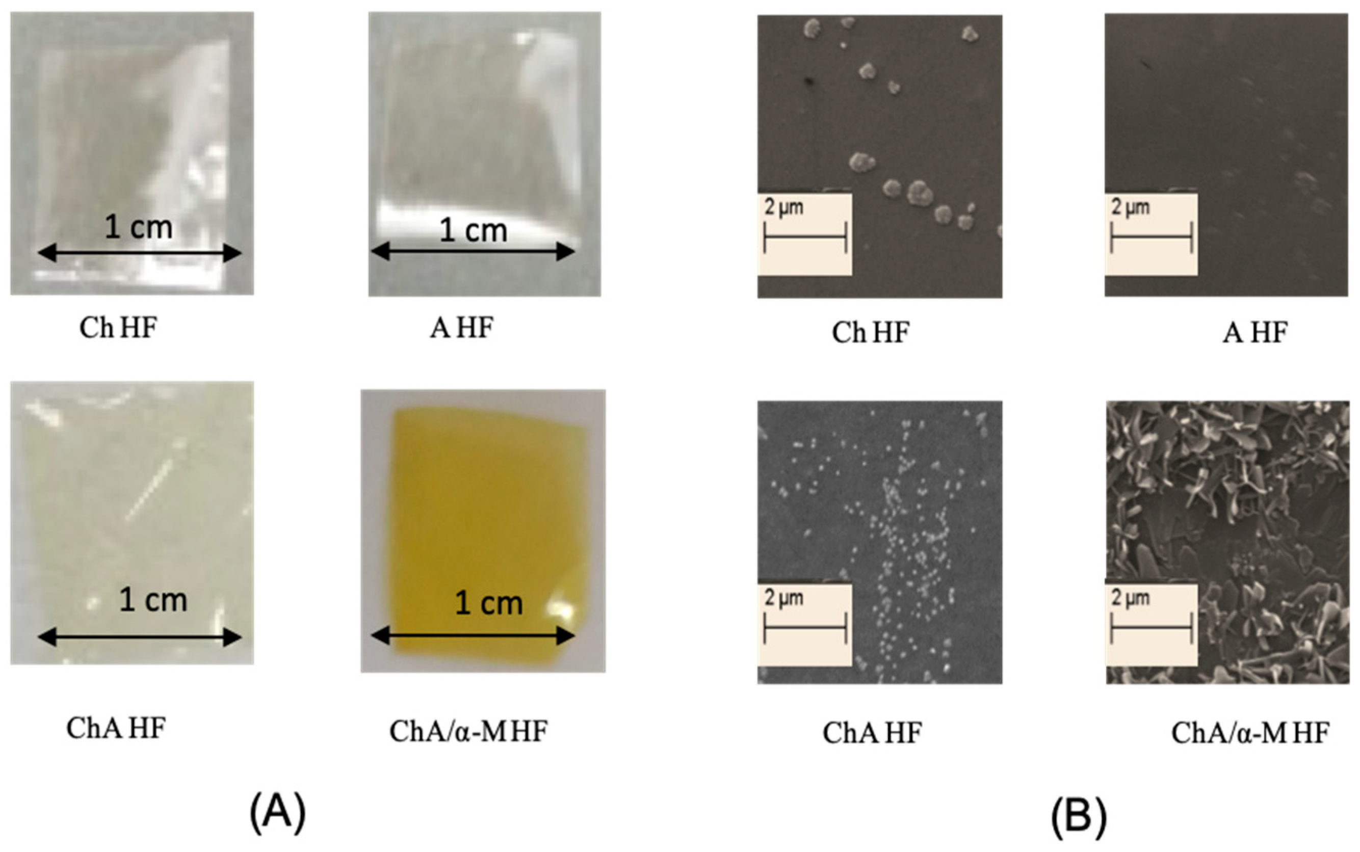
An appropriate carrier is necessary for delivering α-M to the site of action. In this study, ChAlg/α-M HF was prepared as a novel RAS therapy. To prepare ChAlg/α-M HF, firstly, the ChAlg solution was inserted into a propylene box (5 × 5 × 4 cm3) and dried at 60 °C for 24 h to obtain ChAlg HF. Then, α-M (25 mg) in methanol was casted into ChAlg HF [18]. ChAlg HF was successfully prepared using the solvent evaporation method (Figure 2A). The Ch and Alg molecules effectively formed the HF by evaporating the water solvent in a drying process at 60 °C. In addition, ChAlg/α-M HF was successfully prepared with macroscopically smooth.
We next analyzed ChAlg/α-M HF using SEM to confirm that α-M existed in the ChAlg HF. The surface of HFs was observed by SEM at 5000 magnification. The results of SEM clearly showed that α-M existed on the surface of ChAlg HF (Figure 2B) with the halo pattern of Alg–Ch, whether the Alg HF was less homogenous than ChAlg HF. This polyelectrolyte complex film surfaces showed the irregular and porous properties similar to the results in the previous studies [29,30,31,32].
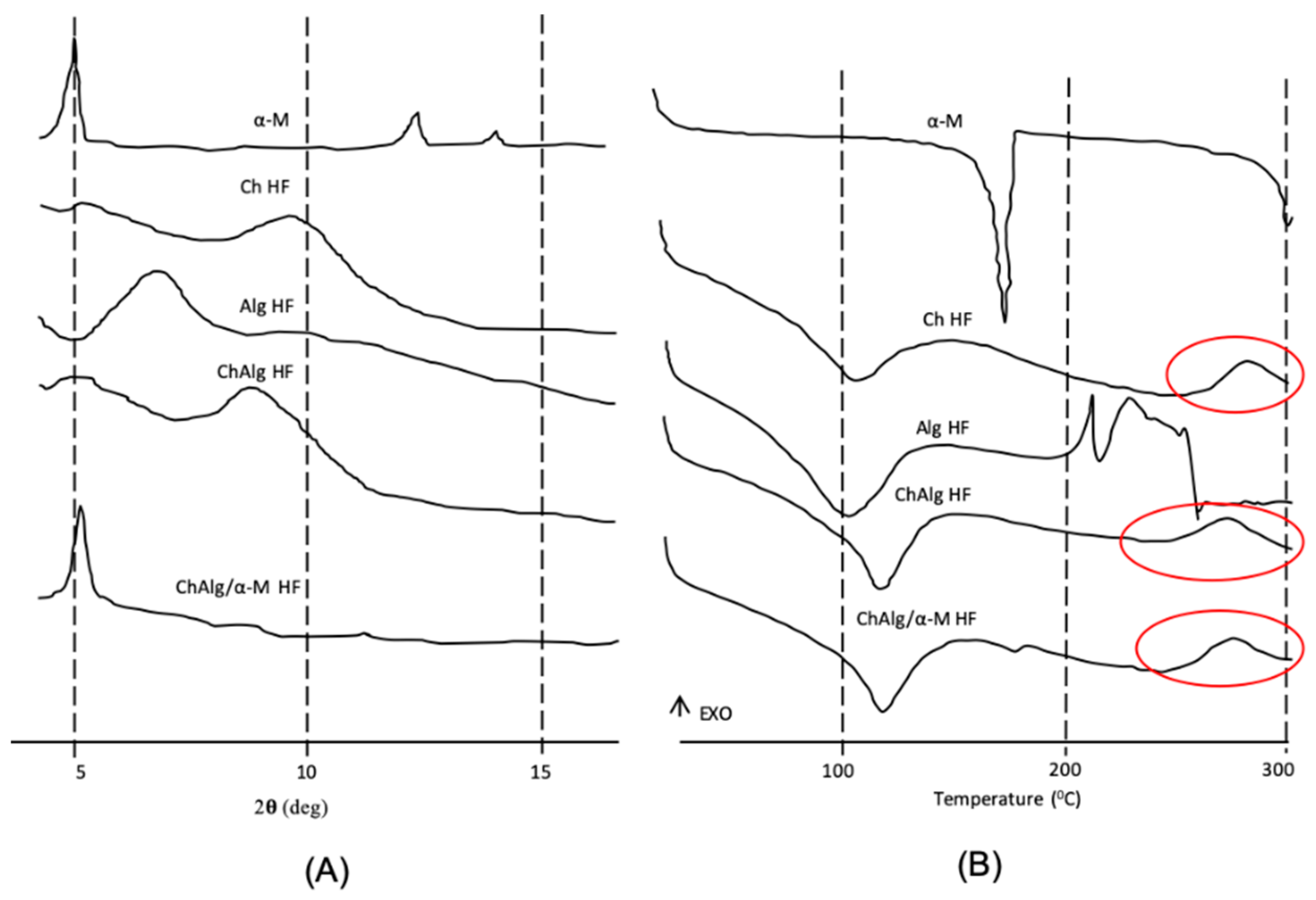
XRD and DSC studies had been done to identify the crystallinity of α-M in HFs. XRD patterns of HFs without α-M (Alg HF, Ch HF, and ChAlg HF) showed a halo pattern, indicating an amorphous form (Figure 3A). In contrast, pure α-Mand ChAlg/α-M HF showed high peaks at around 5°, suggesting that Ch Alg/α-M HF contained α-M. Interestingly, the crystallinity of α-M was decreased in ChAlg/α-M HF as shown by peaks reduction of α-M from around 7.5 to 12.5°. These results corroborate the in silico study which showed a probability to form an interaction with a hydrogen bonding between Ch, Alg, and α-M.
The DSC thermograms presented the endothermic peaks of Ch HF, Alg HF, ChAlg HF, and ChAlg/α-M HF at around 100 °C, representing water evaporation (Figure 3B). This phenomenon maybe related to the amount of free water in HFs. The water molecules in HFs, can be bounded through three polar groups of polymers: A mine, carboxyl, and hydroxyl [33]. The degradation of carboxylic groups in Alg appeared at the temperature range of 200–230 °C. As shown in the DSC thermogram of Ch HF, ChAlg HF, and ChAlg/α-M HF at a temperature range of 270–280 °C. the thermal stability of Alg increased due to complexation between Alg and Ch, representing deacetylation and partial depolymerization of the Ch chains [31]. In addition, a strong endothermic peak of α-M appeared at 179 °C, representing the melting point of α-M. However, it slightly decreased and moved to 180 °C in ChAlg/α-M HF, specifying a lower crystalline of α-M in ChAlg HF compared to pure α-M. The DSC studies imply that α-M interacted with chitosan and alginate in the hydrogel film and these phenomena were supported by previous molecular docking and XRD results.

4.3. Mechanical Properties

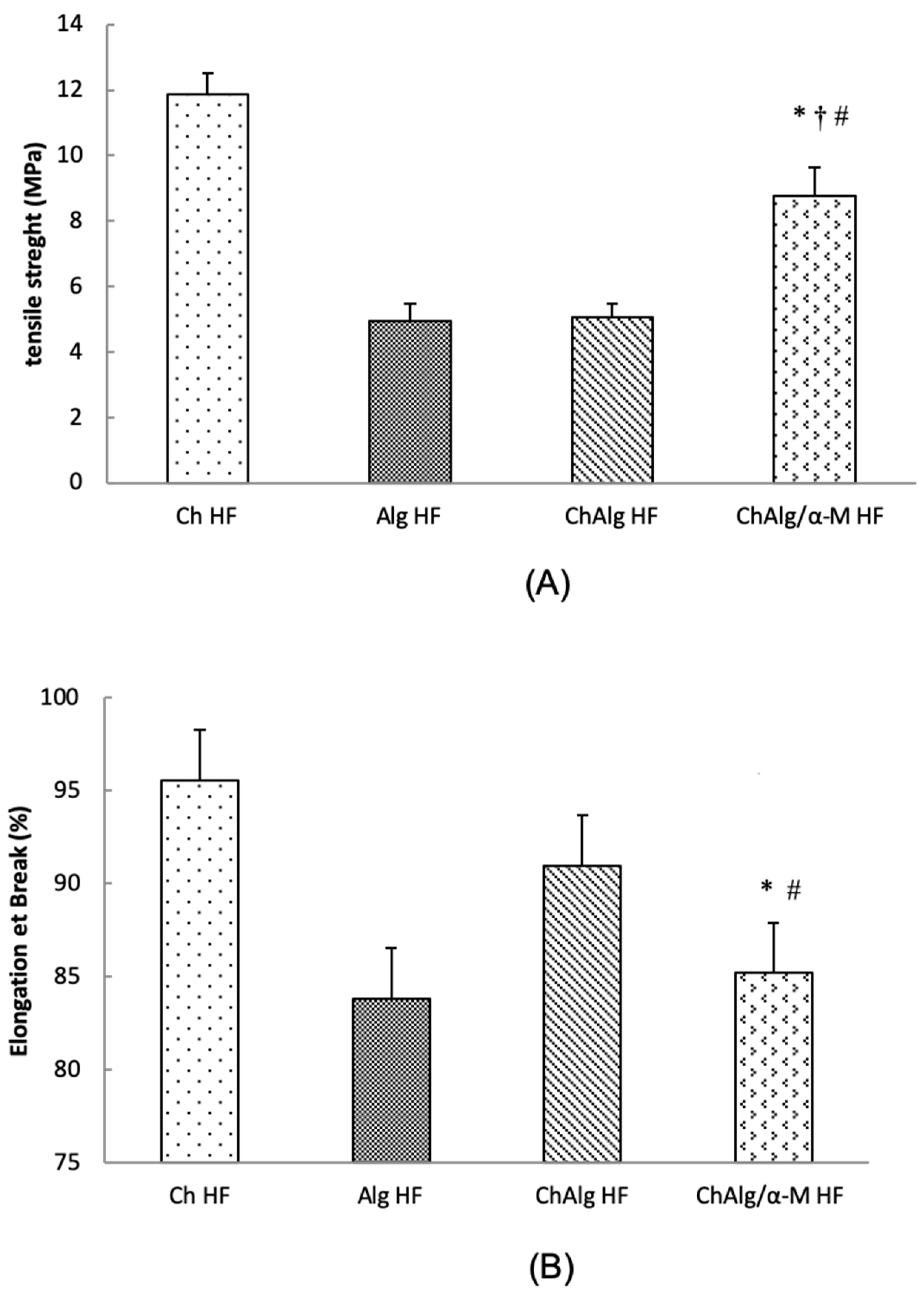
A strong cross linked bond between polymers in HFs will produce a high tensile strength value and an optimum elongation at break [13]. To investigate the effect of α-M in mechanical properties of HFs, tensile strength and elongation at break of HFs were studied. HFs were held between two clips and pulled with the upper clip at a speed of 0.5 mm/s. Then, the results were calculated by tensile strength and elongation at break equations. Figure 4A shows that tensile strength of Ch HF, Alg HF, ChAlg HF, and ChAlg/α-M were 11.88, 4.92, 5.06, and 8.75 MPa, respectively. For the elongation at break study (Figure 4B), Ch HF, Alg HF, ChAlg HF, and ChAlg/α-M HF were 95.57%, 83.82%, 90.98%, and 85.52%, respectively. The tensile strength value is inversely proportional to the value of elongation at break where high tensile resistance will reduce the elasticity [34]. In this study, Ch strengthened the Alg structure and provided the rigid structure of ChAlg/α-M HF, suggesting that a physical interaction occurred between Alg and Ch. The results also imply that α-M increased the tensile strength and decreased the elongation at break of ChAlg HF.

4.4. Degradability Study

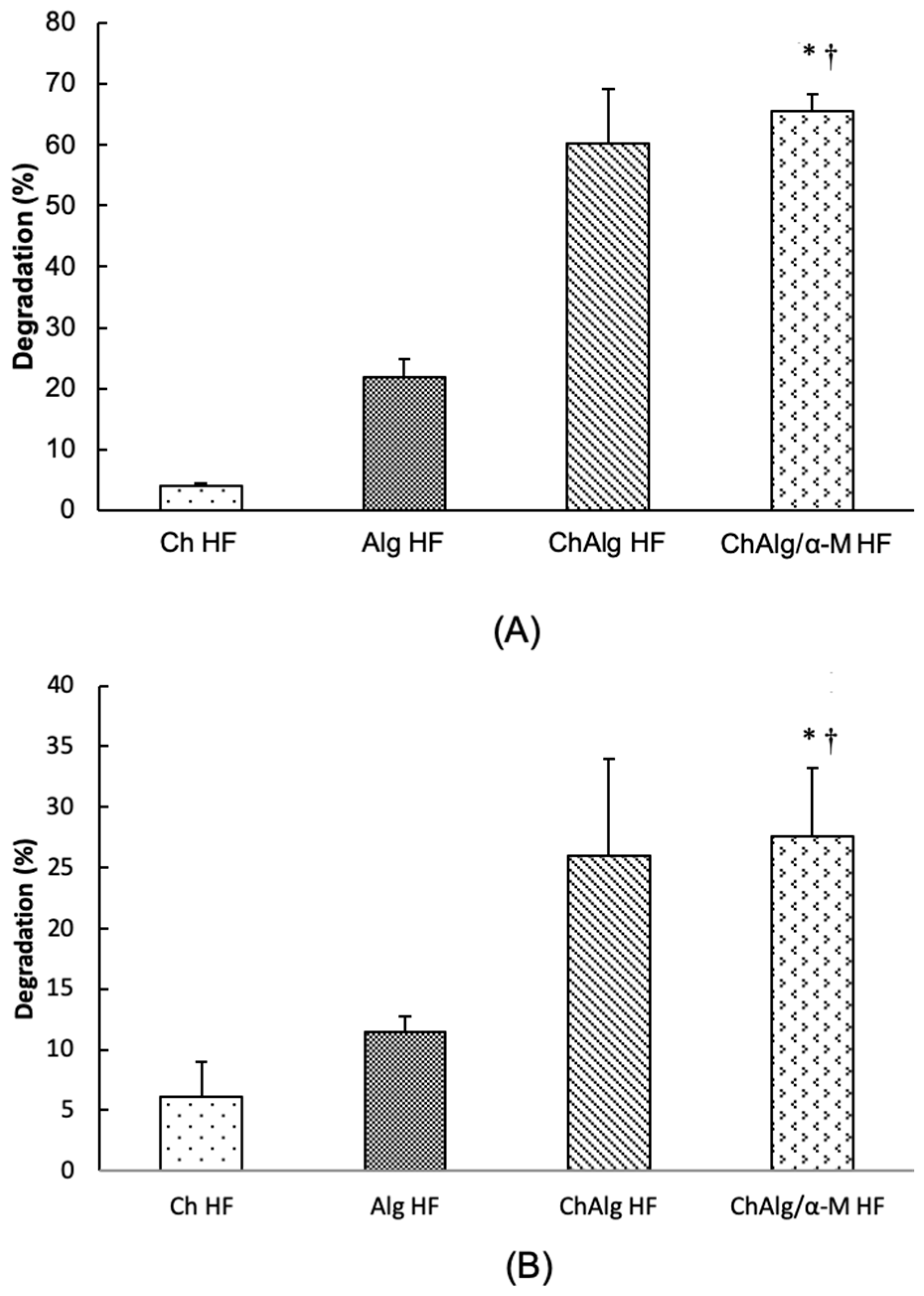
To evaluate the effect of chemicals and enzymes in the degradation of Ch and Alg, degradability studies of HFs were performed using TCA 20% and trypsin. HFs were weighed and soaked in 2 mL of 20% TCA or trypsin solutions, then incubated for 24 h. Figure 5A,B described that not only in TCA, but also in trypsin solutions, ChAlg HF and ChAlg/α-M HF significantly improved the degradability of HFs compared to Ch HF and Alg HF, respectively. In another study, combination of Alg and Ch (2:1) as a microparticle loaded mangosteen rind extract increased 2-fold in releasing α-M from HFs in the colon and intestine compared to a low alginate concentration (0.25:1) [30]. Based on these results, Alg increased the degradability of Ch, and closely related to the release time of α-M from ChAlg HF.

4.5. Swelling Ability

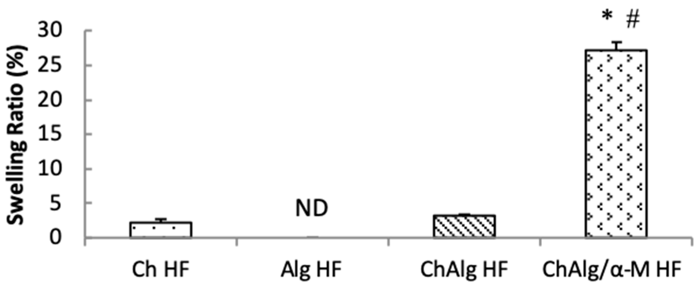
Swelling ability study was conducted to understand the ability of HFs related to liquids absorption in the wound site [35]. In addition, swelling ratio of hydrogel films in RAS therapy is an important parameter for mucoadhesive phenomena. Swollen polymers increase surface contact and mechanical entanglement for hydrogen bonding interactions between the polymer and mucous networks [36,37]. In this study, HFs were weighed and soaked in PBS pH 7.4 for two days. Figure 6 describes that the swelling ratio of Ch HF and ChAlg HF were 2.18 and 3.15, respectively. In addition, Alg HF could not be weighed due to totally dissolved in PBS and did not remain a film formation, indicating that the Alg rapidly dissolve in saliva fluid. The swelling ability of ChAlg polyelectrolyte complexes related to the hydrophilic character of Alg that increase the porosity and surface area of the network leads to improve the swelling ratio of the HF [38,39]. In addition, heparin-based hydrogel containing epidermal growth factor immersed in PBS solution produced swelling values of 14.4 [40]. Ch is a cation which stabilizes the HFs in mucosa surface and strengthen the Alg structure when the HF is swollen and adherent in oral mucosa [41]. Interestingly, the swelling ratio of ChAlg/α-M HF was 27.2 and significantly higher than that of ChAlg HF and Ch HF. α-M in the hydrogel affects the hydrodynamic diameter profile of microgels. When α-M is absorbed in the surface of microgel particle and not in filtrated into matrices of the gel, the gel will be fully swollen [42,43]. In this study, α-M in ChAlg/α-M HF was in the surface of the hydrogel film by intermolecular interaction between α-M at the 21st and 22nd hydrogen atom of Alg increasing the number of water that penetrated the network through hydrogen bond, resulting in the increasing of swelling ratio of ChAlg/α-M HF.

4.6. In Vitro Release Study

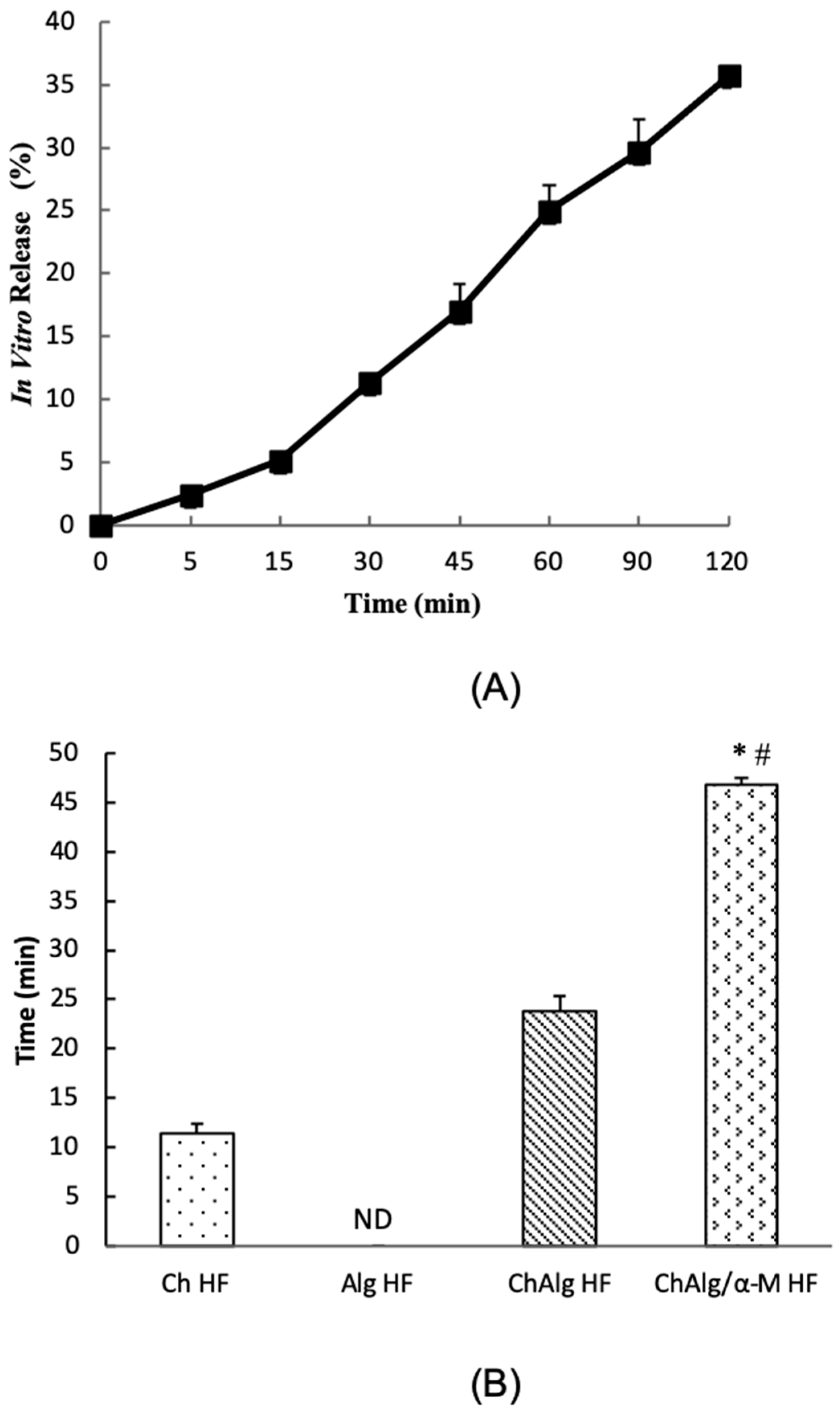
To observe the release mechanism of α-M from ChAlg HF, an in vitro release study of ChAlg/α-M HF was performed in PBS solution (pH 7.4) at 5, 15, 30, 45, 60, 90, and 120 min. The concentrations of α-M were measured by UV spectrophotometry at wavelength of 246 nm [24]. The Q value determines the mechanism of drug release. When Q approaches 0.5, the release mechanism overtakes Fickian diffusion, where the value of Fickian diffusion is 0.5 < Q < 1.0 [25]. Higuchi regression parameters of drug release percentage and quadratic curve of the α-M release time from ChAlg/α-M HF in 2 h confirmed that slope (% min-0,5) and correlation coefficient (Q) were 2.21 ± 0.02 and 0.48 ± 0.09, respectively. The release mechanism of α-M from ChAlg/α-M HF was a matrix type based on Fickian diffusion. As we can see in Figure 7A, the burst release in 2 h was necessary for RAS therapy to provide quick onset of action in oral mucous. Previous study reported that the release of α-M from Ch–Alg microparticle after being immersed in simulated gastric fluid (SGF) and simulated colon fluid (SCF) were burst released for 2 h. The release of α-M in acidic media decreased in the higher Alg concentration, but in neutral condition (in SCF and simulated intestinal fluid/SIF) the presence of Alg increased the α-M release [30]. This phenomenon was caused by the chain relaxation in the electrostatic interaction from the carbonyl group in Alg and amino group in Ch [30,44]. Based on the release properties of α-M, ChAlg/α-M HF is suitable to give an enough time to release the α-M while the ChAlg HF covered the mucous layer.

4.7. Mucoadhesive Study

Finally, to evaluate mucoadhesive properties of ChAlg/α-M HF in the mucous layer, we observed the mucoadhesive time of ChAlg/α-M HF using the fresh mouse mucosa [26]. The mucoadhesive time of Ch HF, ChAlg HF, and ChAlg/α-M HF were 11.5, 23.8, and 46.7 min, respectively (Figure 7B). However, Alg HF was not detected because Alg HF totally dissolved in aqueous liquid. Based on the results, ChAlg/α-M HF significantly improved the mucoadhesive time compared to ChAlg HF. Furthermore, the combination of Ch and Alg in ChAlg HF repaired the mucoadhesive properties of Alg HF. Swelling ratio and adhesive ability of polymers are closely related to the time of attachment abilities of HFs. The combination of polymers increase mechanical bonds between polymers and mucosa [45]. In addition, mucoadhesive ability of Ch based on the formation of hydrogen bond or ionic interaction between the positively charged amino moiety of Ch with negatively charged components of mucus including sialic acid, as well as epithelial surfaces [34,46]. The mucoadhesive polymer system plays an important role in RAS that can cover oral lesions to prevent long-term deterioration of lesions and bacterial proliferation [47]. These results suggest that ChAlg/α-M HF was able to increase the attachment time of ChAlg HF to the mucous layer.

5. Conclusions

Preparation of ChAlg/α-M HF was successfully carried out using the solvent evaporation method. The results of SEM, XRD, and DSC showed that the addition of α-M in ChAlg HF clearly showed that the existence of α-M in ChAlg HF was more homogenous and had a lower crystalline form than pure α-M. In addition, the results of degradability, tensile strength, swelling ratio, and elongation at break confirmed that ChAlg/α-M HF had good physicochemical properties and were suitable for RAS therapy. The in vitro release study showed that the release of α-M from ChAlg/α-M HF showed a release mechanism with the matrix based on Fickian diffusion. Finally, ChAlg/α-M HF was able to prolong the attachment time of ChAlg HF to the mucous layer. These results suggest that ChAlg/α-M HF has the potential as a carrier of α-M for RAS therapy.

Author Contributions

Conceptualization, N.W. and M.M.; methodology, N.W.; software, N.Y.; validation, A.C.; formal analysis, N.Y.; investigation, N.Y.; resources, N.W.; data curation, N.W.; writing—original draft preparation, N.Y.; writing—review and editing, N.W., A.C., and M.M.; visualization, N.Y.; supervision, N.W. and M.M.; project administration, N.Y.; funding acquisition, N.W.

Funding

This research was funded by the Superior Research of Higher Education grant (Penelitian Unggulan Perguruan Tinggi), Ministry of Research and Higher Education, Republic of Indonesia, grant number 1123l/UN6.O/LT/2019.

Acknowledgments

We would like to thank Minister of Research and Higher Education, Republic of Indonesia, for funding this study, Agus Rusdin and Ine Suharyani, for supporting research experiments.

Conflicts of Interest

The authors declare no conflict of interest.

References

  1. Chavan, M.; Jain, H.; Diwan, N.; Khedkar, S.; Shete, A.; Durkar, S. Recurrent aphthous stomatitis: A review. J. Oral Pathol. Med. 2012, 41, 577–583. [Google Scholar] [CrossRef] [PubMed]
  2. Sunarjo, L.; Hendari, R.; Rimbyastuti, H. Manfaat xanthone terhadap kesembuhan ulkus rongga mulut dilihat dari jumlah sel PMN dan fibroblast. Odonto Dent. J. 2015, 2, 14–21. [Google Scholar] [CrossRef]
  3. Kardono, L.B.S.; Basuki, T.; Padmawinata, K. Selected Indonesian Medical Plant Monograph and Description; Penerbit PT Grasindo Gramedia Widiasarana Indonesia: Jakarta, Indonesia, 2003. [Google Scholar]
  4. Zand, N.; Ataie-Fashtami, L.; Djavid, G.E.; Fateh, M.; Alinaghizadeh, M.R.; Fatemi, S.M.; Arbabi-Kalati, F. Relieving pain in minor aphthous stomatitis by a single session of non-thermal carbon dioxide laser irradiation. Lasers Med. Sci. 2009, 24, 515–520. [Google Scholar] [CrossRef] [PubMed]
  5. Obolskiy, D.; Pischel, I.; Siriwatanametanon, N.; Heinrich, M. Garciana mangostana L.: A phytochemical and Pharmacological review. Phytoterapy Res. 2009, 23, 1047–1065. [Google Scholar] [CrossRef] [PubMed]
  6. Zhang, C.; Liu, Y.; Li, W.; Gao, P.; Xiang, D.; Ren, X.; Liu, D. Mucoadhesive buccal film containing ornidazole and dexamethasone for oral ulcers: in vitro and in vivo studies. Pharm. Dev. Technol. 2018, 24, 118–126. [Google Scholar] [CrossRef]
  7. Pedraza-Chaverri, J.; Cárdenas-Rodríguez, N.; Orozco-Ibarra, M.; Pérez-Rojas, J.M. Medicinal properties of mangosteen (Garcinia mangostana). Food Chem. Toxicol. 2008, 46, 3227–3239. [Google Scholar] [CrossRef]
  8. Yaprak Karavana, S.; Güneri, P.; Ertan, G. Benzydamine hydrochloride buccal bioadhesive gels designed for oral ulcers: Preparation, rheological, textural, mucoadhesive and release properties. Pharm. Dev. Technol. 2009, 14, 623–631. [Google Scholar] [CrossRef]
  9. Abdullah, E.; Idris, A.; Saparon, A. Papr reduction using scs-slm technique in stfbc mimo-ofdm. Arpn J. Eng. Appl. Sci. 2017, 12, 3218–3221. [Google Scholar]
  10. Kumar, P.T.S.; Srinivasan, S.; Lakshmanan, V.K.; Tamura, H.; Nair, S.V.; Jayakumar, R. Synthesis, characterization and cytocompatibility studies of α-chitin hydrogel/nano hydroxyapatite composite scaffolds. Int. J. Biol. Macromol. 2011, 49, 20–31. [Google Scholar] [CrossRef]
  11. Qin, Y. Alginate fibers: An overwiew of the production processes and applications in wound management. Polym. Int. 2008, 57, 171–180. [Google Scholar] [CrossRef]
  12. Meng, X.; Tian, F.; Yang, J.; He, C.N.; Xing, N.; Li, F. Chitosan and alginate polyelectrolyte complex membranes and their properties for wound dressing application. J. Mater. Sci. Mater. Med. 2010, 21, 1751–1759. [Google Scholar] [CrossRef] [PubMed]
  13. Rassu, G.; Salis, A.; Porcu, E.P.; Giunchedi, P.; Roldo, M.; Gavini, E. Composite chitosan/alginate hydrogel for controlled release of deferoxamine: A system to potentially treat iron dysregulation diseases. Carbohydr. Polym. 2016, 136, 1338–1347. [Google Scholar] [CrossRef] [PubMed]
  14. Patil, J.S.; Kole, S.G.; Gurav, P.B.; Vilegave, K.V. Natural polymer based mucoadhesive hydrogel beads of nizatidine: Preparation, characterization and evaluation. Indian J. Pharm. Educ. Res. 2016, 50, 159–169. [Google Scholar] [CrossRef]
  15. Kilicarslan, M.; Ilhan, M.; Inal, O.; Orhan, K. Preparation and evaluation of clindamycin phosphate loaded chitosan/alginate polyelectrolyte complex film as mucoadhesive drug delivery system for periodontal therapy. Eur. J. Pharm. Sci. 2018, 123, 441–451. [Google Scholar] [CrossRef]
  16. Menchicchi, B.; Fuenzalida, J.P.; Bobbili, K.B.; Hensel, A.; Swamy, M.J.; Goycoolea, F.M. Structure of chitosan determines its interactions with mucin. Biomacromolecules 2014, 15, 3550–3558. [Google Scholar] [CrossRef]
  17. Muchtaridi, M.; Yusuf, M. Molecular Docking, 1st ed.; Unpad Press: Umedang Regenc, Indonesia, 2018. [Google Scholar]
  18. Wathoni, N.; Motoyama, K.; Higashi, T.; Okajima, M.; Kaneko, T.; Arima, H. Physically crosslinked-sacran hydrogel films for wound dressing application. Int. J. Biol. Macromol. 2016, 89, 465–470. [Google Scholar] [CrossRef]
  19. Wathoni, N.; Motoyama, K.; Higashi, T.; Okajima, M.; Kaneko, T. Enhancement of curcumin wound healing ability by complexation with 2-hydroxypropyl-Y-cyclodextrin in sacran hydrogel film. Int. J. Biol. Macromol. 2017, 98, 268–276. [Google Scholar] [CrossRef]
  20. Wathoni, N.; Hasanah, A.N.; Mohammed, A.F.A.; Pratiwi, E.D.; Mahmudah, R. Accelerated wound healing ability of sacran hydrogel film by keratinocyte growth factor in alloxan-induced diabetic mice. Int. J. Appl. Pharm. 2018, 10, 57–61. [Google Scholar] [CrossRef]
  21. Wathoni, N.; Insani, U.C. Characterization and optimization of natural maltodextrin-based niosome. J. Appl. Pharm. Sci. 2013, 3, 68–71. [Google Scholar]
  22. Wathoni, N.; Rusdiana, T.; Hasanah, A.N.; Muhtadi, A.; Pratiwi, E.D.; Mahmudah, R.; Mohammed, A.F.A.; Okajima, M.; Kaneko, T.; Arima, H. Sacran Hydrogel Film Containing Keratinocyte Growth Factor Accelerates Wound Healing by Stimulating Fibroblast Migration and Re-epithelization. Chem. Pharm. Bull. 2019, 67, 849–854. [Google Scholar] [CrossRef]
  23. Wathoni, N.; Yuan Shan, C.; Yi Shan, W.; Rostinawati, T.; Indradi, R.B.; Pratiwi, R.; Muchtaridi, M. Characterization and antioxidant activity of pectin from Indonesian mangosteen (Garcinia mangostana L.) rind. Heliyon 2019, 5, e02299. [Google Scholar] [CrossRef] [PubMed]
  24. Mohamed, M.I.; Haider, M.; Ali, M.A.M. Buccal Mucoadhesive Films Containing Antihypertensive Drug. J. Chem. Pharm. Res. 2011, 3, 665–686. [Google Scholar]
  25. Motoyama, K.; Tanida, Y.; Hata, K.; Hayashi, T.; Higashi, T.; Ishitsuka, Y.; Kondo, Y.; Irie, T.; Kaneko, S.; Arima, H. Potential Use of a Megamolecular Polysaccharide Sacran as a Hydrogel-Based Sustained Release System. Chem. Pharm. Bull. 2014, 62, 636–641. [Google Scholar] [CrossRef] [PubMed]
  26. Lala, R.; Thorat, A.A.; Gargote, C.S.; Awari, N.G. Preparation of buccoadhesive polymeric film of ketoprofen and its evaluation. Asian J. Pharm. Sci. 2011, 6, 267–274. [Google Scholar]
  27. Smitha, B.; Sridhar, S.; Khan, A.A. Chitosan-sodium alginate polyion complexes as fuel cell membranes. Eur. Polym. J. 2005. [Google Scholar] [CrossRef]
  28. Sæther, H.V.; Holme, H.K.; Maurstad, G.; Smidsrød, O.; Stokke, B.T. Polyelectrolyte complex formation using alginate and chitosan. Carbohydr. Polym. 2008, 74, 813–821. [Google Scholar] [CrossRef]
  29. Yu, C.Y.; Yin, B.C.; Zhang, W.; Cheng, S.X.; Zhang, X.Z.; Zhuo, R.X. Composite microparticle drug delivery systems based on chitosan, alginate and pectin with improved pH-sensitive drug release property. Colloids Surf. B Biointerfaces 2009, 68, 245–249. [Google Scholar] [CrossRef]
  30. Mulia, K.; Halimah, N.; Krisanti, E. Effect of alginate composition on profile release and characteristics of chitosanalginate microparticles loaded with mangosteen extract. In Proceedings of the AIP Conference, Vladivostok, Russia, 18–22 September 2016; American Institute of Physics: College Park, MD, USA, 2017; p. 020010. [Google Scholar]
  31. Kulig, D.; Zimoch-Korzycka, A.; Jarmoluk, A.; Marycz, K. Study on alginate-chitosan complex formed with different polymers ratio. Polymers 2016, 8, 167. [Google Scholar] [CrossRef]
  32. Bhardwaj, N.; Kundu, S.C. Silk fibroin protein and chitosan polyelectrolyte complex porous scaffolds for tissue engineering applications. Carbohydr. Polym. 2011, 85, 325–333. [Google Scholar] [CrossRef]
  33. Neto, C.G.T.; Giacometti, J.A.; Job, A.E.; Ferreira, F.C.; Fonseca, J.L.C.; Pereira, M. Thermal analysis of chitosan based networks. Carbohydr. Polym. 2005, 62, 97–103. [Google Scholar] [CrossRef]
  34. Jacobsen, J.; Meng-Lund, E.; Muff-Westergaard, C.; Sander, C.; Madelung, P. A mechanistic based approach for enhancing buccal mucoadhesion of chitosan. Int. J. Pharm. 2014, 461, 280–285. [Google Scholar]
  35. Sarheed, O.; Rasool, B.K.A.; Abu-gharbieh, E.; Aziz, U.S. An Investigation and Characterization on Alginate Hydogel Dressing Loaded with Metronidazole Prepared by Combined Inotropic Gelation and Freeze-Thawing Cycles for Controlled Release. Aaps Pharmscitech. 2015, 16, 601–609. [Google Scholar] [CrossRef] [PubMed]
  36. Özyazıcı, M.; Fırlak, M.; Tanrıverdı, S.T.; Rençber, S.; Karavana, Y.; Kahraman, M.V. Bioadhesive Gel and Hydrogel Systems for Buccal Delivery of Ketoprofen: Preparation and In vitro Evaluation Studies. Am. J. Drug Deliv. 2015, 2, 078–091. [Google Scholar]
  37. Castel-Molieres, M.; Conzatti, G.; Torrisani, J.; Rouilly, A.; Cavalie, S.; Carrere, N.; Tourrette, A. Influence of Homogenization Technique and Blend Ratio on Chitosan/Alginate Polyelectrolyte Complex Properties. J. Med. Biol. Eng. 2017, 38, 10–21. [Google Scholar] [CrossRef]
  38. Conzatti, G.; Faucon, D.; Castel, M.; Ayadi, F.; Cavalie, S.; Tourrette, A. Alginate/chitosan polyelectrolyte complexes: A comparative study of the influence of the drying step on physicochemical properties. Carbohydr. Polym. 2017, 15, 142–151. [Google Scholar] [CrossRef]
  39. Quraishi, S.; Martins, M.; Barros, A.A.; Gurikov, P.; Raman, S.P.; Smirnova, I.; Duarte, A.R.C.; Reis, R.L. Novel non-cytotoxic alginate–lignin hybrid aerogels as scaffolds for tissue engineering. J. Supercrit. Fluids 2015, 105, 1–8. [Google Scholar] [CrossRef]
  40. Goh, M.C.; Hwang, Y.; Tae, G. Epidermal growth factor loaded heparin-based hydrogel sheet for skin wound healing. Carbohydr. Polym. 2016, 147, 251–260. [Google Scholar] [CrossRef]
  41. Li, X.; Ouyang, J.; Yang, H.; Chang, S. Chitosan modified halloysite nanotubes as emerging porous microspheres for drug carrier. Appl. Clay Sci. 2016, 126, 306–312. [Google Scholar] [CrossRef]
  42. Lazim, A.M.; Eastoe, J.; Bradley, M.; Trickett, K.; Rogers, S.E. Recovery of gold nanoparticles using pH-sensitive microgels. Soft Matter 2010, 6, 2050–2055. [Google Scholar] [CrossRef]
  43. Ahmad, M.; Yamin, B.M.; Lazim, A.M. A study on dispersion and characterisation of α -mangostin loaded pH sensitive microgel systems. Chem. Cent. J. 2013, 7, 1. [Google Scholar] [CrossRef] [Green Version]
  44. Rajendran, A.; Basu, S.K. Alginate-Chitosan Particulate System for Sustained Release of Nimodipine. Trop. J. Pharm. Res. 2009, 8, 433–440. [Google Scholar] [CrossRef] [Green Version]
  45. Das Neves, J.; Sarmento, B. Mucosal delivery of biopharmaceuticals: Biology, challenges and strategies. Mucosal Deliv. Biopharm. Biol. Chall. Strat. 2014, 9781461495, 1–601. [Google Scholar]
  46. Ways, T.M.M.; Lau, W.M.; Khutoryanskiy, V.V. Chitosan and its derivatives for application in mucoadhesive drug delivery systems. Polymers 2018, 10, 267. [Google Scholar] [CrossRef] [PubMed] [Green Version]
  47. Sizílio, R.H.; Galvão, J.G.; Trindade, G.G.G.; Pina, L.T.S.; Andrade, L.N.; Gonsalves, J.K.M.C.; Lira, A.A.M.; Chaud, M.V.; Alves, T.F.R.; Arguelho, M.L.P.M.; et al. Chitosan/pvp-based mucoadhesive membranes as a promising delivery system of betamethasone-17-valerate for aphthous stomatitis. Carbohydr. Polym. 2018, 190, 339–345. [Google Scholar] [CrossRef] [PubMed]
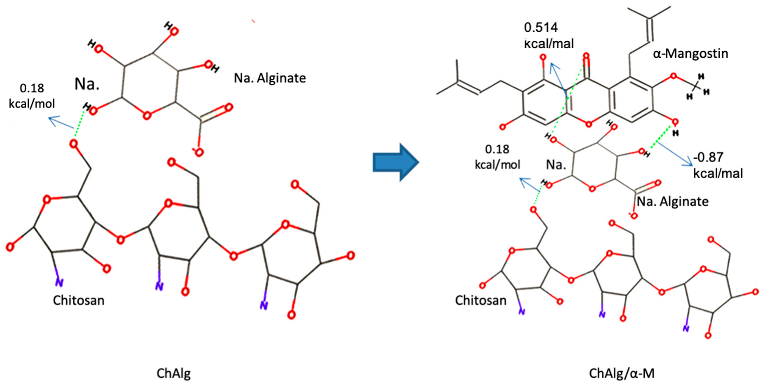
Figure 1. In silico studies of chitosan (Ch), alginate (Alg), and α-Mangostin (α-M). Discovery studio visualizer and AutoDock 1.6 applications were applied for molecular docking studies.
Applsci 09 05235 g001
Figure 2. Macroscopic and microscopic appearances of hydrogel films (HFs). (A) Macroscopic appearances of ChAlg/α-M HF; (B) SEM Analysis of ChAlg/α-M HF. The experiments were performed three times, and representative data are shown.
Applsci 09 05235 g002
Figure 3. Characterization of ChAlg/α-M HF. (A) XRD patterns; (B) DSC thermographs. The experiments were performed three times, and representative data are shown.
Applsci 09 05235 g003
Figure 4. Mechanical properties of ChAlg/α-M HF. (A) Tensile strength; (B) elongation at break. Each value represents the mean ± SEM of three experiments. * p < 0.05, compared to the Ch HF. † p < 0.05, compared to the Alg HF. # p < 0.05, compared to the ChAlg HF.
Applsci 09 05235 g004
Figure 5. Degradability study of ChAlg/α-M HF. (A) Trypsin; (B) TCA (trichloroacetic acid). Each value represents the mean ± SEM of three experiments. * p < 0.05, compared to the Ch HF. † p < 0.05, compared to the Alg HF.
Applsci 09 05235 g005
Figure 6. Swelling ratio of ChAlg/α-M HF. Each value represents the mean ± SEM of three experiments. * p < 0.05, compared to the Ch HF. # p < 0.05, compared to the ChAlg HF.
Applsci 09 05235 g006
Figure 7. In vitro studies of ChAlg/α-M HF. (A) In vitro release of α-M from ChAlg/α-M HF; (B) mucoadhesive study of ChAlg/α-M HF. Each value represents the mean ± SEM of three experiments. * p < 0.05, compared to the Ch HF. # p < 0.05, compared to the ChAlg HF.
Applsci 09 05235 g007