Article

Feasibility of a 3D Surgical Guide Technique for Impacted Supernumerary Tooth Extraction: A Pilot Study with 3D Printed Simulation Models

1
Department of Prosthodontics, School of Dentistry, Pusan National University, Yangsan 46241, Korea
2
Department of Pediatric Dentistry, School of Dentistry, Kyung Hee University, Seoul 02447, Korea
3
Department of Biomedical Engineering, College of Medical Science, Ewha Womans University, Seoul 03760, Korea
4
Department of Pediatric Dentistry, School of Dentistry, Chosun University, Gwangju 61452, Korea
5
Department of Oral and Maxillofacial Surgery, School of Dentistry, Kyung Hee University, Seoul 02447, Korea
*
Author to whom correspondence should be addressed.
Appl. Sci. 2019, 9(18), 3905; https://doi.org/10.3390/app9183905
Submission received: 21 August 2019 / Revised: 4 September 2019 / Accepted: 13 September 2019 / Published: 18 September 2019
(This article belongs to the Special Issue Application of CAD/CAM and 3D Printing Technologies in Dentistry)

Abstract

:
This study aimed to evaluate the feasibility of a 3-dimensional (3D) planned surgical guide technique designed for impacted supernumerary teeth (STs) extraction using 3D printed simulation models. In total, 17 participants from two university hospitals were recruited. Each participant performed surgery with both the conventional and surgical guide techniques. The following parameters were evaluated: (1) The time required for ST extraction, (2) the area of the window opening, and (3) the volume of the material removed. Time required for ST extraction was 213.65 ± 167.45 sec and 114.76 ± 42.87 sec in the conventional and surgical guide techniques, respectively, with significant differences (p = 0.028). The required area of the window opening was 48.10 ± 9.44 mm2 and 45.90 ± 8.17 mm2, respectively, in the conventional and surgical guide techniques, with no significant differences (p > 0.05). The required volume of the material removed was 121.65 ± 43.69 mm3 and 99.12 ± 17.88 mm3 in the conventional and surgical guide techniques, respectively, with significant differences (p = 0.031). The 3D planned surgical guide technique was effective for impacted ST extraction. Within the limits of this study, the surgical guide technique allows us to accomplish minimally invasive surgery within a shorter duration.

1. Introduction

Supernumerary teeth (STs) are developmental anomalies that can affect the eruption of adjacent permanent teeth [1]. Most STs are observed in the anterior region of the maxilla and approximately 75% of STs are impacted [2]. STs can cause various clinical complications, such as ectopic and delayed eruption of permanent teeth, displacement or rotation of the central incisors, and cystic degeneration [1,3]. Thus, early extraction of STs in the mixed dentition is usually recommended prior to occurrence of ST-associated complications.
As STs in the early mixed dentition are often located close to the adjacent permanent teeth, removal of the STs can involve complications [4,5]. Moreover, due to the complex anatomy of the maxillary anterior region, there is a possibility of damaging adjacent permanent tooth germs or removing the wrong tooth [6]. Extraction of impacted ST is often challenging for dental surgeons.
For impacted ST extraction, it is essential to confirm 3-dimensional (3D) localization of the tooth. Precise localization of ST is a critical step during surgery because it can affect the operating time, predictability of the surgery, and the incidence of damage to adjacent structures [7,8]. For this purpose, cone-beam computed tomography (CBCT) is widely used to provide location data of impacted STs. Minimally invasive and efficient surgery can be achieved with the aid of CBCT [9].
Surgical guides fabricated with computer-aided design (CAD) and computer-aided manufacturing (CAM) technology have been shown to be effective in maxillofacial surgeries [10,11]. Efforts have been made to facilitate impacted ST extraction employing CAD/CAM surgical guide surgery [6,7,12]. However, previous reports are limited to case studies and a lack of reports on the feasibility of surgical guide surgery with respect to impacted STs is noted, owing to the limitations in simulating the same conditions of impacted STs in clinical circumstances. Thus, we designed a 3D printed simulation model for impacted ST extraction. A 3D printed simulation model and 3D analysis can facilitate various measurements regarding the amount of window opening or bone removal, and duration of surgery. Therefore, this study aimed to evaluate the feasibility of the surgical guide technique for impacted ST extraction using three-dimensional printed simulation models.

2. Materials and Methods

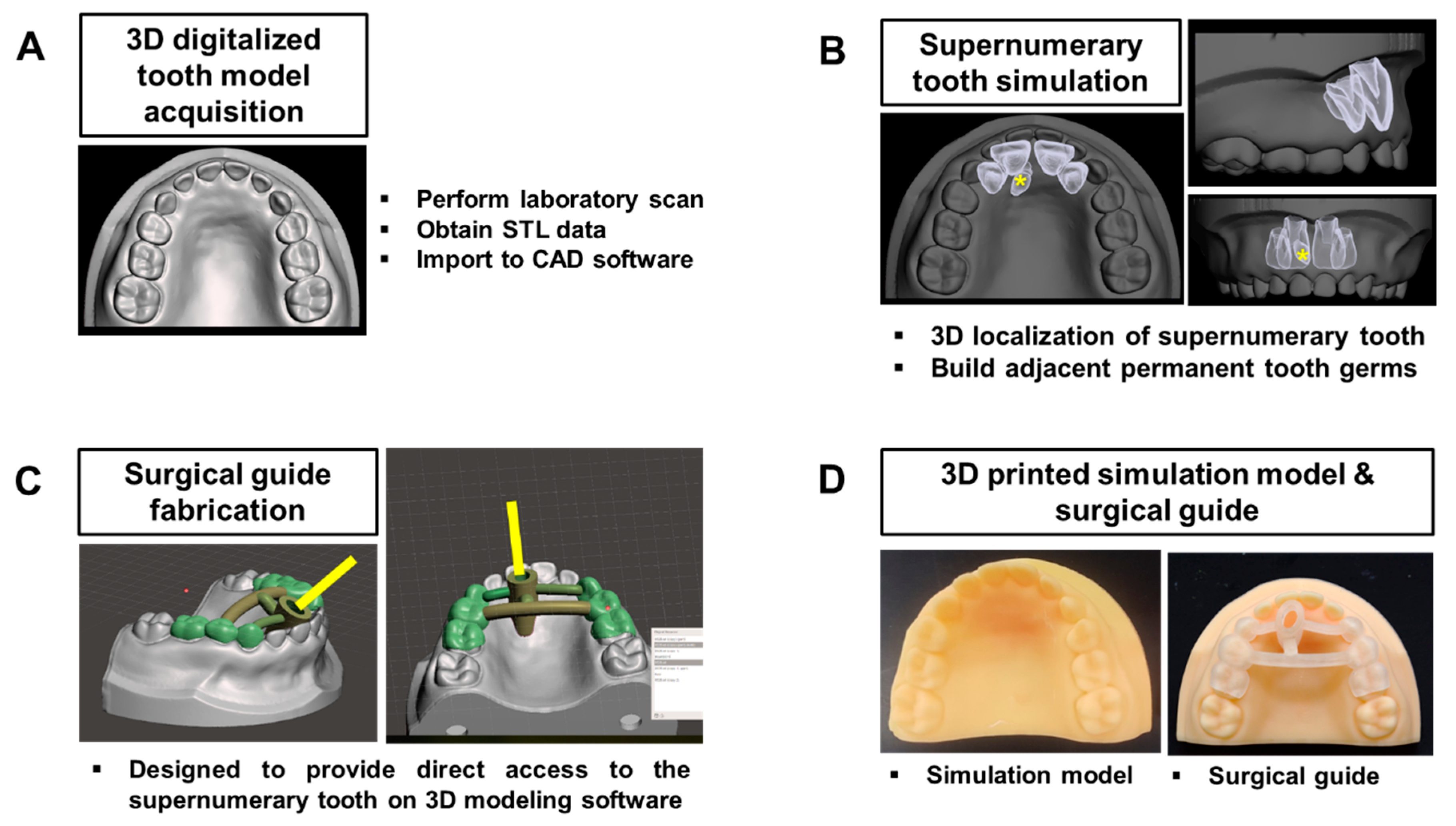
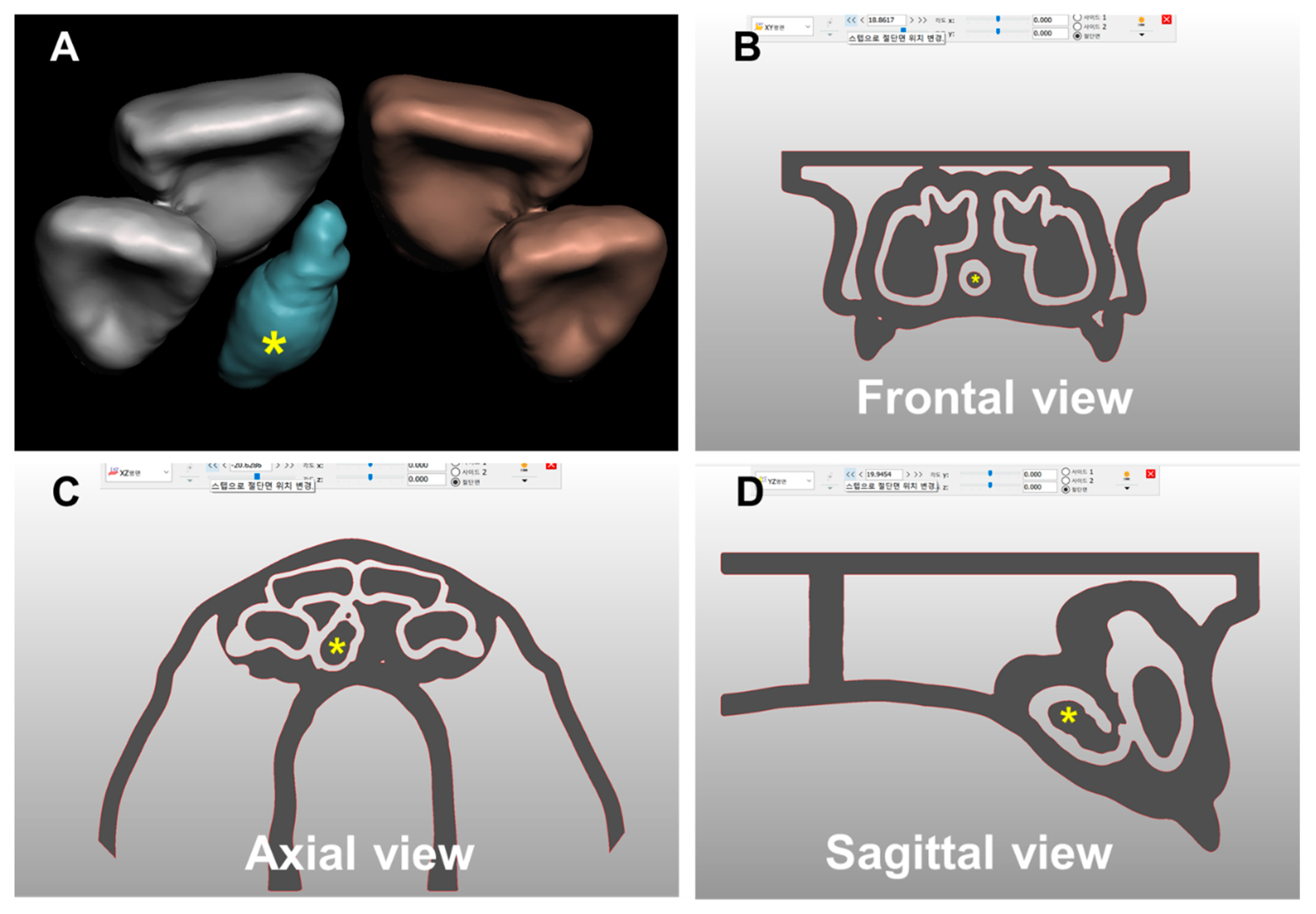
2.1. Development of the 3D Printed Simulation Model and Surgical Guide for ST

The 3D printed simulation model for ST was designed following human anatomy data in primary dentition through a digital workflow (Figure 1). A laboratory scanner (T500, Medit, Seoul, Korea) was used to yield digital scans of the primary dentition model (Dentoform, Nissin, Kyoto, Japan). All scans were saved as an STL file and imported into the 3D modeling software (Meshmixer, Autodesk, San Rafael, CA, USA). We created a digitalized 3D tooth model with 3 mm of uniform thickness, using scanned data of the primary dentition model. In the digitalized 3D tooth model, an impacted ST and adjacent permanent tooth germs were embedded to imitate the natural positional relationship in the primary dentition (Figure 2).
A digitalized surgical guide was made to guide surgical drilling along the 3D position of the impacted ST. The orientation of drilling was designed to avoid damage to the adjacent permanent tooth germs and provide direct access to the impacted ST, minimizing the amount of material removed during surgery.
The 3D printing process was performed at DIO (Dio Co., Busan, Korea). The STL files of the final digitalized 3D tooth model and surgical guide for ST were imported into the 3D printer (DIO PROBO (Dio Co., Busan, Korea)) for 3D printing. Optically cured resin material (DIOnavi-SG, Dio Co., Busan, Korea) was used as the 3D printing material.

2.2. Participant Enrollment

In total, 17 interns and residents participated in this study, with 4 interns and 7 residents from the Department of Pediatric Dentistry, School of Dentistry, Kyung Hee University and 6 residents from that of Chosun University. All participants had previously observed or performed ST extraction surgeries and studied the procedure during their education and training.

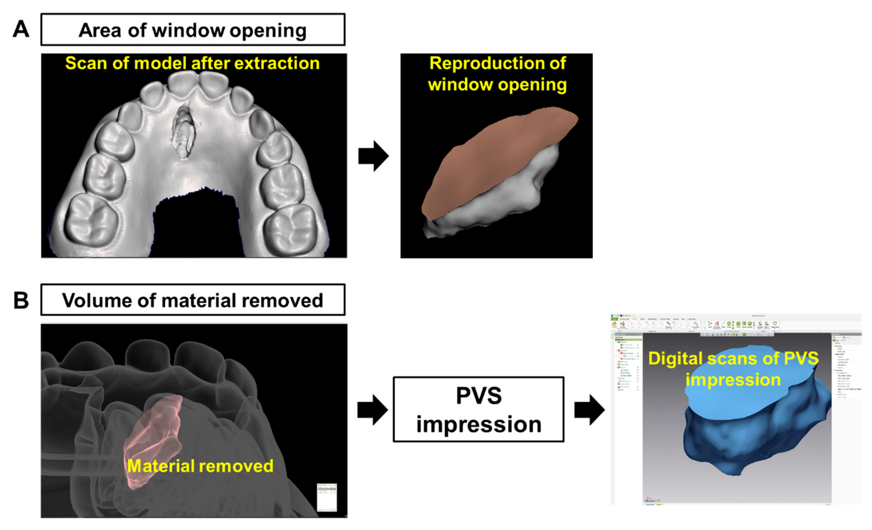
2.3. Evaluating Feasibility of Surgical Guide for Impacted ST Extraction

To evaluate the feasibility of surgical guides for impacted ST extraction, each of the 3D printed tooth models and surgical guides was subjected to a simulated surgical procedure using a low-speed drill with a straight round burr. Initially a wax carver was used for the replacement of a tooth elevator, considering the mechanical strength of the models. The experiment was performed using 2 different sequences: (1) Surgical extraction without surgical guide (conventional technique), and (2) surgical extraction with the surgical guide (surgical guide technique). Each sequence was performed once by each participant. Considering that localization information acquired during the first extraction sequence with the conventional technique could affect the results of the second sequence, the second extraction with the surgical guide technique was performed after two weeks. 3D images of the model were provided to the participants, as substitutes of the CBCT images. For evaluation, each participant extracted an impacted ST using the conventional technique. Subsequently, the participants extracted impacted STs using the surgical guide technique. The following parameters were evaluated: (1) The time required for ST extraction, (2) the area of the window opening, and (3) the 3D volume of the material removed.
The time required for ST extraction was defined as the time from placing a straight round bur onto the tooth model until the ST was completely removed from the model. After ST extraction, the outer surfaces of the models were scanned using a laboratory scanner. The inner surfaces of the models were reproduced as polyvinylsiloxane (PVS) impressions (Imprint 3; 3M ESPE, Maplewood, MN, USA). Next, the impressions were carefully removed and the PVS surfaces were scanned and saved as STL files. All scans were imported into the 3D analysis software (Geomagic Control-X, 3D systems, Morrisville, NY, USA). Finally, we calculated the area of the window opening, and the 3D volume of the material removed, using the software (Figure 3).

2.4. Statistical Analysis

The results are represented as mean ± standard deviation. Data were analyzed using SPSS version 20.0 (SPSS Inc., Chicago, IL, USA). The normalized test was assessed using the Kolmogorov-Smirnov test, which revealed that the data were not normally distributed. Therefore, the data were analyzed using the Wilcoxon signed-rank test. The p-values < 0.05 indicated statistical significance.

3. Results

The average time for the participants to complete ST extraction was 213.65 ± 167.45 sec with the conventional technique and 114.76 ± 42.87 sec with the surgical guide technique (Table 1). Significantly less time was required for impacted ST extraction with the surgical guide technique (p = 0.028). Table 2 shows the results of the area of the window opening and the volume of the material removed for ST extraction. The required area of the window opening was 48.10 ± 9.44 mm2 with the conventional technique and 45.90 ± 8.17 mm2 with the surgical guide technique with no significant differences (p > 0.05). The required volume of the material removed was 121.65 ± 43.69 mm3 using the conventional technique and 99.12 ± 17.88 mm3 using the surgical guide technique. Significantly less material removal was required for impacted ST extraction with the surgical guide technique (p = 0.031).

4. Discussion

3D printing refers to rapid prototyping manufacturing technology that makes products by adding materials layer by layer. Contrary to the use of inks in traditional printers, 3D printers are built with powders using the blueprints made by CAD software [13,14]. Owing to the advances in 3D printing, the surgical guide technique, used in dentistry, has been a remarkable success. With its patient-customizable and reproducible property, it has made dental surgery easier and more predictable with reduced risk [15,16]. The surgical guide technique is also promising for impacted ST extraction.
The results of this study showed that less time was required for completing ST extraction using the surgical guide technique than the conventional technique. The guide technique simplified the whole surgery procedure, resulting in shorter operation time. This may be because the guide technique helped the surgeons understand the precise 3D localization of STs, and provided direct access which increased the surgeons’ confidence in performing the surgery and positively affected the operation time. A previous clinical study of eight patients with impacted STs revealed that the average operation time of the surgical guide technique for impacted ST extraction was approximately 16 min, which was approximately 4 min shorter than that of the conventional technique [17]. In the present study, the surgical guide technique needed approximately 2 min, whereas the conventional technique needed approximately 3.5 min. This difference may be related to the study design. Because the previous study was designed based on patient data, the results were greater than those obtained in this study. This study was designed under controlled conditions without the need for some clinical procedures, such as the flap elevation procedure for impacted ST and the suture procedure after impacted ST extraction. Only patients with deeply impacted ST were included in the previous study and large amounts of osteotomy were conducted to extract impacted ST. Thus, degrees of surgical difficulty could be responsible for the different results. Moreover, the physical strength differences between human bone tissue and our model material should not be ignored.
This study demonstrated that the surgical technique allowed reduced operation times for impacted ST removal. If general anesthesia is planned for impacted ST extraction, the guide technique will reduce the incidence of perioperative complications, as prolonged anesthesia is a predisposing factor of perioperative adverse events [18]. Furthermore, the guide technique can be used for impacted ST extraction also with local anesthesia. Reduced operation time can positively affect children’s behavior in the dental setting. Prolonged dental treatment is correlated with negative behaviors in children [19,20]. In addition, reduced operation time can also decrease the chances of postoperative complications. A previous study regarding lower third molar surgery reported that a prolonged operation time was associated with acute postoperative complications, such as severe pain and swelling [21].
Regarding the area of the window opening, the surgical guide technique did not reduce the size compared with the conventional technique. In fact, impacted ST extraction needs preparation of an access hole to the impacted ST by drilling into the underlying bone tissue. Once exposure of the impacted ST is visible, the hole should be enlarged enough to easily luxate and remove the impacted ST. Because the size of the window opening is mainly determined by the vertical and/or transverse dimensions of the impacted ST, the result may remain unaffected by the adoption of the surgical guide technique.
This study also demonstrated that the surgical guide technique reduced the amount of volume loss of material than the conventional technique did. This finding indicates that adoption of the surgical guide technique can facilitate minimally invasive surgeries for impacted ST extraction. With the aid of the surgical guide, volume loss of the material was reduced, which led to minimal trauma to the adjacent permanent tooth germs. Moreover, these factors may lead to a shortened healing period and reduce the chance of postoperative complications [22].
Interestingly, there were marked differences in the standard deviation of the volume loss of the material between the surgical guide technique and the conventional technique. This finding indicates that the surgical guide technique can reduce the difficulty of the surgery for impacted STs, regardless of the surgeons’ skill level or experience. This finding is in accordance with a previous study of implant placement with a surgical guide, which demonstrated that accurate implant position reproducibility was not influenced by surgeons’ experience level [23]. The study concluded that the surgical guide technique provided better results by surgeons with insufficient experience. Similarly, the effect of the surgical guide did not depend on the surgeons’ skill level or experience in this study.
There were some limitations to this study. First, the design was that of an in vitro study under controlled conditions and the number of participants in the study was relatively small. Next, we considered only one vertical position of impacted STs, which may have affected the degree of surgical difficulty. Additionally, other factors, such as the flap elevation and the suture procedure, were not considered which may have affected the results. Because this study was performed with 3D printed models, reproduction of gingival tissues was technically difficult. Finally, even though the 3D printed model in this study mimicked the natural anatomical structures of children, natural human bone density and strength were difficult to completely reproduce.
In conclusion, we developed a 3D printed simulation model and a 3D planned surgical guide for impacted ST extraction. Within the limits of this study, the 3D planned surgical guide technique was effective for impacted ST extraction. The surgical guide technique for impacted ST extraction allows for better patient acceptance and less patient discomfort. Lastly, the technique is a more preferable alternative treatment, with excellent predictability, for inexperienced oral and maxillofacial surgeons.

Author Contributions

Conceptualization, O.H.N. and H.L.; methodology, O.H.N., H.L., and J.-W.L.; software, H.L.; formal analysis, Y.K.C. and S.C.; data curation, O.H.N. and M.K.J.; writing—original draft. O.H.N. and S.C.C.; project administration, O.H.N.; funding acquisition, O.H.N.

Funding

This research was supported by a grant from Kyung Hee University in 2019 (KHU-20191059) and the APC was funded by a grant from Kyung Hee University in 2019 (KHU-20191059).

Acknowledgments

The authors thank Jang Sun Lee (Dio Ortho R&D team leader) for his time and contributions to the study.

Conflicts of Interest

The authors declare no conflicts of interest. The funders had no role in the design of the study; in the collection, analyses, or interpretation of data; in the writing of the manuscript, or in the decision to publish the results.

References

  1. Rajab, L.D.; Hamdan, M.A. Supernumerary teeth: Review of the literature and a survey of 152 cases. Int. J. Paediatr. Dent. 2002, 12, 244–254. [Google Scholar] [CrossRef]
  2. Marya, C.M.; Kumar, B.R. Familial occurrence of mesiodentes with unusual findings: Case reports. Quintessence Int. 1998, 29, 49–51. [Google Scholar]
  3. Asaumi, J.I.; Shibata, Y.; Yanagi, Y.; Hisatomi, M.; Matsuzaki, H.; Konouchi, H.; Kishi, K. Radiographic examination of mesiodens and their associated complications. Dentomaxillofac. Radiol. 2004, 33, 125–127. [Google Scholar] [CrossRef]
  4. Nam, O.H.; Lee, H.S.; Kim, M.S.; Yun, K.H.; Bang, J.B.; Choi, S.C. Characteristics of mesiodens and its related complications. Pediatr. Dent. 2015, 37, E105–E109. [Google Scholar]
  5. Stokbro, K.; Aagaard, E.; Torkov, P.; Thygesen, T. Virtual planning in orthognathic surgery. Int. J. Oral Maxillofac. Surg. 2014, 43, 957–965. [Google Scholar] [CrossRef]
  6. Wang, W.; Somar, M.; Lv, K. Safer alternative for extraction of impacted supernumerary teeth of a patient in the mixed dentition stage with the aid of an image-guided operating system. Br. J. Oral Maxillofac. Surg. 2017, 55, 551–553. [Google Scholar] [CrossRef]
  7. Wang, J.; Cui, N.H.; Guo, Y.J.; Zhang, W. Navigation-Guided Extraction of Impacted Supernumerary Teeth: A Case Report. J. Oral Maxillofac. Surg. 2017, 75, 1136-e1. [Google Scholar] [CrossRef]
  8. Nam, O.H.; Lee, H.; Kim, M.S.; Lee, H.S.; Choi, S.C. Simplified technique for easy extraction of impacted supernumerary teeth using guided surgery. Quintessence Int. 2017, 48, 563–567. [Google Scholar]
  9. Becker, A.; Chaushu, S.; Casap-Caspi, N. Cone-beam computed tomography and the orthosurgical management of impacted teeth. J. Am. Dent. Assoc. 2010, 141 (Suppl. 3), S14–S18. [Google Scholar] [CrossRef]
  10. Lee, J.W.; Choi, B.J.; Nam, O.H.; Kwon, Y.D. Minimal invasive treatment using patient-specific template for mandibular fractures in children: “Wing-splint” by CAD/CAM technology. Br. J. Oral Maxillofac. Surg. 2016, 54, 1140–1141. [Google Scholar] [CrossRef]
  11. Seruya, M.; Borsuk, D.E.; Khalifian, S.; Carson, B.S.; Dalesio, N.M.; Dorafshar, A.H. Computer-aided design and manufacturing in craniosynostosis surgery. J. Craniofac. Surg. 2013, 24, 1100–1105. [Google Scholar] [CrossRef]
  12. Jo, C.; Bae, D.; Choi, B.; Kim, J. Removal of Supernumerary Teeth Utilizing a Computer-Aided Design/Computer-Aided Manufacturing Surgical Guide. J. Oral Maxillofac. Surg. 2017, 75, 924.e1. [Google Scholar] [CrossRef]
  13. Berman, B. 3-D printing: The new industrial revolution. Bus. Horiz. 2012, 55, 155–162. [Google Scholar] [CrossRef]
  14. Dawood, A.; Marti Marti, B.; Sauret-Jackson, V.; Darwood, A. 3D printing in dentistry. Br. Dent. J. 2015, 219, 521–529. [Google Scholar]
  15. Tahmaseb, A.; Wismeijer, D.; Coucke, W.; Derksen, W. Computer technology applications in surgical implant dentistry: A systematic review. Int. J. Oral Maxillofac. Implants 2014, 29, 25–42. [Google Scholar] [CrossRef]
  16. Oberoi, G.; Nitsch, S.; Edelmayer, M.; Janjić, K.; Müller, A.S.; Agis, H. 3D Printing-Encompassing the Facets of Dentistry. Front. Bioeng. Biotechnol. 2018, 6, 172. [Google Scholar] [CrossRef]
  17. Hu, Y.K.; Xie, Q.Y.; Yang, C.; Xu, G.Z. Computer-designed surgical guide template compared with free-hand operation for mesiodens extraction in premaxilla using “trapdoor” method. Medicine 2017, 96, e7310. [Google Scholar]
  18. Farwell, D.G.; Reilly, D.F.; Weymuller, E.A., Jr.; Greenberg, D.L.; Staiger, T.O.; Futran, N.A. Predictors of perioperative complications in head and neck patients. Arch. Otolaryngol. Head Neck Surg. 2002, 128, 505–511. [Google Scholar] [CrossRef]
  19. Davidovich, E.; Wated, A.; Shapira, J.; Ram, D. The influence of location of local anesthesia and complexity/duration of restorative treatment on children’s behavior during dental treatment. Pediatr. Dent. 2013, 35, 333–336. [Google Scholar]
  20. Getz, T.; Weinstein, P. The effect of structural variables on child behavior in the operatory. Pediatr. Dent. 1981, 3, 262–266. [Google Scholar]
  21. Bello, S.A.; Adeyemo, W.L.; Bamgbose, B.O.; Obi, E.V.; Adeyinka, A.A. Effect of age, impaction types and operative time on inflammatory tissue reactions following lower third molar surgery. Head Face Med. 2011, 7, 8. [Google Scholar] [CrossRef]
  22. Muhonen, A.; Ventä, I.; Ylipaavalniemi, P. Factors predisposing to postoperative complications related to wisdom tooth surgery among university students. J. Am. Coll. Health 1997, 46, 39–42. [Google Scholar] [CrossRef]
  23. Park, S.J.; Leesungbok, R.; Cui, T.; Lee, S.W.; Ahn, S.J. Reliability of a CAD/CAM Surgical Guide for Implant Placement: An In Vitro Comparison of Surgeons’ Experience Levels and Implant Sites. Int. J. Prosthodont. 2017, 30, 367–369. [Google Scholar] [CrossRef]
Figure 1. Digital workflow of the development of the 3D printed simulation model and surgical guide for a supernumerary tooth. (A) A 3-dimensional digitalized tooth model was obtained by laboratory scan of a primary dentition model. (B) A 3D localization of a supernumerary tooth and adjacent permanent tooth germs were determined to imitate the natural positional relationship in the primary dentition. The asterisks refer to the supernumerary tooth. (C) A surgical guide was digitally designed to provide direct access to the 3D position of supernumerary tooth. The bars refer to the 3D predetermined path to access the supernumerary tooth. (D) A 3D printed simulation model and surgical guide were finally fabricated.
Applsci 09 03905 g001
Figure 2. 3D localization of a supernumerary tooth in the 3D digitalized tooth model. (A) A supernumerary tooth was 3-dimensionally localized to imitate the natural positional relationship with adjacent permanent tooth germs, (B) frontal view of the 3D tooth model for the supernumerary tooth, (C) axial view of the 3D tooth model for the supernumerary tooth, and (D) sagittal view of the 3D tooth model for the supernumerary tooth. The asterisks refer to the supernumerary tooth.
Applsci 09 03905 g002
Figure 3. Digital measurements of the area of the window opening and 3D volume of the material removed. (A) Digital scans of models obtained after supernumerary tooth extraction and of the window opening were reproduced. The area of the window opening was calculated using 3D analysis software. Note that the brown circle indicates the extent of the window opening. (B) 3D volume of the material removed was measured by a polyvinylsiloxane (PVS) impression of the inner surface of the model.
Applsci 09 03905 g003
Table 1. Comparison of the time required for supernumerary tooth extraction.
Mean (sec)SD (sec)Zp
Conventional technique213.65167.45
−2.2010.028 1
Surgical guide technique114.7642.87
Wilcoxon signed-rank test, 1 p < 0.05.
Table 2. Required area of the window opening and the volume of the material removed for supernumerary tooth extraction.
Mean (sec)SD (sec)Zp
Area of window opening (mm2)
Conventional technique48.109.44
−0.8760.381
Surgical guide technique45.908.17
Volume of material removed (mm3)
Conventional technique121.6543.69
−2.1540.031 1
Surgical guide technique99.1217.88
Wilcoxon signed-rank test, 1 p < 0.05.

