Article

Non-Thermal Plasma Accelerates Astrocyte Regrowth and Neurite Regeneration Following Physical Trauma In Vitro

1
Center for Brain Injury & Repair, Dept. of Neurosurgery, University of Pennsylvania, Philadelphia, PA 19019, USA
2
Corporal Michael J Crescenz Veteran’s Affairs Medical Center, Philadelphia, PA 19019, USA
3
School of Biomedical Engineering, Drexel University, Philadelphia, PA 19104, USA
4
PLASMANT, University of Antwerp, 2610 Antwerp-Wilrijk, Belgium
5
C. & J. Nyheim Plasma Institute, Drexel University, Philadelphia, PA 19104, USA
6
Department of Microbiology and Immunology, Drexel University College of Medicine, Philadelphia, PA 19102, USA
*
Authors to whom correspondence should be addressed.
Appl. Sci. 2019, 9(18), 3747; https://doi.org/10.3390/app9183747
Submission received: 31 July 2019 / Revised: 31 August 2019 / Accepted: 2 September 2019 / Published: 8 September 2019
(This article belongs to the Special Issue Plasma Medicine Technologies)

Abstract

:
Non-thermal plasma (NTP), defined as a partially ionized gas, is an emerging technology with several biomedical applications, including tissue regeneration. In particular, NTP treatment has been shown to activate endogenous biological processes to promote cell regrowth, differentiation, and proliferation in multiple cell types. However, the effects of this therapy on nervous system regeneration have not yet been established. Accordingly, the current study explored the effects of a nanosecond-pulsed dielectric barrier discharge plasma on neural regeneration. Following mechanical trauma in vitro, plasma was applied either directly to (1) astrocytes alone, (2) neurons alone, or (3) neurons or astrocytes in a non-contact co-culture. Remarkably, we identified NTP treatment intensities that accelerated both neurite regeneration and astrocyte regrowth. In astrocyte cultures alone, an exposure of 20–90 mJ accelerated astrocyte re-growth up to three days post-injury, while neurons required lower treatment intensities (≤20 mJ) to achieve sub-lethal outgrowth. Following injury to neurons in non-contact co-culture with astrocytes, 20 mJ exposure of plasma to only neurons or astrocytes resulted in increased neurite regeneration at three days post-treatment compared to the untreated, but no enhancement was observed when both cell types were treated. At day seven, although regeneration further increased, NTP did not elicit a significant increase from the control. However, plasma exposure at higher intensities was found to be injurious, underscoring the need to optimize exposure levels. These results suggest that growth-promoting physiological responses may be elicited via properly calibrated NTP treatment to neurons and/or astrocytes. This could be exploited to accelerate neurite re-growth and modulate neuron-astrocyte interactions, thereby hastening nervous system regeneration.

1. Introduction

The central nervous system (CNS), consisting of the brain and spinal cord, has a limited capacity to regenerate following injury or neurodegenerative disease [1]. This lack of regenerative capability represents a huge financial burden and heavy strain on the quality of life for patients and caregivers. Each year in the US, more than 80,000 people are afflicted with severe traumatic brain injury (TBI), over 12,000 experience spinal cord injury (SCI), over 500,000 people suffer from stroke, and millions of people are diagnosed with chronic neurodegenerative diseases [2,3]. Following brain injury or neurodegenerative disease, successful regeneration often requires the reformation of long-distance communication fibers, referred to as axons. Long-distance axonal regeneration does not occur in the mature brain, and the formation of a glial scar, occurring after a severe injury, inhibits axon regeneration and cell migration [4]. Strategies to augment axonal regeneration and modulate the post-injury microenvironment are actively being investigated to promote healing and facilitate functional regeneration.
Non-thermal plasma (NTP), sometimes referred to as cold atmospheric plasma, has been investigated to facilitate wound healing and promote tissue regeneration. It is a partially ionized gas that consists of several physical (e.g., electric fields, ultraviolet light) and chemical (e.g., radical species and charged and neutral molecules) constituents [5]. NTP has previously been shown to directly affect biological processes, including the promotion of differentiation of osteoblasts and chondrocytes in vitro and enhancing limb autopod development ex vivo [6,7]. NTP has also been reported to enhance cell proliferation, migration, and tube formation in endothelial cells in vitro [8,9,10,11]. This has been associated with plasma-generated reactive oxygen species (ROS) and stimulation of growth factors including vascular endothelial growth factor (VEGF) and fibroblast growth factor-2 (FGF-2)—factors that stimulate angiogenesis and are crucial for tissue growth and wound healing [10,12,13,14]. Our group has also previously studied the effect of NTP on peripheral artery disease, where stimulation of vasculogenesis and angiogenesis are critical steps for treatment [15]. Following the plasma treatment of excised mice aortic rings, microvessel outgrowth was significantly enhanced. Furthermore, the total RNA isolated from NTP-treated rings showed that VEGF, matrix metalloproteinase-9 (MMP-9), and chemokine (C-X-C motif) ligand 1 (CXCL) 1) were upregulated after treatment over the control aortic rings. Not only are these factors critical for angiogenesis, but VEGF and MMP-9 have also been linked to the stimulation of neuron outgrowth [16,17]. Though the utility of NTP for neuronal regeneration has not been as widely studied, there are reports that NTP can direct in vitro differentiation of neural stem cells and protect against oxidative stress [18,19,20]. These effects have been attributed to NTP-generated nitric oxide (NO) and NTP-stimulated intracellular NO generation via the nitric oxide synthase. Therefore, taken together, we postulate that the NTP treatment of neural cells may stimulate the upregulation of similar factors and enhance neurite growth [20].
Another moderator of neuronal outgrowth and regeneration following injury are localized glial cells in the microenvironment. Glial cells consist of several different cell types, including astrocytes, and promote neuron health and viability by maintaining homeostasis and providing support and protection for neurons. In the healthy CNS, astrocytes maintain neuron health through the secretion of proteins and neurotrophic factors [21]. Post-injury, astrocytes assume a reactive state and form a glial scar—a physical and chemical barrier to sequester the injured area. This barrier also impedes the penetration of regenerating axons. In their reactive state, astrocytes secrete transforming growth factor-beta (TGF-β) and FGF-1 and -2, as well as anti-inflammatory molecules IL-4 and -6 [21]. Therefore, we also propose that the stimulation of astrocytes with NTP under neuronal injury conditions may indirectly affect neuronal outgrowth via increased secretion of these factors by resident astrocytes.
In the present study, we tested the effects of a nanosecond-pulsed dielectric barrier discharge (nspDBD) plasma treatment on neurite regeneration or astrocyte regrowth. Primary cortical neurons or secondary cortical astrocytes, grown in 2D monolayers, were subjected to a scratch-trauma, exposed to NTP, and monitored up to 3 days post-injury and treatment. Neurons and astrocytes were also cultured using Transwell inserts, which kept the two cell types physically separated via a semi-permeable porous membrane (0.4 μm) but allowed for the exchange of astrocyte-secreted factors in the media. For this co-culture assay, NTP was used to treat neurons alone, astrocytes alone, or both the neurons and astrocytes following neuronal scratch. Subsequent neurite regeneration was measured. Our results showed that neural cell regeneration was enhanced across multiple modalities: (1) directly via plasma treatment of astrocytes; (2) directly via plasma treatment of neurons; and (3) indirectly via plasma-stimulation of astrocytes. The increase in neurite outgrowth following low-intensity plasma stimulation in non-contact co-culture was likely due to soluble factors secreted between neurons and astrocytes. Interestingly, we found non-linear effects of NTP, as sub-threshold stimulation did not elicit measurable changes, while high-level stimulation resulted in neuronal or astrocytic cell death. These results suggest that NTP may prove useful in altering the environment following CNS injury if deployed in a targeted capacity.

2. Materials and Methods

2.1. Cortical Neuron Harvest and Culture

Cortical neurons were isolated from embryonic day 18 Sprague Dawley rat pups (Charles River). Briefly, approximately 5–6 cortices were extracted and dissociated using 0.25% Trypsin + EDTA and DNase [22,23,24]. The cells were then plated in polystyrene dishes coated with 20 μg/mL poly-D-lysine (PDL) and 20 μg/mL laminin at a seeding density of 5.0 × 10⁵ cells/cm2 in Neurobasal media (Gibco) supplemented with 2% B-27 and 0.25% L-glutamine. Each iteration of the experiment was done from different isolation to maintain heterogeneity and obtain more reliable results. The neurons extended neurites and formed immature neural networks over 3 days in vitro (DIV), at which time the scratch assay was performed.

2.2. Cortical Astrocyte Harvest and Co-Culture

Cortical astrocytes were isolated from postnatal day 0 Sprague Dawley rat pups (Charles River). Approximately 4 cortices were extracted and dissociated using 0.25% Trypsin + EDTA and DNase [22,23,24]. Astrocytes were passaged and purified over 4 weeks to ensure a pure population of astrocytes (99%), without contaminating neurons. The cells were then plated either in tissue culture-treated well plates for the astrocyte-only cultures or in commercially available Transwell inserts for co-culture experiments (Corning, Corning, NY, USA) at a seeding density of 5.0 × 10⁴ cells/cm2 in DMEM/F-12 medium (Gibco, Dublin, Ireland) supplemented with 10% FBS.
In co-culture studies, astrocytes were plated in Transwells. Concurrently, primary cortical neurons were cultured in separate 24-well plates. After 3 days in culture, when planar cultures for both cell types were established, a scratch assay was performed only on the neuronal cultures, followed by plasma treatment of astrocyte cultures in the Transwell inserts or neuronal cultures in the 24-well plate, depending on the treatment group being tested. Following plasma treatment, the astrocyte-containing inserts were added to the neuron plate and cultured with neurons for the remainder of the experiment using neuronal media as described above.

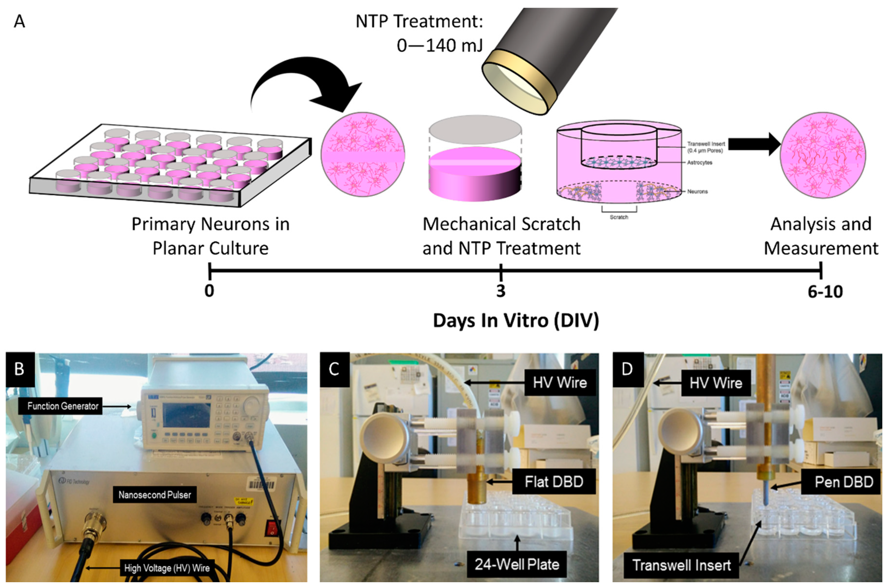
2.3. Scratch Test and NTP Treatment

At 3 DIV, the cellular monolayer (astrocytes or neurons) was subjected to a focal mechanical disruption using a p200 pipette tip (Figure 1A). Astrocytes and neurons/neurites at the site of this scratch injury were physically disrupted, creating a gap across the culture. In this simple and reproducible model, over time, astrocytes regenerate into the gap, and neurons adjacent to the gap re-grow neurites across the gap. Immediately following mechanical disruption, the media was removed, and cells were washed with phosphate buffered saline (PBS). The PBS was then removed, and cultures were treated with NTP using a nanosecond-pulsed dielectric barrier discharge (nspDBD) system (Figure 1B) [25].
Neurons and astrocytes seeded in 24-well plates (1.6 cm diameter per well) while astrocytes alone were seeded in Transwell inserts (0.7 cm diameter) before subjected to NTP treatment. Two dielectric barrier discharge (DBD) electrodes were used in this study for NTP treatment to maximize the treatment area in wells and Transwell inserts. A flat DBD electrode (1.3 cm diameter) was used to treat monolayers of cells in 24-well plates (Figure 1C) while a pen electrode (0.3 cm diameter) was used to treat cells in Transwell inserts (Figure 1D). Both electrodes used a nanosecond pulser (FPB-20-05NM, FID GmBH, Burbach, Germany) to generate a high voltage pulse (29 kV, 2 ns rise times, and 20 ns pulse width), and, therefore, the pulse characteristics were the same. While the electrode geometries are different, several studies have demonstrated that the NTP treatment energy is the most crucial effector of biological response with the DBD plasma system [25,26,27]. Therefore, both the voltage and current of the discharge in the system was measured and energy per plasma pulse for both electrodes was calculated as previously described [28]: 0.9 mJ/pulse for the flat DBD electrode and 1.6 mJ/pulse for the pen electrode. The DBD electrodes were positioned 1 mm above the cells and plasma was generated in the gap between the electrode and the cells for 10 s. Using an external function generator (TG5011LXT, TTI, Fort Worth, TX, USA), the frequency of plasma pulses was adjusted, between 1 and 15 Hz, to control the energy of treatment. Astrocyte-only and neuron-only cultures were treated with 10, 20, 50, 90, or 140 mJ of plasma; the control group was not treated with plasma following the scratch test (n = 3 for each astrocyte culture group, and n = 11–13 for each neuron culture group). Neurons and astrocytes were independently treated in the co-culture group with several combinations of NTP intensities: 0 mJ Neurons/0 mJ Astrocytes, 0 mJ Neurons/20 mJ Astrocytes, 0 mJ Neurons/50 mJ Astrocytes, 20 mJ Neurons/0 mJ Astrocytes, 20 mJ Neurons/20 mJ Astrocytes, and 50 mJ Neurons/50 mJ Astrocytes. In the co-culture control group, neither astrocytes nor neurons were treated with NTP following the scratch test, but astrocyte-containing inserts were added to the neuronal cultures at the same time point as treated groups (n = 4–6 for each co-culture treatment group). Following NTP treatment, 0.5 mL of fresh, complete media was added back to each well. Cells were incubated at 37 °C with 5% CO2 until analysis. Cultures were imaged immediately after plasma treatment and at various time points 1 to 7 days post-injury, as described below.

2.4. Microscopy and Immunocytochemistry

Phase-contrast micrographs were acquired across all cultures using a Nikon Eclipse Ti (QiClick Melville, NY, USA) beginning immediately after scratch application (and plasma treatment if applicable) and then again at 3 and 7 days post-injury (6 and 10 DIV, respectively). At 3 days post plasma treatment (6 DIV), cells were also labeled with antibodies to allow for visualization of axon outgrowth and structure using established immunocytochemistry protocols [23]. Briefly, cultures were fixed with 4% formaldehyde, permeabilized with Triton-X, and labeled for the neuronal marker β-tubulin III (1:500; Sigma T8578, St. Lousi, MO, USA) and HOECHST nuclear counterstain (1:10,000, Invitrogen H21491, Carlsbad, CA, USA), as described previously [23]. Cultures were fluorescently imaged using a Nikon A1RSI laser-scanning confocal microscope.

2.5. Image Quantification, Data Analysis, and Statistical Testing

Images from all culture conditions and time points were analyzed and scored for gap distance and number of cells in the gap using Nikon Elements Basic Research software (4.10.01) and Fiji. Briefly, a grid overlay was used to choose 10 consistent points across the culture in each image to measure the distance between the two edges of the scratch region at the specified point beginning immediately following the scratch assay to obtain the original gap distance. This was accomplished using the phase-contrast images, initially captured in Nikon Elements Basic Research, with the grid overlay implemented in Fiji. The distance of cell migration or process outgrowth (astrocyte cultures) or neurite outgrowth (neuron cultures) was thus measured as the furthest extending cell or neurite across the middle of the gap at each of the 10 grid points by researchers blinded to the experimental group. The percentage of the gap distance remaining was calculated based on the original width of the gap for each treatment and at each time point post-injury. To determine statistical significance, GraphPad Prism 7 was used to perform two-way analysis of variance (ANOVA) on astrocyte-only data, as two variables (time and intensity of plasma stimulation were compared); whereas one-way ANOVA was conducted on neuron-only cultures and co-cultures (only the effect of plasma stimulation was compared). Where significant differences existed between groups, Tukey’s multiple comparisons posthoc test was used in all cases (p < 0.05 required for all statistical tests). Data are represented as the mean percentage of gap closure ± standard error of the mean (SEM).

3. Results

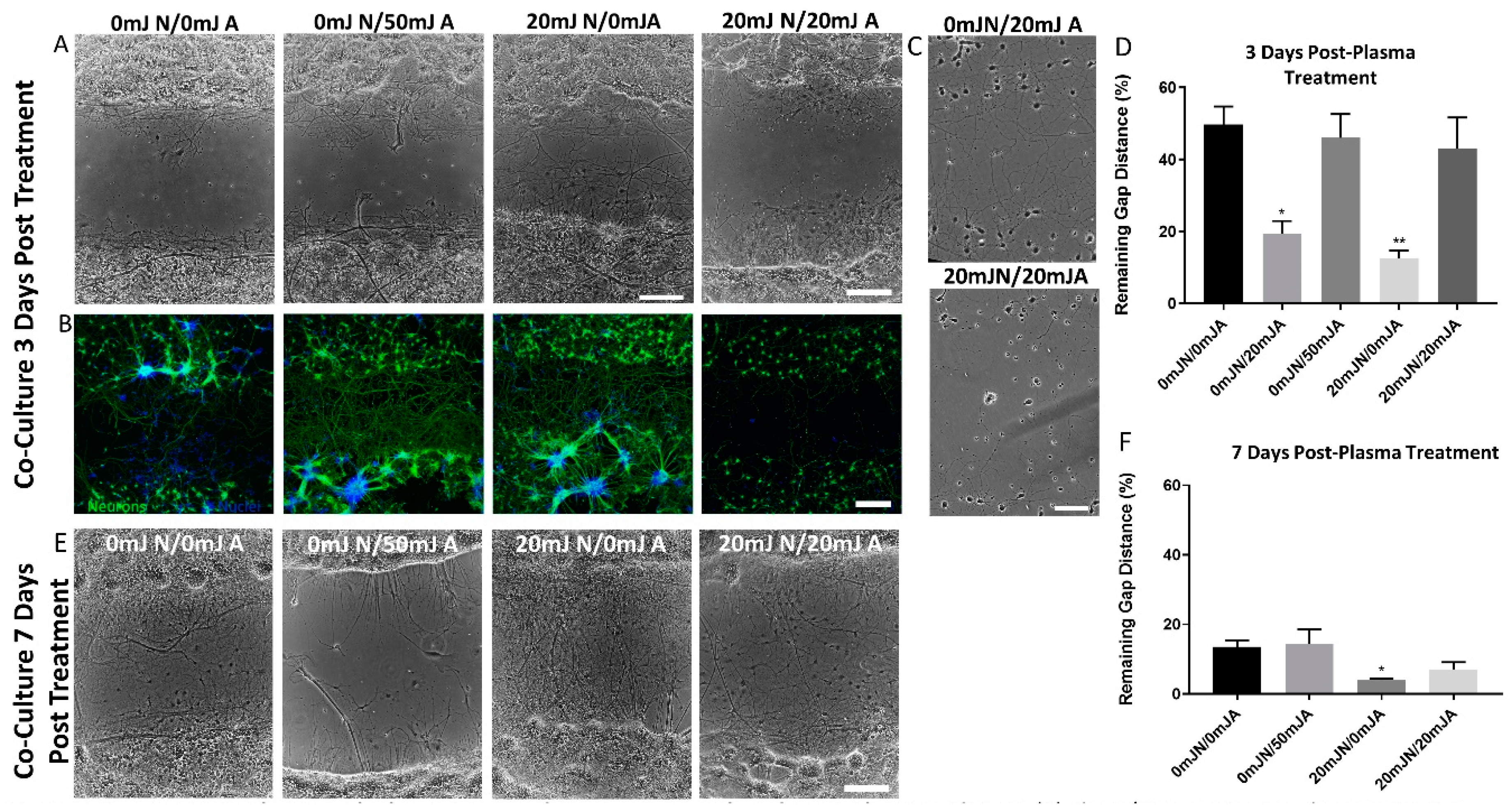
3.1. Direct Treatment of Astrocytes with NTP (20–90 mJ) Led to Increased Astrocyte Infiltration

Planar astrocytes were subjected to a scratch assay and immediately followed with NTP treatment. Regrowth assessment was performed at one, two, and three days post-treatment. Astrocyte cultures subjected to 140 mJ of plasma treatment were completely ablated, resulting in no measurable cells present. Therefore, these were excluded from further analysis. Across all other groups, significant gap reduction was observed over time. Astrocyte ingrowth into the scratch region was observed such that gap length was significantly reduced at each time point relative to previous time points (p < 0.05) (Figure 2). Astrocytes treated with ≤90 mJ plasma exhibited healthy morphology with no signs of distress. At one day post-treatment, cultures treated with both 50 mJ and 90 mJ plasma displayed a statistically significant reduction in mean gap area when compared to the untreated group (p < 0.05 and p < 0.01, respectively). At two days following treatment, cultures treated with 20 mJ plasma along with 50 mJ and 90 mJ exhibited a significant reduction in the mean gap distance (p < 0.05, p < 0.01, and p < 0.05, respectively). Lastly, at three days post-treatment, only cultures treated with 50 mJ plasma resulted in significantly decreased mean gap distance as compared to the control groups (p < 0.05) (Figure 2).

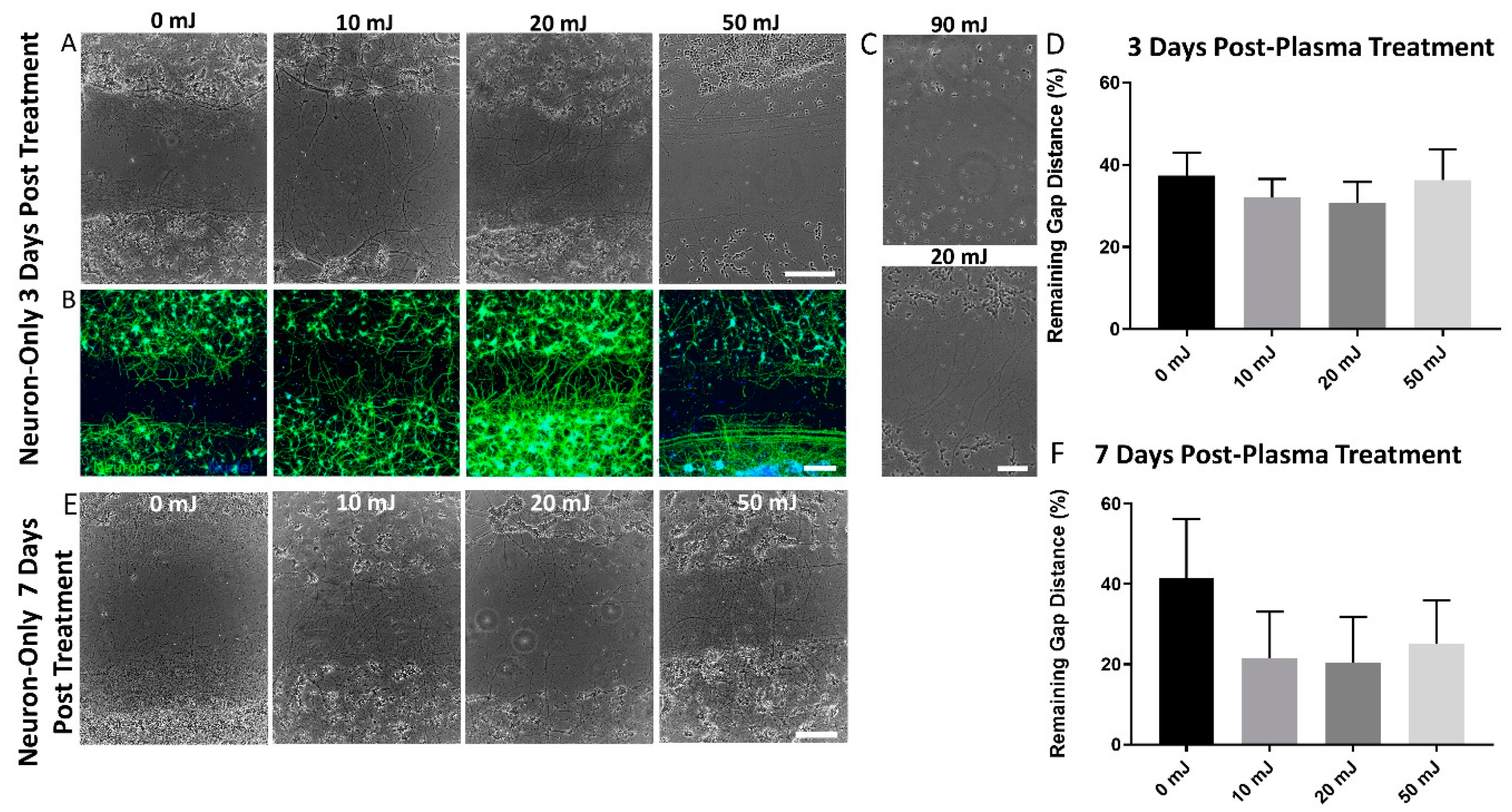
3.2. Neurite Outgrowth was Unaffected by NTP Treatment ≤50 mJ

Planar primary cortical neuron cultures were subjected to a scratch assay and NTP treatment. Neurite infiltration into the gap area was assessed at three and seven days post plasma treatment (6 and 10 DIV, respectively). Neurons appeared to be more sensitive to plasma treatment as treatment with 90 and 140 mJ NTP resulted in widespread cell death (Figure 3C). These groups were excluded from further analysis. Although neurite regeneration significantly increased over time, no statistical differences were found with NTP treatment (Figure 3). However, the trend suggested that treatment with 20 mJ plasma increased neurite outgrowth compared to control groups at 6 DIV (Figure 3). Neurite outgrowth increased over time, as is evident by a reduced percentage gap distance at 10 DIV (7 days post plasma treatment) when compared to 6 DIV, with 10 DIV cultures exhibiting the same trends as those seen at 6 DIV (Figure 3D,F). The immunocytochemical analysis confirmed the neuronal phenotype of surviving cells and was indicative of neurite outgrowth and the health of the neuron-only cultures treated with 0 mJ–50 mJ of plasma at 6 DIV (3 days post-treatment) (Figure 3B).

3.3. The Presence of NTP-Treated Astrocytes Resulted in Increased Neurite Re-Growth

Planar primary neuron cultures were plated in 24-well plates, subjected to a scratch assay, and treated with NTP directly. Independently, planar astrocytes were cultured in Transwell plates and NTP-treated prior to being added to the 24-well plate of neurons. Thus, the astrocytes and neurons were cultured together to allow for indirect contact through secretion of factors. Untreated astrocytes co-cultured with neurons subjected to a scratch assay provided the baseline capacity of astrocytes to promote healing. We observed widespread neuronal death in cultures where both astrocytes and neurons were treated with 50 mJ plasma. Therefore, these groups were excluded from further analysis. Growth was observed over time, and at three days following 20 mJ treatment of either astrocytes or neurons (while the other cell type was untreated), there was significantly increased neurite outgrowth (p < 0.01) (Figure 4). Interestingly, at seven days post-treatment, when both neurons and astrocytes were treated with 20 mJ NTP and co-cultured together, neurite outgrowth was not enhanced as compared to the control group (Figure 4E, F). At three days post-treatment (6 DIV), the immunocytochemical analysis confirmed the neuronal phenotype of cells in the 24-well plate and was indicative of neurite outgrowth and the health of neurons in the co-culture system treated with 0 mJ–50 mJ of plasma (Figure 4B).

4. Discussion

Nervous system trauma and neurodegeneration often result in permanent functional deficits due to the limited regenerative capacity of the brain and spinal cord. In the current study, we assessed the effects of non-thermal plasma treatment on neuroregeneration by performing an in vitro scratch assay on monolayer cultures of astrocytes, neurons, and astrocyte-neuron co-cultures. Immediately following the scratch, cultures were treated with nspDBD plasma, and the effect of treatment on astrocyte re-growth/migration or neurite outgrowth was measured. In the past, typical DBD plasma systems used for biological applications generated non-uniform plasma, characterized by a filamentary structure. These structures are potentially problematic as they may cause localized heating and damage to the surfaces they contact [29,30]. This was largely a result of the applied electric field in the discharge gap as well as the topographically uneven treatment surfaces, such as tissue [29,30,31]. Development of the more sophisticated DBD system, which supplied high voltage over nanosecond rise times was shown to generate a more uniform plasma over uneven surfaces [29,31]. Our group has previously characterized energy-dependent responses of several different cell types (e.g., epithelial, fibroblast, macrophage, etc.) to NTP used in this study [11,28,32,33]. These studies established the plasma parameters and treatment energies used in the present work for stimulation of migration and neurite outgrowth of neurons and astrocytes with minimal lethality.
When astrocyte monolayer cultures were treated with plasma, the infiltration of astrocytes into the gap area was seen over time, as expected (Figure 2B). This suggests that at a lower intensity plasma treatment, cell viability or proliferation were not affected. Interestingly, cultures treated with 50 mJ plasma consistently yielded decreased mean gap distance when compared to time-matched untreated control cultures (Figure 2B). These results suggest that plasma treatment of astrocytes may have persistent effects that lead to increased astrocyte proliferation, migration, or hypertrophy, consequently leading to increased gap closure. Although the mechanism by which this phenomenon is occurring requires further investigation, these results suggest that plasma treatment has a direct effect on astrocytes, which may impact neuron or neurite outgrowth.
Due to the positive effects on cell proliferation and migration seen in astrocyte cultures, as well as the known ability of plasma treatment to increase secretion of growth factors in vitro, direct plasma treatment of neurons was also analyzed. It should be noted that neurite outgrowth, and not neuronal proliferation, was the chosen metric due to the negligible proliferative capabilities of primary cortical neurons, though our measurements also account for any migration of the neuronal somata at the periphery of the scratch zone. Neurite outgrowth did not statistically differ between plasma-treated groups (Figure 3D,F), but a trend was present, suggesting 10 mJ and 20 mJ plasma treatment resulted in greater—and denser—neurite outgrowth and gap closure (Figure 3). A similar trend was observed at a later time point post plasma treatment. While it is possible that direct plasma treatment did not affect the rate of neurite outgrowth, further investigation into the effects of plasma treatment and optimal frequencies on neurons and axons, as well as longer-term implications may be required.
As discussed, direct stimulation of astrocytes resulted in increased infiltration, and the plasma treatment of neurons did not yield a decrease in neurite outgrowth. Therefore, a co-culture system was utilized in which neurons and/or astrocytes could be plasma treated independently, and where the cells do not come in direct contact with one another. Here, neurons in the lower chamber were only exposed to the factors secreted into the media by astrocytes due to the semi-permeable membrane. When only astrocytes in the co-culture system were treated with 50 mJ of plasma, neurite outgrowth occurred at a rate similar to co-cultures that had not been treated—thus, there was no measurable effect of the plasma. However, when only neurons or only astrocytes were treated with a lower intensity of plasma (20 mJ), significant neurite outgrowth and decreased gap distance were observed (Figure 4D,F). Notably, treating both neurons and astrocytes with 20 mJ plasma did not affect neurite outgrowth (Figure 4). These observations suggest an interplay between the stimulation of one population of cells and the presence of another, such as the presence of healthy astrocytes that are secreting growth factors promoting neurite outgrowth. Since increased gap infiltration was observed when both astrocytes and neurons were treated with low plasma intensities independently, it is possible that (1) low treatments of neurons promoted neurite outgrowth, while little to no effect was due to treatment of astrocytes; (2) treatment of astrocytes led to increased secretion of growth factors, yielding increased neurite outgrowth; and (3) stimulation by plasma treatment promoted both neurite outgrowth and the secretion of factors by astrocytes—the combination of which resulted in increased neurite outgrowth. However, overstimulation may not have beneficial effects as observed by a lack of neurite enhancement following the treatment of both astrocyte and neurons in the co-culture. Therefore, highly mediated NTP treatment intensities must be further investigated and optimized to promote neuronal regeneration. Further studies are needed to determine the mechanism responsible for the phenomenon, such as identifying the optimal levels of plasma treatment on neurons and astrocytes and elucidating the mechanisms within each cell type that are responsible for promoting neurite outgrowth.
One possible mechanism by which plasma might enhance neural outgrowth is similar to that seen in stress preconditioning mechanism(s). It is well-documented in the hypoxia/ischemia field that exposure to mild “mini-insults” results in the development of injury tolerance, making neurons more resilient to future damage [34]. Preconditioning hypoxic insults have even been reported to enhance axonal regeneration in sensory and motor neurons [35]. Preconditioning is thought to involve a variety of factors including transcription factors and protein kinases, as well as redox-sensitive proteins [34]. It is possible that plasma treatment could also be generating mild mini-insults that activate the same tolerance pathways as hypoxic/ischemic preconditioning. Since non-thermal plasma contains reactive oxygen and nitrogen species, low exposure levels could induce brief oxidative stress resulting in protection from stronger stress at a later time. In fact, Horiba and colleagues have reported that pre-treating fibroblasts with a plasma-activated media protected them from later oxidative stress [36]. Along these lines, plasma could perhaps be utilized pre-surgery as a way to promote recovery after damaging procedures; however, future studies will be required to assess the efficacy of any putative preconditioning effects of plasma treatment.
Based on our co-culture experiments with NTP-treated astrocytes, plasma may also have utility in the mediation of glial scars, which contribute to the inability of axons in the CNS to regenerate following injury or trauma. A glial scar is comprised of reactive astrocytes that secrete various inhibitory growth factors, leading to defects in the axon growth cone, and, thus, the inability to regenerate. Therefore, glial scars act as both a physical barrier through which axons are unable to penetrate, and a chemical barrier that secretes factors inhibiting axon growth in order to sequester the injured area. There are several factors that are upregulated in the injury micro-environment by many of the cells present in the CNS. These factors include TGFβ-1 and -2, which have been implicated in macrophage-induced glial scarring [37,38]. TGFβ-2 has been associated with the increased production of inhibitory molecules, such as proteoglycans [39,40]. Controlled plasma treatment may affect the expression of TGF-β leading to decreased expression of the proteoglycans, thus, allowing regenerating axons to penetrate through the glial scar [41,42,43]. Additionally, FGF-2 and an inflammatory cytokine, interferon γ (IFNγ) are known to have a role in glial scar induction, with a direct increase in glial scarring correlated to increased levels of IFNγ [44] FGF-2 and IFNγ are believed to modulate one another after injury [45]. Therefore, as previous studies have shown that plasma treatment increases FGF-2 secretion [11], this can be used to decrease the expression of IFNγ that has a known effect on formation of the glial scar. These outcomes suggest that controlled and finely tuned plasma treatment of the chronic glial scar—which is comprised of >95% astrocytes—may create more favorable conditions for regeneration and, thereby, promote axon regeneration across the otherwise inhibitory environment.

5. Conclusions

Our results suggest that astrocytes may be responsive to properly calibrated nanosecond-pulsed dielectric discharge plasma treatment, which can directly enhance their growth as well as improve their ability to enhance neuronal regeneration following trauma. However, non-linear responses were observed with respect to the plasma “dose”, as low intensities (≤10 mJ) did not elicit measurable changes, while high intensities (≥90 mJ) generally resulted in widespread cell death. Intermediate intensities (10 mJ–50 mJ) elicited a physiological response resulting in improved neural cell regeneration across multiple modalities: (1) directly via plasma treatment of astrocytes; (2) directly via plasma treatment of neurons; and (3) indirectly via plasma-stimulation of neurons or astrocytes. Interestingly, increased neurite outgrowth was not observed when both neurons and astrocytes were treated with plasma at the same time, further complicating effective dosages and treatment paradigms. Further work is required to elucidate the mechanism(s) of physiological response to plasma, and, in turn, the mechanism by which astrocyte stimulation led to improved neuronal regeneration. Such studies include cytokine analysis to determine which are being secreted and what their mechanisms of action are. In addition to studies with isolated cell types, more representative models of the neuronal injury microenvironment should be used to investigate the downstream consequences of plasma treatment on multiple cell types and their subsequent effect on each other. These results suggest that NTP may have utility to alter the environment following CNS injury if deployed in a targeted capacity to create more favorable conditions for neurite regeneration. The utilization of plasma treatment following neural injury is a unique and novel approach that may address current challenges in the field of neuroregeneration.

Author Contributions

Conceptualization, K.S.K., A.L., D.K.C., and V.M.; methodology, K.S.K., A.L., D.K.C., and V.M.; validation, K.S.K, A.L., C.E.K.; formal analysis, K.S.K., C.E.K.; investigation, K.S.K., A.L., D.K.C., and V.M.; resources, A.F., D.K.C., V.M.; writing—original draft preparation, K.S.K. and A.L.; writing—review and editing, K.S.K., A.L., D.K.C., and V.M.; visualization, K.S.K. and A.L.; supervision, D.K.C. and V.M.; project administration, K.S.K., A.L., D.K.C., and V.M.; funding acquisition, K.S.K., D.K.C., and V.M.

Funding

Financial support was provided by the National Institutes of Health [U01-NS094340 (Cullen) & F31-NS090746 (Katiyar)] and the Department of Veterans Affairs [RR&D Merit Review I01-RX001097 (Cullen) & BLR&D Merit Review I01-BX003748 (Cullen)].

Acknowledgments

The authors thank Pietro Ranieri and Wisberty Gordian-Velez for their assistance in executing the experiments and data analysis.

Conflicts of Interest

The authors declare no conflict of interest.

References

  1. Yiu, G.; He, Z. Glial inhibition of CNS axon regeneration. Nat. Rev. Neurosci. 2006, 7, 617. [Google Scholar] [CrossRef] [PubMed]
  2. Gabella, B.; Hoffman, R.E.; Marine, W.W.; Stallones, L. Urban and rural traumatic brain injuries in Colorado. Ann. Epidemiol. 1997, 7, 207–212. [Google Scholar] [CrossRef]
  3. Liverman, C.T.; Altevogt, B.M.; Joy, J.E.; Johnson, R.T. Spinal Cord Injury: Progress, Promise, and Priorities; National Academy of Sciences: Washington, DC, USA, 2005. [Google Scholar]
  4. Fawcett, J.W.; Asher, R.A. The glial scar and central nervous system repair. Brain Res. Bull. 1999, 49, 377–391. [Google Scholar] [CrossRef]
  5. Lu, X.; Naidis, G.V.; Laroussi, M.; Reuter, S.; Graves, D.B.; Ostrikov, K. Reactive species in non-equilibrium atmospheric-pressure plasmas: Generation, transport, and biological effects. Phys. Rep. 2016, 630, 1–84. [Google Scholar] [CrossRef] [Green Version]
  6. Steinbeck, M.J.; Chernets, N.; Zhang, J.; Kurpad, D.S.; Fridman, G.; Fridman, A.; Freeman, T.A. Skeletal Cell Differentiation Is Enhanced by Atmospheric Dielectric Barrier Discharge Plasma Treatment. PLoS ONE 2013, 8, e82143. [Google Scholar] [CrossRef] [PubMed]
  7. Chernets, N.; Zhang, J.; Steinbeck, M.J.; Kurpad, D.S.; Koyama, E.; Friedman, G.; Freeman, T.A. Nonthermal atmospheric pressure plasma enhances mouse limb bud survival, growth, and elongation. Tissue Eng. Part A 2014, 21, 300–309. [Google Scholar] [CrossRef] [PubMed]
  8. Brun, P.; Pathak, S.; Castagliuolo, I.; Palù, G.; Brun, P.; Zuin, M.; Cavazzana, R.; Martines, E. Helium generated cold plasma finely regulates activation of human fibroblast-like primary cells. PLoS ONE 2014, 9, e104397. [Google Scholar] [CrossRef]
  9. Miller, V.; Lin, A.; Fridman, G.; Dobrynin, D.; Fridman, A. Plasma Stimulation of Migration of Macrophages. Plasma Process. Polym. 2014, 11, 1193–1197. [Google Scholar] [CrossRef]
  10. Arjunan, K.P.; Friedman, G.; Fridman, A.; Clyne, A.M. Non-thermal dielectric barrier discharge plasma induces angiogenesis through reactive oxygen species. J. R. Soc. Interface 2011, rsif20110220. [Google Scholar] [CrossRef]
  11. Arndt, S.; Unger, P.; Berneburg, M.; Bosserhoff, A.K.; Karrer, S. Cold atmospheric plasma (CAP) activates angiogenesis-related molecules in skin keratinocytes, fibroblasts and endothelial cells and improves wound angiogenesis in an autocrine and paracrine mode. J. Dermatol. Sci. 2018, 89, 181–190. [Google Scholar] [CrossRef]
  12. Kalghatgi, S.; Friedman, G.; Fridman, A.; Clyne, A.M. Endothelial cell proliferation is enhanced by low dose non-thermal plasma through fibroblast growth factor-2 release. Ann. Biomed. Eng. 2010, 38, 748–757. [Google Scholar] [CrossRef] [PubMed]
  13. Kanzler, I.; Tuchscheerer, N.; Steffens, G.; Simsekyilmaz, S.; Konschalla, S.; Kroh, A.; Simons, D.; Asare, Y.; Schober, A.; Bucala, R.; et al. Differential roles of angiogenic chemokines in endothelial progenitor cell-induced angiogenesis. Basic Res. Cardiol. 2013, 108, 310. [Google Scholar] [CrossRef] [PubMed]
  14. Presta, M.; Dell’Era, P.; Mitola, S.; Moroni, E.; Ronca, R.; Rusnati, M. Fibroblast growth factor/fibroblast growth factor receptor system in angiogenesis. Cytokine Growth Factor Rev. 2005, 16, 159–178. [Google Scholar] [CrossRef] [PubMed] [Green Version]
  15. Miller, V.; Lin, A.; Kako, F.; Gabunia, K.; Kelemen, S.; Brettschneider, J.; Fridman, G.; Fridman, A.; Autieri, M. Microsecond-pulsed dielectric barrier discharge plasma stimulation of tissue macrophages for treatment of peripheral vascular disease. Phys. Plasmas (1994 Present) 2015, 22, 122005. [Google Scholar] [CrossRef] [PubMed] [Green Version]
  16. Shubayev, V.I.; Myers, R.R. Matrix metalloproteinase-9 promotes nerve growth factor-induced neurite elongation but not new sprout formation in vitro. J. Neurosci. Res. 2004, 77, 229–239. [Google Scholar] [CrossRef] [PubMed]
  17. Jin, K.; Mao, X.O.; Greenberg, D.A. Vascular endothelial growth factor stimulates neurite outgrowth from cerebral cortical neurons via Rho kinase signaling. J. Neurobiol. 2006, 66, 236–242. [Google Scholar] [CrossRef]
  18. Xiong, Z.; Zhao, S.; Mao, X.; Lu, X.; He, G.; Yang, G.; Chen, M.; Ishaq, M.; Ostrikov, K. Selective neuronal differentiation of neural stem cells induced by nanosecond microplasma agitation. Stem Cell Res. 2014, 12, 387–399. [Google Scholar] [CrossRef]
  19. Yan, X.; Qiao, Y.; Ouyang, J.; Jia, M.; Li, J.; Yuan, F. Protective effect of atmospheric pressure plasma on oxidative stress-induced neuronal injuries: An in vitro study. J. Phys. D Appl. Phys. 2017, 50, 095401. [Google Scholar] [CrossRef]
  20. Zhao, S.; Han, R.; Li, Y.; Lu, C.; Chen, X.; Xiong, Z.; Mao, X. Investigation of the mechanism of enhanced and directed differentiation of neural stem cells by an atmospheric plasma jet: A gene-level study. J. Appl. Phys. 2019, 125, 163301. [Google Scholar] [CrossRef]
  21. Dowell, J.A.; Johnson, J.A.; Li, L. Identification of astrocyte secreted proteins with a combination of shotgun proteomics and bioinformatics. J. Proteome Res. 2009, 8, 4135–4143. [Google Scholar] [CrossRef]
  22. Cullen, D.K.; Gilroy, M.E.; Irons, H.R.; Laplaca, M.C. Synapse-to-neuron ratio is inversely related to neuronal density in mature neuronal cultures. Brain Res. 2010, 1359, 44–55. [Google Scholar] [CrossRef] [PubMed] [Green Version]
  23. Katiyar, K.S.; Winter, C.C.; Struzyna, L.A.; Harris, J.P.; Cullen, D.K. Mechanical elongation of astrocyte processes to create living scaffolds for nervous system regeneration. J. Tissue Eng. Regen. Med. 2017, 11, 2737–2751. [Google Scholar] [CrossRef] [PubMed]
  24. Laplaca, M.C.; Vernekar, V.N.; Shoemaker, J.T.; Cullen, D.K. Three-dimensional neuronal cultures. In Methods in Bioengineering: 3D Tissue Engineering; Artech House: Norwood, MA, USA, 2010; pp. 187–204. [Google Scholar]
  25. Lin, A.; Truong, B.; Patel, S.; Kaushik, N.; Choi, E.H.; Fridman, G.; Fridman, A.; Miller, V. Nanosecond-Pulsed DBD Plasma-Generated Reactive Oxygen Species Trigger Immunogenic Cell Death in A549 Lung Carcinoma Cells through Intracellular Oxidative Stress. Int. J. Mol. Sci. 2017, 18, 966. [Google Scholar] [CrossRef] [PubMed]
  26. Bekeschus, S.; Lin, A.; Fridman, A.; Wende, K.; Weltmann, K.; Miller, V. A Comparison of Floating-Electrode DBD and kINPen Jet: Plasma Parameters to Achieve Similar Growth Reduction in Colon Cancer Cells Under Standardized Conditions. Plasma Chem. Plasma Process. 2018, 38, 1–12. [Google Scholar] [CrossRef]
  27. Lin, A.; Gorbanev, Y.; de Backer, J.; van Loenhout, J.; van Boxem, W.; Lemière, F.; Cos, P.; Dewilde, S.; Smits, E.; Bogaerts, A. Non-Thermal Plasma as a Unique Delivery System of Short-Lived Reactive Oxygen and Nitrogen Species for Immunogenic Cell Death in Melanoma Cells. Adv. Sci. 2019, 6, 1802062. [Google Scholar] [CrossRef] [PubMed]
  28. Lin, A.; Chernets, N.; Han, J.; Alicea, Y.; Dobrynin, D.; Fridman, G.; Freeman, T.A.; Fridman, A.; Miller, V. Non-Equilibrium Dielectric Barrier Discharge Treatment of Mesenchymal Stem Cells: Charges and Reactive Oxygen Species Play the Major Role in Cell Death. Plasma Process. Polym. 2015, 12, 1117–1127. [Google Scholar] [CrossRef]
  29. Ayan, H.; Staack, D.; Fridman, G.; Gutsol, A.; Mukhin, Y.; Starikovskii, A.; Fridman, A.; Friedman, G. Application of nanosecond-pulsed dielectric barrier discharge for biomedical treatment of topographically non-uniform surfaces. J. Phys. D Appl. Phys. 2009, 42. [Google Scholar] [CrossRef]
  30. Ayan, H.; Fridman, G.; Gutsol, A.F.; Vasilets, V.N.; Fridman, A.; Friedman, G. Nanosecond-pulsed uniform dielectric-barrier discharge. IEEE Trans. Plasma Sci. 2008, 36, 5. [Google Scholar] [CrossRef]
  31. Liu, C.; Dobrynin, D.; Fridman, A. Uniform and non-uniform modes of nanosecond-pulsed dielectric barrier discharge in atmospheric air: Fast imaging and spectroscopic measurements of electric field. J. Phys. D Appl. Phys. 2014, 47, 252003. [Google Scholar] [CrossRef]
  32. Lin, A.; Truong, B.; Fridman, G.; Fridman, A.A.; Miller, V. Immune Cells Enhance Selectivity of Nanosecond-Pulsed DBD Plasma Against Tumor Cells. Plasma Med. 2017, 7, 85–96. [Google Scholar] [CrossRef] [Green Version]
  33. Ranieri, P.; Shrivastav, R.; Wang, M.; Lin, A.; Fridman, G.; Fridman, A.; Han, L.H.; Miller, V. Nanosecond pulsed dielectric barrier discharge induced anti-tumor effects propagate through the depth of tissue via intracellular signaling. Plasma Med. 2017, 7, 283–297. [Google Scholar] [CrossRef]
  34. Rybnikova, E.; Samoilov, M. Current insights into the molecular mechanisms of hypoxic pre- and postconditioning using hypobaric hypoxia. Front. Neurosci. 2015, 9, 388. [Google Scholar] [CrossRef] [PubMed] [Green Version]
  35. Cho, Y.; Shin, J.E.; Ewan, E.E.; Oh, Y.M.; Pita-Thomas, W.; Cavalli, V. Activating Injury-Responsive Genes with Hypoxia Enhances Axon Regeneration through Neuronal HIF-1alpha. Neuron 2015, 88, 720–734. [Google Scholar] [CrossRef] [PubMed]
  36. Horiba, M.; Kamiya, T.; Hara, H.; Adachi, T. Cytoprotective effects of mild plasma-activated medium against oxidative stress in human skin fibroblasts. Sci. Rep. 2017, 7, 42208. [Google Scholar] [CrossRef] [PubMed]
  37. Giulian, D.; Woodward, J.; Young, D.G.; Krebs, J.F.; Lachman, L.B. Interleukin-1 injected into mammalian brain stimulates astrogliosis and neovascularization. J. Neurosci. 1988, 8, 2485–2490. [Google Scholar] [CrossRef] [PubMed] [Green Version]
  38. Silver, J.; Miller, J.H. Regeneration beyond the glial scar. Nat. Rev. Neurosci. 2004, 5, 146–156. [Google Scholar] [CrossRef] [PubMed]
  39. Asher, R.A.; Morgenstern, D.A.; Fidler, P.S.; Adcock, K.H.; Oohira, A.; Braistead, J.E.; Levine, J.M.; Margolis, R.U.; Rogers, J.H.; Fawcett, J.W. Neurocan is upregulated in injured brain and in cytokine-treated astrocytes. J. Neurosci. 2000, 20, 2427–2438. [Google Scholar] [CrossRef] [PubMed]
  40. Lagord, C.; Berry, M.; Logan, A. Expression of TGFbeta2 but not TGFbeta1 correlates with the deposition of scar tissue in the lesioned spinal cord. Mol. Cell. Neurosci. 2002, 20, 69–92. [Google Scholar] [CrossRef] [PubMed]
  41. Kim, H.Y.; Kang, S.K.; Park, S.M.; Jung, H.Y.; Choi, B.H.; Sim, J.Y.; Lee, J.K. Characterization and effects of Ar/Air microwave plasma on wound healing. Plasma Process. Polym. 2015, 12, 1423–1434. [Google Scholar] [CrossRef]
  42. Arndt, S.; Landthaler, M.; Zimmermann, J.L.; Unger, P.; Wacker, E.; Shimizu, T.; Li, Y.F.; Morfill, G.E.; Bosserhoff, A.K.; Karrer, S. Effects of cold atmospheric plasma (CAP) on ß-defensins, inflammatory cytokines, and apoptosis-related molecules in keratinocytes in vitro and in vivo. PLoS ONE 2015, 10, e0120041. [Google Scholar] [CrossRef] [PubMed]
  43. Fathollah, S.; Mirpour, S.; Mansouri, P.; Dehpour, A.R.; Ghoranneviss, M.; Rahimi, N.; Naraghi, Z.S.; Chalangari, R.; Chalangari, K.M. Investigation on the effects of the atmospheric pressure plasma on wound healing in diabetic rats. Sci. Rep. 2016, 6, 19144. [Google Scholar] [CrossRef] [PubMed] [Green Version]
  44. Yong, V.W.; Moumdjian, R.; Yong, F.P.; Ruijs, T.C.; Freedman, M.S.; Cashman, N.; Antel, J.P. Gamma-interferon promotes proliferation of adult human astrocytes in vitro and reactive gliosis in the adult mouse brain in vivo. Proc. Natl. Acad. Sci. USA 1991, 88, 7016–7020. [Google Scholar] [CrossRef] [PubMed]
  45. DiProspero, N.A.; Meiners, S.; Geller, H.M. Inflammatory cytokines interact to modulate extracellular matrix and astrocytic support of neurite outgrowth. Exp. Neurol. 1997, 148, 628–639. [Google Scholar] [CrossRef] [PubMed]
Figure 1. Non-thermal plasma (NTP) was applied to neural cultures using a nanosecond-pulsed dielectric barrier discharge (nspDBD) system for all plasma treatments. (A) Primary neurons were plated in 24-well plates, subjected to mechanical disruption via a scratch assay, and treated with NTP 3 days after plating. For the co-culture group, astrocytes cultured in Transwell inserts were added to neuronal cultures at this point, and cell/neurite infiltration into the gap was assessed at 6–10 DIV. (B) A nanosecond-pulser was used to generate and deliver high voltage (HV) pulses to the dielectric barrier discharge (DBD) electrode. A function generator was used to control NTP treatment energy by setting the frequency of pulses and the duration of treatment. A z-positioner was used to fix the position of the (C) flat DBD electrode or the (D) pen DBD electrode 1 mm above the cells treated in 24-well plates or Transwell inserts, respectively.
Applsci 09 03747 g001
Figure 2. NTP treatment of astrocytes resulted in increased cell/process infiltration. (A) Phase-contrast images showing astrocyte infiltration into the gap area 0–3 days post-treatment with 0 mJ, 20 mJ, 50 mJ, and 90 mJ plasma. Dashed lines in images on day 0 and day 1 were added to assist visualization of scratch boundaries. Scale: 250 µm. (B) Astrocyte infiltration was significantly increased over time, as expected. Interestingly, one day post-treatment with 50 mJ and 90 mJ of plasma led to significant astrocyte infiltration into the gap area, two days following treatment, 20 mJ, 50 mJ, and 90 mJ plasma treatment all led to significant reduction in mean gap distance, and three days post-treatment, only 50 mJ plasma resulted in reduced mean gap distance when compared to time-matched untreated control cultures (bars represent standard error of the mean (SEM); * p < 0.05; ** p < 0.01).
Applsci 09 03747 g002
Figure 3. Plasma treatment of neuronal cultures alone did not affect neurite outgrowth into the gap area. (A) Phase/Contrast images showing neurite infiltration into the gap area at three days post-treatment (6 DIV) when treated with 0, 10, 20, and 50 mJ plasma. Scale: 250 μm. While a trend towards increased neurite outgrowth was observed with treatment of 20 mJ plasma, no statistical significance is seen due to plasma treatment. (B) Confocal reconstruction of plasma-treated cultures labeled positively for the neuronal marker, b-tubulin III (green) and nuclear counterstain, HOECHST (blue) three days post-treatment. Qualitative assessment of cultures indicates that neurons and extending neurites remained healthy three days post-treatment when treated with ≤50 mJ plasma. Scale: 150 μm. (C) Higher magnification phase-contrast images clearly showing the difference between cultures where plasma treatment reduced neurite outgrowth (90 mJ treatment) and cultures where plasma treatment increased neurite outgrowth (20 mJ treatment). Scale: 100 μm (D) Quantification of neurite outgrowth at three days post-treatment (6 DIV). No significant difference in neurite outgrowth was observed when compared to control, untreated groups. (E) Phase-contrast images of neuron-only cultures seven days post plasma treatment with 0, 10, 20, and 50 mJ plasma. Scale: 250 μm. (F) Quantification of neurite outgrowth at seven days post-treatment (10 DIV). No significant neurite outgrowth was observed when compared to untreated groups. (D,F) Bars represent SEM.
Applsci 09 03747 g003
Figure 4. Astrocyte stimulation with plasma increased neurite outgrowth under co-culture conditions. (A) Phase/Contrast images showing neurite infiltration into the gap area at three days post-treatment (6 DIV) when neurons were treated with 0 mJ or 20 mJ plasma and astrocytes were treated with 0 mJ or 50 mJ plasma. Scale: 250 μm. (B) Confocal reconstruction of neurons labeled for the neuronal marker, b-tubulin III (green) and nuclear counterstain, HOECHST (blue) at three days post plasma treatment. Qualitative assessment of cultures indicates that neurons and extending neurites remained healthy three days post-treatment when treated with ≤50 mJ plasma. Scale: 150 μm (C) Higher magnification of cultures to show increased, healthy neurite outgrowth three days following no plasma treatment of neurons and 20 mJ treatment of astrocytes (top), and unhealthy cultures following 20 mJ treatment of both neurons and astrocytes (bottom). Scale: 100 μm. (D) Quantification of remaining gap distance at 6 DIV (three days post plasma treatment). When neurons were left untreated but astrocytes were treated with 20 mJ and vice versa, neurite outgrowth into the gap area was significantly increased compared to the untreated control cultures. (E) Phase-contrast images showing neurite outgrowth into the gap area seven days post-treatment (10 DIV) in a subset of treatment groups. Scale: 250 μm. (F) Quantification of the remaining gap distance shows that by seven days post-treatment, cultures where neurons were treated with 20 mJ plasma and astrocytes were untreated exhibited a significantly decreased gap distance than untreated cultures. (D,F) Bars represent SEM; * p < 0.05,** p < 0.01).
Applsci 09 03747 g004

