Article

Exoskeleton for Gait Rehabilitation: Effects of Assistance, Mechanical Structure, and Walking Aids on Muscle Activations

1
Department of Informatics, Bioengineering, Robotics and System Engineering, University of Genoa, 13-16145 Genoa, Italy
2
Spinal Cord Unit, Santa Corona Hospital, ASL2 Savonese, 38-17027 Pietra Ligure (SV), Italy
3
Wyss Center for Bio- and Neuroengineering, CH-1202 Geneva, Switzerland
*
Author to whom correspondence should be addressed.
Indicates equal contribution.
Appl. Sci. 2019, 9(14), 2868; https://doi.org/10.3390/app9142868
Submission received: 1 June 2019 / Revised: 8 July 2019 / Accepted: 12 July 2019 / Published: 18 July 2019

Abstract

:
Several exoskeletons have been developed and increasingly used in clinical settings for training and assisting locomotion. These devices allow people with severe motor deficits to regain mobility and sustain intense and repetitive gait training. However, three factors might affect normal muscle activations during walking: the assistive forces that are provided during walking, the crutches or walker that are always used in combination with the device, and the mechanical structure of the device itself. To investigate these effects, we evaluated eight healthy volunteers walking with the Ekso, which is a battery-powered, wearable exoskeleton. They walked supported by either crutches or a walker under five different assistance modalities: bilateral maximum assistance, no assistance, bilateral adaptive assistance, and unilateral adaptive assistance on each leg. Participants also walked overground without the exoskeleton. Surface electromyography was recorded bilaterally, and the statistical parametric mapping approach and muscle synergies analysis were used to investigate differences in muscular activity across different walking conditions. The lower limb muscle activations while walking with the Ekso were not influenced by the use of crutches or walker aids. Compared to normal walking without robotic assistance, the Ekso reduced the amplitude of activation for the distal lower limb muscles while changing the timing for the others. This depended mainly on the structure of the device, and not on the type or level of assistance. In fact, the presence of assistance did not change the timing of the muscle activations, but instead mainly had the effect of increasing the level of activation of the proximal lower limb muscles. Surprisingly, we found no significant changes in the adaptive control with respect to a maximal fixed assistance that did not account for subjects’ performance. These are important effects to take into careful considerations in clinics where these devices are used for gait rehabilitation in people with neurological diseases.

1. Introduction

In the last few years, powered exoskeletons have been used in clinical practice as rehabilitative tools for improving walking ability in people suffering for neurological diseases or injuries [1,2] and as assistive devices for allowing the most impaired to stand up and walk [3].
As rehabilitative tools, robotic devices assist the physical therapists by providing task-specific, repeatable practice and increased intensity of training [4]. Locomotor training with robotic assistance seems to be more beneficial in individuals following stroke and spinal cord injury (SCI) with respect to other neurological disorders [5]. Some studies reported a marked improvement in lower limb motor functions and walking abilities in incomplete SCI subjects with the use of electromechanical systems [6,7,8,9] and positive effects in the reorganization of spinal locomotor neuronal networks [10]. However, other studies [11,12] found that robot-assisted training does not determine better outcomes than traditional rehabilitation. Thus, while undoubtedly powered exoskeletons can provide non-ambulatory individuals with the ability to walk at modest speed [13], the literature highlights conflicting results for the training potential of these devices, and there is no compelling evidence that robot-assisted gait re-education improves walking function more than other rehabilitative strategies [8,9,14,15,16,17].
Moreover, although exoskeletons are already used as an alternative option to traditional therapy, there is still a lack of knowledge regarding their specific effects on the user locomotor function. We expect that the guidance they provide would restore muscle coordination patterns similar to the physiological human walking. However, little research has been conducted in this direction, and the few studies confirmed the necessity to further investigate this topic [18,19,20,21], suggesting that either the assistive external forces provided during walking [18] or the structure of the device itself [22] can affect normal muscle activations as well as kinetic and kinematic patterns during locomotion. To systematically investigate all these aspects is crucial and at the same time difficult. There are several solutions available for the mechanical structure of the exoskeletons; it is possible to use them with different aids and under different assistive modalities, e.g., unilateral versus bilateral assistance, or maximum assistance versus adaptive assistance based on users’ performance. To the best of our knowledge, these aspects were not deeply investigated with an integrated approach aiming at accounting how each of them influences the overground walking patterns.
This study aimed at filling this gap and had two main objectives. The first was to examine whether and how different assistive modalities influence the muscle patterns during walking. The second was to compare the muscle activations when walking overground with and without an exoskeleton and verify whether the observed differences are due to the mechanical structure of the exoskeleton, the walking aids, or the assistive forces provided by the device.
Our first hypothesis was that the exoskeleton, providing the users with different amounts of assistive forces and requiring the use of walking aids, would induce changes in the amplitude of the muscle activations. More specifically, we hypothesized that the increase of the assistance would induce a decrease of the amplitude of the muscle activations without changing their timing. We will also expect that in individuals with residual walking ability, the adaptive paradigms will determine higher voluntary muscle activations than the maximum fixed assistance, which does not account for the individual ability and does not obey the assistance-as-needed protocol [23].
Our second hypothesis was that the mechanical structure of the exoskeleton would promote changes in the lower-limb muscle activation patterns that have been observed during normal overground walking, as suggested also by [20,21].
With the long-term goal of extending this study to people suffering from neurological diseases or injuries, here we focused on healthy subjects that do not have alterations of their muscle patterns, reducing the possibility of variable and highly subjective responses to each of the investigated aspects. Both hypotheses were tested by analyzing the electromyographic (EMG) patterns generated by eight subjects while walking with and without a wildly used and commercially available exoskeleton: the Ekso (Ekso Bionics, Richmond, CA, USA).
Understanding these aspects will enable us to fully exploit and use the possibilities offered by Ekso in the rehabilitative sessions in a highly personalized way, and will allow defining a path to overcome eventual technological limits.

2. Materials and Methods

2.1. Participants

Eight healthy volunteers (four males, age: 27.1 mean ± 7.4 SD years, height: 174.2 mean ± 10.6 SD, weight: 68.6 mean ± 9.4 SD kg; see Table 1) with no neurological or orthopedic disorders participated in this study.
Subjects were selected in order to fit the criteria needed to use the exoskeleton Ekso: (1) functional upper limb for using the aids, (2) height between 1.50–2 m, (3) weight lower than 100 kg.
The study conformed to the Declaration of Helsinki and was approved by the local ethical committee (Comitato Etico Regione Liguria). All participants signed an informed consent to the analysis and publication of their data for research purposes.

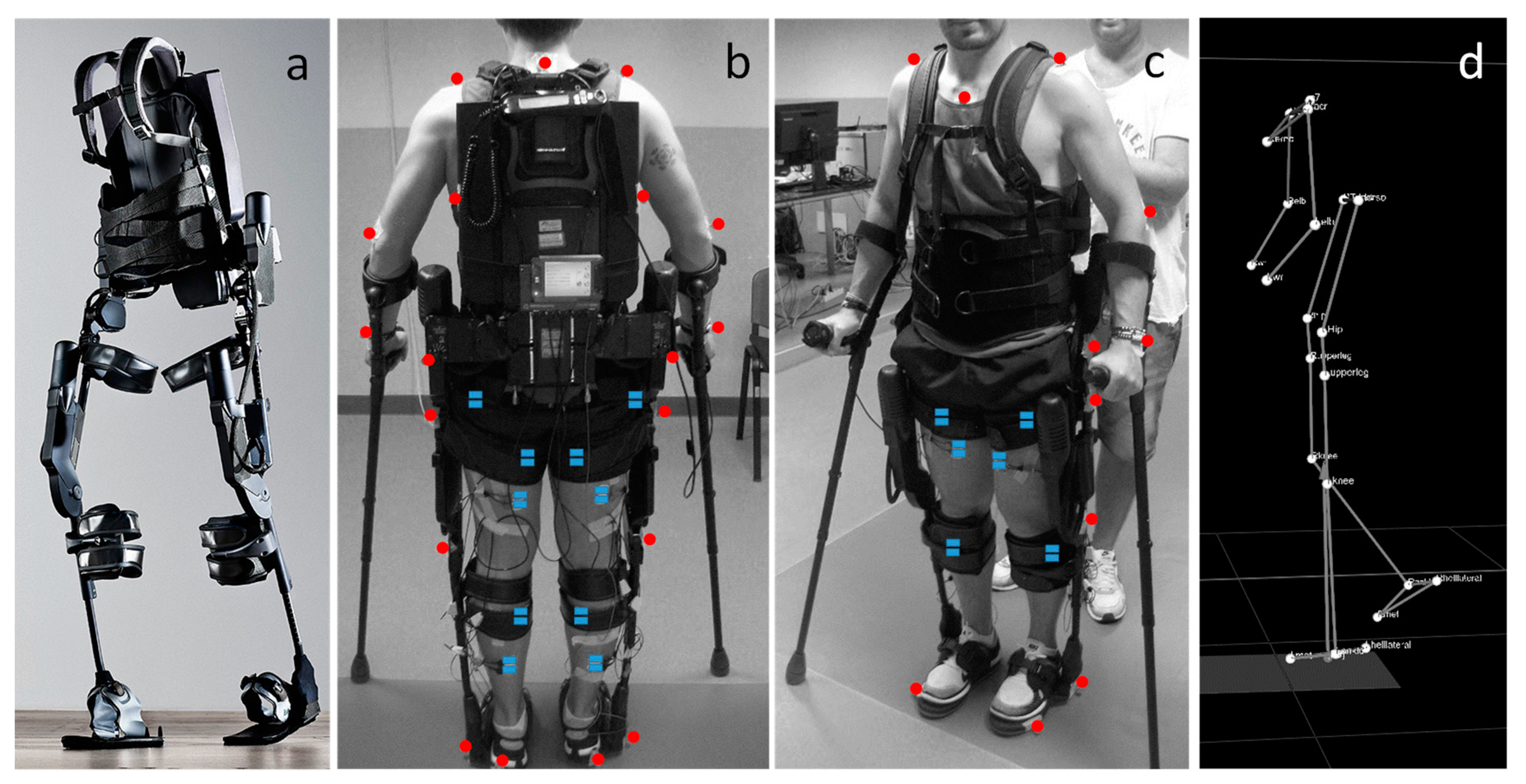
2.2. Apparatus: Brief Description of the Ekso Exoskeleton

The Ekso (Ekso Bionics, Richmond, CA, USA [24], Figure 1a) is a battery-powered, wearable exoskeleton that enables individuals with severe walking impairments to stand up and walk overground.
In this study, we used the model ‘Ekso 1’, which was equipped with four battery-powered motors at the hips and knees, and 15 position sensors. The device includes two powered joints at the hip and knee, and a semi-rigid unpowered ankle joint.
The Ekso is used always with external balancing aids: mainly walkers or crutches. Both aids are characterized by a user interface that enables the subject to interact with the device and select the desired assistance. The device provides assistance to the users by driving their lower limb joints through a predefined trajectory. The exoskeleton has different assistance settings: fixed or adaptive assistance, no assistance, as well as bilateral or unilateral assistance.
Bilateral assistance provides full stance stability and swing assistance (fixed or adaptive) to both legs. Unilateral assistance provides them only for a selected limb, while the contralateral leg is automatically set free i.e., the users voluntary control the swing of that leg with their own strength and coordination, without any help by the device.
With the Fixed assistance setting, a fixed amount of assistance is provided throughout the swing phase of gait; it can vary from 0%, where the device does not provide any assistance, and the subjects should actively move, to a maximum value of 100% corresponding to the maximal assistance that allows the subjects to walk without any voluntary contribution. Thus, depending on the set fixed value, the user is required to use different levels of voluntary control to complete the swing phase. When the Ekso is programmed with a fixed assistance value, it provides a consistent amount of motor output, regardless of the patient’s ability.
The Adaptive assistance setting acts based on the subjects’ performance. In this control strategy, the assistive forces increase as the participant deviates from the desired trajectory [25].
With the No Assistance setting, the Ekso does not provide any assistance to the user, but its inertia is compensated i.e., the users do not feel the weight of the Ekso and voluntary control their walking patterns.
The Ekso is also characterized by three different ways to control the initiation of each step according to the subject skills:
  • First Step: a physical therapist controls the beginning of each step by pushing a button. Usually, this modality is used in the first training session for the familiarization with the device;
  • Active Step: the user controls the beginning of each step by means of an interface, placed on the crutches or on the walker, that communicate with the device.
  • Pro Step: The user controls the beginning of the next step by moving his hips forward and laterally. In this case, when the device recognizes that the user is in the correct position allows the step of the contralateral leg.

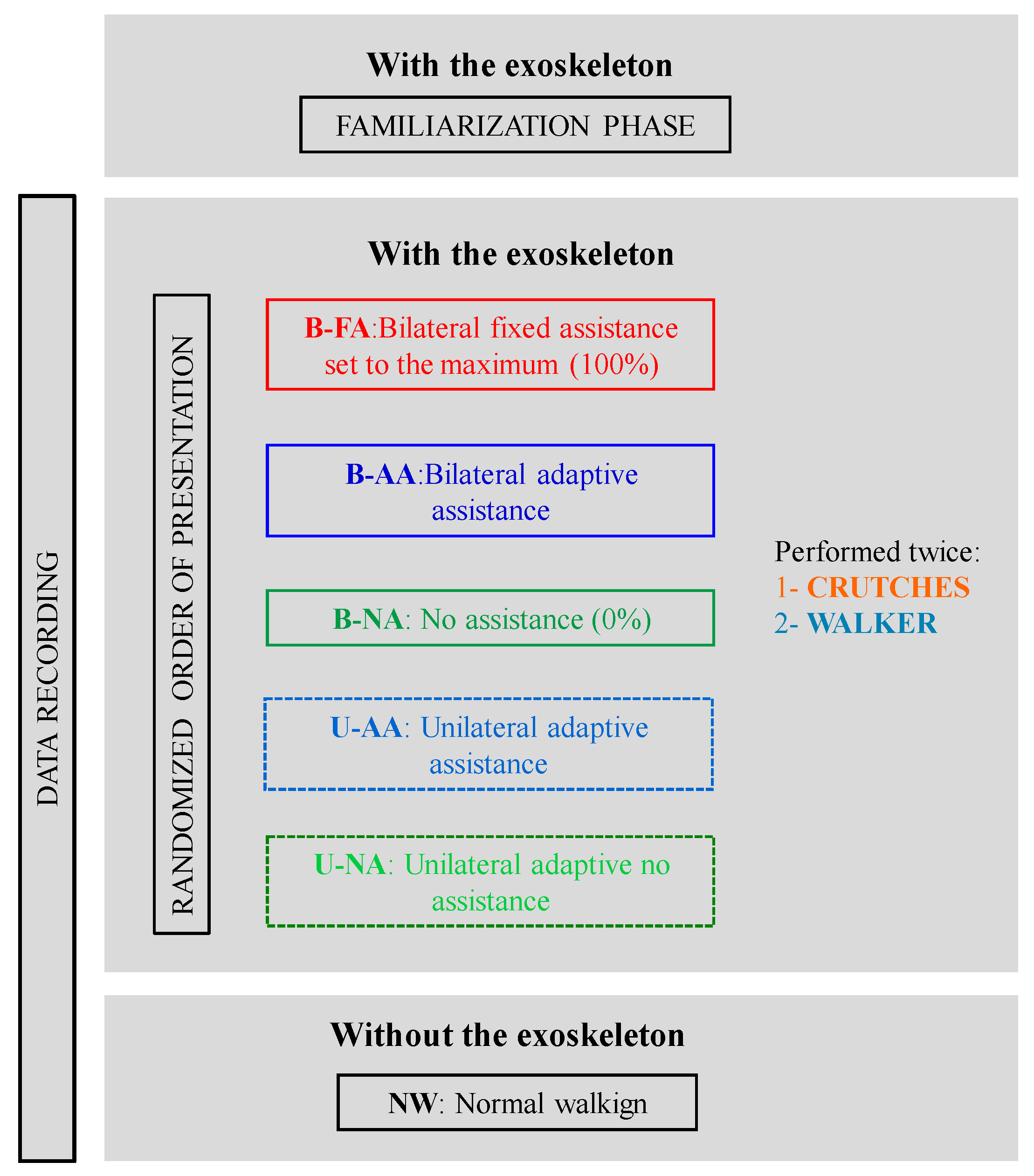
2.3. Experimental Protocol

Subjects were instructed to walk in a pathway eight meters long inside the Santa Corona hospital motion analysis lab (Pietra Ligure, Savona, Italy, see paragraph below). Each participant walked both with and without exoskeleton. In the latter case (normal walking, or NW), subjects were asked to walk barefoot, as naturally as possible, at their preferred speed. In the former case, subjects walked under five different assistive modalities: bilateral fixed assistance set to the maximum (100%, B-FA), no assistance (B-NA), bilateral adaptive assistance (B-AA), unilateral adaptive assistance provided either to the left or the right leg (U-AA, U-NA depending on the assistance or not assistance condition of the considered leg, respectively). The order of presentation of these walking modalities was randomized among subjects (see Figure 2).
To initiate the steps, we used the pro-step mode, and therefore, the subjects were instructed, prior to the start of the experiment, to shift their weight forward and toward a side to trigger the beginning of a step with the contralateral foot. Before the experimental session, an experienced therapist adjusted the hip width as well as the length of the upper and lower leg of the exoskeleton according to the anthropometric characteristics of the subjects and set the gait parameters—step length, step height, and swing time—in order to match the individual natural walking patterns (see Table 2).
For each subject, these parameters were not changed during the experiment, i.e., they were set at the same value for all the assistive modalities.
Since subjects always use either crutches or a walker when walking with the Ekso, in this experiment, all the subjects were tested with both aids. Moreover, three subjects (S2, S7, and S8) were also evaluated when walking without an exoskeleton with both aids to exclude that the differences in the muscular activation patterns when walking with and without the exoskeleton were due to these aids.
We had a familiarization phase of 30 min of walking with the Ekso in the different walking conditions outside the lab setting. Afterwards, in the lab settings, subjects walked 16 m (one time back and forth in the lab) in each condition before starting the recording. In order to avoid the effects of the acceleration and deceleration phases at the beginning and at the end of the walk pathway, we considered for the analysis only strides in the middle of the pathway i.e., the strides during steady walking.
With this procedure, we collected a minimum of 10 strides for the normal walking condition and a minimum of 12 strides for the Ekso-based modalities; half of those started with the right leg, and half started with the left one.

2.4. Data Recording

Muscle activity and kinematic data were recorded both when walking with and without the Ekso. We collected the kinematic data using a motion capture system (SMART DX, BTS Bioengineering, Milan, Italy) that consisted of eight infrared cameras, two video cameras, and reflective spherical passive markers of 15-mm diameter. Subjects walked inside the acquisition volume of the cameras [8 (walking path) × 3 × 2.5 (height) m] and stepped on two force platforms located halfway (Kistler, Kistler Italia, Milan, Italy or P600, BTS Bioengineering, Milan, Italy). In this study, we focused on the EMG patterns that were recorded with surface electromyography (sEMG, POCKETEMG, BTS Bioengineering, Milan, Italy). We used the kinematic data mainly for extracting the phases of the gait cycle. We used the force platforms only to verify the algorithms for estimating the gait cycle (see data analysis), but we did not consider the kinetic data; thus, we analyzed also the strides made outside the force platforms. All the data acquisition systems were hardware synchronized.
As for the sEMG, the data were collected simultaneously from the following eight muscles of both legs (16 muscles in total, Figure 1b,c): tibialis anterior (TA), gastrocnemius medialis (GM), soleus (SOL), rectus femoris (RF), vastus medialis (VM), semitendinosus (ST), biceps femoris (BF), gluteus medius (GLM). The EMG electrodes were positioned according to the guidelines of SENIAM (Surface EMG for Non-Invasive Assessment of Muscles, seniam.org), which is a European project on surface EMG.
As for the kinematic data, in this study, we used only markers at the foot/ankle level for computing the gait cycle events and the marker in C7 for estimating the walking speed (see data analysis). However, for completeness, we are reporting the complete set-up that we designed for future studies with people with complete and incomplete SCI. Specifically, during walking without the exoskeleton, we recorded the position of 28 markers. Of these, 21 were positioned according to the Davis protocol [26] on the spinal process of the sacrum, the spinal process of C7, bilaterally on the acromion, the anterior superior iliac spine (ASIS), the greater trochanter, the lateral epicondyle of the femur, the fibula head, the lateral malleolus, the fifth metatarsal phalangeal joint on the lateral aspect of the foot, and the heel. In both legs, four markers were placed externally, by means of bars 5-cm long in the middle between the greater trochanter and the lateral epicondyle of the femur, and in the middle between the fibula head and the lateral malleolus. The additional seven markers were placed on the sternum, on the head, bilaterally on the lateral epicondyle of the elbow joint, and on the ulnar styloid process. When subjects walked with the Ekso, the markers were positioned differently due to the exoskeleton structure. The markers were placed on the exoskeleton at torso level, in correspondence of the principal joints—hip (in proximity of the spinal process of the greater trochanter), knee (in proximity of the spinal process of the lateral epicondyle of the femur), ankle (in proximity of the spinal process of the lateral malleolus)—and on the lateral aspect of the foot in proximity of the spinal process of the fifth metatarsal phalangeal joint, as well as on the subjects at the level of the spinal process of C7, the sternum, the head, and bilaterally on acromion, elbow, and wrist (Figure 1b–d).

2.5. Data Analysis

Gait event detection and walking speed. The markers trajectories were sampled at 100 Hz. The x, y, and z components were smoothed with a fourth-order Savitzky–Golay filter with a cut-off frequency of 9 Hz [27], which was used to obtain also the subsequent derivative terms.
The gait cycle events, i.e., the heel strike (HS) for initial stance and the toe off (TO) for initial swing, were defined based on the position and speed of the foot. Specifically, we used the algorithm proposed in [28], considering the markers located in correspondence of the fifth metatarsal phalangeal joint and of the lateral malleolus instead of the toe and heel markers. For both normal and exoskeleton-assisted gait, we validated the algorithm by comparing it with the timing of the same gait events, as determined with the use of the force plates, in a preliminary testing and in all the testing trials (15% for Ekso strides and 40% normal strides) where the kinetic data were available. We found a difference <4% for all gait conditions, as reported by [20,29]. These gait events were used to compute the stride duration or gait cycle duration i.e., the time elapsed between two consecutive HS of the same leg (left and right strides). The swing durations were measured as the time between the TO and HS when the considered foot is touching the ground. Here, we reported the percentage of the swing phase with respect to the stride duration. The mean speed of the subjects during assisted and unassisted gait was computed from the speed of the marker on C7 during each stride.
Single muscle activations. EMG data were recorded at 1 kHz, processed using a ninth-order band pass Butterworth filter between 20–450 Hz [30,31]. The filtered data were rectified, and we applied a low-pass fourth-order Butterworth filter with 4-Hz cut-off frequency to obtain their envelope. The EMG envelopes were segmented in correspondence of each gait cycle (from each HS to the following one). Then, we time-interpolated each gait cycle over a time base with 101 points [20].
The position of the electrodes did not change during the entire acquisition (see Figure 1b,c), allowing for direct comparisons of each muscle among all the conditions. To directly compare and average the EMG data from different subjects and legs, we normalized each muscle signal for its maximum value computed over all the acquisitions under the different conditions that we considered in the analyses (e.g., for each subject/leg/muscle = 6 strides × 2 aids × 5 Ekso − modalities + 3 strides × 2 legs for the normal walking). We also verified that a different normalization (e.g., for the median value [32,33]) did not change the main results we obtained.
Muscle synergy analysis. For each participant and walking modality, we extracted muscle synergies from a matrix obtained by the normalized EMG envelopes, by using the non-negative matrix factorization (NNMF) algorithm [34,35]. The NNMF algorithm decomposes the normalized EMG envelopes in a defined number of positive components, or muscle synergies, each composed of weight coefficients (W) and activation profiles (H): the first (W) represents the participation of each muscle in each synergy [36], and the second (H) represents the timing of activity of each muscle synergy.
The NNMF was implemented using the multiplicative update rules [37] with W and H initialized with random non-negative values. The implementation of the NNMF algorithm is based on the minimization of the “Euclidian distance” between the muscle synergies and the combination of W and H.
In order to minimize the possibility that the iterative algorithm converges to a local and not a global minimum, the extraction was repeated 50 times, and we selected the solution explaining the highest overall amount of variance. We obtained eight sets of muscle synergies for each subject in each walking modality. We computed the total variability accounted for (VAF), and for choosing the number of muscle synergies, we adopted a conservative criterion as proposed by Clarks at al. [34] that ensures a strong correspondence between the original and the reconstructed muscles. Specifically, for each subject, we selected the minimum number of synergies that allows explaining the 90% of the variability of each muscle in each phase of the gait cycle [34]. For each participant and walking modality, we retained the muscle synergy set extracted using the most representative number of synergies obtained according to this criterion.
In order to simplify the comparison of the muscle synergies among conditions, the same number of muscle synergies was retained within the same condition across participants; the number was established as the rounded average across participants [33,38]. The NNMF algorithm does not extract muscle synergies in the same order for each subjects and different walking modalities. Therefore, to compare them among modalities, they were ordered according to the similarity of their structure provided by the weight coefficients. For each set of synergies, the weight coefficients were ordered according to their matching with a set of reference weight coefficients by using the highest normalized scalar product between the two vectors, i.e., the scalar products of the two vectors normalized by their norm [39].
The set of reference muscle synergies was obtained by pulling together the weight coefficients of all participants and conditions; then, we used a hierarchical clustering procedure based on the minimization of the Minkowski distance between vectors to categorize them [40]. The number of clusters was equal to the number of extracted muscle synergies across conditions, i.e., four. We obtained the set of reference weight coefficients by averaging the vectors within each cluster [33].

2.6. Statistical Analysis

We investigated the muscle activation patterns of the lower limb during walking overground with the Ekso. We verify two main hypotheses:
Hypothesis 1.
While walking with the exoskeleton, the muscle activations changed depending on two factors: (a) the walking aids, i.e., walker or crutches normally used in combination with the exoskeleton; and (b) the assistance provided by the device.
Hypothesis 2.
The muscle activations changed between walking with and without the exoskeleton, depending on the mechanical structure of the device and the assistance provided by the device.
To verify these hypotheses, we used the statistical parametric mapping (SPM) approach [29,41], which allows analyzing statistical differences among continuous curves, without extracting specific scalar variables. We used the repeated measure Anova approach (rANOVA) (spm1d.org), and we ran this analysis for all the eight bilateral muscles we recorded, as well as for all the activation profiles of the muscle synergies.
Specifically, for answering the first hypothesis, we performed a rANOVA with three within subject factors: ‘assistance’ (five levels: B-FA, B-AA, U-AA, U-NA, B-NA), leg (two levels: right versus left leg), and aids (two levels: walker versus crutches). A main significant effect of the latter factor would support the hypothesis that aids influence the lower limb muscle activations while walking with the exoskeleton. A main effect of the first factor would support the hypothesis that different assistance modalities determine different muscle patterns. We expected this difference to be significant, and we planned (pre-planned comparisons) to further verify (a) the effects of adaptive assistance by investigating changes in the muscle activation patterns with respect to imposed maximal assistance, i.e., by comparing B-AA versus B-FA; (b) the effect of unilateral assistance with respect to bilateral assistance by comparing U-AA and B-AA, as well as U-NA and B-NA; (c) the difference between assistance and no assistance by comparing the assistive modalities (--A: B-FA, B-AA, and U-AA) with the non-assistive modalities (-NA: B-NA, U-NA). For our population of healthy controls, we did not expect any significant difference in the leg factor.
For each condition, in the Ekso-based modalities, 120 strides for each subject were included in the SPM analysis (= six strides × five modalities × two legs × two aids). Specifically, the comparison among the different assistive modalities was based on 24 strides (= six strides × two legs × two aids) per condition per subject. The comparison between legs as well as between aids was based on 60 trials (= six strides × five modalities × two aids/legs)
As for the second hypothesis, we investigated differences in the muscle activations between normal walking and Ekso-assisted walking, considering separately two modalities: full bilateral assistance or no assistance (rANOVA, two factors: gait with and without exoskeleton and left versus right leg). Since we had five strides starting with each leg for the normal walking, we considered the first five strides for each condition, i.e., we considered 20 strides for each subject in both comparisons (five strides × two legs × two walking modalities).
To exclude that eventual differences while walking with the exoskeleton were due to the aids instead of the Ekso mechanical structure, we asked three subjects (S2, S7, S8) to walk also without the exoskeleton with the two aids, and we compared, performing an rANOVA, the three conditions, i.e., walking without the Ekso (a) with no aids, (b) with a walker, and (c) with crutches.
As for the muscle synergies activations profile, we performed an rANOVA with one main factor: walking conditions (with six levels: B-FA, B-AA, U-AA, U-NA, B-NA, and NW). We expected this difference to be significant, and we planned (pre-planned comparisons) to further verify the effects of the Ekso both in terms of the mechanical structure and assistance.
As for the missing data, we used a conservative approach: when a muscle of a subject that was considered was not correctly recorded, we deleted the entire case (i.e., the corresponding subject) from that rANOVA for the single muscle analysis. This happened only in a limited number of cases that were reported in the supplementary materials (see Table S1). Statistical significance was set at the family-wise error rate of α = 0.05.

3. Results

Results are presented and briefly discussed as following: (1) a brief description of the spatio-temporal characteristics of the gait: stance, swing duration, and walking speed; (2) an answer to the first hypothesis with the comparisons of muscle walking patterns in lower limbs when walking with the Ekso under different conditions; and (3) an answer to the second hypothesis with the comparisons between walking patterns with and without the Ekso.

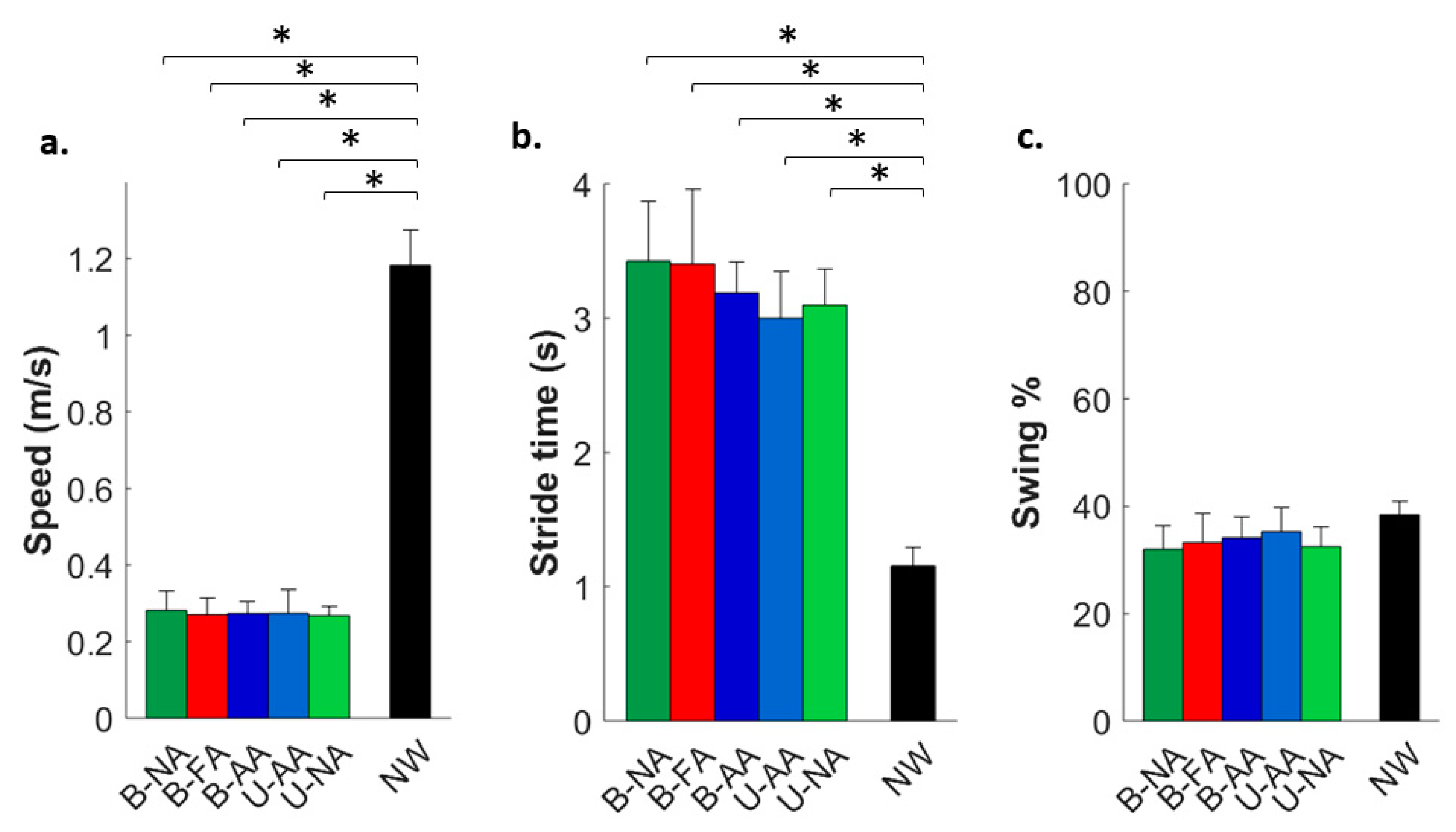
3.1. Spatio-Temporal Characteristics of the Gait

When subjects walked with the Ekso, their walking speed decreased (p < 0.0001 Figure 3a) and the stride time increased (p < 0.0001, Figure 3b).
However, when we time-normalized the gait cycle, the percentage of the swing phase was similar for all the walking conditions with and without the Ekso (p > 0.01, Figure 3c). We noticed a slight (<3%) decrease in the two Ekso unassisted modalities with respect to the Ekso-assisted modalities and the normal walking.

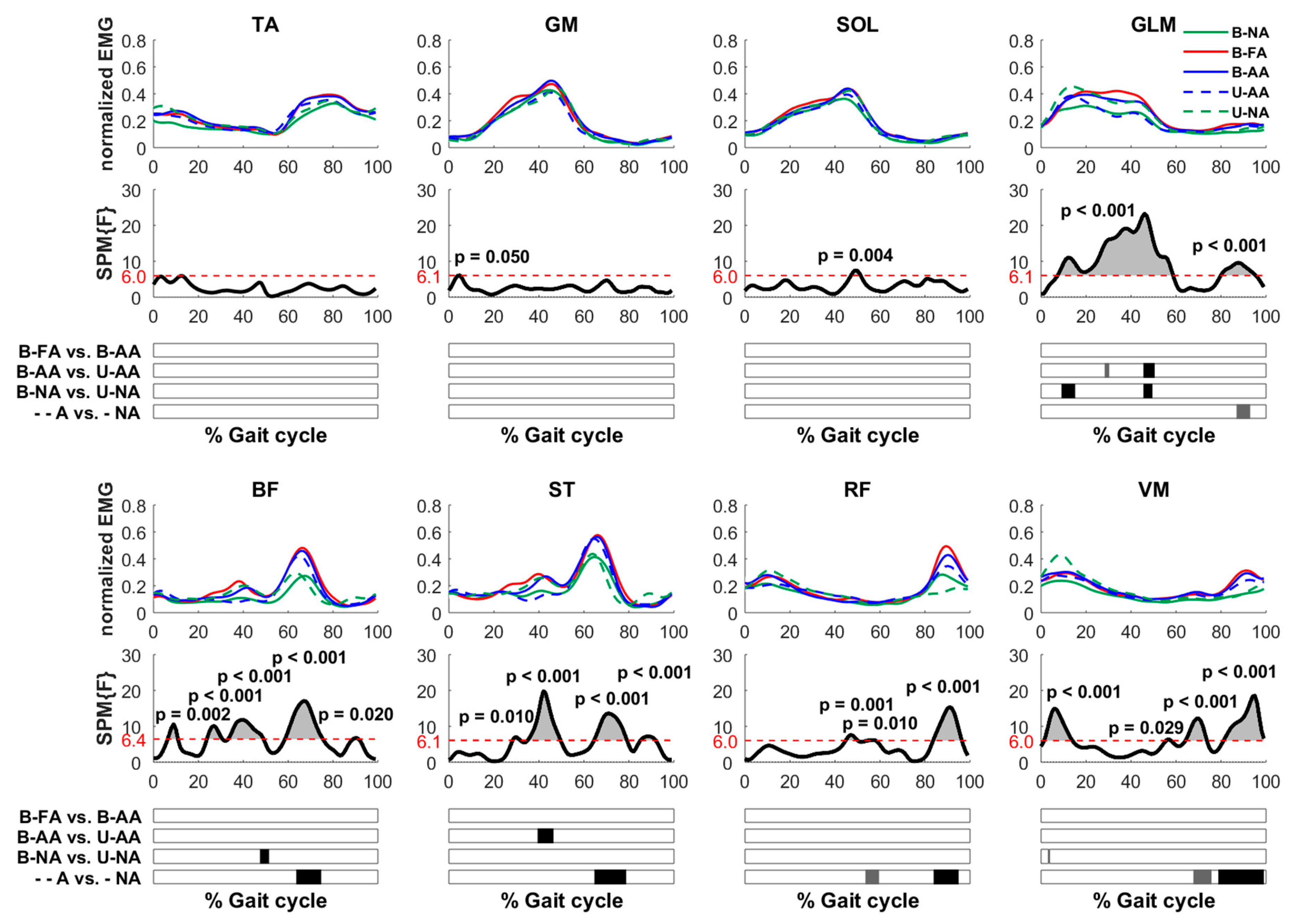
3.2. Muscle Activations When Walking with the Ekso under Different Conditions

As expected, there were no differences (neither main effects nor interactions) in muscle activations between left and right leg during the gait cycle. Moreover, the muscle patterns did not change when walking with the crutches or with the walker, i.e., no significant main effects nor interactions were found for the factor aids (p > 0.05, Figure 4).
When comparing the different assistance modalities (Figure 5), we found that the temporal patterns of activations did not change with the different assistive modalities, i.e., for all the muscles, the shape of the envelopes were highly similar; however, significant differences were observed in terms of level of muscle activations.
Specifically, small or negligible differences were found for the distal lower limb muscles (TA, GM, SOL), while higher and significant differences were found in the other muscles (GLM, BF, ST, RF, VM). When further investigating this result with the pre-planned comparisons, we found that:
  • There was no difference between the muscle patterns between the B-FA, with the Ekso providing a fixed maximum assistance, independently of the subjects’ voluntary contribution, with respect to B-AA with the assistance adapted by the device, depending on the subject’s performance.
  • In the second half of the gait cycle, the level of activations of both the unassisted modalities, B-NA, U-NA, was lower than those of the assistive modalities for the BF, ST RF, VM, and GLM muscles between 60–80% of the gait cycle for the first two muscles (in correspondence with the peak of muscle activation, both p < 0.001) and between 80–100% for the other three (RF and VM p < 0.001; GLM p < 0.05).
  • Under unilateral assistance, both the assisted (U-AA) and unassisted (U-NA) leg had significant differences when compared to the corresponding case where the contralateral leg was in the same assistive condition i.e., B-AA and B-NA, respectively. These differences were mainly evident and significant in the middle of the walking cycle (40–50%) for the GLM muscle in both comparisons (p < 0.001) for BF when comparing the unassisted conditions B-NA versus U-NA (p < 0.001) and for the ST when comparing the assistive conditions B-AA versus U-AA (p < 0.001). Specifically for the unassisted modalities, the activation was mainly higher for the unilateral condition, while it was lower for the assistive modalities.

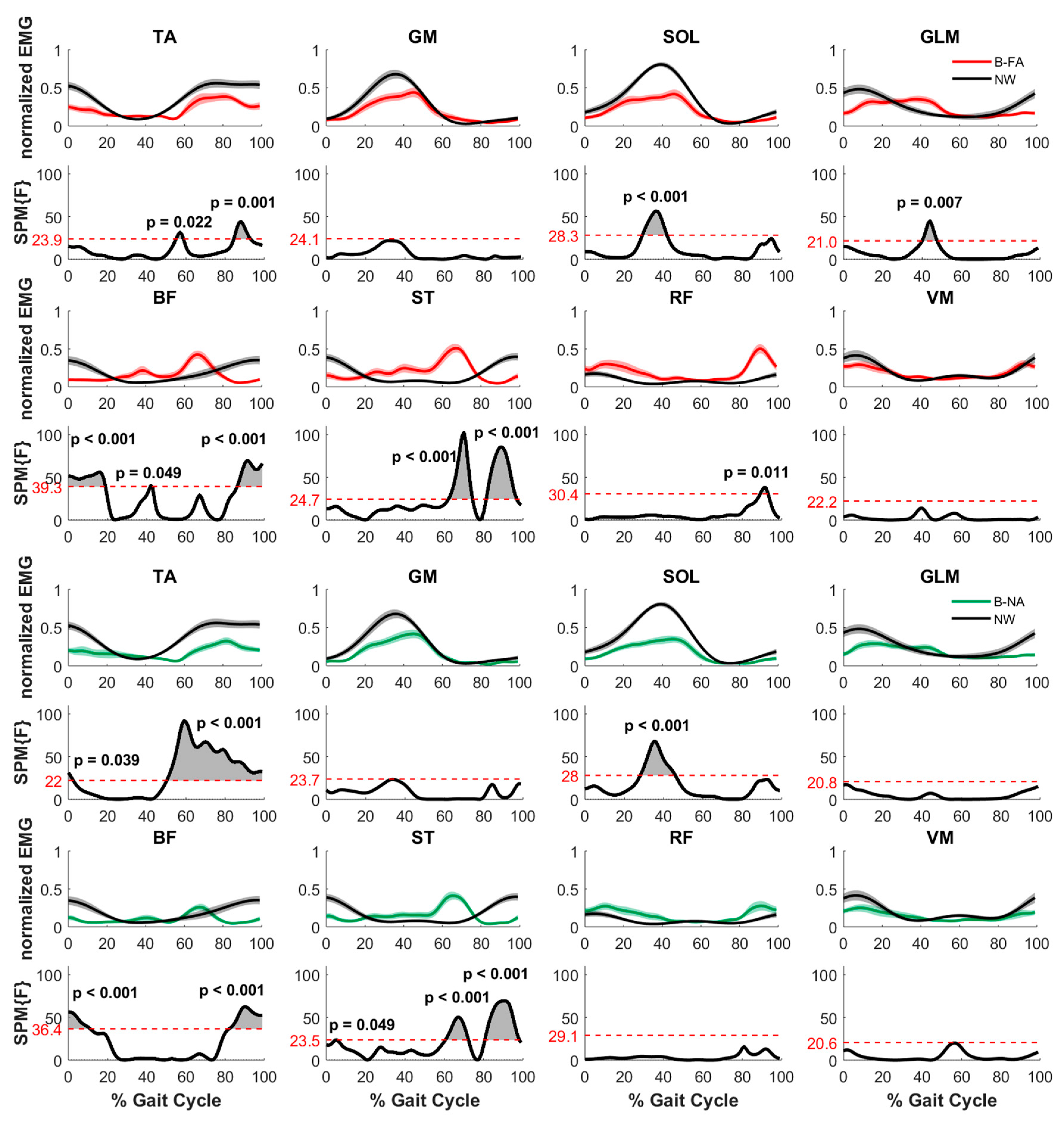
3.3. Muscle Activations When Walking with and without the Ekso

When walking with the Ekso with respect to NW (Figure 6), the activity of the distal lower limb muscles—TA, GM, SOL—decreased, as well as the activity of the GLM, both with (B-FA) and without (B-NA) assistance. These differences were observable mainly in correspondence of the peak of muscle activations, more specifically between 20–40% of the gait cycle for the SOL (p < 0.001) and for the GM (p = 0.05, i.e., at the threshold of significance), in the second half of the gait cycle for the TA (p < 0.02 in the B-FA and p < 0.001 in the B-NA), and between 40–50% for the GLM (p = 0.007 in the B-FA). This decrease of muscle activity was significant for the TA and the SOL, but not for the other two muscles, and it was more marked in absences of Ekso assistance.
Conversely, the RF had higher activations when subjects walked with Ekso at the end (>80%) of the gait cycle, although this difference increased and was statistically significant only when the Ekso assistance was present (p = 0.01). We did not observe any significant difference for the VM. Most interestingly, we observed different timings of activation for the ST. When walking with the Ekso, the peak of activation was between 60–80% of the cycle both in the assisted and unassisted modalities, while in this interval during NW, the ST was not active. This difference was highly significant for both B-FA and B-NA (both p < 0.001). A similar trend, although less marked, was observed for the BF, but the difference between Ekso and normal walking in this part of the gait cycle, i.e., from 60% to 80%, did not reach the threshold for significance. Both the BF and the ST muscles were not active at the beginning (<15%) and at the end (>80%) of the gait cycle, which are time intervals where normally, these muscles are active during walking. At the end of the gait cycle, the difference between NW and B-NA/B-FA was in all cases highly significant (p < 0.001 for both muscles), while at the beginning of the gait cycle, it was significant for the BF (p < 0.001), and for the ST, it was only slightly significant in the NW versus B-NA case (p = 0.049).
We finally verified that these results did not depend on the aids by comparing for three subjects the muscle activations without aids, with a walker, and with crutches (see Supplementary Figure S1) while walking without the Ekso. The only significant difference we found was for the VN muscle at the end of the gait cycle between 90–100%. We also notice a delay on the activation of TA, GM, and SOL, while their deactivation was synchronous with that of walking patterns without aids. This behavior was not present always in all subjects; thus, it did not reach the threshold for significance. None of the differences that were found comparing walking with and without the Ekso were observed in NW when comparing walking with and without the aids.

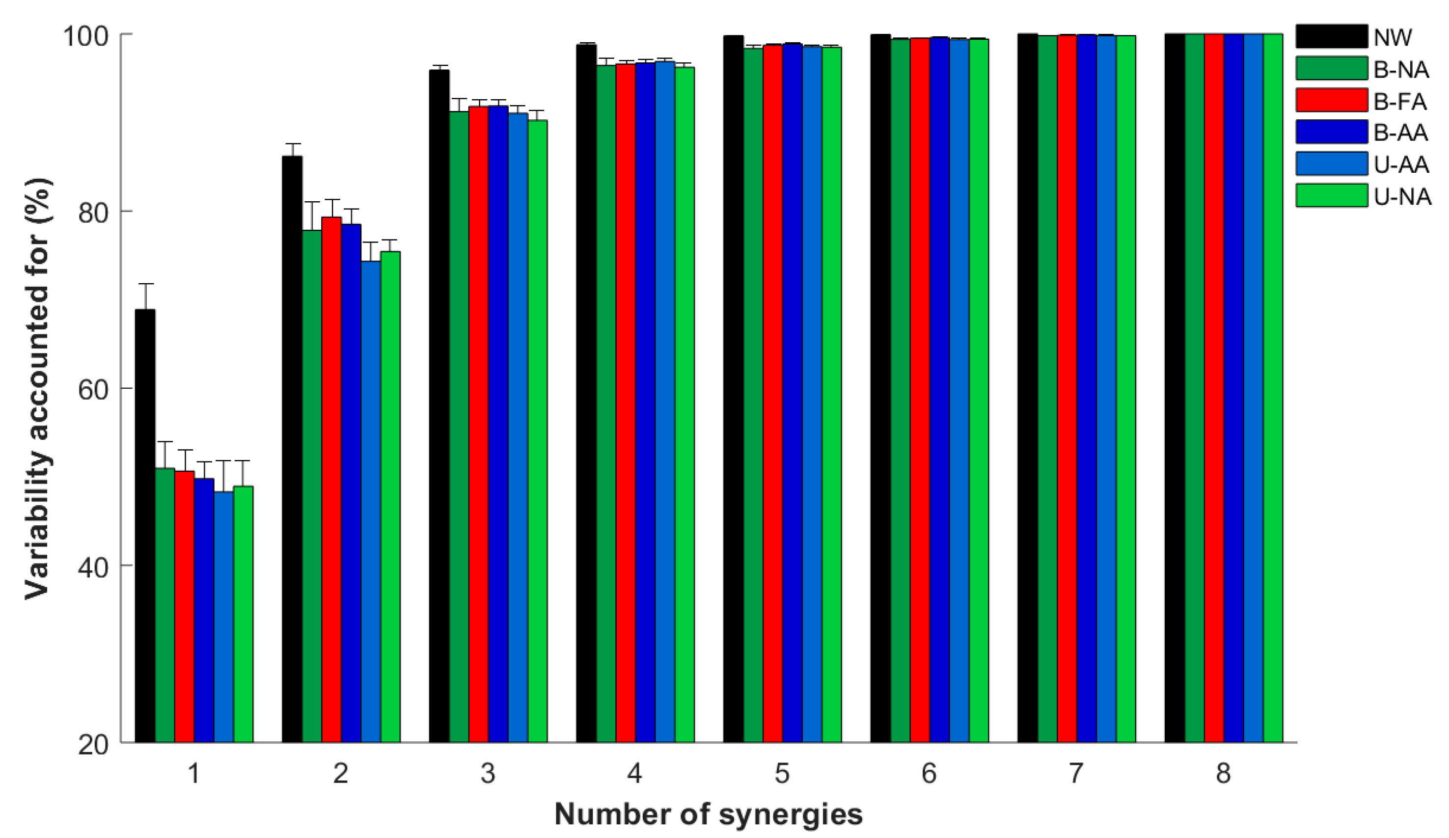
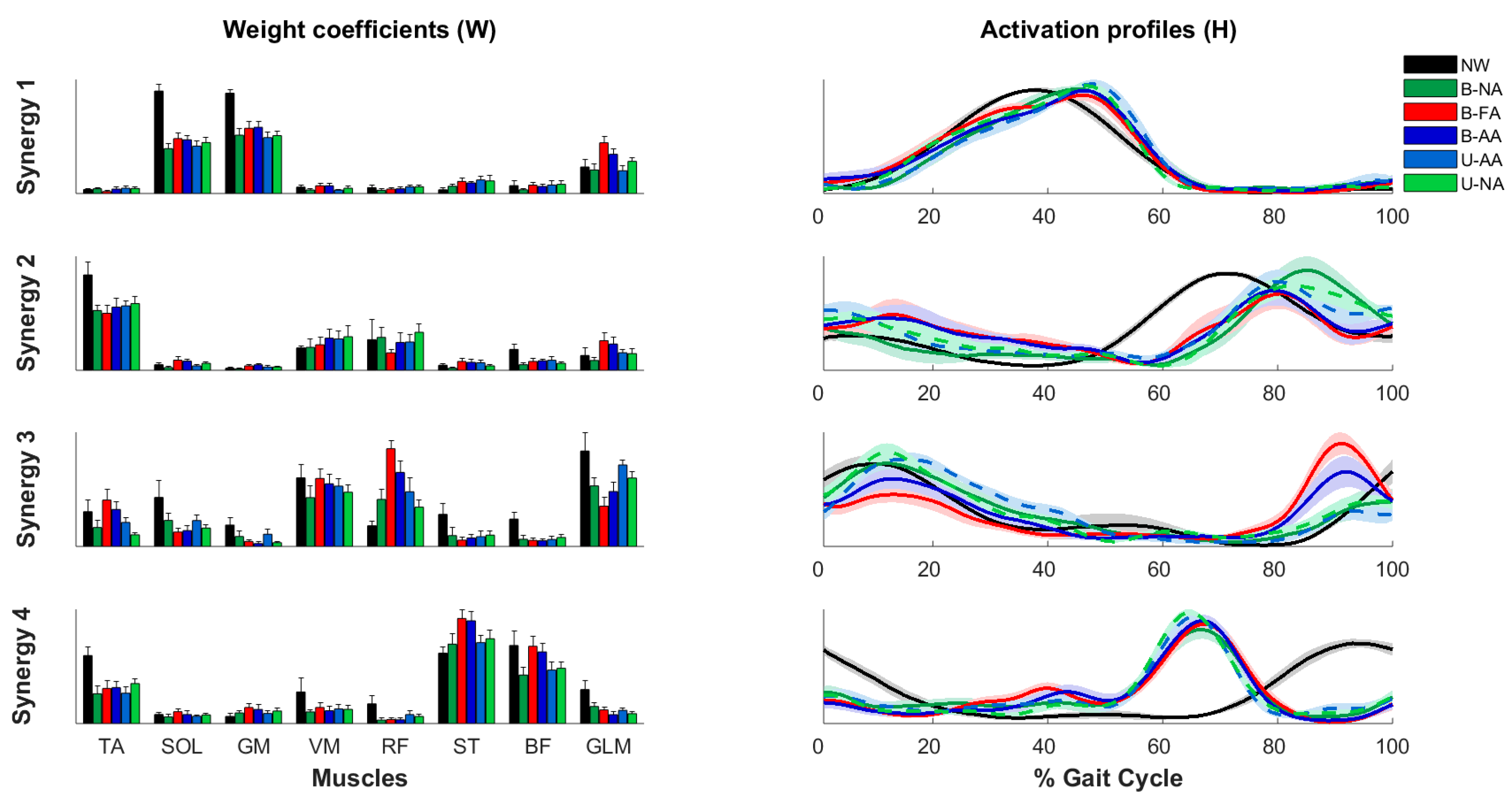
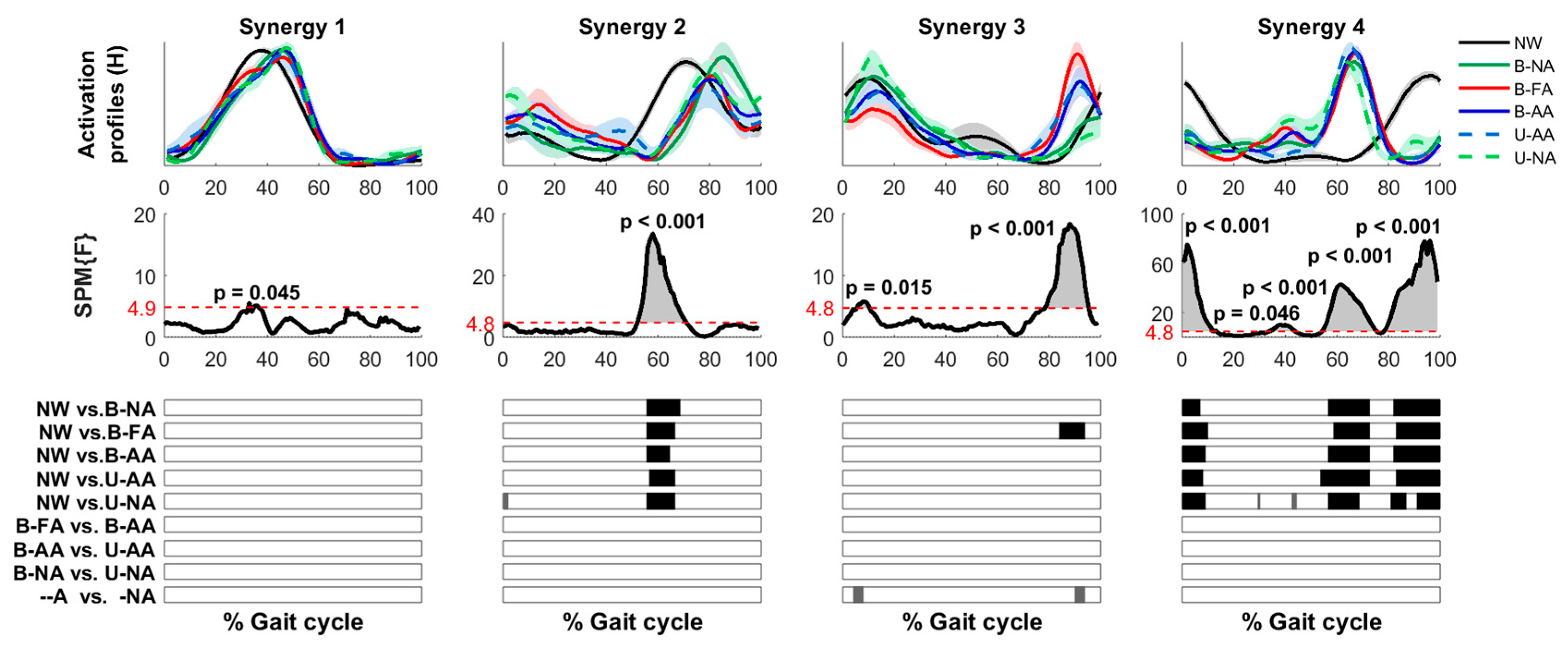
3.4. Muscle Synergies

Four synergies were required to reconstruct muscle activations during walking without the Ekso: (3.6 mean ± 1.2 std). When walking with the Ekso, the number of synergies was (mean ± std): B-NA = 3.87 ± 0.99; B-FA = 3.75 ± 1.75; B-AA = 3.13 ± 1.24; U-AA = 3.00 ± 0.00; U-NA = 3.37 ± 0.74. Notice that the total variability accounted for (Figure 7) based on the number of modules extracted by NNMF was lower for the first synergies when walking with the Ekso than when walking without the Ekso (p < 0.01).
To characterize and compare the synergies’ composition and timing, we extracted four independent modules for all the subjects and walking conditions (Figure 8).
Synergy 1 was due to the simultaneous activations of the ankle plantar flexors GM and SOL, and in all the walking conditions, it was mainly active between 20–60% of the gait cycle, in the late stance providing support to the body for the forward propulsion and for initiating the gait [34,42].
Synergy 2 was dominated by TA, the ankle dorsiflexor. During normal walking without the Ekso, Synergy 2 was active at <20% and more in the second half (>50%) of the gait cycle, with a peak between 60–90%. These activations correspond to the muscle activity for ankle dorsiflexion during and immediately after the heel strike and during the early swing for supporting the ground clearance of the foot [34,42]. When walking with the Ekso, the main peak of activation in all the conditions appeared delayed at about 10% of the gait cycle with respect to walking without the Ekso (p < 0.001, Figure 9).
Synergy 3 was dominated by the knee extensors VM and RF, with the latter also being the knee abductor, and by the GLM, which determines the hip extension and abduction. This synergy was active mainly at the beginning and at the end of the gait cycle, contributing to weight acceptance in the early stance [34,42]. Synergy 3 was quite similar across all the conditions, with and without the Ekso, with the main peak of activity at the end of the gait cycle that in most subjects was anticipated in about 10% in the conditions where participants were wearing the Ekso with respect to when they were not. This behavior reached the threshold for significance for the B-FA modality (p < 0.001, Figure 9).
Synergy 4 was mainly characterized by the ST and BF activity. The activation of this synergy was completely different between walking with and without Ekso. Indeed, in the normal walking without the Ekso, synergy 4 was active at the beginning (<20% of the gait cycle) and at the end (>80%) of the gait cycle, i.e., during stance, contributing to stabilizing the body and flexing the knee, and during the middle/late swing, contributing at decelerating the leg [34,42]. Instead, in all the Ekso walking modalities, synergy 4 was active only between 60–80% of the gait cycle (p < 0.001 in all intervals).

4. Discussion

This study aimed at understanding how the external balancing aids used with the exoskeleton, the structure of the device itself, and the different assistive forces provided for assisting the gait influence the users’ muscular activations when walking overground.
In the following, we discuss the main findings with respect to each of these aspects.

4.1. Comparison between Assistance Modalities with the Ekso

We did not find changes in muscle activations between the two bilateral exoskeleton-assisted modalities (B-FA, B-AA), i.e., comparing maximal and adaptive assistance. This contradicted our original hypothesis. We believed that if the exoskeleton would provide a maximum assistance enabling to walk with the Ekso in a predefined manner, independently from the ability of the user to generate the walking patterns, then the users, being greedy optimizers [23], would allow the Ekso to do all the work, decreasing their voluntary control, and consequently their muscle activations. This did not happen in our experiment, contradicting the basis of the assistance-as-needed approach [23]. The comparison between unassisted and assisted gait further confirmed this finding, since subjects had higher activations in the presence of assistance, i.e., when these activations were not needed for walking, than in the absence of assistance. Indeed, both the B-NA and U-NA subjects had a reduced muscle activation of the BF, ST, RF, and VM when compared to the assisted conditions. The muscle synergies analysis confirmed these results, since the four synergies that were extracted were quite similar among all the walking conditions with the Ekso: in number, in their structure (weight coefficients), and in some activation profiles.
The absence of the greedy optimization might depend on the limited experience of the users with the exoskeleton and because during testing, the overall walking distance was limited by the length of the walking path in the motion analysis lab, resulting in the anticipation of the unavoidable stops and turns at each side of the room. With longer training or with the possibility of walking in longer paths, subjects might exploit the assistive forces, decreasing their muscle activity.
Also, we should consider an alternative explanation: overground walking is different with respect to the treadmill walking (as in [23]), since in the latter case, the motor commands are generated mostly at the spinal level, while walking overground is more mediated by voluntary activity, especially in the early stage of the movement. A future study must further investigate this point.

4.2. Comparison between Normal Walking and Ekso-Assisted Walking

Regardless of the type of assistance, remarkable and significant differences in muscle activation timings were observed in the knee flexor muscles (ST and BF), confirming what was previously observed by [21] for the Ekso and by [43] for the Mindwalker exoskeletons. Specifically, the hamstrings muscles stabilize the body during the stance phase and flex the knee, having a peak of activation during the middle/end of the swing phase [44]; thus, the physiological timing of activation for these muscles is at the beginning and at the end of the gait cycle [45]. However, during the Ekso walking, we found a peak of activation between 60–80% of the gait cycle i.e., at the end of the terminal stance/beginning of the swing phase.
This result was confirmed by the synergies analysis. The muscle synergy S4, which is dominated by the ST and BF muscles, had completely opposite activation patterns when walking without the Ekso with respect to all the walking modalities with the Ekso: S4 was active mainly between 60–80% of the gait cycle instead of at the beginning (<20%) and at the end of the gait cycle (>80%).
Since this pattern was always observed when subjects walked with the Ekso, as well as in the absence of assistance, the change in the activation timing of these muscles might depend on the mechanical structure of the device itself, and not on the type or amount of assistance. More specifically, this abnormal activation might be due to the reduced mobility of the ankle joint of the exoskeleton, which as in many other devices, is semi-rigid and unpowered. This could create an overloading of knee flexors muscles in the push-off phase. The mechanical design of the ankle joint can also explain the reduction in amplitude during the push-off phase in the Ekso walking for the muscle TA, GM, and SOL; a similar observation was also recently reported by [22].
The single muscle analysis did not highlight differences in the remaining muscles, as in [18], while the muscle synergies analysis highlighted that, when walking with the Ekso, unlike in NW, the synergy S3—dominated by VM, RF, and GLM—anticipated the peak of activation by 10% of the gait cycle, while the synergy S2—dominated by the TA—had the peak of activation delayed.
These observations, as explained in the previous point, highlighted that healthy subjects not exposed to prolonged training were inclined to activate the muscles in all the exoskeleton conditions, even when the device provided the user with total assistance.

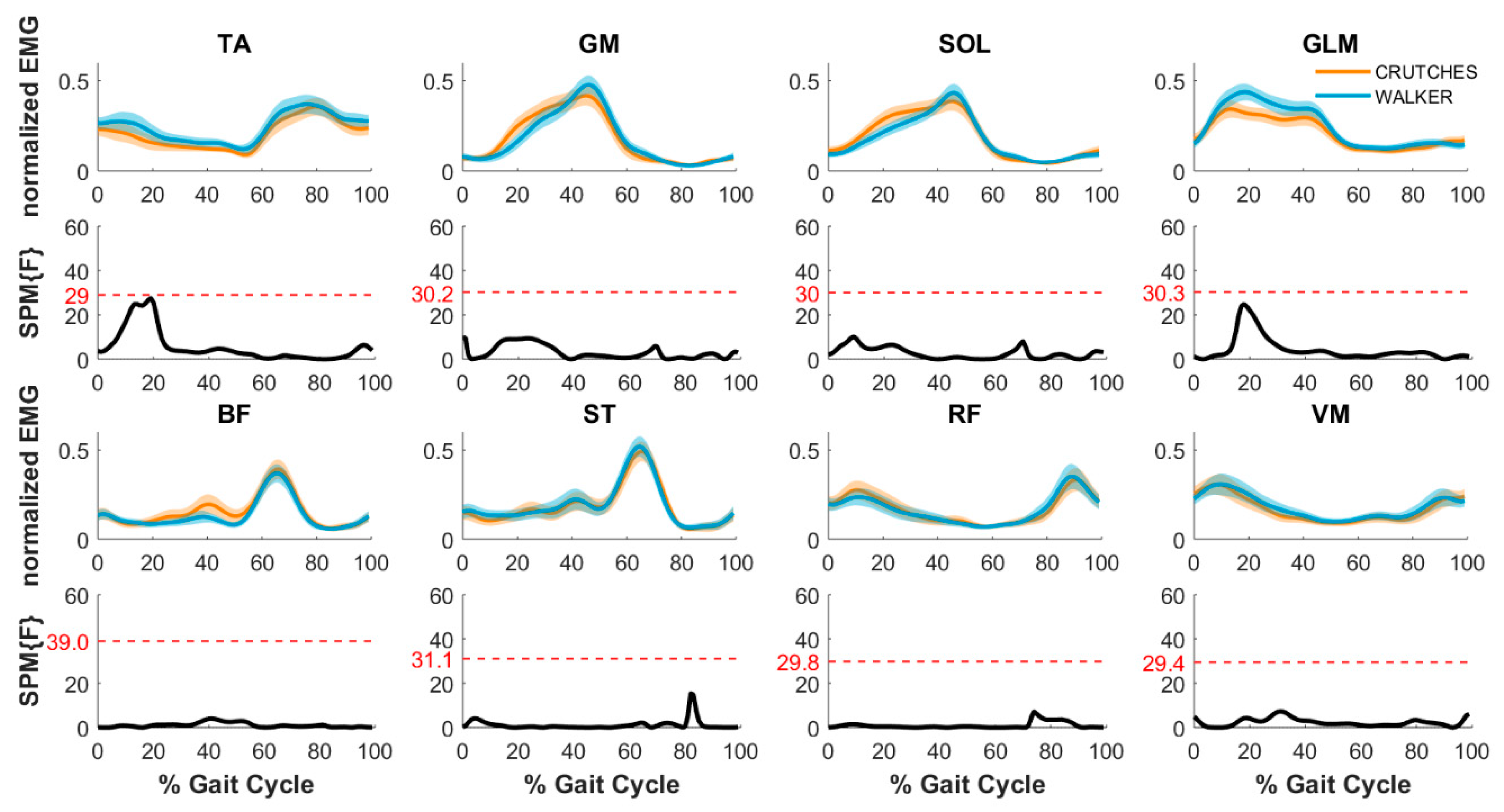
4.3. Walking with the Aids

There were no changes in the muscle activation patterns when the users walked with the walker or the crutches both with and without the Ekso, although in the second case, the small number of subjects limited the power of the analysis. In the latter case, when we had the possibility of comparing normal walking without the Ekso in the absence and presence of the two aids, for all three participants, we observed a reduction in the amplitude of the VM muscle when walking with the aids. This difference could be attributed to the VM being an antigravity extensor muscle, and therefore, as the load decreases because of the aids, the amplitude of the muscle activity decreases [46]. No significant changes were observed in terms of muscle activation timing, although a small delay on the muscle response of the platiflexors muscle was observed for two out of three subjects when walking with the aids. The latter difference may be because at the beginning of the single support phase, the subject’s weight is mainly loaded on the aids. Due to the new load distribution, the activation of the plantar flexor muscles is not required at the beginning of the stance phase, but only in the single support phase, when the load is completely distributed on one leg. The difference disappearing in the Ekso walk is in line with this observation, since in this case, the weight is also supported by the exoskeleton structure. Notice that this difference was not significant for our subjects. In a larger population, it could become significant, but that it was not observed in previous studies suggests that it would probably not, and that this trend is the result of intersubject variability. No other significant differences or trends were observed, confirming that the differences between walking with and without the Ekso were only due to the use of the device itself and not to the presence of the aids.

4.4. Cautionary Notes

The size of the observation pool is small, but in line with the literature. Namely, the number of subjects (eight), since only healthy participants were included, is in line with the field’s standard, e.g., [18,20,47,48,49,50]. Also, the number of strides per subject (10 without and 12 with an Ekso minimum) is low, but again supported by several studies using a similar or even lower number of strides [20,51,52,53,54]. This is because in healthy subjects, the activations of lower limb muscles are highly repeatable, and increasing the number of steps would not modify the outcome [51]. However, a study with a larger observation pool both in terms of strides and subjects is needed to consolidate these findings.

4.5. Future Work and Clinical Applications

Here, we focused on healthy subjects to investigate the effect of the mechanical structure of the Ekso and its different assistive modalities on muscle activity patterns during walking. The reason is that during walking, healthy subjects have highly repeatable activations both individually and as a group. Since neurological diseases and injury might affect the neural control of movements and the ability to walk differently, the muscle activations of the patients could have higher variability in both cases.
However, we performed this study with the goal of understanding the changes induced by the Ekso for identifying subsets of the population that would derive higher or lower benefits from its use. For example, in this study, we found a decrease in distal muscular activity (TA, SOL, and GM muscles) during the push-off phase, in favor of greater activity in the proximal part of the lower limb, with an extra-activation at the level of the muscles’ knee flexors. This suggest that subjects with ankle propulsion deficits would have limited benefits from treatment with this type of exoskeleton, since they need a specific rehabilitative training that enhances the activity of the distal muscles. Moreover, the protocol could be used to test the effects of this and other exoskeletons to provide indications on how to improve their mechanical structure or assistance.
We are also starting to use the proposed protocol with SCI survivors with incomplete injury who have different gait patterns, depending on the level of injury and the type of incompleteness (e.g., ASIA (American Spinal Injury Association) classification B,C,D, [55]). In this case, we are using the proposed protocol to investigate before training the muscle activations during walking with the Ekso, aiming at characterizing the effects of the different assistance modalities to select the most appropriate combination for personalized training. Moreover, the same protocol could then be used to investigate the effects of an exoskeleton-based training on specific patients walking patterns, analyzing the changes in the muscle activities due to practice. Instead, in people with complete SCI who have no muscular activity at the level of the lower limbs, the exoskeleton has undisputed value for allowing the users to maintain the standing posture and walking, with consequences on cardiovascular health and cardiovascular training [15,56]. In this case, while using the exoskeleton, the subjects would be actively contributing to walking with their upper body [57]. Therefore, it would be interesting to propose an evaluation protocol based on similar techniques as well as answering similar questions, but focusing on the muscular activity of the upper limbs and the trunk during walking with exoskeleton.

Supplementary Materials

The following are available online at https://www.mdpi.com/2076-3417/9/14/2868/s1, Figure S1: Comparison between normal walking and walking with walker and crutches, Table S1: Data not available for the analysis.

Author Contributions

Conceptualization, A.D.L., S.M., C.L., L.R., S.G., A.M. and M.C. (Maura Casadio); Methodology, A.D.L. and M.C. (Maura Casadio); Formal Analysis, A.D.L., A.B., G.M., L.P., M.C. (Martina Coscia) and M.C. (Maura Casadio); Investigation, A.D.L. and S.M..; Writing-Original Draft Preparation, A.D.L., A.B., G.M., L.P., M.C. (Martina Coscia) and M.C. (Maura Casadio); Writing-Review & Editing, A.D.L., A.B., S.M., G.M., L.P., M.C. (Martina Coscia), C.L., L.R., S.G., A.M. and (Maura Casadio); Supervision, A.M. and M.C. (Maura Casadio).

Funding

This research was supported by the Marie Curie Integration Grant (REMAKE, FP7-PEOPLE-2012-CIG-334201), the Ministry of Science and Technology, Israel (Joint Israel-Italy lab in Biorobotics “Artificial somatosensation for humans and humanoids”), the Research projects of national interest (ModuLimb, PRIN-2015HFWRYY) and the PhD scholarship of Regione Liguria to Giorgia Marchesi.

Conflicts of Interest

The authors report no conflict of interest.

References

  1. Sale, P.; Franceschini, M.; Waldner, A.; Hesse, S. Use of the robot assisted gait therapy in rehabilitation of patients with stroke and spinal cord injury. Eur. J. Phys. Rehabil. Med. 2012, 48, 111–121. [Google Scholar] [PubMed]
  2. Esquenazi, A.; Packel, A. Robotic-Assisted Gait Training and Restoration. Am. J. Phys. Med. Rehabil. 2012, 91, 217. [Google Scholar] [CrossRef] [PubMed]
  3. Zeilig, G.; Weingarden, H.; Zwecker, M.; Dudkiewicz, I.; Bloch, A.; Esquenazi, A. Safety and tolerance of the ReWalk™ exoskeleton suit for ambulation by people with complete spinal cord injury: A pilot study. J. Spinal Cord Med. 2012, 35, 96–101. [Google Scholar] [CrossRef] [PubMed]
  4. Hornby, T.G.; Zemon, D.H.; Campbell, D. Robotic-Assisted, Body-Weight–Supported Treadmill Training in Individuals Following Motor Incomplete Spinal Cord Injury. Phys. Ther. 2005, 85, 52–66. [Google Scholar] [PubMed]
  5. Tefertiller, C.; Pharo, B.; Evans, N.; Winchester, P. Efficacy of rehabilitation robotics for walking training in neurological disorders: A review. J. Rehabil. Res. Dev. 2011, 48, 387. [Google Scholar] [CrossRef] [PubMed]
  6. Benito-Penalva, J.; Edwards, D.J.; Opisso, E.; Cortes, M.; López-Blázquez, R.; Murillo, N.; Costa, Ú.; Tormos, J.M.; Vidal-Samso, J.; Valls-Solé, J.; et al. Gait Training in Human Spinal Cord Injury Using Electromechanical Systems: Effect of Device Type and Patient Characteristics. Arch. Phys. Med. Rehabil. 2012, 93, 404–412. [Google Scholar] [CrossRef]
  7. Sale, P.; Russo, E.F.; Scarton, A.; Calabrò, R.S.; Masiero, S.; Filoni, S. Training for mobility with exoskeleton robot in spinal cord injury patients: A pilot study. Eur. J. Phys. Rehabil. Med. 2018, 54, 745–751. [Google Scholar] [CrossRef]
  8. Chang, S.-H.; TIRR SCI Clinical Exoskeleton Group; Afzal, T.; Berliner, J.; Francisco, G.E.; Kern, M. Exoskeleton-assisted gait training to improve gait in individuals with spinal cord injury: A pilot randomized study. Pilot Feasibility Stud. 2018, 4, 62. [Google Scholar] [CrossRef] [PubMed]
  9. Nam, K.Y.; Kim, H.J.; Kwon, B.S.; Park, J.-W.; Lee, H.J.; Yoo, A. Robot-assisted gait training (Lokomat) improves walking function and activity in people with spinal cord injury: A systematic review. J. Neuroeng. Rehabil. 2017, 14, 5819. [Google Scholar] [CrossRef] [PubMed]
  10. Stevenson, A.J.; Mrachacz-Kersting, N.; Van Asseldonk, E.; Turner, D.L.; Spaich, E.G. Spinal plasticity in robot-mediated therapy for the lower limbs. J. Neuroeng. Rehabil. 2015, 12, 81. [Google Scholar] [CrossRef] [PubMed]
  11. Dobkin, B.; Apple, D.; Barbeau, H.; Basso, M.; Behrman, A.; Deforge, D.; Ditunno, J.; Dudley, G.; Elashoff, R.; Fugate, L.; et al. Weight-supported treadmill vs over-ground training for walking after acute incomplete SCI. Neurology 2006, 66, 484–493. [Google Scholar] [CrossRef] [PubMed]
  12. Alcobendas-Maestro, M.; Esclarín-Ruz, A.; Casado-López, R.M.; Muñoz-González, A.; Perez-Mateos, G.; Gonzalez-Valdizan, E.; Martin, J.L.R. Lokomat robotic-assisted versus overground training within 3 to 6 months of incomplete spinal cord lesion: Randomized controlled trial. Neurorehabil. Neural Repair 2012, 26, 1058–1063. [Google Scholar] [CrossRef] [PubMed]
  13. Louie, D.R.; Eng, J.J.; Lam, T. Spinal Cord Injury Research Evidence Research, “Gait speed using powered robotic exoskeletons after spinal cord injury: A systematic review and correlational study”. J. Neuroeng. Rehabil. 2015, 12, 82. [Google Scholar] [CrossRef] [PubMed]
  14. Swinnen, E.; Duerinck, S.; Baeyens, J.; Meeusen, R.; Kerckhofs, E. Effectiveness of robot-assisted gait training in persons with spinal cord injury: A systematic review. J. Rehabilitation Med. 2010, 42, 520–526. [Google Scholar] [CrossRef] [PubMed] [Green Version]
  15. Gorgey, A.S.; Wade, R.; Sumrell, R.; Villadelgado, L.; Khalil, R.E.; Lavis, T. Exoskeleton Training May Improve Level of Physical Activity After Spinal Cord Injury: A Case Series. Top. Spinal Cord Inj. Rehabil. 2017, 23, 245–255. [Google Scholar] [CrossRef] [PubMed]
  16. Palermo, A.E.; Maher, J.L.; Baunsgaard, C.B.; Nash, M.S. Clinician-Focused Overview of Bionic Exoskeleton Use After Spinal Cord Injury. Top. Spinal Cord Inj. Rehabil. 2017, 23, 234–244. [Google Scholar] [CrossRef] [PubMed]
  17. Molteni, F.; Gasperini, G.; Gaffuri, M.; Colombo, M.; Giovanzana, C.; Lorenzon, C.; Farina, N.; Cannaviello, G.; Scarano, S.; Proserpio, D.; et al. Wearable robotic exoskeleton for over-ground gait training in sub-acute and chronic hemiparetic stroke patients: Preliminary results. Eur. J. Phys. Rehabil. Med. 2017, 53, 676–684. [Google Scholar] [PubMed]
  18. Moreno, J.C.; Barroso, F.; Farina, D.; Gizzi, L.; Santos, C.; Molinari, M.; Pons, J.L. Effects of robotic guidance on the coordination of locomotion. J. Neuroeng. Rehabil. 2013, 10, 79. [Google Scholar] [CrossRef]
  19. Jin, X.; Cai, Y.; Prado, A.; Agrawal, S.K. Effects of exoskeleton weight and inertia on human walking. In Proceedings of the 2017 IEEE International Conference on Robotics and Automation (ICRA), Singapore, 29 May–3 June 2017; pp. 1772–1777. [Google Scholar]
  20. Labini, F.S.; La Scaleia, V.; D’Avella, A.; Pisotta, I.; Tamburella, F.; Scivoletto, G.; Molinari, M.; Wang, S.; Wang, L.; Van Asseldonk, E.; et al. EMG patterns during assisted walking in the exoskeleton. Front. Hum. Neurosci. 2014, 8, 423. [Google Scholar]
  21. Swank, C.; Wang-Price, S.; Gao, F.; Almutairi, S. Walking in A Robotic Exoskeleton Does Not Mimic Natural Gait (Preprint) Walking with a Robotic Exoskeleton Does Not Mimic Natural Gait: A Within-Subjects Study. JMIR Rehabil. Assist. Technol. 2018, 6, e11023. [Google Scholar] [CrossRef]
  22. Ekkelenkamp, R.; Van Asseldonk, E.H.F.; Veneman, J.F.; Buurke, J.H.; Van Der Helm, F.C.T.; Van Der Kooij, H. The Effects on Kinematics and Muscle Activity of Walking in a Robotic Gait Trainer During Zero-Force Control. IEEE Trans. Neural Syst. Rehabil. Eng. 2008, 16, 360–370. [Google Scholar]
  23. Emken, J.L.; Benitez, R.; Reinkensmeyer, D.J. Human-robot cooperative movement training: Learning a novel sensory motor transformation during walking with robotic assistance-as-needed. J. Neuroeng. Rehabil. 2007, 4, 8. [Google Scholar] [CrossRef] [PubMed]
  24. Ekso Bionics. Ekso Bionics Homepage. 2019. Available online: https://eksobionics.com/ (accessed on 18 July 2019).
  25. Landau, I.D.; Lozano, R.; M’Saad, M.; Karimi, A. Adaptive Control: Algorithms, Analysis AND Applications; Springer Science & Business Media: Berlin, Germany, 2011. [Google Scholar]
  26. Davis, R.B. Clinical gait analysis. IEEE Eng. Med. Biol. Mag. 1988, 7, 35–40. [Google Scholar] [CrossRef] [PubMed]
  27. De Luca, A.; Giannoni, P.; Vernetti, H.; Capra, C.; Lentino, C.; Checchia, G.A.; Casadio, M. Training the Unimpaired Arm Improves the Motion of the Impaired Arm and the Sitting Balance in Chronic Stroke Survivors. IEEE Trans. Neural Syst. Rehabil. Eng. 2016, 25, 873–882. [Google Scholar] [CrossRef] [PubMed]
  28. O’Connor, C.M.; Thorpe, S.K.; O’Malley, M.J.; Vaughan, C.L. Automatic detection of gait events using kinematic data. Gait Posture 2007, 25, 469–474. [Google Scholar] [CrossRef] [PubMed]
  29. De Luca, A.; Vernetti, H.; Capra, C.; Pisu, I.; Cassiano, C.; Barone, L.; Gaito, F.; Danese, F.; Antonio Checchia, G.; Lentino, C.; et al. Recovery and compensation after robotic assisted gait training in chronic stroke survivors. Disabil. Rehabil. Assist. Technol. 2018, 1–13. [Google Scholar] [CrossRef] [PubMed]
  30. Rogers, E.; Polygerinos, P.; Allen, S.; Panizzolo, F.A.; Walsh, C.J.; Holland, D.P. A Quasi-Passive Knee Exoskeleton to Assist During Descent. In Wearable Robotics: Challenges and Trends; Springer: Cham, Switzerland, 2017; pp. 63–67. [Google Scholar]
  31. Naik, G.R.; Al-Ani, A.; Gobbo, M.; Nguyen, H.T. Does Heel Height Cause Imbalance during Sit-to-Stand Task: Surface EMG Perspective. Front. Physiol. 2017, 8, 626. [Google Scholar] [CrossRef]
  32. Cheung, V.C.K.; Piron, L.; Agostini, M.; Silvoni, S.; Turolla, A.; Bizzi, E. Stability of muscle synergies for voluntary actions after cortical stroke in humans. Proc. Natl. Acad. Sci. USA 2009, 106, 19563–19568. [Google Scholar] [CrossRef] [Green Version]
  33. Pellegrino, L.; Coscia, M.; Muller, M.; Solaro, C.; Casadio, M. Evaluating upper limb impairments in multiple sclerosis by exposure to different mechanical environments. Sci. Rep. 2018, 8, 2110. [Google Scholar] [CrossRef] [Green Version]
  34. Clark, D.J.; Ting, L.H.; Zajac, F.E.; Neptune, R.R.; Kautz, S.A. Merging of healthy motor modules predicts reduced locomotor performance and muscle coordination complexity post-stroke. J. Neurophysiol. 2010, 103, 844–857. [Google Scholar] [CrossRef]
  35. Lee, D.D.; Hill, M.; Seung, H.S. Algorithms for Non-negative Matrix Factorization. Adv. Neural Inf. Process. Syst. 2001, 13, 556–562. [Google Scholar]
  36. Ting, L.H.; McKay, J.L. Neuromechanics of muscle synergies for posture and movement. Curr. Opin. Neurobiol. 2007, 17, 622–628. [Google Scholar] [CrossRef] [PubMed] [Green Version]
  37. Flanders, M. Temporal patterns of muscle activation for arm movements in three-dimensional space. J. Neurosci. 1991, 11, 2680–2693. [Google Scholar] [CrossRef] [PubMed]
  38. Coscia, M.; Cheung, V.C.; Tropea, P.; Koenig, A.; Monaco, V.; Bennis, C.; Micera, S.; Bonato, P. The effect of arm weight support on upper limb muscle synergies during reaching movements. J. Neuroeng. Rehabil. 2014, 11, 22. [Google Scholar] [CrossRef] [PubMed]
  39. D’Avella, A.; Saltiel, P.; Bizzi, E. Combinations of muscle synergies in the construction of a natural motor behavior. Nat. Neurosci. 2003, 6, 300–308. [Google Scholar] [CrossRef] [PubMed]
  40. Cheung, V.C.; Turolla, A.; Agostini, M.; Silvoni, S.; Bennis, C.; Kasi, P.; Paganoni, S.; Bonato, P.; Bizzi, E. Muscle synergy patterns as physiological markers of motor cortical damage. Proc. Natl. Acad. Sci. USA 2012, 109, 14652–14656. [Google Scholar] [CrossRef] [Green Version]
  41. Friston, K. A short history of SPM. Stat. Parametr. Mapp. Anal. Funct. Brain Images 2007, 3–9. [Google Scholar] [CrossRef]
  42. Neptune, R.R.; Clark, D.J.; Kautz, S.A. Modular control of human walking: A simulation study. J. Biomech. 2009, 42, 1282–1287. [Google Scholar] [CrossRef] [Green Version]
  43. Wang, S.; Wang, L.; Meijneke, C.; Van Asseldonk, E.; Hoellinger, T.; Cheron, G.; Ivanenko, Y.; La Scaleia, V.; Sylos-Labini, F.; Molinari, M.; et al. Design and control of the MINDWALKER exoskeleton. IEEE Trans. Neural Syst. Rehabil. Eng. 2014, 23, 277–286. [Google Scholar] [CrossRef]
  44. Cheron, G.; Bengoetxea, A.; Pozzo, T.; Bourgeois, M.; Draye, J. Evidence of a preprogrammed deactivation of the hamstring muscles for triggering rapid changes of posture in humans. Electroencephalogr. Clin. Neurophysiol. Mot. Control. 1997, 105, 58–71. [Google Scholar] [CrossRef]
  45. Perry, J.; Davids, J.R. Gait analysis: Normal and pathological function. J. Pediatr. Orthop. 1992, 12, 815. [Google Scholar] [CrossRef]
  46. Clark, B.C.; Manini, T.M.; Ordway, N.R.; Ploutz-Snyder, L.L. Leg muscle activity during walking with assistive devices at varying levels of weight bearing. Arch. Phys. Med. Rehabil. 2004, 85, 1555–1560. [Google Scholar] [CrossRef] [PubMed]
  47. Morone, G.; Iosa, M.; Tamburella, F.; Muzzioli, L.; Pisotta, I.; Moreno, J.C.; Pons, J.L.; Paolucci, S.; Cincotti, F.; Molinari, M. An EMG Pattern Comparison of Exoskeleton vs. End-Effector Robotic Device for Assisted Walking Training BT-Replace, Repair, Restore, Relieve–Bridging Clinical and Engineering Solutions in Neurorehabilitation; Springer: Cham, Switzerland, 2014; pp. 563–567. [Google Scholar]
  48. Hussein, S.; Schmidt, H.; Volkmar, M.; Werner, C.; Helmich, I.; Piorko, F.; Kruger, J.; Hesse, S. Muscle coordination in healthy subjects during floor walking and stair climbing in robot assisted gait training. In Proceedings of the 2008 30th Annual International Conference of the IEEE Engineering in Medicine and Biology Society, Vancouver, BC, Canada, 20–25 August 2008; pp. 1961–1964. [Google Scholar]
  49. Gizzi, L.; Nielsen, J.F.; Felici, F.; Moreno, J.C.; Pons, J.L.; Farina, D. Motor modules in robot-aided walking. J. Neuroeng. Rehabil. 2012, 9, 76. [Google Scholar] [CrossRef] [PubMed]
  50. Cappellini, G.; Ivanenko, Y.P.; Poppele, R.E.; Lacquaniti, F. Motor Patterns in Human Walking and Running. J. Neurophysiol. 2006, 95, 3426–3437. [Google Scholar] [CrossRef] [PubMed] [Green Version]
  51. Arsenault, A.B.; Winter, D.A.; Marteniuk, R.G.; Hayes, K.C. How many strides are required for the analysis of electromyographic data in gait? Scand. J. Rehabil. Med. 1986, 18, 133–135. [Google Scholar]
  52. Lee, S.J.; Hidler, J. Biomechanics of overground vs. treadmill walking in healthy individuals. J. Appl. Physiol. 2008, 104, 747–755. [Google Scholar] [CrossRef] [PubMed]
  53. Shiavi, R.; Frigo, C.; Pedotti, A. Electromyographic signals during gait: Criteria for envelope filtering and number of strides. Med Boil. Eng. 1998, 36, 171–178. [Google Scholar] [CrossRef]
  54. Hof, A.; Elzinga, H.; Grimmius, W.; Halbertsma, J.; Hof, A. Speed dependence of averaged EMG profiles in walking. Gait Posture 2002, 16, 78–86. [Google Scholar] [CrossRef] [Green Version]
  55. Kirshblum, S.C.; Burns, S.P.; Biering-Sorensen, F.; Donovan, W.; Graves, D.E.; Jha, A.; Johansen, M.; Jones, L.; Krassioukov, A.; Mulcahey, M.J.; et al. International standards for neurological classification of spinal cord injury (revised 2011). J. Spinal Cord Med. 2011, 34, 535–546. [Google Scholar] [CrossRef]
  56. Asselin, P.; Knezevic, S.; Kornfeld, S.; Cirnigliaro, C.; Agranova-Breyter, I.; Bauman, W.A.; Spungen, A.M. Heart rate and oxygen demand of powered exoskeleton-assisted walking in persons with paraplegia. J. Rehabil. Res. Dev. 2015, 52, 147–158. [Google Scholar] [CrossRef]
  57. AlAmro, R.A.; Chisholm, A.E.; Williams, A.M.M.; Carpenter, M.G.; Lam, T. Overground walking with a robotic exoskeleton elicits trunk muscle activity in people with high-thoracic motor-complete spinal cord injury. J. Neuroeng. Rehabil. 2018, 15, 109. [Google Scholar] [CrossRef] [PubMed]
Figure 1. (a) Exoskeleton Ekso; (b,c) Participants wearing the Ekso. Red dots indicate the positions of the reflective markers for the motion analysis and the blue rectangles indicate the position of the surface electromyographic (EMG) electrodes. (d) Markers recorded by the motion analysis system (lateral view in the center of the motion analysis lab pathway).
Applsci 09 02868 g001
Figure 2. Experimental protocol.
Applsci 09 02868 g002
Figure 3. (a) Mean ± standard walking speed (m/s); (b) Mean ± standard stride duration (s); (c) Mean ± standard swing phase duration expressed as percentage of the gait cycle. Colors indicate the different walking modalities: black for normal walking without the Ekso (NW); the other colors refer to walking with the Ekso with bilateral no assistance (B-NA, dark green), bilateral full assistance (B-FA, red), bilateral adaptive assistance (B-AA, dark blue), unilateral adaptive assistance (U-AA, light blue), and unilateral no assistance (U-NA, light green). * indicates p < 0.0001.
Applsci 09 02868 g003
Figure 4. First and third rows: Muscle activation patterns (Mean ± SE) during walking inside the Ekso with the walker (blue lines) and crutches (orange lines). We considered the following muscles: muscles tibialis anterior (TA), gastrocnemius medialis (GM), soleus (SOL), rectus femoris (RF), vastus medialis (VM), semitendinosus (ST), biceps femoris (BF), and gluteus medius (GLM). The lines were obtained by averaging all the modalities of walking with the Ekso (B-FA, B-AA, U-AA, B-NA, and U-NA) and the contribution of both legs that were balanced with respect the two aids. Second and fourth rows: Statistical parametric mapping (SPM{F}) statistic as a function of the percentage of gait cycle. The threshold for significance is indicated in red.
Applsci 09 02868 g004
Figure 5. First and fourth rows: Muscle activations patterns during walking inside the Ekso with different assistance modalities. Unilateral assistance was indicated with dashed lines, while bilateral assistance was indicated with continuous lines, and the type of assistance was indicated by the color of the lines: B-FA (red), B-AA, U-AA (blue), B-NA, and U-NA (green). We considered the following muscles: muscles tibialis anterior (TA), gastrocnemius medialis (GM), soleus (SOL), rectus femoris (RF), vastus medialis (VM), semitendinosus (ST), biceps femoris (BF), and gluteus medius (GLM). Second and fifth rows: SPM{F} statistic as a function of the percentage of gait cycle. The lines were obtained by averaging the strides performed with crutches and walkers as well as the strides started with the left and right leg. The threshold for significance is indicated in red. Third and sixth rows: Statistic results of the pre-planned comparison: (1) B-FA vs. B-AA, (2) B-AA vs. U-AA, (3) B-NA vs. U-NA, (4) --A vs. -NA. Black intervals p < 0.001 and grey intervals p < 0.05.
Applsci 09 02868 g005
Figure 6. Rows 1, 3, 5, and 7: Comparison between normal walking (black lines) and bilaterally assistive (B-FA) walking with the Ekso (top panel, rows 1–3, red lines) and unassisted (B-NA) walking with the Ekso (low panel, rows 5–7, green lines) (mean ± SE). We considered the following muscles: muscles tibialis anterior (TA), gastrocnemius medialis (GM), soleus (SOL), rectus femoris (RF), vastus medialis (VM), semitendinosus (ST), biceps femoris (BF), and gluteus medius (GLM). The lines were obtained by averaging the strides performed with crutches and walkers as well as the strides started with the left and right leg. Rows 2, 4, 6, 8: panels show the SPM{F} statistic as a function of the percentage of gait cycle. The threshold for significance is indicated in red.
Applsci 09 02868 g006
Figure 7. Total variability accounted for (mean ± se) based on the number of modules extracted by non-negative matrix factorization (NNMF) for all the investigated walking conditions (normal walking without the Ekso: NW; Ekso-assisted walking: B-NA, B-FA, B-AA, U-AA, U-NA). Colors indicate the different walking modalities: black for normal walking without the Ekso (NW); the other colors refer to walking with the Ekso with bilateral no assistance (B-NA, dark green), bilateral full assistance (B-FA, red), bilateral adaptive assistance (B-AA, dark blue), unilateral adaptive assistance (U-AA, light blue), and unilateral no assistance (U-NA, light green).
Applsci 09 02868 g007
Figure 8. Weights (left panel) and activation coefficients (right panel) of the four muscle synergies when normal walking without the Ekso and when walking with the Ekso: B-NA, B-FA, B-AA, U-AA, and U-NA. Colors and lines indicate the different walking modalities: black solid line for normal walking without the Ekso (NW); the other colors refer to walking with the Ekso with bilateral no assistance (B-NA, dark green solid line), bilateral full assistance (B-FA, red solid line), bilateral adaptive assistance (B-AA, dark blue solid line), unilateral adaptive assistance (U-AA, light blue dotted line), and unilateral no assistance (U-NA, light green dotted line).
Applsci 09 02868 g008
Figure 9. First row: Mean activation profiles of the four muscle synergies for all the walking conditions. Unilateral assistance is indicated with dashed lines, while bilateral assistance is indicated with continuous lines, and the type of assistance is indicated by the color of the lines: B-FA (red), B-AA, U-AA (blue), B-NA, U-NA (green). Second row: SPM{F} statistic as a function of the percentage of gait cycle. The threshold for significance is indicated in red. Third row: Statistic results for the pre-planned comparisons between normal walking and the five Ekso-assisted modalities (NW vs. B-NA; NW vs B-FA; NW vs. B-AA; NW vs. U-AA; NW vs. U-NA ) and for the comparisons between Ekso-assisted modalities (B-FA vs. B-AA; B-AA vs. U-AA; B-NA vs. U-NA; --A vs. -NA). Black intervals p < 0.001 and grey intervals p < 0.05.
Applsci 09 02868 g009
Table 1. Healthy subject’s data; Y: years, M: male, F: female.
Age
[Y]
Gender
[M/F]
Height
[cm]
Weight
[Kg]
S145M17968
S224F17562
S325F15554
S422F18370
S522M17875
S623M18286
S724F15860
S832M18474
Table 2. Gait parameters for the Ekso set for each subject.
Subjects Height
[cm]
Step Length
[inches]
Step Height
[inches]
Swing Time
[s]
S117915.011.35
S217514.511.30
S315513.511.20
S418315.511.40
S517815.011.35
S618215.511.40
S715813.511.20
S818415.511.40

