Review

Perspectives in Liquid-Crystal-Aided Nanotechnology and Nanoscience

School of Physics and Astronomy, University of Manchester, Oxford Road, Manchester M13 9PL, UK
*
Author to whom correspondence should be addressed.
Appl. Sci. 2019, 9(12), 2512; https://doi.org/10.3390/app9122512
Submission received: 29 May 2019 / Revised: 12 June 2019 / Accepted: 14 June 2019 / Published: 20 June 2019
(This article belongs to the Section Nanotechnology and Applied Nanosciences)

Abstract

:

Featured Application

Nanomaterial-doped liquid crystals.

Abstract

The research field of liquid crystals and their applications is recently changing from being largely focused on display applications and optical shutter elements in various fields, to quite novel and diverse applications in the area of nanotechnology and nanoscience. Functional nanoparticles have recently been used to a significant extent to modify the physical properties of liquid crystals by the addition of ferroelectric and magnetic particles of different shapes, such as arbitrary and spherical, rods, wires and discs. Also, particles influencing optical properties are increasingly popular, such as quantum dots, plasmonic, semiconductors and metamaterials. The self-organization of liquid crystals is exploited to order templates and orient nanoparticles. Similarly, nanoparticles such as rods, nanotubes and graphene oxide are shown to form lyotropic liquid crystal phases in the presence of isotropic host solvents. These effects lead to a wealth of novel applications, many of which will be reviewed in this publication.

1. Introduction

For many materials, the transition between the liquid and the solid phase is not a single-step process, but a range of various mesophases, which are called liquid crystals (LCs). LCs are self-organized anisotropic fluids that are thermodynamically located between the isotropic liquid and the crystalline phase, exhibiting the fluidity of liquids as well as the long-range lattice order that can only be found in crystalline solids [1,2,3]. Generally, LCs are composed of anisotropic building blocks (usually of rod or disc shape), which are spontaneously oriented along a specific direction, called the director n [4]. Without an external alignment force, the director of a nematic LC, the simplest phase of LC, whose molecules are only orientationally ordered, is usually spatially changed continuously but randomly over large spatial extensions (except for defects, where the director may vary suddenly and drastically) [5,6].
Normally, one can split LCs into two typical categories, i.e., thermotropic LCs and lyotropic LCs [7,8,9,10]. Thermotropic LCs are usually further distinguished according to the shape of their constituent molecules, being called calamitic for rod-like, discotic for disk-like and sanidic for brick- or lath-like molecules (Figure 1a) [11]. A typical calamitic mesogen is generally composed of a rigid core, often incorporating phenyl and biphenyl groups, and two flexible endgroups, often alkyl or alkoxy chains. A common structure of discotic mesogens is a rigid, disk-like core to which six flexible endgroups are attached. Apart from these conventional mesogens, research attention has been recently focused on the so-called non-conventional LCs [12], which are neither rod- nor disk-shaped. Among them, bent-core LCs [13], in particular, have received attention due to their unique effects of the observation of chirality from achiral molecules, resulting from sterically induced packing of the bent-core mesogens [10], such as ferroelectricity or the formation of helical superstructures in the B7 phase [14]. Thermotropic LCs are commonly constituted by single organic entities or mixtures thereof, which exhibit various mesophases at different temperatures or pressures [15], illustrated in Figure 1b. As the temperature rises, a typical thermotropic LC passes through higher ordered phases, also called soft crystals, the hexatic smectic phases with positional order as well as bond orientational order, through the fluid smectic phases (SmC and SmA), which exhibit both positional and orientational order, and finally to the nematic phase (N) with purely orientational order, into the isotropic phase. The number of different phases observed depends on the chemical composition, symmetry and order of the LC molecules. About 25 different thermotropic phases are known to date, and they are still increasing in number.
In contrast, lyotropic LCs consist of at least two different kinds of components: a collection of anisodiametric molecules and particles dispersed in a suitable solvent (Figure 2). These systems will thus always be mixtures, with the main control variable being the concentration. Unlike thermotropics, the phase transitions of lyotropic LCs are not merely dependent on the temperature of the system, but also, mainly, on the relative concentration of each component [15]. Being mixtures, there will always be two-phase regions at the phase boundaries between two different lyotropic phases. One can distinguish lyotropic LCs into three different kinds: (i) amphiphilic lyotropics, (ii) colloidal lyotropics, and (iii) chromonics, where the constituent molecules are dye molecules in a suitable solvent. As the name indicates, amphiphilic lyotropic LCs are usually composed of amphiphilic molecules upon addition of a solvent [16], often water. As Figure 2 shows, with increasing amphiphile concentration, due to the segregation of hydrophobic and hydrophilic regions, these molecules self-assemble into spherical or rod-like micelles, leading to the formation of the hexagonal phase, cubic phase or lamellar phase. Nevertheless, the solute component of a lyotropic LC need not always be molecular in nature, but may also consist of much larger (solid) particles with anisotropic shapes. It can also be of colloidal size [7], as we will discuss further in Section 3. Such materials would then be known as inorganic liquid crystals, LC clays, but also nanotubes, graphene oxide or biological structures such as viruses (Figure 3).
The fact that the director of LCs, which is often equivalent to the optic axis of the material, is easily influenced by a variety of external stimuli such as mechanical, magnetic, electric, or optic fields, as well as temperature, makes liquid crystals attractive to both the industry and academia [6,17,18,19,20,21,22,23,24,25,26], as exemplified by the $100 billion industry built around displays and large-screen, flat-panel consumer electronics. Due to the great achievement in displays since the 1970s, LCs are one of the most popular materials around the world [27]. However, with the rapid development and impressive advantages of organic light-emitting displays (OLEDs), there is some competition emerging. Over the last decade, more and more scientists have moved their attention away from display materials to a diversity of new fields—for instance, new optical devices, telecommunication, information storage, energy conservation, elastomer robots, sensors, biotechnologies, nano-/micromanipulation, just to name a few [15,28,29,30,31,32,33,34,35,36,37,38,39,40,41,42,43,44,45]. These new fields can be quite different from displays and more stimulating due to their novelty, leading to a new era for LCs, materials design and technology.
At the same time, revolutionary developments have been made in the fields of nanotechnology and nanoscience, leading to the birth of a series of novel nanomaterials [10,47,48,49,50,51,52,53,54,55]. These nanostructured materials, whose size in at least one dimension is in the range from 1 nm to 100 nm, have gained a wealth of academic and industrial attention due to their size-dependent electronic, optical, magnetic, and chemical properties, which are significantly different from those of bulk materials as well as from individual atoms or molecules [55,56,57,58,59,60,61,62]. These nanostructured materials have been extensively applied in nearly every field of science from energy, optics, computing to catalysis, biosciences and medical sciences [48,63,64,65]. Undoubtedly, when these novel nanometre-scale structures encounter liquid crystals, a highly interesting and unique synergy will be observed, leading to an abundance of entirely new and potential applications [2,8,42,66,67,68,69,70].
The addition of nanomaterials to a LC material produces a composite or colloidal dispersion [71,72]. The new materials are expected to behave differently from their individual components (nanomaterials and LCs) both on the microscopic as well as the macroscopic scale [63,64,65,66,67,68,69,70,71,72,73,74,75,76,77]. There are three basic objectives to making a LC–nanomaterial composite system: (i) to modify the primary physical or chemical properties of the pristine LCs [78,79,80]; (ii) manipulating and ordering nanomaterials in LCs to modify the properties of nanomaterials [81]; and (iii) to obtain additional functionalities that are available from neither the LCs nor the nanomaterials in their intrinsic states [82].
A fundamental investigation of the nanophysics of LCs was published by Brochard and de Gennes, who discussed a theory of magnetic nanoparticles suspended in a nematic LCs in 1970 [83]. Later in the same year, Rault et al. reported the first suspension composed of nematic LCs and small magnetic particles [84]. The main idea of these pioneering studies was to control the director of the LC hosts by a coupling between LC molecules and elongated magnetic particles, producing what has been called magneto-nematics, i.e., nematic liquid crystals with ferromagnetic properties. In fact, it should be mentioned that this conclusion is wrong, as the produced system is not ferromagnetic. As the magnetic field is removed, the magnetization does in fact decrease to zero, while it should retain a finite value for ferromagnetics. Nevertheless, one could call the produced system an anisotropic ferrofluid. Still, this idea led the trend of studies on LC–nanomaterial suspensions for the next few decades. With the rapid development of composite LC materials, it was recognized that long-range orientation and interactions in mesophase can lead to a strong impact of nanomaterials on the properties of the LC hosts [85,86,87] and vice versa; the LC matrix can rearrange the orientational and positional order of nanomaterials [88,89]. The coupling of long-range ordering in LCs and the unique properties of dopant nanomaterials allows us to change or even impose unique physical properties on the LC–nanomaterial composites by doping different kinds of nanomaterials in LCs [70].
Up to now, there have been four primary groups of nanomaterials used as dopants in LCs, i.e., metal nanoparticles, ferroelectric nanoparticles, semi-conductor nanoparticles, and carbon nanoparticles. Among these dispersions, improved physical characteristics and novel functionalities can be obtained depending on the physical and chemical characteristics of the dopants as well as the interaction between LCs and nanomaterials [90,91,92,93,94,95]. For example, there are abundant reports declaring that doping metallic, semiconducting, oxide and ferroelectric nanoparticles into LCs can efficiently modify the electrical and mechanical (viscoelastic) properties of LC host, leading to large dielectric and optical anisotropy, low threshold voltage, and an improved electro-optical response [87,92,94,96,97]. In addition, ferroelectric nanoparticles can also increase the order parameter as well as the clearing point of the LC host due to the interaction between the elastic forces of LC molecules and the spontaneous polarization of ferroelectric nanoparticles [86,98,99,100,101]. Doping nematic LCs with ferromagnetic particles can effectively reorient the director of LCs by magnetic fields induced by the coupling of magnetic particles with LC molecules [83,102,103,104,105,106]. At the same time, there have been reports claiming the exact opposite behaviour: an increase in threshold voltage, a slower optical response, and a decrease in order and in transition temperatures. This indicates that the properties of the composite materials are very much dependent on the dopant size, materials employed, preparation conditions and time. It further indicates that a lot more research is needed to fully understand these systems. With largely varying results, even on a qualitative basis, the field of liquid crystal nanoparticle composites is only in its infancy.
Metal and semiconducting nanoparticles have been used to stabilise the mesophase known as the blue phase, effectively broadening the temperature range of this frustrated phase from 1 K to up to 20 K [107,108]. Silica, ferroelectric and metal nanoparticles can effectively influence the electro-optical response of nematic LCs and induce a memory effect, i.e., a residual transmittance can be maintained without external electric fields being applied [109,110,111,112]. Moreover, a frequency-dependent electro-optical response can be obtained in suspensions of metal nanoparticles and nematic LCs due to the coupling of the dielectric properties of the nanoparticles and the LCs [78,113]. It was also reported that carbon nanotubes dispersed in nematic LCs would align along the direction of the LC director, therefore largely changing the conductivity depending on the orientation of the tubes [114]. In fact, it could be shown that the nanotubes can be reoriented elastically by reorienting the director through applied electric or magnetic fields. Fullerenes could effectively improve the switching speed of a nematic host LC [95], and improved electro-optic responses could be induced by doping graphene oxides in LCs [115].
In addition to changing the intrinsic physical properties of the pristine LC materials mentioned above, intriguing photonic functionalities can also be introduced into the composites by combining nanomaterials with LCs, which is considered a promising method for the building of novel metamaterials [116]. Metamaterials are artificial engineered bulks with regular nanostructures that show exotic optical properties [117]. Combining the emerging field of optical metamaterials with LCs provides an extremely attractive quality, i.e., tunability, which is of the utmost importance in applications such as optically addressed spatial light modulators, tunable photonic materials and dynamic holography [118,119,120]. When integrating LCs with metamaterials, one can modify the director alignment of LCs by applying external stimuli and therefore manipulating the overall optical characteristics of the composite [119]. For instance, upon periodically embedding particular metal nanomaterials, such as gold nanoparticles, into LC matrixes, a localized surface plasmon resonance (LSPR) will be obtained, which can be tuned by adjusting the birefringence of the surrounding LC matrix [77,81,121,122,123,124,125]. Apart from the abovementioned technologies, novel photonic properties and applications can be further exploited for nanodopants including semiconducting nanomaterials, dyes, oxides, quantum dots (QDs), etc. [126,127,128,129]. Instead of the SPR effect in the metallic nanomaterial–LC colloidal systems, the main property of interest in these cases is photoluminescence. The optical excitation and emission of these nanomaterials could be tuned effectively through the interaction between nanomaterials and the long-range ordered LC molecules, leading to a series of potential applications such as information storage, displays, LC lasers, etc. [130,131,132,133,134].
At present, one of the central challenges faced by the development of novel LC–nanomaterial composites is reliable assembly of nanoscale building blocks into functional bulk materials by using distorted LCs. It has already been demonstrated that the localisation of a nanoparticle in a distorted LC region allows the decrease of the free energy, and the directional motion of nanomaterials in LCs can be driven by the collective long-range interactions mediated by the LC director field. As a result, the energetic cost of LC defects enables them to entrap and reorient nanomaterials in a reversible manner and with well-manipulated position and orientation [135,136,137,138]. In addition, nanomaterials that are suspended in LCs and have a given anchoring energy at the surface will induce a deformation of the orientation in the surrounding LCs, producing topological defects in the vicinity of the nanoparticle. The elastic distortion exerts a force on neighbouring nanomaterials at a range of up to a few micrometres, resulting in defect lines or points around nanomaterials [139,140,141,142,143,144]. This LC-mediated interaction can be either repulsive or attractive and, as a consequence, can be used as an effective method to manipulate the spatial distance between nanoparticles [88,145,146,147,148]. It is thus anticipated that, by coupling these two LC-mediated effects will result in the assembly of nanomaterials with one-, two- or even three-dimensional (3D) periodic superstructures [143,149,150,151,152,153,154]. Due to the great potential in both fundamental science and applications, this top-down assembly approach, where spontaneous or artificially generated LC textures are utilized as templates for organizing nanoparticles on controlled lattices or confining them in designed defects, has been increasingly coming into the spotlight.
Another interesting research field of LC–nanomaterial technology that is worthwhile to discuss is the self-assembly of lyotropic LCs composed of 1- or 2D anisotropic nanomaterials [3,7]. This topic has been investigated from time to time as people have realized that suspensions of anisodiametric nanomaterials can form LC phases, even at a very low concentration, due to Bernal’s seminal work on suspensions of tobacco mosaic virus [155] and Onsager’s theory of the excluded volume mechanism [156]. However, with the boom in the development of nanotechnology and nanoscience in the last decade, various types of novel nanomaterials have emerged. The size dispersion, shape anisotropy, and surface morphology of the nanomaterials are now well controlled. All of these developments will definitely lead to a dramatic increase in the use of nanoparticle-based LCs. Nowadays, it is well known that suspensions of anisotropic nanorods or nanoplates, viruses, nano-cellulose, carbon nanotubes and graphene oxide can form lyotropic LC phases, which are usually accompanied by surprising physical properties [157,158,159,160,161,162,163,164,165].
In this review, we introduce recent studies on the interactions between LCs and nanomaterials leading to improved or novel physical properties for new applications in various research fields. We first discuss some results on improved electro-optical and other physical properties of LCs by doping with various nanoparticles. This is followed by an outline of the additional exotic photonic functionalities of nanoparticle-doped LCs. In the next section we emphasize the use of LC matrixes for nanoparticle assembly as well as the self-assembly of anisotropic nanoparticles into lyotropic LC phases. In the final part of this review our attention is focused on biological applications of LC–nanoparticle composites, i.e., biosensor and drug delivery systems. We anticipate reaching a wider readership, beyond the liquid crystal community, by presenting some recent examples that are illustrative and representative. The attention of this review is mainly focused on the functionalities of nanomaterial-doped LCs, but the rich LC world also provides further nanotechnology applications such as organic semiconductors, nanoporous materials, ion conducting materials, etc. Readers who are interested in these areas can find more information in a recently published review by Kato et al. [166] and the book by Li [167].

2. Modification of Physical Properties of LC Materials by Nanodopants

LCs have been well identified with the applications in commercial displays due to their outstanding electro-optic performance, which is a consequence of their anisotropy of the dielectric constant and the optical refractive index in combination with their elasticity and ready response to external stimuli. However, with the rapid development of technology, liquid crystal displays (LCDs) can no longer meet the increasingly demanding visual requirements with traditional LC mixtures. In the search for LCDs with lower driving voltage, higher colour contrast ratio and shorter response times, novel materials need to be employed to meet increasing demands. At the same time, for progress in other research and technology areas, such as novel optical devices, advanced materials, nanotechnologies and biosciences, LCs with improved physical properties are needed. Currently, one of the effective primary solutions is introducing various functional nanodopants into existing high-performance LC materials. As reported by many studies, nanodopants, such as ferromagnetic, ferroelectric, semiconducting particles of various shapes, as well as carbon nanoparticles, can effectively modify electro-optic responses as well as other physical characteristics of LCs.
Since Reznikov et al. first reported that dispersing low concentrations of submicron ferroelectric particles (Sn2P2S6) in commercial nematic LCs (ZLI-4801) could enhance the dielectric response, lower the operating voltage and induce a linear response to the applied electric field [168], ferroelectric nanoparticle dispersions have been studied extensively as dopants for LC modifications. It was argued that the dispersion of ferroelectric nanoparticles can induce an enhancement of the orientational order of LCs because of the coupling between the spontaneous polarization of the ferroelectric nanoparticles and the liquid crystal via an elastic field, and hence effectively improve the electro-optical properties of LCs [86,169,170] through increased molecular order. Recently, Singh et al. systemically investigated the physical characteristics, including dielectric constants, threshold voltage and elastic constants, of the composite consisting of the nematic LC 6CHBT and ferroelectric nanoparticles of barium titanate (BaTiO3) [94]. The ferroelectric nanoparticle–LC composite in their system showed an improved electro-optical characteristic, accompanied by an increase in the dielectric anisotropy as well as a decrease in the threshold voltage. Mishra et al. found that the physical properties of suspensions of ferroelectric nanoparticles of barium titanate dispersed in the nematic liquid crystal 5CB are largely dependent on the concentration of nanoparticles [171]. For samples with a low concentration (<1 wt %), the threshold voltage, elastic constants, dielectric coefficients and ionic conductivity could be effectively changed. For those with high concentrations (>1 wt %), the changes were relatively small. Although there have been numerous papers proposing that doping ferroelectric nanoparticles will improve the physical properties of the dispersion, the experimental results still appear sketchy, sometimes even contradictory, and improvements are not straightforward to achieve. As pointed out above, the effect of ferroelectric particle doping strongly depends on the preparation conditions, time, size of the dopant particles, and various other uncontrollable parameters; for example, barium titanate exhibits a particle size-dependent transition between ferroelectric and paraelectric behaviour, whose transition, in addition, depends on the actual milling process employed to produce the nanoparticles. Glushchenko et al. found that doping ferroelectric nanoparticles (BaTiO3) did not change the threshold voltage of nematic LCs (5CB) [169], while Klein et al. even presented an increase in the Fréedericksz threshold voltage and no change in the dielectric anisotropy [172]. The reasons for the different behaviour observed may be the uncontrollable ionic contamination, broad size distribution and irregular shapes of particles, poor dispersibility, etc. For example, it has been reported that large ferroelectric particles (>100 nm) can form a polydomain structure but very small particles (<10 nm) may lose their ferroelectricity [98].
A recent study by Zangana et al. systemically investigated the dependence of the electric-optic and dielectric properties of the LC (nematic 5CB and ferroelectric SmC* Felix M4851/050), suspended [173] with barium titanate nanoparticles of different size and concentration (Figure 4). They demonstrated that the ferroelectricity of nanoparticles was highly dependent on the particles’ sizes, and the particles did not influence the nematic LC properties unless the concentration was high enough (>0.5 vol %), where a reduction of electro-optic response time was observed, which was due to the enhancement of dielectric anisotropy. Dubey et al. did a low-frequency dielectric investigation of a Schiff-based LC compound dispersed with BaTiO3 nanoparticles [174]. They found that the doped LC–ferroelectric nanoparticle suspension exhibited a temperature- and frequency-dependent dielectric permittivity. In addition, due to the spontaneous dipole moment, a huge number of free ions in LCs were captured by the ferroelectric nanodopants, leading to a diminishing of the Maxwell–Wagner–Sillars effect. This will in fact have a pronounced effect on the threshold voltage, and ion capture would be expected to reduce the threshold voltage. Alternatively, this implies that when using a pure, commercially cleaned liquid crystal, the ion concentration is already low, such that only minute effects on the threshold voltage would be expected, explaining some of the discrepancies reported in different publications. Furthermore, the authors found in general that the conductivity, dielectric anisotropy and relaxation frequency of the compound decreased due to the strong interaction between BaTiO3 particles and LC molecules.
It seems that ferroelectric nanoparticles do modify the physical properties of LC matrixes, sometimes leading to an improvement in the electro-optic performance; however, due to the complicated size-dependent ferroelectricity and aggregation behaviour of ferroelectric nanoparticles in LCs, additional research is necessary.
Except for ferroelectric nanoparticles, inorganic nanoparticles, such as metallic nanoparticles, quantum dots (QDs) and oxides, can also effectively modify the physical parameters of LC materials, sometimes leading to an improvement in the electrical and electro-optic performance.
Just like ferroelectric nanoparticles, metallic nanoparticles, such as gold nanoparticles, are expected to potentially improve the physical properties of liquid crystal materials due to the anticipation that the electrostatic interactions between LC molecules and metal nanoparticles can significantly enhance the orientational order of LCs [175,176,177,178,179,180,181]. The argument of performance enhancement through an increase in orientational order has been used in many publications to explain the observed results. Nevertheless, to our knowledge such an increase has not been demonstrated quantitatively by a strongly reliable experimental method, such as X-ray diffraction at equal reduced temperatures, for example, with values for the neat and the doped system being well outside the limits of error. Recently, Elkhalgi et al. investigated the thermodynamics, electro-optic response and dielectric permittivity of a nematic LC (6CHBT)-gold nanoparticle composite [182] (Figure 5). It was found that the threshold voltage required for switching LC molecules from homogeneous to homeotropic alignment was decreased, while the first phase transition temperature (isotropic to nematic) as well as the dielectric permittivity for the composite, was raised due to the increased nematic ordering of the host LC medium. Apart from that, the presence of gold nanoparticles also promoted the reorientational motion of LC molecules by applying an electric field. Furthermore, an increase of dielectric permittivity and ionic conductivity was found in the gold nanoparticle-columnar discotic LC (HAT4) suspensions [183]. Silver nanoparticles are also quite popular among the metal nanoparticles and have been widely used as nanodopants for the modification of LC physical properties [179] due to their high electric and thermal conductivity, nano-scale calorimetric effect, excellent chemical stability, and various medical and catalysts applications [184,185,186]. The thermodynamic, dielectric and electro-optic characteristics of a nematic LC (6CHBT) mixed with silver nanoparticles with different sizes have been studied by Tripathi et al. They found that the first-order phase transition temperature of the mixture was marginally increased, while the dielectric permittivity, and elastic constants decreased compared to the pure LC [187].
QDs are semiconductor nanoparticles in the range from two to 10 nanometres in diameter. They are usually composed of thousands of atoms of group II and VI elements (e.g., CdSe, CdTe, ZnO), group III and V elements (e.g., InP, InAs), and group IV–VI elements (e.g., PbS) [188]. As a semiconductor material, the electrons of QDs will be promoted from the valence band to the conduction band by the irradiation of photons with energies larger than the band gap. Since these electrons are not stable in the conduction band, they will transit back to the valence band and recombine with the holes. As a result, the extra energy will be emitted as photons with a wavelength related to the bandgap. According to the developed bulk Wannier-Hamiltonian model of Luis Brus [189,190], this bandgap is dependent on the size of the employed QDs. Their unique size- and shape-dependent electro-optic characteristics made QDs the focal point of research in various fields including photonics, sensors, medicine, information storage, etc.
The special physical properties have made QDs a promising nanodopant for modifying physical properties and improving the electro-optic responses of LC materials [191,192,193,194]. Singh et al. recently found that the inclusion of QDs (CdSe-QDs) would effectively influence phase transition temperatures and enhance the dielectric anisotropy of nematic LCs (4PP4OB). Moreover, it increased the transition voltage for switching LC molecules from homogeneous alignment to homeotropic alignment, decreased the splay elastic constant, and increased the relaxation frequency of the LC matrix [195]. Roy et al. reported that nematic LC 1832A dispersed with QDs (InP/ZnS) exhibited a promoted molecule alignment, leading to an enhanced non-linear optical anisotropy. They also found that the physical characteristics of the composite, including elastic constants, switching time, and viscosity, were distinctly decreased, while faster electro-optical response, enhanced birefringence and larger dielectric anisotropy were observed [196].
Oxides such as zinc and titanium oxide nanoparticles have also regularly been used as dopants in LC materials due to their exotic electrical and optical properties. Numerous works focusing on the physical properties of LCs with oxide entities demonstrated new or improved electro-optic performance, including altered dielectric anisotropy, lower threshold voltage or shorter response and relaxation time [197,198,199]. In a recent publication, Oh et al. demonstrated the effects on the physical characteristics of nematic LCs (ZSM-50087XX, JNC) by doping with titanium silicon oxide (TiSiO4) nanoparticles. The authors found that the electro-optic characteristics of the composite were apparently improved. By increasing the doping concentration, the electro-optic properties can be further improved until the doping concentration was too large (0.2 wt %), which would influence the LC behaviour and deteriorate the electro-optic behaviour. In addition, the authors also demonstrated that the dispersed TiSiO4 nanoparticles could effectively improve the thermal stability of the pristine LC system, capture impurity ions and thus lead to hysteresis-free voltage-transmittance characteristics [200].
Another kind of nanomaterial that has been widely used as a dopant for influencing the physical properties of LC materials is carbon nanomaterials. Carbon nanomaterials, including fullerene, carbon nanotube, graphene and graphene oxides, have attracted great attention due to their exotic physical and chemical properties.
Fullerenes are hollow spherical molecules of nano-sized diameter, generally composed of 60–70 carbon atoms (C60, C70). Among the various fullerenes, C60 has been studied particularly extensively. The molecule of C60 is a strong acceptor capable of accepting from one to six electrons to form the corresponding anions [201]. In order to improve the electrical and electro-optic properties of LC materials, great efforts have been made to attach C60 covalently to mesogens as well as to dope C60 into LC matrices [95,202,203,204]. The recent, significant developments in fullerene-containing LCs have been reviewed by Zhang et al. [205].
In 1991, the discovery of quasi-1D carbon nanotubes (CNTs) was reported by Iijima [206], which ignited scientific interest in 1D nanostructures. CNTs are crystalline allotropes of carbon with atoms hexagonally arranged in curved lattices, forming hollow cylinders with diameters on the nanometre scale and aspect ratios up to 108. The CNTs composed of a single layer are called Single-Walled CNTs (SWCNTs), which can be either metallic or semi-conductive, depending on the diameter and rolling angle, while the ones formed by several concentric cylinders are called Multi-Walled CNTs (MWCNTs). Due to their exceptional anisotropic thermal, electrical, optical and mechanical properties, CNTs have been found to have great potential as additives to LCs, which will result in the improvement of electro-optic properties. A few papers have demonstrated the beneficial effects of CNTs doping on the physical properties of LC materials, such as reduced amplitude of residual DC, altered dielectric anisotropy, lower threshold voltage and shorter response time [207,208,209,210,211,212,213]. This improved electro-optical performance can be attributed to the higher dielectric anisotropy of the suspension, due to the extremely high permittivity and anisotropy of CNT inclusions as well as the ion-trapping effect of CNTs reducing the free ionic concentration of the solvent, which leads to an increase in the effective applied cell voltage (a reduction of electric field shielding). The inclusion of CNTs in LCs can also lead to other modifications of physical properties, such as viscosity, elastic constants and phase transition temperature, to name just a few. Very recently, Singh et al. reported the enhancement of orientational order, increase of dielectric anisotropy and decrement of threshold voltage in the composite of SWCNTs and nematic LCs (3017) [214,215] (Figure 6). However, just like ferroelectric nanoparticles, the experimental results of the CNT-LC switching behaviour are not consistent. Some authors claimed neither a decrease in threshold voltage nor an increase in dielectric anisotropy was found in respective composites. On the contrary, a higher concentration of CNTs could even increase the switching voltage [216,217].
Another allotrope of carbon, graphene, which is a monolayer of sp2-hybridized carbon atoms arranged into a 2D honeycomb lattice, has been one of the most attractive nanomaterials since its discovery in 2004 [218]. Due to the excellent mechanical, electrical, optical, thermal and electronic properties, such as high thermal conductivity, charge mobility, mechanical strength, optical transmittance and chemical stability, graphene has been widely applied in a variety of applications such as electronics, energy storage, biomedical engineering, new composite materials, or waste water management. However, graphene is known for its notoriously poor dispersibility and solubility in solvents, organic liquids, including LCs, which greatly inhibits their use in LC composites so far, despite large efforts having been made to increase dispersibility via surface treatment and surface decoration with covalently bound mesogens. In contrast to graphene, graphene oxide (GO), obtained by treating graphite with strong oxiders, is readily dispersible in solvents, including water, due to its hydrophilic nature. In fact, in certain concentration ranges, GO actually forms a lyotropic liquid crystal in many such solvents, as well as in water. Although GO is electrically insulating, it can be converted into conducting graphene (rGO) by chemical or thermal reduction methods, at least partially regaining the conductive properties of neat graphene.
So far, a series of studies have shown some improvement in the electro-optic performance of thermotropic LCs doped with graphene oxide (GO) [219,220,221,222,223,224]. The reason for this improvement may be the coupling of electrical dipoles on the surface of the GO with the ones of mesogens and the trapping of free ionic contamination by GO. Zangana et al. have dispersed GO of different sizes in a nematic LC (5CB) with a wide range of concentrations (Figure 7). They found that the LC molecules would be strongly anchored on the surface of GO flakes. As a result, the threshold voltage and elastic constants were strongly increased, while the elastically driven electro-optic off-response time decreased when increasing the concentration of GO flakes [225,226]. Similar results in GO-doped nematic LC (E5CN7) were reported by Dalir et al. They found that the capacity of charge storage was expanded, while the mobility of free ions and the diffusion coefficient were significantly decreased due to the strong interaction between LC molecules and GO flakes [227]. In another publication, they also reported that GO doping would lead to an increase in the nematic-isotropic phase transition temperature, which could be shifted by changing the GO concentration [224]. Moreover, Lapanik et al. reported a 30–50% reduction of the threshold voltage and switching time in partially reduced graphene oxide (PRGO)-doped nematic LCs and a 20–25% increment of the spontaneous polarization in PRGO-doped ferroelectric LCs [228]. Ozgan et al. found that the dielectric anisotropy of nematic LC (6CB) would be increased with an increasing concentration of GO [115]. In a very recent investigation, Mrukiewicz et al. systematically investigated the physical properties of nematic LC (5CB) dispersed with GO at a wide concentration range from 0.05 to 0.3 wt %. They observed a decrease in the threshold voltage in electro-optic and dielectric spectroscopy measurements. This was attributed to the disrupted homogeneous alignment induced by the strong π–π stacking effect between the graphene oxide and the LC molecules’ benzene rings. In addition, they presented the dependence of the first-order phase transition temperature, elastic constants, dielectric permittivity and switching duration on the GO doping concentration [229]. Additionally, except for fullerenes, CNTs and GOs, modified physical properties of LC hosts can also arise from other carbon nanoparticles including carbon dots [230], detonation nanodiamonds [231] and graphene quantum dots [232].
A small number of added nanomaterials in LCs can produce composite materials with modified physical properties and improved electro-optical performance—for example, a shorter response time, a higher birefringence, a lower threshold voltage, a larger dielectric anisotropy, better contrast, enhanced nonlinear-optical properties, etc., which has great potential for the design of next-generation LC equipment including LC displays, tunable LC lasers and filters, electro-optical switchers and shutters, nonlinear-optical valves for photonic information processing systems, telecommunication and many other aspects of modern technology. At present, though, experimental results are still inconclusive, lacking reproducibility. Thus, further research is needed in the near future, both experimental as well as theoretical, to identify the main underlying aspects that lead to the modification of liquid-crystal-based composite materials’ properties, as well as the causes for the range of behaviours observed. Developing a good understanding of the latter will clear the way for an optimized development of novel materials for improved future technologies.

3. Photonic Applications

Surface plasmon waves, which are the collective oscillation in electron density at the interface of a dielectric and a metallic material, have received great attention since the 1960s [233]. They offer the opportunity to merge electronics and photonics at nanoscale dimensions due to their particular capability to guide and localize electromagnetic waves in sub-wavelength metallic structures, and have been widely exploited in various research fields including transforming photo-conversion processes [234,235], improving weak fluorescence signals for biological imaging [236,237], enabling nano-lasers or spontaneous amplified irradiation [238,239], realizing photo-thermal conversion [240] and so on. Moreover, a series of plasmonic devices, such as light sources, waveguides, lenses, filters, or dynamic colour displays, have already been demonstrated [241,242,243,244].
In the past few decades, metal nanoparticles have already been widely utilized as functional building blocks for obtaining new generations of nanomaterials with tunable surface plasmonic resonance (SPR). Metal nanoparticles show SPR when stimulated by electromagnetic radiation of an appropriate wavelength due to the quantum confinement effect of free electrons. In particular, gold nanoparticles have received great attention and been found to be pivotal for SPR applications such as scanning near-field optical microscopy [245] and biosensors [246] for several reasons. First, the plasmonic resonance of most metals occurs in the ultraviolet (UV) region of the spectrum, while gold shows this resonance in the visible region. Secondly, gold is chemically inert and does not oxidize, which is a very important property in optical applications since oxide layers will drastically complicate the calculation of optical properties. Thirdly, gold nanoparticles are compatible with biological systems, which will be quite important for investigations and in vivo analysis. Last but not least, there are now many new physical and chemical methods for the synthesis of gold nanoparticles with a wide range of diameters and shapes [247].
The optical characteristics of gold nanoparticles in visible and near-infrared (NIR) wavelength ranges are mainly determined by the collective response of electrons. The strong interactions between the incident electro-magnetic field and free electron clouds at the gold nanoparticle’s surface will displace the electron clouds from their equilibrium position, thus inducing surface polarization charges that act as a restoring force on the electron clouds. This collective oscillation of free electrons will enhance the electric field that is produced at the interfaces of nanoparticles, and also confine it to a scale shorter than the wavelength of the incident electromagnetic field. At a macroscopic scale, such resonant excitation will lead to both strong scattering and absorption of light [247,248,249].
As a result, SPR is highly dependent on the diameter, shape, and dielectric constant of gold nanoparticles as well as the surrounding medium [248,250,251,252,253], implying that the SPR of gold nanoparticles can be tuned by changing these parameters. Although recent progress in the fabrication of nanomaterials has enabled us to produce metal nanoparticles with arbitrary size and shape, it is still time-consuming and costly to design plasmonic architectures with complicated structures for different optical applications. New methods for externally tuning SPR are still in demand. Among the various methods, one feasible approach is combining plasmonic units with host matrixes whose dielectric characteristics can be manipulated through external stimuli [254]. To this end, LCs have come into the spotlight. LCs hold great potential for enabling effective manipulation over the optic characteristics of plasmonic architectures due to their large refractive index anisotropy, real-time response to the external stimuli and the unique ability to act as dynamic templates for the self-assembly of nanomaterials. As a result, LCs have been widely used to manipulate SPR effects, not only in nanoparticles, but also in binary gratings, antenna arrays, patterned conductive surfaces and metamaterials.
To date, a wealth of gold nanoparticle–LC composites with tunable SPR have been reported [81,82,124,125,181,254,255,256,257,258,259,260,261,262,263]. In recent years, among the different kinds of gold nanoparticles, due to the anisotropic mechanical and optical properties, gold nanorods have especially attracted attention [263,264]. Very recently, a LC modulated tunable filter based on SPR was designed and discussed by Li et al. [265]. The filter consisted of Au nanorod arrays and an Au film separated by a dielectric ITO glass layer. The numerical simulation demonstrated that the filter could achieve double absorption peaks at NIR wavelengths, which were generated by the strong plasmonic coupling between the plasmon-induced transparency mode of the Au nanorod dimer and the cavity mode of the Au cavity structure. A tunable band range of 160 nm was confirmed. On the other hand, apart from the size and shape of single nanoparticles, the SPR also depends on the interparticle spacing and spatial structure of nanoparticles. Rozic et al. demonstrated a self-organization of gold nanorods into end-to-end chains by using oriented linear arrays of smetic A defects (see Figure 8). These nano-chains led to strongly anisotropic SPR absorption, which varied from 530 nm to 920 nm when end-to-end coupled gold nanorods were employed [266].
Another possible SPR effect, which is attractive but as yet largely unexploited, arises from the design and realization of tunable emissive materials, in which the SPR-related losses of light can be compensated for by the corresponding enhancement effect. For instance, in [238], nanoparticles composed of gold cores and dye-doped silica shells could efficiently offset the loss of SPR through the gain of dyes, thus enabling surface plasmon amplification by stimulated emission of radiation. Based on this idea, the authors of [267] successfully realized the control of both the intensity and decay rates of fluorescence by combining the absorption of SPR and the fluorescence anisotropy of dyes (Figure 9). In this system, the LC medium acts as an active template, imposing orientational ordering on the nanorods and mediating synchronous switching of the anisotropic particle orientation. Although gold nanoparticles have been widely used as plasmonic building blocks for various plasmonic optical applications, they are rarely applied in applications such as optical shutters, smart windows or spatial light modulators. The main reason is due to the confinement of the size and shape. The SPR peaks of these gold nanoparticles are normally limited to within the near-infrared (NIR) spectral range. One effective solution to this problem was put forward by Sheetah et al. [268]. They developed a guest–host mesostructured composite in which dye molecules and plasmonic nanorods spontaneously aligned either parallel or orthogonally to the director of the LC (5CB and AMLC-0010) host. Such a system allows the feasible tunability of the SPR peaks from the NIR to the visible spectral range. By changing the geometry of nanoparticles and different kinds of LC materials (nematic and cholesteric LCs), the authors also showed that the switching time of the composite could be tuned from milliseconds to seconds and even the polarization of the transmitted light could be manipulated according to the application demands. Another solution, which was proposed by the same group, was co-dispersing different anisotropic nanoparticles in LCs [269]. The authors used gold nanorods with varying aspect ratios in a nematic LC host (5CB). By applying appropriate surface treatments to the particles, the nanorods oriented either parallel or orthogonal to the LC director. With the application of an electric field, the nanorods would rotate their short or long axes due to elastic coupling between particles and the LC director, thus leading to a tunable SPR spectral range as well as polarization-dependent optical characteristics.
Recent progress has demonstrated that SPR also exists in monolayers of graphene [270], graphene ribbons [271] and other 2D materials [272,273]. Compared to the SPR in gold nanoparticles, the SPR in graphene possesses lower propagation loss and stronger mode confinement in the mid-IR spectra [218,274,275]. Reshetnyak et al. recently discussed the theoretical possibility of manipulating the SPR effect of graphene monolayers and ribbons by modulating the dielectric properties of the neighbouring LC media [276]. According to their modelling, the graphene monolayers/ribbons were coated with a LC layer and, by altering the applied electric field or the surface anchoring, the LC director would be reoriented and thus vary the SPR spectra due to changes in the dielectric characteristics of the LC layer on the graphene monolayers/ribbons and the polarization of the incident light.
On the other hand, due to the enhanced light absorption of SPR, gold nanoparticles and graphene can also act as heat nano-sources when excited by a specific wavelength of light, i.e., act as photothermal agents. Over the past decade, the photothermal effect has been a fertile ground for fundamentally new scientific research. Among the various inorganic (e.g., metal and carbon nanomaterials) and organic (e.g., indocyanine green and polyaniline) photothermal agents, gold nanoparticles and graphene have attracted particular attention due to their high photothermal conversion efficiency as well as their NIR-absorbing spectra, which make them extraordinarily popular in biological applications [277,278,279,280]. Remote manipulation of LCs through the photothermal effects induced by particular nanomaterials is an emerging field of promising materials [281,282,283,284,285]. The functional nanoparticle–LC composites are driven by light when the photothermal nanomaterial absorbs the light energy and converts it into heat, leading to a local increase in temperature. Consequently, the remotely triggered local temperature increase induces morphological and physical changes [23]. Recently, Palermo et al. presented an optical manipulation of the plasmonic thermal effect induced by randomly distributed gold nanoparticles coupled with a nematic LCs director field (E7) as an active medium. The authors reoriented the LC director by employing a photoalignment material, which produced changes in both the thermal conductivity and the refractive index of the LCs surrounding the nanoparticles, thus enabling optical control of their SPR and of the resulting heating [286]. Moreover, a tunable helical pitch and a reversible handedness inversion of cholesteric LCs (CLCs), enabled by the photothermal effect, were demonstrated by Wang et al. The authors dispersed mesogen-functionalized gold nanorods into CLCs. Upon irradiation with NIR light, the photothermal effect of the dispersed gold nanorods commenced, triggering the change of the helical pitch and the handedness inversion of the helical superstructure from left-handedness to right-handedness through an untwisted transient state (see Figure 10). The effect is based on the temperature dependence of the pitch of the helical superstructure of the cholesteric (chiral nematic) liquid crystal. To achieve an inversion of the helical handedness, special materials or mixtures are needed, which exhibit a temperature-induced twist inversion. Together with the optical properties of the cholesteric phase, known as selective reflection, the structure reflects left- or right-handed circular polarized light, depending on the handedness of the CLC helix, while the wavelength of the reflected circular polarized light depends on the pitch and average refractive index of the helix. By removing the NIR irradiation, the reverse process would spontaneously occur [287]. In other recent work, the authors designed an adaptive smart window whose optical transmittance could be switched between transparent and opaque states due to the phase transition between the SmA* and the N* phase induced by the photothermal effect of dispersed graphene (Figure 10c) [288].
Light-driven phase transitions in LCs are a fascinating topic from both the scientific and the technological point of view. Apart from the photothermal effect [289], such phase transition can also be obtained by the photoisomerization ability of azo benzene dyes [290,291,292,293,294], which, at the same time, is also usually used for fabricating LC lasers [32,295,296,297,298]. However, because these dyes are not nanoparticles, they will not be discussed within the framework of this review. However, since we have mentioned LC lasers on several occasions, it is sensible to go into somewhat more detail by discussing quantum dots (QDs) in this respect.
As we mentioned above, QDs are semiconductor nanoparticles with unique physical characteristics such as high quantum yields, large absorption cross-section, tunability of fluorescence emission, excellent colour purity, broad excitation spectra, good stability and long lifetimes. As a result, QDs have already been widely used as a nanodopant in LC materials for introducing additional photonic functionalities to LCs including LC lasers. LC lasers are usually made by doping laser dyes (e.g., DCM) into cholesteric LCs (CLCs), which, due to the selective reflection, are photonic band gap materials. A CLC has a 1D modulation of the refractive index due to its unique self-organized helical superstructures, in which rod-like LC molecules self-assemble into monomolecular layers, with their long axes continuously rotating along the helical axis, perpendicular to the director. The helical superstructure is described by its pitch length P and the helical handedness, where P is the distance along the helix axis over which the director rotates 360°, while the helix handedness is the direction of rotation. According to Bragg’s law, such helical superstructures can selectively reflect circularly polarized light with the same handedness as the helix and the reflection wavelength is determined by its pitch λ = <n>P, where <n> is the average refractive index. Thus, CLCs are usually considered a 1D photonic crystal with an adjustable photonic bandgap (PBGs) via the temperature dependence of the pitch P. After doping the gain medium (e.g., dyes and QDs), the CLC helix acts as a resonator necessary for laser emission, which can be tuned by various external stimuli. Compared to conventional LC lasers, which are fabricated by dispersing laser dyes into CLCs, LC lasers based on QDs can lase emissions with improved features such as narrower line width, lower lasing threshold and low intensity to noise ratios, as well as high stability due to the 3D quantum confinement effect of QDs [131,299,300,301] (Figure 11). On the other hand, amplified spontaneous emission (ASE) from colloids of QDs dispersed in LCs has also been investigated widely [301]. Cao et al. reported an ASE effect of QDs doped into polymer-dispersed LCs (PDLCs). They found that, due to the multiple scattering in the LC–polymer matrix, the dwell time and path distance of light in the active media was drastically increased, thus leading to enhanced ASE and s decreased threshold. Such a QD-PDLC system is promising for applications such as random fibre lasers and laser amplifiers [302]. Recently, tunable ASE in graphene QD-doped CLCs have also been realized by the authors [303].
Nanomaterials with SPR effects, fluorescence emission and other particular optical properties can introduce unique photonic functionalities to LC materials. Vice versa, LCs with large refractive index and long-range elastic anisotropies can function as dynamic medium for externally tuning the optical properties of nanomaterials. Such multifunctional nanomaterial–LC composites pave the way for the realization of various novel optic devices.

4. LC-Aided-Assembly and Self-Assembly of Nanomaterials

As we have seen, nanomaterials show unique physical and chemical properties that are significantly different from bulk materials as well as from individual atoms and molecules. However, single nanomaterial entities cannot be directly applied in functional devices. The generation of macroscopic assemblies of nanomaterials with controlled localization and orientation is necessary for their practical application. A common method is to use a “bottom-up” approach whereby functional nanomaterials are designed and directed through self-organization into a particular structure, which requires the targeted ability to manipulate interactions between nanomaterials. To this end, the properties of self-organization of LCs may be exploited. The unique ability of LC materials to self-assemble into long-range ordered superstructures has been widely used as a potential method for synthesizing various functional materials with complicated spatial structures. By virtue of the chemical reactions performed in aqueous lyotropic LCs, different mesoporous materials have been successfully obtained [304]. In addition, thermotropic LCs can also act as an appropriate medium to design “top-down” approaches. LCs composed of anisotropic molecules possess a long-range orientational or even positional order and a high susceptibility to various external stimuli. Thanks to these properties, anisotropic torques and forces will be imposed on the micro-additives when they are dispersed in a LC matrix, which will also direct them on a macroscopic scale or even arrange their position on specific micro-scale locations. In the past several decades, a large number of studies have demonstrated the interactions between microparticles in LCs [145,147,148,305,306] as well as between microparticles and LC defects [137,307,308]. Some of them even reported the successful fabrication of complex periodic superstructures consisting of microparticles [146,149,150,154,308,309]. However, when it comes to the nanoscale, the problem becomes much more complicated because of the uncontrolled aggregation as well as the limited number of effective characterization methods.
Nanoparticles localized in distorted LC regions can effectively decrease the free energy of the system due to the release of the elastic energy generated by the volume occupied by the nanoparticles. In the presence of a distortion gradient, the nanoparticles tend to be automatically dragged into the centre of the distortion [310,311,312,313]. As a result, topological defects, which are the cores surrounded by strongly elastically distorted regions, are usually used as templates for the bottom-up assembly of nanoparticles [314]. Moreover, in the case of anisotropic nanoparticles, the surface anchoring energy plays a vital role in the orientation of nanoparticles. With tangential anchoring, the particles tend to align along the nematic director, while, with radial anchoring, the alignment of the particles tends to be perpendicular to the director. Furthermore, nanoparticles suspended in LCs with a given anchoring energy at the surface will induce a local deformation of LC director field. According to the studies of Koenig et al. [142], the elastic distortion exerts a weak force (<5 kBT) on neighbouring nanoparticles at a range of up to a few micrometres. Coupling the LC-mediated elastic interaction between nanoparticles with the long-range drag force exerted by the distortion gradient of director field, nanoparticle assemblies with ordered mesostructures in LCs are expected to be obtained, which will potentially be useful in applications connected with the fields of photonics and electronics [128,254,261,263,264,315,316,317,318,319].
It has been reported that gold nanoparticles can be aligned along linear disclinations [316,320] and trapped in the centre of toric focal conic defects of smectic phases [321]. Moreover, they can be captured by the topological defects induced by the inclusion of microparticles in nematic phases [311] (Figure 12). Such periodically organized gold nanoparticles are especially interesting for optical applications due to their intriguing SPR effects. Recently, Rozic et al. demonstrated that the orientation of gold nanorods with a large range of diameters could be oriented along the direction of the linear arrays of smectic A defects, so-called smectic oily streaks. The gold nanorods self-organized into chains, increasing the density of nanorods. The gap between nanorods could be controlled through the balance between the steric repulsion and the van der Waals attraction. Furthermore, it was found that these nanorods were more likely to be trapped by the smectic dislocations with respect to ribbon-like defects. Such special microstructures of gold nanorods could lead to an electromagnetic coupling controlled by light polarization [266].
Additionally, as mentioned above, CNTs are famous for their remarkable anisotropic thermal, electrical, optical and mechanical characteristics as well as their exceptional thermal and chemical stability and have attracted interest in relation to various applications such as sensors, electronic devices, conductive and transparent films, as well as strong, light and conductive fibres. The realization of macroscopic ordered assemblies of CNTs is significant for the development of electronics, new materials and energy storage devices. In the past few years, some studies have reported on the ordered alignment and assemblies of CNTs in lyotropic LCs [322,323,324]. CNTs dispersed in films of polymerizable lyotropic LC surfactants can be aligned in the film thickness direction by the alignment of a suitable host mesophase under an external magnetic field [325], and filaments of uniformly aligned CNTs can be extracted from CNT-dispersed lyotropic LCs [326]. In a recent investigation, Kasprzak et al. reported that, by using photo-polymerization, a large group of SWCNT-based polymer nanocomposites was obtained from polymerizable quaternary ammonium surfactants. Efficient dispersion of CNTs within cylindrical micelles was observed due to the physical adsorption of the nonpolar tails of the surfactants. After photo-polymerization, the composites with SWCNTs are organized into ordered nanostructures with both lamellar and gyroid nanostructures [327]. Apart from this, the alignment and assembly of CNTs in traditional thermotropic LCs [2,328,329,330,331] and macromolecular LCs [332,333] have also been reported.
Apart from the relatively simple one- (organized by nematics, smectics and cholesterics) and two- (columnar phases) dimensional nanoparticle structures, the structurally rich world of LCs also provides phases with complicated spatial modulation of density and molecular orientation, periodic in three (blue and cubic phases) dimensions [334]. As a distinct class of frustrated phases, Blue Phases (BPs) are composed of double-twist cylinders, energetically favoured at high enough molecular chirality, but always leading to geometric frustration since it is impossible to tile the whole 3D space with double-twist cylinders without introducing disclination lines. These disclination lines either self-assemble into a 3D periodic regular lattice (BPI and BPII) or remain amorphously distorted (BPIII), which make BPs potential candidates for the 3D template of trapping sites for nanoparticles [335]. According to the computer simulations of Stratford et al. [318], nanoparticle–BPLC composites can give rise to regular crystals, glasses, percolating gels, isolated clusters, twisted rings and undulating colloidal ropes (Figure 13a). The wide variety of structures can be tuned via the concentration and anchoring condition of particles as well as external stimuli. Such a new class of composites will be promising for switchable, multistable devices for optical technologies such as smart glass and e-paper. Furthermore, particle trapping can also substantially increase the thermal stability range of the blue phase, by a factor of two or more [107,108,336] (Figure 13b). Apart from the disclinations in BPs, other defects such as focal conic domains in smectic LCs [321], fingerprint textures in CLCs [337,338,339], or even the LC–water interface [340], as well as the topological defects induced by microparticles [311], can also be used to trap nanoparticles and produce periodic arrays.
On the other hand, it is well known that 1D or 2D anisotropic nanoparticles, such as CNTs and GOs, can act as LC building blocks in isotropic solvents and will entropically form lyotropic liquid crystalline phases above a critical volume fraction due to the excluded volume interactions, according to Onsager’s theory. According to the simple steric model, the molecular configurations are dominated by steric effects, which are dependent on the molecule concentration. In a dilute solution, the molecular configuration behaves isotropically due to the large free volume. In a condensed solution, the free volume is inhibited by the concentrated inclusions and, as a consequence, the suspension becomes liquid crystalline. In such suspensions, the nanoparticles exhibit the long-range orientational order with the thermodynamic stability [341]. Such nanoparticle LCs are particularly interesting and hold great potential in industrial applications such as thin films, fibres and various multifunctional composites. A variety of different materials have recently been studied to an increasing extent, such as carbon nanotubes, graphene oxide, nanorods and nanowires, viruses, clays and nanocrystalline cellulose.
Because of their anisotropic shape, CNTs may form lyotropic nematic LC phases when they are suspended in an isotropic liquid medium at a sufficiently high concentration [342,343,344]. At the same time, such carbon nanotube liquid crystals (CNTLCs) can also be formed with the assistance of external forces such as magnetic field [345], electric field [331], and especially shear [346]. Nevertheless, suspending CNTs at a high concentration in a solvent is not always straightforward. CNTs are notoriously difficult to disperse in any medium and the long-term stability of the dispersion is poor because of the strong attraction from van der Waals forces between CNTs. To obtain a homogeneous suspension of CNTs, various chemical approaches have been employed [347,348,349,350,351]. In addition, CNTLCs usually show numerous topological defects that do not coarsen with time due to their length and waviness [352,353], as well as the high viscosity (Figure 14a–d). This is unfavourable for the optimum performance of CNTs and thus seriously hinders their applications. Methods such as mechanical shearing [354], as well as changing the morphology of CNTs [355], have been applied to obtain homogeneously aligned CNTLCs. CNTs are excellent conductors and have exceptional conductivity anisotropy. The electrical conductivity along the main axis of the tube can typically be several orders of magnitude larger than that in the tube’s radial direction. As a consequence, with the assistance of self-assembly CNTLCs, CNT films with specific structures, vertically aligned CNT arrays or horizontally aligned CNT arrays, can be obtained, which will have considerable potential to be used as transparent electrodes [356,357,358,359]. Compared to the traditional ITO electrodes, which are suffering from difficulties such as the brittleness of ITO, the rising cost of indium, and the high vacuum and temperature processing during the production, CNT electrodes have great advantages in terms of their flexibility, low cost, safe fabrication process and special conductivity anisotropy, which make them a good substitute for ITO in the LCD industries [360,361]. CNT films can also act as alignment layers to orient LC molecules via the CNT substrates due to the orientational structure and the π–π interaction between them [212,362,363,364] (Figure 14e,f). Apart from that, CNTs also show anisotropic optical responses due to the anisotropy and the graphite π electrons. When the polarization of the incident light is perpendicular to the tube axis, there will be no attenuation; in contrast, if they are parallel to each other, there will be strong absorption. Such anisotropic absorption not only exists in the visible region, but also in ultraviolet and infrared regions. This optical property produces a highly aligned CNT film to be used in various optical applications [365,366,367] (Figure 14i,j). Last but not least, CNTLCs can also be used to fabricate continuous and multifilament fibres. These lightweight fibres show a uniform morphology with few defects and a high degree of CNT alignment (Figure 14g,h). They have excellent mechanical, electrical and thermal properties and can be applied in the field of smart and strong materials including smart textiles, conductive composites and electric wires [368,369,370].
Furthermore, since GO has an extremely large aspect ratio, it is expected that aqueous GO dispersions can also self-assemble into lyotropic LCs according to Onsager’s theory (Figure 15a,b). Although there have been a fair number of studies focused on the liquid crystallinity of aqueous GO dispersions, it should be noticed that the first relevant study for graphene LCs was that of Behabtu et al., who dispersed graphene in chlorosulfonic acid [163]. One should recall that graphene is hardly soluble in water and organic solvents, which severely limits its application in LCs. Instead, GO has long been known to disperse well in water due to its tunable amphiphilic properties. In 2011, Kim et al. first reported the liquid crystallinity of GO [164]. The liquid crystalline properties of GO dispersions have since been studied intensively. Early investigations found that the liquid crystalline properties of GO dispersions are highly dependent on the concentration [165,371] and aspect ratio of GO flakes [372]. A high concentration of GO can lead to a “pseudo” lamellar phase [165] and a tunable structural colour can be obtained by varying the concentration [371], while a large aspect ratio of GO flakes can efficiently decrease the critical concentration for the spontaneous formation of the nematic phase [373]. Furthermore, Jalili et al. found that the ability of GO to form lyotropic LCs is largely due to the polarity and the capacity of the solvents to form hydrogen bonds with GO flakes [374], while the groups of Xu et al. [375] and Tkacz et al. [376] discovered that the LC behaviour of GO dispersions was also related to the salt concentration and pH value, respectively. Kim et al. [164] and Shen et al. [377] showed that the macroscopic alignment of GO LC could be induced by magnetic and/or electric fields. Very recently, a systematic analysis of the electro-optical response of GO LCs under alternating fields was presented by Guerrero et al. [378] and a shear-induced stripe assembly was reported by Hong et al. [379]. Due to these intriguing physical properties, various potential applications based on GO LCs (Figure 15c–e), such as GO LC gels with “shape memory” effect [380], photoluminescence imaging [381], nanocomposites [382,383,384], sensitive strain sensors [385], transparent electrodes [386], conductive fibres [387,388,389,390], multifunctional yarns [391] and membranes [392], flame-retardant nanocoatings [393], optical devices [394] and biomedical applications [395], have been anticipated and some of them have been realized. More relevant information can be found in a recent review published by Sasikala et al. [396].
Before the conclusion of this section, it is necessary to introduce another nanomaterial that can self-assemble into cholesteric lyotropic LCs, i.e., cellulose nanocrystals (Figure 16). Cellulose nanocrystals are rod-like nanoparticles with a relatively high aspect ratio (from 10 to over 100) that can be extracted from an extensive range of materials including wood, bacteria, algae and tunicate [397,398,399]. Cellulose LCs have attracted much interest due to their mechanical and optical properties. The individual cellulose nanocrystal (CNC) rods are transparent across the visible spectrum and have low density and excellent mechanical strength. It is well known that suspensions of cellulose with an appropriate concentration can spontaneously form a CLC phase [159,400,401]. However, it is still not completely understood how the transfer of chirality from asymmetric carbons in the molecular structure of CNCs to the macroscopic scale of the LC phases occurs. A recent study by Usov et al. confirmed that the mechanism of chirality transfer involves multiple steps, including a geometric effect whereby the CNCs acquire a uniformly right-handed twisted morphology and transfer this to the helical modulation of the macroscopic phase [402]. The induced helical pitch of the cholesteric CNC suspension is also intricate. Although many studies have demonstrated a decrease of pitch with increasing CNC concentration [403,404,405], the CNCs in all these cases are electrostatically charged. It is thus unlikely that the decrease of the pitch is solely due to the concentration increase. It may also be related to electrostatic screening effects [406,407,408]. So far, the manipulation of the pitch has been successfully realized by controlling various parameters including the concentration of ions [409], different kinds of solvents [410], ultrasound treatment [411], etc. However, the methods for controlling the orientation of the helix axis are still limited. Recently, a cholesteric CNC film with controllable helix was achieved by Petesic et al. The authors successfully controlled the cholesteric domains by using small commercial magnets. By manipulating the direction of the magnetic field, the orientation of the helix can be adjusted from 0° to 17°, leading to interesting optical properties [412]. The geometrical characteristics of CNCs [413], as well as the nature of the solvents [414], appear to play an important role in the behaviour of pitch. A most desirable feature of the cholesteric CNC suspensions is the drying to solid films. The helical superstructure will not vanish due to kinetic arrest; in contrast, the pitch of the helix will be decreased to the range of a visible selective reflection, making the films like photonic crystal paper [415] that can be used as a colorimetric humidity sensor [416] or temperature and PH sensor [409], for optical encryption [417,418], in chiral plasmonics [419], as light scattering shutters [420], as nano-templates [421], and even in mirrorless lasing [160]. Recently, Rofouie et al. reported a cholesteric CNC photonic semi-spherical film with a polydomain morphology where the thickness, structure and optical properties of the film changed along the surface, leading to a broadband reflection covering the entire spectral range [422].

5. Biological Applications: Biosensors and Drug Delivery

5.1. Biosensors

With increasingly improved medical techniques, it is expected that people will live longer. However, the current circumstances are not that promising. Due to the emerging problem of a “graying society” as well as the rapid spread of “illnesses of affluence,” chronic conditions such as overweight, hypertension, heart disease, diabetes, etc. are still severely threatening people’s health. Therefore, the advancement of more compelling techniques for immune detection of different kinds of disease biomarkers is necessary. LCs, which are susceptible to weak external stimuli, such as chemistry, solvent vapour, humidity or surface forces, have received increased attention for use in biological sensing since Abbott and his co-workers initiated the research field of using LC materials as sensing elements for detecting biomolecules [426]. The key idea of this study is using a nematic LC film with a specific alignment, either homogeneous or homeotropic, at an interface with an aqueous phase. Under polarizing optical microscopy, the LCs will show a particular texture. By introducing some “impurity” molecules into the aqueous phase, the alignment of the LC molecules at the interface will be altered due to the binding between the biomolecules and the doped impurity, thus leading to a texture transition (Figure 17). This transition is easily seen by the naked eye.
Such LC-based biosensors can localize biomolecules to the micrometre scale and the procedure can be executed under ambient light even without an external electric power source. All these advantages lend LC-based biosensors great potential for the next generation of high-sensitivity, low-cost and label-free bioassays. Up to now, various investigations of LC-based biosensors have been reported [427,428,429,430,431,432]. Very recently, Lee et al. reported a label-free protein (bovine serum albumin, BSA) quantitative detection by a dual-frequency LC (DFLC)-based biosensor [433]. The spectra of the dielectric properties of DFLC in the presence of BSA at varied concentrations were measured and analysed. They demonstrated that the difference in dielectric properties between the low- and high-frequency regimes was related to the concentration of BSA, which permitted a quantitative measurement of the BSA concentration.
The simplicity and versatility of LC-based biosensors and their sensitivity is restricted by the size and amount of biomolecules, which may restrict their applications in the bioassay range of low concentrations and trace molecules. It is thus significant to seek signal-enhancement strategies for LC-based sensors to circumvent the problem. Several studies have demonstrated that the disruption of the LC orientation can be enhanced by introducing nanoparticles, thus leading to an amplified optical signal [434,435,436,437,438,439,440] (Figure 18a,b). A recent study by Wei et al. demonstrated a nickel nanoparticle-assisted LC cell system that can be used to visualized the enzymatic activities between cholylglycine hydrolase (CGH) and cholylglycine (CG). The trace doping of nickel nanoparticles induced a uniform homeotropic LC alignment that could be disturbed by introducing CG to the binding-immobilized CGH surface. Such a system had a detection limit at the “10 pM” level, and could quantitatively define a working range from 0.1 nM to 1 μM [439]. The investigation by Kim et al. reported a different biosensor based on self-reporting and self-regulating LCs (Figure 18c,d). In this interesting work, the authors showed that a range of stimuli, including temperature, mechanical shear as well as cationic amphiphiles, can induce a continuous or transient release of microcargo (micro-particles or -droplets) that initially is trapped within the LC matrix. By pre-programming through an interplay of elasticity, electrical double layers, buoyancy and shear forces in diverse geometries, the LC matrixes could self-report and self-regulate their chemical response to targeted chemical, physical and biological events in particular ways. These LC materials can detect the weak mechanical shear stress generated by motile bacteria and then respond in a self-regulated manner via a feedback loop in order to release the minimum amount of biocidal agent required to cause bacterial cell death [441].

5.2. Drug Delivery

As one of the key features providing biological systems with unique physical and chemical properties, the self-assembly of biological materials, such as amphiphilic lipids, has been the focus of an increasing number of studies. Lyotropic LC droplets with diameters in the range of 100 nm, usually referred to as cubosomes and hexosomes, are potential candidates for constructing novel matrices mimicking biological systems in the formation of new nanoparticle carriers for delivering drugs and nutrition. These self-assembled nanostructures are spontaneously formed by biocompatible amphiphilic lipids, such as phospholipids, monoglycerides, glycolipids and urea-based lipids, mainly in water, and contained in a thin shell of amphiphilic block co-polymer (Figure 19).
Unlike traditional liposomes, which can only load a hydrophilic drug cargo and release it to the target immediately once the lipid membrane is broken, these LC nanoparticles have cores with equal amounts of polar and non-polar regimes, and thus can carry either hydrophilic or hydrophobic cargo, or even both simultaneously. At the same time, the interior drugs are protected from chemical and enzymatic degradation during the passage through the digestive system to the site of release, leading to a sustained release over time [15]. Due to such a slow release, the drugs are inclined to accumulate as much as possible in sickness-related tissues or cells to generate a maximal effect and reduce the negative effects on human bodies such as the toxicity of the drugs, making these LC colloidal nanoparticles particularly interesting in medical treatments [443,444]. Solid lipid nanoparticles (SLNs) have also attracted great attention as cosmetic and pharmaceutical formulations [445]. Due to their relatively small particle size but large surface area, SLNs possess very strong adhesive characteristics. This can induce film formation on the human skin, which may be helpful to keep the skin moist and even restore the previously damaged protective lipid film on the body through occlusion. Incorporation of active ingredients into a solid lipid matrix can provide protection against hydrolytic degradation, allowing drug release at a controlled rate. This implies either a transient or a continuous drug release, depending on the particle size and polymorphic transitions of the lipid matrix [446]. Continuous release is critical for active ingredients that may irritate the body if the concentration is too high or when a prolonged period of drug transport time is desired, whereas transient release will be helpful to improve the penetration of drugs. In addition to LC phases and solid lipids, a large variety of materials can be fabricated into drug-carrier nanoparticles. For instance, both biodegradable and non-biodegradable polymers have been used as drug carrier materials [447].

6. Summary

In this paper, we summarized the influence of nanoparticle doping on the electro-optical and other physical properties, of liquid crystals, including molecule alignment, viscosity, clearing point, or elastic constants, just to name a few of the properties introduced. Various kinds of nanodopants, such as ferroelectric nanoparticles, noble metallic nanoparticles, semiconductor nanoparticles and carbon nanoparticles, have been presented to induce modifications of the physical properties of LCs. However, these studies create an inconsistent picture of the beneficial effects of nanoparticles on the physical properties of LC hosts. This may be attributed to the effect of different nanoparticles’ size, shape, dispersibility and functionalization. To thoroughly understand the influence of nanoparticle doping on the physical properties of LCs, further systematic investigations are required. We further demonstrated the exciting photonic functionalities, including SPR effect, photothermal effect and lasing, of LC–nanoparticle composites. The intrinsic optical properties of nanoparticles can be effectively modified by a LC medium, leading to a variety of multifunctional photonic applications.
Various nanoparticle mesostructures, obtained either within distorted areas of LCs or topological defects, were discussed. The self-organization-assisted nanoparticle assembly within the disclination lines will effectively eliminate high-energy volume areas and reduce the elastic-free energy density of the LC system. Such an effect can even lead to a stabilization of the 3D blue phase defect lattice.
Additionally, we demonstrated the self-assembly of nanoparticles into lyotropic LC phases. Colloidal suspensions of anisotropic particles, including rods, tubes, disks, flexible chains and wires, can self-organize from the isotropic disordered to the nematic ordered phase as the concentration exceeds a critical value according to Onsager’s model. These lyotropic LC materials have intriguing physical properties and can be applied to various functional devices.
Finally, we briefly introduce the practical applications of LC–nanoparticle composites in biological systems. The aspects of biological sensors and drug delivery systems are of particular interest, and it is anticipated that this area of novel liquid crystal research will attract much interest in the liquid crystal community and soft matter community in particular.

Author Contributions

Y.S. conceived and wrote the manuscript, and prepared the figures and reproduction permissions. I.D. helped with discussing and writing the manuscript.

Funding

This research received no external funding.

Acknowledgments

Y.S. would like to thank the China Scholarship Council (CSC).

Conflicts of Interest

The authors declare no conflict of interest.

References

  1. Tschierske, C. Liquid Crystals: Materials Design and Self-Assembly; Springer Science & Business Media: Berlin, Germany, 2012; Volume 318. [Google Scholar]
  2. Bisoyi, H.K.; Kumar, S. Liquid-crystal nanoscience: An emerging avenue of soft self-assembly. Chem. Soc. Rev. 2011, 40, 306–319. [Google Scholar] [CrossRef] [PubMed]
  3. Kato, T.; Mizoshita, N.; Kishimoto, K. Functional liquid-crystalline assemblies: Self-organized soft materials. Angew. Chem. Int. Ed. 2005, 45, 38–68. [Google Scholar] [CrossRef] [PubMed]
  4. Ohzono, T.; Fukuda, J.I. Zigzag line defects and manipulation of colloids in a nematic liquid crystal in microwrinkle grooves. Nat. Commun. 2012, 3, 701. [Google Scholar] [CrossRef] [PubMed]
  5. Chuang, I.; Durrer, R.; Turok, N.; Yurke, B. Cosmology in the laboratory: Defect dynamics in liquid crystals. Science 1991, 251, 1336–1342. [Google Scholar] [CrossRef] [PubMed]
  6. Pieranski, P.; Yang, B.; Burtz, L.-J.; Camu, A.; Simonetti, F. Generation of umbilics by magnets and flows. Liq. Cryst. 2013, 40, 1593–1608. [Google Scholar] [CrossRef]
  7. Dierking, I.; Al-Zangana, S. Lyotropic liquid crystal phases from anisotropic nanomaterials. Nanomaterials 2017, 7, 305. [Google Scholar] [CrossRef] [PubMed]
  8. Saliba, S.; Mingotaud, C.; Kahn, M.L.; Marty, J.-D. Liquid crystalline thermotropic and lyotropic nanohybrids. Nanoscale 2013, 5, 6641–6661. [Google Scholar] [CrossRef]
  9. Laschat, S.; Baro, A.; Steinke, N.; Giesselmann, F.; Hägele, C.; Scalia, G.; Judele, R.; Kapatsina, E.; Sauer, S.; Schreivogel, A.; et al. Discotic liquid crystals: From tailor-made synthesis to plastic electronics. Angew. Chem. Int. Ed. 2007, 46, 4832–4887. [Google Scholar] [CrossRef]
  10. Hegmann, T.; Qi, H.; Marx, V.M. Nanoparticles in liquid crystals: Synthesis, self-assembly, defect formation and potential applications. J. Inorg. Organomet. Polym. Mater. 2007, 17, 483–508. [Google Scholar] [CrossRef]
  11. Dierking, I. Textures of Liquid Crystals; John Wiley & Sons: Hoboken, NJ, USA, 2003. [Google Scholar]
  12. Tschierske, C. Non-conventional liquid crystals—the importance of micro-segregation for self-organisation. J. Mater. Chem. 1998, 8, 1485–1508. [Google Scholar] [CrossRef]
  13. Takezoe, H.; Eremin, A. Bent-Shaped Liquid Crystals: Structures and Physical Properties; CRC Press: Boca Raton, FL, USA, 2017. [Google Scholar]
  14. Coleman, D.; Fernsler, J.; Chattham, N.; Nakata, M.; Takanishi, Y.; Körblova, E.; Link, D.; Shao, R.-F.; Jang, W.; Maclennan, J. Polarization-modulated smectic liquid crystal phases. Science 2003, 301, 1204–1211. [Google Scholar] [CrossRef] [PubMed]
  15. Lagerwall, J.P.F.; Scalia, G. A new era for liquid crystal research: Applications of liquid crystals in soft matter nano-, bio- and microtechnology. Curr. Appl. Phys. 2012, 12, 1387–1412. [Google Scholar] [CrossRef]
  16. Park, H.-S.; Lavrentovich, O.D. Lyotropic chromonic liquid crystals: Emerging applications. Liq. Cryst. Beyond Disp. 2012. [Google Scholar] [CrossRef]
  17. Zheng, Z.G.; Li, Y.; Bisoyi, H.K.; Wang, L.; Bunning, T.J.; Li, Q. Three-dimensional control of the helical axis of a chiral nematic liquid crystal by light. Nature 2016, 531, 352–356. [Google Scholar] [CrossRef] [PubMed]
  18. Li, S.-S.; Shen, Y.; Chang, Z.-N.; Li, W.-S.; Xu, Y.-C.; Fan, X.-Y.; Chen, L.-J. Dynamic cholesteric liquid crystal superstructures photoaligned by one-step polarization holography. Appl. Phys. Lett. 2017, 111, 231109. [Google Scholar] [CrossRef]
  19. Zheng, Z.G.; Zola, R.S.; Bisoyi, H.K.; Wang, L.; Li, Y.; Bunning, T.J.; Li, Q. Controllable dynamic zigzag pattern formation in a soft helical superstructure. Adv. Mater. 2017, 29, 1701903. [Google Scholar] [CrossRef] [PubMed]
  20. Bisoyi, H.K.; Bunning, T.J.; Li, Q. Stimuli-driven control of the helical axis of self-organized soft helical superstructures. Adv. Mater. 2018, 30, e1706512. [Google Scholar] [CrossRef] [PubMed]
  21. Shen, Y.; Xu, Y.C.; Ge, Y.H.; Jiang, R.G.; Wang, X.Z.; Li, S.S.; Chen, L.J. Photoalignment of dye-doped cholesteric liquid crystals for electrically tunable patterns with fingerprint textures. Opt. Express 2018, 26, 1422–1432. [Google Scholar] [CrossRef]
  22. Subacius, D.; Bos, P.J.; Lavrentovich, O.D. Switchable diffractive cholesteric gratings. Appl. Phys. Lett. 1997, 71, 1350–1352. [Google Scholar] [CrossRef] [Green Version]
  23. Bisoyi, H.K.; Urbas, A.M.; Li, Q. Soft materials driven by photothermal effect and their applications. Adv. Opt. Mater. 2018, 6, 1800458. [Google Scholar] [CrossRef]
  24. Ma, L.-L.; Li, S.-S.; Li, W.-S.; Ji, W.; Luo, B.; Zheng, Z.-G.; Cai, Z.-P.; Chigrinov, V.; Lu, Y.-Q.; Hu, W.; et al. Rationally designed dynamic superstructures enabled by photoaligning cholesteric liquid crystals. Adv. Opt. Mater. 2015, 3, 1691–1696. [Google Scholar] [CrossRef]
  25. Lin, Y.; Yang, Y.; Shan, Y.; Gong, L.; Chen, J.; Li, S.; Chen, L. Magnetic nanoparticle-assisted tunable optical patterns from spherical cholesteric liquid crystal bragg reflectors. Nanomaterials 2017, 7, 376. [Google Scholar] [CrossRef] [PubMed]
  26. O’Neill, M.; Kelly, S.M. Ordered materials for organic electronics and photonics. Adv. Mater. 2010, 23, 566–584. [Google Scholar] [CrossRef] [PubMed]
  27. Sano, S.; Miyama, T.; Takatoh, K.; Kobayashi, S. Enhancement of the characteristics of lcds by doping nanoparticles: Reduction of the operating voltage, viscosity, and response times. In Proceedings of the Integrated Optoelectronic Devices 2006, San Jose, CA, USA, 21–26 January 2006; SPIE: Bellingham, WA, USA, 2006; p. 613501. [Google Scholar]
  28. Stannarius, R. Liquid crystals: More than display fillings. Nat. Mater. 2009, 8, 617–618. [Google Scholar] [CrossRef] [PubMed]
  29. Ryabchun, A.; Bobrovsky, A. Cholesteric liquid crystal materials for tunable diffractive optics. Adv. Opt. Mater. 2018, 6, 1800335. [Google Scholar] [CrossRef]
  30. Li, W.-S.; Ma, L.-L.; Gong, L.-L.; Li, S.-S.; Yang, C.; Luo, B.; Hu, W.; Chen, L.-J. Interlaced cholesteric liquid crystal fingerprint textures via sequential uv-induced polymer-stabilization. Opt. Mater. Express 2016, 6, 19–28. [Google Scholar] [CrossRef]
  31. Li, W.-S.; Shen, Y.; Chen, Z.-J.; Cui, Q.; Li, S.-S.; Chen, L.-J. Demonstration of patterned polymer-stabilized cholesteric liquid crystal textures for anti-counterfeiting two-dimensional barcodes. Appl. Opt. 2017, 56, 601–606. [Google Scholar] [CrossRef]
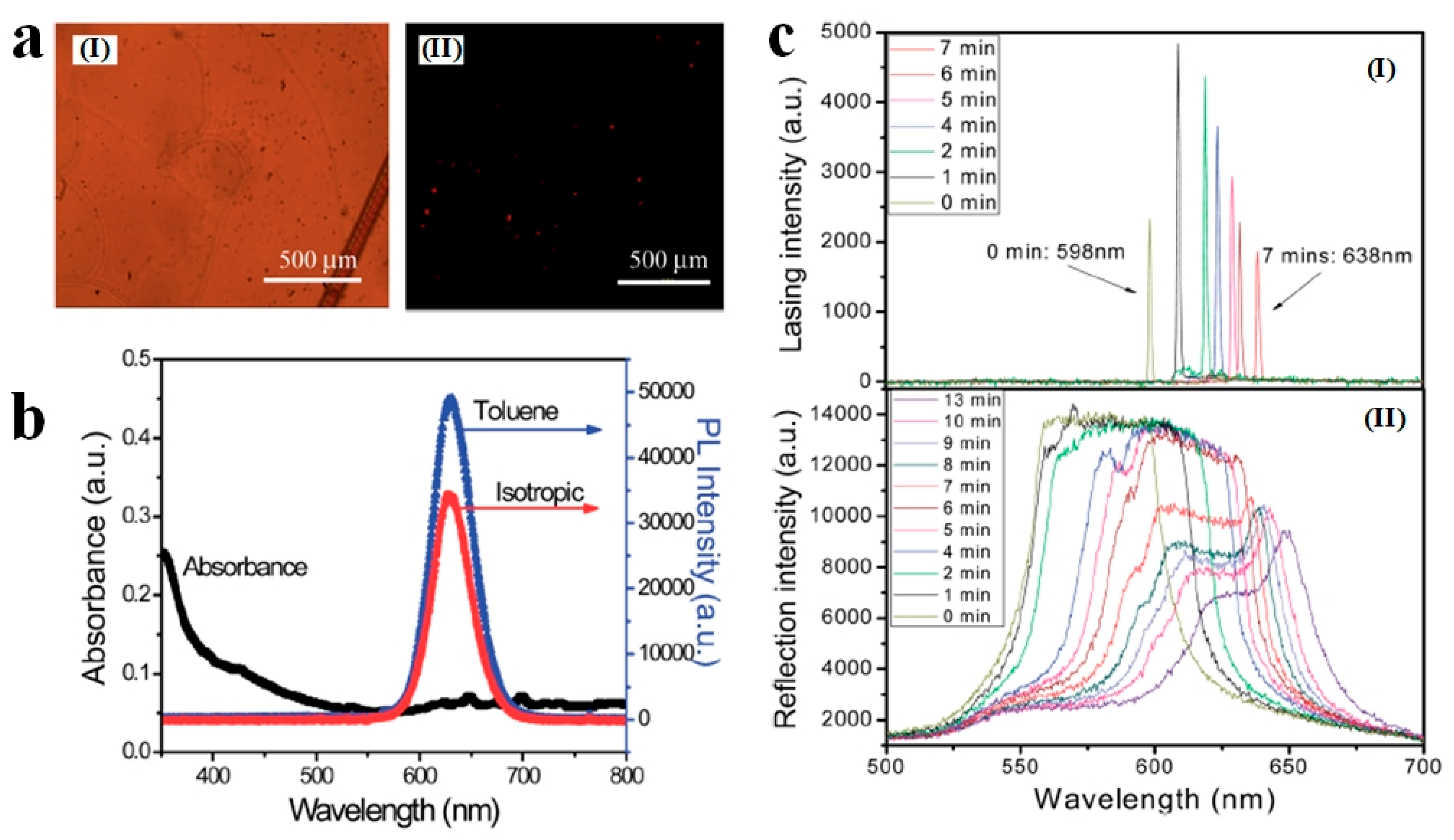
  32. Huang, W.; Yuan, C.L.; Shen, D.; Zheng, Z.G. Dynamically manipulated lasing enabled by a reconfigured fingerprint texture of a cholesteric self-organized superstructure. J. Mater. Chem. C 2017, 5, 6923–6928. [Google Scholar] [CrossRef]
  33. Price, A.D.; Schwartz, D.K. DNA hybridization-induced reorientation of liquid crystal anchoring at the nematic liquid crystal/aqueous interface. J. Am. Chem. Soc. 2008, 130, 8188–8194. [Google Scholar] [CrossRef]
  34. Sivakumar, S.; Wark, K.L.; Gupta, J.K.; Abbott, N.L.; Caruso, F. Liquid crystal emulsions as the basis of biological sensors for the optical detection of bacteria and viruses. Adv. Funct. Mater. 2009, 19, 2260–2265. [Google Scholar] [CrossRef]
  35. Chen, C.-H.; Yang, K.-L. Detection and quantification of DNA adsorbed on solid surfaces by using liquid crystals. Langmuir 2010, 26, 1427–1430. [Google Scholar] [CrossRef]
  36. Spicer, P.T. Progress in liquid crystalline dispersions: Cubosomes. Curr. Opin. Colloid Interface Sci. 2005, 10, 274–279. [Google Scholar] [CrossRef]
  37. Sagalowicz, L.; Leser, M.E.; Watzke, H.J.; Michel, M. Monoglyceride self-assembly structures as delivery vehicles. Trends Food Sci. Technol. 2006, 17, 204–214. [Google Scholar] [CrossRef]
  38. Yaghmur, A.; Glatter, O. Characterization and potential applications of nanostructured aqueous dispersions. Adv. Colloid Interface Sci. 2009, 147–148, 333–342. [Google Scholar] [CrossRef]
  39. Mushenheim, P.C.; Trivedi, R.R.; Weibel, D.B.; Abbott, N.L. Using liquid crystals to reveal how mechanical anisotropy changes interfacial behaviors of motile bacteria. Biophys. J. 2014, 107, 255–265. [Google Scholar] [CrossRef] [PubMed]
  40. Zhou, S.; Sokolov, A.; Lavrentovich, O.D.; Aranson, I.S. Living liquid crystals. Proc. Natl. Acad. Sci. USA 2014, 111, 1265. [Google Scholar] [CrossRef] [PubMed]
  41. Kim, Y.H.; Yoon, D.K.; Jeong, H.S.; Lavrentovich, O.D.; Jung, H.-T. Smectic liquid crystal defects for self-assembling of building blocks and their lithographic applications. Adv. Funct. Mater. 2011, 21, 610–627. [Google Scholar] [CrossRef]
  42. Tschierske, C. Liquid crystal engineering—new complex mesophase structures and their relations to polymer morphologies, nanoscale patterning and crystal engineering. Chem. Soc. Rev. 2007, 36, 1930–1970. [Google Scholar] [CrossRef]
  43. Zhou, S. Living liquid crystals. In Lyotropic Chromonic Liquid Crystals: From Viscoelastic Properties to Living Liquid Crystals; Zhou, S., Ed.; Springer International Publishing: Cham, Switzerland, 2017; pp. 77–92. [Google Scholar]
  44. Wang, L.; Ge, S.; Hu, W.; Nakajima, M.; Lu, Y. Tunable reflective liquid crystal terahertz waveplates. Opt. Mater. Express 2017, 7, 2023. [Google Scholar] [CrossRef]
  45. Kularatne, R.S.; Kim, H.; Boothby, J.M.; Ware, T.H. Liquid crystal elastomer actuators: Synthesis, alignment, and applications. J. Polym. Sci. B Polym. Phys. 2017, 55, 395–411. [Google Scholar] [CrossRef]
  46. Oldenbourg, R.; Wen, X.; Meyer, R.B.; Caspar, D.L.D. Orientational distribution function in nematic tobacco-mosaic-virus liquid crystals measured by X-ray diffraction. Phys. Rev. Lett. 1988, 61, 1851–1854. [Google Scholar] [CrossRef] [PubMed]
  47. Daniel, M.-C.; Astruc, D. Gold nanoparticles:  Assembly, supramolecular chemistry, quantum-size-related properties, and applications toward biology, catalysis, and nanotechnology. Chem. Rev. 2004, 104, 293–346. [Google Scholar] [CrossRef] [PubMed]
  48. Kim, F.S.; Ren, G.; Jenekhe, S.A. One-dimensional nanostructures of π-conjugated molecular systems: Assembly, properties, and applications from photovoltaics, sensors, and nanophotonics to nanoelectronics. Chem. Mater. 2010, 23, 682–732. [Google Scholar] [CrossRef]
  49. Hamley, I.W. Nanotechnologie mit weichen Materialien. Angew. Chem. 2003, 115, 1730–1752. [Google Scholar] [CrossRef]
  50. Anisa, M.; Abdallah, S.D.; Peter, A.S. ‘Mind the gap’: Science and ethics in nanotechnology. Nanotechnology 2003, 14, R9. [Google Scholar]
  51. Gupta, S.M.; Tripathi, M. A review of TO2 nanoparticles. Chin. Sci. Bull. 2011, 56, 1639. [Google Scholar] [CrossRef]
  52. Koch, U.; Fojtik, A.; Weller, H.; Henglein, A. Photochemistry of semiconductor colloids. Preparation of extremely small ZnO particles, fluorescence phenomena and size quantization effects. Chem. Phys. Lett. 1985, 122, 507–510. [Google Scholar] [CrossRef]
  53. Özgür, Ü.; Alivov, Y.I.; Liu, C.; Teke, A.; Reshchikov, M.A.; Doğan, S.; Avrutin, V.; Cho, S.J.; Morkoç, H. A comprehensive review of ZnO materials and devices. J. Appl. Phys. 2005, 98, 041301. [Google Scholar] [CrossRef] [Green Version]
  54. Zhao, Y.; Li, C.; Liu, X.; Gu, F.; Jiang, H.; Shao, W.; Zhang, L.; He, Y. Synthesis and optical properties of TiO2 nanoparticles. Mater. Lett. 2007, 61, 79–83. [Google Scholar] [CrossRef]
  55. Burda, C.; Chen, X.; Narayanan, R.; El-Sayed, M.A. Chemistry and properties of nanocrystals of different shapes. Chem. Rev. 2005, 105, 1025–1102. [Google Scholar] [CrossRef]
  56. Liu, J.; He, H.; Xiao, D.; Yin, S.; Ji, W.; Jiang, S.; Luo, D.; Wang, B.; Liu, Y. Recent advances of plasmonic nanoparticles and their applications. Materials 2018, 11, 1833. [Google Scholar] [CrossRef] [PubMed]
  57. Zhang, S.; Pelligra, C.I.; Feng, X.; Osuji, C.O. Directed assembly of hybrid nanomaterials and nanocomposites. Adv. Mater. 2018, 30, 1705794. [Google Scholar] [CrossRef] [PubMed]
  58. Meng, Q.B.; Fu, C.H.; Einaga, Y.; Gu, Z.Z.; Fujishima, A.; Sato, O. Assembly of highly ordered three-dimensional porous structure with nanocrystalline TiO2 semiconductors. Chem. Mater. 2002, 14, 83–88. [Google Scholar] [CrossRef]
  59. Furube, A.; Du, L.; Hara, K.; Katoh, R.; Tachiya, M. Ultrafast plasmon-induced electron transfer from gold nanodots into TiO2 nanoparticles. J. Am. Chem. Soc. 2007, 129, 14852–14853. [Google Scholar] [CrossRef] [PubMed]
  60. Robel, I.; Kuno, M.; Kamat, P.V. Size-dependent electron injection from excited CdSe quantum dots into TiO2 nanoparticles. J. Am. Chem. Soc. 2007, 129, 4136–4137. [Google Scholar] [CrossRef] [PubMed]
  61. Hyun, B.-R.; Zhong, Y.-W.; Bartnik, A.C.; Sun, L.; Abruña, H.D.; Wise, F.W.; Goodreau, J.D.; Matthews, J.R.; Leslie, T.M.; Borrelli, N.F. Electron injection from colloidal PbS quantum dots into titanium dioxide nanoparticles. ACS Nano 2008, 2, 2206–2212. [Google Scholar] [CrossRef] [PubMed]
  62. Tripathi, A.K.; Singh, M.K.; Mathpal, M.C.; Mishra, S.K.; Agarwal, A. Study of structural transformation in TiO2 nanoparticles and its optical properties. J. Alloy Compd. 2013, 549, 114–120. [Google Scholar] [CrossRef]
  63. Chik, M.W.; Hussain, Z.; Zulkefeli, M.; Tripathy, M.; Kumar, S.; Majeed, A.B.A.; Byrappa, K. Polymer-wrapped single-walled carbon nanotubes: A transformation toward better applications in healthcare. Drug Deliv. Transl. Res. 2018, 9, 578–594. [Google Scholar] [CrossRef]
  64. Farokhzad, O.C.; Langer, R. Impact of nanotechnology on drug delivery. ACS Nano 2009, 3, 16–20. [Google Scholar] [CrossRef]
  65. Grzelczak, M.; Vermant, J.; Furst, E.M.; Liz-Marzán, L.M. Directed self-assembly of nanoparticles. ACS Nano 2010, 4, 3591–3605. [Google Scholar] [CrossRef]
  66. Goodby, J.W.; Saez, I.M.; Cowling, S.J.; Görtz, V.; Draper, M.; Hall, A.W.; Sia, S.; Cosquer, G.; Lee, S.-E.; Raynes, E.P. Transmission and amplification of information and properties in nanostructured liquid crystals. Angew. Chem. Int. Ed. 2008, 47, 2754–2787. [Google Scholar] [CrossRef] [PubMed]
  67. Qi, H.; Hegmann, T. Impact of nanoscale particles and carbon nanotubes on current and future generations of liquid crystal displays. J. Mater. Chem. 2008, 18, 3288–3294. [Google Scholar] [CrossRef]
  68. Blanc, C.; Coursault, D.; Lacaze, E. Ordering nano- and microparticles assemblies with liquid crystals. Liq. Cryst. Rev. 2013, 1, 83–109. [Google Scholar] [CrossRef]
  69. Shivakumar, U.; Mirzaei, J.; Feng, X.; Sharma, A.; Moreira, P.; Hegmann, T. Nanoparticles: Complex and multifaceted additives for liquid crystals. Liq. Cryst. 2011, 38, 1495–1514. [Google Scholar] [CrossRef]
  70. Dierking, I. Nanomaterials in liquid crystals. Nanomaterials 2018, 8, 453. [Google Scholar] [CrossRef]
  71. Garbovskiy, Y.A.; Glushchenko, A.V. Liquid crystalline colloids of nanoparticles. In Solid State Physics; Academic Press: Cambridge, MA, USA, 2010; Volume 62, pp. 1–74. [Google Scholar]
  72. Poulin, P.; Stark, H.; Lubensky, T.C.; Weitz, D.A. Novel colloidal interactions in anisotropic fluids. Science 1997, 275, 1770. [Google Scholar] [CrossRef]
  73. Khoo, I.; Chen, K.; Williams, Y.Z. Orientational photorefractive effect in undoped and cdse nanorods-doped nematic liquid crystal—Bulk and interface contributions. IEEE J. Sel. Top. Quantum Electron. 2006, 12, 443–450. [Google Scholar] [CrossRef]
  74. Khoo, I.C.; Williams, Y.Z.; Lewis, B.; Mallouk, T. + − Photorefractive CdSe and gold nanowire-doped liquid crystals and polymer-dispersed-liquid-crystal photonic crystals. Mol. Cryst. Liq. Cryst. 2006, 446, 233–244. [Google Scholar] [CrossRef]
  75. Kinkead, B.; Hegmann, T. Effects of size, capping agent, and concentration of CdSe and CdTe quantum dots doped into a nematic liquid crystal on the optical and electro-optic properties of the final colloidal liquid crystal mixture. J. Mater. Chem. 2010, 20, 448–458. [Google Scholar] [CrossRef]
  76. Bartkiewicz, S.; Matczyszyn, K.; Miniewicz, A.; Kajzar, F. High gain of light in photoconducting polymer–nematic liquid crystal hybrid structures. Opt. Commun. 2001, 187, 257–261. [Google Scholar] [CrossRef]
  77. Müller, J.; Sönnichsen, C.; von Poschinger, H.; von Plessen, G.; Klar, T.A.; Feldmann, J. Electrically controlled light scattering with single metal nanoparticles. Appl. Phys. Lett. 2002, 81, 171–173. [Google Scholar] [CrossRef]
  78. Tomohiro, M.; Jirakorn, T.; Hiroyuki, S.; Yoshio, S.; Yukihide, S.; Naoki, T.; Shunsuke, K. Fast switching of frequency modulation twisted nematic liquid crystal display fabricated by doping nanoparticles and its mechanism. Jpn. J. Appl. Phys. 2004, 43, 2580. [Google Scholar]
  79. Rajiv, M.; Sat Prakash, Y.; Abhishek Kumar, S.; Abhishek Kumar, M.; Kamal Kumar, P.; Prashant, K.S.; Avinash Chand, P. Zinc oxide (1% Cu) nanoparticle in nematic liquid crystal: Dielectric and electro-optical study. Jpn. J. Appl. Phys. 2009, 48, 101501. [Google Scholar]
  80. Li, X.; Yang, C.; Wang, Q.; Jia, D.; Hu, L.; Peng, Z.; Xuan, L. Enhanced birefringence for metallic nanoparticle doped liquid crystals. Opt. Commun. 2013, 286, 224–227. [Google Scholar] [CrossRef]
  81. Park, S.Y.; Stroud, D. Surface-enhanced plasmon splitting in a liquid-crystal-coated gold nanoparticle. Phys. Rev. Lett. 2005, 94, 217401. [Google Scholar] [CrossRef] [PubMed]
  82. Kossyrev, P.A.; Yin, A.; Cloutier, S.G.; Cardimona, D.A.; Huang, D.; Alsing, P.M.; Xu, J.M. Electric field tuning of plasmonic response of nanodot array in liquid crystal matrix. Nano Lett. 2005, 5, 1978–1981. [Google Scholar] [CrossRef] [PubMed]
  83. Brochard, F.; De Gennes, P. Theory of magnetic suspensions in liquid crystals. J. Phys. 1970, 31, 691–708. [Google Scholar] [CrossRef]
  84. Rault, J.; Cladis, P.E.; Burger, J.P. Ferronematics. Phys. Lett. A 1970, 32, 199–200. [Google Scholar] [CrossRef]
  85. Kaczmarek, M.; Buchnev, O.; Nandhakumar, I. Ferroelectric nanoparticles in low refractive index liquid crystals for strong electro-optic response. Appl. Phys. Lett. 2008, 92, 103307. [Google Scholar] [CrossRef] [Green Version]
  86. Li, F.; Buchnev, O.; Cheon, C.I.; Glushchenko, A.; Reshetnyak, V.; Reznikov, Y.; Sluckin, T.J.; West, J.L. Orientational coupling amplification in ferroelectric nematic colloids. Phys. Rev. Lett. 2006, 97, 147801. [Google Scholar] [CrossRef]
  87. Kaur, S.; Singh, S.P.; Biradar, A.M.; Choudhary, A.; Sreenivas, K. Enhanced electro-optical properties in gold nanoparticles doped ferroelectric liquid crystals. Appl. Phys. Lett. 2007, 91, 023120. [Google Scholar] [CrossRef]
  88. Yada, M.; Yamamoto, J.; Yokoyama, H. Direct observation of anisotropic interparticle forces in nematic colloids with optical tweezers. Phys. Rev. Lett. 2004, 92, 185501. [Google Scholar] [CrossRef]
  89. Smalyukh, I.I.; Kuzmin, A.N.; Kachynski, A.V.; Prasad, P.N.; Lavrentovich, O.D. Optical trapping of colloidal particles and measurement of the defect line tension and colloidal forces in a thermotropic nematic liquid crystal. Appl. Phys. Lett. 2005, 86, 021913. [Google Scholar] [CrossRef] [Green Version]
  90. Lapanik, A.; Rudzki, A.; Kinkead, B.; Qi, H.; Hegmann, T.; Haase, W. Electrooptical and dielectric properties of alkylthiol-capped gold nanoparticle–ferroelectric liquid crystal nanocomposites: Influence of chain length and tethered liquid crystal functional groups. Soft Matter 2012, 8, 8722–8728. [Google Scholar] [CrossRef]
  91. Shukla, R.K.; Feng, X.; Umadevi, S.; Hegmann, T.; Haase, W. Influence of different amount of functionalized bulky gold nanorods dopant on the electrooptical, dielectric and optical properties of the flc host. Chem. Phys. Lett. 2014, 599, 80–85. [Google Scholar] [CrossRef]
  92. Joshi, T.; Kumar, A.; Prakash, J.; Biradar, A.M. Low power operation of ferroelectric liquid crystal system dispersed with zinc oxide nanoparticles. Appl. Phys. Lett. 2010, 96, 253109. [Google Scholar] [CrossRef] [Green Version]
  93. Lisetski, L.N.; Minenko, S.S.; Zhukov, A.V.; Shtifanyuk, P.P.; Lebovka, N.I. Dispersions of carbon nanotubes in cholesteric liquid crystals. Mol. Cryst. Liq. Cryst. 2009, 510, 43–50. [Google Scholar] [CrossRef]
  94. Singh, U.B.; Dhar, R.; Dabrowski, R.; Pandey, M.B. Enhanced electro-optical properties of a nematic liquid crystals in presence of BaTiO3 nanoparticles. Liq. Cryst. 2014, 41, 953–959. [Google Scholar] [CrossRef]
  95. San, S.E.; Okutan, M.; Köysal, O.; Yerli, Y. Carbon nanoparticles in nematic liquid crystals. Chin. Phys. Lett. 2008, 25, 212. [Google Scholar] [CrossRef]
  96. Zhao, D.; Xu, L.; Shang, Y.; Li, X.; Guo, L. Facet-dependent electro-optical properties of cholesteric liquid crystals doped with Cu2O nanocrystals. Nano Res. 2018, 11, 4836–4845. [Google Scholar] [CrossRef]
  97. Williams, Y.; Chan, K.; Park, J.H.; Khoo, I.C.; Lewis, B.; Mallouk, T.E. Electro-Optical and Nonlinear Optical Properties of Semiconductor Nanorod Doped Liquid Crystals. In Proceedings of the Optics and Photonics 2005, San Diego, CA, USA, 22 July 2005; SPIE: Bellingham, WA, USA, 2005; p. 593613. [Google Scholar]
  98. Garbovskiy, Y.; Glushchenko, A. Ferroelectric nanoparticles in liquid crystals: Recent progress and current challenges. Nanomaterials 2017, 7, 361. [Google Scholar] [CrossRef]
  99. Shelestiuk, S.M.; Reshetnyak, V.Y.; Sluckin, T.J. Frederiks transition in ferroelectric liquid-crystal nanosuspensions. Phys. Rev. E 2011, 83, 041705. [Google Scholar] [CrossRef] [Green Version]
  100. Kurochkin, O.; Buchnev, O.; Iljin, A.; Park, S.K.; Kwon, S.B.; Grabar, O.; Yu, R. A colloid of ferroelectric nanoparticles in a cholesteric liquid crystal. J. Opt. A Pure Appl. Opt. 2009, 11, 024003. [Google Scholar] [CrossRef]
  101. Lopatina, L.M.; Selinger, J.V. Maier-saupe-type theory of ferroelectric nanoparticles in nematic liquid crystals. Phys. Rev. E 2011, 84, 041703. [Google Scholar] [CrossRef]
  102. Chen, S.-H.; Amer, N.M. Observation of macroscopic collective behavior and new texture in magnetically doped liquid crystals. Phys. Rev. Lett. 1983, 51, 2298–2301. [Google Scholar] [CrossRef]
  103. Martínez-Miranda, L.J.; McCarthy, K.; Kurihara, L.K.; Harry, J.J.; Noel, A. Effect of the surface coating on the magnetic nanoparticle smectic-a liquid crystal interaction. Appl. Phys. Lett. 2006, 89, 161917. [Google Scholar] [CrossRef]
  104. Kopčanský, P.; Tomašovičová, N.; Koneracká, M.; Závišová, V.; Timko, M.; Džarová, A.; Šprincová, A.; Éber, N.; Fodor-Csorba, K.; Tóth-Katona, T.; et al. Structural changes in the 6chbt liquid crystal doped with spherical, rodlike, and chainlike magnetic particles. Phys. Rev. E 2008, 78, 011702. [Google Scholar] [CrossRef]
  105. Mertelj, A.; Lisjak, D.; Drofenik, M.; Čopič, M. Ferromagnetism in suspensions of magnetic platelets in liquid crystal. Nature 2013, 504, 237. [Google Scholar] [CrossRef]
  106. Mertelj, A.; Osterman, N.; Lisjak, D.; Čopič, M. Magneto-optic and converse magnetoelectric effects in a ferromagnetic liquid crystal. Soft Matter 2014, 10, 9065–9072. [Google Scholar] [CrossRef]
  107. Hiroyuki, Y.; Yuma, T.; Kosuke, K.; Hitoshi, K.; Tetsuya, T.; Akihiko, F.; Susumu, K.; Hirotsugu, K.; Masanori, O. Nanoparticle-stabilized cholesteric blue phases. Appl. Phys. Express 2009, 2, 121501. [Google Scholar]
  108. Karatairi, E.; Rožič, B.; Kutnjak, Z.; Tzitzios, V.; Nounesis, G.; Cordoyiannis, G.; Thoen, J.; Glorieux, C.; Kralj, S. Nanoparticle-induced widening of the temperature range of liquid-crystalline blue phases. Phys. Rev. E 2010, 81, 041703. [Google Scholar] [CrossRef] [Green Version]
  109. Prakash, J.; Choudhary, A.; Kumar, A.; Mehta, D.S.; Biradar, A.M. Nonvolatile memory effect based on gold nanoparticles doped ferroelectric liquid crystal. Appl. Phys. Lett. 2008, 93, 112904. [Google Scholar] [CrossRef]
  110. Dolgov, L.O.; Yaroshchuk, O.V. Electrooptic properties of liquid crystals filled with silica nanoparticles of different sorts. Colloid Polym. Sci. 2004, 282, 1403–1408. [Google Scholar] [CrossRef]
  111. Basu, R. Soft memory in a ferroelectric nanoparticle-doped liquid crystal. Phys. Rev. E 2014, 89, 022508. [Google Scholar] [CrossRef] [Green Version]
  112. Kempaiah, R.; Liu, Y.; Nie, Z.; Basu, R. Giant soft-memory in liquid crystal nanocomposites. Appl. Phys. Lett. 2016, 108, 083105. [Google Scholar] [CrossRef]
  113. Shiraishi, Y.; Toshima, N.; Maeda, K.; Yoshikawa, H.; Xu, J.; Kobayashi, S. Frequency modulation response of a liquid-crystal electro-optic device doped with nanoparticles. Appl. Phys. Lett. 2002, 81, 2845–2847. [Google Scholar] [CrossRef]
  114. Dierking, I.; Scalia, G.; Morales, P.; LeClere, D. Aligning and reorienting carbon nanotubes with nematic liquid crystals. Adv. Mater. 2004, 16, 865–869. [Google Scholar] [CrossRef]
  115. Özgan, Ş.; Eskalen, H.; Tapkıranlı, Y. Thermal and electro-optic properties of graphene oxide-doped hexylcyanobiphenyl liquid crystal. J. Theor. Appl. Phys. 2018, 12, 169–176. [Google Scholar] [CrossRef] [Green Version]
  116. Gardner, D.F.; Evans, J.S.; Smalyukh, I.I. Towards reconfigurable optical metamaterials: Colloidal nanoparticle self-assembly and self-alignment in liquid crystals. Mol. Cryst. Liq. Cryst. 2011, 545, 1227–1245. [Google Scholar] [CrossRef]
  117. Valentine, J.; Zhang, S.; Zentgraf, T.; Ulin-Avila, E.; Genov, D.A.; Bartal, G.; Zhang, X. Three-dimensional optical metamaterial with a negative refractive index. Nature 2008, 455, 376. [Google Scholar] [CrossRef] [PubMed]
  118. Zheludev, N.I.; Kivshar, Y.S. From metamaterials to metadevices. Nat. Mater. 2012, 11, 917–924. [Google Scholar] [CrossRef] [PubMed]
  119. Si, G.; Leong, E.S.P.; Jiang, X.; Lv, J.; Lin, J.; Dai, H.; Liu, Y.J. All-optical, polarization-insensitive light tuning properties in silver nanorod arrays covered with photoresponsive liquid crystals. Phys. Chem. Chem. Phys. 2015, 17, 13223–13227. [Google Scholar] [CrossRef] [PubMed]
  120. Nemati, A.; Wang, Q.; Hong, M.; Teng, J. Tunable and reconfigurable metasurfaces and metadevices. Opto Electron. Adv. 2018, 1, 180009. [Google Scholar] [CrossRef]
  121. Liu, Y.J.; Hao, Q.; Smalley, J.S.T.; Liou, J.; Khoo, I.C.; Huang, T.J. A frequency-addressed plasmonic switch based on dual-frequency liquid crystals. Appl. Phys. Lett. 2010, 97, 091101. [Google Scholar] [CrossRef]
  122. Link, S.; El-Sayed, M.A. Optical properties and ultrafast dynamics of metallic nanocrystals. Annu. Rev. Phys. Chem. 2003, 54, 331–366. [Google Scholar] [CrossRef] [PubMed]
  123. Hutter, E.; Fendler, J.H. Exploitation of localized surface plasmon resonance. Adv. Mater. 2004, 16, 1685–1706. [Google Scholar] [CrossRef]
  124. Chu, K.C.; Chao, C.Y.; Chen, Y.F.; Wu, Y.C.; Chen, C.C. Electrically controlled surface plasmon resonance frequency of gold nanorods. Appl. Phys. Lett. 2006, 89, 103107. [Google Scholar] [CrossRef] [Green Version]
  125. Evans, P.R.; Wurtz, G.A.; Hendren, W.R.; Atkinson, R.; Dickson, W.; Zayats, A.V.; Pollard, R.J. Electrically switchable nonreciprocal transmission of plasmonic nanorods with liquid crystal. Appl. Phys. Lett. 2007, 91, 043101. [Google Scholar] [CrossRef]
  126. Singh, D.P.; Gupta, S.K.; Srivastava, A.; Manohar, R. The phenomenon of induced photoluminescence in ferroelectric mesophase. J. Lumin. 2013, 139, 60–63. [Google Scholar] [CrossRef]
  127. Tong, X.; Zhao, Y. Liquid-crystal gel-dispersed quantum dots:  Reversible modulation of photoluminescence intensity using an electric field. J. Am. Chem. Soc. 2007, 129, 6372–6373. [Google Scholar] [CrossRef] [PubMed]
  128. Rodarte, L.A.; Cisneros, F.; Hein, E.J.; Ghosh, S.; Hirst, S.L. Quantum dot/liquid crystal nanocomposites in photonic devices. Photonics 2015, 2, 855–864. [Google Scholar] [CrossRef]
  129. Danilov, V.V.; Artem’ev, M.V.; Baranov, A.V.; Ermolaeva, G.M.; Utkina, N.A.; Khrebtov, A.I. Fluorescence of semiconductor nanorods in liquid-crystal composites. Opt. Spectrosc. 2008, 105, 306–309. [Google Scholar] [CrossRef]
  130. Ozaki, M.; Kasano, M.; Ganzke, D.; Haase, W.; Yoshino, K. Mirrorless lasing in a dye-doped ferroelectric liquid crystal. Adv. Mater. 2002, 14, 306–309. [Google Scholar] [CrossRef]
  131. Chen, L.-J.; Lee, C.-R.; Chu, C.-L. Surface passivation assisted lasing emission in the quantum dots doped cholesteric liquid crystal resonating cavity with polymer template. RSC Adv. 2014, 4, 52804–52807. [Google Scholar] [CrossRef]
  132. Lee, C.-R.; Lin, S.-H.; Guo, J.-W.; Lin, J.-D.; Lin, H.-L.; Zheng, Y.-C.; Ma, C.-L.; Horng, C.-T.; Sun, H.-Y.; Huang, S.-Y. Electrically and thermally controllable nanoparticle random laser in a well-aligned dye-doped liquid crystal cell. Opt. Mater. Express 2015, 5, 1469–1481. [Google Scholar] [CrossRef]
  133. Rodarte, A.L.; Gray, C.; Hirst, L.S.; Ghosh, S. Spectral and polarization modulation of quantum dot emission in a one-dimensional liquid crystal photonic cavity. Phys. Rev. B 2012, 85, 035430. [Google Scholar] [CrossRef] [Green Version]
  134. Wu, K.-J.; Chu, K.-C.; Chao, C.-Y.; Chen, Y.-F.; Lai, C.-W.; Kang, C.-C.; Chen, C.-Y.; Chou, P.-T. CdS nanorods imbedded in liquid crystal cells for smart optoelectronic devices. Nano Lett. 2007, 7, 1908–1913. [Google Scholar] [CrossRef]
  135. Ruhwandl, R.W.; Terentjev, E.M. Long-range forces and aggregation of colloid particles in a nematic liquid crystal. Phys. Rev. E 1997, 55, 2958–2961. [Google Scholar] [CrossRef]
  136. Stark, H. Physics of colloidal dispersions in nematic liquid crystals. Phys. Rep. 2001, 351, 387–474. [Google Scholar] [CrossRef]
  137. Pires, D.; Fleury, J.-B.; Galerne, Y. Colloid particles in the interaction field of a disclination line in a nematic phase. Phys. Rev. Lett. 2007, 98, 247801. [Google Scholar] [CrossRef] [PubMed]
  138. Samitsu, S.; Takanishi, Y.; Yamamoto, J. Molecular manipulator driven by spatial variation of liquid-crystalline order. Nat. Mater. 2010, 9, 816. [Google Scholar] [CrossRef] [PubMed]
  139. Kuksenok, O.V.; Ruhwandl, R.W.; Shiyanovskii, S.V.; Terentjev, E.M. Director structure around a colloid particle suspended in a nematic liquid crystal. Phys. Rev. E 1996, 54, 5198–5203. [Google Scholar] [CrossRef]
  140. Mondain-Monval, O.; Dedieu, J.C.; Gulik-Krzywicki, T.; Poulin, P. Weak surface energy in nematic dispersions: Saturn ring defects and quadrupolar interactions. Eur. Phys. J. B Condens. Matter Complex. Syst. 1999, 12, 167–170. [Google Scholar] [CrossRef]
  141. Stark, H. Director field configurations around a spherical particle in a nematic liquid crystal. Eur. Phys. J. B Condens. Matter Complex. Syst. 1999, 10, 311–321. [Google Scholar] [CrossRef] [Green Version]
  142. Koenig, G.M.; de Pablo, J.J.; Abbott, N.L. Characterization of the reversible interaction of pairs of nanoparticles dispersed in nematic liquid crystals. Langmuir 2009, 25, 13318–13321. [Google Scholar] [CrossRef] [PubMed]
  143. Škarabot, M.; Muševič, I. Direct observation of interaction of nanoparticles in a nematic liquid crystal. Soft Matter 2010, 6, 5476–5481. [Google Scholar] [CrossRef]
  144. Tomar, V.; Roberts, T.F.; Abbott, N.L.; Hernández-Ortiz, J.P.; de Pablo, J.J. Liquid crystal mediated interactions between nanoparticles in a nematic phase. Langmuir 2012, 28, 6124–6131. [Google Scholar] [CrossRef]
  145. Kotar, J.; Vilfan, M.; Osterman, N.; Babič, D.; Čopič, M.; Poberaj, I. Interparticle potential and drag coefficient in nematic colloids. Phys. Rev. Lett. 2006, 96, 207801. [Google Scholar] [CrossRef]
  146. Škarabot, M.; Ravnik, M.; Žumer, S.; Tkalec, U.; Poberaj, I.; Babič, D.; Osterman, N.; Muševič, I. Interactions of quadrupolar nematic colloids. Phys. Rev. E 2008, 77, 031705. [Google Scholar] [CrossRef]
  147. Vilfan, M.; Osterman, N.; Čopič, M.; Ravnik, M.; Žumer, S.; Kotar, J.; Babič, D.; Poberaj, I. Confinement effect on interparticle potential in nematic colloids. Phys. Rev. Lett. 2008, 101, 237801. [Google Scholar] [CrossRef] [PubMed]
  148. Chernyshuk, S.B.; Lev, B.I. Elastic interaction between colloidal particles in confined nematic liquid crystals. Phys. Rev. E 2010, 81, 041701. [Google Scholar] [CrossRef] [PubMed] [Green Version]
  149. Muševič, I.; Škarabot, M.; Tkalec, U.; Ravnik, M.; Žumer, S. Two-dimensional nematic colloidal crystals self-assembled by topological defects. Science 2006, 313, 954. [Google Scholar] [CrossRef] [PubMed]
  150. Škarabot, M.; Ravnik, M.; Žumer, S.; Tkalec, U.; Poberaj, I.; Babič, D.; Osterman, N.; Muševič, I. Two-dimensional dipolar nematic colloidal crystals. Phys. Rev. E 2007, 76, 051406. [Google Scholar] [CrossRef] [PubMed]
  151. Muševič, I.; Škarabot, M. Self-assembly of nematic colloids. Soft Matter 2008, 4, 195–199. [Google Scholar] [CrossRef]
  152. Ognysta, U.; Nych, A.; Nazarenko, V.; Škarabot, M.; Muševič, I. Design of 2d binary colloidal crystals in a nematic liquid crystal. Langmuir 2009, 25, 12092–12100. [Google Scholar] [CrossRef]
  153. Prathap Chandran, S.; Mondiot, F.; Mondain-Monval, O.; Loudet, J.C. Photonic control of surface anchoring on solid colloids dispersed in liquid crystals. Langmuir 2011, 27, 15185–15198. [Google Scholar] [CrossRef]
  154. Nych, A.; Ognysta, U.; Škarabot, M.; Ravnik, M.; Žumer, S.; Muševič, I. Assembly and control of 3d nematic dipolar colloidal crystals. Nat. Commun. 2013, 4, 1489. [Google Scholar] [CrossRef]
  155. Bawden, F.C.; Pirie, N.W.; Bernal, J.D.; Fankuchen, I. Liquid crystalline substances from virus-infected plants. Nature 1936, 138, 1051. [Google Scholar] [CrossRef]
  156. Onsager, L. The effects of shape on the interaction of colloidal particles. Ann. N. Y. Acad. Sci. 1949, 51, 627–659. [Google Scholar] [CrossRef]
  157. Li, L.S.; Walda, J.; Manna, L.; Alivisatos, A.P. Semiconductor nanorod liquid crystals. Nano Lett. 2002, 2, 557–560. [Google Scholar] [CrossRef]
  158. Li, L.S.; Alivisatos, A.P. Semiconductor nanorod liquid crystals and their assembly on a substrate. Adv. Mater. 2003, 15, 408–411. [Google Scholar] [CrossRef]
  159. Revol, J.F.; Bradford, H.; Giasson, J.; Marchessault, R.H.; Gray, D.G. Helicoidal self-ordering of cellulose microfibrils in aqueous suspension. Int. J. Biol. Macromol. 1992, 14, 170–172. [Google Scholar] [CrossRef]
  160. Lagerwall, J.P.F.; Schütz, C.; Salajkova, M.; Noh, J.; Hyun Park, J.; Scalia, G.; Bergström, L. Cellulose nanocrystal-based materials: From liquid crystal self-assembly and glass formation to multifunctional thin films. NPG Asia Mater. 2014, 6, e80. [Google Scholar] [CrossRef]
  161. Shimoda, H.; Oh, S.J.; Geng, H.Z.; Walker, R.J.; Zhang, X.B.; Mcneil, L.E.; Zhou, O. Self-assembly of carbon nanotubes. Adv. Mater. 2002, 14, 899–901. [Google Scholar] [CrossRef]
  162. Song, W.; Kinloch, I.A.; Windle, A.H. Nematic liquid crystallinity of multiwall carbon nanotubes. Science 2003, 302, 1363. [Google Scholar] [CrossRef]
  163. Behabtu, N.; Lomeda, J.R.; Green, M.J.; Higginbotham, A.L.; Sinitskii, A.; Kosynkin, D.V.; Tsentalovich, D.E.; Parravasquez, A.N.G.; Schmidt, J.; Kesselman, E. Spontaneous high-concentration dispersions and liquid crystals of graphene. Nat. Nanotechnol. 2010, 5, 406–411. [Google Scholar] [CrossRef] [PubMed]
  164. Kim, J.E.; Han, T.H.; Lee, S.H.; Kim, J.; Ahn, C.W.; Yun, J.M.; Kim, S.O. Graphene oxide liquid crystals. Angew. Chem. 2011, 50, 3043–3047. [Google Scholar] [CrossRef] [PubMed]
  165. Xu, Z.; Gao, C. Aqueous liquid crystals of graphene oxide. ACS Nano 2011, 5, 2908–2915. [Google Scholar] [CrossRef] [PubMed]
  166. Kato, T.; Uchida, J.; Ichikawa, T.; Sakamoto, T. Functional liquid crystals towards the next generation of materials. Angew. Chem. Int. Ed. 2018, 57, 4355–4371. [Google Scholar] [CrossRef] [PubMed]
  167. Li, Q. Nanoscience with Liquid Crystals; Springer: Berlin, Germany, 2016. [Google Scholar]
  168. Reznikov, Y.; Buchnev, O.; Tereshchenko, O.; Reshetnyak, V.; Glushchenko, A.; West, J. Ferroelectric nematic suspension. Appl. Phys. Lett. 2003, 82, 1917–1919. [Google Scholar] [CrossRef]
  169. Glushchenko, A.; Cheon, C.I.; West, J.; Li, F.; Büyüktanir, E.; Reznikov, Y.; Buchnev, A. Ferroelectric particles in liquid crystals: Recent frontiers. Mol. Cryst. Liq. Cryst. 2006, 453, 227–237. [Google Scholar] [CrossRef]
  170. Blach, J.F.; Saitzek, S.; Legrand, C.; Dupont, L.; Henninot, J.F.; Warenghem, M. BaTiO3 ferroelectric nanoparticles dispersed in 5CB nematic liquid crystal: Synthesis and electro-optical characterization. J. Appl. Phys. 2010, 107, 074102. [Google Scholar] [CrossRef]
  171. Mishra, M.; Dabrowski, R.S.; Dhar, R. Thermodynamical, optical, electrical and electro-optical studies of a room temperature nematic liquid crystal 4-pentyl-4′-cyanobiphenyl dispersed with barium titanate nanoparticles. J. Mol. Liq. 2016, 213, 247–254. [Google Scholar] [CrossRef]
  172. Klein, S.; Richardson Robert, M.; Greasty, R.; Jenkins, R.; Stone, J.; Thomas Michael, R.; Sarua, A. The influence of suspended nanoparticles on the frederiks threshold of the nematic host. Philos. Trans. R. Soc. A Math. Phys. Eng. Sci. 2013, 371, 20120253. [Google Scholar] [CrossRef] [PubMed]
  173. Al-Zangana, S.; Turner, M.; Dierking, I. A comparison between size dependent paraelectric and ferroelectric BaTiO3 nanoparticle doped nematic and ferroelectric liquid crystals. J. Appl. Phys. 2017, 121, 085105. [Google Scholar] [CrossRef]
  174. Dubey, R.; Mishra, A.; Singh, K.N.; Alapati, P.R.; Dhar, R. Electric behaviour of a schiff’s base liquid crystal compound doped with a low concentration of BaTiO3 nanoparticles. J. Mol. Liq. 2017, 225, 496–501. [Google Scholar] [CrossRef]
  175. Prasad, S.K.; Kumar, M.V.; Shilpa, T.; Yelamaggad, C.V. Enhancement of electrical conductivity, dielectric anisotropy and director relaxation frequency in composites of gold nanoparticle and a weakly polar nematic liquid crystal. RSC Adv. 2014, 4, 4453–4462. [Google Scholar] [CrossRef]
  176. Sridevi, S.; Prasad, S.K.; Nair, G.G.; D’Britto, V.; Prasad, B.L.V. Enhancement of anisotropic conductivity, elastic, and dielectric constants in a liquid crystal-gold nanorod system. Appl. Phys. Lett. 2010, 97, 151913. [Google Scholar] [CrossRef]
  177. Pandey, A.S.; Dhar, R.; Kumar, S.; Dabrowski, R. Enhancement of the display parameters of 4′-pentyl-4-cyanobiphenyl due to the dispersion of functionalised gold nano particles. Liq. Cryst. 2011, 38, 115–120. [Google Scholar] [CrossRef]
  178. Holt, L.A.; Bushby, R.J.; Evans, S.D.; Burgess, A.; Seeley, G. A 106-fold enhancement in the conductivity of a discotic liquid crystal doped with only 1% (w/w) gold nanoparticles. J. Appl. Phys. 2008, 103, 063712. [Google Scholar] [CrossRef]
  179. Singh, U.B.; Dhar, R.; Dabrowski, R.; Pandey, M.B. Influence of low concentration silver nanoparticles on the electrical and electro-optical parameters of nematic liquid crystals. Liq. Cryst. 2013, 40, 774–782. [Google Scholar] [CrossRef]
  180. Liu, B.; Ma, Y.; Zhao, D.; Xu, L.; Liu, F.; Zhou, W.; Guo, L. Effects of morphology and concentration of cus nanoparticles on alignment and electro-optic properties of nematic liquid crystal. Nano Res. 2017, 10, 618–625. [Google Scholar] [CrossRef]
  181. Khatua, S.; Manna, P.; Chang, W.-S.; Tcherniak, A.; Friedlander, E.; Zubarev, E.R.; Link, S. Plasmonic nanoparticles−liquid crystal composites. J. Phys. Chem. C 2010, 114, 7251–7257. [Google Scholar] [CrossRef]
  182. Elkhalgi, H.H.M.; Khandka, S.; Singh, U.B.; Pandey, K.L.; Dabrowski, R.; Dhar, R. Dielectric and electro-optical properties of a nematic liquid crystalline material with gold nanoparticles. Liq. Cryst. 2018, 45, 1795–1801. [Google Scholar] [CrossRef]
  183. Mishra, M.; Kumar, S.; Dhar, R. Effect of high concentration of colloidal gold nanoparticles on the thermodynamic, optical, and electrical properties of 2, 3, 6, 7, 10, 11-hexabutyloxytryphenylene discotic liquid crystalline material. Soft Matter 2017, 15, 34–44. [Google Scholar] [CrossRef]
  184. Gittins, D.I.; Bethell, D.; Schiffrin, D.J.; Nichols, R.J. A nanometre-scale electronic switch consisting of a metal cluster and redox-addressable groups. Nature 2000, 408, 67. [Google Scholar] [CrossRef]
  185. Haynes, C.L.; McFarland, A.D.; Zhao, L.; Van Duyne, R.P.; Schatz, G.C.; Gunnarsson, L.; Prikulis, J.; Kasemo, B.; Käll, M. Nanoparticle optics:  The importance of radiative dipole coupling in two-dimensional nanoparticle arrays. J. Phys. Chem. B 2003, 107, 7337–7342. [Google Scholar] [CrossRef]
  186. Crooks, R.M.; Zhao, M.; Sun, L.; Chechik, V.; Yeung, L.K. Dendrimer-encapsulated metal nanoparticles:  Synthesis, characterization, and applications to catalysis. Acc. Chem. Res. 2001, 34, 181–190. [Google Scholar] [CrossRef]
  187. Tripathi, P.; Mishra, M.; Kumar, S.; Dabrowski, R.; Dhar, R. Dependence of physical parameters on the size of silver nano particles forming composites with a nematic liquid crystalline material. J. Mol. Liq. 2018, 268, 403–409. [Google Scholar] [CrossRef]
  188. Mirzaei, J.; Reznikov, M.; Hegmann, T. Quantum dots as liquid crystal dopants. J. Mater. Chem. 2012, 22, 22350–22365. [Google Scholar] [CrossRef] [Green Version]
  189. Brus, L.E. Electron–electron and electron-hole interactions in small semiconductor crystallites: The size dependence of the lowest excited electronic state. J. Chem. Phys. 1984, 80, 4403–4409. [Google Scholar] [CrossRef]
  190. Brus, L. Electronic wave functions in semiconductor clusters: Experiment and theory. J. Phys. Chem. 1986, 90, 2555–2560. [Google Scholar] [CrossRef]
  191. Singh, U.B.; Dhar, R.; Pandey, A.S.; Kumar, S.; Dabrowski, R.; Pandey, M.B. Electro-optical and dielectric properties of cdse quantum dots and 6chbt liquid crystals composites. AIP Adv. 2014, 4, 117112. [Google Scholar] [CrossRef]
  192. Konshina, E.A.; Galin, I.F.; Shcherbinin, D.P.; Gavrish, E.O. Study of dynamics and relaxation optical response of nematic liquid crystals doped with CdSe/ZnS quantum dots. Liq. Cryst. 2014, 41, 1229–1234. [Google Scholar] [CrossRef]
  193. Gupta, S.K.; Singh, D.P.; Tripathi, P.K.; Manohar, R.; Varia, M.; Sagar, L.K.; Kumar, S. CdSe quantum dot-dispersed DOBAMBC: An electro-optical study. Liq. Cryst. 2013, 40, 528–533. [Google Scholar] [CrossRef]
  194. Rastogi, A.; Pathak, G.; Srivastava, A.; Herman, J.; Manohar, R. Cd1−x ZnxS/ZnS core/shell quantum dots in nematic liquid crystals to improve material parameter for better performance of liquid crystal based devices. J. Mol. Liq. 2018, 255, 93–101. [Google Scholar] [CrossRef]
  195. Singh, U.B.; Pandey, M.B.; Dhar, R.; Verma, R.; Kumar, S. Effect of dispersion of cdse quantum dots on phase transition, electrical and electro-optical properties of 4pp4ob. Liq. Cryst. 2016, 43, 1075–1082. [Google Scholar] [CrossRef]
  196. Roy, A.; Pathak, G.; Herman, J.; Inamdar, S.R.; Srivastava, A.; Manohar, R. InP/ZnS quantum-dot-dispersed nematic liquid crystal illustrating characteristic birefringence and enhanced electro-optical parameters. Appl. Phys. A 2018, 124, 273. [Google Scholar] [CrossRef]
  197. Tripathi, P.K.; Misra, A.K.; Manohar, S.; Gupta, S.K.; Manohar, R. Improved dielectric and electro-optical parameters of ZnO nano-particle (8% Cu2+) doped nematic liquid crystal. J. Mol. Struct. 2013, 1035, 371–377. [Google Scholar] [CrossRef]
  198. Ye, W.; Yuan, R.; Dai, Y.; Gao, L.; Pang, Z.; Zhu, J.; Meng, X.; He, Z.; Li, J.; Cai, M.; et al. Improvement of image sticking in liquid crystal display doped with γ-Fe2O3 nanoparticles. Nanomaterials 2017, 8, 5. [Google Scholar] [CrossRef] [PubMed]
  199. Elkhalgi, H.H.M.; Khandka, S.; Yadav, N.; Dhar, R.; Dabrowski, R. Effects of manganese (ii) titanium oxide nano particles on the physical properties of a room temperature nematic liquid crystal 4-(trans-4′-n-hexylcyclohexyl) isothiocyanatobenzene. J. Mol. Liq. 2018, 268, 223–228. [Google Scholar] [CrossRef]
  200. Oh, C.-W.; Park, E.-G.; Park, H.-G. Enhanced electro-optical properties in titanium silicon oxide nanoparticle doped nematic liquid crystal system. Surf. Coat. Technol. 2019, 360, 50–55. [Google Scholar] [CrossRef]
  201. Li, C.-Z.; Yip, H.-L.; Jen, A.K.Y. Functional fullerenes for organic photovoltaics. J. Mater. Chem. 2012, 22, 4161–4177. [Google Scholar] [CrossRef]
  202. San, S.E.; Köysal, O.; Okutan, M. Laser-induced dielectric anisotropy of a hybrid liquid crystal composite made up of methyl red and fullerene C60. J. Non-Cryst. Solids 2005, 351, 2798–2801. [Google Scholar] [CrossRef]
  203. Okutan, M.; Eren San, S.; Basaran, E.; Yakuphanoglu, F. Determination of phase transition from nematic to isotropic state in carbon nano-balls’ doped nematic liquid crystals by electrical conductivity-dielectric measurements. Phys. Lett. A 2005, 339, 461–465. [Google Scholar] [CrossRef]
  204. Shukla, R.K.; Raina, K.K.; Haase, W. Fast switching response and dielectric behaviour of fullerene/ferroelectric liquid crystal nanocolloids. Liq. Cryst. 2014, 41, 1726–1732. [Google Scholar] [CrossRef]
  205. Zhang, X.; Yu, B.; Cong, H. Recent developments in fullerene-containing thermotropic liquid crystals. Curr. Org. Chem. 2017, 21, 1600–1611. [Google Scholar] [CrossRef]
  206. Iijima, S. Helical microtubules of graphitic carbon. Nature 1991, 354, 56. [Google Scholar] [CrossRef]
  207. Baik, I.-S.; Jeon, S.Y.; Lee, S.H.; Park, K.A.; Jeong, S.H.; An, K.H.; Lee, Y.H. Electrical-field effect on carbon nanotubes in a twisted nematic liquid crystal cell. Appl. Phys. Lett. 2005, 87, 263110. [Google Scholar] [CrossRef] [Green Version]
  208. Chi-Yen, H.; Chao-Yuan, H.; Hung-Chih, P.; Kuang-Yao, L. Electrooptical responses of carbon nanotube-doped liquid crystal devices. Jpn. J. Appl. Phys. 2005, 44, 8077. [Google Scholar]
  209. Chen, H.-Y.; Lee, W. Suppression of field screening in nematic liquid crystals by carbon nanotubes. Appl. Phys. Lett. 2006, 88, 222105. [Google Scholar] [CrossRef]
  210. Chen, H.-Y.; Lee, W.; Clark, N.A. Faster electro-optical response characteristics of a carbon-nanotube-nematic suspension. Appl. Phys. Lett. 2007, 90, 033510. [Google Scholar] [CrossRef]
  211. Lee, W.; Wang, C.-Y.; Shih, Y.-C. Effects of carbon nanosolids on the electro-optical properties of a twisted nematic liquid-crystal host. Appl. Phys. Lett. 2004, 85, 513–515. [Google Scholar] [CrossRef]
  212. Kumar, J.; Manjuladevi, V.; Gupta, R.K.; Kumar, S. Fast response in TN liquid-crystal cells: Effect of functionalised carbon nanotubes. Liq. Cryst. 2016, 43, 488–496. [Google Scholar] [CrossRef]
  213. Shukla, R.K.; Chaudhary, A.; Bubnov, A.; Raina, K.K. Multi-walled carbon nanotubes-ferroelectric liquid crystal nanocomposites: Effect of cell thickness and dopant concentration on electro-optic and dielectric behaviour. Liq. Cryst. 2018, 45, 1672–1681. [Google Scholar] [CrossRef]
  214. Singh, D.; Bahadur Singh, U.; Bhushan Pandey, M.; Dabrowski, R.; Dhar, R. Improvement of orientational order and display parameters of liquid crystalline material dispersed with single-wall carbon nanotubes. Mater. Lett. 2018, 216, 5–7. [Google Scholar] [CrossRef]
  215. Singh, D.; Singh, U.B.; Pandey, M.B.; Dabrowski, R.; Dhar, R. Enhancement in electro-optical parameters of nematic liquid crystalline material with swcnts. Opt. Mater. 2018, 84, 16–21. [Google Scholar] [CrossRef]
  216. Schymura, S.; Scalia, G. On the effect of carbon nanotubes on properties of liquid crystals. Phil. Trans. R. Soc. A 2013, 371, 20120261. [Google Scholar] [CrossRef]
  217. Scalia, G.; Lagerwall, J.P.F.; Schymura, S.; Haluska, M.; Giesselmann, F.; Roth, S. Carbon nanotubes in liquid crystals as versatile functional materials. Phys. Status Solidi 2007, 244, 4212–4217. [Google Scholar] [CrossRef]
  218. Novoselov, K.S.; Geim, A.K.; Morozov, S.V.; Jiang, D.; Zhang, Y.; Dubonos, S.V.; Grigorieva, I.V.; Firsov, A.A. Electric field effect in atomically thin carbon films. Science 2004, 306, 666. [Google Scholar] [CrossRef] [PubMed]
  219. Gökçen, M.; Yıldırım, M.; Köysal, O. Dielectric and ac electrical conductivity characteristics of liquid crystal doped with graphene. Eur. Phys. J. Appl. Phys. 2012, 60, 30104. [Google Scholar] [CrossRef]
  220. Alam, T.M.; Pearce, C.J. Impact of graphene incorporation on the orientational order of graphene/liquid crystal composites. Chem. Phys. Lett. 2014, 592, 7–13. [Google Scholar] [CrossRef]
  221. Basu, R. Effects of graphene on electro-optic switching and spontaneous polarization of a ferroelectric liquid crystal. Appl. Phys. Lett. 2014, 105, 112905. [Google Scholar] [CrossRef] [Green Version]
  222. Basu, R.; Garvey, A.; Kinnamon, D. Effects of graphene on electro-optic response and ion-transport in a nematic liquid crystal. J. Appl. Phys. 2015, 117, 074301. [Google Scholar] [CrossRef] [Green Version]
  223. Basu, R.; Kinnamon, D.; Garvey, A. Nano-electromechanical rotation of graphene and giant enhancement in dielectric anisotropy in a liquid crystal. Appl. Phys. Lett. 2015, 106, 201909. [Google Scholar] [CrossRef] [Green Version]
  224. Javadian, S.; Dalir, N.; Kakemam, J. Non-covalent intermolecular interactions of colloidal nematic liquid crystals doped with graphene oxide. Liq. Cryst. 2017, 44, 1341–1355. [Google Scholar] [CrossRef]
  225. Al-Zangana, S.; Iliut, M.; Turner, M.; Vijayaraghavan, A.; Dierking, I. Properties of a thermotropic nematic liquid crystal doped with graphene oxide. Adv. Opt. Mater. 2016, 4, 1541–1548. [Google Scholar] [CrossRef]
  226. Al-Zangana, S.; Iliut, M.; Boran, G.; Turner, M.; Vijayaraghavan, A.; Dierking, I. Dielectric spectroscopy of isotropic liquids and liquid crystal phases with dispersed graphene oxide. Sci. Rep. 2016, 6, 31885. [Google Scholar] [CrossRef]
  227. Dalir, N.; Javadian, S.; Kakemam, J.; Yousefi, A. Evolution of electro-chemical and electro-optical properties of nematic liquid crystal doped with graphene oxide. J. Mol. Liq. 2018, 265, 398–407. [Google Scholar] [CrossRef]
  228. Lapanik, V.; Timofeev, S.; Haase, W. Electro-optic properties of nematic and ferroelectric liquid crystalline nanocolloids doped with partially reduced graphene oxide. Phase Transit. 2016, 89, 133–143. [Google Scholar] [CrossRef]
  229. Mrukiewicz, M.; Kowiorski, K.; Perkowski, P.; Mazur, R.; Djas, M. Threshold voltage decrease in a thermotropic nematic liquid crystal doped with graphene oxide flakes. Beilstein J. Nanotechnol. 2019, 10, 71–78. [Google Scholar] [CrossRef] [PubMed]
  230. Shukla, R.K.; Mirzaei, J.; Sharma, A.; Hofmann, D.; Hegmann, T.; Haase, W. Electro-optic and dielectric properties of a ferroelectric liquid crystal doped with chemically and thermally stable emissive carbon dots. RSC Adv. 2015, 5, 34491–34496. [Google Scholar] [CrossRef]
  231. Lapanik, V.; Lugouskiy, A.; Timofeev, S.; Haase, W. Influence of the size and the attached organic tail of modified detonation nanodiamond on the physical properties of liquid crystals. Liq. Cryst. 2014, 41, 1332–1338. [Google Scholar] [CrossRef]
  232. Cho, M.-J.; Park, H.-G.; Jeong, H.-C.; Lee, J.-W.; Jung, Y.H.; Kim, D.-H.; Kim, J.-H.; Lee, J.-W.; Seo, D.-S. Superior fast switching of liquid crystal devices using graphene quantum dots. Liq. Cryst. 2014, 41, 761–767. [Google Scholar] [CrossRef]
  233. Raether, H. Surface excitations. In Excitation of Plasmons and Interband Transitions by Electrons; Springer: Berlin, Germany, 1980; pp. 116–171. [Google Scholar]
  234. Song, J.-H.; Atay, T.; Shi, S.; Urabe, H.; Nurmikko, A.V. Large enhancement of fluorescence efficiency from cdse/zns quantum dots induced by resonant coupling to spatially controlled surface plasmons. Nano Lett. 2005, 5, 1557–1561. [Google Scholar] [CrossRef] [PubMed]
  235. Zhang, H.; Li, Y.; Ivanov, I.A.; Qu, Y.; Huang, Y.; Duan, X. Plasmonic modulation of the upconversion fluorescence in nayf4:Yb/tm hexaplate nanocrystals using gold nanoparticles or nanoshells. Angew. Chem. Int. Ed. 2010, 49, 2865–2868. [Google Scholar] [CrossRef]
  236. Fu, Y.; Zhang, J.; Lakowicz, J.R. Plasmon-enhanced fluorescence from single fluorophores end-linked to gold nanorods. J. Am. Chem. Soc. 2010, 132, 5540–5541. [Google Scholar] [CrossRef]
  237. Kneipp, K.; Wang, Y.; Kneipp, H.; Perelman, L.T.; Itzkan, I.; Dasari, R.R.; Feld, M.S. Single molecule detection using surface-enhanced raman scattering (sers). Phys. Rev. Lett. 1997, 78, 1667–1670. [Google Scholar] [CrossRef]
  238. Noginov, M.A.; Zhu, G.; Belgrave, A.M.; Bakker, R.; Shalaev, V.M.; Narimanov, E.E.; Stout, S.; Herz, E.; Suteewong, T.; Wiesner, U. Demonstration of a spaser-based nanolaser. Nature 2009, 460, 1110. [Google Scholar] [CrossRef]
  239. Stockman, M.I. Spasers explained. Nat. Photonics 2008, 2, 327. [Google Scholar] [CrossRef]
  240. Huang, X.; Jain, P.K.; El-Sayed, I.H.; El-Sayed, M.A. Plasmonic photothermal therapy (pptt) using gold nanoparticles. Lasers Med. Sci. 2007, 23, 217. [Google Scholar] [CrossRef]
  241. Ebbesen, T.W.; Lezec, H.J.; Ghaemi, H.F.; Thio, T.; Wolff, P.A. Extraordinary optical transmission through sub-wavelength hole arrays. Nature 1998, 391, 667. [Google Scholar] [CrossRef]
  242. Fang, N.; Lee, H.; Sun, C.; Zhang, X. Sub–diffraction-limited optical imaging with a silver superlens. Science 2005, 308, 534–537. [Google Scholar] [CrossRef]
  243. Liu, Z.; Lee, H.; Xiong, Y.; Sun, C.; Zhang, X. Far-field optical hyperlens magnifying sub-diffraction-limited objects. Science 2007, 315, 1686. [Google Scholar] [CrossRef] [PubMed]
  244. Shao, L.; Zhuo, X.; Wang, J. Advanced plasmonic materials for dynamic color display. Adv. Mater. 2018, 30, 1704338. [Google Scholar] [CrossRef] [PubMed]
  245. Kalkbrenner, T.; Ramstein, M.; Mlynek, J.; Sandoghdar, V. A single gold particle as a probe for apertureless scanning near-field optical microscopy. J. Microsc. 2001, 202, 72–76. [Google Scholar] [CrossRef] [PubMed]
  246. Lazarides, A.A.; Lance Kelly, K.; Jensen, T.R.; Schatz, G.C. Optical properties of metal nanoparticles and nanoparticle aggregates important in biosensors. J. Mol. Struct. 2000, 529, 59–63. [Google Scholar] [CrossRef]
  247. Myroshnychenko, V.; Rodríguez-Fernández, J.; Pastoriza-Santos, I.; Funston, A.M.; Novo, C.; Mulvaney, P.; Liz-Marzán, L.M.; García de Abajo, F.J. Modelling the optical response of gold nanoparticles. Chem. Soc. Rev. 2008, 37, 1792–1805. [Google Scholar] [CrossRef] [Green Version]
  248. Choudhary, A.; Singh, G.; Biradar, A.M. Advances in gold nanoparticle–liquid crystal composites. Nanoscale 2014, 6, 7743–7756. [Google Scholar] [CrossRef]
  249. Prasad, P.N. Nanophotonics; John Wiley & Sons: Hoboken, NJ, USA, 2004. [Google Scholar]
  250. Jensen, T.; Kelly, L.; Lazarides, A.; Schatz, G.C. Electrodynamics of noble metal nanoparticles and nanoparticle clusters. J. Clust. Sci. 1999, 10, 295–317. [Google Scholar] [CrossRef]
  251. Kelly, K.L.; Coronado, E.; Zhao, L.L.; Schatz, G.C. The optical properties of metal nanoparticles:  The influence of size, shape, and dielectric environment. J. Phys. Chem. B 2003, 107, 668–677. [Google Scholar] [CrossRef]
  252. Chen, B.; Zeng, X.; Baumeister, U.; Ungar, G.; Tschierske, C. Liquid crystalline networks composed of pentagonal, square, and triangular cylinders. Science 2005, 307, 96. [Google Scholar] [CrossRef] [PubMed]
  253. Mock, J.J.; Barbic, M.; Smith, D.R.; Schultz, D.A.; Schultz, S. Shape effects in plasmon resonance of individual colloidal silver nanoparticles. J. Chem. Phys. 2002, 116, 6755–6759. [Google Scholar] [CrossRef]
  254. De Sio, L.; Caputo, R.; Cataldi, U.; Umeton, C. Broad band tuning of the plasmonic resonance of gold nanoparticles hosted in self-organized soft materials. J. Mater. Chem. 2011, 21, 18967–18970. [Google Scholar] [CrossRef]
  255. Caldwell, M.E.; Yeatman, E.M. Surface-plasmon spatial light modulators based on liquid crystal. Appl. Opt. 1992, 31, 3880–3891. [Google Scholar] [CrossRef] [PubMed] [Green Version]
  256. Wang, Y. Voltage-induced color-selective absorption with surface plasmons. Appl. Phys. Lett. 1995, 67, 2759–2761. [Google Scholar]
  257. Wang, Y.; Russell, S.D.; Shimabukuro, R.L. Voltage-induced broad-spectrum reflectivity change with surface-plasmon waves. J. Appl. Phys. 2004, 97, 023708. [Google Scholar]
  258. Koenig, G.M.; Meli, M.-V.; Park, J.-S.; de Pablo, J.J.; Abbott, N.L. Coupling of the plasmon resonances of chemically functionalized gold nanoparticles to local order in thermotropic liquid crystals. Chem. Mater. 2007, 19, 1053–1061. [Google Scholar] [CrossRef]
  259. Hsu, L.-H.; Lo, K.-Y.; Huang, S.-A.; Huang, C.-Y.; Yang, C.-S. Irreversible redshift of transmission spectrum of gold nanoparticles doped in liquid crystals. Appl. Phys. Lett. 2008, 92, 181112. [Google Scholar]
  260. Pratibha, R.; Park, K.; Smalyukh, I.I.; Park, W. Tunable optical metamaterial based on liquid crystal-gold nanosphere composite. Opt. Express 2009, 17, 19459–19469. [Google Scholar] [CrossRef] [PubMed]
  261. Liu, Q.; Cui, Y.; Gardner, D.; Li, X.; He, S.; Smalyukh, I.I. Self-alignment of plasmonic gold nanorods in reconfigurable anisotropic fluids for tunable bulk metamaterial applications. Nano Lett. 2010, 10, 1347–1353. [Google Scholar] [CrossRef] [PubMed]
  262. Liu, Q.; Senyuk, B.; Tang, J.; Lee, T.; Qian, J.; He, S.; Smalyukh, I.I. Plasmonic complex fluids of nematiclike and helicoidal self-assemblies of gold nanorods with a negative order parameter. Phys. Rev. Lett. 2012, 109, 088301. [Google Scholar] [CrossRef] [PubMed]
  263. Liu, Q.; Yuan, Y.; Smalyukh, I.I. Electrically and optically tunable plasmonic guest–host liquid crystals with long-range ordered nanoparticles. Nano Lett. 2014, 14, 4071–4077. [Google Scholar] [CrossRef] [PubMed]
  264. Zhang, Y.; Liu, Q.; Mundoor, H.; Yuan, Y.; Smalyukh, I.I. Metal nanoparticle dispersion, alignment, and assembly in nematic liquid crystals for applications in switchable plasmonic color filters and e-polarizers. ACS Nano 2015, 9, 3097–3108. [Google Scholar] [CrossRef]
  265. Li, D.; Liu, F.; Ren, G.J.; Fu, P.; Yao, J.Q. Liquid crystal-modulated tunable filter based on coupling between plasmon-induced transparency and cavity mode. Opt. Eng. 2018, 57, 097101. [Google Scholar] [CrossRef]
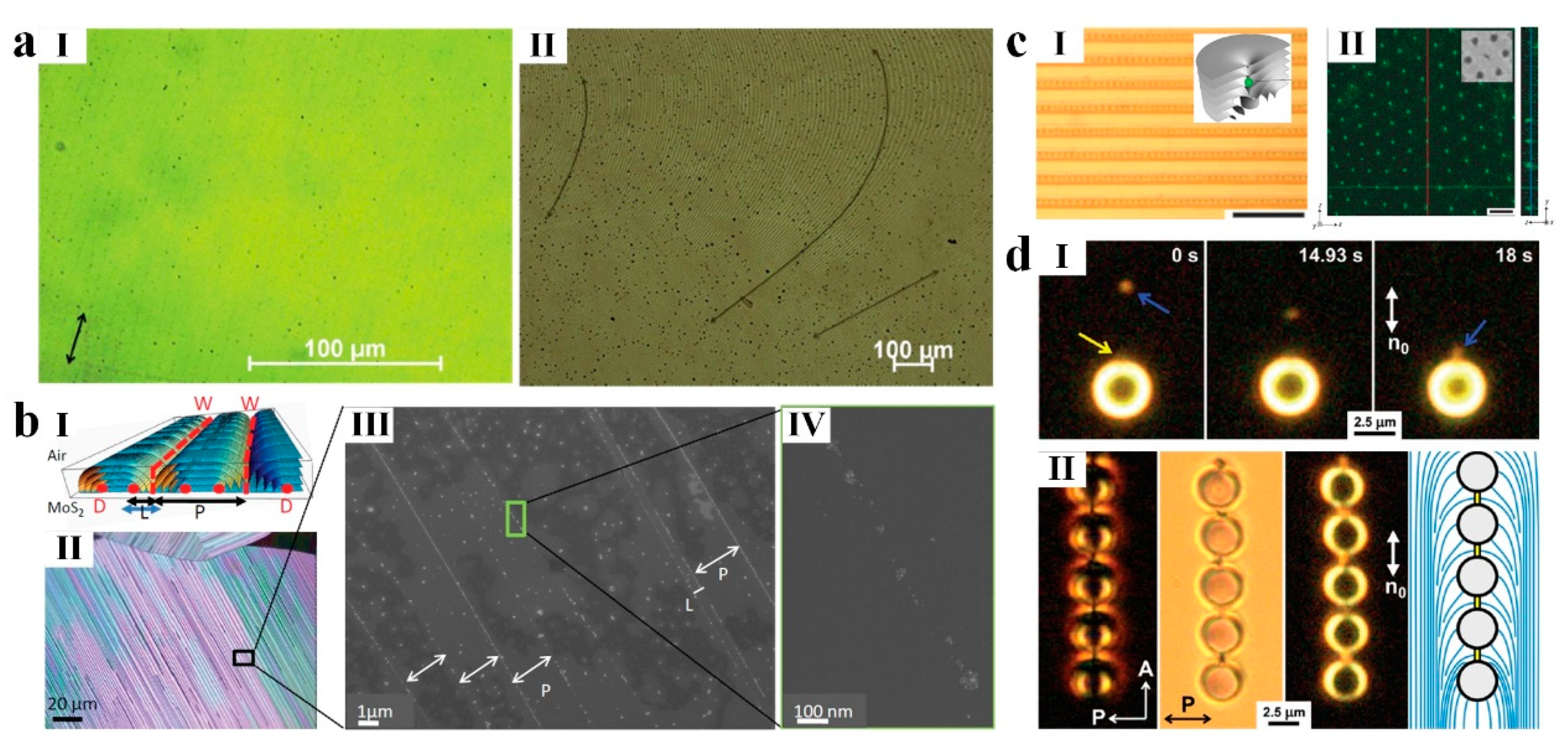
  266. Rožič, B.; Fresnais, J.; Molinaro, C.; Calixte, J.; Umadevi, S.; Lau-Truong, S.; Felidj, N.; Kraus, T.; Charra, F.; Dupuis, V.; et al. Oriented gold nanorods and gold nanorod chains within smectic liquid crystal topological defects. ACS Nano 2017, 11, 6728–6738. [Google Scholar] [CrossRef]
  267. Jiang, L.; Mundoor, H.; Liu, Q.; Smalyukh, I.I. Electric switching of fluorescence decay in gold–silica–dye nematic nanocolloids mediated by surface plasmons. ACS Nano 2016, 10, 7064–7072. [Google Scholar] [CrossRef]
  268. Sheetah, G.H.; Liu, Q.; Senyuk, B.; Fleury, B.; Smalyukh, I.I. Electric switching of visible and infrared transmission using liquid crystals co-doped with plasmonic gold nanorods and dichroic dyes. Opt. Express 2018, 26, 22264–22272. [Google Scholar] [CrossRef]
  269. Sheetah, G.H.; Liu, Q.; Smalyukh, I.I. Self-assembly of predesigned optical materials in nematic codispersions of plasmonic nanorods. Opt. Lett. 2016, 41, 4899–4902. [Google Scholar] [CrossRef]
  270. Jablan, M.; Soljačić, M.; Buljan, H. Plasmons in graphene: Fundamental properties and potential applications. Proc. IEEE 2013, 101, 1689–1704. [Google Scholar] [CrossRef]
  271. Ju, L.; Geng, B.; Horng, J.; Girit, C.; Martin, M.; Hao, Z.; Bechtel, H.A.; Liang, X.; Zettl, A.; Shen, Y.R.; et al. Graphene plasmonics for tunable terahertz metamaterials. Nat. Nanotechnol. 2011, 6, 630. [Google Scholar] [CrossRef] [PubMed]
  272. Garciía de Abajo, F.J. Special Issue “2d Materials for Nanophotonics”; ACS Publications: Washington, DC, USA, 2017. [Google Scholar]
  273. Li, Y.; Li, Z.; Chi, C.; Shan, H.; Zheng, L.; Fang, Z. Plasmonics of 2d nanomaterials: Properties and applications. Adv. Sci. 2017, 4, 1600430. [Google Scholar] [CrossRef] [PubMed]
  274. Jablan, M.; Buljan, H.; Soljačić, M. Plasmonics in graphene at infrared frequencies. Phys. Rev. B 2009, 80, 245435. [Google Scholar] [CrossRef] [Green Version]
  275. García de Abajo, F.J. Graphene plasmonics: Challenges and opportunities. ACS Photonics 2014, 1, 135–152. [Google Scholar] [CrossRef]
  276. Reshetnyak, V.Y.; Bunning, T.J.; Evans, D.R. Using liquid crystals to control surface plasmons. Liq. Cryst. 2018, 45, 2010–2021. [Google Scholar] [CrossRef]
  277. Zhu, M.; Baffou, G.; Meyerbröker, N.; Polleux, J. Micropatterning thermoplasmonic gold nanoarrays to manipulate cell adhesion. ACS Nano 2012, 6, 7227–7233. [Google Scholar] [CrossRef] [PubMed]
  278. Stehr, J.; Hrelescu, C.; Sperling, R.A.; Raschke, G.; Wunderlich, M.; Nichtl, A.; Heindl, D.; Kürzinger, K.; Parak, W.J.; Klar, T.A.; et al. Gold nanostoves for microsecond DNA melting analysis. Nano Lett. 2008, 8, 619–623. [Google Scholar] [CrossRef] [PubMed]
  279. Hirsch, L.R.; Stafford, R.J.; Bankson, J.A.; Sershen, S.R.; Rivera, B.; Price, R.E.; Hazle, J.D.; Halas, N.J.; West, J.L. Nanoshell-mediated near-infrared thermal therapy of tumors under magnetic resonance guidance. Proc. Natl. Acad. Sci. USA 2003, 100, 13549. [Google Scholar] [CrossRef] [PubMed]
  280. Huschka, R.; Neumann, O.; Barhoumi, A.; Halas, N.J. Visualizing light-triggered release of molecules inside living cells. Nano Lett. 2010, 10, 4117–4122. [Google Scholar] [CrossRef] [PubMed]
  281. Sun, Y.; Evans, J.S.; Lee, T.; Senyuk, B.; Keller, P.; He, S.; Smalyukh, I.I. Optical manipulation of shape-morphing elastomeric liquid crystal microparticles doped with gold nanocrystals. Appl. Phys. Lett. 2012, 100, 241901. [Google Scholar] [CrossRef] [Green Version]
  282. Fong, W.-K.; Hanley, T.L.; Thierry, B.; Kirby, N.; Waddington, L.J.; Boyd, B.J. Controlling the nanostructure of gold nanorod–lyotropic liquid-crystalline hybrid materials using near-infrared laser irradiation. Langmuir 2012, 28, 14450–14460. [Google Scholar] [CrossRef] [PubMed]
  283. Fong, W.-K.; Hanley, T.L.; Thierry, B.; Tilley, A.; Kirby, N.; Waddington, L.J.; Boyd, B.J. Understanding the photothermal heating effect in non-lamellar liquid crystalline systems, and the design of new mixed lipid systems for photothermal on-demand drug delivery. Phys. Chem. Chem. Phys. 2014, 16, 24936–24953. [Google Scholar] [CrossRef]
  284. Pezzi, L.; De Sio, L.; Veltri, A.; Placido, T.; Palermo, G.; Comparelli, R.; Curri, M.L.; Agostiano, A.; Tabiryan, N.; Umeton, C. Photo-thermal effects in gold nanoparticles dispersed in thermotropic nematic liquid crystals. Phys. Chem. Chem. Phys. 2015, 17, 20281–20287. [Google Scholar] [CrossRef] [PubMed] [Green Version]
  285. Luciano De, S.; Tiziana, P.; Roberto, C.; Maria Lucia, C.; Nelson, T.; Timothy, J.B. Plasmonic photoheating of gold nanorods in thermo-responsive chiral liquid crystals. J. Opt. 2016, 18, 125005. [Google Scholar]
  286. Palermo, G.; Cataldi, U.; De Sio, L.; Bürgi, T.; Tabiryan, N.; Umeton, C. Optical control of plasmonic heating effects using reversible photo-alignment of nematic liquid crystals. Appl. Phys. Lett. 2016, 109, 191906. [Google Scholar] [CrossRef]
  287. Wang, L.; Gutierrez-Cuevas, K.G.; Urbas, A.; Li, Q. Near-infrared light-directed handedness inversion in plasmonic nanorod-embedded helical superstructure. Adv. Opt. Mater. 2016, 4, 247–251. [Google Scholar] [CrossRef]
  288. Wang, L.; Bisoyi, H.K.; Zheng, Z.; Gutierrez-Cuevas, K.G.; Singh, G.; Kumar, S.; Bunning, T.J.; Li, Q. Stimuli-directed self-organized chiral superstructures for adaptive windows enabled by mesogen-functionalized graphene. Mater. Today 2017, 20, 230–237. [Google Scholar] [CrossRef]
  289. Gutierrez-Cuevas, K.G.; Wang, L.; Xue, C.; Singh, G.; Kumar, S.; Urbas, A.; Li, Q. Near infrared light-driven liquid crystal phase transition enabled by hydrophobic mesogen grafted plasmonic gold nanorods. Chem. Commun. 2015, 51, 9845–9848. [Google Scholar] [CrossRef]
  290. Ikeda, T.; Tsutsumi, O. Optical switching and image storage by means of azobenzene liquid-crystal films. Science 1995, 268, 1873. [Google Scholar] [CrossRef]
  291. Natansohn, A.; Rochon, P. Photoinduced motions in azo-containing polymers. Chem. Rev. 2002, 102, 4139–4176. [Google Scholar] [CrossRef] [PubMed]
  292. Tanaka, D.; Ishiguro, H.; Shimizu, Y.; Uchida, K. Thermal and photoinduced liquid crystalline phase transitions with a rod–disc alternative change in the molecular shape. J. Mater. Chem. 2012, 22, 25065–25071. [Google Scholar] [CrossRef]
  293. Lin, T.-H.; Li, Y.; Wang, C.-T.; Jau, H.-C.; Chen, C.-W.; Li, C.-C.; Bisoyi, H.K.; Bunning, T.J.; Li, Q. Red, green and blue reflections enabled in an optically tunable self-organized 3d cubic nanostructured thin film. Adv. Mater. 2013, 25, 5050–5054. [Google Scholar] [CrossRef]
  294. Fernandez-Palacio, F.; Poutanen, M.; Saccone, M.; Siiskonen, A.; Terraneo, G.; Resnati, G.; Ikkala, O.; Metrangolo, P.; Priimagi, A. Efficient light-induced phase transitions in halogen-bonded liquid crystals. Chem. Mater. 2016, 28, 8314–8321. [Google Scholar] [CrossRef] [PubMed]
  295. Coles, H.; Morris, S. Liquid-crystal lasers. Nat. Photonics 2010, 4, 676. [Google Scholar] [CrossRef]
  296. Zheng, Z.-G.; Liu, B.-W.; Zhou, L.; Wang, W.; Hu, W.; Shen, D. Wide tunable lasing in photoresponsive chiral liquid crystal emulsion. J. Mater. Chem. C 2015, 3, 2462–2470. [Google Scholar] [CrossRef]
  297. Furumi, S.; Yokoyama, S.; Otomo, A.; Mashiko, S. Phototunable photonic bandgap in a chiral liquid crystal laser device. Appl. Phys. Lett. 2004, 84, 2491–2493. [Google Scholar] [CrossRef]
  298. Lucchetta, D.E.; Criante, L.; Francescangeli, O.; Simoni, F. Light amplification by dye-doped holographic polymer dispersed liquid crystals. Appl. Phys. Lett. 2004, 84, 4893–4895. [Google Scholar] [CrossRef]
  299. Chen, L.-J.; Lin, J.-D.; Huang, S.-Y.; Mo, T.-S.; Lee, C.-R. Thermally and electrically tunable lasing emission and amplified spontaneous emission in a composite of inorganic quantum dot nanocrystals and organic cholesteric liquid crystals. Adv. Opt. Mater. 2013, 1, 637–643. [Google Scholar] [CrossRef]
  300. Chen, L.-J.; Lin, J.-D.; Lee, C.-R. An optically stable and tunable quantum dot nanocrystal-embedded cholesteric liquid crystal composite laser. J. Mater. Chem. C 2014, 2, 4388–4394. [Google Scholar] [CrossRef]
  301. Gao, S.; Zhang, C.; Liu, Y.; Su, H.; Wei, L.; Huang, T.; Dellas, N.; Shang, S.; Mohney, S.E.; Wang, J.; et al. Lasing from colloidal inp/zns quantum dots. Opt. Express 2011, 19, 5528–5535. [Google Scholar] [CrossRef] [PubMed]
  302. Mingxuan, C.; Yating, Z.; Xiaoxian, S.; Yongli, C.; Haiting, Z.; Chao, Y.; Haitao, D.; Guang, L.; Guizhong, Z.; Jianquan, Y. Enhanced amplified spontaneous emission in a quantum dot-doped polymer-dispersed liquid crystal. Nanotechnology 2016, 27, 26LT01. [Google Scholar]
  303. Mingxuan, C.; Siwei, Y.; Yating, Z.; Xiaoxian, S.; Yongli, C.; Haiting, Z.; Yu, Y.; Guqiao, D.; Guizhong, Z.; Jianquan, Y. Tunable amplified spontaneous emission in graphene quantum dots doped cholesteric liquid crystals. Nanotechnology 2017, 28, 245202. [Google Scholar]
  304. Cuiqing, W.; Dairong, C.; Xiuling, J. Lyotropic liquid crystal directed synthesis of nanostructured materials. Sci. Technol. Adv. Mater. 2009, 10, 023001. [Google Scholar]
  305. Chernyshuk, S.B.; Lev, B.I. Theory of elastic interaction of colloidal particles in nematic liquid crystals near one wall and in the nematic cell. Phys. Rev. E 2011, 84, 011707. [Google Scholar] [CrossRef] [PubMed] [Green Version]
  306. Eskandari, Z.; Silvestre, N.M.; Tasinkevych, M.; Telo da Gama, M.M. Interactions of distinct quadrupolar nematic colloids. Soft Matter 2012, 8, 10100–10106. [Google Scholar] [CrossRef] [Green Version]
  307. West, J.L.; Glushchenko, A.; Liao, G.; Reznikov, Y.; Andrienko, D.; Allen, M.P. Drag on particles in a nematic suspension by a moving nematic-isotropic interface. Phys. Rev. E 2002, 66, 012702. [Google Scholar] [CrossRef] [PubMed] [Green Version]
  308. Yoshida, H.; Asakura, K.; Fukuda, J.; Ozaki, M. Three-dimensional positioning and control of colloidal objects utilizing engineered liquid crystalline defect networks. Nat. Commun. 2015, 6, 7180. [Google Scholar] [CrossRef] [PubMed]
  309. Loudet, J.-C.; Barois, P.; Poulin, P. Colloidal ordering from phase separation in a liquid-crystalline continuous phase. Nature 2000, 407, 611. [Google Scholar] [CrossRef]
  310. Voloschenko, D.; Pishnyak, O.P.; Shiyanovskii, S.V.; Lavrentovich, O.D. Effect of director distortions on morphologies of phase separation in liquid crystals. Phys. Rev. E 2002, 65, 060701. [Google Scholar] [CrossRef]
  311. Senyuk, B.; Evans, J.S.; Ackerman, P.J.; Lee, T.; Manna, P.; Vigderman, L.; Zubarev, E.R.; van de Lagemaat, J.; Smalyukh, I.I. Shape-dependent oriented trapping and scaffolding of plasmonic nanoparticles by topological defects for self-assembly of colloidal dimers in liquid crystals. Nano Lett. 2012, 12, 955–963. [Google Scholar] [CrossRef] [PubMed]
  312. Senyuk, B.; Smalyukh, I.I. Elastic interactions between colloidal microspheres and elongated convex and concave nanoprisms in nematic liquid crystals. Soft Matter 2012, 8, 8729–8734. [Google Scholar] [CrossRef] [Green Version]
  313. Jose, R.; Skačej, G.; Sastry, V.S.S.; Žumer, S. Colloidal nanoparticles trapped by liquid-crystal defect lines: A lattice monte carlo simulation. Phys. Rev. E 2014, 90, 032503. [Google Scholar] [CrossRef] [PubMed]
  314. Lagerwall, J.P.; Scalia, G. Liquid Crystals with Nano and Microparticles; World Scientific: Singapore, 2017. [Google Scholar]
  315. Milette, J.; Cowling, S.J.; Toader, V.; Lavigne, C.; Saez, I.M.; Bruce Lennox, R.; Goodby, J.W.; Reven, L. Reversible long range network formation in gold nanoparticle-nematic liquid crystal composites. Soft Matter 2012, 8, 173–179. [Google Scholar] [CrossRef]
  316. Coursault, D.; Grand, J.; Zappone, B.; Ayeb, H.; Lévi, G.; Félidj, N.; Lacaze, E. Linear self-assembly of nanoparticles within liquid crystal defect arrays. Adv. Mater. 2012, 24, 1461–1465. [Google Scholar] [CrossRef] [PubMed]
  317. Bitar, R.; Agez, G.; Mitov, M. Cholesteric liquid crystal self-organization of gold nanoparticles. Soft Matter 2011, 7, 8198–8206. [Google Scholar] [CrossRef]
  318. Stratford, K.; Henrich, O.; Lintuvuori, J.S.; Cates, M.E.; Marenduzzo, D. Self-assembly of colloid-cholesteric composites provides a possible route to switchable optical materials. Nat. Commun. 2014, 5, 3954. [Google Scholar] [CrossRef] [Green Version]
  319. Coursault, D.; Blach, J.-F.; Grand, J.; Coati, A.; Vlad, A.; Zappone, B.; Babonneau, D.; Lévi, G.; Félidj, N.; Donnio, B.; et al. Tailoring anisotropic interactions between soft nanospheres using dense arrays of smectic liquid crystal edge dislocations. ACS Nano 2015, 9, 11678–11689. [Google Scholar] [CrossRef]
  320. Milette, J.; Relaix, S.; Lavigne, C.; Toader, V.; Cowling, S.J.; Saez, I.M.; Lennox, R.B.; Goodby, J.W.; Reven, L. Reversible long-range patterning of gold nanoparticles by smectic liquid crystals. Soft Matter 2012, 8, 6593–6598. [Google Scholar] [CrossRef]
  321. Yoon, D.K.; Choi, M.C.; Kim, Y.H.; Kim, M.W.; Lavrentovich, O.D.; Jung, H.-T. Internal structure visualization and lithographic use of periodic toroidal holes in liquid crystals. Nat. Mater. 2007, 6, 866. [Google Scholar] [CrossRef]
  322. Scalia, G.; von Bühler, C.; Hägele, C.; Roth, S.; Giesselmann, F.; Lagerwall, J.P.F. Spontaneous macroscopic carbon nanotube alignment via colloidal suspension in hexagonal columnar lyotropic liquid crystals. Soft Matter 2008, 4, 570–576. [Google Scholar] [CrossRef] [Green Version]
  323. Schymura, S.; Enz, E.; Roth, S.; Scalia, G.; Lagerwall, J.P.F. Macroscopic-scale carbon nanotube alignment via self-assembly in lyotropic liquid crystals. Synth. Met. 2009, 159, 2177–2179. [Google Scholar] [CrossRef]
  324. Sousa, M.; Cloutier, S.; Jian, K.; Weissman, B.; Hurt, R.; Crawford, G. Patterning lyotropic liquid crystals as precursors for carbon nanotube arrays. Appl. Phys. Lett. 2005, 87, 173115. [Google Scholar] [CrossRef]
  325. Mauter, M.S.; Elimelech, M.; Osuji, C.O. Nanocomposites of vertically aligned single-walled carbon nanotubes by magnetic alignment and polymerization of a lyotropic precursor. ACS Nano 2010, 4, 6651–6658. [Google Scholar] [CrossRef]
  326. Schymura, S.; Dölle, S.; Yamamoto, J.; Lagerwall, J. Filament formation in carbon nanotube-doped lyotropic liquid crystals. Soft Matter 2011, 7, 2663–2667. [Google Scholar] [CrossRef] [Green Version]
  327. Kasprzak, C.R.; Scherzinger, E.T.; Sarkar, A.; Miao, M.; Porcincula, D.H.; Madriz, A.M.; Pennewell, Z.M.; Chau, S.S.; Fernando, R.; Stefik, M.; et al. Ordered nanostructures of carbon nanotube–polymer composites from lyotropic liquid crystal templating. Macromol. Chem. Phys. 2018, 219, 1800197. [Google Scholar] [CrossRef]
  328. Scalia, G. Alignment of carbon nanotubes in thermotropic and lyotropic liquid crystals. ChemPhysChem 2010, 11, 333–340. [Google Scholar] [CrossRef]
  329. Ji, Y.; Huang, Y.Y.; Terentjev, E.M. Dissolving and aligning carbon nanotubes in thermotropic liquid crystals. Langmuir 2011, 27, 13254–13260. [Google Scholar] [CrossRef] [PubMed]
  330. Lynch, M.D.; Patrick, D.L. Organizing carbon nanotubes with liquid crystals. Nano Lett. 2002, 2, 1197–1201. [Google Scholar] [CrossRef]
  331. Tie, W.; Yang, G.H.; Bhattacharyya, S.S.; Lee, Y.H.; Lee, S.H. Electric-field-induced dispersion of multiwalled carbon nanotubes in nematic liquid crystal. J. Phys. Chem. C 2011, 115, 21652–21658. [Google Scholar] [CrossRef]
  332. Bisoyi, H.K.; Kumar, S. Carbon nanotubes in triphenylene and rufigallol-based room temperature monomeric and polymeric discotic liquid crystals. J. Mater. Chem. 2008, 18, 3032–3039. [Google Scholar] [CrossRef]
  333. Ahir, S.V.; Huang, Y.Y.; Terentjev, E.M. Polymers with aligned carbon nanotubes: Active composite materials. Polymer 2008, 49, 3841–3854. [Google Scholar] [CrossRef] [Green Version]
  334. Lavrentovich, O.D. Liquid crystals, photonic crystals, metamaterials, and transformation optics. Proc. Natl. Acad. Sci. USA 2011, 108, 5143. [Google Scholar] [CrossRef]
  335. Ravnik, M.; Alexander, G.P.; Yeomans, J.M.; Žumer, S. Three-dimensional colloidal crystals in liquid crystalline blue phases. Proc. Natl. Acad. Sci. USA 2011, 108, 5188. [Google Scholar] [CrossRef] [PubMed]
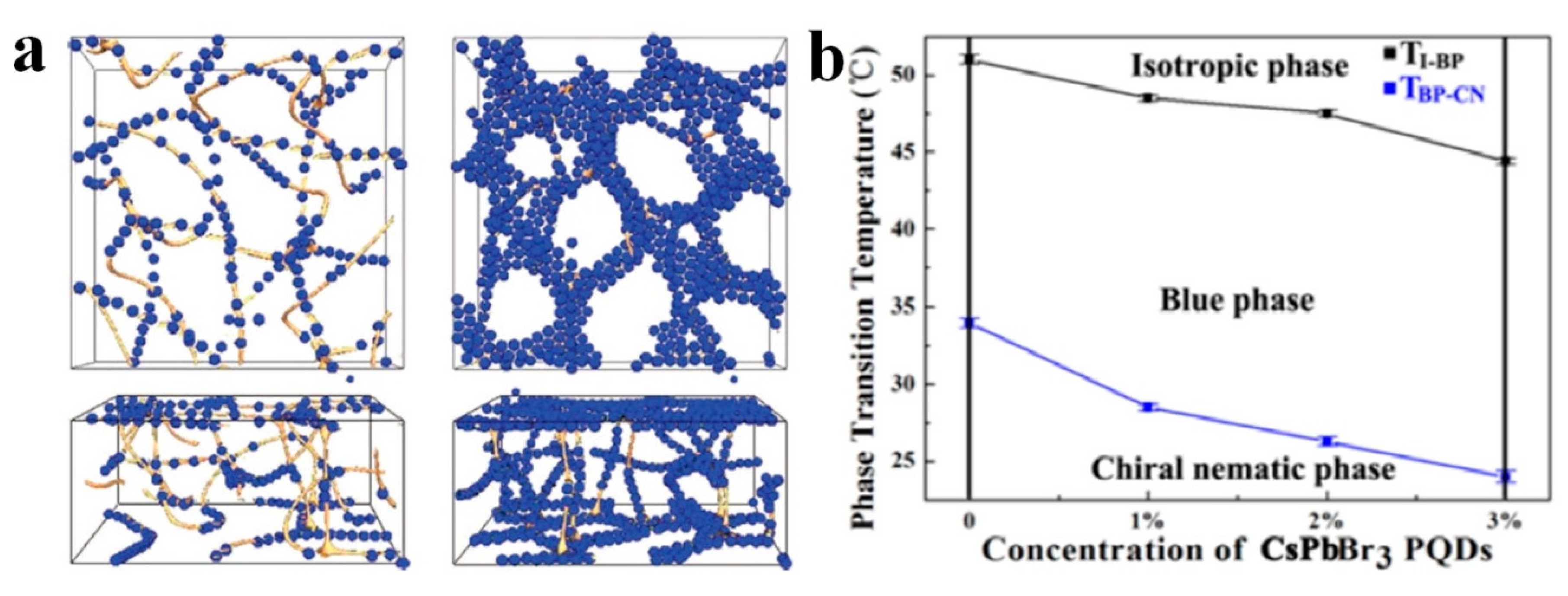
  336. Lin, P.; Yan, Q.; Wei, Z.; Chen, Y.; Chen, F.; Huang, Z.; Li, X.; Wang, H.; Wang, X.; Cheng, Z. All-inorganic perovskite quantum dots stabilized blue phase liquid crystals. Opt. Express 2018, 26, 18310–18319. [Google Scholar] [CrossRef] [PubMed]
  337. Mitov, M.; Portet, C.; Bourgerette, C.; Snoeck, E.; Verelst, M. Long-range structuring of nanoparticles by mimicry of a cholesteric liquid crystal. Nat. Mater. 2002, 1, 229. [Google Scholar] [CrossRef] [PubMed]
  338. Michel, M.; Christian, B.; François de, G. Fingerprint patterning of solid nanoparticles embedded in a cholesteric liquid crystal. J. Phys. Condens. Matter 2004, 16, S1981. [Google Scholar]
  339. Pendery, J.S.; Merchiers, O.; Coursault, D.; Grand, J.; Ayeb, H.; Greget, R.; Donnio, B.; Gallani, J.-L.; Rosenblatt, C.; Félidj, N.; et al. Gold nanoparticle self-assembly moderated by a cholesteric liquid crystal. Soft Matter 2013, 9, 9366–9375. [Google Scholar] [CrossRef]
  340. Rahimi, M.; Roberts, T.F.; Armas-Pérez, J.C.; Wang, X.; Bukusoglu, E.; Abbott, N.L.; de Pablo, J.J. Nanoparticle self-assembly at the interface of liquid crystal droplets. Proc. Natl. Acad. Sci. USA 2015, 112, 5297. [Google Scholar] [CrossRef]
  341. Chang, C.; Zhao, Y.; Liu, Y.; An, L. Liquid crystallinity of carbon nanotubes. RSC Adv. 2018, 8, 15780–15795. [Google Scholar] [CrossRef] [Green Version]
  342. Bravo-Sanchez, M.; Simmons, T.J.; Vidal, M.A. Liquid crystal behavior of single wall carbon nanotubes. Carbon 2010, 48, 3531–3542. [Google Scholar] [CrossRef]
  343. Lu, L.; Chen, W. Large-scale aligned carbon nanotubes from their purified, highly concentrated suspension. ACS Nano 2010, 4, 1042–1048. [Google Scholar] [CrossRef] [PubMed]
  344. Song, W.; Windle, A.H. Isotropic−nematic phase transition of dispersions of multiwall carbon nanotubes. Macromolecules 2005, 38, 6181–6188. [Google Scholar] [CrossRef]
  345. Steinert, B.W.; Dean, D.R. Magnetic field alignment and electrical properties of solution cast pet–carbon nanotube composite films. Polymer 2009, 50, 898–904. [Google Scholar] [CrossRef]
  346. Hobbie, E.K.; Fry, D.J. Nonequilibrium phase diagram of sticky nanotube suspensions. Phys. Rev. Lett. 2006, 97, 036101. [Google Scholar] [CrossRef] [PubMed]
  347. Davis, V.A.; Ericson, L.M.; Parra-Vasquez, A.N.G.; Fan, H.; Wang, Y.; Prieto, V.; Longoria, J.A.; Ramesh, S.; Saini, R.K.; Kittrell, C.; et al. Phase behavior and rheology of swnts in superacids. Macromolecules 2004, 37, 154–160. [Google Scholar] [CrossRef]
  348. Rai, P.K.; Pinnick, R.A.; Parra-Vasquez, A.N.G.; Davis, V.A.; Schmidt, H.K.; Hauge, R.H.; Smalley, R.E.; Pasquali, M. Isotropic−nematic phase transition of single-walled carbon nanotubes in strong acids. J. Am. Chem. Soc. 2006, 128, 591–595. [Google Scholar] [CrossRef]
  349. Pénicaud, A.; Poulin, P.; Derré, A.; Anglaret, E.; Petit, P. Spontaneous dissolution of a single-wall carbon nanotube salt. J. Am. Chem. Soc. 2005, 127, 8–9. [Google Scholar] [CrossRef]
  350. Baskaran, D.; Mays, J.W.; Bratcher, M.S. Noncovalent and nonspecific molecular interactions of polymers with multiwalled carbon nanotubes. Chem. Mater. 2005, 17, 3389–3397. [Google Scholar] [CrossRef]
  351. Haggenmueller, R.; Rahatekar, S.S.; Fagan, J.A.; Chun, J.; Becker, M.L.; Naik, R.R.; Krauss, T.; Carlson, L.; Kadla, J.F.; Trulove, P.C.; et al. Comparison of the quality of aqueous dispersions of single wall carbon nanotubes using surfactants and biomolecules. Langmuir 2008, 24, 5070–5078. [Google Scholar] [CrossRef]
  352. Zhang, S.; Kinloch, I.A.; Windle, A.H. Mesogenicity drives fractionation in lyotropic aqueous suspensions of multiwall carbon nanotubes. Nano Lett. 2006, 6, 568–572. [Google Scholar] [CrossRef] [PubMed]
  353. Zhang, S.; Li, Q.; Kinloch, I.A.; Windle, A.H. Ordering in a droplet of an aqueous suspension of single-wall carbon nanotubes on a solid substrate. Langmuir 2010, 26, 2107–2112. [Google Scholar] [CrossRef] [PubMed]
  354. Zamora-Ledezma, C.; Blanc, C.; Maugey, M.; Zakri, C.; Poulin, P.; Anglaret, E. Anisotropic thin films of single-wall carbon nanotubes from aligned lyotropic nematic suspensions. Nano Lett. 2008, 8, 4103–4107. [Google Scholar] [CrossRef] [PubMed]
  355. Song, W.; Windle, A.H. Size-dependence and elasticity of liquid-crystalline multiwalled carbon nanotubes. Adv. Mater. 2008, 20, 3149–3154. [Google Scholar] [CrossRef]
  356. Fu, W.; Liu, L.; Jiang, K.; Li, Q.; Fan, S. Super-aligned carbon nanotube films as aligning layers and transparent electrodes for liquid crystal displays. Carbon 2010, 48, 1876–1879. [Google Scholar] [CrossRef]
  357. Roussel, F.; Brun, J.-F.; Allart, A.; Huang, L.; O’Brien, S. Horizontally-aligned carbon nanotubes arrays and their interactions with liquid crystal molecules: Physical characteristics and display applications. AIP Adv. 2012, 2, 012110. [Google Scholar] [CrossRef]
  358. Park, H.-G.; Lee, M.-J.; Kim, K.; Seo, D.-S. Transparent conductive single wall carbon nanotube network films for liquid crystal displays. ECS Solid State Lett. 2012, 1, R31–R33. [Google Scholar] [CrossRef]
  359. Rajasekharan, R.; Dai, Q.; Wilkinson, T.D. Electro-optic characteristics of a transparent nanophotonic device based on carbon nanotubes and liquid crystals. Appl. Opt. 2010, 49, 2099–2104. [Google Scholar] [CrossRef]
  360. Yu, L.; Shearer, C.; Shapter, J. Recent development of carbon nanotube transparent conductive films. Chem. Rev. 2016, 116, 13413–13453. [Google Scholar] [CrossRef]
  361. Song, Y.I.; Lee, J.W.; Kim, T.Y.; Jung, H.J.; Jung, Y.C.; Suh, S.J.; Yang, C.-M. Performance-determining factors in flexible transparent conducting single-wall carbon nanotube film. Carbon Lett. 2013, 14, 255–258. [Google Scholar] [CrossRef] [Green Version]
  362. Lee, H.; Lee, J.-H.; Huynh, C.P.; Hawkins, S.C.; Musameh, M.; Kim, D.H.; Lee, S.H.; Choi, J. Orientational and electro-optical properties of liquid crystal aligned with a directly spinnable carbon nanotube web. Liq. Cryst. 2015, 42, 322–327. [Google Scholar] [CrossRef]
  363. Russell, J.M.; Oh, S.; LaRue, I.; Zhou, O.; Samulski, E.T. Alignment of nematic liquid crystals using carbon nanotube films. Thin Solid Film 2006, 509, 53–57. [Google Scholar] [CrossRef]
  364. Liu, Y.; Lim, Y.J.; Kundu, S.; Lee, S.H.; Lee, G.-D. Super-fast switching of twisted nematic liquid crystals with a single-wall-carbon-nanotube-doped alignment layer. J. Korean Phys. Soc. 2015, 66, 952–958. [Google Scholar] [CrossRef]
  365. Ren, L.; Pint, C.L.; Booshehri, L.G.; Rice, W.D.; Wang, X.; Hilton, D.J.; Takeya, K.; Kawayama, I.; Tonouchi, M.; Hauge, R.H.; et al. Carbon nanotube terahertz polarizer. Nano Lett. 2009, 9, 2610–2613. [Google Scholar] [CrossRef] [PubMed]
  366. Shoji, S.; Suzuki, H.; Zaccaria, R.P.; Sekkat, Z.; Kawata, S. Optical polarizer made of uniaxially aligned short single-wall carbon nanotubes embedded in a polymer film. Phys. Rev. B 2008, 77, 153407. [Google Scholar] [CrossRef]
  367. Byeong Gyun, K.; Young Jin, L.; Kwang-Un, J.; Kyu, L.; Young Hee, L.; Seung Hee, L. A tunable carbon nanotube polarizer. Nanotechnology 2010, 21, 405202. [Google Scholar]
  368. Behabtu, N.; Young, C.C.; Tsentalovich, D.E.; Kleinerman, O.; Wang, X.; Ma, A.W.K.; Bengio, E.A.; ter Waarbeek, R.F.; de Jong, J.J.; Hoogerwerf, R.E.; et al. Strong, light, multifunctional fibers of carbon nanotubes with ultrahigh conductivity. Science 2013, 339, 182. [Google Scholar] [CrossRef]
  369. Zhang, S.; Koziol, K.K.K.; Kinloch, I.A.; Windle, A.H. Macroscopic fibers of well-aligned carbon nanotubes by wet spinning. Small 2008, 4, 1217–1222. [Google Scholar] [CrossRef] [PubMed]
  370. Ericson, L.M.; Fan, H.; Peng, H.; Davis, V.A.; Zhou, W.; Sulpizio, J.; Wang, Y.; Booker, R.; Vavro, J.; Guthy, C.; et al. Macroscopic, neat, single-walled carbon nanotube fibers. Science 2004, 305, 1447. [Google Scholar] [CrossRef]
  371. Li, P.; Wong, M.; Zhang, X.; Yao, H.; Ishige, R.; Takahara, A.; Miyamoto, M.; Nishimura, R.; Sue, H.-J. Tunable lyotropic photonic liquid crystal based on graphene oxide. ACS Photonics 2014, 1, 79–86. [Google Scholar] [CrossRef]
  372. Dan, B.; Behabtu, N.; Martinez, A.; Evans, J.S.; Kosynkin, D.V.; Tour, J.M.; Pasquali, M.; Smalyukh, I.I. Liquid crystals of aqueous, giant graphene oxide flakes. Soft Matter 2011, 7, 11154–11159. [Google Scholar] [CrossRef]
  373. Aboutalebi, S.H.; Gudarzi, M.M.; Zheng, Q.B.; Kim, J.-K. Spontaneous formation of liquid crystals in ultralarge graphene oxide dispersions. Adv. Funct. Mater. 2011, 21, 2978–2988. [Google Scholar] [CrossRef]
  374. Jalili, R.; Aboutalebi, S.H.; Esrafilzadeh, D.; Konstantinov, K.; Moulton, S.E.; Razal, J.M.; Wallace, G.G. Organic solvent-based graphene oxide liquid crystals: A facile route toward the next generation of self-assembled layer-by-layer multifunctional 3d architectures. ACS Nano 2013, 7, 3981–3990. [Google Scholar] [CrossRef] [PubMed]
  375. Xu, Z.; Gao, C. Graphene chiral liquid crystals and macroscopic assembled fibres. Nat. Commun. 2011, 2, 571. [Google Scholar] [CrossRef] [Green Version]
  376. Tkacz, R.; Oldenbourg, R.; Mehta, S.B.; Miansari, M.; Verma, A.; Majumder, M. Ph dependent isotropic to nematic phase transitions in graphene oxide dispersions reveal droplet liquid crystalline phases. Chem. Commun. 2014, 50, 6668–6671. [Google Scholar] [CrossRef] [PubMed]
  377. Shen, T.-Z.; Hong, S.-H.; Song, J.-K. Electro-optical switching of graphene oxide liquid crystals with an extremely large kerr coefficient. Nat. Mater. 2014, 13, 394. [Google Scholar] [CrossRef]
  378. Arenas-Guerrero, P.; Delgado, Á.V.; Jiménez, M.L. Analysis of the electro-optical response of graphene oxide dispersions under alternating fields. Carbon 2018, 144, 395–401. [Google Scholar] [CrossRef]
  379. Hong, S.-H.; Shen, T.-Z.; Song, J.-K. Shear-induced assembly of graphene oxide particles into stripes near surface. Liq. Cryst. 2018, 45, 1303–1311. [Google Scholar] [CrossRef]
  380. Guo, F.; Kim, F.; Han, T.H.; Shenoy, V.B.; Huang, J.; Hurt, R.H. Hydration-responsive folding and unfolding in graphene oxide liquid crystal phases. ACS Nano 2011, 5, 8019–8025. [Google Scholar] [CrossRef] [PubMed]
  381. Senyuk, B.; Behabtu, N.; Pacheco, B.G.; Lee, T.; Ceriotti, G.; Tour, J.M.; Pasquali, M.; Smalyukh, I.I. Nonlinear photoluminescence imaging of isotropic and liquid crystalline dispersions of graphene oxide. ACS Nano 2012, 6, 8060–8066. [Google Scholar] [CrossRef]
  382. Qi, B.; Yuan, Z.; Lu, S.; Liu, K.; Li, S.; Yang, L.; Yu, J. Mechanical and thermal properties of epoxy composites containing graphene oxide and liquid crystalline epoxy. Fibers Polym. 2014, 15, 326–333. [Google Scholar] [CrossRef]
  383. Lu, S.; Li, S.; Yu, J.; Yuan, Z.; Qi, B. Epoxy nanocomposites filled with thermotropic liquid crystalline epoxy grafted graphene oxide. RSC Adv. 2013, 3, 8915–8923. [Google Scholar] [CrossRef]
  384. Naficy, S.; Jalili, R.; Aboutalebi, S.H.; Gorkin Iii, R.A.; Konstantinov, K.; Innis, P.C.; Spinks, G.M.; Poulin, P.; Wallace, G.G. Graphene oxide dispersions: Tuning rheology to enable fabrication. Mater. Horiz. 2014, 1, 326–331. [Google Scholar] [CrossRef]
  385. Wu, X.; Hou, K.; Huang, J.; Wang, J.; Yang, S. Graphene-based cellular materials with extremely low density and high pressure sensitivity based on self-assembled graphene oxide liquid crystals. J. Mater. Chem. C 2018, 6, 8717–8725. [Google Scholar] [CrossRef]
  386. Zamora-Ledezma, C.; Puech, N.; Zakri, C.; Grelet, E.; Moulton, S.E.; Wallace, G.G.; Gambhir, S.; Blanc, C.; Anglaret, E.; Poulin, P. Liquid crystallinity and dimensions of surfactant-stabilized sheets of reduced graphene oxide. J. Phys. Chem. Lett. 2012, 3, 2425–2430. [Google Scholar] [CrossRef]
  387. Park, H.; Lee, K.H.; Kim, Y.B.; Ambade, S.B.; Noh, S.H.; Eom, W.; Hwang, J.Y.; Lee, W.J.; Huang, J.; Han, T.H. Dynamic assembly of liquid crystalline graphene oxide gel fibers for ion transport. Sci. Adv. 2018, 4, eaau2104. [Google Scholar] [CrossRef] [PubMed] [Green Version]
  388. Jalili, R.; Aboutalebi, S.H.; Esrafilzadeh, D.; Shepherd, R.L.; Chen, J.; Aminorroaya-Yamini, S.; Konstantinov, K.; Minett, A.I.; Razal, J.M.; Wallace, G.G. Scalable one-step wet-spinning of graphene fibers and yarns from liquid crystalline dispersions of graphene oxide: Towards multifunctional textiles. Adv. Funct. Mater. 2013, 23, 5345–5354. [Google Scholar] [CrossRef]
  389. Xu, Z.; Gao, C. Graphene in macroscopic order: Liquid crystals and wet-spun fibers. Acc. Chem. Res. 2014, 47, 1267–1276. [Google Scholar] [CrossRef] [PubMed]
  390. Kim, I.H.; Yun, T.; Kim, J.-E.; Yu, H.; Sasikala, S.P.; Lee, K.E.; Koo, S.H.; Hwang, H.; Jung, H.J.; Park, J.Y.; et al. Mussel-inspired defect engineering of graphene liquid crystalline fibers for synergistic enhancement of mechanical strength and electrical conductivity. Adv. Mater. 2018, 30, 1803267. [Google Scholar] [CrossRef]
  391. Aboutalebi, S.H.; Jalili, R.; Esrafilzadeh, D.; Salari, M.; Gholamvand, Z.; Aminorroaya Yamini, S.; Konstantinov, K.; Shepherd, R.L.; Chen, J.; Moulton, S.E.; et al. High-performance multifunctional graphene yarns: Toward wearable all-carbon energy storage textiles. ACS Nano 2014, 8, 2456–2466. [Google Scholar] [CrossRef]
  392. Mahalingam, D.K.; Wang, S.; Nunes, S.P. Graphene oxide liquid crystal membranes in protic ionic liquid for nanofiltration. ACS Appl. Nano Mater. 2018, 1, 4661–4670. [Google Scholar] [CrossRef]
  393. Kim, H.; Kim, D.W.; Vasagar, V.; Ha, H.; Nazarenko, S.; Ellison, C.J. Polydopamine-graphene oxide flame retardant nanocoatings applied via an aqueous liquid crystalline scaffold. Adv. Funct. Mater. 2018, 28, 1803172. [Google Scholar] [CrossRef]
  394. Kim, M.; Park, J.H.; Shahini, S.; Yamamoto, J.; Campidelli, S.; Kim, Y.S.; Scalia, G. Graphene: A new liquid crystal for high performance electro-optic applications. In Liquid Crystals XXII; International Society for Optics and Photonics: Bellingham, WA, USA, 2018; p. 107350N. [Google Scholar]
  395. Huang, X.; He, J.; Sun, K.; Chen, Y.; Zha, Z.; Zhou, C. Liquid crystal behavior and cytocompatibility of graphene oxide dispersed in sodium alginate solutions. Carbon 2018, 129, 258–269. [Google Scholar] [CrossRef]
  396. Sasikala, S.P.; Lim, J.; Kim, I.H.; Jung, H.J.; Yun, T.; Han, T.H.; Kim, S.O. Graphene oxide liquid crystals: A frontier 2d soft material for graphene-based functional materials. Chem. Soc. Rev. 2018, 47, 6013–6045. [Google Scholar] [CrossRef]
  397. Klemm, D.; Kramer, F.; Moritz, S.; Lindström, T.; Ankerfors, M.; Gray, D.; Dorris, A. Nanocelluloses: A new family of nature-based materials. Angew. Chem. Int. Ed. 2011, 50, 5438–5466. [Google Scholar] [CrossRef] [PubMed]
  398. Shoda, M.; Sugano, Y. Recent advances in bacterial cellulose production. Biotechnol. Bioprocess. Eng. 2005, 10, 1. [Google Scholar] [CrossRef]
  399. Eichhorn, S.J. Cellulose nanowhiskers: Promising materials for advanced applications. Soft Matter 2011, 7, 303–315. [Google Scholar] [CrossRef]
  400. Li, Y.; Jun-Yan Suen, J.; Prince, E.; Larin, E.M.; Klinkova, A.; Thérien-Aubin, H.; Zhu, S.; Yang, B.; Helmy, A.S.; Lavrentovich, O.D.; et al. Colloidal cholesteric liquid crystal in spherical confinement. Nat. Commun. 2016, 7, 12520. [Google Scholar] [CrossRef] [Green Version]
  401. Marchessault, R.; Morehead, F.; Walter, N. Liquid crystal systems from fibrillar polysaccharides. Nature 1959, 184, 632–633. [Google Scholar] [CrossRef]
  402. Usov, I.; Nyström, G.; Adamcik, J.; Handschin, S.; Schütz, C.; Fall, A.; Bergström, L.; Mezzenga, R. Understanding nanocellulose chirality and structure–properties relationship at the single fibril level. Nat. Commun. 2015, 6, 7564. [Google Scholar] [CrossRef] [PubMed]
  403. Hirai, A.; Inui, O.; Horii, F.; Tsuji, M. Phase separation behavior in aqueous suspensions of bacterial cellulose nanocrystals prepared by sulfuric acid treatment. Langmuir 2009, 25, 497–502. [Google Scholar] [CrossRef] [PubMed]
  404. Schütz, C.; Agthe, M.; Fall, A.B.; Gordeyeva, K.; Guccini, V.; Salajková, M.; Plivelic, T.S.; Lagerwall, J.P.F.; Salazar-Alvarez, G.; Bergström, L. Rod packing in chiral nematic cellulose nanocrystal dispersions studied by small-angle x-ray scattering and laser diffraction. Langmuir 2015, 31, 6507–6513. [Google Scholar] [CrossRef] [PubMed]
  405. Shafeiei-Sabet, S.; Hamad, W.Y.; Hatzikiriakos, S.G. Influence of degree of sulfation on the rheology of cellulose nanocrystal suspensions. Rheol. Acta 2013, 52, 741–751. [Google Scholar] [CrossRef]
  406. Dong, X.M.; Kimura, T.; Revol, J.-F.; Gray, D.G. Effects of ionic strength on the isotropic−chiral nematic phase transition of suspensions of cellulose crystallites. Langmuir 1996, 12, 2076–2082. [Google Scholar] [CrossRef]
  407. Araki, J.; Kuga, S. Effect of trace electrolyte on liquid crystal type of cellulose microcrystals. Langmuir 2001, 17, 4493–4496. [Google Scholar] [CrossRef]
  408. Song, W.; Lee, J.-K.; Gong, M.S.; Heo, K.; Chung, W.-J.; Lee, B.Y. Cellulose nanocrystal-based colored thin films for colorimetric detection of aldehyde gases. ACS Appl. Mater. Interfaces 2018, 10, 10353–10361. [Google Scholar] [CrossRef] [PubMed]
  409. Kelly, J.A.; Shukaliak, A.M.; Cheung, C.C.Y.; Shopsowitz, K.E.; Hamad, W.Y.; MacLachlan, M.J. Responsive photonic hydrogels based on nanocrystalline cellulose. Angew. Chem. Int. Ed. 2013, 52, 8912–8916. [Google Scholar] [CrossRef]
  410. Bruckner, J.R.; Kuhnhold, A.; Honorato-Rios, C.; Schilling, T.; Lagerwall, J.P.F. Enhancing self-assembly in cellulose nanocrystal suspensions using high-permittivity solvents. Langmuir 2016, 32, 9854–9862. [Google Scholar] [CrossRef]
  411. Beck, S.; Bouchard, J.; Berry, R. Controlling the reflection wavelength of iridescent solid films of nanocrystalline cellulose. Biomacromolecules 2011, 12, 167–172. [Google Scholar] [CrossRef]
  412. Frka-Petesic, B.; Guidetti, G.; Kamita, G.; Vignolini, S. Controlling the photonic properties of cholesteric cellulose nanocrystal films with magnets. Adv. Mater. 2017, 29, 1701469. [Google Scholar] [CrossRef]
  413. Beck-Candanedo, S.; Roman, M.; Gray, D.G. Effect of reaction conditions on the properties and behavior of wood cellulose nanocrystal suspensions. Biomacromolecules 2005, 6, 1048–1054. [Google Scholar] [CrossRef] [PubMed]
  414. Dong, Y.-m.; Mao, W.; Wang, H.-w.; Zhao, Y.-q.; Li, X.-j.; Bi, D.-x.; Yang, L.-l.; Ge, Q.; Fang, X. Measurement of critical concentration for mesophase formation of chitosan derivatives in both aqueous and organic solutions. Polym. Int. 2006, 55, 1444–1449. [Google Scholar] [CrossRef]
  415. Parker, R.M.; Guidetti, G.; Williams, C.A.; Zhao, T.; Narkevicius, A.; Vignolini, S.; Frka-Petesic, B. The self-assembly of cellulose nanocrystals: Hierarchical design of visual appearance. Adv. Mater. 2018, 30, 1704477. [Google Scholar] [CrossRef] [PubMed]
  416. Zhang, Y.P.; Chodavarapu, V.P.; Kirk, A.G.; Andrews, M.P. Structured color humidity indicator from reversible pitch tuning in self-assembled nanocrystalline cellulose films. Sens. Actuators B Chem. 2013, 176, 692–697. [Google Scholar] [CrossRef]
  417. Zhang, Y.P.; Chodavarapu, V.P.; Kirk, A.G.; Andrews, M.P. Nanocrystalline cellulose for covert optical encryption. J. Nanophotonics 2012, 6, 063516. [Google Scholar] [CrossRef]
  418. Bardet, R.; Roussel, F.; Coindeau, S.; Belgacem, N.; Bras, J. Engineered pigments based on iridescent cellulose nanocrystal films. Carbohydr. Polym. 2015, 122, 367–375. [Google Scholar] [CrossRef] [PubMed]
  419. Majoinen, J.; Hassinen, J.; Haataja, J.S.; Rekola, H.T.; Kontturi, E.; Kostiainen, M.A.; Ras, R.H.A.; Törmä, P.; Ikkala, O. Chiral plasmonics using twisting along cellulose nanocrystals as a template for gold nanoparticles. Adv. Mater. 2016, 28, 5262–5267. [Google Scholar] [CrossRef]
  420. Almeida, P.L.; Kundu, S.; Borges, J.P.; Godinho, M.H.; Figueirinhas, J.L. Electro-optical light scattering shutter using electrospun cellulose-based nano- and microfibers. Appl. Phys. Lett. 2009, 95, 043501. [Google Scholar] [CrossRef]
  421. Shopsowitz, K.E.; Qi, H.; Hamad, W.Y.; MacLachlan, M.J. Free-standing mesoporous silica films with tunable chiral nematic structures. Nature 2010, 468, 422. [Google Scholar] [CrossRef]
  422. Rofouie, P.; Alizadehgiashi, M.; Mundoor, H.; Smalyukh, I.I.; Kumacheva, E. Self-assembly of cellulose nanocrystals into semi-spherical photonic cholesteric films. Adv. Funct. Mater. 2018, 28, 1803852. [Google Scholar] [CrossRef]
  423. Majoinen, J.; Kontturi, E.; Ikkala, O.; Gray, D.G. Sem imaging of chiral nematic films cast from cellulose nanocrystal suspensions. Cellulose 2012, 19, 1599–1605. [Google Scholar] [CrossRef]
  424. Shopsowitz, K.E.; Stahl, A.; Hamad, W.Y.; MacLachlan, M.J. Hard templating of nanocrystalline titanium dioxide with chiral nematic ordering. Angew. Chem. 2012, 124, 6992–6996. [Google Scholar] [CrossRef]
  425. Li, Y.; Prince, E.; Cho, S.; Salari, A.; Mosaddeghian Golestani, Y.; Lavrentovich, O.D.; Kumacheva, E. Periodic assembly of nanoparticle arrays in disclinations of cholesteric liquid crystals. Proc. Natl. Acad. Sci. USA 2017, 114, 2137. [Google Scholar] [CrossRef] [PubMed]
  426. Gupta, V.K.; Skaife, J.J.; Dubrovsky, T.B.; Abbott, N.L. Optical amplification of ligand-receptor binding using liquid crystals. Science 1998, 279, 2077. [Google Scholar] [CrossRef] [PubMed]
  427. Brake, J.M.; Daschner, M.K.; Luk, Y.-Y.; Abbott, N.L. Biomolecular interactions at phospholipid-decorated surfaces of liquid crystals. Science 2003, 302, 2094. [Google Scholar] [CrossRef]
  428. Hsiao, Y.-C.; Sung, Y.-C.; Lee, M.-J.; Lee, W. Highly sensitive color-indicating and quantitative biosensor based on cholesteric liquid crystal. Biomed. Opt. Express 2015, 6, 5033–5038. [Google Scholar] [CrossRef] [PubMed] [Green Version]
  429. Lin, C.-H.; Lee, M.-J.; Lee, W. Bovine serum albumin detection and quantitation based on capacitance measurements of liquid crystals. Appl. Phys. Lett. 2016, 109, 093703. [Google Scholar] [CrossRef]
  430. Lee, M.-J.; Chang, C.-H.; Lee, W. Label-free protein sensing by employing blue phase liquid crystal. Biomed. Opt. Express 2017, 8, 1712–1720. [Google Scholar] [CrossRef]
  431. Qi, L.; Hu, Q.; Kang, Q.; Yu, L. Fabrication of liquid-crystal-based optical sensing platform for detection of hydrogen peroxide and blood glucose. Anal. Chem. 2018, 90, 11607–11613. [Google Scholar] [CrossRef]
  432. Zhang, J.; Su, X.; Yang, D.; Luan, C. Label-free liquid crystal biosensor for cecropin b detection. Talanta 2018, 186, 60–64. [Google Scholar] [CrossRef]
  433. Lin, C.-M.; Wu, P.-C.; Lee, M.-J.; Lee, W. Label-free protein quantitation by dielectric spectroscopy of dual-frequency liquid crystal. Sens. Actuators B Chem. 2019, 282, 158–163. [Google Scholar] [CrossRef]
  434. Tan, H.; Yang, S.; Shen, G.; Yu, R.; Wu, Z. Signal-enhanced liquid-crystal DNA biosensors based on enzymatic metal deposition. Angew. Chem. Int. Ed. 2010, 49, 8608–8611. [Google Scholar] [CrossRef] [PubMed]
  435. Hartono, D.; Qin, W.J.; Yang, K.-L.; Yung, L.-Y.L. Imaging the disruption of phospholipid monolayer by protein-coated nanoparticles using ordering transitions of liquid crystals. Biomaterials 2009, 30, 843–849. [Google Scholar] [CrossRef] [PubMed]
  436. Liao, S.; Qiao, Y.; Han, W.; Xie, Z.; Wu, Z.; Shen, G.; Yu, R. Acetylcholinesterase liquid crystal biosensor based on modulated growth of gold nanoparticles for amplified detection of acetylcholine and inhibitor. Anal. Chem. 2012, 84, 45–49. [Google Scholar] [CrossRef] [PubMed]
  437. Yang, S.; Liu, Y.; Tan, H.; Wu, C.; Wu, Z.; Shen, G.; Yu, R. Gold nanoparticle based signal enhancement liquid crystal biosensors for DNA hybridization assays. Chem. Commun. 2012, 48, 2861–2863. [Google Scholar] [CrossRef] [PubMed]
  438. Zhao, D.; Peng, Y.; Xu, L.; Zhou, W.; Wang, Q.; Guo, L. Liquid-crystal biosensor based on nickel-nanosphere-induced homeotropic alignment for the amplified detection of thrombin. ACS Appl. Mater. Interfaces 2015, 7, 23418–23422. [Google Scholar] [CrossRef] [PubMed]
  439. Wei, Y.; Jang, C.-H. Visualization of cholylglycine hydrolase activities through nickel nanoparticle-assisted liquid crystal cells. Sens. Actuators B Chem. 2017, 239, 1268–1274. [Google Scholar] [CrossRef]
  440. Choudhary, A.; George, F.T.; Li, G. Conjugation of nanomaterials and nematic liquid crystals for futuristic applications and biosensors. Biosensors 2018, 8, 69. [Google Scholar] [CrossRef] [PubMed]
  441. Kim, Y.-K.; Wang, X.; Mondkar, P.; Bukusoglu, E.; Abbott, N.L. Self-reporting and self-regulating liquid crystals. Nature 2018, 557, 539–544. [Google Scholar] [CrossRef] [PubMed]
  442. Barauskas, J.; Johnsson, M.; Tiberg, F. Self-assembled lipid superstructures:  Beyond vesicles and liposomes. Nano Lett. 2005, 5, 1615–1619. [Google Scholar] [CrossRef] [PubMed]
  443. Vallamkondu, J.; Corgiat, E.; Buchaiah, G.; Kandimalla, R.; Reddy, P. Liquid crystals: A novel approach for cancer detection and treatment. Cancers 2018, 10, 462. [Google Scholar] [CrossRef] [PubMed]
  444. Malmsten, M. Soft drug delivery systems. Soft Matter 2006, 2, 760–769. [Google Scholar] [CrossRef]
  445. Nikam, S.; Chavan, M.; Sharma, P.H. Solid lipid nanoparticles: A lipid based drug delivery. Nanotechnology 2014, 1, 5. [Google Scholar]
  446. Volkhard Jenning, S.H.G. Encapsulation of retinoids in solid lipid nanoparticles (sln). J. Microencapsul. 2001, 18, 149–158. [Google Scholar] [CrossRef] [PubMed]
  447. Müller-Goymann, C.C. Physicochemical characterization of colloidal drug delivery systems such as reverse micelles, vesicles, liquid crystals and nanoparticles for topical administration. Eur. J. Pharm. Biopharm. 2004, 58, 343–356. [Google Scholar] [CrossRef] [PubMed]
Figure 1. (a) The various shapes of LC molecules. (b) A typical illustration of different thermotropic LC phases observed on heating from crystalline state. Reproduced with permission from [7]. Copyright (2017) MDPI.
Applsci 09 02512 g001
Figure 2. A typical lyotropic LC phase diagram of amphiphilic molecules dissolved in a solvent. Cubic phases may be observed at different positions in the phase diagram. At very high concentrations, the inverse phases are located. Reproduced with permission from [7]. Copyright (2017) MDPI.
Applsci 09 02512 g002
Figure 3. (a) SEM image of a collection of tobacco mosaic virus particles. Scale bar: 0.2 μm. Reproduced from Wikimedia Commons, with no author name supplied. (b) The orientational order parameter of the viruses increases as their concentration in the solvent is increased. Reproduced with permission from [46]. Copyright (1988) American Physical Society.
Applsci 09 02512 g003
Figure 4. (a) A Schlieren texture (left) of 0.5 vol % BaTiO3 + 5CB observed by polarizing optical microscopy, POM. Observation between parallel polarizers (right) provides evidence that the dispersed particles are collected in the topological defects. (b,c) Permittivity components ε′ and ε′, respectively. (d) The relationship between the electro-optic response time and the concentration of BaTiO3 with (d) the field on-time and (e) the off-time for larger (ferroelectric) and smaller (non-ferroelectric) particles. (f) The relationship between the concentration of BaTiO3 and threshold voltage Vth, and (g) splay elastic constant K11. Reproduced with permission from [173]. Copyright (2017) AIP.
Applsci 09 02512 g004
Figure 5. (a) Variation of longitudinal (ε’) and transverse (ε′) components of the relative dielectric permittivity with temperature for pure LC and gold-nanoparticle-dispersed 6CHBT. (b) Frequency dependence of the longitudinal component of loss (ε″) for pure and gold-nanoparticle-dispersed 6CHBT. Reproduced with permission from [182]. Copyright (2018) Taylor & Francis.
Applsci 09 02512 g005
Figure 6. (a) Transmission voltage function of the sample device with single-walled carbon nanotube doping. (b) Relative permittivities (transverse (ε′) and longitudinal (ε′) components) with temperature for pure and dispersed samples. Reproduced with permission from [215]. Copyright (2018) ELSEVIER.
Applsci 09 02512 g006
Figure 7. (a) Threshold voltage (top left), splay elastic constant (top right), and electro-optic response time (bottom) as functions of GO concentration in 5CB, for small and large sizes. (b) Schematic illustration of the Fréedericksz transition of a device cell filled with a GO flakes dispersed nematic liquid crystal. Reproduced with permission from [225]. Copyright (2016) John Wiley and Sons.
Applsci 09 02512 g007
Figure 8. Extinction measurement of gold-nanorods (GNRs)/8CB samples deposited on a PVA polymer with (a,d) nonpolarized incident light and with an incident light polarized (b,e) parallel and (c,f) perpendicular to the oily streaks. Reproduced with permission from [266]. Copyright (2017) American Chemistry Society.
Applsci 09 02512 g008
Figure 9. Electric switching of SPR-enhanced fluorescence intensity of gold–silica–dye nanoparticles (GSDs) in a nematic LC matrix. (a) Fluorescence micrograph showing the GSD–LC composite in a planar glass cell. (b) Fluorescence spectrum of GSDs in the nematic LC. (c) Polarization-dependent fluorescence intensity. (d) Voltage-dependent fluorescence of GSDs in a nematic LC. (e) Switching of GSDs’ fluorescence intensity by an electric field. (f) Voltage-dependent ON and OFF switching times obtained from the change of transmission I-I0 versus time curves. Reproduced with permission from [267]. Copyright (2016) American Chemical Society.
Applsci 09 02512 g009
Figure 10. (a) Schematic figure of the photothermal process of reversible handedness inversion. (b) Dynamic photonic reflection colours of self-organized helical superstructure upon NIR irradiation. Reproduced with permission from [287]. Copyright (2016) John Wiley and Sons. (c) Polymer-stabilized graphene-containing CLC thin film as a photothermal material. Reproduced with permission from [288]. Copyright (2017) ELSEVIER.
Applsci 09 02512 g010
Figure 11. (a) POM (I) and fluorescence microscope (II) images of the QD-CLC cell. (b) Fluorescence emission and absorption spectra of the 8.7 wt % QDs dissolved in toluene and the measured fluorescence emission of the QD-CLC cell in the isotropic phase (red curve). (c) Optical tuning of the lasing emission of the QD-CLC laser with the added chiral-azobenzene moiety under UV irradiation (I) and optically induced red-shift of the reflection band of the chiral-azobenzene-added QD-CLC under UV irradiation (II). Reproduced with permission from [300]. Copyright (2014) Royal Society of Chemistry.
Applsci 09 02512 g011
Figure 12. (a) POM images of (I) a 0.1 wt % Au dispersion of AuNPs in the smectic A phase showing an array of very faint lines in the direction indicated by the double-headed arrow, and (II) 1.0 wt % Au dispersion of AuNPs in the smectic phase between homeotropic glass slides showing an example of a curved domain where the black arrows indicate the direction of AuNP arrays. Reproduced with permission from [320]. Copyright (2012) The Royal Society of Chemistry. (b) Schematic illustration of a linear array of straight parallel oily streaks (I); (II) POM image of a single array of 8CB film on MoS2. (III) SEM image of the MoS2 substrate with gold-nanoparticle chains and (IV) the enlarged view. Reproduced with permission from [316]. Copyright (2012) John Wiley and Sons. (c) Optical microscopy image (I) of the dense population of toric focal conic domains (TFCDs) on Si microchannels (Inset is a schematic diagram of a silica particle trapped in the centre of a TFCD); (II) Confocal microscopy image of fluorescent silica particles trapped in TFCDs. Reproduced with permission from [321]. Copyright (2007) Springer Nature. (d) A sequence of dark-field images showing a gold-nanorod moving into a topological singularity near the microsphere (I) and polarizing, bright-field, dark-field microscopy images and a schematic diagram showing chains of microsphere-gold nanoparticle dimers aligned along the director (II). Reproduced with permission from [311]. Copyright (2012) American Chemical Society.
Applsci 09 02512 g012
Figure 13. (a) Snapshots of the steady states obtained when a dispersion of colloids with different concentration in the isotropic phase is placed in a sandwich geometry and then quenched into a regime where BP I is stable in the bulk. Reproduced with kind permission from [318]. Copyright (2014) Springer Nature. (b) The phase diagram for the BPLC/nanoparticle composite. Reproduced with permission from [336]. Copyright (2018) Optical Society of America.
Applsci 09 02512 g013
Figure 14. (a) Optical images of a CNT nematic LC schlieren texture. (b) Optical images of a dried CNTLC phase. SEM images of topological disclinations of strength +1/2 (c) and −1/2 are observable (d). Reproduced with permission from [162]. Copyright (2003) The American Association for the Advancement of Science. (e) A schematic illustration of the aligning LC molecules by a CNT film. (f) The SEM images of the CNT alignment film on a glass substrate. Reproduced with permission from [356]. Copyright (2010) ELSEVIER. (g) Winding drums with collected fibres. (h) SEM images of corresponding CNT fibres. Reproduced with permission from [368]. Copyright (2013) The American Association for the Advancement of Science. (i) Sketch of an optical CNT polarizer. (j) Schematic cell structures of a TN-LCD by the aligned CNT polarizer. Reproduced with permission from [341]. Copyright (2018) Royal Society of Chemistry.
Applsci 09 02512 g014
Figure 15. (a) POM images and (b) macroscopic photographs between crossed polarizers of GO aqueous dispersions in planar cells with mass fraction fm of 5 × 10⁻⁴, 1 × 10⁻³, 3 × 10⁻³, 5 × 10⁻³, 8 × 10⁻³, 1 × 10⁻² (from 1 to 6). Reproduced with permission from [165]. Copyright (2011) American Chemistry Society. (c) Schematic illustration of GO LC membrane in protic ionic liquid for nanofiltration. Reproduced with permission from [392]. Copyright (2018) American Chemistry Society. (d) SEM images of an as-spun GO fibre. Reproduced with permission from [387]. Copyright (2013) John Wiley and Sons. (e) Illustration of fire ignition retardation behaviour for (I) neat PU foam and (II) PDA/GO-coated PU foam. Reproduced with permission from [393]. Copyright (2018) John Wiley and Sons.
Applsci 09 02512 g015
Figure 16. (a) TEM images of dried CNCs and the formation of a CLC phase and its coexistence with the isotropic phase in a CNC suspension (upper image). The typical CLC fingerprint appearance and the helical arrangement of nanorods are illustrated below. Reproduced with permission from [160,400,421,423]. Copyright (2010), (2012), (2014), (2016) Springer Nature. (b) Colour change observed when a dried CNC film is immersed in water and the corresponding spectrum. Reproduced with permission from [416]. Copyright (2013) Elsevier. (c) Nanoparticle templating of the CNC-derived helical structure. Reproduced with permission from [424,425]. Copyright (2012) John Wiley and Sons and (2017) National Academy of Sciences.
Applsci 09 02512 g016
Figure 17. (a) Schematic illustration of the LC-based biosensor. (bd) Optical image and cartoon representation of the anchoring transition of 5CB and the state of the aqueous-5CB interface immediately after injection of a dispersion of vesicles formed from 0.1 mM L-α-dilauroyl phosphatidylcholine (L-DLPC) in tris-buffered saline, (c) after ~10–20 min of contact with the vesicle dispersion of L-DLPC and (d) after 2 h of contact. Reproduced with permission from [427]. Copyright (2003) The American Association for the Advancement of Science.
Applsci 09 02512 g017
Figure 18. (a) The orientations of 5CB in the LC cells fabricated with DMOAP-coated glass slides (upper slides) and modified slides (IIII; lower): (I) (3-aminopropyl)trimethoxysilane (APS)/DMOAP-coated glass slide, (II) streptavidin alkaline phosphatase (Sv-ALP) modified glass slide, and (III) silver-deposited glass slide. (b) Optical images under crossed polarizers of LC cells with 5CB sandwiched between a DMOAP-coated glass slide and a silver-deposited glass slide with complementary target DNA at concentrations of (I) 0, (II) 0.1, (III) 1, (IV) 10, (V) 100 pM, and (VI) two-base mismatched DNA at concentration of 100 pM. Reproduced with permission from [434]. Copyright (2010) John Wiley and Sons. (c) Sequential micrographs (IIII) and corresponding illustrations (IVVI) of a LC film containing microdroplets before (I,IV) and after addition of cationic amphiphile DTAB to the overlying aqueous phase, at 0 (II,V) and 60 min (III,VI). (d) Release of microcargo from LCs by the heat of a human finger or by motile bacteria that generate interfacial shear stresses and interact with microcargo in a self-regulated manner. Reproduced with permission from [441]. Copyright (2018) Springer Nature.
Applsci 09 02512 g018
Figure 19. TEM images of different nonlamellar lipid nanoparticles containing (ad) reversed bicontinuous, (e,f) sponge, and (g,h) reversed hexagonal lyotropic LC phases. Reproduced with permission from [442]. Copyright (2005) American Chemical Society.
Applsci 09 02512 g019

