Article

Evaluation of Quality of Eggs from Hens Kept in Caged and Free-Range Systems Using Traditional Methods and Ultra-Weak Luminescence

by
Ilona Gałązka-Czarnecka
1,
Ewa Korzeniewska
2,*,
Andrzej Czarnecki
1,
Michał Sójka
1,
Paweł Kiełbasa
3 and
Tomasz Dróżdź
3
1
Faculty of Biotechnology and Food Sciences, Lodz University of Technology, 90-924 Łódź, Poland
2
Institute of Electrical Engineering Systems, Lodz University of Technology, 90-924 Łódź, Poland
3
Faculty of Production and Power Engineering, University of Agriculture in Krakow, 30-149 Kraków, Poland
*
Author to whom correspondence should be addressed.
Appl. Sci. 2019, 9(12), 2430; https://doi.org/10.3390/app9122430
Submission received: 26 April 2019 / Revised: 5 June 2019 / Accepted: 10 June 2019 / Published: 14 June 2019

Abstract

:
The paper presents the results of an evaluation of the quality of eggs from laying hens kept in caged and free range systems using traditional methods and ultra-weak luminescence (USL). It was found that the tested eggs were fresh and were characterized by the required quality, as demonstrated by analysis of the egg white and egg yolk. Eggs from free-range laying hens were characterized by an eight-fold higher emission of photons compared to eggs from caged hens, and they had over three times higher content of natural antioxidants in the form of carotenoids. Most probably, the higher number of photons emitted is associated with a higher content of biologically active substances in the material under study. Photon emission also varies in different ways depending on the specific hen breeding system. Differences in time in the identified maximum values of photon emission result from the composition of individual parts of the egg. Different times in which the emission peaks occurred for free-range eggs and for caged eggs were observed. The application of the USL method in order to confirm its usefulness in the assessment of food quality requires further research.

1. Introduction

Poland is a significant producer of consumer eggs in the European Union. The annual output is more than nine billion eggs. Most often, the choice of eggs is determined by the price and size, therefore, the most popular on the market are eggs from the cage system in the sizes M and L. In recent years, consumers have begun to pay more and more attention to the origin of the eggs and rearing of the chickens. There has been a gradual increase in interest in more expensive eggs of free range origin. Consumers perceive these eggs as traditional food, because hens have access to a chicken run and a much more varied diet. This type of rearing has a substantial impact on the eggs’ composition. Consumers usually associate the quality of eggs with their freshness and the color of their yolk. However, the quality of eggs is a broader concept, and, although it is partly shaped by the hen-housing conditions, many other factors affect it. These include genetic factors, like breed and line of laying hens (which affects the size and color of the shell, as well as nutritional values [1]), rearing conditions [2], nutrition (feed composition, access to free range), age of laying hens [3] (older hens lay eggs of greater weight, but the quality of their shell is worse [4,5]), and time and storage conditions for eggs [6,7,8]. Eggs undergo numerous analyses and tests to determine their freshness, technological suitability, quality of egg white and egg yolk, nutritional composition, and heavy metal pollution.
Currently, intensive work on an objective and quantitative measure of the health status and freshness of chicken eggs is underway. Pilot studies on food parameterization with methods using fluorescence decay recorded using a time-correlated single photon counting (TCSPC) method were carried out on already processed products observing photon emission variation. The phenomenon of photon emission has been discovered in many microscopic and macroscopic systems [9,10], including lipid systems, bacteria [11], yeast proliferating bacteria [12], leukocytes [13], nerve cells [14], mitochondria and chloroplasts [15], tumor cells [16], hepatic tissue [17], renal tissue [18], and body fluids [19]. Recent studies indicate that ultra-low photon emission seems to be a good method for analyzing the interaction of nanoparticles with various biological objects [20]. The measurement of radiated light is carried out in the visible spectrum with the highest sensitivity (>10−17 W). As the next stage of research, the authors of the paper predict egg testing using optical coherence tomography (OCT) [21].

2. Materials and Methods

2.1. Research Methodology

Selected characteristics of egg yolk (n = 60) and egg white (n = 60) from free-range hens and caged hens were defined in order to determine their freshness and quality (for this purpose, the pH, color at La Roche point—YCF scale, yolk color fan, and Haugh units were determined). In egg yolks, the content of tocopherols (n = 60) and carotenoids (n = 60) was measured using liquid chromatography methods, the color parameters were determined by the CIELab method (n = 60). The measurement of egg photons was carried out using the phenomenon of ultra-weak luminescence (n = 60). The statistical analysis of the obtained results was carried out using the Statistica 10 program.

2.2. Raw Material

The tested materials were Class A washed eggs from caged hens (eggs C) (marked 3PL12091305) with weight class M (56.2 g ± 1.8) and from free-range hens (eggs F) (marked 1-PL-12071302) with weight class L (69.8 g ± 2.1). The egg shell was of a uniform color, clean, and without damage. Eggs after breaking were characterized by a specific odor, and a dense, transparent white. No fertile eggs were observed.

2.3. Determination of Tocopherols and Carotenoids

Yolks were manually separated from the whites. Three yolks of each type of egg were combined and mixed. Samples and standard solutions were prepared directly before analysis. Standards of tocopherols and carotenoids were purchased from Sigma-Aldrich (Saint Louis, MO, USA).
Tocopherols were determined in accordance with the method described in Reference [22].
Samples of yolk were weighed (0.2 g) and dissolved in 2 mL of 2-propanol. Vortex-mixed samples were centrifuged (5 min, 20,000 RCF, 4 °C) and directly injected (10 μL) onto an HPLC column.
A liquid chromatograph (Shimadzu, Kyoto, Japan), comprising degasser of mobile phase DG 3014 (Ecom, Prague, Czech Republic), pump LC-20AC, autosampler SIL-AC, thermostat CTO-10AS, and fluorescence detector RF-10A XL (set at emission wavelength of 325 nm with an excitation at 295 nm), was coupled with a column Synergi 4u Hydro 150 × 4.6 mm, particle size 5 µm) (Phenomenex, Torrance, CA, USA). A mixture of methanol and water 1:1 (v/v) as the mobile phase was used, with a flow rate of 1.25 mL/min. Tocopherol isomers were identified by comparing their retention times with those of corresponding standards, and quantified by comparing the peak height with that of the standard.
The contents of carotenoids in yolks were measured using HPLC according to the method described by Ruth et al. [23]. A total 2.5 g of the homogenized samples were placed in a plastic tube. Extraction was performed twice with acetone. The supernatants were evaporated to dryness under a stream of N2 at 50 °C, dissolved in chloroform:methanol (1:1 v/v). Each extraction step was performed by vortex mixing for 2 min, followed by centrifugation at 15,000 RCF, 10 min, 4 °C (MPW-360R, MPW MED. INSTRUMENTS, Warsaw, Poland). The carotenoid extracts (injection volume 20 μL) were analyzed on an HPLC system Knauer (Berlin, Germany) comprising a degasser of mobile phase Manager 5000, Pump 1000, and autosampler model 3935 (maintained at 6 °C). A Gemini (5u C18110A, 150 × 4.60 mm 5 µm, Phenomenex, Torrance, CA, USA) column was used. The column was housed in a temperature of 40 °C. Carotenoids were detected using a PDA detector model 2800 and quantified at 445 nm. A gradient system was applied with methanol:water (90:10 v/v) as eluent A and acetonitrile:2-propanol (63:37 v/v) as eluent B, with the flow rate ml/min. Carotenoids were identified by comparing their retention times with those of corresponding standards, and quantified by comparing the peak height with that of the standard.

2.4. Quantification of Color

The CIELAB color scale is a commonly used color measurement in the food industry, as its perception of color is closest to the human eye. To determine the color in the yolk, a Konica Minolta Chroma Meter CR-400 (aperture 8 mm) (Konica Minolta, Tokyo, Japan) with Spectra Magic NX software was used. The instrument was calibrated using a Minolta calibration plate CR-A43 (Konica Minolta, Tokyo, Japan). The CIELAB coordinates (L*, a*, b*) were measured. The parameter a* takes positive values for reddish colors and negative values for greenish ones, whereas b* takes positive values for yellowish colors and negative values for bluish ones. L* is an approximate measurement of luminosity, which is the property according to which each color can be considered as equivalent to a member of the grey scale, between black and white [24]. The analysis was performed on sixty samples from each type of egg (n = 60).
In addition, yolk color was evaluated visually by means of the 15 point La Roche color scale standard (Hoffman–La Roche, Basel, Switzerland).

2.5. Haugh Unit Calculation

Haugh unit (HU) values were calculated according to Reference [25]. After weighing, the eggs were broken onto a glass break-out table for albumen height measurement. Albumen height was measured by averaging three measurements carried out at different points of the thick albumen, and approximately 10 mm from the yolk and the edge of the thick albumen with a tripod micrometer. The HU of poultry eggs was determined using the following equation:
H U = 100 · log ( h 1.7 · w 0.37 + 7.6 ) ,
where:
  • h : height of the albumen (mm),
  • w : weight of egg (g).

2.6. pH Measurement

After the eggs had been broken, the albumen was separated from the yolk. The pH of the egg albumen and yolk was measured using a pH meter with temperature sensor Model 215 Meter (Denver Instrument, Bohemia, NY, USA).

2.7. Registration of Ultra-Weak Photonic Emission

The research was carried out using a proprietary measurement system consisting of elements that enabled photon registration based on the electromagnetic radiation emitted from living organisms. A photomultiplier HAMAMATSU type R4220 (Hamamatsu Photonics KK, Hamamatsu, Japan) was used for registration and photon detector.
The light-emitting sample is placed in the measuring chamber. The chamber is thermally stabilized depending on the type of tests performed. The light-emitting sample is placed in a lightproof measuring chamber. The lightproof chamber is equipped with a shield system for periodically obscuring photon registration Consequently, the measurement and emission noise value are measured alternately. This is especially important when the intensity of the emitted radiation is residual and the recorded signal is not much different from the measurement noise.
The single photon counting method was used to determine the ultra-low photon emission. The time interval of the single photon counting operation was set experimentally in each case, depending on the material tested (egg shell, egg white, egg yolk).
The minimum length of the sample residence interval in the lightproof chamber was assumed to be the correct time when the difference in the number of photons counted between two immediately adjacent 1 min intervals was less than 10%. The result of the measurement of ultra-low photonic emission is the absolute difference between the number of photons registered by the photomultiplier in the lightproof chamber with the material and the number of photons registered by the photomultiplier in this chamber without material, according to L = A – B (photons), where: L = number of photons emitted by the tested sample, A = number of photons emitted by a sample placed in a lightproof chamber, B = number of indications (photons) generated by an empty lightproof chamber. Calibration of the sensor was carried out each time on the day of the measurements, and consisted of determining the ratio of the system response to the standard dose of radiation according to Equation (2). This results from the test procedure for which the department is accredited by the research laboratory of the Polish Center for Accreditation no. AB 169, and it is necessary to ensure an appropriate budget for the uncertainty of the measurement results:
K = A o B D ,
where:
  • K —calibration coefficient—if the calibration factor is in the range of (0.8–1), the measuring system is considered to be efficient and ready for measurement.
  • A o —number of photons counted in the interval of 500 s, with reference forcing,
  • B —number of photons registered in an empty lightproof chamber,
  • D —known dose used for calibration (400 photons).
After starting the measuring system in the first phase with a time interval of 120 s, stabilization of the system occurred to prevent interference arising from temporary destabilization of standard conditions. After the initial phase, the main (measuring) phase ensued, with a time interval of 500 s. The frequency of the result recording was 4 Hz, i.e., each recorded result was the sum of photons counted within 0.25 s. These parameters were determined as mentioned above by preliminary experiments, but also included the minimum exposure time necessary for observations to lead to statistically significant test results. The main phase was followed by the final phase of the measurement, in which a stop occurs, but not by the operation of the measuring sequence. The whole measurement process was monitored in real time by our original application made in the LabView program.
Eggs from cage (industrial) and free-range (traditional) housed hens were analyzed. Due to the different mass of the analyzed eggs, the results (expressed as a standardized number of photons) were converted to an egg weight of 60 g. An analysis was made of photons of 60 eggs from each type of culture in duplicate—a total of 240 samples for each part of the egg. The weight of a single egg yolk and egg white sample from each egg, which was placed in a lightproof chamber, was 5 g, whereas, in the case of the eggshell, the whole egg was placed in the lightproof chamber.

2.8. Statistical Analysis

Results are presented as mean ± standard deviation (SD). Statistical comparisons were performed using the single factor ANOVA with the least significant difference (LSD) test. Differences were considered significant at p < 0.05. The calculations were performed using the software STATISTICA for Windows (version 10, Statsoft, Krakow, Poland).

3. Results and Discussion

3.1. Biochemical Measurement

Marked quality indicators show the freshness of both eggs from caged hens (eggs C) and free range hens (eggs F). Haugh units in the case of C and F egg whites were, respectively, 84.6 JH and 78.60 JH. The quality of protein in fresh eggs should be above 60 JH. Thus, all eggs tested, regardless of the housing conditions were characterized as having high quality egg whites. The measured pH of the C egg white was 8.11 and 8.19 for F eggs, and egg yolks (respectively 6.12, 6.07) confirm their quality for consumption and freshness. Selected qualitative features of egg yolks from caged hens (eggs C) and free range hens (eggs F) are presented in Table 1.
An important factor determining the commercial quality of eggs is the yolk color, which is conditioned by the content of dyes (carotenoids). Consumers and industry prefer intensely stained egg yolks. The color is influenced by the method of feeding layers, the content of dyes in the feed, and access to free-range vegetation. In cage hens, feed supplements are used to color egg yolks in the form of synthetic dyes (canthaxanthin, apocarotenoic acid derivatives, e.g., beta-apo-8′-carotenic acid ethyl ester and others) or natural ones. Red pepper fruits containing capsanthin or oleoresins extracted from edible raw materials rich in lutein (e.g., calendula) are used for laying hens. On the other hand, in free-range hen housing, hens can consume both feed enriched with colorants and, at the same time, depending on the season, enrich their diet with vegetation (including young false oat grass, white cress, red fescue, alfalfa, and dandelion) abundant in natural carotenoids [26,27,28].
It was determined that, in the case of eggs from caged hens, the yolk index was 8.00 points, and for from free-range hens, 9.00 points. Lewko and Gornowicz in their research show that the egg yolk index for eggs from free range hens is variable; it can be below 8 points and it is affected by poor vegetation on the catwalk, conditioned by seasonality [29]. The eggs analyzed were from September, so the laying hens from free-range housing probably had access mostly to green vegetation. In the present study, it was shown that the content of lutein was six times higher in egg yolks from free-range laying hens in comparison to eggs from caged hens, and the sum of carotenoids more than three times higher (Table 2). Lutein is naturally found in alfalfa, shrubs, and the green parts of plants, i.e., products to which free-range layers can have access, especially during the summer and early autumn. In addition, lutein is found in maize, which is usually a component of fodder, and can be found in various proportions along with synthetic dyes. β-carotene was not identified in egg yolks from free-range hens. The level of carotenoids in the diet has the greatest influence on the content of lutein in yolks. Surai et al. demonstrated that higher content of carotenoids in the laying diet affects the color intensity and lutein level in egg yolks [30,31]. Hammershøj et al. showed that carrot supplementation increased the yolk content of lutein (>1.5-fold) and β-carotene (>100-fold) [32]. Other studies have shown that the content of carotenoids is higher in eggs of ecological husbandry compared to eggs of other systems (free range, barn, and caged). Similarly, total carotenoid concentrations of wild bird egg yolks were significantly higher compared to yolks from farm-reared birds, because wild birds have better access to various sources of natural carotenoids [33,34].
In turn, the total content of the tocopherols in eggs F was 84 μg/g, and in eggs C was 8% higher at 91 μg/g (Table 2). Analyzing the qualitative composition of individual tocopherols, it was noticed that the content of α-tocopherol was higher (by 15%) in eggs from caged hens compared to eggs from free-range hens. In contrast, the sum of β and γ tocopherol was 3 times higher, and δ-tocopherol 2.7 higher in F eggs in comparison to C eggs.
It was found that the proportion of red (a*) in egg yolk F was higher by 13% compared to egg yolk C. Similarly, in the case of the yellow color in egg yolks from free range hens, a 12% higher parameter b* was observed in comparison to egg yolks from caged hens. There were no significant differences in the psychrometric brightness (L*) between the egg yolks C and F.
In the present study, the higher share of yellow (a*) and red (b*) confirmed the higher content of lutein and the sum of carotenoids in the egg yolk from free-range layers. Because yolk color is significantly influenced by the content and ratio of lutein to zeaxanthin, which in the case of eggs F was 8:1, respectively, and in the case of eggs C was 1.3:1 (Table 3).The higher proportion of lutein affects the color and shades it in the yellow-orange direction, allowing it to obtain pigmentation on the La Roche scale, and 9–10 (points). With a higher content of zeaxanthin, the yolk would be more stained, and on the La Roche scale and YCF scale, pigmentation could be obtained up to 11 points. Carotenoid dyes (including β-carotene, lutein, and zeaxanthin) and tocopherols are among the most important hydrophobic antioxidants. Some carotenoids strengthen the immune system of the body and are perceived to be important compounds of pro-health significance. Carotenoids are characterized by high activity against reactive oxygen species as well as free radicals. In addition, a diet rich in lutein helps to prevent the development of cataracts and macular degeneration.

3.2. Photon Emission

Considering the quality requirements of contemporary food and the degree of awareness of the potential consumer, the identification of the features of the offered product is fundamental.
The aim is to eliminate cost-intensive research in favor of technology that allows similar parameterization of the product, but at the same time registers the synergistic effect of various product features that indicate its quality, origin, or production method. Logical relationships between the number of photons emitted by the studied food products and the concentration of biologically active substances and the degree of processing in them were noticed. A higher number of photons emitted is probably associated with pro-health values of food [35]. Attempts have also been made to use ultra-weak luminescence (USL) to assess the quality of Jonagold apples [36]. Research conducted by many authors has confirmed the validity of research connected with fluorescence atrophy recording by the time-correlated single photon counting (TCSPC) method correlated with time [37]. In the current paper, the counting of the photons was done by PMT method (photomultiplier tube method) [38,39]. In the case of chicken eggs, of which the production takes place in various systems of maintenance and various feeding regimes, parameterizing them in terms of quality is particularly important.
The tests analyzed the spontaneous luminescence properties of eggs, which were produced in two rearing systems, i.e., caged and free-range systems. The total number of photons registered during the entire measurement for an experimentally determined cycle, which was 600 s, was on average 366.5 for eggs from cage hens, and 441 for eggs for free-range eggs. Thus, the difference in absolute values was as much as 74.5 units, which confirmed the accuracy of the selection and the suitability of the method for identifying eggs from different farming systems. Taking into account the unitary total photon emission considering the mass of eggs, it was found that it was 7.18 photon/s for eggs from free-range hens, and was 0.871 photon/s higher than the unit emission recorded for eggs from hens from cage housing. Usa et al. [40] found periodic changes in the intensity of ultra-weak luminescence, associated with the spontaneous surface electric potential for the developing soy root (with hypocotyl). There are several papers on the oscillatory processes of ultra-weak spontaneous luminescence of plant organisms.
In the analyzed period, changes in photon emission were determined by dividing the time of exposure of the material (total photon emission measurement time—”x” axis) into smaller time periods of 100 s, which allowed the time structure of the photon emissions of the examined eggs to be quantified.
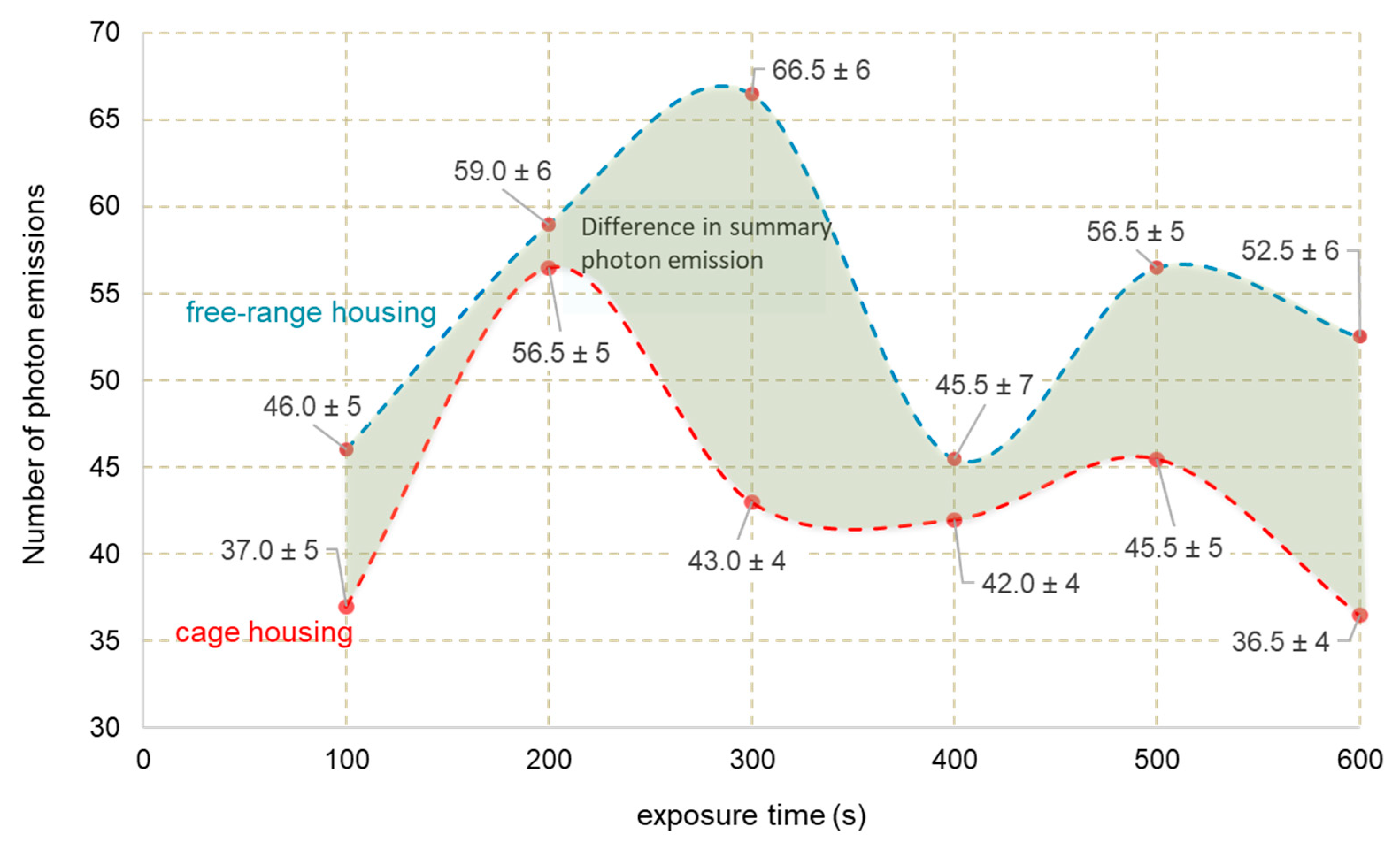
The applied measurement system generated the result of total photon emission in intervals of 0.25 s. Figure 1 shows the photon emission from fresh eggs in 100 s time intervals (dots marked with assigned values). The dashed line in the drawings describes the emission trend and is not the result of measurements. It was observed that eggs obtained from free-range eggs (blue line) in each of the analyzed compartments were characterized by a higher degree of emission compared to caged eggs (red line).
The maximum relative difference of 29.1% was recorded in the case of a time interval of 100 to 200 s, where the number of photons emitted from eggs from free-range housing was 85.5, and eggs from caged housing, 60.5 units. Therefore, this time interval is crucial from the point of view of egg identification on the basis of type of housing. In both of the analyzed cases, photon emission changed in the measured time interval, although it should be noted that the characteristics of changes in the number of emitted photons in the case of eggs from caged hens were more diverse, and photon emissions less stable. The changes relate to the periodic variability of photon emission intensity.
When analyzing the dynamics of photon emission in relation to the exposure time, it should be noted that in both analyzed cases it was on average about 17% for each of the six 100 s time intervals. Therefore, the dynamics of photon emission cannot be used as a measure differentiating the examined eggs, because of the criteria assumed in the research.
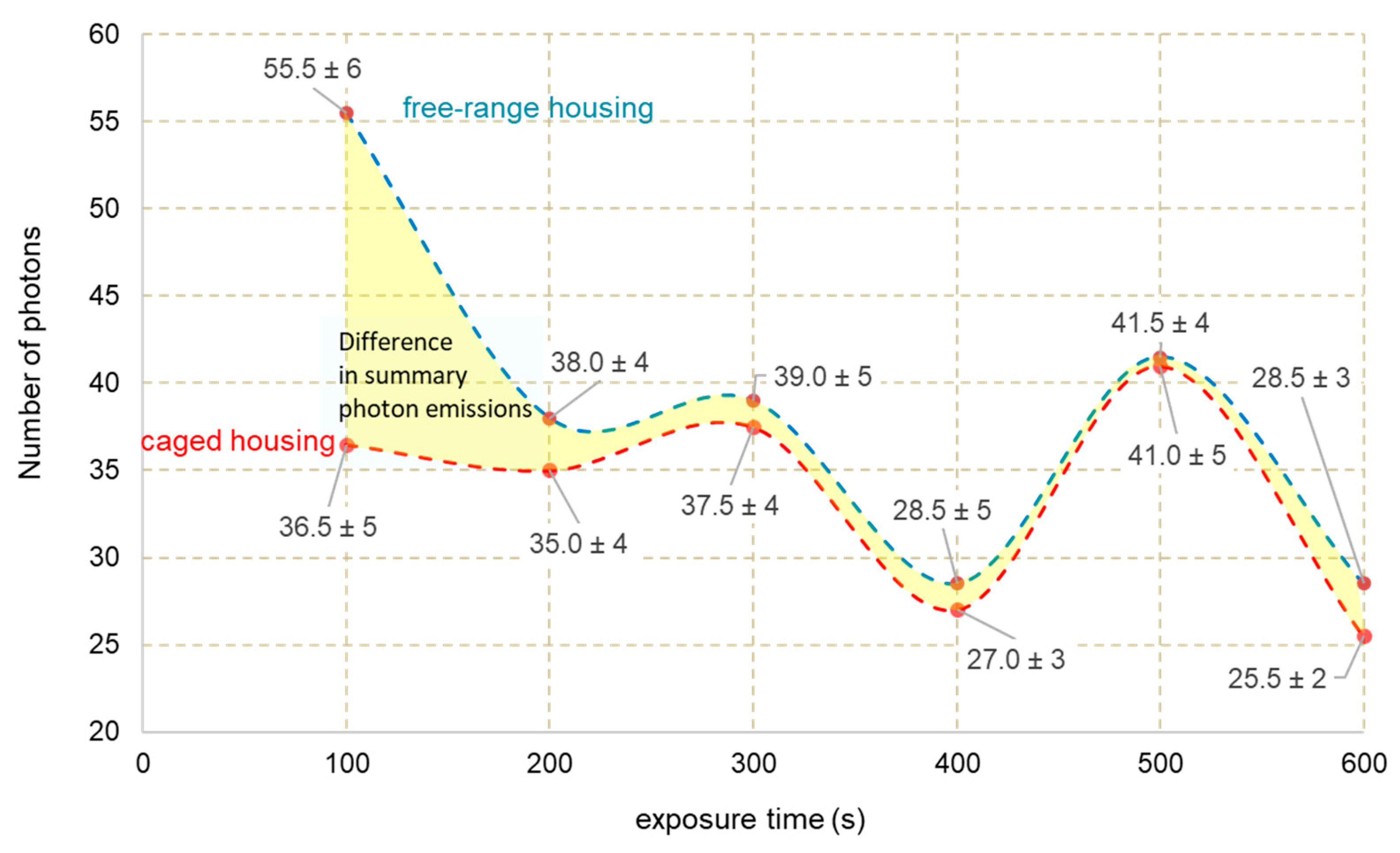
Due to the production processes, qualitative identification of the substrate should be carried out with division into product components. Figure 2 shows the structure of recorded photons during the experiment from egg yolks obtained from hens from caged hens (dashed line—red color), and egg yolks obtained from free-range hens (dashed line—blue).
The results presented were similar to those of whole eggs for emission time intervals of 100 s, which allowed for precise determination of photon emission variation over the time, the course of which may be an important factor in parameterization. It was found that the biggest difference in photon emission between the tested egg groups occurred in the third exposure interval, i.e., 201–300 s, amounting to 35.3%, which, in absolute terms, broke out on 23.5 photons, which can be used to identify the origin of egg yolks. It should be noted that throughout the experiment’s time interval, the emission of photons from egg yolks from free range hens was higher than the parameter recorded among egg yolks from caged eggs. Therefore, it should be stated that the obtained results were similar to the results obtained using traditional methods (sum of carotenoids, β + γ-tocopherol, δ-tocopherol), where the total number of photons emitted was 326, 65 photons higher than the emission recorded for egg yolks from cage-housing hens, which confirms the correctness of the choice of the assessment method and its possible practical use.
The unitary average photon emission for egg yolks from free-range hens was 0.54 photon/s, and for egg yolk from hens from caged hens, 0.43 photon/s. Analyzing the dynamics of photon emissions, it was observed that for egg yolks from hens of cage housing, the second time interval (101–200 pp.) is crucial, where photon emission accounted for over 21% of their total number.
In the case of egg yolks from free-range hens, the key time-related interval was 201–300 s, where the sum of identified photons was 20% of the total number of photons counted in the entire 600 s measurement cycle. The differentiation of the photon emission structure during the material exposure indicates a differentiation of the properties of the tested material, however, the parameterization of these changes requires additional research.
A slightly different photon emission curve as a time function in relation to the above-mentioned research objects was characterized by egg white of hens (Figure 3), because there was a large convergence in the shape of the photon emission curve between the egg white obtained from hen eggs from caged housing (red line) and the egg white from free-range housing (blue line). This statement does not refer to the sum of recorded photons in the first time interval (0 to 100 s). In the interval where the relative difference in the number of photon emissions by the egg white between the analyzed hen housing groups was 34.2%, the total number of photons allows the differentiation of egg white in terms of housing. However, it also should be noted that the emission of photons from egg white from free-range hens was higher throughout the whole experiment’s time interval, which coincides with the results obtained using an alternative identification method and confirms the suitability of the method based on counting individual photons.
In the analyzed case, the emission of photons was very clearly oscillatory (large changes in photon emission in time) regardless of whether the analyzed whites comes from free-range or cage eggs. It was found that the highest emission, constituting 24% of total photon emission for egg whites from free-range hens, occurred during the first time interval, while for egg whites from hens from caged housing, it occurred during the fifth time interval (401–500 s). This diversity could be the basis for further research, providing the possibility of specifying boundary parameters of the identification method of the albumen according to the origin of eggs. It should be emphasized that, despite a total difference in photon emission between the analyzed egg whites of 28 photons, photon emission characteristics as a time function of egg whites did not show significant variation.

4. Conclusions

The results obtained by the ultra-weak luminescence method indicate the variability of the tested eggs. However, the USL method makes it impossible to compare the freshness of the raw material, as the tested eggs have a comparable quality of consumption and freshness, despite significant differences in the obtained photon emissivity results. The emission of photons from eggs from free-range housing was eight times higher than eggs from caged housing. At the same time, it was observed that eggs from free range hens, characterized by higher photon emission, have more than three times higher carotenoid content compared to eggs from caged hens. The obtained results confirm the assumption that a higher number of photons emitted is probably associated with a higher content of biologically active substances.
Photon emission also varies depending on the specific hen breeding system. Differences in time in the identified maximum values of photon emission result from the composition of individual parts of the egg. It is related to the specificity of metabolic changes in the examined cells. Despite statistically significant differences in the number of photons counted in the specified time intervals, no intermediate values, average fluctuation values, or effective value of the number of registered photons emitted from the tested eggs were analyzed depending on the method of hen breeding. It will be part of the future analyses. The studies identified time points in which the difference in the total number of photons between the analyzed groups of eggs was the largest. This could be useful in practice. Ultra-low photon emission of biological material, such as food, in connection with other analytical methods, could be used in the future to evaluate the quality of food and to explain the processes occurring during processing and storage of food. Moreover, the use of the USL method to confirm its usefulness requires further research.

Author Contributions

Conceptualization, E.K., I.G.-C. and P.K.; methodology, E.K., I.G.-C., A.C., M.S., P.K. and T.D.; validation, E.K., I.G.-C., A.C., P.K. and T.D.; formal analysis, E.K., I.G.-C., A.C., M.S., P.K. and T.D. investigation, E.K., I.G.-C., A.C., M.S., P.K. and T.D.; data curation, E.K., I.G.-C., A.C., M.S., P.K. and T.D.; writing—original draft preparation, E.K., I.G.-C., A.C., P.K. and T.D.; writing—review and editing, E.K., I.G.-C., A.C. and P.K.; visualization, E.K., P.K. and T.D.; supervision, E.K., I.G.-C. and P.K.

Funding

This research received no external funding.

Conflicts of Interest

The authors declare no conflict of interest.

References

  1. Krawczyk, J. Effect of layer age and egg production level on changes in quality of eggs from hens of conservation breeds and commercial hybrids. Ann. Anim. Sci. 2009, 9, 185–193. [Google Scholar]
  2. Dudek, M.; Rabsztyn, A. Egg quality of dual-purpose hens intended for small-scale farming. Acta Sci. Pol. Zootech. 2011, 10, 3–12. [Google Scholar]
  3. Wang, J.; Yue, H.; Wu, S.; Zhang, H.; Qi, G. Nutritional modulation of health, egg quality and environmental pollution of the layers. Anim. Nutr. 2017, 3, 91–96. [Google Scholar] [CrossRef]
  4. Batkowska, J.; Brodacki, A. Selected quality traits of eggs and the productivity of newly created laying hen hybrids dedicated to an extensive rearing system. Arch. Anim. Breed. 2017, 60, 87–93. [Google Scholar] [CrossRef] [Green Version]
  5. Şekeroğlu, A.; Altuntaş, E. Effects of egg weight on egg quality Characteristics. J. Sci. Food Agric. 2009, 89, 379–383. [Google Scholar] [CrossRef]
  6. Shin, D.; Narciso-Gaytán, C.; Regenstein, J.M.; Sánchez-Plata, M.X. Effect of various refrigeration temperatures on quality of shell eggs. J. Sci. Food Agric. 2012, 92, 1341–1345. [Google Scholar] [CrossRef] [PubMed]
  7. Batkowska, J.; Brodacki, A.; Knaga, S. Quality of Laying Hen Eggs During Storage Depending on Egg Weight and Type of Cage System (Conventional vs. Furnished Cages). Ann. Anim. Sci. 2014, 3, 707–719. [Google Scholar] [CrossRef]
  8. Wang, X.C.; Wu, S.; Zhang, H.; Yue, H.; Qi, G.; Li, J. Effect of dietary protein sources and storage temperatures on egg internal quality of stored shell eggs. Anim. Nutr. 2015, 1, 299–304. [Google Scholar] [CrossRef] [PubMed]
  9. Oziemblowski, M.; Dróżdż, M.; Kiełbasa, P.; Dróżdż, T.; Gąsiorski, A.; Nawara, P.; Tabor, S. Ultra weak luminescence (USL) as a potential method for evaluating the quality of traditional food. Przeglad Elektrotechniczny 2017, 94, 104–107. [Google Scholar]
  10. Borc, R.; Jaśkowska, A.; Dudziak, A. Ultra-Low Photon Emission from Living Systems; Lublin University of Technology: Lublin, Poland, 2015. [Google Scholar]
  11. Laager, F.M.; Becker, N.M.; Park, S.H.; Soh, K.S. Effects of Lac operon activation, deletion of the Yhha gene, and the removal of oxygen on the ultraweak photon emission of Escherichia coli. Electromagn. Biol. Med. 2009, 28, 240–249. [Google Scholar] [CrossRef] [PubMed]
  12. Nerudová, M.; Červinková, K.; Hašek, J.; Cifra, M. Optical spectral analysis of ultra-weak photon emission from tissue culture and yeast cells. Proc. SPIE 2015, 9450. [Google Scholar] [CrossRef]
  13. Van Wijk, E.P.A.; Van der Greef, J.; Van Wijk, R. Photon counts statistics in leukocyte cell dynamics. J. Phys. Conf. Ser. 2011, 329. [Google Scholar] [CrossRef]
  14. Salari, V.; Valian, H.; Bassereh, H.; Bókkon, I.; Barkhordari, A. Ultraweak photon emission in the brain. J. Integr. Neurosci. 2015, 14, 419–429. [Google Scholar] [CrossRef] [PubMed]
  15. Hideg, E.; Kobayashi, M.; Inaba, H. Delayed Fluorescence and Ultraweak Light Emission from Isolated Chloroplasts (Comparison of Emission Spectra and Concentration Dependence). Plant Cell Physiol. 1992, 33, 689–693. [Google Scholar]
  16. Karbowski, L.M.; Murugan, N.J.; Dotta, B.T.; Persinger, M.A. Only 1% Melanoma Proportion in Non-Malignant Cells Exacerbates Photon Emissions: Implications for Tumor Growth and Metastases. Int. J. Cancer Res. Mol. Mech. 2015, 1. [Google Scholar] [CrossRef]
  17. Kim, J.D.; Lim, J.; Sung, B.; Soh, K.S. Biophoton Emission from Rat Liver. J. Korean Phys. Soc. 2003, 42, 427–430. [Google Scholar]
  18. Zhang, J.G.; Zhong, L.F.; Zhang, M.; Xia, Y.X. Protection effects of procaine on oxidative stress and toxicities of renal cortical slices from rats caused by cisplatin in vitro. Arch. Toxicol. 1992, 66, 354–358. [Google Scholar] [CrossRef]
  19. Kobayashi, M.; Takeda, M.; Sato, T.; Yamazaki, Y.; Kaneko, K.; Ito, K.-I.; Kato, H.; Inaba, H. In vivo imaging of spontaneous ultraweak photon emission from a rat’s brain correlated with cerebral energy metabolism and oxidative stress. Neurosci. Res. 1999, 34, 103–113. [Google Scholar] [CrossRef]
  20. Hossu, M.; Ma, L.; Zou, X.; Chen, W. Enhancement of biophoton emission of prostate cancer cells by Ag nanoparticles. Cancer Nanotechnol. 2013, 4, 21–26. [Google Scholar] [CrossRef] [Green Version]
  21. Gocławski, J.; Sekulska-Nalewajko, J.; Korzeniewska, E.; Piekarska, A. The use of optical coherence tomography for the evaluation of textural changes of grapes exposed to pulsed electric field. Comput. Electron. Agric. 2017, 142, 29–40. [Google Scholar] [CrossRef]
  22. Gliszczynska-Świgło, A.; Sikorska, E. Simple reversed-phase liquid chromatography method for determination of tocopherols in edible plant oils. J. Chromatogr. 2004, 1048, 195–198. [Google Scholar] [CrossRef]
  23. Ruth, S.; Alewijn, M.; Rogers, K.; Newton-Smith, E.; Tena, N.; Bollen, M.; Koot, A. Authentication of organic and conventional eggs by carotenoid profiling. Food Chem. 2011, 126, 1299–1305. [Google Scholar] [CrossRef]
  24. Pathare, P.B.; Opara, U.L.; Al-Said, F.A.J. Colour Measurement and Analysis in Fresh and Processed Foods: A Review. Food Bioprocess. Technol. 2013, 6, 36–60. [Google Scholar] [CrossRef]
  25. Aboonajmi, M.; Amir, S.; Najafabadi, T.A.; Kondo, N. Quality Assessment of Poultry Egg Based on Visible–Near Infrared Spectroscopy and Radial Basis Function Networks. Int. J. Food Prop. 2016, 19, 1163–1172. [Google Scholar] [CrossRef]
  26. Karadas, F.; Grammenidis, E.; Surai, P.F.; Acamovic, T.; Sparks, N.H.C. Effects of carotenoids from lucerne, marigold and tomato on egg yolk pigmentation and carotenoid composition. Br. Poult. Sci. 2006, 47, 561–566. [Google Scholar] [CrossRef]
  27. Lokaewmanee, K.; Yamauchi, K.; Okuda, N. Effects of dietary red pepper on egg yolk colour and histological intestinal morphology in laying hens. J. Anim. Physiol. Anim. Nutr. 2009, 97, 986–995. [Google Scholar] [CrossRef]
  28. Islam, K.M.S.; Khalil, M.; Männer, K.; Raila, J.; Rawel, H.; Zentek, J.; Schweigert, F.J. Lutein specific relationships among some spectrophotometric and colorimetric parameters of chicken egg yolk. J. Poult. Sci. 2017, 54, 271–277. [Google Scholar] [CrossRef]
  29. Lewko, L.; Gornowicz, E. Effect of Housing System on Egg Quality in Laying Hensvol. Ann. Anim. Sci. 2011, 11, 607–616. [Google Scholar] [CrossRef]
  30. Surai, P.F.; Speake, B.K.; Sparks, N.H.C. Carotenoids in avian nutrition and embryonic development. 1. Absorption, availability and levels in plasma and egg yolk. J. Poult. Sci. 2001, 38, 1–27. [Google Scholar] [CrossRef]
  31. Surai, P.F.; Fisinin, I.V.; Karadas, F. Antioxidant systems in chick embryo development. Part 1. Vitamin E, carotenoids and selenium. Anim. Nutr. 2016, 1, 1–11. [Google Scholar] [CrossRef]
  32. Hammershøj, M.; Steenfeldt, S. Organic egg production. II: The quality of organic eggs is influenced by hen genotype, diet and forage material analyzed by physical parameters, functional properties and sensory evaluation. Anim. Feed Sci. Technol. 2015, 208, 182–197. [Google Scholar] [CrossRef]
  33. Schlatterer, J.; Breithaupt, D.E. Xanthophylls in Commercial Egg Yolks Quantification and Identification by HPLC and LC-(APCI)MS Using a C30 Phase. J. Agric. Food Chem. 2006, 54, 2267–2273. [Google Scholar] [CrossRef] [PubMed]
  34. Karadas, F.; Møller, A.P.; Karageçili, M.R. A comparison of fat-soluble antioxidants in wild and farm-reared chukar partridges (Alectoris chukar). Comp. Biochem. Physiol. 2017, 208, 89–94. [Google Scholar] [CrossRef] [PubMed]
  35. Kiełbasa, P.; Dróżdż, T.; Nawara, P.; Dróżdż, M. The use of bio-photons emission for the quality parameterization of food products. Przeglad Elektrotechniczny 2017, 93, 153–156. [Google Scholar]
  36. Trzyniec, K.; Kiełbasa, P.; Oziemblowski, M.; Dróżdż, M.; Nawara, P.; Posyłek, Z.; Leja, R. Using photons emission to evaluate the quality of apples. Przeglad Elektrotechniczny 2017, 93, 183–186. [Google Scholar]
  37. Becker, W. Fluorescence lifetime imaging by multi-dimensional time correlated single photon counting. Med. Photonics 2015, 27, 41–61. [Google Scholar] [CrossRef]
  38. Narayana, P.S.; Varalakshmi, D.; Pullaiah, T. Research Methodology in Plant Science; Scientific Publishers: Jodhpur, India, 2016. [Google Scholar]
  39. De Haas, J.T.M.; Dorenbos, P. Methods for Accurate Measurement of the Response of Photomultiplier Tubes and Intensity of Light Pulses. IEEE Trans. Nucl. Sci. 2011, 58, 1290–1296. [Google Scholar] [CrossRef]
  40. Usa, M.; Kobayashi, M.; Scott, R.Q.; Hiratsuka, R.; Maeda, T.; Inaba, H. Ultraweak biophoton emission and bioelectrical activity in plant tissues. In Biological Luminescence; World Scientific Publishing: Singapore, 1990; pp. 117–130. [Google Scholar]
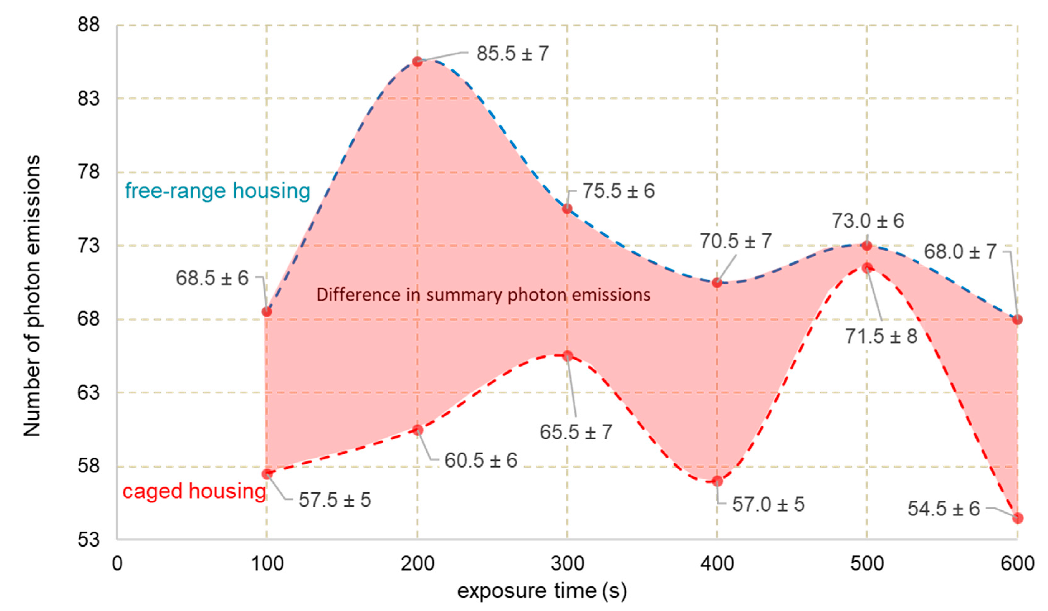
Figure 1. Characteristics of photon emissions as a function of the exposure time of whole eggs obtained from free-range housing (blue) and caged housing (red).
Applsci 09 02430 g001
Figure 2. Characteristics of photon emission as a function of exposure time of egg yolks obtained from free-range housing (blue) and cage housing (red).
Applsci 09 02430 g002
Figure 3. Characteristics of photon emissions as a function of exposure time in egg white obtained from free-range housing (blue) and caged housing (red).
Applsci 09 02430 g003
Table 1. Selected quality features of eggs, n = 60.
FeaturesEggs CEggs F
egg yolk
Color of yolk8.00 ± 0.309.00 ± 0.20
pH6.12 ± 0.206.07 ± 0.08
egg albumen
Haugh units84.60 ± 0.5978.60 ± 0.36
pH8.11 ± 0.228.19 ± 0.38
Table 2. The content of carotenoids [μg/g] and tocopherols [μg/g] in egg yolk from caged hens (C eggs) and free range hens (eggs F), n = 60.
SampleCarotenoidsTotal
Unknown PigmentLuteinZeaxanthinβ-Carotene
Eggs F0.7 ± 0.134.2 a ± 1.34.3 a ± 0.1nd39.1 a ± 1.4
Eggs Cnd5.7 b ± 0.24.4 a ± 0.11.1 ± 0.011.2 b ± 0.2
Tocopherol
αβ + γΔ
Eggs F77.8 a ± 1.55.2 a ± 0.10.8 a ± 0.083.7 a ± 1.7
Eggs C89.9 b ± 2.41.7 b ± 0.00.3 b ± 0.091.3 b ± 2.5
nd: not detected; averages marked with different letters (a, b), within the column, are significantly different (p < 0.05, LSD test).
Table 3. Color parameters of egg yolks from caged hens (eggs C) and free range hens (eggs F), n = 60.
SampleYolk Color Parameters
L* (C)a* (C)b* (C)
Eggs F51.61 a ± 2.453.36 a ± 0.1545.69 a ± 1.79
Eggs C49.60 a ± 2.272.93 b ± 0.7440.05 b ± 1.33
L*—psychometric brightness of color, a*—amount of red, b*—amount of yellow, averages marked with different letters (a, b), within the column, are significantly different (p < 0.05, LSD test).