A Novel Method for Assessing Regional Tendon Stiffness and Its Significance

Abstract
1. Introduction
2. Materials and Methods
2.1. Ethics Statement
2.2. Participants
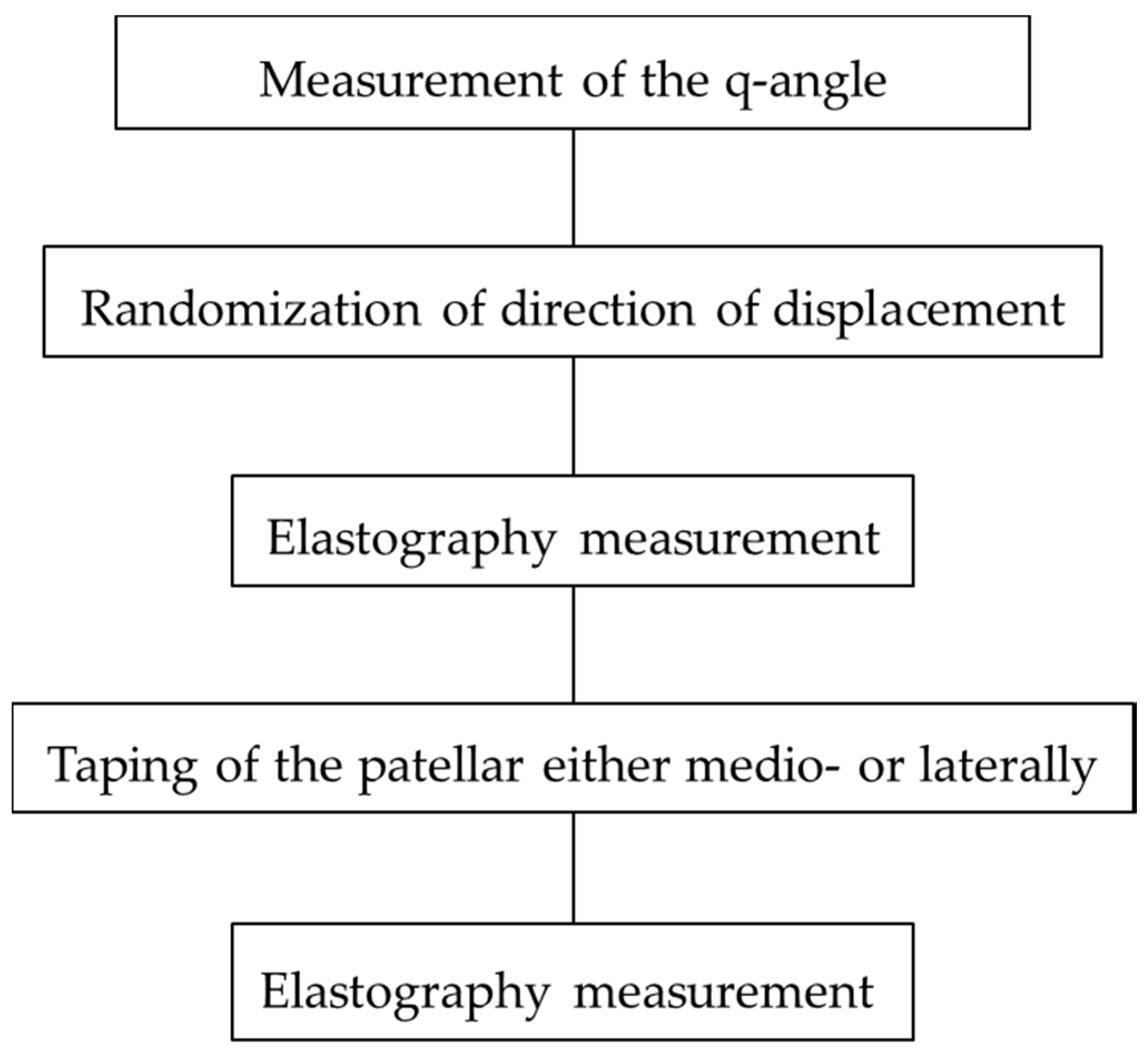
2.3. Procedure
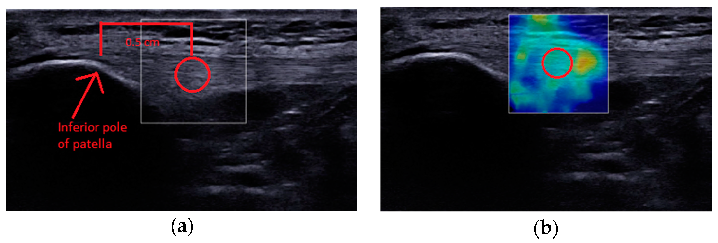
2.3.1. Elastography Measurement
2.3.2. Patellar Positions
2.3.3. Data Extraction
2.3.4. Reliability Test
2.3.5. Statistical Analysis
3. Results
3.1. Demographic Data
3.2. Reliability Test
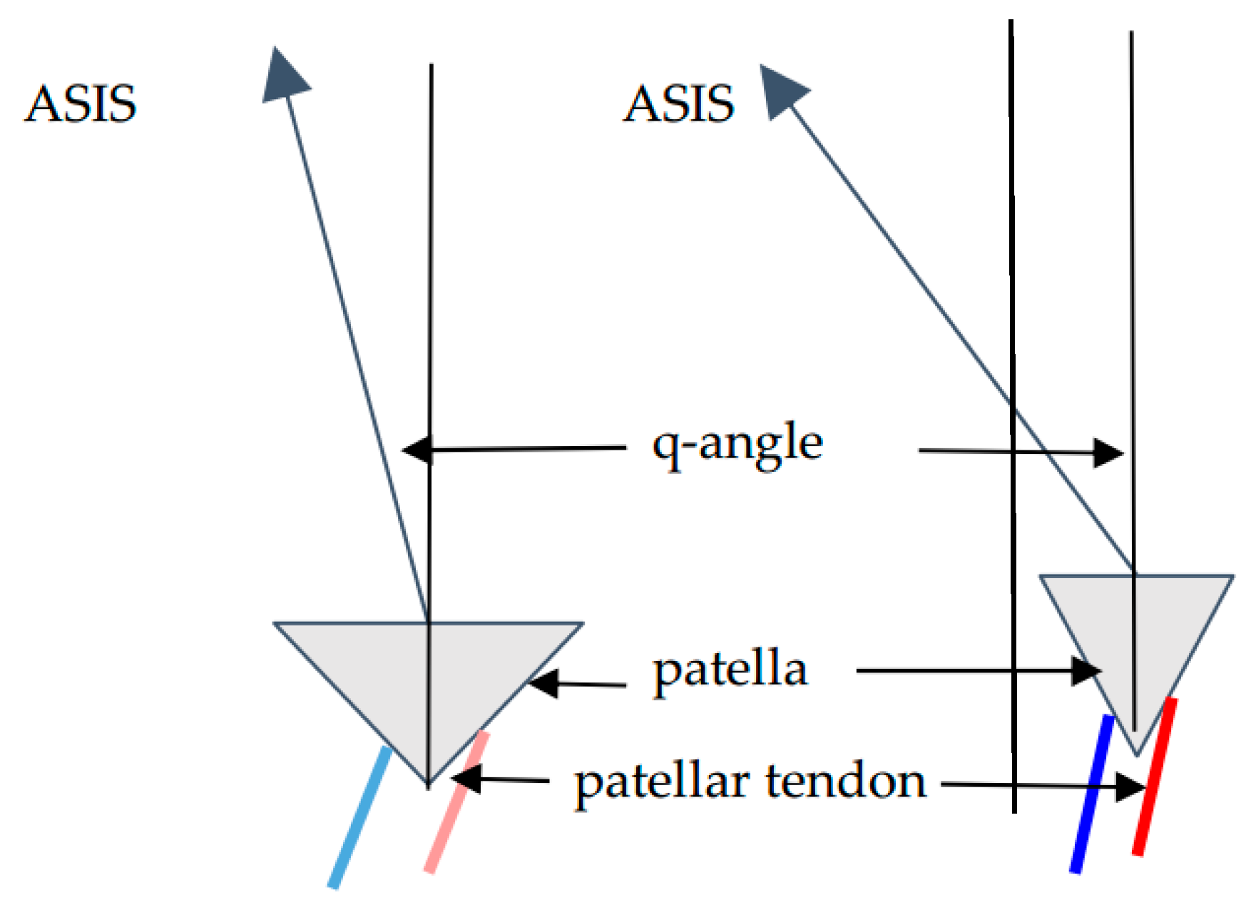
3.3. Effects of q-Angle on Percentage Changes in Tendon Shear Modulus
3.4. Effect of Laterally Displaced Patellar Position on the Shear Elastic Modulus of Patellar Tendon
3.5. Effect of Medially Displaced Patellar Position on the Shear Elastic Modulus of Patellar Tendon
4. Discussion
5. Conclusions
Author Contributions
Funding
Acknowledgments
Conflicts of Interest
References
- Kot, B.C.W.; Zhang, Z.J.; Lee, A.W.C.; Leung, V.Y.F.; Fu, S.N. Elastic modulus of muscle and tendon with shear wave ultrasound elastography: Variations with different technical settings. PLoS ONE 2012, 7, e44348. [Google Scholar] [CrossRef] [PubMed]
- Nordez, A.; Hug, F. Muscle shear elastic modulus measured using supersonic shear imaging is highly related to muscle activity level. J Appl. Physiol. 2010, 108, 1389–1394. [Google Scholar] [CrossRef] [PubMed]
- Bercoff, J.; Tanter, M.; Fink, M. Supersonic shear imaging: A new technique for soft tissues elasticity mapping. IEEE Trans. Ultrason. Ferroelectr. Freq. Control 2004, 51, 396–409. [Google Scholar] [CrossRef] [PubMed]
- Zhang, Z.J.; Fu, S.N. Shear elastic modulus on patellar tendon captured from supersonic shear imaging: Correlation with tangent traction modulus computed from material testing system and test–retest reliability. PLoS ONE 2013, 8, e68216. [Google Scholar] [CrossRef] [PubMed]
- Cook, J.L.; Khan, K.M.; Kiss, Z.S.; Griffiths, L. Patellar tendinopathy in junior basketball players: A controlled clinical and ultrasonographic study of 268 patellar tendons in players aged 14-18 years. Scand. J. Med. Sci. Sports 2000, 10, 216–220. [Google Scholar] [CrossRef] [PubMed]
- Fredberg, U.; Stengaard-Pedersen, K. Chronic tendinopathy tissue pathology, pain mechanisms, and etiology with a special focus on inflammation. Scand. J. Med. Sci. Sports 2008, 18, 3–15. [Google Scholar] [CrossRef] [PubMed]
- Grob, K.; Manestar, M.; Filgueira, L.; Ackland, T.; Gilbey, H.; Kuster, M.S. New insight in the architecture of the quadriceps tendon. J. Exp. Orthop. 2006, 3, 32. [Google Scholar] [CrossRef] [PubMed]
- Powers, C.M.; Chen, Y.J.; Farrokhi, S.; Lee, T.Q. Role of peripatellar retinaculum in transmission of forces within the extensor mechanism. J. Bone Jt. Surg. Am. 2006, 88, 2042–2048. [Google Scholar]
- Crema, M.D.; Cortinas, L.G.; Lima, G.B.P.; Abdalla, R.J.; Ingham, S.J.M.; Skaf, A.Y. Magnetic Resonance imaging-based morphological and alignment assessment of the patellofemoral joint and its relationship to proximal patellar tendinopathy. Skeletal. Radiol. 2018, 47, 231–349. [Google Scholar] [CrossRef] [PubMed]
- Zhang, Z.J.; Ng, G.Y.F.; Lee, W.C.; Fu, S.N. Increase in passive muscle tension of the quadriceps muscle heads in jumping athletes with patellar tendinopathy. Scand. J. Med. Sci. Sports 2017, 27, 1099–1104. [Google Scholar] [CrossRef] [PubMed]
- Becker, I.; Baxter, G.D.; Woodley, S.J. The vastus lateralis muscle: An anatomical investigation. Clin. Anat. 2010, 23, 575–585. [Google Scholar] [CrossRef] [PubMed]
- Culvenor, A.G.; Cook, J.L.; Warden, S.J.; Crossley, K.M. Infrapatellar fat pad size, but not patellar alignment, is associated with patellar tendinopathy. Scand. J. Med. Sci. Sports 2011, 21, e405–e411. [Google Scholar] [CrossRef] [PubMed]
- Coombes, B.K.; Tucker, K.; Vicenzino, B.; Vuvan, V.; Mellor, R.; Heales, L.; Nordez, A.; Hug, F. Achilles and patellar tendinopathy display opposite changes in elastic properties: A shear wave elastography study. Scand. J. Med. Sci. Sports 2018, 28, 1201–1208. [Google Scholar] [CrossRef] [PubMed]
- Varadarajan, K.M.; Gill, T.J.; Freiberg, A.A.; Rubash, H.E.; Li, G. Patellar tendon orientation and patellar tracking in male and female knees. J. Orthop. Res. 2010, 28, 322–328. [Google Scholar] [CrossRef] [PubMed]
- Livingston, L.A. The quadriceps angle: A review of the literature. J. Orthop. Sports Phys. Ther. 1998, 2, 105–109. [Google Scholar] [CrossRef] [PubMed]
- Hungerford, D.S.; Barry, M. Biomechanics of the patellofemoral joint. Clin. Orthop. Relat. Res. 1979, 144, 9–15. [Google Scholar] [CrossRef]
- De Zordo, T.; Fink, C.; Feuchtner, G.M.; Smekal, V.; Reindl, M.; Klauser, A.S. Real-time sonoelastography findings in healthy Achilles tendons. AJR Am. J. Roentgenol. 2009, 193, W134–W138. [Google Scholar] [CrossRef] [PubMed]
- Mitrogiannis, L.; Barbouti, A.; Kanavaros, P.; Paraskevas, G.; Kitsouli, A.; Mitrogiannis, G.; Kitsoulis, P. Cadaveric-biomechanical study on medial retinaculum: Its stabilizing role for the patella against lateral dislocation. Folia Morphol. 2018. [Google Scholar] [CrossRef] [PubMed]
- Toumi, H.; Higashiyama, I.; Suzuki, D.; Kumai, T.; Bydder, G.; McGonagle, D.; Emery, P.; Fairclough, J.; Benjamin, M. Regional variations in human patellar trabecular architecture and the structure of the proximal patellar tendon enthesis. J. Anat. 2006, 208, 47–57. [Google Scholar] [CrossRef] [PubMed]
- Wren, T.A.; Lindsey, D.P.; Beaupre, G.S.; Carter, D.R. Effects of creep and cyclic loading on the mechanical properties and failure of human Achilles tendons. Ann. Biomed. Eng. 2003, 31, 710–721. [Google Scholar] [CrossRef] [PubMed]
- Thambyah, A.; Thiagarajan, P.; Goh, J.C. Biomechanical study on the effect of twisted human patellar tendon. Clin. Biomech. 2000, 15, 756–760. [Google Scholar] [CrossRef]
- Hansen, P.; Bojsen-Moller, J.; Aagaard, P.; Kjaer, M.; Magnusson, S.P. Mechanical properties of the human patellar tendon, in vivo. Clin. Biomech. 2006, 21, 54–58. [Google Scholar] [CrossRef] [PubMed]
- Ophir, J.; Cespedes, I.; Ponnekanti, H.; Yazdi, Y.; Li, X. Elastography: A quantitative method for imaging the elasticity of biological tissues. Ultrason. Imaging 1991, 13, 111–134. [Google Scholar] [CrossRef] [PubMed]


| Demographic | Males (n = 21) | Females (n = 19) | p |
|---|---|---|---|
| Age (year) | 21.0 ± 1.9 | 21.8 ± 2.6 | 0.12 |
| Height (cm) | 173.4 ± 6.3 | 161.0 ± 5.1 | 0.001 * |
| Weight (kg) | 63.4 ± 7.5 | 52.3 ± 8.1 | 0.001 * |
| Body mass index (kg/m2) | 21.1 ± 2.1 | 20.1 ± 2.3 | 0.056 |
| q-angle (degrees) | 16.9 ± 3.4 | 20.4 ± 3.2 | 0.001 * |
| Patella Position | Males | Females | ||
|---|---|---|---|---|
| Lateral Half | Medial Half | Lateral Half | Medial Half | |
| Laterally displaced | 0.29 | 0.17 | 0.47 * | −0.48 * |
| Medially displaced | 0.01 | −0.23 | 0.24 | −0.15 |
| Patella Position | Shear modulus (KPa) Males (n = 20) | Shear modulus (KPa) Females (n = 20) | ||
|---|---|---|---|---|
| Lateral Half | Medial Half | Lateral Half | Medial Half | |
| Neutral | 242.1 ± 92.6 | 293.4 ± 85.4 | 281.5 ± 131.0 | 303.3 ± 121.8 |
| Laterally displaced | 307.9 ± 150.9 | 356.6 ± 147.4 * | 244.3 ± 110.3 * | 341.9 ± 155.1 * |
| Neutral | 275.8 ± 104.7 | 341.2 ± 104.9 | 271.0 ± 96.8 | 290.7 ± 118.3 |
| Medially displaced | 280.8 ± 130.5 | 367.8 ± 83.4 | 263.3 ± 105.0 | 341.4 ± 140.7 * |
© 2018 by the authors. Licensee MDPI, Basel, Switzerland. This article is an open access article distributed under the terms and conditions of the Creative Commons Attribution (CC BY) license (http://creativecommons.org/licenses/by/4.0/).
Share and Cite
Fu, S.N.; Wang, H.-K.; Huang, C. A Novel Method for Assessing Regional Tendon Stiffness and Its Significance. Appl. Sci. 2018, 8, 1156. https://doi.org/10.3390/app8071156
Fu SN, Wang H-K, Huang C. A Novel Method for Assessing Regional Tendon Stiffness and Its Significance. Applied Sciences. 2018; 8(7):1156. https://doi.org/10.3390/app8071156
Chicago/Turabian StyleFu, Siu Ngor, Hsing-Kuo Wang, and Chen Huang. 2018. "A Novel Method for Assessing Regional Tendon Stiffness and Its Significance" Applied Sciences 8, no. 7: 1156. https://doi.org/10.3390/app8071156
APA StyleFu, S. N., Wang, H.-K., & Huang, C. (2018). A Novel Method for Assessing Regional Tendon Stiffness and Its Significance. Applied Sciences, 8(7), 1156. https://doi.org/10.3390/app8071156