Article

Bio-Corrosion Behavior of Ceramic Coatings Containing Hydroxyapatite on Mg-Zn-Ca Magnesium Alloy

1
Jiangsu Provincial Key Laboratory for Interventional Medical Devices, Huaiyin Institute of Technology, Huaian 223003, China
2
College of Materials Science and Engineering, Nanjing Tech University, Nanjing 211800, China
*
Author to whom correspondence should be addressed.
These authors contributed equally to this work and should be considered co-first authors.
Appl. Sci. 2018, 8(4), 569; https://doi.org/10.3390/app8040569
Submission received: 21 February 2018 / Revised: 31 March 2018 / Accepted: 4 April 2018 / Published: 6 April 2018
(This article belongs to the Section Materials Science and Engineering)

Abstract

:
Ceramic coatings containing hydroxyapatite (HA) were fabricated on a biodegradable Mg66Zn29Ca5 magnesium alloy through micro-arc oxidation by adding HA particles into the electrolytes. The phase composition and surface morphology of the coatings were characterized by X-ray diffraction and scanning electron microscopy analyses, respectively. Electrochemical experiments and immersion tests were performed in Hank’s solution at 37 °C to measure the corrosion resistance of the coatings. Blood compatibility was evaluated by in vitro blood platelet adhesion tests and static water contact angle measurement. The results show that the typical ceramic coatings with a porous structure were prepared on the magnesium alloy surface with the main phases of MgO and MgSiO3 and a small amount of Mg3(PO4)2 and HA. The optimal surface morphology appeared at HA concentration of 0.4 g/L. The electrochemical experiments and immersion tests reveal a significant improvement in the corrosion resistance of the ceramic coatings containing HA compared with the coatings without HA or bare Mg66Zn29Ca5 magnesium alloy. The static water contact angle of the HA-containing ceramic coatings is 18.7°, which is lower than that of the coatings without HA (40.1°). The in vitro blood platelet adhesion tests indicate that the HA-containing ceramic coatings possess improved blood compatibility compared with the coatings without HA. Utilizing HA-containing ceramic coatings may be an effective way to improve the surface biocompatibility and corrosion resistance of magnesium alloys.

1. Introduction

Traditional permanent implants consisting of stainless steel, titanium and its alloys are important in the field of metallic implant materials for their maintaining mechanical integrity and good biocompatibility during the bone healing period [1]. However, metallic materials may cause the release of toxic corrosion products [2,3] and allergens [4,5]; it need a second surgery for implant removal after the healing of damaged tissues. As a result, biodegradable implants for biomedical application have attracted great attention in recent years [6,7,8]. Common biodegradable implants are made of polymers with unsatisfactory mechanical strength [7,9,10]. Magnesium and its alloys are now becoming a priority due to their low density, high specific strength and good biocompatibility, especially their biodegradability and non-toxicity to the human body [11,12,13]. The elastic modulus of magnesium is approximately 45 GPa [14], which is similar to that of human bone (3–20 GPa); this avoids stress shielding effects when implanted into the human body [15,16]. However, magnesium and its alloys are considerably active metals in chloride containing physiological environments including human body fluid or blood plasma [13]. Therefore, the severe corrosion and accelerated degradation rate of magnesium and its alloys have limited their biomedical application.
In general, the corrosion resistance of magnesium and its alloys can be improved by (i) tailoring the grain size [17,18] or the microstructure and texture [19] of the base material by alloying and (ii) forming coatings via surface treatments on the substrates to obtain protective ceramics, polymers or composite layers [20]. Considering the requirements of biocompatibility, Mg-Zn-Ca alloys are promising for implant applications due to the fact that elements of Mg, Zn, and Ca can not only improve the mechanical properties but also participate in metabolic processes. As the low solubility of many elements in magnesium, the alloying of magnesium has a limitation for application. Therefore, the surface treatments are of high significance. Chemical conversion coatings are common protective coatings on magnesium and its alloys, such as calcium phosphate-containing layer [21,22] and fluoride phosphate-containing layer [23,24], which are biodegradable coatings for biomedical applications. Ion implantation has also been conducted as a technique to improve the corrosion resistance of magnesium alloy. However, the improvements were not significant measured in NaCl solution or Hank’s solution, e.g., Al, Zr, or Ti ions implanted in AZ91 [25] and Ta ion in AZ31 alloy [26].
Micro-arc oxidation (MAO), also known as plasma electrolytic oxidation or anodic spark deposition, is a novel, promising surface treatment, which has been developed and applied to magnesium and its alloys [27,28,29,30,31]. This technique has become one of the most prospective surface treatments due to the good adhesion and corrosion resistance of ceramic coatings. However, the application of these coatings on biodegradable implant materials must be further confirmed.
Hydroxyapatite (HA, Ca10(PO4)6(OH)2) is a naturally occurring mineral of calcium apatite and is used as a biomedical material. HA exhibits excellent biocompatibility and bioactivity due to its structural and chemical similarities to bone minerals [32,33,34]. HA coatings are degradable in physiological environment and show good early interaction between the implant and the tissue [35]. Much research has been done on the fabrication of HA coatings on magnesium and its alloys; preparation methods include electrodeposition [36,37,38], aerosol deposition [39], chemical solution deposition [40], sol-gel method [41], and other processes [42,43]. It should be pointed that the poor bonding strength, corrosion resistance, and inhomogeneity of simplex HA coatings still restrict their application.
To the best of our knowledge, only few studies have reported the preparation of coatings containing HA via MAO [44]. Therefore, in this paper, a research on fabrication a HA-containing coating has been made by method of MAO technique. The effects of HA particles added into the electrolyte solution on the surface morphology, thickness, and hardness of the coatings were determined. Properties, including corrosion resistance and blood compatibility, of the coatings with and without HA were also investigated.

2. Experimental

2.1. Preparation of the Coatings

High-purity (>99.9 wt %) of Mg, Zn, and Ca were melted under an argon atmosphere in an induction furnace to achieve magnesium alloy with a composition of Mg66Zn29Ca5 (in atomic percentage). The test specimen was a disc with a diameter of 14 mm and a thickness of 3 mm. Prior to MAO, the specimens were ground with SiC papers (from 150 to 2000 grits), and polished with Al2O3 aqueous suspension to an average surface roughness of Ra = 0.8 µm. The specimens were then ultrasonically rinsed in acetone and ethanol, and finally dried using nitrogen.
The electrolyte solution for MAO, which mainly consisted of Na2SiO3 (10 g/L), Na3PO4 (3 g/L), NaOH (2 g/L), and Glycerin (10 mL/L) was prepared with deionized water. And various concentrations of HA powder with mean size of 0.5 µm were then added into the electrolyte. The concentrations of HA added into the electrolyte were selected as 0, 0.2, 0.4, and 0.6 g/L. Note that the electrolyte should be ultrasonically oscillated for 10 min before MAO to ensure that the HA particles are well dispersed throughout the electrolyte.
As a device used for MAO, the equipment (WHD-30) was composed of a pulsed power supply, electrolytic bath, and stirring and cooling system that can automatically control electrolyte temperature to <40 °C. In our experiment, constant voltage mode was adopted and the electric parameters were fixed as follows: 400 V anodic voltage, 80 V cathodic voltage, 1500 Hz frequency, and 50% duty cycle. The MAO coatings were deposited on the magnesium alloy specimens for 5 min.

2.2. Characterization of the Coatings

The morphologies of the coatings were characterized by scanning electron microscopy (SEM, Hitachi S-3000N) coupled with an energy dispersion spectroscopy (EDS). The phase composition was characterized by X-ray diffraction (XRD, D8 Advance) with Cu Κα in scanning angles of 20°–70°.
The thickness and microhardness of the coatings were examined using an eddy-current coating thickness gauge (Minitest 600B) and micro Vickers equipment (HXD-1000TMC), respectively. Measurements were carried out for 10 times, and the average values were determined.
Electrochemical measurements were performed in Hank’s solution at 37 °C by using an electrochemical workstation (CHI 660D). Hank’s solution was synthesized in the laboratory and composed of 8 g/L NaCl, 0.40 g/L KCl, 0.10 g/L MgCl2·6H2O, 0.10 g/L MgSO4·7H2O, 0.14 g/L CaCl2, 0.06 g/L Na2HPO4·2H2O, 0.06 g/L KH2PO4 and 1 g/L glucose. The experimental set-up comprised a conventional three-electrode cell containing a working electrode, a platinum sheet as the counter electrode, and an Ag-AgCl electrode as reference. The area of the working electrode that was exposed in the solution was 1.54 cm2. The potentiodynamic polarization curves of the samples were measured at a scan rate of 1 mV/s. Prior to each test, the specimen was immersed in the solution for 10 min to stabilize the open circuit potential (OCP).
The immersion corrosion tests were performed in Hank’s solution for 20 d at a constant temperature of 37 °C. Every day, the samples were removed from the solution, rinsed ultrasonically with deionized water, and then dried. An electronic balance (FA 1004) was used to weigh the mass before and after the immersion, and mass loss curves were obtained by calculating the relative mass loss rate.
The static water contact angles of the coatings were measured by a video optical contact angle measuring instrument (DSA 25) to study the wetting properties.
In vitro blood platelet adhesion test was investigated to identify the blood compatibility of the coatings. Platelet-rich plasma (PRP) was prepared by centrifuging the whole blood for 15 min at a rate of 1000 rpm. The samples with and without HA coatings were ultrasonically rinsed with ethanol and deionized water for 10 min, respectively. The samples were then dried and placed into culture plates. The PRP was dropped on the surface of the samples and incubated in an electro-heating standing-temperature cultivator (Thermo 3110) at 37 °C for 2 h. After incubation, the samples were rinsed thrice with normal saline to remove non-adherent platelets. The platelets that adhered on the samples were fixed in 2.5% glutaraldehyde solutions for 1.5 h at room temperature, and the samples were dehydrated in a gradient ethanol/distilled water mixture (50%, 60%, 70%, 80%, 90%, and 100%) for 10 min each and then dried in air for more than 48 h. The morphology and distribution of the platelets were observed by SEM. The platelet shape change was evaluated from high-magnification micrographs. At least 50 platelets were analyzed for each sample.

3. Results and Discussion

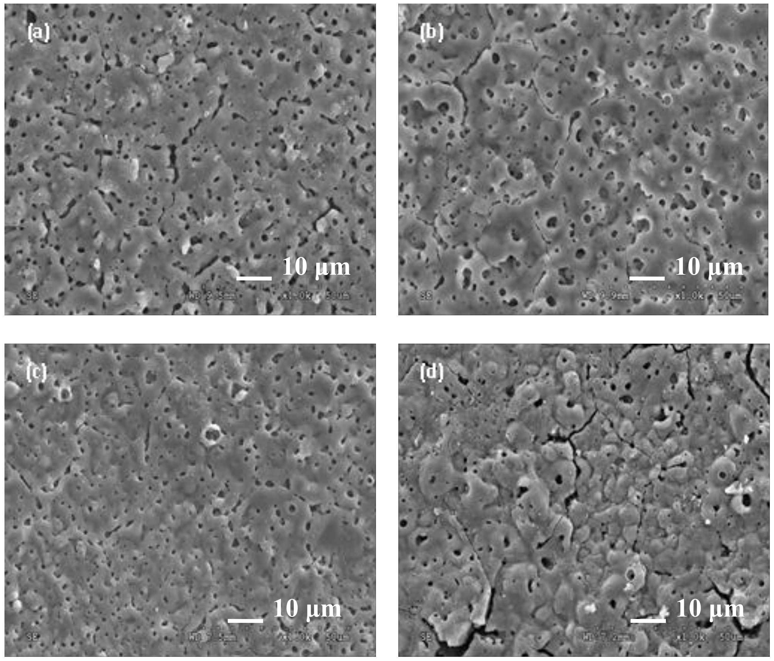
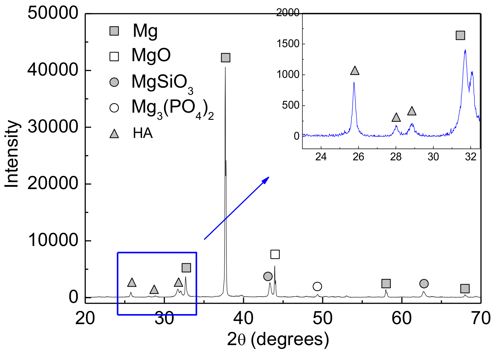
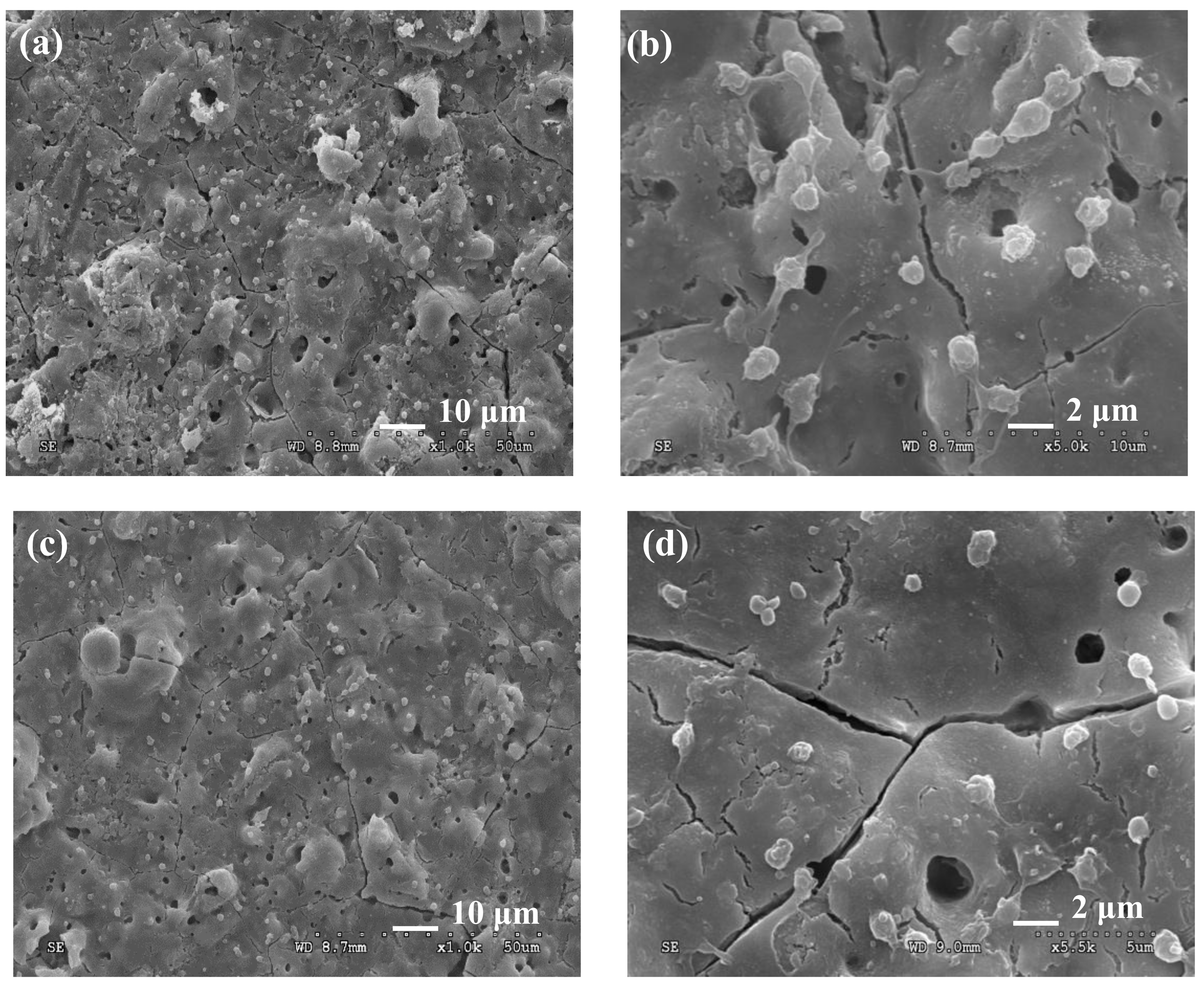
3.1. Microstructure, Thickness, and Micro-Hardness of the Coatings

Figure 1 shows the surface morphologies of the coatings formed at various concentrations of HA. One can see that the coatings are porous with a large number of lamellar, spherical pieces, presenting a typical feature of MAO coating. It should be noted that HA additive may considerably influence the surface morphologies. Figure 1b,c display the morphology of the coating added with 0.2 and 0.4 g/L HA in the electrolyte, respectively. Interestingly, samples prepared in the electrolyte with HA exhibited fewer cracks, smoother surface, and smaller pore size compared with samples without additional HA, as shown in Figure 1a.
The electrical conductivity of the HA-containing complex electrolyte slightly decreased after the addition of a small amount of insulating HA particles. The voltage distribution on the specimen became somewhat low. Thus, instantaneous breakdown energy was small, and the discharge channel was narrow. Furthermore, the HA particles may enter into the discharge channel with the HA-suspended complex electrolyte, which raises the dielectric breakdown potential of the growing oxide film, resulting in the formation of smooth surface morphology.
As HA concentrations were increased to 0.6 g/L, on the one hand, the original uniform pore-structure gradually disappeared and was accompanied by the appearance of few large holes; on the other hand, some deep penetrating cracks were generated, and a small amount of white, sintered granules adhered to the surface of the coatings, as evidenced in Figure 1d. Thus, the surface quality of the coating decreased as HA concentrations were increased. This observation may be due to the large number of insulating HA particles that may hinder the movement of conductive ions in the electrolyte, resulting in the decrease in both the electrochemical reaction rate and migration of charged particles in the solution. Moreover, in the HA-suspended complex electrolyte, extensive heat that was generated during the electrical breakdown process cannot be easily spread, decreasing the cooling capacity of molten material.
The XRD pattern of the HA-containing coatings is shown in Figure 2. The coating consists mainly of MgO, MgSiO3, and a small amount of Mg3(PO4)2 and HA. HA cannot be easily identified from the full spectrum because of its small amount. In the inset in Figure 2, some characteristic diffraction peaks of HA are presented, indicating that HA was incorporated into the coatings.
The average micro-hardness of the HA-containing coatings (0.4 g/L, hereinafter) was approximately 215.8 ± 5.3 HV, slightly higher than that of the coatings without HA additive (hereinafter referred as MAO coating) which was about 194.7 ± 4.8 HV. The hardness of the HA-containing coatings might be enhanced by some dispersed HA particles as the second phase within the coatings. The thickness of the HA-containing coatings was 13.5 ± 0.2 μm; as for the MAO coatings, the thickness was about 12.9 ± 0.2 μm. There was no obvious variation in the thickness which might be interpreted as the same electric parameters during the MAO process.

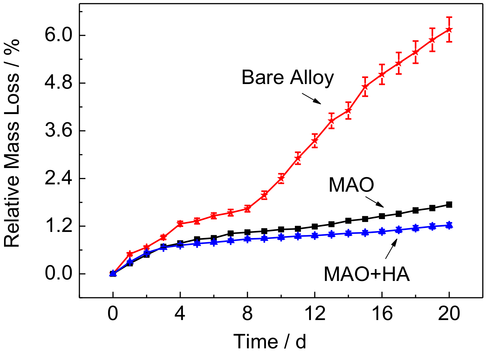
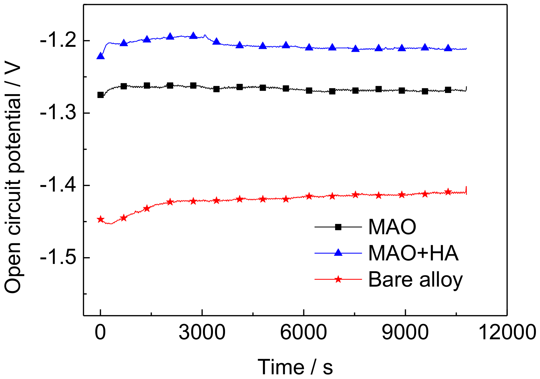
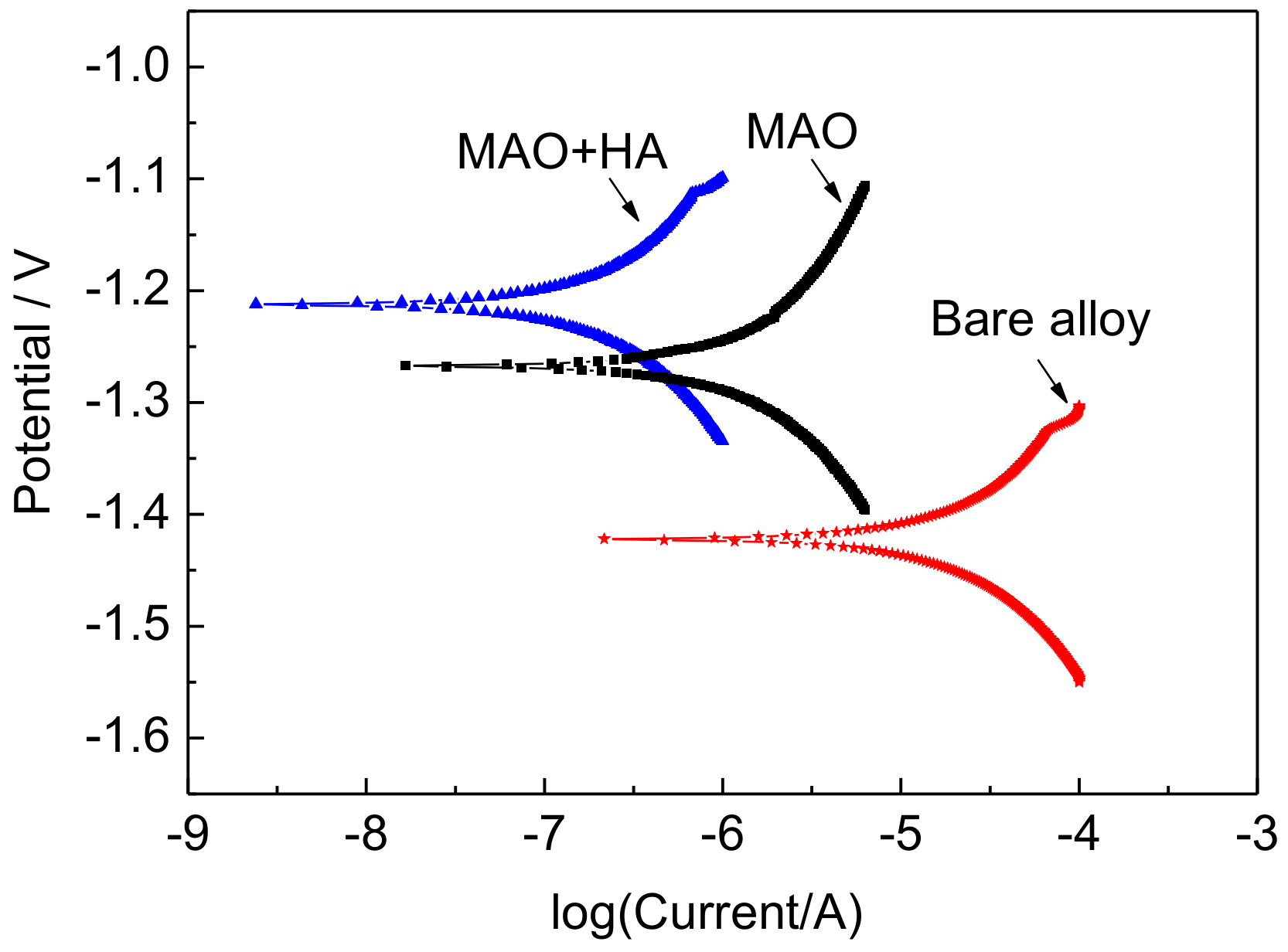
3.2. Effect of HA on the Corrosion Resistance of the Coatings

Figure 3 illustrates the mass loss curves of the coatings and Mg66Zn29Ca5 alloy immersed in Hank’s solution at 37 °C for 20 d. When the bare Mg66Zn29Ca5 alloy was soaked in Hank’s solution, corrosion occurred immediately on the surface due to the rapid dissolution of active Mg and Ca. However, the coated alloys both present a relatively slow mass loss rate compared with bare Mg66Zn29Ca5 alloy. This result indicates that the coatings can effectively protect the substrate to avoid further corrosion.
The mass loss of the coatings tended to linearly increase at the early immersion stage (approximately 3 d), but subsequently slowed down. It is well known that there is a loose and porous layer on the surface of coatings [29]. The corrosion medium can easily permeate into the loose superficial layer and spread out, resulting in severe corrosion. With the increase of immersion time, the dense inner layer would prevent corrosive medium further permeation, which leads to a slow corrosion rate; on the other hand, the coating may achieve a self-protection by the corrosion products obtained via a spontaneous mineralization in Hank’s solution [45], resulting in a slow corrosion rate as well. The mass loss rate was much lower in the HA-containing coating than that in MAO coating. As previous analysis, few cracks and pores were found in the HA-containing coatings, which inhibited the permeation of the corrosion medium, leading to good corrosion resistance.
The measurement of the OCP changes with time (OCP-t) can be employed to understand the chemical stability and corrosion process that occurs on the sample surface. The recorded OCP-t curves of Mg66Zn29Ca5 alloys after soaking in Hank’s solution for up to 3 h are plotted in Figure 4. All of the samples exhibited a similar trend in the evolution of OCP: the potentials moved rapidly toward the positive direction at the initial stage, and then OCP was slightly increased (bare alloy) or maintained constant (HA-containing coating, MAO coatings). The rapid increase in OCP at the initial stage of immersion indicates the formation of a certain kind passivation layer on the sample surfaces. The bare alloy, especially, requires more time to reach a stable potential, indicating a slow rate of establishing equilibrium between corrosion and protection. Commonly, more positive OCP implies that the alloy is comparatively more passive. Therefore, the electrochemical activity of different samples follows the order bare alloy > MAO > MAO + HA.
Figure 5 shows the potentiodynamic polarization curves of HA-containing coating, MAO coating, and bare Mg66Zn29Ca5 alloy in Hank’s solution at 37 ± 0.5 °C. The corrosion potential (Ecorr) and corrosion current density (Icorr) can be calculated from polarization curves through a software using the Tafel extrapolation method. The fitting results are listed in Table 1. Both coatings in Hank’s solution show good anti-corrosion property. The Ecorr of the coatings, e.g., HA-containing coating (−1.215 V) and MAO coating (−1.263 V), significantly increased in comparison with that of bare Mg66Zn29Ca5 alloy (−1.422 V); the Icorr of the coatings reduced remarkably with values of 6.138 × 10⁻⁸ A·cm−2 and 8.831 × 10⁻⁷ A·cm−2, respectively, manifesting better corrosion resistance than Mg66Zn29Ca5 alloy.
For the coatings, the Ecorr of the HA-containing coating presented 48 mV positively shift and the Icorr decreased by about 1 order of magnitude compared with MAO coating, revealing an improvement in anti-corrosion property.

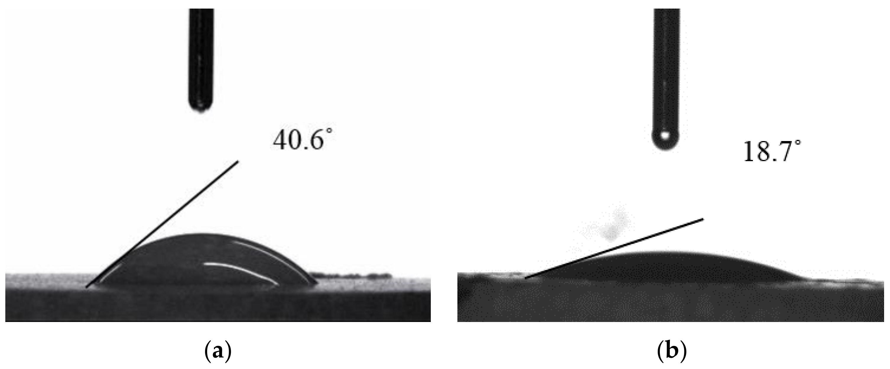
3.3. Effect of HA Particles on the Blood Compatibility of the Coatings

Figure 6 shows the effect of HA particles on the amount and shape of blood platelet adhesion of the coatings. It can be seen that less blood platelet counts were observed on the HA-containing coating compared with that of the MAO coating. Furthermore, the platelets on the MAO coating were unevenly dispersed, with some regions exhibiting higher platelet density than those on the HA-containing coating. Generally, the sample with less platelet density adhering on the surface indicates improved blood compatibility [46]. The SEM micrographs also provided information on the morphology of adhered platelets and their activation state. The platelets on the HA-containing coating were round or oval without pseudopodia-like protrusions. However, platelets on the MAO coating were spread as dendritic with one or more pseudopodia. These results indicate that the HA-containing coating possessed good blood compatibility.
Figure 7 shows the change in the static water contact angles of the coatings with and without HA particles. The water contact angle of the MAO coating was 40.6° whereas that of the HA-containing coating decreased to approximately 18.7°, indicating that the HA-containing coating possessed improved hydrophilicity. Normally, the percentages of round platelets and the total number of adhering platelets are related to the degree of hydrophiticity: the more hydrophilic the surface is, the lower the number of platelets adhering to it and the smaller are the changes in platelet shape. The improved hydrophilicity causes a less tendency of absorption in the protein, blood platelet, and blood clotting factors and resulting improvement in the blood compatibility of implant material [47,48].

4. Conclusions

An HA-containing coating was fabricated on a biodegradable Mg66Zn29Ca5 magnesium alloy via MAO. The XRD analysis showed that the coating consists mainly of MgO, MgSiO3, and a small amount of Mg3(PO4)2 and HA, indicating that the HA can be incorporated into the coating. The average micro-hardness of the HA-containing coating (0.4 g/L) was approximately 215.8 HV, which was slightly higher than that of the coating without HA additive (approximately 194.7 HV). The Ecorr of the HA-containing coating significantly increased, whereas the Icorr was remarkably reduced compared with bare Mg66Zn29Ca5, manifesting an improved corrosion resistance. Lesser count and uniformity of blood platelets were observed on the HA-containing coating compared with those of the MAO coating. The static water contact angle of the HA-containing ceramic coating was 18.7°, which was lower than that of the coating without HA (40.1°), demonstrating an improved blood compatibility in one respect. The HA-containing ceramic coatings may be an effective way to simultaneously improve the surface biocompatibility and corrosion resistance of magnesium alloys.

Acknowledgments

This work is supported by National Science Foundation of China (51775221).

Author Contributions

Guang-Hong Zhou and Hong-Yan Ding conceived and designed the experiments; Guo-Qing Wang and Hong Li performed MAO and bio-corrosion experiments and analyzed the data. Tao Liu performed the cell experiments. Hong-Yan Ding and Hong Li wrote the paper.

Conflicts of Interest

The authors declare no conflict of interest.

References

  1. Shadanbaz, S.; Dias, G.J. Calcium phosphate coatings on magnesium alloys for biomedical applications: A review. Acta Biomater. 2012, 8, 20–30. [Google Scholar] [CrossRef] [PubMed]
  2. Merritt, K.; Brown, S.A. Release of hexavalent chromium from corrosion of stainless steel and cobalt-chromium alloys. J. Biomed. Mater. Res. 1995, 29, 627–633. [Google Scholar] [CrossRef] [PubMed]
  3. Yang, J.; Merritt, K. Detection of antibodies against corrosion products in patients after Co-Cr total joint replacements. J. Biomed. Mater. Res. 1994, 28, 1249–1258. [Google Scholar] [CrossRef] [PubMed]
  4. Almpanis, G.C.; Tsigkas, G.G.; Koutsojannis, C.; Mazarakis, A.; Kounis, G.N.; Kounis, N.G. Nickel allergy, Kounis syndrome and intracardiac metal devices. Int. J. Cardiol. 2010, 145, 364–365. [Google Scholar] [CrossRef] [PubMed]
  5. Dietrich, K.A.; Mazoochian, F.; Summer, B.; Reinert, M.; Ruzicka, T.; Thomas, P. Intolerance reactions to knee arthroplasty in patients with nickel/cobalt allergy and disappearance of symptoms after revision surgery with titanium-based endoprostheses. J. Dtsch. Dermatol. Ges. 2009, 7, 410–413. [Google Scholar] [CrossRef] [PubMed]
  6. Mani, G.; Feldman, M.D.; Patel, D.; Agrawal, C.M. Coronary stents: A materials perspective. Biomaterials 2007, 28, 1689–1710. [Google Scholar] [CrossRef] [PubMed]
  7. Piskin, E. Biodegradable polymers as biomaterials. J. Biomater. Sci. Polym. Ed. 1995, 6, 775–795. [Google Scholar] [CrossRef] [PubMed]
  8. Song, G.; Song, S. A Possible Biodegradable Magnesium Implant Material. Adv. Eng. Mater. 2007, 9, 298–302. [Google Scholar] [CrossRef]
  9. Gupta, A.P.; Kumar, V. New emerging trends in synthetic biodegradable polymers—Polylactide: A critique. Eur. Polym. J. 2007, 43, 4053–4074. [Google Scholar] [CrossRef]
  10. Li, W.; Cooper, J.A., Jr.; Mauck, R.L.; Tuan, R.S. Fabrication and characterization of six electrospun poly(α-hydroxy ester)-based fibrous scaffolds for tissue engineering applications. Acta Biomater. 2006, 2, 377–385. [Google Scholar] [CrossRef] [PubMed]
  11. Xu, L.; Pan, F.; Yu, G.; Yang, L.; Zhang, E.; Yang, K. In vitro and in vivo evaluation of the surface bioactivity of a calcium phosphate coated magnesium alloy. Biomaterials 2009, 30, 1512–1523. [Google Scholar] [CrossRef] [PubMed]
  12. Witte, F.; Fischer, J.; Nellesen, J.; Crostack, H.; Kaese, V.; Pisch, A.; Beckmann, F.; Windhagen, H. In vitro and in vivo corrosion measurements of magnesium alloys. Biomaterials 2006, 27, 1013–1018. [Google Scholar] [CrossRef] [PubMed]
  13. Kannan, M.B.; Raman, R.K.S. In vitro degradation and mechanical integrity of calcium-containing magnesium alloys in modified-simulated body fluid. Biomaterials 2008, 29, 2306–2314. [Google Scholar] [CrossRef] [PubMed]
  14. Zeng, R.; Dietzel, W.; Witte, F.; Hort, N.; Blawert, C. Progress and Challenge for Magnesium Alloys as Biomaterials. Adv. Eng. Mater. 2008, 10, B3–B14. [Google Scholar] [CrossRef]
  15. Witte, F.; Ulrich, H.; Rudert, M.; Willbold, E. Biodegradable magnesium scaffolds: Part 1: Appropriate inflammatory response. J. Biomed. Mater. Res. A 2007, 81, 748–756. [Google Scholar] [CrossRef] [PubMed]
  16. Staiger, M.P.; Pietak, A.M.; Huadmai, J.; Dias, G. Magnesium and its alloys as orthopedic biomaterials: A review. Biomaterials 2006, 27, 1728–1734. [Google Scholar] [CrossRef] [PubMed]
  17. Aung, N.N.; Zhou, W. Effect of grain size and twins on corrosion behaviour of AZ31B magnesium alloy. Corros. Sci. 2010, 52, 589–594. [Google Scholar] [CrossRef]
  18. Wang, H.; Estrin, Y.; Zúberová, Z. Bio-corrosion of a magnesium alloy with different processing histories. Mater. Lett. 2008, 62, 2476–2479. [Google Scholar] [CrossRef]
  19. Pu, Z.; Song, G.L.; Yang, S.; Outeiro, J.C.; Dillon, O.W., Jr.; Puleo, D.A.; Jawahir, I.S. Grain refined and basal textured surface produced by burnishing for improved corrosion performance of AZ31B Mg alloy. Corros. Sci. 2012, 57, 192–201. [Google Scholar] [CrossRef]
  20. Gray, J.E.; Luan, B. Protective coatings on magnesium and its alloys—A critical review. J. Alloy. Compd. 2002, 336, 88–113. [Google Scholar] [CrossRef]
  21. Yanovska, A.; Kuznetsov, V.; Stanislavov, A.; Danilchenko, S.; Sukhodub, L. Calcium–phosphate coatings obtained biomimetically on magnesium substrates under low magnetic field. Appl. Surf. Sci. 2012, 258, 8577–8584. [Google Scholar] [CrossRef]
  22. Tan, L.; Wang, Q.; Geng, F.; Xi, X.; Qiu, J.; Yang, K. Preparation and characterization of Ca-P coating on AZ31 magnesium alloy. Trans. Nonferr. Metals Soc. China 2010, 20, s648–s654. [Google Scholar] [CrossRef]
  23. Chiu, K.Y.; Wong, M.H.; Cheng, F.T.; Man, H.C. Characterization and corrosion studies of fluoride conversion coating on degradable Mg implants. Surf. Coat. Technol. 2007, 202, 590–598. [Google Scholar] [CrossRef]
  24. Wu, L.; Dong, J.; Ke, W. Potentiostatic deposition process of fluoride conversion film on AZ31 magnesium alloy in 0.1 M KF solution. Electrochim. Acta 2013, 105, 554–559. [Google Scholar] [CrossRef]
  25. Liu, C.; Xin, Y.; Tian, X.; Chu, P.K. Corrosion behavior of AZ91 magnesium alloy treated by plasma immersion ion implantation and deposition in artificial physiological fluids. Thin Solid Films 2007, 516, 422–427. [Google Scholar] [CrossRef]
  26. Wang, X.; Zeng, X.; Wu, G.; Yao, S.; Lai, Y. Effects of tantalum ion implantation on the corrosion behavior of AZ31 magnesium alloys. J. Alloy. Compd. 2007, 437, 87–92. [Google Scholar] [CrossRef]
  27. Fischerauer, S.F.; Kraus, T.; Wu, X.; Tangl, S.; Sorantin, E.; Hänzi, A.C.; Löffler, J.F.; Uggowitzer, P.J.; Weinberg, A.M. In vivo degradation performance of micro-arc-oxidized magnesium implants: A micro-CT study in rats. Acta Biomater. 2013, 9, 5411–5420. [Google Scholar] [CrossRef] [PubMed]
  28. Wang, Y.M.; Guo, J.W.; Shao, Z.K.; Zhuang, J.P.; Jin, M.S.; Wu, C.J.; Wei, D.Q.; Zhou, Y. A metasilicate-based ceramic coating formed on magnesium alloy by microarc oxidation and its corrosion in simulated body fluid. Surf. Coat. Technol. 2013, 219, 8–14. [Google Scholar] [CrossRef]
  29. Gu, Y.; Chen, C.; Bandopadhyay, S.; Ning, C.; Zhang, Y.; Guo, Y. Corrosion mechanism and model of pulsed DC microarc oxidation treated AZ31 alloy in simulated body fluid. Appl. Surf. Sci. 2012, 258, 6116–6126. [Google Scholar] [CrossRef]
  30. Gu, X.N.; Li, N.; Zhou, W.R.; Zheng, Y.F.; Zhao, X.; Cai, Q.Z.; Ruan, L. Corrosion resistance and surface biocompatibility of a microarc oxidation coating on a Mg–Ca alloy. Acta Biomater. 2011, 7, 1880–1889. [Google Scholar] [CrossRef] [PubMed]
  31. Wang, Y.M.; Wang, F.H.; Xu, M.J.; Zhao, B.; Guo, L.X.; Ouyang, J.H. Microstructure and corrosion behavior of coated AZ91 alloy by microarc oxidation for biomedical application. Appl. Surf. Sci. 2009, 255, 9124–9131. [Google Scholar] [CrossRef]
  32. Viswanath, B.; Shastry, V.V.; Ramamurty, U.; Ravishankar, N. Effect of calcium deficiency on the mechanical properties of hydroxyapatite crystals. Acta Mater. 2010, 58, 4841–4848. [Google Scholar] [CrossRef]
  33. Descamps, M.; Hornez, J.C.; Leriche, A. Manufacture of hydroxyapatite beads for medical applications. J. Eur. Ceram. Soc. 2009, 29, 369–375. [Google Scholar] [CrossRef]
  34. Cui, W.; Li, X.; Xie, C.; Zhuang, H.; Zhou, S.; Weng, J. Hydroxyapatite nucleation and growth mechanism on electrospun fibers functionalized with different chemical groups and their combinations. Biomaterials 2010, 31, 4620–4629. [Google Scholar] [CrossRef] [PubMed]
  35. Yari, S.A.; Shokrgozar, M.A.; Homaeigohar, S.S.; Khavandi, A. Biological evaluation of partially stabilized zirconia added HA/HDPE composites with osteoblast and fibroblast cell lines. J. Mater. Sci. Mater. Med. 2008, 19, 2359–2365. [Google Scholar] [CrossRef] [PubMed]
  36. YSong, W.; Shan, D.Y.; Han, E.H. Electrodeposition of hydroxyapatite coating on AZ91D magnesium alloy for biomaterial application. Mater. Lett. 2008, 62, 3276–3279. [Google Scholar]
  37. Jamesh, M.; Kumar, S.; Narayanan, T.S.N.S. Electrodeposition of hydroxyapatite coating on magnesium for biomedical applications. J. Coat. Technol. Res. 2012, 9, 495–502. [Google Scholar] [CrossRef]
  38. Wang, H.X.; Guan, S.K.; Wang, X.; Ren, C.X.; Wang, L.G. In vitro degradation and mechanical integrity of Mg–Zn–Ca alloy coated with Ca-deficient hydroxyapatite by the pulse electrodeposition process. Acta Biomater. 2010, 6, 1743–1748. [Google Scholar] [CrossRef] [PubMed]
  39. Hahn, B.; Park, D.; Choi, J.; Ryu, J.; Yoon, W.; Choi, J.; Kim, H.; Kim, S. Aerosol deposition of hydroxyapatite–chitosan composite coatings on biodegradable magnesium alloy. Surf. Coat. Technol. 2011, 205, 3112–3118. [Google Scholar] [CrossRef]
  40. Hiromoto, S.; Tomozawa, M.; Maruyama, N. Fatigue property of a bioabsorbable magnesium alloy with a hydroxyapatite coating formed by a chemical solution deposition. J. Mech. Behav. Biomed. Mater. 2013, 25, 1–10. [Google Scholar] [CrossRef] [PubMed]
  41. Rojaee, R.; Fathi, M.; Raeissi, K. Controlling the degradation rate of AZ91 magnesium alloy via sol–gel derived nanostructured hydroxyapatite coating. Mater. Sci. Eng. C 2013, 33, 3817–3825. [Google Scholar] [CrossRef] [PubMed]
  42. Tomozawa, M.; Hiromoto, S.; Harada, Y. Microstructure of hydroxyapatite-coated magnesium prepared in aqueous solution. Surf. Coat. Technol. 2010, 204, 3243–3247. [Google Scholar] [CrossRef]
  43. Hiromoto, S.; Tomozawa, M. Hydroxyapatite coating of AZ31 magnesium alloy by a solution treatment and its corrosion behavior in NaCl solution. Surf. Coat. Technol. 2011, 205, 4711–4719. [Google Scholar] [CrossRef]
  44. Sreekanth, D.; Rameshbabu, N. Development and characterization of MgO/hydroxyapatite composite coating on AZ31 magnesium alloy by plasma electrolytic oxidation coupled with electrophoretic deposition. Mater. Lett. 2012, 68, 439–442. [Google Scholar] [CrossRef]
  45. Hahn, J.J.; McGowan, N.G.; Heimann, R.L.; Barr, T.L. Modification and characterization of mineralization surface for corrosion protection. Surf. Coat. Technol. 1998, 109, 403–407. [Google Scholar] [CrossRef]
  46. Zhang, L.; Lv, P.; Huang, Z.Y.; Lin, S.P.; Chen, D.H.; Pan, S.R.; Chen, M. Blood compatibility of La2O3 doped diamond-like carbon films. Diam. Relat. Mater. 2008, 17, 1922–1926. [Google Scholar] [CrossRef]
  47. Kim, K.; Yu, M.; Zong, X.; Chiu, J.; Fang, D.; Seo, Y.; Hsiao, B.S.; Chu, B.; Hadjiargyrou, M. Control of degradation rate and hydrophilicity in electrospun non-woven poly(d,l-lactide) nanofiber scaffolds for biomedical applications. Biomaterials 2003, 24, 4977–4985. [Google Scholar] [CrossRef]
  48. Soria, J.M.; Ramos, C.M.; Bahamonde, O.; Cruz, D.M.G.; Sánchez, M.S.; Esparza, M.A.G.; Casas, C.; Guzmán, M.; Navarro, X.; Ribelles, J.L.G.; et al. Influence of the substrate’s hydrophilicity on the in vitro Schwann cells viability. J. Biomed. Mater. Res. A 2007, 83, 463–470. [Google Scholar] [CrossRef] [PubMed]
Figure 1. Surface morphologies of coatings formed at hydroxyapatite (HA) concentrations of (a) 0; (b) 0.2; (c) 0.4; and (d) 0.6 g/L.
Applsci 08 00569 g001
Figure 2. X-ray diffraction patterns of the coatings formed at HA concentration of 0.4 g/L.
Applsci 08 00569 g002
Figure 3. Mass loss curves of the coatings immersed in Hank’s solution at 37 °C for 20 d.
Applsci 08 00569 g003
Figure 4. Evolution of open circuit potential in Hank’s solution at 37 °C as a function of exposure time for the coatings.
Applsci 08 00569 g004
Figure 5. Potentiodynamic polarization curves obtained in Hank’s solution at 37 °C.
Applsci 08 00569 g005
Figure 6. SEM images of blood platelet adhesion on the (a,b) micro-arc oxidation (MAO) coating and (c,d) HA-containing coating.
Applsci 08 00569 g006
Figure 7. Water contact angle of coatings without (a) and with (b) HA.
Applsci 08 00569 g007
Table 1. Fitting results of polarization curves for bare and coated alloy.
SampleEcorr/VIcorr/(A·cm−2)
Bare alloy−1.4222.546 × 10−4
MAO coating−1.2638.831 × 10−7
HA-containing coating−1.2156.138 × 10−8

