Article

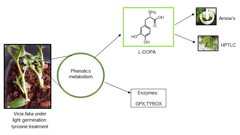
L-DOPA Trends in Different Tissues at Early Stages of Vicia faba Growth: Effect of Tyrosine Treatment

by
Claudia A. Oviedo-Silva
1,*,
Mhartyn Elso-Freudenberg
1 and
Mario Aranda-Bustos
2
1
Departamento de Química, Facultad de Ciencias, Universidad del Bio-Bio, Concepción 4081112, Chile
2
Departamento de Ciencia y Tecnología de los Alimentos, Facultad de Farmacia, Universidad de Concepción, Concepción 4089100, Chile
*
Author to whom correspondence should be addressed.
Appl. Sci. 2018, 8(12), 2431; https://doi.org/10.3390/app8122431
Submission received: 26 October 2018 / Revised: 23 November 2018 / Accepted: 27 November 2018 / Published: 30 November 2018
(This article belongs to the Section Chemical and Molecular Sciences)

Abstract

:

Featured Application

The potential applications of this work are in human health and in plant pest control. In humans: L-DOPA is the first-line treatment for Parkinson’s disease. In plants, L-DOPA has insecticidal and allelochemical properties.

Abstract

The nonprotein amino acid Levo-3,4-dihydroxyphenylalanine (L-DOPA) has insecticidal, allelochemical, and antiparkinsonian effects. The aim of this research was to assess L-DOPA content in different tissues of Vicia faba (cv. Super Agua Dulce), and to verify if treatment with the phenolic amino acid L-4-hydroxyphenylalanine (tyrosine) had an effect on such content. Under light germination, control and tyrosine-treated early seedling stages of V. faba were studied and L-DOPA was quantified spectrophotometrically (Arnow’s method) and by high-performance thin-layer chromatography (HPTLC), as well. Additionally, tyrosinase (TYROX) and guaiacol peroxidase (GPX) activities (considered markers of a phenolic compounds metabolism) were quantified as germination proceeded. Different organs (roots, sprouts, and seeds) and different developmental stages were considered. Steady high L-DOPA concentrations were found in untreated sprouts and roots compared to seeds, as time progressed. While TYROX activity was not detected in these experiments, GPX had diverse trends. In control tissues, GPX increased in seed tissue as germination progressed, whereas in roots and sprouts, a decreasing GPX activity was observed. Tyrosine exposure decreased L-DOPA content, and decreased or did not change GPX activity (depending on the organ). Both Arnow’s and HPTLC methods were consistent in terms of tendencies, except for the scarce contents found in seeds, in which HPTLC was more sensitive. The richest source of L-DOPA was found in shoots (untreated), reaching as high as 125 mg g−1 DW (12% in DW) (the highest content reported in fava bean seedlings until now), whereas the smallest L-DOPA content was found in seeds. The importance of light germination conditions is discussed in terms of L-DOPA yield and from a physiological perspective. It is concluded that V. faba (cv. Super Agua Dulce) shoots are a good source of L-DOPA and that tyrosine addition (0.55 mM) decreases L-DOPA content in actively growing tissues (shoots and roots).

Graphical Abstract

1. Introduction

Levo 3,4-dihydroxyphenylalanine (L-DOPA) therapy is necessary for Parkinson’s disease for its powerful antiparkinsonian effect [1]. L-DOPA is a nonprotein amino acid with biological activity, found in animals and in some plants [2]. It is the precursor of catecholamine type neurotransmitters (dopamine, norepinephrine, and epinephrine), being involved in melanin synthesis as well [3]. Lately, L-DOPA has drawn much attention in the field of plants for its acknowledged defensive role against insects [4]. In addition, it is considered an allelochemical, i.e., a compound released into the environment affecting the growth and development of neighboring plants [1,5,6].
L-DOPA has raised particular interest in legumes, particularly in the Fabaceae family, as reports describe a relatively high content in such plants [7,8]. Additionally, for Parkinson’s patients, fava bean consumption would be even better than the drug in terms of motor performance and side effects [1]. Moreover, carbidopa (a peripheral inhibitor of the enzyme DOPA decarboxylase) has been found in fava bean as well [9]. Thus, different tissues of fava bean have been analyzed for L-DOPA, and as much as 6.0–6.75% dry weight (DW) (60–67.5 mg g−1 DW) has been found in young pods of cultivars Diana and Alameda, respectively [10], and contents in whole seedlings in the order of 0.2% DW (2 mg g−1 DW) [11].
L-DOPA would be produced via the oxidation of the phenolic amino acid L-4-hydroxyphenylalanine (tyrosine) by the enzyme tyrosinase (tyrosine oxidase: TYROX) [12]. In turn, tyrosine comes in plants from the shikimic acid pathway and TYROX would be involved in L-DOPA synthesis in non-Cariophyllalles plants [13]. TYROX is involved in other polyphenolics and melanin synthesis, as well [6]. L-DOPA is a melanin biosynthetic precursor (present in plant and animal tissues). In general, TYROX are multicopper monophenol monooxygenases (EC 1.14.18.1), which use oxygen as cosubstrate to catalyze the o-hydroxylation of phenols (i.e., tyrosine to L-DOPA step); and the subsequent oxidation of the formed o-diphenols to the corresponding o-quinones as well. Thus, using the aromatic amino acid L-tyrosine as substrate, the ultimate tyrosinase-catalyzed reaction product is dopaquinone [13]. This o-quinone can undergo chemically spontaneous and also enzymatic reactions, which in turn can further convert it into melanin. Therefore, TYROX is considered a key enzyme for melanin synthesis [14] and has also been associated with stress responses to the environment, as melanin plays a survival function [15].
L-DOPA is also a precursor of phenylpropanoids in plants; the phenylpropanoid pathway is an important metabolic route, as it synthesizes phenolic compounds, including lignin [16]. Indeed, in plants, TYROX plays an important role in modifying and hardening the protective exterior layer of, for instance, seed envelopes, and as an agent against invasive organisms [16].
In plants, a common early response to environmental changes is the antioxidant enzymatic pathway, with roles in signal transduction and pathogen defense, producing protective biomolecules such as melanin and lignin [17]. GPX (Guaiacol peroxidase, (EC 1.11.1.7)) is a heme-containing protein that oxidizes certain phenolic substrates at the expense of H2O2 [18]. GPX is considered a marker phenolic compounds metabolism and antioxidant response. Indeed, GPX has a role in the biosynthesis of lignin, the defense against biotic stresses, decomposition of indole-3-acetic acid (IAA), among others [19,20], and is widely accepted as a ‘stress enzyme’. Amongst the various antioxidant enzymes, GPX can be considered one of the key ones, since both of its extra and intracellular forms participate in the breakdown of H2O2 [15,20].
In an effort to stimulate L-DOPA production in fava bean, Randhir et al. [21] studied L-DOPA content in seedlings, primed with oregano extract, lactoferrin, and a fish protein hydrolysate (dark germination). They found a positive effect of priming seeds with all those former elicitors, finding a fast response in lactoferrin primed seedlings, with a L-DOPA content of 3.5% DW approx (data transformed from the author using 90% water content). On the other hand, Raghavendra et al. [12] reported that in Mucuna pruriens suspended tissue cultures, the phenolic amino acid L-tyrosine increased L-DOPA production, finding that the highest concentration of L-DOPA obtainable was 9.47 ± 0.70% DW (40.13-fold increase). Although this latter study achieved a 40-fold L-DOPA content increase by adding tyrosine to cell tissue cultures of M. pruriens, no information is available in terms of tyrosine effects on DOPA content in legume germinating seeds. Moreover, there is no evidence on whether such an interaction involves the tyrosine related enzymes formerly described.
The aim of this research is to verify if tyrosine treatment has an effect on L-DOPA content in different tissues at the early developmental stages of Vicia faba. Concomitant monitoring of TYROX and GPX activities, as related markers of phenolic metabolism, was recorded as well. The plants’ response to the treatment in terms of growth rate was measured too. In addition, the L-DOPA content was assessed and compared using two different analytical methods.

2. Materials and Methods

Solvents were of HPLC grade. USP standard of L-DOPA and Guaiacol 98% were obtained from Sigma-Aldrich (St. Louis, MO, USA). Ethanol, H2O2 30% p.a., Dichloromethane 99.9%, Isopropanol 99.8%, Acetic acid 96%, water LiChrosolv® LC-MS grade, and HPTLC plates silica gel were obtained from Merck (Darmstadt, Germany), and formic acid 98% from Fluka (Buchs, Switzerland). Polyvinylpyrrolidone (PVP) was obtained from Calbiochem (Selangor, Malaysia).

2.1. Experimental Setup

V. faba seeds (Super Agua Dulce variety, Agroflora®) and coconut fiber (Cocomix, BioBizz®) were obtained at local market suppliers. Control and tyrosine-treated seeds were primed for 48 h in distilled water and tyrosine (0.55 mM), respectively. They were watered each 3–4 days with 50 mL per tray, with distilled water and tyrosine solution (0.55 mM), respectively. The experiment was performed in triplicate. Each tray contained 300 g of coconut fiber and 20 seeds (sawn at surface, under natural light conditions in the laboratory).

2.2. Plant Extracts

Three different organs were considered: Roots, sprouts and seeds. Their respective enzymatic activities, L-DOPA content, protein content, and dry mass (lyophilized) were followed in each seedling developmental stage: 3–4 days (A); 8–9 days (B) or 2–4 leaves (C, 15 days approx.).
The activity of enzymes described below was determined spectrophotometrically from the accumulation of the respective product using a Shimadzu UV-1240 spectrophotometer and presented as mmol min−1 g−1 dry weight. One gram of each tissue was manually ground with a mortar in 3–6 mL of cold extraction buffer (0.05 M potassium phosphate buffer pH 6.5, 0.5% polyvinylpyrrolidone). Samples were centrifuged at 11,000× g for 10 min and stored on ice. The resulting supernatant was the extract used in all enzyme assays.

2.3. Enzyme Assays

2.3.1. TYROX

Dopachrome production in each organ enzyme extract was followed spectrophotometrically at 475 nm (modified from Kampmann et al. [22]). Assays were conducted at 25 °C, in a final volume of 1 mL reaction mixture that was prepared as follows: 50 μL of fava bean organ extract was added to 950 μL of 10 mM L-DOPA (in 0.05 M phosphate buffer, pH 6.0) to start the reaction. Enzyme activity was determined from the linear portion of the curve, using a Dopachrome ε475 = 3.6 mM−1 cm−1; proper blanks were considered and a positive control for tyrosine oxidase (Beta vulgaris roots) was run in parallel as well.

2.3.2. GPX

Tetraguaiacol was followed at 470 nm. The assay was performed as follows (modified from Jebara et al. [23]): Briefly, to a final volume of 1 mL (25 °C), a reaction mixture consisting of 930 μL 0.05 M phosphate buffer (pH 7.0), 50 mM guaiacol, 50 μL of fava bean organ extract, and 20 μL 0.2 mM H2O2 were added. Enzyme activity was calculated at a fixed time interval of 0.5 min, determined from the linear portion of the curve, using a tetraguaiacol ε470: 26.6 mM−1 cm−1 [23,24], (corresponding blanks were run in parallel).

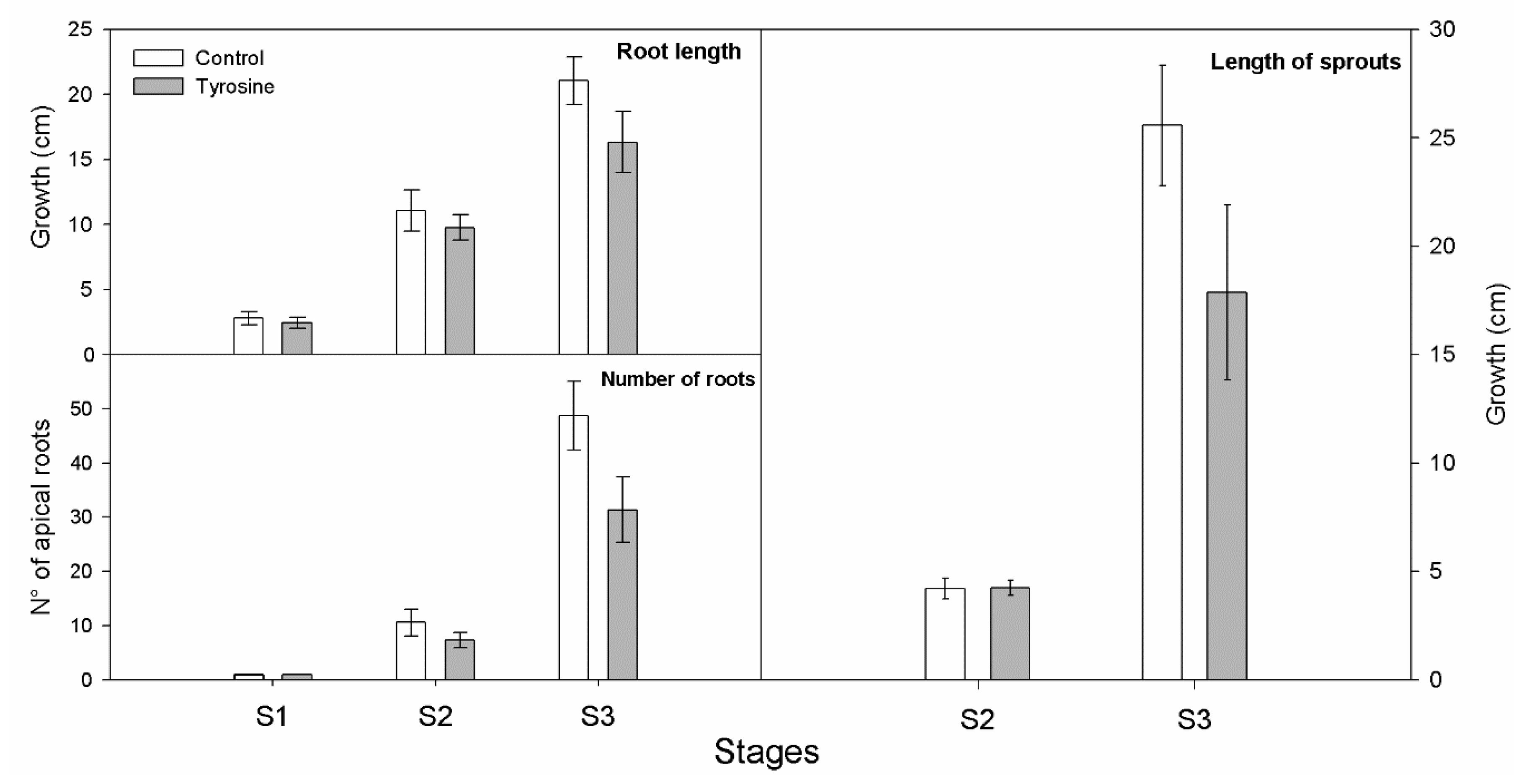
2.4. Growth Rate Record

Growth rates were determined using the respective photographic records with a reference scale in centimeters. These measurements were performed with the software ImageJ (1.49v, USA) [25] for three parameters: Root length, sprout length, and number of roots. The root and sprout length were determined from the longest root and the longest shoot apices to the cotyledon stalk, respectively. Regarding the number of roots, each apex of primary and secondary roots was labeled by a dot and then quantified.

2.5. L-DOPA Content in V. faba

Plant organ extracts (obtained as the extracts for enzymatic activity) were used to determine the L-DOPA content using Arnow’s method [26]. L-DOPA was further quantified using ultraviolet coupled high-performance thin-layer chromatography HPTLC–UV. Briefly, lyophilized samples of each organ were ground to a fine powder (Bertin Instruments, Precellys 24 homogenizer, ceramic balls, for 30 s at 5000 rpm) and extracted at room temperature, as follows. Each sample (50–200 mg) was extracted with 1:1 formic acid–ethanol (for 2 h, 2 mL) under continuous agitation (120 rpm). Afterwards, samples were centrifuged for 10 min at 10,000× g and supernatants were saved. The resulting pellets were resuspended in new 1:1 formic acid-ethanol (2 h, 2 mL, 120 rpm), followed by the centrifugation step (10 min at 11,000× g). Thus, pellets were repeatedly (4 times) subjected to the same extraction cycle. Finally, pellets were extracted for the last time over a longer period of time in 1:1 formic acid-ethanol (2 mL, overnight, and 120 rpm). The five supernatants involved in the overall process were pooled and finally centrifuged (10 min 10,000× g). These collected supernatants were used as the respective fava bean extracts loaded on the HPTLC plates for further analysis.
Fava bean extract samples and standard solutions were applied by means of Automatic TLC Sampler 4 (ATS 4) from Planar Chromatography Manager CAMAG (Muttenz, Switzerland), with the following settings for 18 tracks per plate: Band length 6.0 mm, track distance 10.5 mm, applied volume range loaded of 2–20 uL, dosage speed 100 nL s−1, and application position x-axis 10.0 mm and y-axis 10.0 mm each. Chromatography was carried out in a 20 × 10 cm twin trough chamber (CAMAG) using 10 mL of dichloromethane:isopropanol:acetic acid:water as 2:2:1:1 (v/v/v/v) as mobile phase, up to a migration distance of 80 mm. After development, the plate was dried in a stream of warm air for 1 min. Detection was performed with TLC Scanner 3 (CAMAG) in UV-absorption mode at 282 nm with a slit dimension of 5.0 mm × 0.2 mm and a scanning speed of 20 mm s−1. All instruments were controlled via software platform winCats 1.4.6 (CAMAG).

2.6. Statistical Analysis

Differences between control and tyrosine treatments in terms of their L-DOPA content, GPX activity, and growth records were assessed by Tukey’s test (P < 0.05). These analyses were performed with the InfoStat/L software package (FCA-UNC, Argentina). A Pearson correlation analysis was performed to assess the relationship between the HPTLC and Arnow’s method for L-DOPA quantification, using Statistica 7.0 (Stat Soft Inc., Tulsa, OK, USA).

3. Results and Discussion
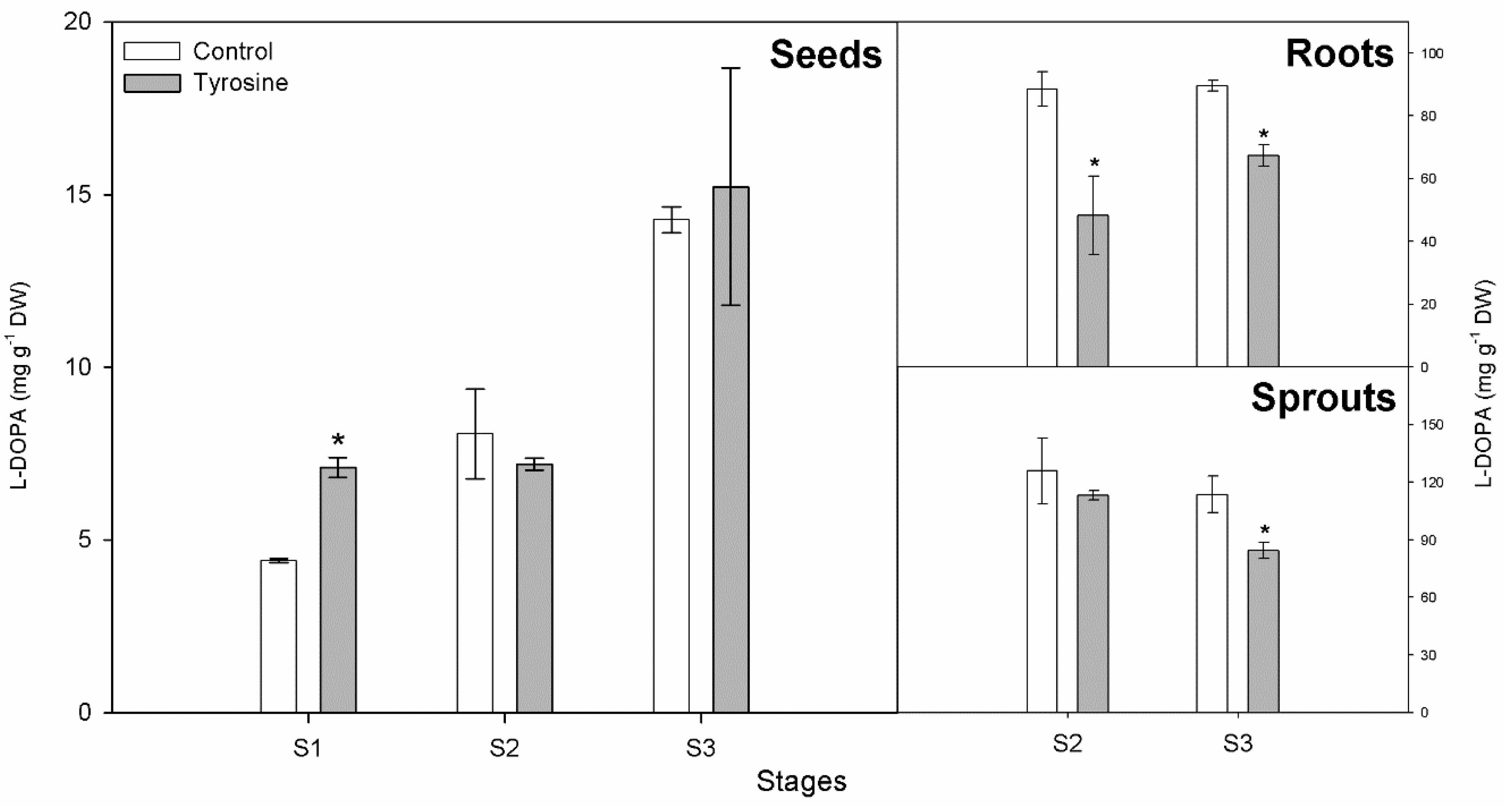
3.1. L-DOPA Content

With regard to L-DOPA concentration in control tissues: Although low contents were found in seeds, an increasing tendency in time could be observed until stage S3 (2–4 leaves emerged, 15 days approx), reaching concentrations up to 14.2 ± 0.38 mg g−1 DW (1.42 ± 0.03% DW) (Figure 1). Steady high concentrations were found in sprouts and roots as time progressed. Indeed, concentrations as high as 125.87 ± 17.18 mg g−1 DW (12.5 ± 1.7% DW) were observed in sprouts (S2 stage: 8–9 days), i.e., 10 times higher than in seeds, sprouts being the richest sources of L-DOPA found (Figure 1). This is in accordance with Goyoaga [11], who observed similar trends as germination took place in a V. faba embryo axis with a concentration range of 20.3 mg g−1 DW (at day 0) up to approximately 80 mg g−1 DW at 9 days, in light germination as well.
The L-DOPA content decreased with tyrosine treatment at almost all stages of roots and sprouts. In seeds, no significant differences in L-DOPA concentrations were detected at almost all stages, except for stage S1 (3–4 days) (Figure 1), in which a slight increase in such content was found.
It must be noted that light exposure during germination seems to be an important factor in the accumulation of L-DOPA, since Shetty et al. [27], Randhir et al. [21], and Vattem et al. [28] describe a totally different trend, i.e., decreasing L-DOPA content at the early stages of V. faba being germinated in darkness (values in the order of 4 mg g−1 to 1.5 mg g−1 FW in from day 0 to day 8, respectively). Indeed, they report L-DOPA contents that decrease gradually as plant growth proceeds, which suggests that L-DOPA is only synthesized, or even that it could be possibly only imported, at the early developmental stages, in dark germination [28,29]. On the contrary, increasing DOPA contents were observed in light germinating faba bean (4 to 7 mg g−1 FW from day 12 to day 16, respectively) [30]. Moreover, Wichers et al. [31] reported an apparent light effect in L-DOPA content in Mucuna pruriens cell liquid culture (a threefold increase as compared with dark condition). In that sense, our results agree with those of Goyoaga et al. [11] and Wichers et al. [31] in that they both worked with legumes in light environments, finding, in Goyoaga’s study, a very high L-DOPA content in germinating faba bean harvested 9 days after imbibition (80 mg g−1 DW) [11] and, in Wichers et al. [31], comparing a light versus a dark condition, finding more than three times L-DOPA under light culturing experiments with suspended Mucuna cells in liquid medium.
From a physiological perspective, light enhanced L-DOPA production is consistent with developmental needs, i.e., more L-DOPA content needed as the plantule emerges above ground as an herbivory preventive strategy. Therefore, if L-DOPA maximal yield potential is sought, dark germination should be avoided in the experimental set ups.
Recently, in a long-term greenhouse light germinating study, the highest L-DOPA concentration found in V. faba seedlings harvested after 15 days of germination was 13.3 mg g−1 DW [32]. It must be noted that the study weighed the whole seedling and did not separate individual tissues at that first stage of plant development, which could be one of the causes of a lower value than the former ones. Additionally, the same study determined L-DOPA content in leaf, weighing just that specific organ tissue, finding 10.5 mg g−1 DW. In other legumes belonging to the Mucuna gender, relatively high L-DOPA has been detected in different organs, such as leaves and seeds, containing as much as 1% DW and 4–7% DW L-DOPA, respectively [33,34,35]. Ramya and Thaakur [36] compiled L-DOPA content in other genera, such as Canavalia, Glycine, Vigna, and others, with relatively low concentrations (0.2–1.9% DW).
Regarding tyrosine availability in plants, recent findings suggest that enhanced tyrosine supply would enhance betalain synthesis (other DOPA-derived secondary metabolite) in betalain-producing Caryophyllales [13]. Our results do not prove any stimulatory growth effect (Figure 2), nor an enhanced L-DOPA content. On the contrary, we found that tyrosine treatment decreased DOPA content, and this could be related to tyrosine degradation as an energy source (catabolic pathway). Tyrosine-free levels can vary inside plants, and its catabolism has the highest amino acid energy yield feeding the tricarboxillic acid cycle [37]. Additionally, it must be noted that another potential route for tyrosine catabolism has been suggested via L-DOPA catabolism by Ellis et al. [38]. Other possible final destinations of tyrosine have to be considered as well, such as protein synthesis and other molecules, because tyrosine is the precursor of many other diverse metabolites, so its final destiny can be molecules such as isoquinoline alkaloids, lignin, and tyramine, among others [13].
As stated earlier, environmental stress is associated with both enzyme activity and secondary metabolite accumulation. Faba bean is a Nitrogen fixing plant and tyrosine contains Nitrogen; hence, there is a possibility that seedlings could have experienced stress by Nitrogen supplementation. Our results cannot support such an association. Indeed, considering GPX as stress marker (see Section 3.3), no increment in this enzyme activity or in the L-DOPA content was found (see Figure 1). This latter finding is in agreement with a recent report that describes that a wide range of an inorganic fertilizer supplementation (calcium ammonium nitrate) in faba bean does not reflect any significant difference in L-DOPA content [32].
Studies in which tyrosine is provided to cultured cells (of other plant species) have shown an increment in L-DOPA content. Apart from the study of Raghavendra et al. [12] performed in Mucuna tissue culture, a high biotransformation rate of tyrosine to L-DOPA in liquid medium cultured cells of Portulaca grandiflora has been reported as well [39].

3.2. Comparison of Methods for L-DOPA Determination

Both Arnow’s and HPTLC methods for L-DOPA determination were consistent in terms of tendencies and magnitudes found, except for the scarce content found in seeds. In the latter, the HPTLC procedure was more sensitive than Arnow’s (3–5-fold).
Although HPTLC consistently yielded a higher content of L-DOPA than Arnow’s (Table 1) in all V. faba organs analyzed (probably due to the more efficient extraction employed), Arnow’s method for L-DOPA determination seems to be a valuable tool in screening L-DOPA content. Indeed, it is a simple, inexpensive, and robust method for an evaluation of this compound. Hence, if a relatively high content of L-DOPA is detected in faba bean by simple mortar extraction and Arnow’s method, a much higher concentration should be expected with more exhaustive extractions and sensitive techniques, such as HPTLC.
In seeds, the correlation between L-DOPA content measured by HPTLC and Arnow’s (Vis-spectrophotometry), respectively, was estimated by the Pearson correlation method. In this case, a good correlation between the resulting measurements was observed: 0.96 (y = 0.4787x − 1.43 R2 = 0.93), Arnow’s L-DOPA content being 31% of the respective HPTLC values.

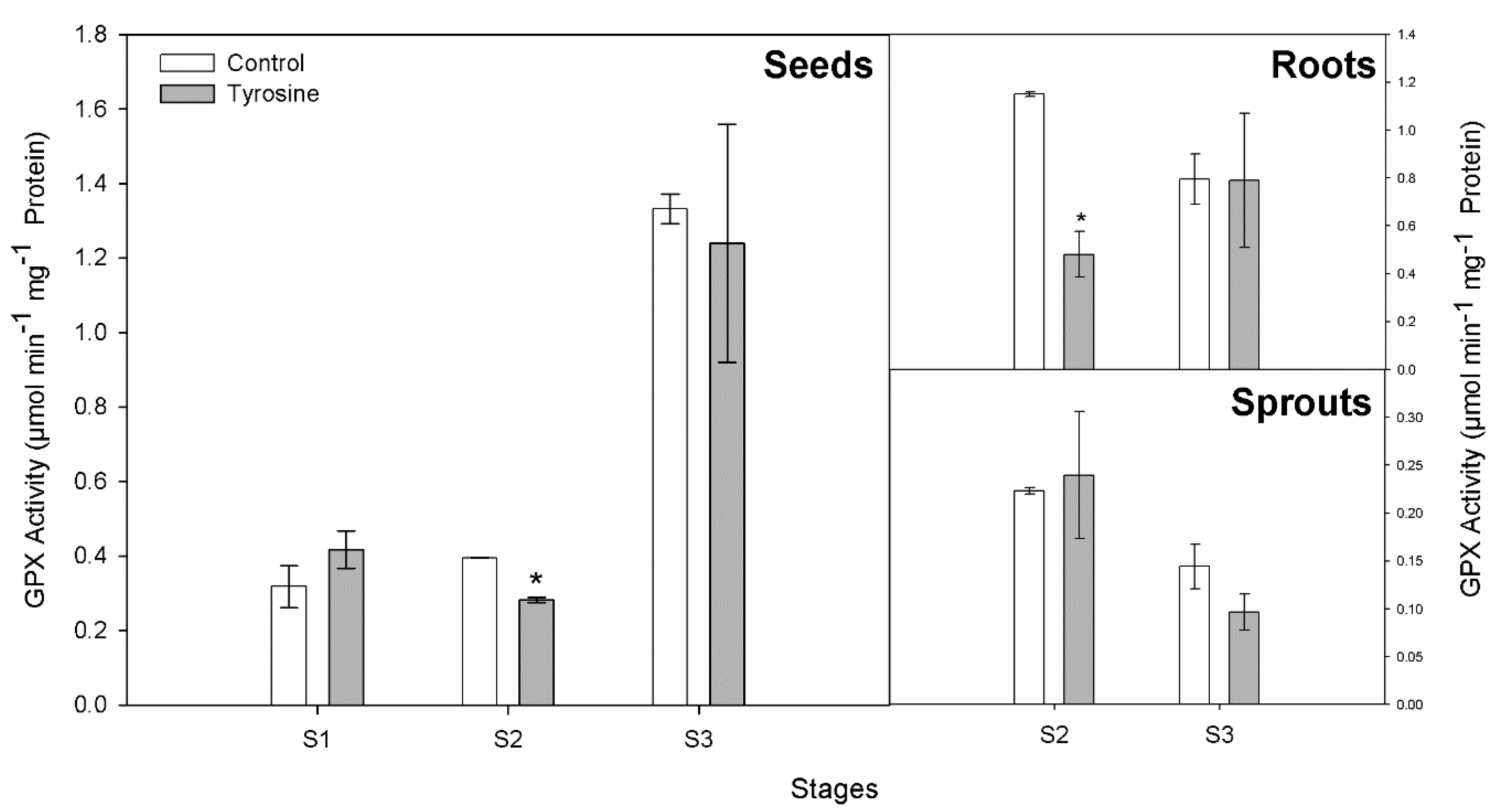
3.3. GPX and TYROX Activities

In control tissues, GPX only increased in the seed tissue as germination progressed, until stage S3 (2–3 leaves) (Figure 3). This in agreement with Shetty et al. [7,27]. In roots and sprouts, a decreasing GPX activity was observed (Figure 3).
GPX elicitation has been described as an early indicator of environmental changes in plants [15]. Indeed, the elevation of GPX activity has been reported as the only more general response to stimuli, such as metal exposure in V. faba [40]. Interestingly, Kabbadj [41] found that different cultivars of fava bean had different GPX responses to water deficit, and only the Reina Mora cultivar exhibited an increase in GPX activity.
Our results do not reflect any increased GPX activity with tyrosine treatment. On the contrary, GPX activity decreased or did not vary, depending on the organ (Figure 3). GPX has been proposed as a phenolic marker metabolism; thus, precursors derived from shikiimate route could be used by GPX for phenolic polymerization in structural development processes (germination) [29]. In our experiment, tyrosine treatment implies abundant phenolics, suggesting no need of increment in GPX activity. TYROX activity was not detected in all our experiments. The positive control for tyrosine oxidase (Beta vulgaris roots) was run in parallel in all samples and yielded proper activities 0.6 mmol min−1 g−1 DW.

4. Conclusions

Overall, it can be stated that to the best of our knowledge, the occurrence of such concentrated L-DOPA content in specific organ tissues has never been reported. Sprouts of V. faba (var. Super Agua Dulce) have the highest L-DOPA content detected until now 125 mg g−1 DW (12% DW). It can be also concluded that tyrosine exposure (0.55 mM) does not enhance L-DOPA content and that the high L-DOPA content found in the latest stage of growth studied (sprouts) does not seem to be related to GPX or TYROX activities. In terms of L-DOPA quantification, both Arnow’s and HPTLC methods were consistent in terms of tendencies and magnitudes found, except for the scarce contents found in seeds, in which HPTLC was more sensitive.

Author Contributions

Formal analysis, C.A.O.-S.; Funding acquisition, C.A.O.-S.; Investigation, C.A.O.-S.; Methodology, C.A.O.-S., M.E.-F. and M.A.-B.; Project administration, C.A.O.-S.; Software, M.E.-F.; Supervision C.A.O.-S.; Validation, C.O.; Writing—original draft, C.O. and M.E.-F.; Writing—review and editing, C.A.O.-S.

Funding

This research was funded by DIUBB grant No. 165006 3/R and Escuela de Graduados Research Assistant grant No. 352/649/2017 from Universidad del Bio-Bio, and by FONDEQUIP grant No. 130209. The APC was funded by Universidad del Bio-Bio.

Conflicts of Interest

Claudia Oviedo-Silva, Mhartyn Elso-Freudenderga, and Mario Aranda-Bustos declare that they have no conflict of interest.

References

  1. Patil, S.A.; Apine, O.A.; Surwase, S.N.; Jadhav, J.P. Biological sources of L-DOPA: An alternative approach. Adv. Parkinson Dis. 2013, 2, 81–87. [Google Scholar] [CrossRef]
  2. Gautam, M.; Chandel, M.; Azmi, W. Therapeutic role of L-DOPA produced as a secondary metabolite from different legumes and plant sources. Ann. Phytomed. 2012, 1, 1–8. [Google Scholar]
  3. Rios, M.; Habecker, B.; Sasaoka, T.; Eisenhofer, G.; Tian, H.; Landis, S.; Roffler-Tarlov, S. Catecholamine synthesis is mediated by tyrosinase in the absence of tyrosine hydroxylase. J. Neurosci. 1999, 19, 3519–3526. [Google Scholar] [CrossRef] [PubMed]
  4. Gish, M.; Mescher, M.C.; De Moraes, C.M. Targeted predation of extrafloral nectaries by insects despite localized chemical defenses. Proc. R. Soc. B 2015, 282, 20151835. [Google Scholar] [CrossRef] [PubMed]
  5. Sharma, N. L-DOPA Inhibited Early Root Growth in Rice Involves Biochemical Alterations in Macromolecules and Associated Hydrolytic Enzymes. Ann. Plant Sci. 2015, 4, 1109–1115. [Google Scholar]
  6. Soares, A.R.; Marchiosi, R.; Siqueira-Soares, R.D.; Barbosa de Lima, R.; Dantas dos Santos, W.; Ferrarese-Filho, O. The role of L-DOPA in plants. Plant Signal. Behav. 2014, 9, 28275. [Google Scholar] [CrossRef] [PubMed]
  7. Shetty, P.; Atallah, M.T.; Shetty, K. Enhancement of total phenolic, L-DOPA and proline contents in germinating fava bean (Vicia faba) in response to bacterial elicitors. Food Biotechnol. 2001, 15, 47–67. [Google Scholar] [CrossRef]
  8. Inamdar, S.; Joshi, S.; Jadhav, J.; Bapat, V. Innovative use of intact seeds of Mucuna monosperma Wight for improved yield of L-DOPA. Nat. Prod. Bioprospecting 2012, 2, 16–20. [Google Scholar] [CrossRef]
  9. Mohseni Mehran, S.M.; Golshani, B. Simultaneous Determination of Levodopa and Carbidopa from Fava Bean, Green Peas and Green Beans by High Performance Liquid Gas Chromatography. J. Clin. Diagn. Res. 2013, 7, 1004–1007. [Google Scholar] [CrossRef]
  10. Burbano, C.; Cuadrado, C.; Muzquiz, M.; Cubero, J.I. Variation of favism-inducing factors (vicine, convicine and L-DOPA) during pod development in Vicia faba L. Plant Foods Hum. Nutr. 1995, 47, 265–274. [Google Scholar] [CrossRef] [PubMed]
  11. Goyoaga, C.; Burbano, C.; Cuadrado, C.; Varela, A.; Guillamón, E.; Pedrosa, M.; Muzquiz, M. Content and distribution of vicine, convicine and L-DOPA during germination and seedling growth of two Vicia faba L. varieties. Eur. Food Res. Technol. 2008, 227, 1537–1542. [Google Scholar] [CrossRef]
  12. Raghavendra, S.; Ramesh, C.K.; Kumar, V.; Moinuddin Khan, M.H. Elicitors and precursor induced effect on L-DOPA production in suspension cultures of Mucuna pruriens L. Front. Life Sci. 2011, 5, 127–133. [Google Scholar] [CrossRef]
  13. Schenck, C.A.; Maeda, H.A. Tyrosine biosynthesis, metabolism, and catabolism in plants. Phytochemistry 2018, 149, 82–102. [Google Scholar] [CrossRef] [PubMed]
  14. Yoruk, R.; Marshall, M.R. A survey on the potential mode of inhibition for oxalic acid on polyphenol oxidase. J. Food Sci. 2003, 68, 2479–2485. [Google Scholar] [CrossRef]
  15. Gill, S.S.; Tuteja, N. Reactive oxygen species and antioxidant machinery in abiotic stress tolerance in crop plants. Plant Physiol. Biochem. 2010, 48, 909–930. [Google Scholar] [CrossRef] [PubMed]
  16. Siqueira-Soares, R.D.; Soares, A.R.; Parizotto, A.V.; Ferrarese, M.D.; Ferrarese-Filho, O. Root growth and enzymes related to the lignification of maize seedlings exposed to the allelochemical L-DOPA. Sci. World J. 2013, 2013, 134237. [Google Scholar] [CrossRef] [PubMed]
  17. Li, J.L.; Sulaiman, M.; Beckett, R.P.; Minibayeva, F.V. Cell wall peroxidases in the liverwort Dumortiera hirsuta are responsible for extracellular superoxide production, and can display tyrosinase activity. Physiol. Plant. 2010, 138, 474–484. [Google Scholar] [CrossRef] [PubMed]
  18. Sharma, P.; Jha, A.B.; Dubey, R.S.; Pessarakli, M. Reactive oxygen species, oxidative damage, and antioxidative defense mechanism in plants under stressful conditions. J. Bot. 2012, 2012, 1–26. [Google Scholar] [CrossRef]
  19. Saeidian, S. Kinetics properties of polyphenol oxidase in hawthorn (Crataegus spp.). Int. Lett. Nat. Sci. 2014, 25, 29–38. [Google Scholar] [CrossRef]
  20. O’Brien, J.A.; Daudi, A.; Butt, V.S.; Bolwell, G.P. Reactive oxygen species and their role inplant defense and cell wall metabolism. Planta 2012, 236, 765–779. [Google Scholar] [CrossRef] [PubMed]
  21. Randhir, R.; Shetty, P.; Shetty, K. L-DOPA and total phenolic stimulation in dark germinated fava bean in response to peptide and phytochemical elicitors. Process Biochem. 2002, 37, 1247–1256. [Google Scholar] [CrossRef]
  22. Kampmann, M.; Boll, S.; Kossuch, J.; Bielecki, J.; Uhl, S.; Kleiner, B.; Wichmann, R. Efficient immobilization of mushroom tyrosinase utilizing whole cells from Agaricus bisporus and its application for degradation of bisphenol A. Water Res. 2014, 57, 295–303. [Google Scholar] [CrossRef] [PubMed]
  23. Lin, C.C.; Kao, C.H. NaCl induced changes in ionically bound peroxidase activity in roots of rice seedlings. Plant Soil 1999, 216, 147. [Google Scholar] [CrossRef]
  24. Jebara, S.; Jebara, M.; Limam, F.; Aouani, M.E. Changes in ascorbate peroxidase, catalase, guaiacol peroxidase and superoxide dismutase activities in common bean (Phaseolus vulgaris) nodules under salt stress. J. Plant Physiol. 2005, 162, 929–936. [Google Scholar] [CrossRef] [PubMed]
  25. Schneider, C.A.; Rasband, W.S.; Eliceiri, K.W. NIH Image to ImageJ: 25 years of image analysis. Nat. Methods 2012, 9, 671–675. [Google Scholar] [CrossRef] [PubMed] [Green Version]
  26. Arnow, L.E. Colorimetric determination of the components of 3,4-dihydroxyphenylalanine-tyrosine mixtures. J. Biol. Chem. 1937, 118, 531–537. [Google Scholar]
  27. Shetty, P.; Atallah, M.T.; Shetty, K. Effects of UV treatment on the proline-linked pentose phosphate pathway for phenolics and L-DOPA synthesis in dark germinated Vicia faba. Process Biochem. 2002, 37, 1285–1295. [Google Scholar] [CrossRef]
  28. Vattem, D.; Randhir, R.; Shetty, K. Cranberry phenolics-mediated elicitation of antioxidant enzyme response in fava bean (Vicia faba) sprouts. J. Food Biochem. 2005, 29, 41–70. [Google Scholar] [CrossRef]
  29. Ray, H.; Georges, F. A genomic approach to nutritional, pharmacological and genetic issues of faba bean (Vicia faba): Prospects for genetic modifications. GM Crops 2010, 1, 99–106. [Google Scholar] [CrossRef] [PubMed]
  30. Randhir, R.; Shetty, K. Light-mediated fava bean (Vicia faba) response to phytochemical and protein elicitors and consequences on nutraceutical enhancement and seed vigour. Process Biochem. 2003, 38, 945–952. [Google Scholar] [CrossRef]
  31. Wichers, H.J.; Wijnsma, R.; Visser, J.F.; Malingré, T.M.; Huizing, H.J. Production of L-DOPA by cell suspension cultures of Mucuna pruriens. Plant Cell Tissue Organ Cult. 1985, 4, 75–82. [Google Scholar] [CrossRef]
  32. Etemadi, F.; Hashemi, M.; Randhir, R.; ZandVakili, O.; Ebadi, A. Accumulation of L-DOPA in various organs of faba bean and influence of drought, nitrogen stress, and processing methods on L-DOPA yield. Crop J. 2018. [CrossRef]
  33. Fujii, Y.; Shibuya, T.; Yasuda, T. L-3, 4-dihydroxyphenylalanine as an allelochemical candidate from Mucuna pruriens (L.) DC. var. utilis. Agric. Boil. Chem. 1991, 55, 617–618. [Google Scholar] [CrossRef]
  34. Prakash, D.; Abhishek Niranjan, S.K.; Tewari, D. Some nutritional properties of the seeds of three Mucuna species. Int. J. Food Sci. Nutr. 2001, 52, 79–82. [Google Scholar] [CrossRef] [PubMed]
  35. Siddhuraju, P.; Becker, K. Rapid reversed-phase high performance liquid chromatographic method for the quantification of L-DOPA (L-3,4-dihydroxyphenylalanine), non-methylated and methylated tetrahydroisoquinoline compounds from Mucuna beans. Food Chem. 2001, 72, 389–394. [Google Scholar] [CrossRef]
  36. Ramya, K.B.; Thaakur, S. Herbs containing L-DOPA: An update. Anc. Sci. Life 2007, 27, 50. [Google Scholar] [PubMed]
  37. Hildebrandt, T.M.; Nesi, A.N.; Araújo, W.L.; Braun, H.P. Amino acid catabolism in plants. Mol. Plant 2015, 8, 1563–1579. [Google Scholar] [CrossRef] [PubMed]
  38. Ellis, B.E. Catabolic ring-cleavage of tyrosine in plant cell cultures. Planta 1973, 111, 113–118. [Google Scholar] [CrossRef] [PubMed]
  39. Rani, N.; Joy, B.; Abraham, T.E. Cell suspension cultures of Portulaca grandiflora. as potent catalysts for biotransformation of L-tyrosine into L-DOPA, an anti-Parkinson’s drug. Pharm. Boil. 2007, 45, 48–53. [Google Scholar] [CrossRef]
  40. Nadgórska-Socha, A.; Kafel, A.; Kandziora-Ciupa, M.; Gospodarek, J.; Zawisza-Raszka, A. Accumulation of heavy metals and antioxidant responses in Vicia faba plants grown on monometallic contaminated soil. Environ. Sci. Pollut. Res. 2013, 20, 1124–1134. [Google Scholar] [CrossRef] [PubMed]
  41. Kabbadj, A.; Makoudi, B.; Mouradi, M.; Pauly, N.; Frendo, P.; Ghoulam, C. Physiological and biochemical responses involved in water deficit tolerance of nitrogen-fixing Vicia faba. PLoS ONE 2017, 12, e0190284. [Google Scholar] [CrossRef] [PubMed]
Figure 1. Effect of tyrosine treatment on Levo-3,4-dihydroxyphenylalanine (L-DOPA) content in Vicia faba seeds, roots, and sprouts at the early stages of development: S1: 3–4 days, S2: 8–9 days, and S3: 2–4 leaves. The bars indicate ± standard error (n = 3). * indicates significant differences between controls and tyrosine treatments according to Tukey (P < 0.05).
Applsci 08 02431 g001
Figure 2. Effect of tyrosine treatment on root length, number of roots, and length of sprouts in V. faba at the early stages of development: S1: 3–4 days, S2: 8–9 days, and S3: 2–4 leaves. Bars indicate ± standard error (n = 3). No significant differences between controls and tyrosine treatments were found, Tukey (P < 0.05).
Applsci 08 02431 g002
Figure 3. Effect of tyrosine treatment on guaiacol peroxidase (GPX) activity in V. faba seeds, roots, and sprouts at the early stages of development: S1: 3–4 days, S2: 8–9 days, and S3: 2–4 leaves. The bars indicate ± standard error (n = 3). “*” indicates significant differences between controls and tyrosine treatments according to Tukey (P < 0.05).
Applsci 08 02431 g003
Table 1. L-DOPA content (mg g−1 dry matter) in different V. faba tissues.
StagesArnowHPTLC
SeedRootSproutSeedRootSprout
S10.90 ± 0.11**4.40 ± 0.06**
S21.90 ± 0.1669.43 ± 3.0581.25 ± 2.918.07 ± 1.3088.50 ± 5.54125.87 ± 17.18
S35.69 ± 0.2669.45 ± 5.3389.28 ± 2.0614.27 ± 0.3889.67 ± 1.76113.60 ± 9.53
* Nonexistent organ.

