1. Introduction
Tracheotomy is a fundamental procedure to ensure airway patency in patients with severe airway obstruction [
1]. The main approaches are surgical and percutaneous, and the choice can be challenging, depending on the specific patient case and several factors, including age, which serve as discriminating criteria [
2,
3]. The surgical approach consists of a direct incision and dissection of the cervical soft tissues to expose the trachea and create the airway opening, and is generally considered more adaptable to varied clinical scenarios and complex anatomy, making it the standard technique in many pediatric settings [
1,
2,
4,
5].
In contrast, the percutaneous approach involves the sequential dilation of the tracheal tract via a small incision using a guidewire-based technique, and is often associated with shorter procedural time, reduced wound infections, and improved cosmetic outcomes compared with open methods. Technical challenges associated with pediatric patients involve the risk of posterior tracheal wall perforation and the correct identification of relevant anatomical landmarks, such as tracheal rings, due to the flexibility of underdeveloped cartilages and the small size of the airways [
1,
2,
3].
While percutaneous tracheotomy is routinely performed in adult patients because of its minimally invasive nature, decreased cost, and safety profile compared to the open procedure [
2,
6], its adoption in pediatric patients has been slower. This is mainly due to anatomical and physiological differences that increase both procedural difficulty and potential risks. In recent years, percutaneous tracheotomy has gained acceptance in selected pediatric patients, generally considered safe in children older than 10 years, and in some pediatric hospitals, it has been successfully performed in children as young as 5 years [
2]. Among the available percutaneous techniques, the Frova screw technique is a method of tracheal access typically performed under bronchoscopic guidance to ensure correct placement and avoid injury to surrounding structures. In this technique, a self-tapping plastic screw is gently rotated through the anterior tracheal wall, typically at the level of the second or third tracheal rings, allowing safe entry into the tracheal lumen [
1,
2,
7].
Given the complexity of this procedure, medical simulation plays a crucial role by providing a safe environment in which both technical and non-technical skills can be developed and refined, thereby reducing the risk of errors without compromising patient safety [
8,
9,
10]. Despite the growing recognition of the importance of medical simulation, opportunities for hands-on practice in rare but high-risk pediatric interventions, such as percutaneous tracheotomy with the Frova screw, remain limited [
11].
In response to the above specific training need, and following the successful development of the task trainer designed to support the training of pediatric surgical tracheotomy by Brogi et al. [
12], a collaboration was established between the Department of Industrial Engineering of the University of Florence and the Respiratory Endoscopy Unit of Meyer Children’s Hospital IRCCS within the T3Ddy Laboratory, leading to the development of an advanced physical simulator for percutaneous tracheotomy using a specially designed screw-type dilator (PercuTwist, Rüsch, Kernen, Germany) for the Frova screw technique [
7].
2. State of the Art and Commercial Analysis
The literature regarding simulators for pediatric front-of-neck access (FONA) is scarce, and most studies introduce significant simplification from both an anatomical and functional standpoint. Kovatch et al. described a simulator for FONA training, obtained by modifying a commercial manikin through the introduction of a softer tracheal structure [
13]. Other studies have reported the development of rigid tracheal models produced using 3D printing, generally in polylactic acid (PLA) [
14] or photopolymer resins [
15,
16,
17,
18], primarily intended for cricothyrotomy training.
The models described in the literature typically reproduce only the cricothyroid membrane or limited portions of the laryngeal structures and therefore do not adequately represent the anatomical complexity of the cervical region. Overall, they do not include key anatomical elements such as tracheal rings and surrounding soft tissue structures, nor do they integrate systems for bleeding simulation. Furthermore, none of the described models are designed to allow the insertion of a bronchoscope for internal visualization during the procedure. Collectively, the available simulators primarily focus on simplified anatomical representations of emergency airway access, highlighting the need for more comprehensive models capable of reproducing both the anatomical complexity of the pediatric neck and the procedural workflow required for bronchoscope-guided percutaneous tracheotomy.
An analysis of currently available commercial simulators shows that, although they allow training in FONA procedures, including percutaneous tracheotomy, they still present several limitations. The representation of the anatomical stratification of the cervical region is generally simplified, with a transition between skin and trachea that is often unrealistic, due to the limited reproduction of subcutaneous tissue. In addition, relevant anatomical elements such as clearly palpable tracheal rings and surrounding cervical structures are frequently absent or only partially reproduced. A further key limitation concerns the lack of material differentiation between the tracheal wall and cartilaginous rings, which are typically represented as a single homogeneous structure, resulting in reduced tactile realism. Bleeding simulation systems are also rarely integrated. Finally, most models do not allow for bronchoscope insertion for procedure monitoring, thereby reducing procedural realism. A detailed comparison between commercially available simulators and the model proposed in this study is reported in
Table 1. None of the commercially available models provides a dedicated, anatomically accurate, multi-layered simulator for percutaneous tracheotomy. High-fidelity manikins are generally designed for comprehensive clinical scenarios or emergency airway management, with a primary focus on intubation or cricothyrotomy, and therefore lack the level of anatomical detail and tissue interaction required for percutaneous tracheotomy training. Conversely, low- and mid-fidelity task trainers are typically limited to isolated technical skills and do not reproduce key features such as layered cervical tissues, bleeding simulation, interaction with cartilaginous structures, or procedural verification through bronchoscope insertion.
3. Materials and Methods
3.1. Layout Definition
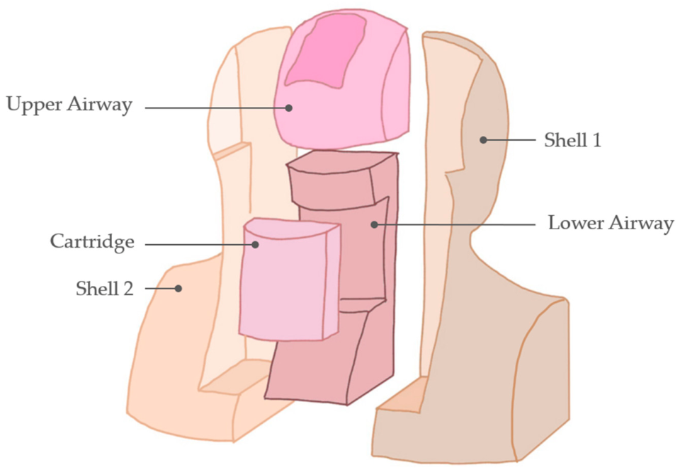
The proposed simulator aims to address the main limitations of currently available models by reproducing the relevant cervical structures involved in the procedure and the multilayer tissue structure for a realistic dissection, while allowing the endoluminal monitoring. The overall layout of the simulator and the selection of the reproduced anatomical structures were defined in consultation with clinicians, whose feedback contributed to ensuring the clinical relevance of the design. Referring to
Figure 1, the simulator features a modular structure consistent with approaches proposed in the literature [
12,
19] to allow easy replacement of the disposable Cartridge corresponding to the simulation site and worn-out parts. Moreover, this approach reduces maintenance and resetting costs between simulation sessions and enables future integration of novel cartridges representing additional clinical scenarios.
The simulator reproduces the anatomy of a generic 8-year-old patient in neck hyperextension and features an open oral cavity to allow insertion of a rigid bronchoscope for endoluminal monitoring throughout the procedure. The reusable structure of the simulator consists of two Shells housing the Upper Airway component, in which the Tongue and the Teeth are embedded, as well as the Lower Airway component, into which the Cartridge is inserted. The entire structure is covered with a 5 mm-thick silicone layer simulating the Skin, also serving as a containment element enhancing the stability of internal components. To further ensure structural integrity during simulation sessions, a non-slip Back Cover is secured to the Shells, immobilizing the Skin and preventing the simulator from slipping on the working surface.
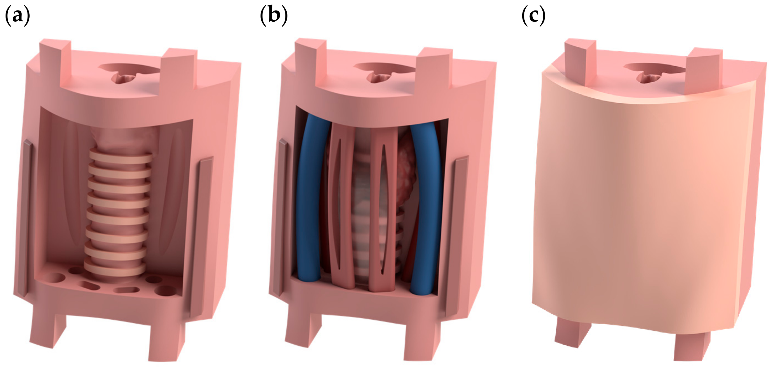
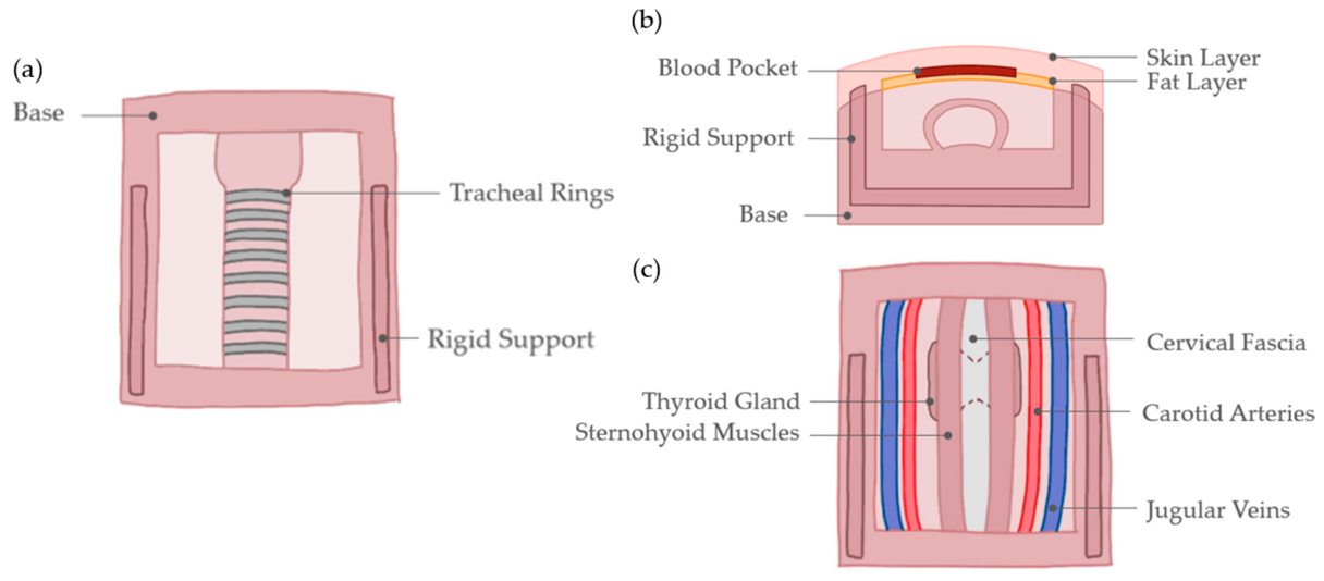
The Cartridge, whose layout is reported in
Figure 2, integrates several key anatomical elements of the cervical region, embedded in a tracheal-shaped Base that extends from the vocal cords to the carina. Tracheal rings are also forecast due to their crucial role in the palpation phase of the procedure. A Rigid Support is incorporated into the Cartridge’s Base matrix to provide stability and prevent structural collapse during procedural execution. The elements embedded into the Base include Thyroid Gland, Cervical Fascia, Sternohyoid Muscles, and major vascular structures such as the Jugular Veins and Carotid Arteries. The Cartridge is covered by a bi-component Skin Layer simulating realistic cutaneous stratification. Between the skin and subcutaneous layers, a Pocket allows the insertion of blood-simulating liquid.
3.2. Modeling
In the present study, generic digital models of the trachea, thyroid gland, and cricoid and thyroid cartilages were obtained from an online repository [
20], were used to develop the simulator. Similarly, the external structure of the patient, including the cervical region, was generated using an anatomical model representative of an average 8-year-old pediatric patient without anatomical anomalies, obtained from the certified platform Daz Studio (Daz 3D, Salt Lake City, UT, USA). The model was selected to reflect average anatomical characteristics, and its dimensions were further verified through comparison with anatomical data reported in the literature [
21,
22,
23].
The external structure was subsequently processed using predefined morphing tools in Daz Studio to adapt the model into a neck hyperextension posture to enable the introduction of a rigid bronchoscope into the oral cavity.
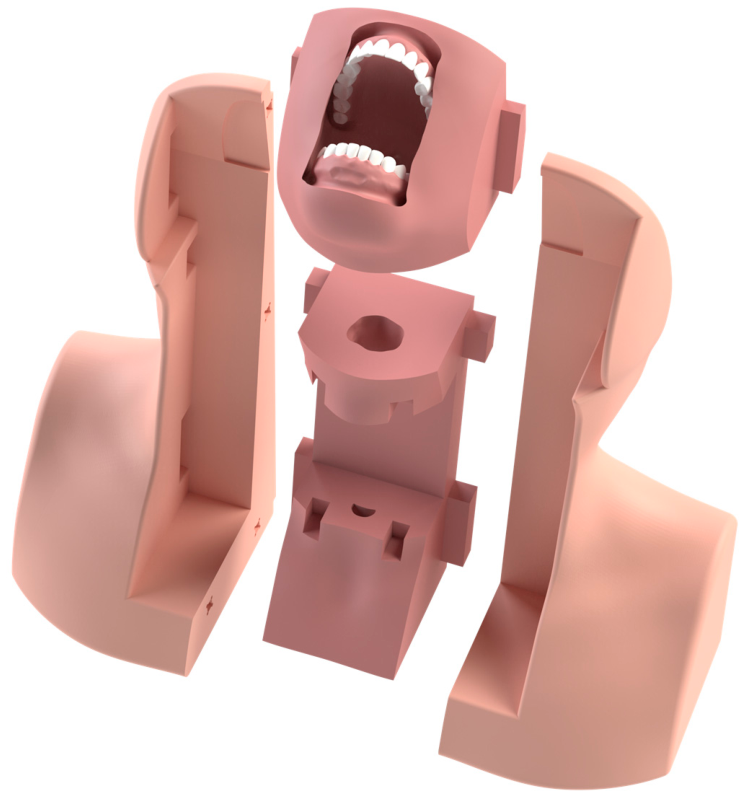
Additional modeling steps were performed using Geomagic Design X (3D Systems Inc., Rock Hill, SC, USA). The process ensured accurate alignment between the internal anatomical elements and the externally modeled structures generated in Daz Studio, followed by the subsequent geometric modifications required to complete the model. Based on the previously defined layout, the reusable structure was designed with the two rigid Shells housing the Upper and Lower Airway modules in dedicated cavities and held together by magnets for easy disassembly when needed (
Figure 3).
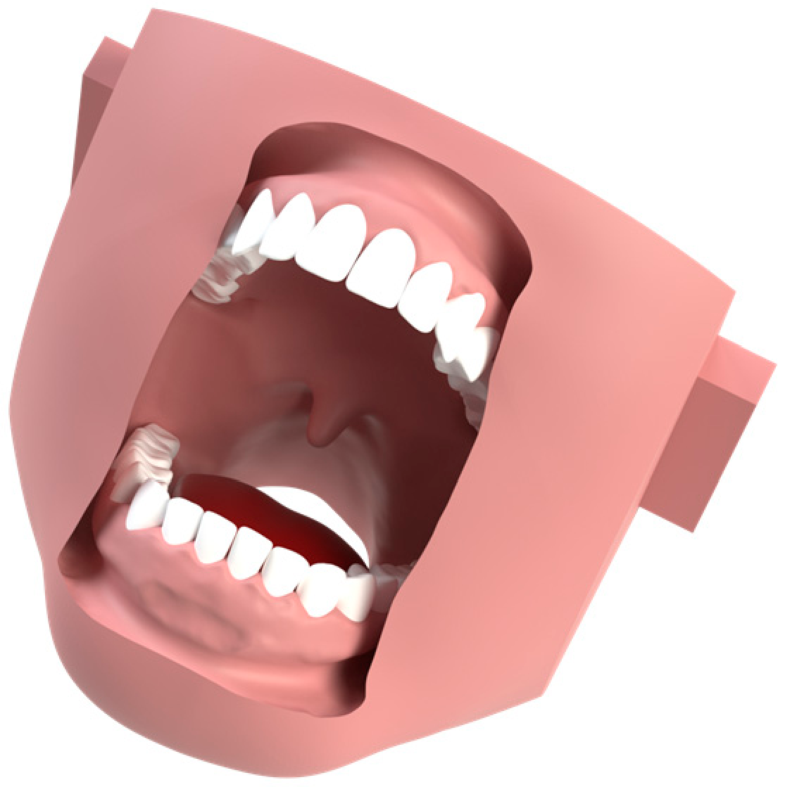
The Upper Airway component embeds two rigid structures representing the Teeth. These elements are integrated during the fabrication process using an exposed gyroid reinforcement, a design approach derived from the literature [
12], which allows the casting material to penetrate the lattice structure. During casting, the material infiltrates this structure, mechanically anchoring the rigid components to the surrounding soft matrix and ensuring their stable integration. The Tongue is modeled as a separate component within the oral cavity, ensuring realism during bronchoscope insertion and accurate reproduction of the overall anatomy. The final configuration of the Upper Airway embedding the Teeth and the Tongue is shown in
Figure 4.
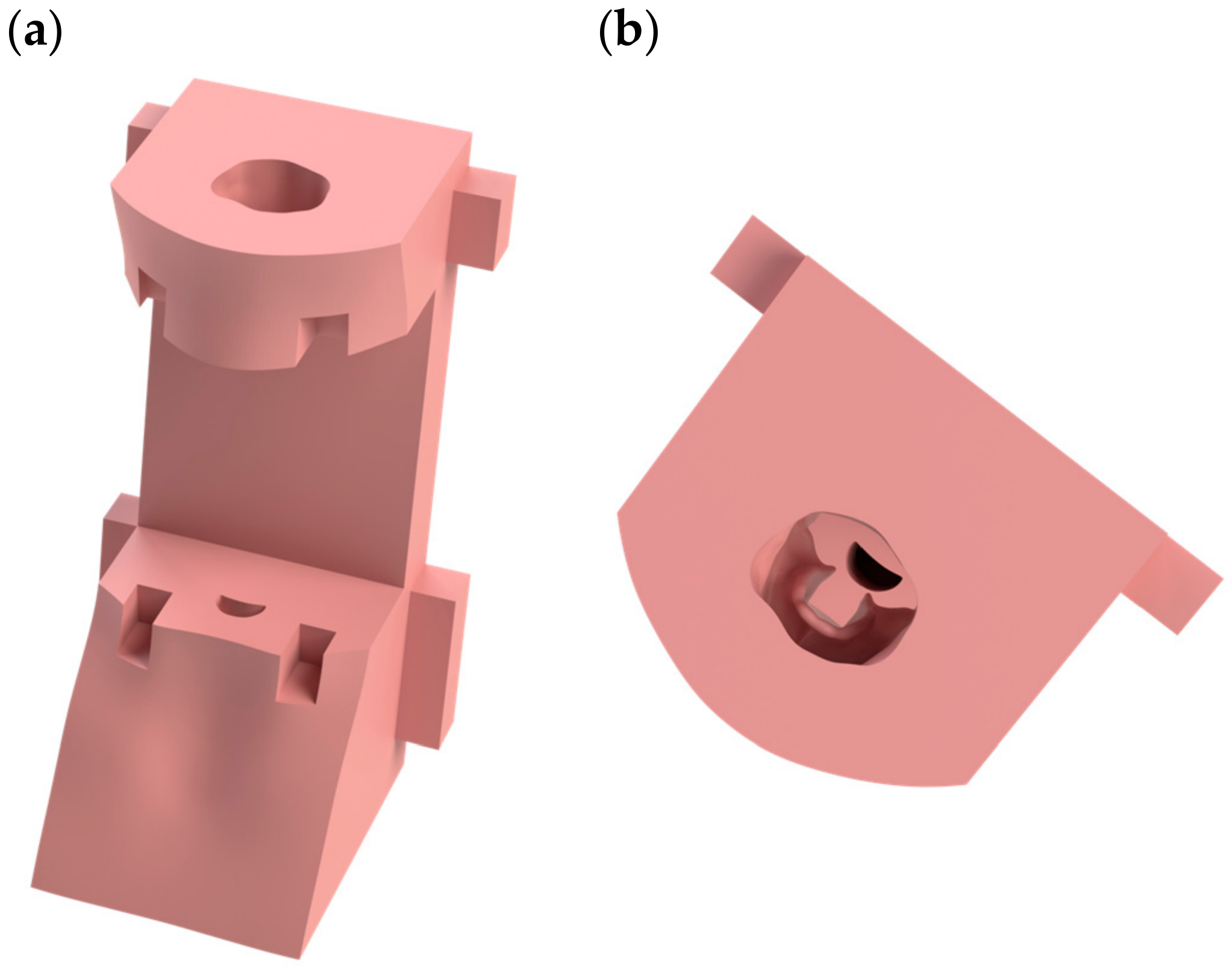
The Lower Airway component (
Figure 5) is modeled to allow the Cartridge to snap into place in a dedicated slot. It replicates the section of the tracheal lumen extending from the laryngeal inlet to the cricoid cartilage, as well as the distal portion of the trachea down to the carina, for greater realism. The central section of the trachea, however, is reproduced within the Cartridge, as detailed further below.
The Cartridge represents the core of the simulator, realistically replicating the neck region extending between the cricoid cartilage and the tracheal carina. Tracheal lumen dimensions were derived from the literature [
22], as well as the tracheal rings that were manually modeled to ensure proper alignment and high anatomical accuracy [
24]. The Cartridge’s structure, in accordance with the previously defined layout, consists of a Base representing the tracheal segment and embedding a Rigid Support to avoid collapse and provide stability during simulation. The structure is incorporated during fabrication, employing the exposed gyroid technique mentioned above. The anatomical structures reproduced in the Cartridge include the Thyroid Gland, Sternohyoid Muscles, Carotid Arteries, Jugular Veins, and Cervical Fascia for further realism in tissue stratification. Dimensions are consistent with those reported in the literature [
23,
25,
26]. A bi-component Skin Layer reproducing the skin and subcutaneous tissue covers the Cartridge and consists of two variable-thickness layers, iteratively optimized in collaboration with clinicians to provide realistic tactile feedback. The final configuration features a 5 mm-thick peripheral frame serving as a reference for alignment with the edges of the Cartridge’s Base, and a bi-layer central region consisting of a 2 mm-thick skin layer and a 1 mm-thick subcutaneous layer. A sealed pocket is created between these two components, into which a blood-mimicking fluid is introduced to enhance procedural realism. The Cartridge’s final configuration is reported in
Figure 6.
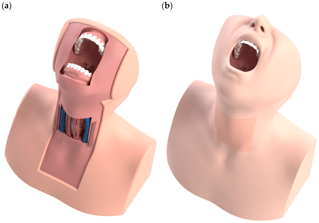
Finally, the simulator’s Skin consists of a 5 mm-thick layer replicating the appearance of a standard patient, with correct alignment ensured by predefined shape references. Stability during simulation sessions is ensured by the presence of a flap that fits in a channel between the back of the Shells and a Back Cover secured with screws and featuring a non-slip layer. The final configuration of the simulator is reported in
Figure 7.
3.3. Material Selection and Fabrication
Material selection was carried out consistently with T3Ddy Laboratory’s prior experience [
12,
19] and in consultation with medical experts. An exception was made for the tracheal body, including the tracheal rings, for which no clear guidance was available in the literature and for which T3Ddy Laboratory had no prior fabrication experience. To address this gap, three prototypes were developed using the same silicone matrix, with tracheal rings fabricated, respectively, from polylactic acid (PLA), thermoplastic polyurethane (TPU), and Elastic 50A stereolithography resin. These materials were selected due to their differing mechanical properties, which result in distinct tactile responses. The samples were tested and evaluated blindly by a panel of four expert clinicians in percutaneous tracheotomy from the Respiratory Unit of the Meyer Children’s Hospital IRCSS, who assessed the tactile feedback of the samples through direct palpation based on their clinical experience. PLA was unanimously identified as the most suitable material in terms of tactile perception, rigidity, and procedural realism.
Components were fabricated using a combination of additive manufacturing, molding, and casting techniques. Rigid elements, including Shells, Tracheal Rings, Cartridge’s Rigid Support, and molds, were fabricated in PLA using Prusa Research (Prague, Czech Republic) fused deposition modeling (FDM) 3D printers. Soft components were manufactured using platinum-cure silicones of varying hardness and silicone foam, ensuring realistic haptic feedback and tissue behavior during simulation.
Molds were designed for the fabrication of the soft components, following commonly adopted principles in silicone casting and mold design [
12]. In the case of the Upper Airway component, the presence of multiple undercuts generated by the dental structures made the use of a rigid mold unsuitable for demolding. To address this issue, a flexible silicone mold was developed, enabling compliance of both the cast component and the mold to facilitate demolding while preserving anatomical details. To enable the production of this flexible mold, dedicated master molds were designed. Materials used for each component and respective costs are listed in
Table 2. Costs refer to raw material expenses only and exclude design time, labor, and equipment depreciation.
3.4. Assembly
Before framing the Cartridge into the reusable part of the simulator, careful assembly of each Cartridge’s component must be done. Within the Base, embedding Tracheal Rings and the Rigid Support, Thyroid Gland, Cervical Fascia, Carotid Arteries, Jugular Veins, and Sternohyoid Muscles are glued using a silicone-based adhesive. Correct anatomical positioning is ensured by the presence of alignment references and slots to guide component placement. The bi-component Skin Layer covering the Cartridge is then assembled by attaching the edges of the subcutaneous layer to the outer skin layer. This generates a central pocket to contain the blood-mimicking fluid. Once filled, the pocket is sealed to prevent undesired leakage, and the Skin Layer is glued to the Cartridge’s edges using silicone adhesive, ensuring a stable assembly throughout the procedure.
Once the Tongue has been glued into the oral cavity of the Upper Airway component, the reusable part of the simulator can be easily assembled by framing the Upper and Lower Airway components into the magnetic-coupling Shells.
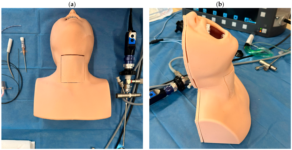
The Cartridge is positioned within the Lower Airway, and the entire simulator is covered with the Skin. Finally, the non-slip Back Cover is secured using screws, ensuring a robust and durable assembly (
Figure 8).
The overall stability of the simulator is ensured by the Shells, further supported by the Skin and the Back Cover, which secures the system in place during a simulation session. In addition, friction between the silicone Cartridge and the silicone Lower Airway prevents unintended detachment or movement, ensuring that the Cartridge remains properly positioned throughout the procedure. At the same time, the inherent compliance of the silicone allows for smooth replacement of the used Cartridge without requiring removal of the screwed Back Cover.
In addition to the very smooth Cartridge replacement between subsequent simulation sessions, the simulator’s modular architecture allows selective replacement of other components in case of accidental damage or wear and tear, without requiring an entire substitution.
4. Validation
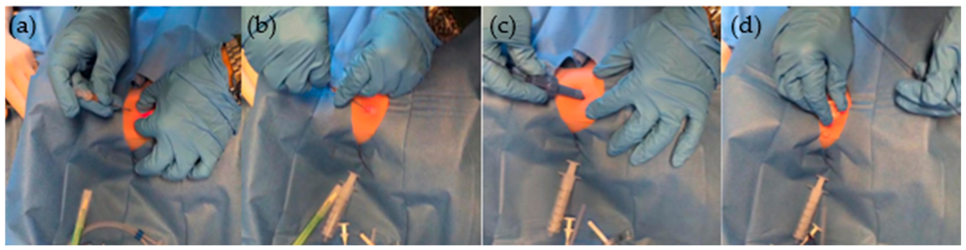
Validation was performed during a structured theoretical and practical course in pediatric operative bronchoscopy held by Meyer Children’s Hospital IRCCS. The course was open to attendings and residents from different specialties, including anaesthesiology, intensive care, pediatric surgery, neonatology, emergency medicine, otorhinolaryngology, and pediatrics, with participation based on voluntary registration. The course aimed to enable participants to acquire competencies in pediatric difficult airway management, including percutaneous tracheostomy using a specially designed screw-type dilator performed on the developed simulator (
Figure 9;
Video S1).
All participants were organized in pairs, and each performed the procedure twice, alternating roles: once as the primary operator and once as the assistant who handled the bronchoscope. The procedure was conducted in a realistic clinical scenario within a simulation environment using standard surgical equipment for percutaneous tracheotomy with the Frova screw technique, and all clinicians completed the full procedure under the supervision of experienced physicians with extensive expertise in tracheotomy procedures.
The evaluation was carried out using a questionnaire based on previously published studies [
12,
19], which describe similar approaches for the validation of pediatric manikins and are consistent with routine clinical practice. The assessment was qualitative and grounded in the clinical experience of the participants and included 15 items reported in
Table 3, using a 5-point Likert scale (1 = strongly disagree, 2 = disagree, 3 = neutral, 4 = agree, 5 = strongly agree). The questionnaire was designed to assess four key aspects: the anatomical realism of the simulator (S1–S3), the fidelity of the simulated scenario (S4–S6), interaction with surgical instruments (S7–S9), and the perceived clinical relevance for training in difficult pediatric airway management (S10–S15). Sections regarding personal data, professional expertise level, and previous experience with the procedure were also included, as well as a comment section.
5. Results
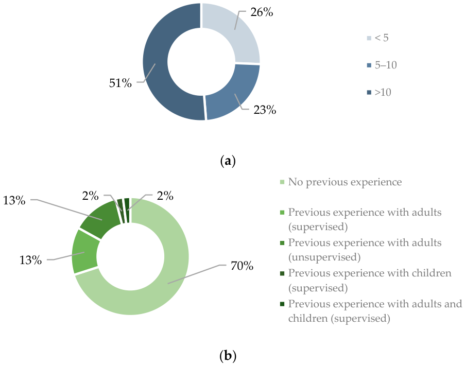
The mean age of the 39 participants who completed the questionnaire was 43.3 years. 36% (14 out of 39) were male, and 64% (25 out of 39) were female. They were all attendings, although none of them had experience performing Frova’s PercuTwist dilational percutaneous tracheotomy on pediatric patients without supervision.
Specifically, 70% (27 out of 39) had no previous experience, while 13% (5 out of 39) had performed percutaneous tracheotomy using the Frova screw technique on adult patients under supervision, and 13% (5 out of 39) without supervision.
In addition, there was one participant who had previous experience performing the procedure on both adult and pediatric patients under supervision, and likewise, only one participant had performed the procedure on pediatric patients under supervision (
Figure 10).
Mean scores for each statement are reported in
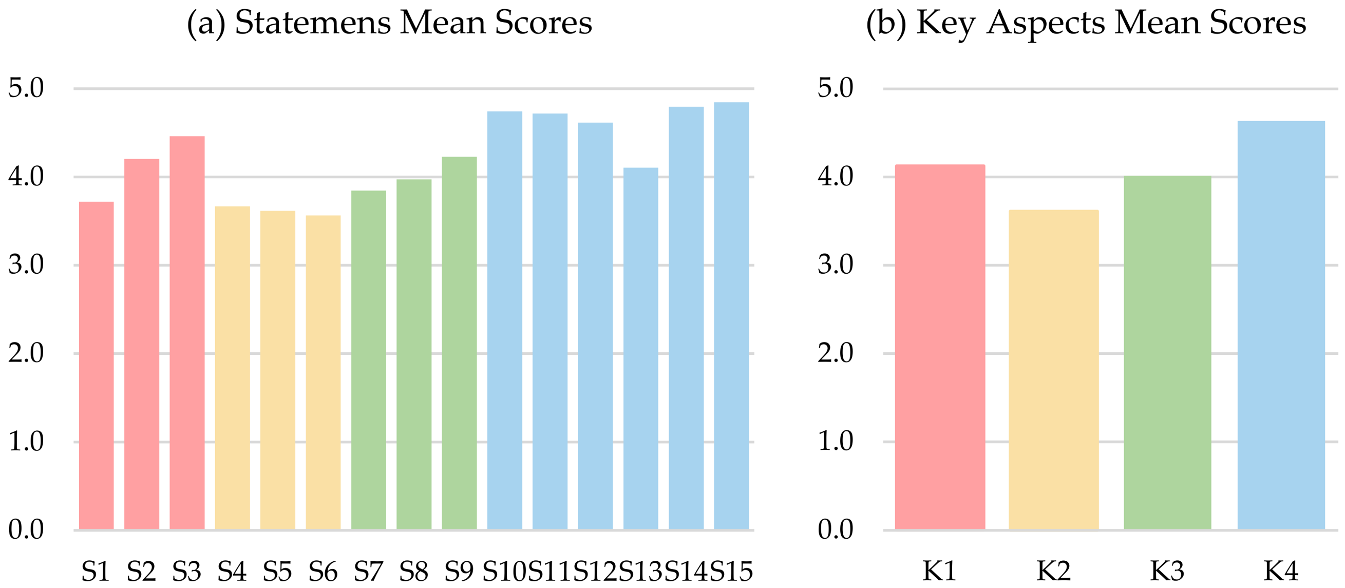
Figure 11, together with the mean values calculated for each of the four key aspects (simulator realism, procedural fidelity, surgical instruments interaction, and educational relevance). The simulator obtained an overall score of 4.2/5, which indicates a high level of perceived realism and educational value among participants.
A subgroup analysis was also performed based on previous experience in percutaneous tracheotomy. The mean overall score was 4.2/5 among participants without experience and 4.1/5 among those with experience, indicating comparable evaluations of the simulator’s realism and perceived educational value across different levels of clinical expertise.
6. Discussion
The results indicate an overall positive perception of the proposed simulator as a training tool for pediatric percutaneous tracheotomy using Frova’s PercuTwist dilational percutaneous tracheostomy. Across the 15 questionnaire items, mean scores ranged from 3.6 to 4.7 on a 5-point Likert scale, reflecting a generally high level of satisfaction in terms of anatomical fidelity, procedural interaction, and educational value.
Concerning anatomical realism (S1–S3), participants reported good agreement with the fidelity of the simulated anatomy. The geometry of the simulated region (S2) and the representation of the endoscopic view (S3) received relatively high scores (4.2 and 4.5, respectively), suggesting that both the external and internal visual references were considered accurate for procedural guidance. The item concerning the anatomical dimensions of the simulator (S1) received a slightly lower rating (3.7), indicating that while the model was generally perceived as anatomically accurate, some discrepancies in scale may exist. This observation is consistent with the single qualitative comment provided by a participant, which suggested that the overall dimensions appeared slightly larger than those expected for an 8-year-old patient. It should be noted that the anatomical structures used for the simulator were derived from publicly available anatomical datasets and scaled according to morphometric values reported in the literature, thus representing an average 8-year-old pediatric anatomy. The perceived discrepancy may therefore be related to the partial configuration of the simulator, which reproduces only the cervical region rather than a full-body manikin. In the absence of the surrounding anatomical context, the proportions of the simulated region may appear larger than expected.
The fidelity of the simulated scenario (S4–S6) reported satisfactory ratings as well. The realism of palpation of the tracheal rings (S4) achieved a mean score of 3.7, indicating that participants generally found the tactile identification of anatomical landmarks realistic, although further improvements may enhance their definition. The feedback provided by the cutaneous layer during incision (S5) and the representation of bleeding (S6) both received scores around 3.6, representing the lowest ratings among all questionnaire items. These results suggest that while the simulator provides an acceptable baseline level of procedural realism, refinement of the soft tissue layers and fluid simulation system could improve the perceived fidelity of the surgical environment.
The category related to interaction with surgical instruments (S7–S9) demonstrated adequate results. The haptic feedback during manipulation (S7) obtained a mean score of 3.8, while the tracheal feedback during insertion of the Frova device (S8) was rated at 4.0, highlighting a satisfactory level of tactile realism, representing a relevant achievement given the technical complexity of reproducing tracheal mechanical feedback in a physical simulator. Notably, the technical feasibility of introducing the Frova screw into the trachea (S9) achieved one of the highest scores (4.2), indicating that the simulator successfully reproduces the mechanical interaction between the device and the airway.
The highest scores were observed regarding the perceived clinical relevance for training (S10–S15). Participants strongly agreed that the simulator represents an appropriate tool for training (S10), may have an impact on increasing patient safety (S11), and may improve procedural speed and success rate (S12), with positive reflections on clinical practice, all with mean scores from 4.6 to 4.7. Similarly, a score of 4.1 on S13 suggests that users’ perceived ability to perform pediatric percutaneous tracheotomy with the Frova screw improved after training on the simulator. The high level of accordance with the importance of using the simulator for teaching new procedures before transferring to clinical practice (S14) and for maintaining procedural skills among both experienced physicians and trainees (S15) further highlights the strong educational potential of the developed simulator.
These results suggest a high perceived value of simulation-based training in pediatric airway management, where procedural opportunities on real patients are rare and associated with significant risk.
7. Conclusions
Overall, data demonstrate that the simulator achieves a satisfactory balance between anatomical realism, procedural interaction, and didactic effectiveness. While certain aspects, particularly the realism of the skin layers and the scaling of anatomical dimensions, may benefit from further refinement, the strong endorsement from participants confirms the simulator’s utility as a valuable training tool.
The validation strategy adopted in this study relied on a structured questionnaire designed to capture expert qualitative assessment of anatomical and procedural realism. This approach represents a methodological choice consistent with the study’s primary objective of evaluating perceived realism, which inherently depends on the clinical experience and judgment of trained physicians. In this context, subjective evaluation is not an arbitrary assessment but rather an expert-informed appraisal grounded in real clinical practice.
Objective performance metrics such as procedure time, error rates, or success rates were not included in the present study, as their measurement during a single training session would primarily reflect individual participant performance rather than the intrinsic characteristics of the simulator. However, the integration of such metrics in future studies conducted over repeated training sessions would allow the analysis of learning curves and provide meaningful insight into the educational effectiveness of the simulator. Longitudinal assessment of performance improvement over time would therefore represent a valuable next step in evaluating the training potential of the proposed model. Moreover, the application of comparable performance metrics to commercially available simulators would enable structured benchmarking and comparative evaluation.
The integration of sensor-based technologies may be considered in future developments, particularly in the context of longitudinal studies aimed at evaluating learning curves, where objective acquisition of procedural metrics could provide additional quantitative support to educational outcomes.
This study does have some limitations. Among these, no formal assessment of construct validity or transfer validity was conducted through direct comparison with real clinical performance. Although all participants were attending physicians with varying levels of experience in percutaneous tracheotomy, this heterogeneity was not specifically analyzed to evaluate the simulator’s ability to discriminate between different levels of procedural expertise. The assessment of these aspects would require structured comparative studies and, regarding transfer validity, longitudinal designs with repeated measurements over time and analysis of learning curves.
Another limitation lies in the perceived scaling of the simulator, which may be influenced by the absence of a full-body anatomical context, potentially affecting users’ perception of overall proportions. Minor adjustments may therefore be considered in future developments to better align visual and tactile perception with clinical expectations.
Lower scores regarding the realism of cutaneous feedback during incision suggest that the current configuration of the skin layers may not fully reproduce the tactile response encountered in clinical practice. From a development perspective, future iterations of the simulator will focus on improving the multilayered structure of the soft tissues representing the skin, including the subcutaneous layer, to enhance tactile stability during incision and manipulation. This modification may contribute to more realistic interaction with surgical instruments and improve the fidelity of tactile feedback.
Similarly, the bleeding simulation system remains limited in reproducing realistic intraoperative bleeding patterns. Future developments will therefore focus on optimizing this component to improve procedural realism.
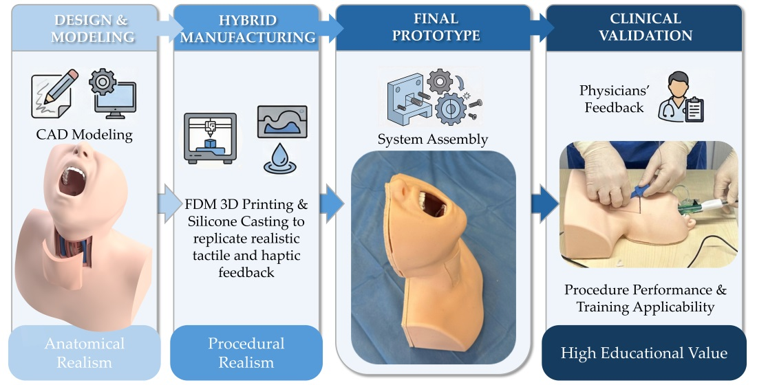
In conclusion, the proposed pediatric tracheotomy simulator represents a promising tool for training in percutaneous tracheotomy using Frova’s PercuTwist dilational percutaneous tracheostomy. The model demonstrated anatomical and functional realism and was perceived by clinicians as highly valuable for procedural education. Its integration into simulation-based training programs may contribute to improved operator confidence, skill acquisition, and ultimately safer management of complex pediatric airway emergencies.
Author Contributions
Conceptualization, M.M., M.B., L.P. and P.S.; methodology, G.V., M.M. and M.B.; validation, G.V., M.M., M.B., L.P., R.B., S.A., M.D.M. and P.S.; formal analysis, G.V. and M.M.; investigation, G.V., M.M., M.B. and L.P.; resources, M.M. and M.B.; writing—original draft preparation, G.V. and M.M.; writing—review and editing, M.B.; visualization, G.V. and M.M.; supervision, Y.V. and P.S.; project administration, Y.V. All authors have read and agreed to the published version of the manuscript.
Funding
This research received no external funding.
Institutional Review Board Statement
Ethical review and approval were waived for this study due to the anonymous and voluntary nature of the questionnaire administered to healthcare professionals, with no patient data collected, consistent with the Italian Personal Data Protection Code (Legislative Decree no. 196/2003, updated by Legislative Decree no. 101/2018) and EU GDPR Article 4(1) and Recital 26. Participation was voluntary and anonymous. The study was conducted in accordance with the principles of the Declaration of Helsinki and in compliance with applicable institutional and national guidelines for non-clinical research.
Informed Consent Statement
Participation in the study was voluntary, and informed consent was implied through completion of the questionnaire.
Data Availability Statement
Data presented in this study are available on request from the corresponding author.
Conflicts of Interest
The authors declare no conflicts of interest.
References
- de Régloix, S.B.; Maurin, O.; Crambert, A.; Bonfort, G.; Clément, P.; Pons, Y.; Kossowski, M. Trachéotomie. EMC Tech. Chir. 2016, 11, 1–15. [Google Scholar] [CrossRef]
- Markov, S.S.; Markova, P.P.; Madzarova-Nikolova, K.I. Pediatric Tracheotomy: Modern Surgical Techniques, Challenges, and Clinical Considerations. Children 2025, 12, 637. [Google Scholar] [CrossRef] [PubMed]
- Watters, K.F. Tracheostomy in Infants and Children. Respir. Care 2017, 62, 799–825. [Google Scholar] [CrossRef]
- Westphal, K.; Maeser, D.; Scheifler, G.; Lischke, V.; Byhahn, C. PercuTwist: A New Single-Dilator Technique for Percutaneous Tracheostomy. Anesth. Analg. 2003, 96, 229. [Google Scholar] [CrossRef]
- De Leyn, P.; Bedert, L.; Delcroix, M.; Depuydt, P.; Lauwers, G.; Sokolov, Y.; Van Meerhaeghe, A.; Van Schil, P. Tracheotomy: Clinical Review and Guidelines. Eur. J. Cardio-Thorac. Surg. 2007, 32, 412–421. [Google Scholar] [CrossRef]
- Susanto, I. Comparing Percutaneous Tracheostomy with Open Surgical Tracheostomy. BMJ 2002, 324, 3–4. [Google Scholar] [CrossRef] [PubMed]
- Frova, G.; Quintel, M. A New Simple Method for Percutaneous Tracheostomy: Controlled Rotating Dilation: A Preliminary Report. Intensive Care Med. 2002, 28, 299–303. [Google Scholar] [CrossRef]
- Cardoso, S.A.; Suyambu, J.; Iqbal, J.; Cortes Jaimes, D.C.; Amin, A.; Sikto, J.T.; Valderrama, M.; Aulakh, S.S.; Ramana, V.; Shaukat, B.; et al. Exploring the Role of Simulation Training in Improving Surgical Skills Among Residents: A Narrative Review. Cureus 2023, 15, e44654. [Google Scholar] [CrossRef]
- Clarke, E. Virtual Reality Simulation—The Future of Orthopaedic Training? A Systematic Review and Narrative Analysis. Adv. Simul. 2021, 6, 2. [Google Scholar] [CrossRef]
- Elendu, C.; Amaechi, D.C.; Okatta, A.U.; Amaechi, E.C.; Elendu, T.C.; Ezeh, C.P.; Elendu, I.D. The Impact of Simulation-Based Training in Medical Education: A Review. Medicine 2024, 103, e38813. [Google Scholar] [CrossRef]
- Ghandourah, H.; Townsi, N.; Abualjdyel, M.; Khan, M.A.; Seebran, N.; Mowalled, S.; El Jamal, W.M.; Kashqari, H.; Selati, S. Assessing and Enhancing Pediatric Residents’ Knowledge and Skills in Tracheostomy Care through Simulation-Based Training. Front. Pediatr. 2025, 13, 1551517. [Google Scholar] [CrossRef] [PubMed]
- Brogi, M.; Mencarelli, M.; Serio, P.; Puggelli, L.; Baggi, R.; Avenali, S.; Amoretti, F.; Volpe, Y. Simulation of Pediatric Surgical Tracheotomy: Development of a High-Fidelity Modular Task Trainer—ScienceDirect. Clin. Simul. Nurs. 2026, 110, 101867. [Google Scholar] [CrossRef]
- Kovatch, K.J.; Powell, A.R.; Green, K.; Reighard, C.L.; Green, G.E.; Gauger, V.T.; Rooney, D.M.; Zopf, D.A. Development and Multidisciplinary Preliminary Validation of a 3-Dimensional-Printed Pediatric Airway Model for Emergency Airway Front-of-Neck Access Procedures. Anesth. Analg. 2020, 130, 445–451. [Google Scholar] [CrossRef]
- Doucet, G.; Ryan, S.; Bartellas, M.; Parsons, M.; Dubrowski, A.; Renouf, T. Modelling and Manufacturing of a 3D Printed Trachea for Cricothyroidotomy Simulation. Cureus 2017, 9, e1575. [Google Scholar] [CrossRef] [PubMed]
- Song, J.; Ock, J.; Kim, W.-J.; Park, Y.-S.; Kim, N.; Kim, S.-H. Enhancing Cricothyroidotomy Training for Novices Using Three-Dimensional-Printed Patient-Specific Models of a Patient with Obesity. J. Med. Syst. 2025, 49, 75. [Google Scholar] [CrossRef] [PubMed]
- Duggan, L.V.; Lockhart, S.L.; Romano, K.R.; Weingart, S.D.; Levitan, R.M.; Brindley, P.G. Front-of-Neck Airway Meets Front-of-Neck Simulation: Improving Cricothyroidotomy Skills Using a Novel Open-Access Three-Dimensional Model and the Airway App. Can. J. Anaesth. J. Can. Anesth. 2017, 64, 1079–1081. [Google Scholar] [CrossRef]
- Huang, J.; Licatino, L.K.; Ocariz, S.; Warner, P.A.; Sims, C.R. A Novel Approach to Emergency Airway Simulation Using a 3D-Printed Cricothyrotomy Task Trainer. J. Educ. Perioper. Med. 2024, 23, e670. [Google Scholar] [CrossRef]
- Narváez Moreno, C.A.; Maureira Moreno, V.; Papuzinski, A.C.; Zúñiga San Martín, S. Effectiveness of a 3D Printed Model in Emergency Front-of-Neck Access Training: A Comparative Study with the Porcine Laryngotracheal Model. Colomb. J. Anestesiol. 2024, 53, e1122. [Google Scholar] [CrossRef]
- Puggelli, L.; Mencarelli, M.; Serio, P.; Furferi, R.; Amoretti, F.; Volpe, Y. Development, Implementation, and Evaluation of a 3D-Printed High-Fidelity Pediatric Mannequin with Expected Hard-to-Intubate Airway. Eng. Rep. 2025, 7, e12975. [Google Scholar] [CrossRef]
- Larynx Model for Studies: Speech Therapy and Oncology Head and Neck by ErikaMaia Fonomediatech|Download Free STL Model. Available online: https://www.printables.com/model/829978-larynx-model-for-studies-speech-therapy-and-oncolo (accessed on 16 September 2025).
- Cozza, P.; Stirpe, G.; Condó, R.; Donatelli, M. Craniofacial and Body Growth: A Cross-Sectional Anthropometric Pilot Study on Children during Prepubertal Period. Eur. J. Paediatr. Dent. 2005, 6, 90–96. [Google Scholar]
- Santarelli, C.; Puggelli, L.; Carfagni, M.; Governi, L. Preliminary Study of a High-Fidelity Simulator for the Management of Paediatric Tracheal Pathologies. In Proceedings of the Design Tools and Methods in Industrial Engineering II, Rome, Italy, 9–10 September 2021; Rizzi, C., Campana, F., Bici, M., Gherardini, F., Ingrassia, T., Cicconi, P., Eds.; Springer International Publishing: Berlin/Heidelberg, Germany, 2022; pp. 773–784. [Google Scholar]
- Zimmermann, M.B.; Hess, S.Y.; Molinari, L.; De Benoist, B.; Delange, F.; Braverman, L.E.; Fujieda, K.; Ito, Y.; Jooste, P.L.; Moosa, K.; et al. New Reference Values for Thyroid Volume by Ultrasound in Iodine-Sufficient Schoolchildren: A World Health Organization/Nutrition for Health and Development Iodine Deficiency Study Group Report. Am. J. Clin. Nutr. 2004, 79, 231–237. [Google Scholar] [CrossRef]
- Furlow, P.W.; Mathisen, D.J. Surgical Anatomy of the Trachea. Ann. Cardiothorac. Surg. 2018, 7, 255–260. [Google Scholar] [CrossRef]
- Semmler, L.; Weberruß, H.; Baumgartner, L.; Pirzer, R.; Oberhoffer-Fritz, R. Vascular Diameter and Intima-Media Thickness to Diameter Ratio Values of the Carotid Artery in 642 Healthy Children. Eur. J. Pediatr. 2021, 180, 851–860. [Google Scholar] [CrossRef]
- Sayin, M.M.; Mercan, A.; Koner, O.; Ture, H.; Celebi, S.; Sozubir, S.; Aykac, B. Internal Jugular Vein Diameter in Pediatric Patients: Are the J-Shaped Guidewire Diameters Bigger than Internal Jugular Vein? An Evaluation with Ultrasound. Paediatr. Anaesth. 2008, 18, 745–751. [Google Scholar] [CrossRef]
Figure 1.
Simulator’s layout.
Figure 1.
Simulator’s layout.
Figure 2.
Cartridge’s layout—frontal view showing the trachea with tracheal rings (a), top view showing bi-component Skin Layer containing blood-mimicking fluid (b), frontal view of the Cartridge displaying all internal structures (c).
Figure 2.
Cartridge’s layout—frontal view showing the trachea with tracheal rings (a), top view showing bi-component Skin Layer containing blood-mimicking fluid (b), frontal view of the Cartridge displaying all internal structures (c).
Figure 3.
Exploded view of the rigid Shells and the Upper and Lower Airway modules.
Figure 3.
Exploded view of the rigid Shells and the Upper and Lower Airway modules.
Figure 4.
Upper Airway embedding the Teeth and the Tongue.
Figure 4.
Upper Airway embedding the Teeth and the Tongue.
Figure 5.
Lower Airway component—side view (a) and top view (b), where the epiglottis can be appreciated.
Figure 5.
Lower Airway component—side view (a) and top view (b), where the epiglottis can be appreciated.
Figure 6.
Cartridge’s Base (pink) and Tracheal Rings (cream color) with the alignment references provided by the Rigid Support (dark pink) (a); fully assembled Cartridge containing the Thyroid Gland, in red, placed over the Tracheal Rings, the Cervical Fascia in transparency, the Sternohyoid Muscles in dark pink, and Arteries and Veins shown in red and blue respectively (b); fully assembled Cartridge covered by the bi-component layer with the Skin Layer visible in deeper pink (c).
Figure 6.
Cartridge’s Base (pink) and Tracheal Rings (cream color) with the alignment references provided by the Rigid Support (dark pink) (a); fully assembled Cartridge containing the Thyroid Gland, in red, placed over the Tracheal Rings, the Cervical Fascia in transparency, the Sternohyoid Muscles in dark pink, and Arteries and Veins shown in red and blue respectively (b); fully assembled Cartridge covered by the bi-component layer with the Skin Layer visible in deeper pink (c).
Figure 7.
Final simulator without the Skin and Cartridge’s Skin Layer: the Cartridge (described in
Figure 6b) is integrated into the Lower Airway component, and the Upper Airway Component contains the Teeth, both shown in pink (
a); fully assembled manikin covered by the Skin (in light pink) and Cartridge’s Skin Layer, shown in a deeper pink (
b).
Figure 7.
Final simulator without the Skin and Cartridge’s Skin Layer: the Cartridge (described in
Figure 6b) is integrated into the Lower Airway component, and the Upper Airway Component contains the Teeth, both shown in pink (
a); fully assembled manikin covered by the Skin (in light pink) and Cartridge’s Skin Layer, shown in a deeper pink (
b).
Figure 8.
Assembled simulator: (a) front view in the operating position; (b) lateral view in the upright position, showing the Back Cover secured with screws to the posterior side of the Shells.
Figure 8.
Assembled simulator: (a) front view in the operating position; (b) lateral view in the upright position, showing the Back Cover secured with screws to the posterior side of the Shells.
Figure 9.
Usage of the percutaneous tracheostomy simulator during hands-on sessions: palpation and tracheal puncture (a), guide wire insertion (b), Frova screw insertion (c), and cannula positioning (d).
Figure 9.
Usage of the percutaneous tracheostomy simulator during hands-on sessions: palpation and tracheal puncture (a), guide wire insertion (b), Frova screw insertion (c), and cannula positioning (d).
Figure 10.
Attendings’ professional expertise in terms of years (a); Previous experience with percutaneous tracheotomy using the Frova screw technique on real patients (b).
Figure 10.
Attendings’ professional expertise in terms of years (a); Previous experience with percutaneous tracheotomy using the Frova screw technique on real patients (b).
Figure 11.
Mean scores for the 15 questionnaire sentences (S) evaluating the simulator (a), and mean scores obtained in the four key aspects (K) corresponding to the realism of the simulator (pink—S1–S3), the procedural fidelity (yellow—S4–S6), surgical instruments interaction (green—S7–S9) and the overall educational relevance (blue—S10–S15) (b).
Figure 11.
Mean scores for the 15 questionnaire sentences (S) evaluating the simulator (a), and mean scores obtained in the four key aspects (K) corresponding to the realism of the simulator (pink—S1–S3), the procedural fidelity (yellow—S4–S6), surgical instruments interaction (green—S7–S9) and the overall educational relevance (blue—S10–S15) (b).
Table 1.
Comparison of commercial simulators and the proposed pediatric tracheotomy simulator.
Table 1.
Comparison of commercial simulators and the proposed pediatric tracheotomy simulator.
| | Our Model | Pediatric HAL S2225
(Gaumard) | AirSim Child Combo Bronchi X (TruCorp) | TraumaChild
(SimuLab) | Smart Airway Child (TruCorp) | Tracheotomy Trainer SKM172 (Sakamoto Model Corp.) | SimuCric
(SimuLab) | SimBaby with Tracheostomy (Laerdal) | Infant Tracheostomy Care Manikin
(Simulaids) | LM-106 Tracheostomy Management (KOKEN) | HAL Pediatric (S312/S157) (Gaumard) | Infant Airway/Nurse Training Baby (3D Scientific) |
|---|
| Percutaneous tracheotomy | Yes | Yes | Yes | Yes | Yes | Yes | Yes | No (tracheostomy care) | No (tracheostomy care) | No (tracheostomy care) | No (tracheostomy care) | No (tracheostomy care) |
| Cervical structures (vessels, muscles, thyroid) | Yes | No | No | No | No | No | No | No | No | No | No | No |
| Trachea and palpable tracheal rings | Yes | Yes | Yes | Partial (approximate) | Yes | Yes | Yes | Partial (limited palpability) | Partial (limited palpability) | Partial (limited palpability) | Partial (limited palpability) | Partial (limited palpability) |
Material
differentiation between tracheal wall and rings | Yes | No | No | No | No | No | No | No | No | No | No | No |
| Skin Stratification | Yes | No | No | No | No | No | No | No | No | No | No | No |
| Bleeding Simulation | Yes | No | No | Yes | No | No | No | No | No | No | No | No |
| Bronchoscope insertion | Yes | Partial (not designed for) | Yes | No | Partial (optional) | No (absence of oral cavity) | No (absence of oral cavity) | No | No | No | Yes | No |
Table 2.
Materials used for each component and respective costs.
Table 2.
Materials used for each component and respective costs.
| Component | Material | Cost (€) |
|---|
| Reusable Parts | | |
| Shell 1 | PLA | 4.56 |
| Shell 2 | PLA | 4.56 |
| Teeth | PLA | 0.28 |
| Lower Airway | Shore A 10 platinum-cure silicone | 14.70 |
| Upper Airway | Shore A 10 platinum-cure silicone | 10.08 |
| Tongue | Platinum-cure silicone foam | 0.32 |
| Skin | Shore A 8 platinum-cure silicone | 25.41 |
| Back Cover | PLA and Shore A 10 platinum-cure silicone | 1.30 |
| Disposable Cartridge | | |
| Rigid Support | PLA | 0.11 |
| Tracheal Rings | PLA | 0.01 |
| Base | Shore A 12 platinum-cure silicone | 4.20 |
| Thyroid Gland | Platinum-cure silicone foam | 0.23 |
| Carotid Arteries | Shore 00–10 platinum-cure silicone | 0.84 |
| Jugular Veins | Shore 00–10 platinum-cure silicone | 1.26 |
| Cervical Fascia | Shore 00–10 platinum-cure silicone | 0.46 |
| Sternohyoid Muscles | Shore A 8 platinum-cure silicone | 0.84 |
| Skin Layer | Shore A 8 platinum-cure silicone | 1.05 |
| Subcutaneous Layer | Shore A 8 platinum-cure silicone | 0.42 |
| Molds | | |
| Rigid Molds | PLA | 34.78 |
| Upper Airway’s Mold | Shore A 10 platinum-cure silicone | 25.66 |
| | Total | 131.07 |
Table 3.
Sentences reported in the questionnaire.
Table 3.
Sentences reported in the questionnaire.
| | Sentence |
|---|
| S1 | The anatomical dimensions of the simulator accurately reproduce those of an 8-year-old patient. |
| S2 | The anatomical geometry of the simulated region is faithfully reproduced. |
| S3 | The representation of the endoscopic view is accurate. |
| S4 | The simulated region feels realistic upon palpation, particularly regarding the tracheal rings. |
| S5 | The skin layer provides realistic feedback during incision. |
| S6 | Bleeding is adequately reproduced and contributes to the realism of the simulation. |
| S7 | The haptic/tactile feedback is adequate. |
| S8 | Tracheal feedback during insertion of the Frova screw is realistic. |
| S9 | Insertion of the Frova screw into the trachea is technically feasible. |
| S10 | The simulator is an appropriate tool for training. |
| S11 | Training on the simulator improves patient safety. |
| S12 | Training on the simulator improves procedural speed and success rate. |
| S13 | My ability to perform percutaneous tracheostomy with the Frova screw in pediatric patients improved after using the simulator. |
| S14 | The use of the simulator is recommended for learning new procedures before performing them on real patients. |
| S15 | The simulator is recommended for both attending physicians and residents to improve and maintain procedural skills. |
| Disclaimer/Publisher’s Note: The statements, opinions and data contained in all publications are solely those of the individual author(s) and contributor(s) and not of MDPI and/or the editor(s). MDPI and/or the editor(s) disclaim responsibility for any injury to people or property resulting from any ideas, methods, instructions or products referred to in the content. |




 ,
,