Review

Rethinking Acne Vulgaris: The Gut–Skin Axis as a Central Mechanism and Therapeutic Target

1
Faculty of Medicine, Wroclaw Medical University, Ludwika Pasteura 1, 50-367 Wrocław, Poland
2
University Clinical Hospital in Wroclaw, Borowska 213, 50-556 Wrocław, Poland
3
Department of Microbiology, Faculty of Medicine, Wroclaw Medical University, Chałubińskiego 4, 50-368 Wrocław, Poland
*
Author to whom correspondence should be addressed.
Appl. Sci. 2026, 16(9), 4527; https://doi.org/10.3390/app16094527
Submission received: 2 April 2026 / Revised: 24 April 2026 / Accepted: 30 April 2026 / Published: 4 May 2026
(This article belongs to the Special Issue Bioactive Natural Compounds: From Discovery to Applications)

Abstract

Acne vulgaris is a chronic inflammatory disease of the pilosabaceous unit with a multifactorial pathogenesis involving sebaceous gland activity, follicular hyperkeratinization, microbial dysbiosis, and immune dysregulation. Increasing attention has been given to the role of the skin and gut microbiome, as well as the gut–skin axis, although their clinical significance has not yet been fully explained. This review critically evaluates the current evidence regarding the use of probiotics, prebiotics, and synbiotics in the treatment of acne. Available studies suggest that microbiome-targeted interventions may influence inflammatory pathways, microbial composition, and metabolic regulators such as IGF-1 and mTORC1. Some clinical trials indicate improvements in acne severity and skin parameters following oral or local interventions. However, the evidence is heterogeneous and limited by small sample sizes, short study durations, and variability in formulations and outcomes. Therefore, although microbiome-based strategies may have potential as adjunctive therapy, their clinical efficacy remains uncertain. Further, well-designed, large-scale studies are needed to determine their role in dermatological practice.

1. Introduction

Acne vulgaris is a chronic inflammatory disorder of the pilosebaceous unit [1,2]. The etiology of the condition is multifactorial and involves a complex interaction between follicular hyperkeratosis, excessive sebum production, and the pro-inflammatory activity of Cutibacterium acnes (C. acnes) [3]. Clinically, this manifests as non-inflammatory open and closed comedones, as well as inflammatory lesions including papules, pustules, and—in severe cases—nodules and cysts [1,2]. Persistent inflammation may also lead to secondary symptoms such as scarring, erythema, and post-inflammatory hyperpigmentation. In addition to physical symptoms, the psychological impact is also significant. Studies indicate that over 50% of patients experience psychological distress, including clinical depression and social anxiety, which can drastically reduce quality of life [4,5]. The global burden of acne is extensive, affecting approximately 9.4% of the population [1,2]. It occurs in up to 85–90% of teenagers, but it is increasingly recognized as a condition that affects adults [1,6,7]. The prevalence is approximately 50.9% among women compared to 42.5% among men aged 20–29, and remains as high as 26.3% in women versus 12.0% in men aged 40–49 [6].
Despite its high prevalence, standard therapies based on long-term use of antibiotics and retinoids present several limitations. The major problem is the global increase in antibiotic resistance. Some studies indicate that more than 60% of C. acnes strains are resistant to commonly used macrolides and tetracyclines [8,9]. Moreover, the side effects of systemic therapies and their potential to disrupt the host’s natural microbiota highlight the need for alternative solutions. Current research increasingly suggests a role for the “gut–skin axis” in the pathogenesis of acne. Within this bidirectional communication network, intestinal dysbiosis may promote systemic inflammation [10]. This microbial imbalance has been associated with increased epithelial permeability, allowing the translocation of gut bacteria and toxic metabolites into the circulation. Such processes may contribute to activation of the immune response, including effector T cells and the release of pro-inflammatory cytokines, thereby contributing to chronic systemic inflammation. In addition, dysbiosis has been linked to the activation of the mechanistic target of rapamycin complex 1 (mTORC1) signaling pathway and elevated levels of insulin-like growth factor (IGF-1). These pathways are thought to influence forkhead box protein O1 (FoxO1) expression and metabolic regulation, potentially promoting sebaceous gland hyperplasia and lipogenesis. Furthermore, the gut–brain–skin axis facilitates the release of substance P and neuropeptides. These mediators, upon entering the circulation through the compromised barrier, may enhance inflammatory signaling, including interleukin-1 (IL-1), interleukin 6 (IL-6), and tumor necrosis factor α (TNF-α), which have been implicated in the exacerbation of acne lesions [10,11,12].
Based on these pathophysiological mechanisms, microbiome-targeted interventions represent a promising therapeutic strategy for people with acne [13,14]. Probiotics, defined as live microorganisms, may exert beneficial effects by restoring microbial homeostasis and directly strengthening the intestinal epithelial barrier. This structural reinforcement may help prevent the translocation of pro-inflammatory metabolites and bacteria into the systemic circulation. Furthermore, certain probiotic strains have been shown to modulate immune response by inhibiting interleukin production, which may contribute to reduced sebaceous gland activity and lipogenesis [13,14]. Complementing this action, prebiotics act as substrates that selectively stimulate the growth and activity of beneficial bacteria naturally present in the body. By supporting the fermentation process, prebiotics increase the endogenous production of short-chain fatty acids (SCFAs), which, as mentioned earlier, are essential for maintaining systemic immune tolerance. When administered as synbiotics, these components are thought to act synergistically, potentially enhancing the restoration of the gut–skin axis relative to their individual effects. However, the available evidence remains heterogeneous, and further research is needed to confirm these effects in clinical settings. Therefore, these agents may represent a multifaceted alternative to traditional antimicrobial therapies.
Although research has significantly expanded our understanding of the pathogenesis of acne vulgaris, there remains a significant gap in integrating gut dysbiosis, skin inflammation, and microbiome-targeted therapies into a coherent mechanistic model. A growing body of evidence suggests that acne can be more accurately viewed not only as a localized skin disease but as a manifestation of systemic dysregulation within the gut–skin axis. Therefore, this review aims to evaluate microbiome-based interventions within this framework, emphasizing their role in modulating the interrelated pathways involved in the development of acne. By synthesizing current evidence from the perspective of the gut–skin axis, this work highlights the interplay of microbial, immunological, and metabolic processes, providing a more integrated understanding of the pathogenesis of acne and potential therapeutic strategies.

2. Pathogenesis of Acne and the Role of Microbiome

Acne vulgaris has a complex etiology and develops due to several factors. The most important ones include increased sebaceous gland activity, abnormal keratinization of the follicular duct, C. acnes proliferation, and inflammation. The presence of these factors contributes to the development of acne. Genetic predisposition, hormonal changes, stress, and diet may increase the severity of the condition. These factors collectively contribute to disease development [15,16]. Recent studies also strongly highlight the role of the skin and gut microbiota and their intercommunication in the pathogenesis of acne [17,18].

2.1. Sebaceous Gland Activity and Follicular Keratinization

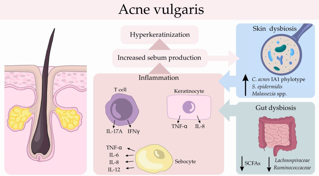
Sebum is a natural, oily substance produced by the sebaceous glands. It is composed of triglycerides (57.5%), wax esters (26%), squalene (12%), and cholesterols (4%) [19]. It plays a key role in maintaining the integrity of the skin barrier by reducing transepidermal water loss. It also has antimicrobial activity, which is driven by immunomodulatory effects of certain fatty acids. Sebaceous gland activity and sebum production are influenced by several factors, i.e., hormones, diet, and environmental factors. Among these, androgens—the primary regulators of sebum production—are considered to play the most significant role in the pathogenesis of acne [20]. In particular, dihydrotestosterone (DHT), which is formed from testosterone via type 1 5α-reductase, appears to be especially important, as increased activity of this enzyme has been observed in acne-prone skin, particularly within the pilosebaceous unit [18]. Furthermore, DHT binds to the androgen receptors (ARs) in sebaceous glands and stimulates production of sebum, thereby creating favorable conditions for the growth of C. acnes and promoting follicular hyperkeratinization. This results in the accumulation of corneocytes in the follicle and the formation of comedones. Consequently, the combination of excessive sebum production and follicular hyperkeratinization may lead to obstruction within the follicle and create conditions for the development of the disease [21,22]. Another factor that has been implicated in acne development is the Western diet, characterized by high intake of foods with a high glycemic index. Such dietary patterns may increase insulin levels, which in turn are associated with elevated levels of insulin-like growth factor 1 (IGF-1) [23]. IGF-1 is a potent activator of dehydroepiandrosterone sulfate (DHEA-S) synthesis in gonads and an inducer of testosterone conversion to DHT in the skin. DHT binds to ARs located in sebocytes within the sebaceous gland, leading to increased sebum production. Excess sebum promotes the colonization of C. acnes, which, in combination with hyperkeratinization and inflammation, creates an environment for acne development. Furthermore, IGF-1 binds to the IGF-1 receptors located in keratinocytes and stimulates sebaceous lipogenesis, thereby resulting in increased sebum production [18,24]. Studies have shown that a diet rich in milk and dairy products can lead to excessive insulin stimulation. This process is driven by high levels of branched-chain amino acids (BCAAs), whose activity leads to elevated IGF-1 levels and, consequently, to sebum overproduction [23]. Furthermore, a Western diet may not only lead to an increase in the quantity of sebum but also to a change in its composition [19,25]. The altered composition of sebum, especially the amount of monounsaturated fatty acids, squalene and triglycerides, also has a comedogenic effect, contributing to the clogging of hair follicles. Sebum also plays a role in inflammation [26]. It stimulates the over-proliferation of C. acnes and increases the production of proinflammatory cytokines, such as IL-1 (Figure 1). Changes in the quantity and composition of sebum not only irritate the hair follicle but also provide a breeding ground for specific strains of bacteria, leading to skin dysbiosis.
Acne pathogenesis cannot be attributed to one specific factor. Rather, it is driven by interaction between processes that reinforce each other (Figure 1). Excess sebum, stimulated by androgens and the Western diet, together with hyperkeratinization, contributes to follicular obstruction. Moreover, sebum creates conditions that promote colonization of C. acnes and production of pro-inflammatory cytokines, which both play a role in this skin disorder. This highlights the complex and multidirectional nature of acne.

2.2. The Gut–Skin Axis: Integrated Role of Skin and Gut Microbiomes in Acne Pathogenesis

Both the skin and gut microbiome play an important role in maintaining host homeostasis. Disruptions in these microbial communities may contribute to inflammatory processes and barrier dysfunction, which appear to be involved in acne pathogenesis.

2.2.1. Specifics of the Skin Microbiome

The skin is the largest organ of the human body and harbors a rich community of microorganisms that play a vital role in proper skin functions. Depending on the skin’s physiological state, age, and environmental components like diet or antibiotic use, the composition of skin microbiota may temporarily change [27]. The predominant bacteria in the sebaceous-rich areas are Cutibacterium spp., Staphylococcus spp., and Corynebacterium spp. [17,28]. The skin microbiota is not limited to bacteria, but also includes fungi. Among them, the most predominant is Malassezia spp., which commonly occurs in sebaceous-gland-rich areas [29].
Among these skin commensals, C. acnes is considered a major contributor to acne development. C. acnes is a Gram-positive anaerobic bacterium located deep in the sebaceous follicles and belongs to the skin microbiota. Based on morphological characteristics, biochemical activity, virulence factors, as well as differences in immunogenic and inflammatory activity within this species, we distinguish six phylotypes: IA1, IA2, IB, IC, II, and III [30,31]. The 1A1 phylotype is the most predominant in acne patients’ skin and can produce virulence factors that stimulate T cells to release IL-17A and IFNγ (Figure 1). These pro-inflammatory cytokines activate the immune response, leading to inflammation, which is one of the main factors in acne pathogenesis [21,32]. Under physiological conditions, C. acnes is crucial for maintaining a healthy skin barrier. Furthermore, a product of its metabolic processes is propionic acid, which, by lowering the skin’s pH, prevents the colonization of pathogenic bacteria [33]. Meanwhile, C. acnes is critically involved in the pathogenesis of acne by increasing the activity of sebaceous glands, promoting their proliferation, and contributing to the development of inflammation. It increases sebum production by stimulating diacylglycerol acyltransferase, leading not only to an exacerbation of seborrhea but also, by using energy from sebum, to its own overproliferation [28]. This vicious cycle contributes to the complexity of acne pathogenesis, in which many factors interact with one another. For many years, acne has been associated with an increased abundance of this member of the skin microbiota. However, recent studies have shed new light on the subject, suggesting that the abundance of C. acnes does not differ between healthy and acne-affected skin. Instead, the difference lies in the distribution of the species’ phylotypes [34,35]. It is now known that C. acnes contributes to the development of acne primarily through a loss of diversity among its phylotypes, rather than through overgrowth [30,31,33,36]. Studies have shown that the IA1 phylotype predominates in acne-prone skin compared to healthy skin. Of all six, it has the highest biofilm-forming capacity and the most virulent profile. Its dominance leads to the production of exopolysaccharide, which binds to corneocytes and promotes the formation of comedones [37].
Furthermore, its biofilm-forming capabilities increase lipase activity, activating inflammation in sebum [35]. Other C. acnes phylotypes appear to be less associated with acne. It has been shown that phylotypes IB and II, which exhibit a significantly lower ability to form biofilms, predominate on healthy skin [16,36]. Other studies have shown that C. acnes increases local inflammation by stimulating sebocytes to release pro-inflammatory cytokines such as TNF-α, IL-6, interleukin 8 (IL-8), and interleukin 12 (IL-12). It also stimulates keratinocytes to release neutrophil-attracting cytokines through activation of toll-like receptor 2 (TLR2) receptors. Through the NOD-like receptor protein 3 (NLRP3) inflammasome, C. acnes stimulates IL-1β secretion.
Coagulase-negative staphylococci (CNS) are a major component of the skin microbiota, with Staphylococcus epidermidis as the predominant species. S. epidermidis, a Gram-positive, facultative anaerobe, colonizes all skin regions, including dry, moist, and sebaceous areas, and is considered a commensal bacterium [38,39]. S. epidermidis helps maintain skin homeostasis through a variety of mechanisms, including colonization resistance, immune modulation, and maintaining proper skin barrier integrity. These beneficial strains produce phenol-soluble modulins (PSMs) that have anti-microbial properties against pathogenic Staphylococcus aureus, which is commonly linked to skin disorders involving skin barrier disruption, such as atopic dermatitis or wounds [40]. In addition, S. epidermidis can produce autoinducing peptides that inhibit S. aureus accessory gene regulator quorum-sensing system, thereby limiting colonization of these pathogenic bacteria. Moreover, S. epidermidis has an impact on the cutaneous immune system by promoting immune priming and supporting tissue repair due to the production of lipoteichoic acid. Syringomyelinase secreted by S. epidermidis promotes ceramide production from keratinocytes, thereby preventing transcutaneous water loss and maintaining proper skin barrier integrity. However, under certain conditions, S. epidermidis can act as a pathogenic bacterium due to biofilm formation and the extracellular serine protease (EcpA protease), which can exacerbate barrier disruption [29,38,41].
Interactions between skin microbiota are crucial for skin homeostasis, as dysbiosis between S. epidermidis and C. acnes is considered an important factor in acne pathogenesis [42]. For example, S. epidermidis has been shown to limit C. acnes colonization by producing antimicrobial peptides (AMPs), including epidermin or lipoteichoic acids. S. epidermidis has also been reported to reduce inflammatory response through the regulation of miRNA expression and modulation of Toll-like receptor signaling in keratinocytes. A lower abundance of S. epidermidis in acne skin compared to healthy skin supports the theory of skin dysbiosis in acne [43,44].
Malassezia spp., the most abundant fungi on skin, are predominant in sebaceous-rich areas such as the face and scalp. The role of Malassezia spp. in the skin microbiota remains unclear, as it is commonly linked with skin disorders such as dandruff [40]. These fungi can also potentially lead to inflammatory acne. Through the hydrolysis of sebum triglycerides into free fatty acids, Malassezia may promote follicular hyperkeratinization. These species have also been shown to stimulate polymorphonuclear chemotaxis and increase the production of pro-inflammatory cytokines in keratinocytes and monocytes [28,45].
Overall, the balance between skin microbiota is essential for maintaining homeostasis, and its disruption may contribute to acne development. Although C. acnes is closely associated with acne pathogenesis, interactions between skin commensals also play an important role. Current studies suggest that the skin microbiota influences acne pathogenesis, but further research is needed to better understand these interactions and their direct impact on the disease [32].

2.2.2. Specifics of the Gut Microbiome

The gut microbiota consists of a wide range of microorganisms that inhabit the digestive tract, including bacteria, archaea, viruses, and fungi, together with their genetic material and metabolic products. It represents the largest and most densely populated microbial community in the human body, with estimates suggesting up to 1011 microbial cells and over a thousand bacterial species. The gut microbiota is mainly composed of a few dominant phyla, such as Firmicutes, Bacteroidetes, Actinobacteria, and Proteobacteria [46,47,48]. Together, Firmicutes and Bacteroidetes account for 90%for the 90% of the gut microbial population. Key representatives of Firmicutes include genera like Lactobacillus, Bacillus, Clostridium, Enterococcus, and Ruminicoccus. Bacteroidetes are mainly represented by Bacteroides and Prevotella. Members of Actinobacteria include genera such as Bifidobacteria, Atopobium, and Collinsella, while Proteobacteria are primarily represented by Enterobacteriaceae [49]. The structure of the gut microbiota is shaped by multiple factors, including age, genetics, immune function, and physiological conditions, as well as environmental influences. These microorganisms play an important role in maintaining overall balance in the body by supporting the intestinal barrier, protecting against pathogens, contributing to metabolic processes, and regulating immune responses. As a result, their effects extend beyond the gut, influencing systemic host health [48,50]. Metabolites formed by the gut microbiota during the fermentation of dietary fiber and other non-digestible carbohydrates are mainly SCFAs, which are considered key products of microbial activity in the gut. The most common SCFAs are acetate, propionate, and butyrate [51]. These compounds are important for maintaining intestinal balance, as they support epithelial cell function, help preserve the integrity of the intestinal barrier, and influence immune responses. Butyrate, in particular, plays a central role by strengthening tight junctions and promoting mucin production, which reduces the risk of bacterial translocation. Beyond their local effects in the gut, SCFAs are also involved in regulating systemic inflammation, partly through interactions with G protein-coupled receptors and by affecting gene expression via inhibition of histone deacetylases. Because of these properties, they may indirectly influence skin conditions, including acne, where chronic low-grade inflammation and immune dysregulation are known to play a role [52,53,54].

2.2.3. Gut–Skin Axis

The skin microbiome plays a key role in the development of skin lesions, and without it, the skin cannot function as a separate system. However, recent studies emphasize that skin homeostasis is closely linked to systemic health, particularly through bidirectional communication known as the gut–skin axis. More and more evidence suggests that the gut microbiota also significantly influences skin homeostasis through immunological and biochemical pathways [10,18]. Intestinal bacteria, including those of the genera Bacteroides, Bifidobacterium, Cutibacterium, Eubacterium, Lactobacillus, and Prevotella, are capable of producing SCFAs. These metabolites possess anti-inflammatory activity, mainly through limiting inflammatory cell proliferation, migration, adhesion, and cytokine production. They also modulate immune cell activity by inhibiting histone deacetylases and interfering with nuclear factor kappa-light-chain-enhancer of activated B cells (NF-κB) signaling, which affects both activation and apoptosis of immune cells. Through these mechanisms, SCFAs promote the expansion of regulatory cells involved in skin processes such as differentiation and wound healing, and may additionally influence skin physiology more directly via translocation of microbial metabolites to the skin. Moreover, SCFAs also strengthen the epithelial barrier function and reduce intestinal permeability. Their involvement in acne has been confirmed by their regulatory role in the mTOR pathway [10,27,49,55].
This pathway is involved in skin cell growth and differentiation, contributing to the maintenance of a healthy epidermal barrier and skin homeostasis. The gut microbiome, by interacting with this pathway, may indirectly influence the pathomechanism of acne, leading to increased sebum gland activity, lipogenesis, and keratinocyte proliferation [18]. This may also affect the integrity of the intestinal barrier and the composition of the gut microbiota, involving the mTOR pathway [10,43,56]. Recent studies suggest that patients with acne exhibit changes in the composition of their gut microbiota, with increased abundance of Bacteroidetes and a concurrent decrease in the number of bacteria belonging to the Firmicutes, Clostridium, Clostridiales, Lachnospiraceae, and Ruminococcaceae compared to healthy controls. Among the depleted genera are those that are the primary producers of SCFAs affecting the mTOR pathway (Figure 1) [18].
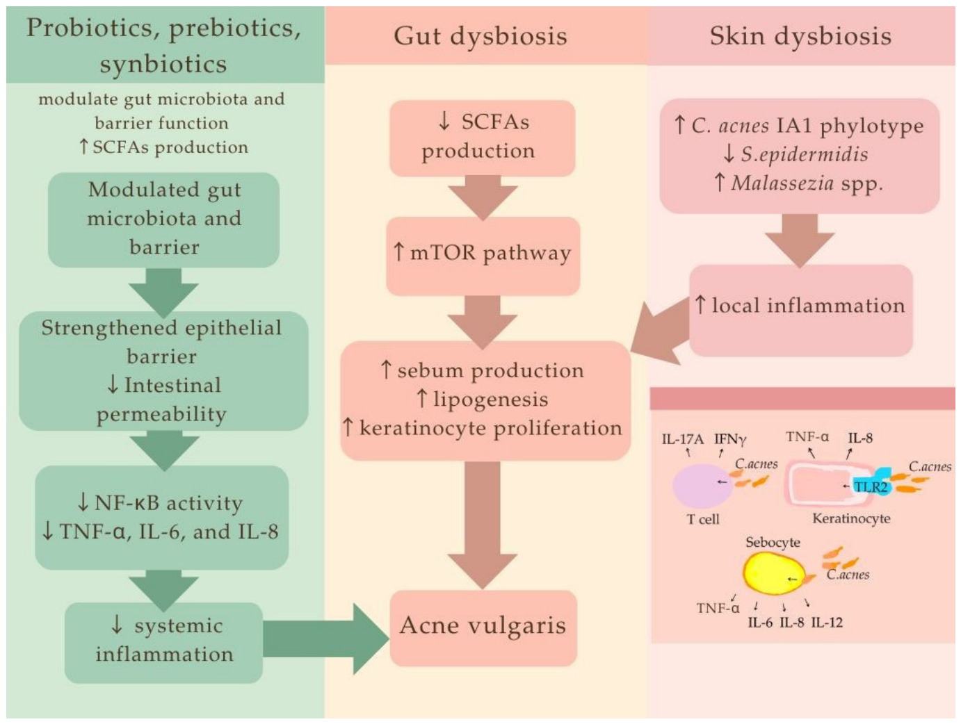
Studies available in the literature indicate a close link between skin homeostasis and the state of the gut. They demonstrate that the activity of the gut microbiome plays a significant role in the pathogenesis of acne. The mTOR pathway is considered to play a key role in this connection, as these commensal bacteria can influence it either directly or indirectly. However, despite these interesting findings, no biomarkers directly correlated with acne severity have been identified to date [18,43,49]. The role of the gut–skin axis in the pathogenesis of acne is illustrated in Figure 2.
In summary, these findings support the view that the gut–skin axis is not only an interconnected pathway but also a key integrative mechanism linking microbial dysbiosis to immune system activation and metabolic disorders in acne.

3. Mechanisms of Action of Microbiome-Based Interventions in Acne

3.1. Microbiota and Metabolome Modulation

Microbiome-targeted interventions, including probiotics, prebiotics, and synbiotics, exert their effects through a variety of interrelated mechanisms that go beyond direct antimicrobial action. The gut–skin axis provides a framework for understanding these interventions as modulators of systemic homeostasis, influencing microbial composition, immune signaling, and metabolic regulation. Rather than acting through isolated pathways, their effects appear to converge on key processes related to acne pathogenesis, including inflammation, sebaceous gland activity, and hormonal signaling. This integrative perspective provides a more comprehensive understanding of how microbiome modulation may contribute to the treatment of acne.
Probiotics, defined as live microorganisms, exhibit direct antibacterial activity against acne-associated pathobionts. One of the primary mechanisms involves the secretion of antimicrobial peptides that confer a competitive advantage to the producing strains [57]. In contrast, prebiotics serve as energy sources that promote the growth of specific beneficial microorganisms, thereby modulating both gut and skin microbiota [58]. When combined in a single formulation, synbiotics act in a manner analogous to their individual components, but with potentially enhanced efficacy due to the supportive effects of prebiotics [59]. Therefore, interventions targeting the microbiome can be viewed as strategies aimed at restoring the homeostasis of the gut–skin axis, rather than as measures that act solely through isolated antimicrobial or anti-inflammatory effects.
From a mechanistic perspective, it is useful to distinguish between oral and topical supplements. Oral products primarily act on the gut microbiota and can indirectly affect skin health through the gut–skin axis. In contrast, topical preparations exert their effects locally, directly shaping the skin microbiota at the site of application. Regarding direct antimicrobial action, cell-free supernatants (CFS) from probiotic cultures have been investigated in the context of acne pathobionts. They contain a broad mixture of antimicrobial compounds, including bacteriocins, organic acids, and other metabolites [60]. The CFS of Lactobacillus paraplantarum THG-G10 has been shown to inhibit the growth of C. acnes and induce deleterious morphological alterations in bacterial cell membranes [61]. Similarly, the supernatant of Lactobacillus paracasei LPH01 demonstrates inhibitory activity against C. acnes, reducing cellular metabolic activity, disrupting membrane integrity, and downregulating lipase gene expression [62].
When bacteriocins are investigated independently, their use has also shown beneficial effects in acne. Topical application of a formulation containing 1% bacteriocins derived from Bacillus. subtilis has been shown to reduce the absolute abundance of S. aureus in acne-affected areas in human subjects [63], while strains such as Lactobacillus salivarius LS03 produce bacteriocins that directly inhibit the growth of C. acnes in vitro [64]. Other competitive mechanisms include reducing the adhesion of pathobionts to host cells. In a murine model, the Weissella viridescens UCO-SMC3 strain reduced the adhesion of C. acnes to keratinocytes and lowered pathogen loads in skin lesions when applied topically. Interestingly, oral administration of W. viridescens UCO-SMC3 induced more pronounced changes in the immune response to C. acnes than topical treatment, while being less effective in reducing bacterial loads in the skin, likely due to indirect effects at this site of application [65].
Evidence from in vivo studies further supports the antimicrobial potential of probiotics. In a clinical trial, strains such as Lactobacillus rhamnosus GG, Lactobacillus plantarum WCFS1, and Lactobacillus pentosus KCA1 were shown to significantly reduce the abundance of Staphylococci—targeted as pathobionts—alongside increasing the relative abundance of Lactobacilli, when applied to acne-affected facial skin topically. Although no major changes were detected in the relative abundance of Cutibacterium spp. in vivo, the selected strains demonstrated the ability to inhibit the growth of both C. acnes and S. aureus when tested in vitro [66].
Among prebiotics, the most studied oral supplements are inulin and fructooligosaccharides (FOS), which generally show similar properties. Both consistently stimulate the growth of Bifidobacterium, often making it a dominant genus in fecal samples [67]. Similarly, galactooligosaccharides (GOS) have been shown to stimulate Bifidobacterium spp. and Lactobacillus spp. across numerous studies [68,69,70]. However, the effects of these prebiotics are not uniform and depend strongly on an individual’s baseline microbiota composition.
For instance, a study comparing individuals with different Bacteroides/Bifidobacterium (Ba/Bi) ratios showed that responses to prebiotics varied between high (H) and low (L) ratio groups. In the H group, FOS increased the abundance of butyrate-producing bacteria, leading to higher levels of butyrate and related metabolites, suggesting a greater functional benefit. In contrast, inulin’s effects were more metabolic than compositional: the L group showed increased propionate production and enhanced glycolysis, while the H group exhibited enrichment in amino acid metabolism and aminoglycolysis [71].
Beyond classical prebiotics, polyphenol-rich foods—such as tea, berries, grapes, and pomegranates—also exhibit prebiotic-like effects. They can increase the abundance of Lactobacillus and Bifidobacterium while reducing potentially harmful genera such as Clostridium [72]. These effects can also be observed in a more targeted manner through compounds like epigallocatechin gallate (EGCG) [73].
Certain polyphenols, such as resveratrol, may be particularly relevant in metabolic contexts. In high-fat diet models, resveratrol induces specific taxonomic shifts, including increased Blautia and decreased Desulfovibrio and Lachnospiraceae. These changes are associated with anti-obesity effects in animal studies [74]. Given that acne and obesity share underlying mechanisms, such as impaired glucose metabolism and elevated androgen levels, these interventions may hold potential relevance for acne.
Regarding topically applied prebiotics, Li et al. demonstrated that they can influence not only the composition of the skin microbiota but also its metabolome [75]. The application of prebiotic-containing cosmetics led to an increase in metabolic pathways related to sugar and sugar acid degradation, including the lactose degradation pathway, with effects largely attributed to inulin fermentation. Notably, the intervention was associated with an increased abundance of commensal species such as Staphylococcus equorum, Streptococcus mitis, and Haemophilus desiderata SP1, alongside a reduction in potential pathogens such as Pseudomonas stutzeri and Staphylococcus anadarae. Beneficial alterations in metabolites—including long- and medium-chain fatty acids, fatty acid esters, and fatty acyls—were also observed, all of which contribute to skin barrier integrity [76]. Additionally, in reconstructed human epithelium models, short-chain FOS at a 1% concentration promoted the growth of S. epidermidis while inhibiting C. acnes and S. aureus, whereas GOS at a 5% concentration stimulated S. epidermidis and concurrently inhibited S. aureus [77,78].
Lastly, Min et al. demonstrated that supplementation with a synbiotic formulation containing Bifidobacterium lactis, Lactobacillus acidophilus, and Bacillus coagulans, along with compounds such as Commiphora mukul (guggul extract), epigallocatechin gallate (EGCG), significantly improved non-cystic acne lesions. It was also observed when a myoinositol-based herbal supplement was administered. For the synbiotic group, improvement was accompanied by an increase in beneficial bacterial taxa, including Faecalibacterium prausnitzii and Blautia spp., alongside a reduction in potentially pathogenic species such as Clostridioides difficile, Collinsella spp., and Bilophila wadsworthia [79].
Overall, probiotics exert antimicrobial effects through multiple complementary mechanisms, including the secretion of antimicrobial metabolites, modulation of the immune response, and reduction in pathobiont adhesion. While in vitro and other preclinical studies consistently demonstrate inhibitory effects against acne-associated bacteria such as C. acnes and S. aureus, clinical findings remain more variable and appear to be highly strain- and population-dependent. Prebiotics exert beneficial effects through microbiota modulation, though their impact is strongly influenced by baseline microbial composition and host metabolic status. These effects may collectively contribute to rebalancing dysregulated gut–skin axis signaling. Furthermore, the majority of mechanistic evidence is derived from experimental models, highlighting a gap in standardized clinical trials directly linking microbiota-targeted interventions to acne outcomes.

3.2. Cytokine Modulation and Inflammation Control

Considering that acne vulgaris is a chronic inflammatory skin disease, targeting immune dysregulation has emerged as a promising therapeutic target. The bidirectional interaction between the intestinal microbiota and skin homeostasis is mediated through modulation of the immune system [80]. In this context, oral supplements, including probiotics, prebiotics, and synbiotics, may exert anti-inflammatory effects and restore intestinal barrier integrity, thereby contributing to acne improvement [18]. These effects are largely mediated through the modulation of cytokine networks implicated in acne pathogenesis.
Both probiotics and prebiotics primarily reduce pro-inflammatory cytokines (IL-1β, IL-6, IL-8, TNF-α) associated with sebaceous gland dysfunction and Th17-mediated inflammation, while enhancing anti-inflammatory pathways, particularly through increased IL-10 production [81,82,83,84,85]. Notably, prebiotics may exert immunomodulatory effects via both microbiota-dependent and microbiota-independent mechanisms [86].
In murine models of acne, L. rhamnosus significantly reduced IL-1β, IL-6, and TNF-α levels, leading to an improvement in acne-like symptoms [81]. Similar immunomodulatory effects have been observed across other probiotic strains. L. rhamnosus strains markedly suppressed the production of TNF-α, IL-6, and IL-8 in lipopolysaccharide-stimulated macrophages [82], while L. paracasei reduced TNF-α and IL-1β levels [83]. Furthermore, L. plantarum and Lactobacillus fermentum inhibited IL-6 and TNF-α secretion while enhancing the production of the anti-inflammatory cytokine interleukin 10 (IL-10) in macrophages [84]. Finally, the B. subtilis strain significantly decreased IL-8 production under various stress conditions in vitro [85]. These findings are consistent with the established role of pro-inflammatory cytokines in acne pathogenesis.
For instance, IL-1β is abundantly expressed in inflammatory acne lesions, particularly around pilosebaceous follicles, where it colocalizes with tissue macrophages. Similarly, IL-6 and IL-8 are produced by sebocytes and keratinocytes in response to C. acnes through TLR2 and toll-like receptor 4 (TLR4)-dependent signaling pathways [87,88,89]. These cytokines promote differentiation of naïve CD4+ T cells into Th17 cells, thereby amplifying inflammation, and are upregulated in sebocyte supernatant. Moreover, sebocytes functionally interact with C. acnes during the maturation of dendritic cells, leading to the generation of antigen-presenting cells that preferentially prime Th17 cells, thus establishing a self-perpetuating inflammatory loop [88,90].
In turn, TNF-α directly influences sebaceous gland activity by promoting lipogenesis in sebocytes. It occurs through activation of the c-Jun N-terminal kinase (JNK) and phosphoinositide-3-kinase–protein kinase B/Akt (PI3K/Akt) signaling pathways, leading to the upregulation of fatty acid synthase (FAS) and Sterol regulatory element-binding protein 1 (SREBP-1) expression, and thereby contributing to the development of seborrhea [91]. Taken together, the relationship between IL-1β, IL-6, IL-8, and TNF-α and sebum production in acne forms an inflammatory feedback loop, where these cytokines both respond to and perpetuate the inflammatory environment that drives sebaceous gland dysfunction.
Moreover, IL-10 deficiency represents a key immunological abnormality in acne patients [92]. Peripheral blood mononuclear cells (PBMCs) isolated from these individuals exhibit diminished IL-10 secretion, which is associated with a decreased capacity of CD14+ cells to phagocytose C. acnes. Importantly, supplementation with exogenous IL-10 enabled restoring the phagocytic function of these cells, highlighting the beneficial role of this cytokine [92]. Despite these generally beneficial effects, the available evidence is not entirely consistent. Some strain-specific effects are paradoxical, as certain strains have been reported to increase pro-inflammatory cytokine levels while still contributing to improvements in acne outcomes. Supplementation with L. paracasei NCC 2461 (ST11) in a randomized controlled trial by Gueniche et al. was associated with significantly increased circulating levels of transforming growth factor β (TGF-β) compared to controls, alongside reduced skin sensitivity and improved skin barrier recovery [93].
Notably, TGF-β is implicated in acne susceptibility and scarring. In acne lesions, it contributes to Th17 differentiation, whereas in atrophic scars, elevated TGF-β1 appears to drive abnormal extracellular matrix remodeling, including degradation of collagen and elastic fibers with incomplete repair [94,95].
The beneficial clinical effects observed may be attributed to additional strain-specific mechanisms, such as enhanced IL-10 production. L. paracasei NCC 2461 (ST11) promotes the expansion of a CD4+ T cell population closely resembling regulatory T cells, characterized by high secretion of both TGF-β and IL-10 [93].
Similar inconsistencies have also been observed in studies on prebiotics. In the context of FOS and inulin, the simultaneous induction of both pro-inflammatory cytokines (TNF-α, IL-1β) and anti-inflammatory mediators (IL-10) in human peripheral blood monocytes was observed [96].
Conversely, an in vitro study demonstrated that human milk oligosaccharides (HMOs) and GOS attenuated TNF-α, IL-1β, and other pathogen-induced inflammatory cytokines, including IL-8, monocyte chemoattractant protein-1 (MCP-1), and macrophage inflammatory protein-3α, in a model of the human immature intestine [97].
Overall, these discrepancies may be attributed to differences in experimental conditions, including the type and dose of probiotics and prebiotics used, as well as the specific cell culture models employed in some cases. Additionally, the chain length of oligosaccharides classified as prebiotics appears to play a significant role in determining whether prebiotics elicit predominantly pro-inflammatory or anti-inflammatory responses [98]. Beyond their effects on cytokine modulation, prebiotics may also exert additional immunomodulatory functions, particularly at the level of the intestinal barrier.
For instance, they contribute to the maintenance of epithelial barrier integrity by upregulating tight junction proteins, including zonula occludens-1 (ZO-1), occludin, and claudins, as well as by increasing mucin production. Mucins constitute a primary physical barrier against pathogens and toxins and contain antimicrobial components such as defensins and immunoglobulin A (IgA) [99].
Moreover, polyphenols, a subset of which may be considered prebiotics, exhibit both antioxidant properties and direct immunomodulatory effects. During immune responses, phagocytic cells such as neutrophils and macrophages generate reactive oxygen species (ROS) to eliminate pathogens; however, excessive ROS production can also damage the immune cells themselves. In this context, polyphenols support immune function by protecting immune cells from oxidative stress [100].
Importantly, the activity of polyphenols extends beyond antioxidant effects. They can bind to receptors that are highly expressed on immune cells, including the aryl hydrocarbon receptor (AhR), retinoic acid receptor (RAR), and RIG-I-like receptors (RLR). Through these interactions, they initiate immunometabolic pathways involving mitochondria-centered mechanisms, ultimately helping to alleviate chronic low-grade inflammation [101]. These complementary mechanisms may be particularly beneficial in the context of acne.
In addition to polyphenols, gut microbial metabolites derived from tryptophan, such as indole and indole-3-acetic acid (IAA), also act as ligands of the AhR, thereby attenuating pro-inflammatory cytokine production associated with acne [81,102]. For instance, supplementation with L. rhamnosus in murine models enhanced the production of these metabolites, which was associated with reduced levels of IL-1β, IL-6, and TNF-α, and improvement of acne-like symptoms. Notably, these effects were abolished following administration of AhR antagonists, highlighting the central role of this pathway [81]. Moreover, these mechanisms further support the role of the gut–skin axis as a key mediator of systemic influences on acne.
While most research has focused on orally administered interventions, topical approaches have likewise demonstrated immunomodulatory potential. Topically applied probiotics also confirm notable immunomodulatory effects, acting both through antimicrobial activity against C. acnes and via direct interactions with keratinocytes and epithelial cells. Studies indicate that they can suppress IL-8 production in these cells, thereby attenuating the inflammatory cascade initiated by C. acnes. Additionally, glycerol fermentation by S. epidermidis has been shown to inhibit the growth of C. acnes. As active components in topical formulations, species such as Lactococcus contribute to reducing inflammation by decreasing the release of mediators responsible for vasodilation, edema, and mast cell degranulation [103].
It should be emphasized that considerably more research on probiotics and prebiotics has been conducted under in vitro conditions and via oral administration than on topical applications. Further studies are needed to better characterize the role of oral and topical formulations in inflammation control, particularly in the context of acne vulgaris.

3.3. Insulin Sensitivity and Lipogenesis

Oral supplementation influences systemic metabolism and may, in this way, contribute to acne severity. Their effects are largely mediated through the regulation of insulin sensitivity and lipid profile regulation, which in turn is linked to hormonal balance and sebogenesis.
Oral administration of L. rhamnosus SP1 (LSP1) in patients with acne was shown to normalize skin expression of insulin signaling genes in acne areas. Notably, LSP1 modulates key components of this pathway, including IGF-1 and FOXO1. These molecules stimulate both sebaceous lipogenesis and androgen receptor signaling [104].
Additionally, supplementation with a synbiotic formulation influenced hormone levels associated with acne pathogenesis, with reductions observed in androgens or their precursors. Interestingly, in the same study, a myo-inositol-based formulation resulted in a significant decrease in 17-hydroxyprogesterone (17-OHP) and androstenedione—an effect not observed in the synbiotic group [79].
Oral supplementation with L. plantarum CJLP55 improved acne lesions, reduced sebum triglyceride production, and enhanced skin hydration by increasing ceramide 2 levels—the principal sphingolipid responsible for maintaining the epidermal lipid barrier. These effects appear to be primarily mediated through modulation of IGF-1 signaling [105].
Interestingly, IGF-1 and FOXO1 are also implicated in the pharmacological action of oral isotretinoin, suggesting that probiotics may exert acne-improving effects through mechanisms partially overlapping with established therapies, but potentially without their associated adverse effects [106].
Evidence for the beneficial effects of probiotics on glycemic control has also been provided in two umbrella meta-analyses. The first, encompassing 47 meta-analyses, demonstrated that supplementation significantly reduced fasting plasma glucose, fasting plasma insulin, homeostasis model assessment of insulin resistance (HOMA-IR), and glycated hemoglobin (HbA1c) [62]. Consistently, a second umbrella review of 48 meta-analyses confirmed these findings, reporting reductions in fasting plasma glucose, HbA1c, HOMA-IR, and insulin levels following probiotic supplementation [107]. It should be noted, however, that some studies may have been included more than once, both within and between the umbrella reviews. This overlap could slightly overestimate the reported effects.
Additionally, probiotic supplementation has been associated with modest but statistically significant improvements in lipid profiles, although findings across studies are not entirely consistent. Evidence from an umbrella meta-analysis of 38 meta-analyses indicates that probiotics significantly reduce total cholesterol (TC), triglycerides (TG), and low-density lipoprotein cholesterol (LDL-C) [108]. However, the effects on TC and TG levels appear to be primarily observed in individuals with elevated baseline lipid levels, whereas these effects are not consistently seen in normolipidemic individuals [109].
In contrast to probiotics, findings related to prebiotics appear to be more nuanced both in the context of glucose metabolism and lipid profile. Nonetheless, prebiotics still positively influence certain anthropometric parameters. In a study by Dall’Oglio et al., an oral supplementation with FOS and GOS resulted in significant metabolic improvements in women with acne. Specifically, fasting blood glucose levels decreased by 10%, and total cholesterol levels were reduced by 13% by the end of the intervention period [110].
Supporting these findings, a 4-week intervention with EGCG, a polyphenol present in 1500 mg of decaffeinated green tea extract, led to a significant reduction in total cholesterol levels in the treatment group, but not in the placebo group. Notably, this decrease in cholesterol was accompanied by a reduction in lesion counts on the forehead, cheeks, and overall facial area [111].
In contrast, a 2025 meta-analysis of 11 trials involving prediabetic adults found no significant effects of prebiotic supplementation on fasting glucose, insulin, HbA1c, total cholesterol, triglycerides, or low- and high-density lipoprotein cholesterol. However, a modest but statistically significant reduction in body fat percentage (−1.27%) was observed [112].
This effect may be relevant in the context of acne, given that a prospective case–control study reported significantly higher body fat percentage, BMI, and fat mass in individuals with acne compared to healthy controls, across both sexes [113].
Additionally, research in overweight and obese populations has demonstrated that prebiotic supplementation can reduce total cholesterol and LDL-cholesterol levels, with particularly pronounced effects on triglycerides in individuals with diabetes [114].
Overall, probiotics and prebiotics appear to exert population-dependent effects, potentially offering therapeutic value in acne-related metabolic dysregulation. Although the available evidence on metabolic outcomes remains mixed, probiotics seem to consistently improve glucose metabolism and insulin sensitivity. Effects on lipid profiles, observed for both probiotics and prebiotics, tend to be more pronounced in individuals exhibiting baseline features of metabolic syndrome, such as hyperlipidemia or diabetes. Notably, studies specifically involving individuals with acne appear to demonstrate relatively consistent reductions in total cholesterol levels. Additionally, supplementation with these agents may support the management of overweight and obesity, conditions that frequently co-occur with acne. However, heterogeneity across studies, including differences in strains, dosages, and study populations, limits direct comparability.
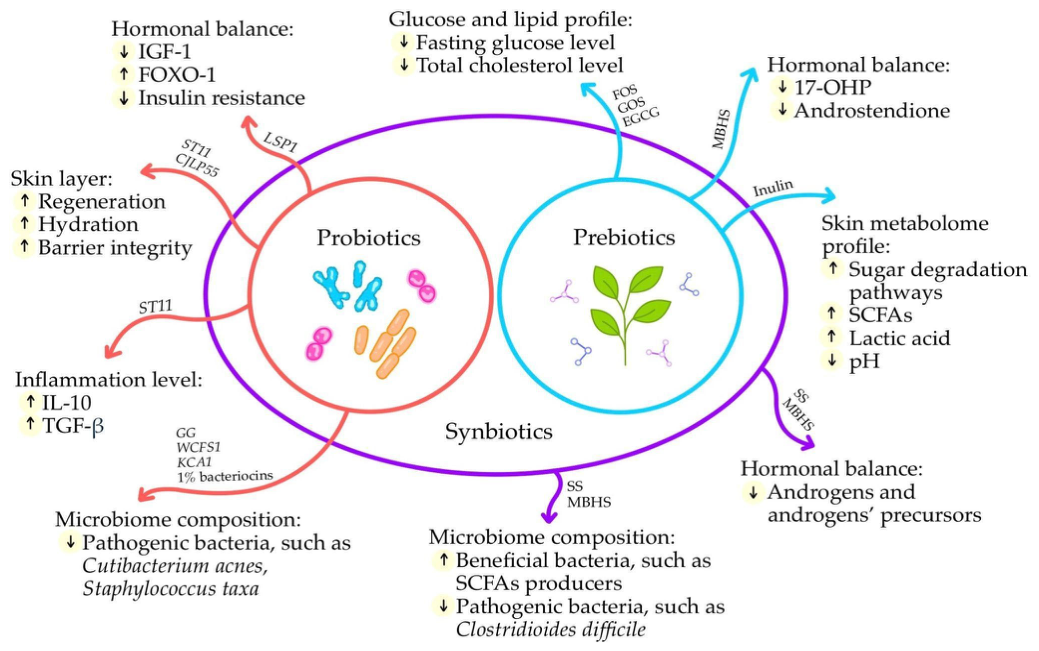
A summary of the potential mechanisms of action of probiotics, prebiotics, and synbiotics is presented in Figure 3.

4. Applications of Probiotics, Prebiotics, Postbiotics, and Synbiotics in the Treatment of Acne

Gut dysbiosis may contribute to the development and progression of various dermatological disorders. For this reason, therapeutic strategies targeting this complex interaction should receive particular attention [49,115,116]. Not only gut microbiota but also skin dysbiosis affects dermatological conditions such as atopic dermatitis, acne, psoriasis and skin cancer [49,117]. Growing public interest in the microbiome and its role in the development of dermatoses has led to an increase in the number of topical products designed to enhance microbial diversity on the skin surface [117]. Moreover, oral products aimed at gut microbiome modulation have also gained considerable attention. This trend reflects a growing body of evidence supporting systemic probiotic supplementation as a promising therapeutic approach for skin diseases [104,118]. The following section reviews the impact of modifying the microbiome using probiotics, prebiotics, postbiotics, and synbiotics in the treatment of acne.

4.1. Oral Probiotics

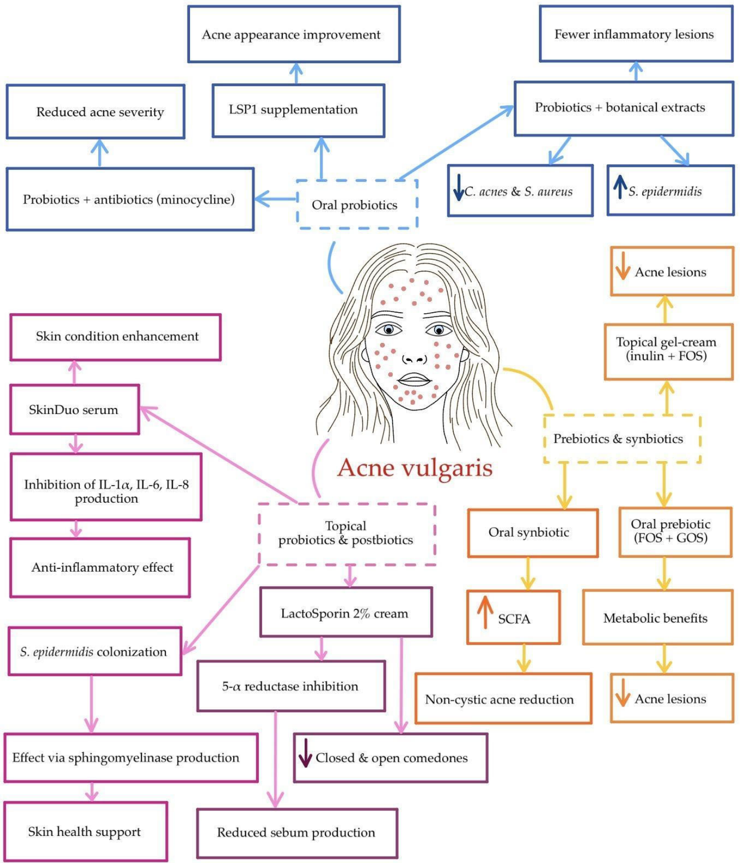
In the management of moderate to severe acne, systemic antibiotic therapy with tetracyclines is commonly used. This class includes agents such as minocycline, doxycycline, and sarecycline [119]. Beyond its antimicrobial activity against both Gram-positive and Gram-negative bacteria, minocycline also exhibits antioxidant, immunomodulatory, anti-apoptotic, and anti-inflammatory effects [120]. In addition to systemic administration, topical minocycline represents an emerging and potentially safer alternative for the treatment of inflammatory lesions in acne vulgaris, for example, in the form of a 4% foam (FMX101) [121,122,123,124]. Despite this, oral minocycline and doxycycline remain among the most frequently administered antibiotics by dermatologists [125]. The systemic use of tetracyclines is associated with an increased risk of adverse events, including Candida vulvovaginitis, C. difficile infection, and pharyngitis [119]. Moreover, minocycline therapy in acne vulgaris has been linked to a marked decline in gut microbial diversity, with an approximate 10% decrease in Bifidobacteriaceae and Lactobacillaceae. This alteration may persist even after antibiotic discontinuation, suggesting that such therapy could contribute to long-term gut dysbiosis [126]. Furthermore, patients treated with minocycline showed a depletion of probiotic gut species, namely B. adolescentis, Bifidobacterium breve, Bifidobacterium pseudolongum, and L. salivarius [127]. The aforementioned dysbiosis may potentially be corrected through the administration of oral probiotics. These include species from the genera Bifidobacterium, Lactococcus, and Lactobacillus, as well as Saccharomyces boulardii [42].
In this context, it is of particular interest whether probiotics can be used in acne management as part of combination therapy with antibiotics. This issue was addressed in a study involving 45 female participants. The prospective, randomized, open-label clinical trial assessed the effectiveness and tolerability of an acne treatment regimen combining minocycline with probiotic supplementation, compared with minocycline alone or probiotics alone. The intensity of disease among the patients was classified as mild to moderate [13]. It was demonstrated that, after 12 weeks, a combination of Lactobacillus delbrueckii subsp. bulgaricus, Bifidobacterium bifidum, and Lactobacillus acidophilus with minocycline significantly reduced acne severity [128]. These findings suggest a potential synergistic interaction between the probiotic formulation and the antibiotic, potentially enhancing its anti-inflammatory effects. Moreover, it was concluded that probiotic supplementation may reduce the risk of adverse effects [13]. Other studies also indicate the potential of probiotics to mitigate adverse effects, such as antibiotic-associated diarrhea [129,130]. The Western diet is an inseparable part of the industrialized lifestyle and is typically rich in simple sugars and dairy products [131,132,133,134]. It can modulate hormonal factors involved in the development of acne. Several studies have demonstrated that a high-glycemic-index diet (GI > 55) correlates not only with elevated postprandial insulin levels but also with increased concentrations of IGF-1 [131]. Consequently, carbohydrate intake and insulin levels may contribute to both the onset and severity of acne [135,136]. Fabbrocini et al. investigated the effects of supplementation with the LSP1 strain on acne severity in adults, as well as on insulin signaling-related genes in the skin. This pilot, randomized, double-blind, placebo-controlled clinical trial included 14 female and 6 male participants. After 12 weeks of liquid LSP1 supplementation at a dose of 3 × 10⁹ CFU/day (75 mg/day), IGF1 gene expression decreased, whereas FOXO1 gene expression increased. Additionally, an improvement in acne appearance was noted, likely resulting from the normalization of IGF1 and FOXO1 gene activity mediated by LSP1 [104].
Probiotics combined with plant extracts may serve as an adjunctive therapy for acne management. This correlation was observed by Rinaldi et al. in an 8-week randomized clinical trial involving 114 participants [137]. Patients were divided into four groups. Group I received a placebo. Group II received a dietary supplement containing probiotics (Lacticaseibacillus casei LC03 DSM 27537, Ligilactobacillus salivarius LS03 DSM 22776, and B. breve BR03 DSM 16604) together with botanical compounds (Echinacea extract and lupeol from Solanum melongena L.). Group III received only the plant extracts, and Group IV received only the probiotics. Data analysis revealed that patients in Group II exhibited a decline in the abundance of S. aureus and C. acnes, and an elevation in S. epidermidis levels. Moreover, participants receiving the combination therapy showed the greatest reduction in surface-level inflammatory lesions [137].

4.2. Topical Postbiotics and Probiotics

In addition to oral preparations, topical products are receiving increasing attention. Among the latest dermatology-focused skincare trends are microbiome-targeted cosmetics. These products support and enhance the diversity of the skin microbiota. Their formulations may contain probiotics, prebiotics, or postbiotics, which exert beneficial local effects on the microorganisms residing on human skin [117].
The term “postbiotic” was clearly defined by the International Scientific Association for Probiotics and Prebiotics (ISAPP) in 2019 as a “preparation of inanimate microorganisms and/or their components that confers a health benefit on the host” [138]. Postbiotics are added to a wide range of topical skincare products, including serums, creams, and cleansing agents. Their primary aim is to improve skin health by strengthening cutaneous immunity and maintaining the skin’s microbiological balance [139]. LactoSporin® 2% cream (Sabinsa Corporation, Bengaluru, India) is a topically applied postbiotic whose therapeutic effect has been assessed in a randomized, open-label, comparative clinical trial. The product inhibited 5-alpha reductase in vitro, contributing to reduced sebum production. It also showed antimicrobial activity. Moreover, improvements in both closed and open comedones occurred more rapidly compared to a 2% benzoyl peroxide (BP) gel in in vivo studies. Based on the analyzed data, LactoSporin may represent a potential alternative for the treatment of mild to moderate acne vulgaris [140]. However, it is worth considering whether a larger sample size than the 64 participants who completed the trial might have influenced the observed results. Despite growing interest in topical probiotics for the treatment of skin disorders, regulatory frameworks that clearly define their classification remain limited [141,142,143]. In practice, such products are categorized as cosmetics, primarily intended for skin care, cleansing, or esthetic purposes [142,143]. At the same time, studies report that topical probiotics may have applications in medicine for the treatment of various skin conditions, including atopic dermatitis, wound healing, and the reduction in inflammatory responses [144,145,146]. Among the most commonly used probiotic strains in cosmetic products are L. plantarum, Lactobacillus casei, B. subtilis, Lactococcus lactis, and L. acidophilus [143].
Acne-prone skin has a distinct microbiome, and current topical acne therapies, including retinoids and BP, often paradoxically result in both irritation and dryness by disrupting the cutaneous barrier [147,148]. In individuals with acne, the skin generally exhibits reduced ceramide levels in the stratum corneum [148,149]. In vivo studies have shown that the use of ceramide-containing formulations improves the condition of acne-prone skin while minimizing the side effects of conventional topical acne treatments [148]. For this reason, investigating how the skin microbiota may contribute to ceramide production and enhance the integrity of the host’s skin barrier is of particular interest. Zheng et al. demonstrated in mouse models that the commensal S. epidermidis markedly reduces transepidermal water loss (TEWL) and elevates ceramide levels in damaged skin upon colonization. This effect is mediated by the production of sphingomyelinase by S. epidermidis, which not only helps prevent skin dehydration but also provides essential metabolites for the bacteria. These findings suggest that topical probiotics containing S. epidermidis may have potential in supporting skin health [150].
Another preparation directly relevant to acne-prone skin is SkinDuo. It is an anti-acne topical serum containing Lactiplantibacillus plantarum strains combined with natural enhancers. The product was evaluated on human skin models ex vivo and validated on the skin of 10 healthy volunteers. The investigated serum demonstrated anti-inflammatory effects by inhibiting the production of IL-1α, IL-8, and IL-6. The formulation also enhances the skin condition in acne-like models. Furthermore, a reduction in lipid synthesis and a decrease in the viability of acne-associated bacteria (C. acnes) were observed in primary human sebocytes [151]. The experimental interventions and assessed outcomes are summarized in Table 1.

4.3. Prebiotics and Synbiotics

Probiotic, postbiotic, and prebiotic therapies can complement standard anti-acne treatments. Among these approaches, probiotics are currently the most intensively studied, while postbiotics and prebiotics are considered complementary interventions [152].
An open-label, prospective clinical trial was conducted on acne-prone skin using a topical gel-cream containing prebiotics like inulin and FOS [153]. The study evaluated the effectiveness of treatment for mild to moderate acne and its effects on the skin microbiome. Interestingly, results showed an increase in the abundance of C. acnes HL050PA2 (a strain linked to healthy skin), together with a rise in S. epidermidis, which may suppress pathogenic C. acnes strains [153,154]. A substantial reduction in inflammatory, non-inflammatory, and total acne lesions was observed after six weeks of use. In this context, the tested topical product may support favorable changes in the skin microbiome and represents a novel alternative among acne treatment strategies [153].
A case involving oral prebiotic supplementation—100 mg/day of FOS and 500 mg/day of GOS—in adult women with acne showed a beneficial effect on metabolic measures. Participants in the open-label, prospective pilot trial received the dietary supplement for three months without any modifications to their usual diet. Improvements were observed in the number of acne lesions as well as reductions in total cholesterol levels and beneficial effects on glycemic parameters [110].
Synbiotics remain new products in acne treatment, and only a limited number of studies have explored this area. Min M. et al. demonstrated in a randomized, prospective clinical trial how an oral synbiotic affected the number of acne lesions in individuals with non-cystic acne. An increase in SCFA levels was observed, associated with an increased abundance of bacteria responsible for their production in the gut, along with an improvement in acne lesions [79]. The role of SCFAs is currently investigated in inflammatory skin conditions, including acne. However, their beneficial effects depend on how they are administered [53]. Some studies report that topical application of SCFAs induces the expression of cytokines, whereas subcutaneous administration has an inhibitory effect [155]. Another study investigated intradermal injection of C. acnes and S. epidermidis in the presence of sucrose. This sugar selectively stimulated the probiotic S. epidermidis to produce SCFAs, which potentially inhibited the growth of C. acnes, suggesting novel therapeutic approaches [156]. Taken together, these results support the use of oral probiotics both as a treatment option and as an adjunct in the management of acne vulgaris. Promising evidence indicates that probiotics may be particularly favorable in individuals with impaired insulin signaling. The aforementioned findings also suggest that probiotic-containing dietary supplements may improve the condition of acne-prone skin. Other combinations of probiotics with plant extracts remain largely unexplored and need further investigation, as they may confer similar beneficial effects in acne.
A comparable gap in the literature on topical postbiotics and probiotics is evident. In addition, the relatively small sample sizes may limit the generalizability of the available findings. Future studies should include larger sample sizes, ideally several hundred participants. Such studies could focus on other commensal species residing on human skin that may exhibit similar positive properties, such as topical probiotics containing S. epidermidis. Identifying specific formulations that effectively support the skin microbiota would also be valuable. Direct clinical evidence supporting the use of the anti-acne topical serum SkinDuo as a microbiome-modulating therapy is still lacking.
Moreover, observations of oral prebiotic supplementation similarly highlight the importance of further research, not only in the context of acne but also in other metabolic diseases. Due to the role of SCFAs in inflammatory skin conditions, combined studies involving intestinal stimulation of SCFA production along with intradermal or subcutaneous SCFA therapy would be of considerable interest. It would also be useful to explore other synbiotic combinations, which could broaden the horizons of available therapeutic approaches for acne. A summary of the clinical evidence supporting microbiome modulation as a therapeutic approach for acne treatment is presented in Figure 4 and Table 2.
Importantly, the variability in clinical outcomes may reflect differences in the baseline gut–skin axis dysfunction among individual patients, suggesting that future therapeutic strategies should stratify patients into groups based on their microbiome and metabolic profiles.

5. Limitations

Despite a growing body to evidence pointing to the role of microbiome modulation in the treatment of acne, the findings summarized in this review have several significant limitations that may affect the reliability of the current conclusions. Therefore, it should be interpreted with great caution.
A significant portion of the available studies are based on relatively small groups, often with less than 50 participants. Such a limited sample size reduces statistical power and increases the risk of error. Furthermore, many studies have a short duration (typically 4–12 weeks), which is insufficient to assess the long-term efficacy and durability of treatment effects in the case of a chronic and recurrent condition such as acne vulgaris.
Furthermore, the available literature is highly heterogeneous. There is significant variability in the probiotic strains, prebiotic substances, synbiotic formulations, doses, and routes of administration used. This heterogeneity significantly limits comparability between studies and makes it difficult to identify consistent therapeutic effects or develop standard treatment protocols. Importantly, microbiome-related effects are often strain-specific and therefore cannot be generalized across different interventions.
It is also important to note the methodological limitations that further weaken the strength of the evidence. Many studies lack proper randomization, placebo control, or double-blind design. Furthermore, the assessment of outcomes is not always standardized, and some studies rely on subjective or invalid acne severity scales. These issues carry a risk of bias and may lead to an overestimation of treatment efficacy.
Finally, data on the long-term safety and potential adverse effects of microbiome-targeted interventions remain limited. This is particularly relevant for oral supplementation, which may induce permanent changes in the composition of the gut microbiota. Furthermore, potential interactions between microbiome-based therapies and conventional acne treatments—such as retinoids, antibiotics, or hormonal therapies—have not been adequately studied.
In summary, based on available literature, definitive conclusions regarding the efficacy and safety of microbiome modulation in the treatment of acne cannot be made. Therefore, these limitations underscore the need for more rigorous, standardized and long-term clinical trials.

6. Conclusions

Acne vulgaris is increasingly recognized as a systemic immunometabolic disorder in which hormonal, microbial, immunological, and environmental factors interact through the gut–skin axis. A growing body of evidence suggests that dysbiosis within this axis may contribute to the development of acne by linking microbial imbalances to immune system activation and metabolic signaling pathways, such as IGF-1 and mTORC1.
Microbiome-targeted interventions, including probiotics, prebiotics, and synbiotics, have emerged as promising strategies for modulating these pathways. Available studies suggest that such therapies may reduce lesion counts, improve skin barrier function, and exert systemic metabolic and anti-inflammatory effects. Furthermore, their potential to mitigate adverse effects associated with conventional treatments, particularly antibiotics, makes them attractive candidates for adjunctive therapy. Microbiological dysbiosis, disturbances in metabolite production, and immune system activation interact in this context, affecting key pathways such as IGF-1 and mTORC1, which ultimately influence sebaceous gland activity and inflammatory processes.
However, despite these encouraging and promising results, current scientific evidence remains insufficient to support the routine use of microbiome-based therapies as standalone treatments for acne. Significant limitations, such as small study populations, short treatment durations, and incomplete analysis of mechanisms of action, necessitate cautious interpretation of available data.
Future research should prioritize large-scale, well-designed randomized controlled trials with standardized methodologies, longer follow-up periods, and precise characterization of the microbial strains involved in this process, in conjunction with the host response. A deeper understanding of the gut–skin axis, identification of patient subgroups, and investigation of optimal doses appear essential for translating these approaches into clinical practice.
Based on current scientific knowledge, microbiome-based interventions should be viewed primarily as adjunctive strategies—based on mechanisms of action and aimed at restoring the homeostasis of the gut–skin axis—rather than as standalone treatments. Their future role in acne treatment will likely depend on identifying specific patient subgroups, precisely characterizing microbial strains, and integrating them with individually tailored therapeutic approaches.

Author Contributions

Conceptualization, A.D.-M. and K.Ł.; methodology, software, K.Ł. and P.L.; validation, A.D.-M.; investigation, K.Ł., P.L., J.S., J.L. and A.D.-M.; resources, K.Ł., P.L., J.S., J.L. and A.D.-M.; data curation, K.Ł., P.L., J.S., J.L. and A.D.-M.; writing—original draft preparation K.Ł., P.L., J.S., J.L. and A.D.-M.; writing—review and editing, K.Ł., P.L., J.S., J.L. and A.D.-M.; visualization, K.Ł. and P.L.; supervision, A.D.-M.; project administration, A.D.-M.; funding acquisition, A.D.-M. All authors have read and agreed to the published version of the manuscript.

Funding

This research received no external funding.

Institutional Review Board Statement

Not applicable.

Informed Consent Statement

Not applicable.

Data Availability Statement

No new data were created or analyzed in this study.

Conflicts of Interest

The authors declare no conflicts of interest.

References

  1. Heng, A.H.S.; Chew, F.T. Systematic Review of the Epidemiology of Acne Vulgaris. Sci. Rep. 2020, 10, 5754. [Google Scholar] [CrossRef]
  2. Tan, J.K.L.; Bhate, K. A Global Perspective on the Epidemiology of Acne. Br. J. Dermatol. 2015, 172, 3–12. [Google Scholar] [CrossRef]
  3. Zaenglein, A.L.; Pathy, A.L.; Schlosser, B.J.; Alikhan, A.; Baldwin, H.E.; Berson, D.S.; Bowe, W.P.; Graber, E.M.; Harper, J.C.; Kang, S.; et al. Guidelines of Care for the Management of Acne Vulgaris. J. Am. Acad. Dermatol. 2016, 74, 945–973.e33. [Google Scholar] [CrossRef]
  4. Alqahtani, A.; Alsaab, W.I.; Altulahi, B. Psychological Impact of Acne Vulgaris on the Young Saudi Population. Cureus 2021, 13, e20509. [Google Scholar] [CrossRef]
  5. Samuels, D.V.; Rosenthal, R.; Lin, R.; Chaudhari, S.; Natsuaki, M.N. Acne Vulgaris and Risk of Depression and Anxiety: A Meta-Analytic Review. J. Am. Acad. Dermatol. 2020, 83, 532–541. [Google Scholar] [CrossRef] [PubMed]
  6. Collier, C.N.; Harper, J.C.; Cantrell, W.C.; Wang, W.; Foster, K.W.; Elewski, B.E. The Prevalence of Acne in Adults 20 Years and Older. J. Am. Acad. Dermatol. 2008, 58, 56–59. [Google Scholar] [CrossRef]
  7. Perkins, A.C.; Maglione, J.; Hillebrand, G.G.; Miyamoto, K.; Kimball, A.B. Acne Vulgaris in Women: Prevalence across the Life Span. J. Women’s Health 2012, 21, 223–230. [Google Scholar] [CrossRef]
  8. Karadag, A.S.; Aslan Kayıran, M.; Wu, C.Y.; Chen, W.; Parish, L.C. Antibiotic Resistance in Acne: Changes, Consequences and Concerns. J. Eur. Acad. Dermatol. Venereol. 2021, 35, 73–78. [Google Scholar] [CrossRef] [PubMed]
  9. Walsh, T.R.; Efthimiou, J.; Dréno, B. Systematic Review of Antibiotic Resistance in Acne: An Increasing Topical and Oral Threat. Lancet Infect. Dis. 2016, 16, e23–e33. [Google Scholar] [CrossRef] [PubMed]
  10. Salem, I.; Ramser, A.; Isham, N.; Ghannoum, M.A. The Gut Microbiome as a Major Regulator of the Gut-Skin Axis. Front. Microbiol. 2018, 9, 1459. [Google Scholar] [CrossRef]
  11. Melnik, B.C. Dietary Intervention in Acne: Attenuationof Increased MTORC1 Signaling Promoted by Western Diet. Dermato-Endocrinology 2012, 4, 20–32. [Google Scholar] [CrossRef] [PubMed]
  12. Smith, R.N.; Mann, N.J.; Braue, A.; Mäkeläinen, H.; Varigos, G.A. A Low-Glycemic-Load Diet Improves Symptoms in Acne Vulgaris Patients: A Randomized Controlled Trial. Am. J. Clin. Nutr. 2007, 86, 107–115. [Google Scholar] [CrossRef]
  13. Jung, G.W.; Tse, J.E.; Guiha, I.; Rao, J. Prospective, Randomized, Open-Label Trial Comparing the Safety, Efficacy, and Tolerability of an Acne Treatment Regimen with and without a Probiotic Supplement and Minocycline in Subjects with Mild to Moderate Acne. J. Cutan. Med. Surg. 2013, 17, 114–122. [Google Scholar] [CrossRef]
  14. Szántó, M.; Dózsa, A.; Antal, D.; Szabó, K.; Kemény, L.; Bai, P. Targeting the Gut-Skin Axis—Probiotics as New Tools for Skin Disorder Management? Exp. Dermatol. 2019, 28, 1210–1218. [Google Scholar] [CrossRef] [PubMed]
  15. Deng, Y.; Wang, F.; He, L. Skin Barrier Dysfunction in Acne Vulgaris: Pathogenesis and Therapeutic Approaches. Med. Sci. Monit. 2024, 30, e945336. [Google Scholar] [CrossRef]
  16. Dreno, B.; Dekio, I.; Baldwin, H.; Demessant, A.L.; Dagnelie, M.A.; Khammari, A.; Corvec, S. Acne Microbiome: From Phyla to Phylotypes. J. Eur. Acad. Dermatol. Venereol. 2024, 38, 657–664. [Google Scholar] [CrossRef] [PubMed]
  17. Dréno, B.; Dagnelie, M.A.; Khammari, A.; Corvec, S. The Skin Microbiome: A New Actor in Inflammatory Acne. Am. J. Clin. Dermatol. 2020, 21, 18–24. [Google Scholar] [CrossRef]
  18. Sánchez-Pellicer, P.; Navarro-Moratalla, L.; Núñez-Delegido, E.; Ruzafa-Costas, B.; Agüera-Santos, J.; Navarro-López, V. Acne, Microbiome, and Probiotics: The Gut-Skin Axis. Microorganisms 2022, 10, 1303. [Google Scholar] [CrossRef]
  19. Li, D.; Zhou, Z.; Yang, X.; Zhang, Q.; Xu, J.; Zouboulis, C.C.; Xiang, Q.; Zhang, S. A Comprehensive Review: The Bidirectional Role of Sebum in Skin Health. Bioengineering 2025, 12, 1333. [Google Scholar] [CrossRef]
  20. Behayaa, H.R.; Juda, T.M.; Mohammed, S.B. The Effect of Androgen Hormones in Acne Pathogenesis: A Review. Med. J. Babylon 2022, 19, 345–349. [Google Scholar] [CrossRef]
  21. Gollnick, H.P.M. From New Findings in Acne Pathogenesis to New Approaches in Treatment. J. Eur. Acad. Dermatol. Venereol. 2015, 29, 1–7. [Google Scholar] [CrossRef] [PubMed]
  22. Vasam, M.; Korutla, S.; Bohara, R.A. Acne Vulgaris: A Review of the Pathophysiology, Treatment, and Recent Nanotechnology Based Advances. Biochem. Biophys. Rep. 2023, 36, 101578. [Google Scholar] [CrossRef]
  23. González-Mondragón, E.A.; Ganoza-Granados, L.D.C.; Eréndira Toledo-Bahena, M.; Valencia-Herrera, A.M.; Duarte-Abdala, M.R.; Camargo-Sánchez, K.A.; Mena-Cedillos, C.A.; Toledo-Bahena, M.E. Acne and Diet: A Review of Pathogenic Mechanisms. Bol. Med. Hosp. Infant. Mex. 2022, 79, 83–90. [Google Scholar] [CrossRef]
  24. Faruga-Lewicka, W.; Kardas, M. The Influence of Insulin-like Growth Factor IGF-1 on the Course of Acne Vulgaris. Aesthetic Cosmetol. Med. 2022, 11, 105–108. [Google Scholar] [CrossRef]
  25. Zouboulis, C.C.; Coenye, T.; He, L.; Kabashima, K.; Kobayashi, T.; Niemann, C.; Nomura, T.; Oláh, A.; Picardo, M.; Quist, S.R.; et al. Sebaceous Immunobiology—Skin Homeostasis, Pathophysiology, Coordination of Innate Immunity and Inflammatory Response and Disease Associations. Front. Immunol. 2022, 13, 1029818. [Google Scholar] [CrossRef]
  26. Del Rosso, J.Q.; Kircik, L. The Primary Role of Sebum in the Pathophysiology of Acne Vulgaris and Its Therapeutic Relevance in Acne Management. J. Dermatol. Treat. 2024, 35, 2296855. [Google Scholar] [CrossRef]
  27. Lee, H.J.; Kim, M. Skin Barrier Function and the Microbiome. Int. J. Mol. Sci. 2022, 23, 13071. [Google Scholar] [CrossRef]
  28. Xu, H.; Li, H. Acne, the Skin Microbiome, and Antibiotic Treatment. Am. J. Clin. Dermatol. 2019, 20, 335–344. [Google Scholar] [CrossRef]
  29. Scharschmidt, T.C.; Segre, J.A. Skin Microbiome and Dermatologic Disorders. J. Clin. Investig. 2025, 135, e184315. [Google Scholar] [CrossRef] [PubMed]
  30. Cheung, C.T.; Lancien, U.; Corvec, S.; Mengeaud, V.; Mias, C.; Véziers, J.; Khammari, A.; Dréno, B. Pro-Inflammatory Activity of Cutibacterium acnes Phylotype IA1 and Extracellular Vesicles: An in Vitro Study. Exp. Dermatol. 2024, 33, e15150. [Google Scholar] [CrossRef] [PubMed]
  31. Corvec, S.; Dagnelie, M.-A.; Khammari, A.; Dréno, B. Taxonomy and Phylogeny of Cutibacterium (Formerly Propionibacterium) Acnes in Inflammatory Skin Diseases Taxonomie et Phylogénie de Cutibacterium (Ex-Propionibacterium) Acnes et Pathologies Inflammatoires Cutanées. Ann. Dermatol. Venereol. 2019, 146, 26–30. [Google Scholar] [CrossRef] [PubMed]
  32. Niedźwiedzka, A.; Micallef, M.P.; Biazzo, M.; Podrini, C. The Role of the Skin Microbiome in Acne: Challenges and Future Therapeutic Opportunities. Int. J. Mol. Sci. 2024, 25, 11422. [Google Scholar] [CrossRef] [PubMed]
  33. Dréno, B.; Pécastaings, S.; Corvec, S.; Veraldi, S.; Khammari, A.; Roques, C. Cutibacterium acnes (Propionibacterium acnes) and Acne Vulgaris: A Brief Look at the Latest Updates. J. Eur. Acad. Dermatol. Venereol. 2018, 32, 5–14. [Google Scholar] [CrossRef]
  34. Mayslich, C.; Grange, P.A.; Dupin, N. Cutibacterium acnes as an Opportunistic Pathogen: An Update of Its Virulence-Associated Factors. Microorganisms 2021, 9, 303. [Google Scholar] [CrossRef]
  35. Cruz, S.; Vecerek, N.; Elbuluk, N. Targeting Inflammation in Acne: Current Treatments and Future Prospects. Am. J. Clin. Dermatol. 2023, 24, 681–694. [Google Scholar] [CrossRef]
  36. Mias, C.; Thouvenin, M.D.; Gravier, E.; Dalmon, S.; Bouyer, K.; Alvarez, S.; Mengeaud, V.; Ribet, V.; Bessou-Touya, S.; Duplan, H. Change in Cutibacterium acnes Phylotype Abundance and Improvement of Clinical Parameters Using a New Dermocosmetic Product Containing Myrtus Communis and Celastrol Enriched Plant Cell Culture Extracts in Patients with Acne Vulgaris. J. Eur. Acad. Dermatol. Venereol. 2023, 37, 20–25. [Google Scholar] [CrossRef] [PubMed]
  37. Cavallo, I.; Sivori, F.; Truglio, M.; De Maio, F.; Lucantoni, F.; Cardinali, G.; Pontone, M.; Bernardi, T.; Sanguinetti, M.; Capitanio, B.; et al. Skin Dysbiosis and Cutibacterium acnes Biofilm in Inflammatory Acne Lesions of Adolescents. Sci. Rep. 2022, 12, 21104. [Google Scholar] [CrossRef]
  38. Brown, M.M.; Horswill, A.R. Staphylococcus epidermidis—Skin Friend or Foe? PLoS Pathog. 2020, 16, e1009026. [Google Scholar] [CrossRef]
  39. Fournière, M.; Latire, T.; Souak, D.; Feuilloley, M.G.J.; Bedoux, G. Staphylococcus epidermidis and Cutibacterium acnes: Two Major Sentinels of Skin Microbiota and the Influence of Cosmetics. Microorganisms 2020, 8, 1752. [Google Scholar] [CrossRef]
  40. Flowers, L.; Grice, E.A. The Skin Microbiota: Balancing Risk and Reward. Cell Host Microbe 2020, 28, 190–200. [Google Scholar] [CrossRef]
  41. Severn, M.M.; Horswill, A.R. Staphylococcus epidermidis and Its Dual Lifestyle in Skin Health and Infection. Nat. Rev. Microbiol. 2023, 21, 97–111. [Google Scholar] [CrossRef] [PubMed]
  42. Dessinioti, C.; Katsambas, A. The Microbiome and Acne: Perspectives for Treatment. Dermatol. Ther. 2024, 14, 31–44. [Google Scholar] [CrossRef]
  43. Lee, Y.B.; Byun, E.J.; Kim, H.S. Potential Role of the Microbiome in Acne: A Comprehensive Review. J. Clin. Med. 2019, 8, 987. [Google Scholar] [CrossRef]
  44. Wojciechowska, K.; Szewczyk, K.D.S. The Skin Microbiome and Bioactive Compounds: Mechanisms of Modulation, Dysbiosis, and Dermatological Implications. Molecules 2025, 30, 4363. [Google Scholar] [CrossRef]
  45. Piazzesi, A.; Scanu, M.; Ciprandi, G.; Putignani, L. Modulations of the Skin Microbiome in Skin Disorders: A Narrative Review from a Wound Care Perspective. Int. Wound J. 2024, 21, e70087. [Google Scholar] [CrossRef]
  46. Luqman, A.; Hassan, A.; Ullah, M.; Naseem, S.; Ullah, M.; Zhang, L.; Din, A.U.; Ullah, K.; Ahmad, W.; Wang, G. Role of the Intestinal Microbiome and Its Therapeutic Intervention in Cardiovascular Disorder. Front. Immunol. 2024, 15, 1321395. [Google Scholar] [CrossRef]
  47. Mehta, I.; Juneja, K.; Nimmakayala, T.; Bansal, L.; Pulekar, S.; Duggineni, D.; Ghori, H.K.; Modi, N.; Younas, S. Gut Microbiota and Mental Health: A Comprehensive Review of Gut-Brain Interactions in Mood Disorders. Cureus 2025, 17, e81447. [Google Scholar] [CrossRef] [PubMed]
  48. Van Hul, M.; Cani, P.D.; Petitfils, C.; De Vos, W.M.; Tilg, H.; El-Omar, E.M. What Defines a Healthy Gut Microbiome? Gut 2024, 73, 1893–1908. [Google Scholar] [CrossRef]
  49. De Pessemier, B.; Grine, L.; Debaere, M.; Maes, A.; Paetzold, B.; Callewaert, C. Gut-Skin Axis: Current Knowledge of the Interrelationship between Microbial Dysbiosis and Skin Conditions. Microorganisms 2021, 9, 353. [Google Scholar] [CrossRef] [PubMed]
  50. Jandhyala, S.M.; Talukdar, R.; Subramanyam, C.; Vuyyuru, H.; Sasikala, M.; Reddy, D.N. Role of the Normal Gut Microbiota. World J. Gastroenterol. 2015, 21, 8787–8803. [Google Scholar] [CrossRef]
  51. Blaak, E.E.; Canfora, E.E.; Theis, S.; Frost, G.; Groen, A.K.; Mithieux, G.; Nauta, A.; Scott, K.; Stahl, B.; van Harsselaar, J.; et al. Short Chain Fatty Acids in Human Gut and Metabolic Health. Benef. Microbes 2020, 11, 411–455. [Google Scholar] [CrossRef] [PubMed]
  52. Thursby, E.; Juge, N. Introduction to the Human Gut Microbiota. Biochem. J. 2017, 474, 1823–1836. [Google Scholar] [CrossRef]
  53. Xiao, X.; Hu, X.; Yao, J.; Cao, W.; Zou, Z.; Wang, L.; Qin, H.; Zhong, D.; Li, Y.; Xue, P.; et al. The Role of Short-Chain Fatty Acids in Inflammatory Skin Diseases. Front. Microbiol. 2023, 13, 1083432. [Google Scholar] [CrossRef]
  54. Wu, H.; Mu, C.; Xu, L.; Yu, K.; Shen, L.; Zhu, W. Host-Microbiota Interaction in Intestinal Stem Cell Homeostasis. Gut Microbes 2024, 16, 2353399. [Google Scholar] [CrossRef]
  55. Ji, X.; Wu, S.; Zhao, D.; Bai, Q.; Wang, Y.; Gong, K.; Zheng, H.; Zhu, M. Revealing the Impact of Gut Microbiota on Acne Through Mendelian Randomization Analysis. Clin. Cosmet. Investig. Dermatol. 2024, 17, 383–393. [Google Scholar] [CrossRef] [PubMed]
  56. Criton, V.J.S.; Joy, S.; Criton, V.J.S.; Joy, S. Beyond Skincare Routines: Follow Your Gut to Healthy Skin—A Review of the Interplay between Gut Microbiome and Skin. J. Ski. Sex. Transm. Dis. 2024, 6, 5–12. [Google Scholar] [CrossRef]
  57. Sugrue, I.; Ross, R.P.; Hill, C. Bacteriocin Diversity, Function, Discovery and Application as Antimicrobials. Nat. Rev. Microbiol. 2024, 22, 556–571. [Google Scholar] [CrossRef] [PubMed]
  58. Sanders, M.E.; Merenstein, D.J.; Reid, G.; Gibson, G.R.; Rastall, R.A. Probiotics and Prebiotics in Intestinal Health and Disease: From Biology to the Clinic. Nat. Rev. Gastroenterol. Hepatol. 2019, 16, 605–616. [Google Scholar] [CrossRef]
  59. Pandey, K.R.; Naik, S.R.; Vakil, B.V. Probiotics, Prebiotics and Synbiotics—A Review. J. Food Sci. Technol. 2015, 52, 7577–7587. [Google Scholar] [CrossRef]
  60. Mani-López, E.; Arrioja-Bretón, D.; López-Malo, A. The Impacts of Antimicrobial and Antifungal Activity of Cell-Free Supernatants from Lactic Acid Bacteria in Vitro and Foods. Compr. Rev. Food Sci. Food Saf. 2022, 21, 604–641. [Google Scholar] [CrossRef]
  61. Cha, H.; Kim, S.-K.; Kook, M.; Yi, T.-H. Lactobacillus Paraplantarum THG-G10 as a Potential Anti-Acne Agent with Anti-Bacterial and Anti-Inflammatory Activities. Anaerobe 2020, 64, 102243. [Google Scholar] [CrossRef] [PubMed]
  62. Xu, D.; Fu, L.; Pan, D.; Chu, Y.; Feng, M.; Lu, Y.; Yang, C.; Wang, Y.; Xia, J.; Sun, G. Role of Probiotics/Synbiotic Supplementation in Glycemic Control: A Critical Umbrella Review of Meta-Analyses of Randomized Controlled Trials. Crit. Rev. Food Sci. Nutr. 2024, 64, 1467–1485. [Google Scholar] [CrossRef] [PubMed]
  63. Alessandrini, G.; Mercuri, S.; Martella, A.; Ferrara, F.; Simonetti, V.; Trifirò, C.; Emanuele, E. Topical Application of Bacteriocins from Bacillus Subtilis Promotes Staphylococcus Aureus Decolonization in Acneic Skin and Improves the Clinical Appearance of Mild-to-Moderate Acne. Adv. Dermatol. Allergol. 2023, 40, 115–118. [Google Scholar] [CrossRef]
  64. Deidda, F.; Amoruso, A.; Nicola, S.; Graziano, T.; Pane, M.; Mogna, L. New Approach in Acne Therapy. J. Clin. Gastroenterol. 2018, 52, S78–S81. [Google Scholar] [CrossRef]
  65. Espinoza-Monje, M.; Campos, J.; Alvarez Villamil, E.; Jerez, A.; Dentice Maidana, S.; Elean, M.; Salva, S.; Kitazawa, H.; Villena, J.; García-Cancino, A. Characterization of Weissella Viridescens UCO-SMC3 as a Potential Probiotic for the Skin: Its Beneficial Role in the Pathogenesis of Acne Vulgaris. Microorganisms 2021, 9, 1486. [Google Scholar] [CrossRef]
  66. Lebeer, S.; Oerlemans, E.F.M.; Claes, I.; Henkens, T.; Delanghe, L.; Wuyts, S.; Spacova, I.; van den Broek, M.F.L.; Tuyaerts, I.; Wittouck, S.; et al. Selective Targeting of Skin Pathobionts and Inflammation with Topically Applied Lactobacilli. Cell Rep. Med. 2022, 3, 100521. [Google Scholar] [CrossRef]
  67. Gibson, G.R.; Beatty, E.R.; Wang, X.; Cummings, J.H. Selective Stimulation of Bifidobacteria in the Human Colon by Oligofructose and Inulin. Gastroenterology 1995, 108, 975–982. [Google Scholar] [CrossRef]
  68. Ben, X.-M.; Li, J.; Feng, Z.-T.; Shi, S.-Y.; Lu, Y.-D.; Chen, R.; Zhou, X.-Y. Low Level of Galacto-Oligosaccharide in Infant Formula Stimulates Growth of Intestinal Bifidobacteria and Lactobacilli. World J. Gastroenterol. 2008, 14, 6564–6568. [Google Scholar] [CrossRef] [PubMed]
  69. Knol, J.; Scholtens, P.; Kafka, C.; Steenbakkers, J.; Gro, S.; Helm, K.; Klarczyk, M.; Schöpfer, H.; Böckler, H.-M.; Wells, J. Colon Microflora in Infants Fed Formula with Galacto- and Fructo-Oligosaccharides: More like Breast-Fed Infants. J. Pediatr. Gastroenterol. Nutr. 2005, 40, 36–42. [Google Scholar] [CrossRef]
  70. Fernández, J.; Moreno, F.J.; Olano, A.; Clemente, A.; Villar, C.J.; Lombó, F. A Galacto-Oligosaccharides Preparation Derived From Lactulose Protects Against Colorectal Cancer Development in an Animal Model. Front. Microbiol. 2018, 9, 2004. [Google Scholar] [CrossRef]
  71. Yin, P.; Du, T.; Yi, S.; Zhang, C.; Yu, L.; Tian, F.; Chen, W.; Zhai, Q. Response Differences of Gut Microbiota in Oligofructose and Inulin Are Determined by the Initial Gut Bacteroides/Bifidobacterium Ratios. Food Res. Int. 2023, 174, 113598. [Google Scholar] [CrossRef] [PubMed]
  72. Rakhra, G.; Malhotra, R.; Prasad, P.; Sahu, J.K.; Rakhra, G.; Khan, T.U.; Khan, M.U.; Rastogi, S. Synergistic Effects of Polyphenols and Gut Microbiota-Derived Metabolites on Inflammation and Metabolic Syndrome: A Review. Mol. Nutr. Food Res. 2026, 70, e70360. [Google Scholar] [CrossRef] [PubMed]
  73. Liu, Z.; de Bruijn, W.J.C.; Bruins, M.E.; Vincken, J.-P. Reciprocal Interactions between Epigallocatechin-3-Gallate (EGCG) and Human Gut Microbiota In Vitro. J. Agric. Food Chem. 2020, 68, 9804–9815. [Google Scholar] [CrossRef]
  74. Wang, P.; Gao, J.; Ke, W.; Wang, J.; Li, D.; Liu, R.; Jia, Y.; Wang, X.; Chen, X.; Chen, F.; et al. Resveratrol Reduces Obesity in High-Fat Diet-Fed Mice via Modulating the Composition and Metabolic Function of the Gut Microbiota. Free Radic. Biol. Med. 2020, 156, 83–98. [Google Scholar] [CrossRef]
  75. Li, M.; Mao, J.; Diaz, I.; Kopylova, E.; Melnik, A.V.; Aksenov, A.A.; Tipton, C.D.; Soliman, N.; Morgan, A.M.; Boyd, T. Multi-Omic Approach to Decipher the Impact of Skincare Products with Pre/Postbiotics on Skin Microbiome and Metabolome. Front. Med. 2023, 10, 1165980. [Google Scholar] [CrossRef] [PubMed]
  76. Ananthapadmanabhan, K.P.; Mukherjee, S.; Chandar, P. Stratum Corneum Fatty Acids: Their Critical Role in Preserving Barrier Integrity during Cleansing. Int. J. Cosmet. Sci. 2013, 35, 337–345. [Google Scholar] [CrossRef]
  77. Le Bourgot, C.; Meunier, C.; Gaio, E.; Murat, V.; Micheletto, M.; Tedesco, E.; Benetti, F. Effects of Short Chain Fructo-Oligosaccharides on Selected Skin Bacteria. Sci. Rep. 2022, 12, 9702. [Google Scholar] [CrossRef]
  78. Petrov, A.; Ćorović, M.; Milivojević, A.; Simović, M.; Banjanac, K.; Pjanović, R.; Bezbradica, D. Prebiotic Effect of Galacto-Oligosaccharides on the Skin Microbiota and Determination of Their Diffusion Properties. Int. J. Cosmet. Sci. 2022, 44, 309–319. [Google Scholar] [CrossRef]
  79. Min, M.; Afzal, N.; Maloh, J.; Dulai, A.S.; Ahmad, N.; Pinzauti, D.; Sivamani, R.K. Prospective Comparative Study of an Oral Synbiotic and a Myoinositol-Based Herbal Supplement in Modifying Hormone Levels and the Gut Microbiome in Non-Cystic Acne. Dermatol. Ther. 2025, 15, 1331–1350. [Google Scholar] [CrossRef]
  80. Zhao, Y.; Yu, C.; Zhang, J.; Yao, Q.; Zhu, X.; Zhou, X. The Gut-skin Axis: Emerging Insights in Understanding and Treating Skin Diseases through Gut Microbiome Modulation (Review). Int. J. Mol. Med. 2025, 56, 210. [Google Scholar] [CrossRef]
  81. Huang, Y.; Huang, Y.; Xia, D.; Liu, L.; Xiong, X.; Ouyang, Y.; Deng, Y. Lactobacillus Rhamnosus Ameliorates Acne Vulgaris in SD Rats via Changes in Gut Microbiota and Associated Tryptophan Metabolism. Front. Immunol. 2023, 14, 1293048. [Google Scholar] [CrossRef]
  82. Chornchoem, P.; Tandhavanant, S.; Saiprom, N.; Preechanukul, A.; Thongchompoo, N.; Sensorn, I.; Chantratita, W.; Chantratita, N. Metagenomic Evaluation, Antimicrobial Activities, and Immune Stimulation of Probiotics from Dietary Supplements and Dairy Products. Sci. Rep. 2025, 15, 11537. [Google Scholar] [CrossRef]
  83. Sun, K.-Y.; Xu, D.-H.; Xie, C.; Plummer, S.; Tang, J.; Yang, X.F.; Ji, X.H. Lactobacillus Paracasei Modulates LPS-Induced Inflammatory Cytokine Release by Monocyte-Macrophages via the up-Regulation of Negative Regulators of NF-KappaB Signaling in a TLR2-Dependent Manner. Cytokine 2017, 92, 1–11. [Google Scholar] [CrossRef] [PubMed]
  84. Kook, S.-Y.; Chung, E.-C.; Lee, Y.; Lee, D.W.; Kim, S. Isolation and Characterization of Five Novel Probiotic Strains from Korean Infant and Children Faeces. PLoS ONE 2019, 14, e0223913. [Google Scholar] [CrossRef] [PubMed]
  85. Rhayat, L.; Maresca, M.; Nicoletti, C.; Perrier, J.; Brinch, K.S.; Christian, S.; Devillard, E.; Eckhardt, E. Effect of Bacillus Subtilis Strains on Intestinal Barrier Function and Inflammatory Response. Front. Immunol. 2019, 10, 564. [Google Scholar] [CrossRef]
  86. McLoughlin, R.F.; Berthon, B.S.; Jensen, M.E.; Baines, K.J.; Wood, L.G. Short-Chain Fatty Acids, Prebiotics, Synbiotics, and Systemic Inflammation: A Systematic Review and Meta-Analysis. Am. J. Clin. Nutr. 2017, 106, 930–945. [Google Scholar] [CrossRef] [PubMed]
  87. Qin, M.; Pirouz, A.; Kim, M.-H.; Krutzik, S.R.; Garbán, H.J.; Kim, J. Propionibacterium acnes Induces IL-1β Secretion via the NLRP3 Inflammasome in Human Monocytes. J. Investig. Dermatol. 2014, 134, 381–388. [Google Scholar] [CrossRef]
  88. Kim, J.; Ochoa, M.-T.; Krutzik, S.R.; Takeuchi, O.; Uematsu, S.; Legaspi, A.J.; Brightbill, H.D.; Holland, D.; Cunliffe, W.J.; Akira, S.; et al. Activation of Toll-like Receptor 2 in Acne Triggers Inflammatory Cytokine Responses. J. Immunol. 2002, 169, 1535–1541. [Google Scholar] [CrossRef]
  89. Huang, Y.-C.; Yang, C.-H.; Li, T.-T.; Zouboulis, C.C.; Hsu, H.-C. Cell-Free Extracts of Propionibacterium acnes Stimulate Cytokine Production through Activation of P38 MAPK and Toll-like Receptor in SZ95 Sebocytes. Life Sci. 2015, 139, 123–131. [Google Scholar] [CrossRef]
  90. Mattii, M.; Lovászi, M.; Garzorz, N.; Atenhan, A.; Quaranta, M.; Lauffer, F.; Konstantinow, A.; Küpper, M.; Zouboulis, C.C.; Kemeny, L.; et al. Sebocytes Contribute to Skin Inflammation by Promoting the Differentiation of T Helper 17 Cells. Br. J. Dermatol. 2018, 178, 722–730. [Google Scholar] [CrossRef]
  91. Choi, J.J.; Park, M.Y.; Lee, H.J.; Yoon, D.-Y.; Lim, Y.; Hyun, J.W.; Zouboulis, C.C.; Jin, M. TNF-α Increases Lipogenesis via JNK and PI3K/Akt Pathways in SZ95 Human Sebocytes. J. Dermatol. Sci. 2012, 65, 179–188. [Google Scholar] [CrossRef]
  92. Caillon, F.; O’Connell, M.; Eady, E.A.; Jenkins, G.R.; Cove, J.H.; Layton, A.M.; Mountford, A.P. Interleukin-10 Secretion from CD14+ Peripheral Blood Mononuclear Cells Is Downregulated in Patients with Acne Vulgaris. Br. J. Dermatol. 2010, 162, 296–303. [Google Scholar] [CrossRef]
  93. Gueniche, A.; Philippe, D.; Bastien, P.; Reuteler, G.; Blum, S.; Castiel-Higounenc, I.; Breton, L.; Benyacoub, J. Randomised Double-Blind Placebo-Controlled Study of the Effect of Lactobacillus Paracasei NCC 2461 on Skin Reactivity. Benef. Microbes 2014, 5, 137–145. [Google Scholar] [CrossRef]
  94. Kelhälä, H.-L.; Palatsi, R.; Fyhrquist, N.; Lehtimäki, S.; Väyrynen, J.P.; Kallioinen, M.; Kubin, M.E.; Greco, D.; Tasanen, K.; Alenius, H.; et al. IL-17/Th17 Pathway Is Activated in Acne Lesions. PLoS ONE 2014, 9, e105238. [Google Scholar] [CrossRef]
  95. Moon, J.; Yoon, J.Y.; Yang, J.H.; Kwon, H.H.; Min, S.; Suh, D.H. Atrophic Acne Scar: A Process from Altered Metabolism of Elastic Fibres and Collagen Fibres Based on Transforming Growth Factor-Β1 Signalling. Br. J. Dermatol. 2019, 181, 1226–1237. [Google Scholar] [CrossRef]
  96. Capitán-Cañadas, F.; Ortega-González, M.; Guadix, E.; Zarzuelo, A.; Suárez, M.D.; de Medina, F.S.; Martínez-Augustin, O. Prebiotic Oligosaccharides Directly Modulate Proinflammatory Cytokine Production in Monocytes via Activation of TLR4. Mol. Nutr. Food Res. 2014, 58, 1098–1110. [Google Scholar] [CrossRef]
  97. Newburg, D.S.; Ko, J.S.; Leone, S.; Nanthakumar, N.N. Human Milk Oligosaccharides and Synthetic Galactosyloligosaccharides Contain 3′-, 4-, and 6′-Galactosyllactose and Attenuate Inflammation in Human T84, NCM-460, and H4 Cells and Intestinal Tissue Ex Vivo. J. Nutr. 2016, 146, 358–367. [Google Scholar] [CrossRef] [PubMed]
  98. Del Fabbro, S.; Calder, P.C.; Childs, C.E. Microbiota-Independent Immunological Effects of Non-Digestible Oligosaccharides in the Context of Inflammatory Bowel Diseases. Proc. Nutr. Soc. 2020, 79, 468–478. [Google Scholar] [CrossRef]
  99. Omer, M.; Awad, H.O.; Omer, O.I.; Mohamed, I.O. Preclinical Evidence for Prebiotic Therapy in Ulcerative Colitis: Mechanisms, Efficacy, and Translational Perspectives. Nutr. Rev. 2026, nuag011. [Google Scholar] [CrossRef] [PubMed]
  100. Amir Aslani, B.; Ghobadi, S. Studies on Oxidants and Antioxidants with a Brief Glance at Their Relevance to the Immune System. Life Sci. 2016, 146, 163–173. [Google Scholar] [CrossRef] [PubMed]
  101. Ferreira, C.; Vieira, P.; Sá, H.; Malva, J.; Castelo-Branco, M.; Reis, F.; Viana, S. Polyphenols: Immunonutrients Tipping the Balance of Immunometabolism in Chronic Diseases. Front. Immunol. 2024, 15, 1360065. [Google Scholar] [CrossRef] [PubMed]
  102. Huang, Y.; Liu, L.; Hao, Z.; Chen, L.; Yang, Q.; Xiong, X.; Deng, Y. Potential Roles of Gut Microbial Tryptophan Metabolites in the Complex Pathogenesis of Acne Vulgaris. Front. Microbiol. 2022, 13, 942027. [Google Scholar] [CrossRef] [PubMed]
  103. Goodarzi, A.; Mozafarpoor, S.; Bodaghabadi, M.; Mohamadi, M. The Potential of Probiotics for Treating Acne Vulgaris: A Review of Literature on Acne and Microbiota. Dermatol. Ther. 2020, 33, e13279. [Google Scholar] [CrossRef]
  104. Fabbrocini, G.; Bertona, M.; Picazo, Ó.; Pareja-Galeano, H.; Monfrecola, G.; Emanuele, E. Supplementation with Lactobacillus Rhamnosus SP1 Normalises Skin Expression of Genes Implicated in Insulin Signalling and Improves Adult Acne. Benef. Microbes 2016, 7, 625–630. [Google Scholar] [CrossRef] [PubMed]
  105. Kim, M.-J.; Kim, K.-P.; Choi, E.; Yim, J.-H.; Choi, C.; Yun, H.-S.; Ahn, H.-Y.; Oh, J.-Y.; Cho, Y. Effects of Lactobacillus Plantarum CJLP55 on Clinical Improvement, Skin Condition and Urine Bacterial Extracellular Vesicles in Patients with Acne Vulgaris: A Randomized, Double-Blind, Placebo-Controlled Study. Nutrients 2021, 13, 1368. [Google Scholar] [CrossRef]
  106. Melnik, B.C. Isotretinoin and FoxO1: A Scientific Hypothesis. Dermato-Endocrinology 2011, 3, 141–165. [Google Scholar] [CrossRef]
  107. Zarezadeh, M.; Musazadeh, V.; Faghfouri, A.H.; Sarmadi, B.; Jamilian, P.; Jamilian, P.; Tutunchi, H.; Dehghan, P. Probiotic Therapy, a Novel and Efficient Adjuvant Approach to Improve Glycemic Status: An Umbrella Meta-Analysis. Pharmacol. Res. 2022, 183, 106397. [Google Scholar] [CrossRef]
  108. Zarezadeh, M.; Musazadeh, V.; Faghfouri, A.H.; Roshanravan, N.; Dehghan, P. Probiotics Act as a Potent Intervention in Improving Lipid Profile: An Umbrella Systematic Review and Meta-Analysis. Crit. Rev. Food Sci. Nutr. 2023, 63, 145–158. [Google Scholar] [CrossRef]
  109. Su, D.; Liu, Y.; Zhang, L.; Zhao, S.; Wang, Y.; Bian, R.; Xu, B.; Chen, X.; Xu, X. Potential Value of Probiotics on Lipid Profiles in Hyperlipidemia and Healthy Participants: Systematic Review and Meta-Analysis. Altern. Ther. Health Med. 2024, 30, 84–89. [Google Scholar]
  110. Dall’Oglio, F.; Milani, M.; Micali, G. Effects of Oral Supplementation with FOS and GOS Prebiotics in Women with Adult Acne: The “S.O. Sweet” Study: A Proof-of-Concept Pilot Trial. Clin. Cosmet. Investig. Dermatol. 2018, 11, 445–449. [Google Scholar] [CrossRef]
  111. Lu, P.; Hsu, C.H. Does Supplementation with Green Tea Extract Improve Acne in Post-Adolescent Women? A Randomized, Double-Blind, and Placebo-Controlled Clinical Trial. Complement. Ther. Med. 2016, 25, 159–163. [Google Scholar] [CrossRef] [PubMed]
  112. Hosseini, E.; Mokhtari, Z.; Askari, G. Effect of Prebiotic Supplementation on Health Status in Adults with Prediabetes: A Systematic Review and Meta-Analysis of Randomized Clinical Trials. J. Nutr. 2025, 155, 2485–2496. [Google Scholar] [CrossRef] [PubMed]
  113. Tamer, F. Do Patients with Acne Tend to Have Increased Body Fat? Comparison of Body Composition Analysis of Patients with Acne Vulgaris and Healthy Individuals: A Prospective Case Control Study. Arch. Dermatol. Res. 2024, 316, 602. [Google Scholar] [CrossRef]
  114. Beserra, B.T.S.; Fernandes, R.; do Rosario, V.A.; Mocellin, M.C.; Kuntz, M.G.F.; Trindade, E.B.S.M. A Systematic Review and Meta-Analysis of the Prebiotics and Synbiotics Effects on Glycaemia, Insulin Concentrations and Lipid Parameters in Adult Patients with Overweight or Obesity. Clin. Nutr. 2015, 34, 845–858. [Google Scholar] [CrossRef]
  115. Parhizkar, E.; Vosough, P.; Baneshi, M.; Keshavarzi, A.; Lohrasbi, P.; Taghizadeh, S.; Savardashtaki, A. Probiotics and Gut Microbiota Modulation: Implications for Skin Health and Disease Management. Arch. Microbiol. 2025, 207, 68. [Google Scholar] [CrossRef]
  116. Baglama, Š.Š.; Trčko, K. Skin and Gut Microbiota Dysbiosis in Autoimmune and Inflammatory Skin Diseases. Acta Dermatovenerol. Alp. Pannonica Adriat. 2022, 31, 105–109. [Google Scholar] [CrossRef]
  117. Han, J.H.; Kim, H.S. Skin Deep: The Potential of Microbiome Cosmetics. J. Microbiol. 2024, 62, 181–199. [Google Scholar] [CrossRef] [PubMed]
  118. Searle, T.; Al-Niaimi, F.; Ali, F.R. Modulation of the Microbiome: A Paradigm Shift in the Treatment of Acne. Clin. Exp. Dermatol. 2025, 50, 2357–2364. [Google Scholar] [CrossRef]
  119. Reynolds, R.V.; Yeung, H.; Cheng, C.E.; Cook-Bolden, F.; Desai, S.R.; Druby, K.M.; Freeman, E.E.; Keri, J.E.; Stein Gold, L.F.; Tan, J.K.L.; et al. Guidelines of Care for the Management of Acne Vulgaris. J. Am. Acad. Dermatol. 2024, 90, 1006.e1–1006.e30. [Google Scholar] [CrossRef]
  120. Nazarian, S.; Akhondi, H. Minocycline. In StatPearls; StatPearls Publishing: Tampa, FL, USA, 2023. [Google Scholar]
  121. Onge, E.S.; Mobley, W.C. Minocycline Topical Foam: A New Drug for the Treatment of Acne. Ann. Pharmacother. 2021, 55, 105–110. [Google Scholar] [CrossRef]
  122. Paik, J. Topical Minocycline Foam 4%: A Review in Acne Vulgaris. Am. J. Clin. Dermatol. 2020, 21, 449–456. [Google Scholar] [CrossRef]
  123. Gold, L.S.; Dhawan, S.; Weiss, J.; Draelos, Z.D.; Ellman, H.; Stuart, I.A. A Novel Topical Minocycline Foam for the Treatment of Moderate-to-Severe Acne Vulgaris: Results of 2 Randomized, Double-Blind, Phase 3 Studies. J. Am. Acad. Dermatol. 2019, 80, 168–177. [Google Scholar] [CrossRef] [PubMed]
  124. Martins, A.M.; Marto, J.M.; Johnson, J.L.; Graber, E.M. A Review of Systemic Minocycline Side Effects and Topical Minocycline as a Safer Alternative for Treating Acne and Rosacea. Antibiotics 2021, 10, 757. [Google Scholar] [CrossRef]
  125. Barbieri, J.S.; Bhate, K.; Hartnett, K.P.; Fleming-Dutra, K.E.; Margolis, D.J. Trends in Oral Antibiotic Prescription in Dermatology, 2008 to 2016. JAMA Dermatol. 2019, 155, 290–297. [Google Scholar] [CrossRef] [PubMed]
  126. Moura, I.B.; Grada, A.; Spittal, W.; Clark, E.; Ewin, D.; Altringham, J.; Fumero, E.; Wilcox, M.H.; Buckley, A.M. Profiling the Effects of Systemic Antibiotics for Acne, Including the Narrow-Spectrum Antibiotic Sarecycline, on the Human Gut Microbiota. Front. Microbiol. 2022, 13, 901911. [Google Scholar] [CrossRef]
  127. Ghannoum, M.A.; Long, L.; Bunick, C.G.; Del Rosso, J.Q.; Gamal, A.; Tyring, S.K.; McCormick, T.S.; Grada, A. Sarecycline Demonstrated Reduced Activity Compared to Minocycline against Microbial Species Representing Human Gastrointestinal Microbiota. Antibiotics 2022, 11, 324. [Google Scholar] [CrossRef] [PubMed]
  128. Borrego-Ruiz, A.; Borrego, J.J. Nutritional and Microbial Strategies for Treating Acne, Alopecia, and Atopic Dermatitis. Nutrients 2024, 16, 3559. [Google Scholar] [CrossRef]
  129. Goodman, C.; Keating, G.; Georgousopoulou, E.; Hespe, C.; Levett, K. Probiotics for the Prevention of Antibiotic-Associated Diarrhoea: A Systematic Review and Meta-Analysis. BMJ Open 2021, 11, e043054. [Google Scholar] [CrossRef]
  130. Liao, W.; Chen, C.; Wen, T.; Zhao, Q. Probiotics for the Prevention of Antibiotic-Associated Diarrhea in Adults: A Meta-Analysis of Randomized Placebo-Controlled Trials. J. Clin. Gastroenterol. 2020, 55, 469–480. [Google Scholar] [CrossRef]
  131. Meixiong, J.; Ricco, C.; Vasavda, C.; Ho, B.K. Diet and Acne: A Systematic Review. JAAD Int. 2022, 7, 95–112. [Google Scholar] [CrossRef]
  132. Dodds, M.; Bodemer, A.; Shields, B.E. What’s Diet Got to Do With It? Basic and Clinical Science Behind Diet and Acne. Cutis 2022, 110, 13–16. [Google Scholar] [CrossRef]
  133. Taha, S.; Shakhshir, M.; Zyoud, S.H. The Mediterranean Diet and Acne Vulgaris: A Systematic Review and Meta-Analysis. Nutr. Metab. 2025, 22, 132. [Google Scholar] [CrossRef]
  134. Taha, S.; Shakhshir, M.; Zyoud, S.H. Acne Vulgaris and Adherence to the Mediterranean Diet among University Students: A Case-control Study. J. Health Popul. Nutr. 2024, 43, 41. [Google Scholar] [CrossRef] [PubMed]
  135. Hasrat, N.H.; Al-Yassen, A.Q. The Relationship Between Acne Vulgaris and Insulin Resistance. Cureus 2023, 15, e34241. [Google Scholar] [CrossRef] [PubMed]
  136. Conforti, C.; Agozzino, M.; Emendato, G.; Fai, A.; Fichera, F.; Marangi, G.F.; Neagu, N.; Pellacani, G.; Persichetti, P.; Segreto, F.; et al. Acne and Diet: A Review. Int. J. Dermatol. 2022, 61, 930–934. [Google Scholar] [CrossRef] [PubMed]
  137. Rinaldi, F.; Marotta, L.; Mascolo, A.; Amoruso, A.; Pane, M.; Giuliani, G.; Pinto, D. Facial Acne: A Randomized, Double-Blind, Placebo-Controlled Study on the Clinical Efficacy of a Symbiotic Dietary Supplement. Dermatol. Ther. 2022, 12, 577–589. [Google Scholar] [CrossRef]
  138. Salminen, S.; Collado, M.C.; Endo, A.; Hill, C.; Lebeer, S.; Quigley, E.M.M.; Sanders, M.E.; Shamir, R.; Swann, J.R.; Szajewska, H.; et al. The International Scientific Association of Probiotics and Prebiotics (ISAPP) Consensus Statement on the Definition and Scope of Postbiotics. Nat. Rev. Gastroenterol. Hepatol. 2021, 18, 649–667. [Google Scholar] [CrossRef]
  139. Prajapati, S.K.; Lekkala, L.; Yadav, D.; Jain, S.; Yadav, H. Microbiome and Postbiotics in Skin Health. Biomedicines 2025, 13, 791. [Google Scholar] [CrossRef]
  140. Majeed, M.; Majeed, S.; Nagabhushanam, K.; Mundkur, L.; Rajalakshmi, H.R.; Shah, K.; Beede, K. Novel Topical Application of a Postbiotic, LactoSporin®, in Mild to Moderate Acne: A Randomized, Comparative Clinical Study to Evaluate Its Efficacy, Tolerability and Safety. Cosmetics 2020, 7, 70. [Google Scholar] [CrossRef]
  141. Tamer, F.; Kekilli, M. Exploring the Therapeutic Potential of Topical Probiotics in Dermatological Diseases: A Comprehensive Review of Clinical Studies. J. Dtsch. Dermatol. Ges. 2024, 22, 1195–1204. [Google Scholar] [CrossRef]
  142. Lee, G.R.; Maarouf, M.; Hendricks, A.J.; Lee, D.E.; Shi, V.Y. Topical Probiotics: The Unknowns behind Their Rising Popularity. Dermatol. Online J. 2019, 25, 5–6. [Google Scholar] [CrossRef]
  143. Chilicka, K.; Dzieńdziora-Urbińska, I.; Szyguła, R.; Asanova, B.; Nowicka, D. Microbiome and Probiotics in Acne Vulgaris—A Narrative Review. Life 2022, 12, 422. [Google Scholar] [CrossRef] [PubMed]
  144. Flint, E.; Ahmad, N.; Rowland, K.; Hildebolt, C.; Raskin, D. Topical Probiotics Reduce Atopic Dermatitis Severity: A Systematic Review and Meta-Analysis of Double-Blind, Randomized, Placebo-Controlled Trials. Cureus 2024, 16, e70001. [Google Scholar] [CrossRef]
  145. Bădăluță, V.A.; Curuțiu, C.; Dițu, L.M.; Holban, A.M.; Lazăr, V. Probiotics in Wound Healing. Int. J. Mol. Sci. 2024, 25, 5723. [Google Scholar] [CrossRef] [PubMed]
  146. Alves, A.C.; Martins, S.M.d.S.B.; Belo, J.V.T.; Lemos, M.V.C.; Lima, C.E.d.M.C.; da Silva, C.D.; Zagmignan, A.; da Silva, L.C.N. Global Trends and Scientific Impact of Topical Probiotics in Dermatological Treatment and Skincare. Microorganisms 2024, 12, 2010. [Google Scholar] [CrossRef]
  147. Habeebuddin, M.; Karnati, R.K.; Shiroorkar, P.N.; Nagaraja, S.; Asdaq, S.M.B.; Anwer, M.K.; Fattepur, S. Topical Probiotics: More Than a Skin Deep. Pharmaceutics 2022, 14, 557. [Google Scholar] [CrossRef]
  148. Draelos, Z.D.; Baalbaki, N.; Colon, G.; Dréno, B. Ceramide-Containing Adjunctive Skin Care for Skin Barrier Restoration During Acne Vulgaris Treatment. J. Drugs Dermatol. 2023, 22, 554–558. [Google Scholar] [CrossRef]
  149. Pappas, A.; Kendall, A.C.; Brownbridge, L.C.; Batchvarova, N.; Nicolaou, A. Seasonal Changes in Epidermal Ceramides Are Linked to Impaired Barrier Function in Acne Patients. Exp. Dermatol. 2018, 27, 833–836. [Google Scholar] [CrossRef]
  150. Zheng, Y.; Hunt, R.L.; Villaruz, A.E.; Fisher, E.L.; Liu, R.; Liu, Q.; Cheung, G.Y.C.; Li, M.; Otto, M. Commensal Staphylococcus epidermidis Contributes to Skin Barrier Homeostasis by Generating Protective Ceramides. Cell Host Microbe 2022, 30, 301–313.e9. [Google Scholar] [CrossRef]
  151. Podrini, C.; Schramm, L.; Marianantoni, G.; Apolinarska, J.; McGuckin, C.; Forraz, N.; Milet, C.; Desroches, A.L.; Payen, P.; D’Aguanno, M.; et al. Topical Administration of Lactiplantibacillus Plantarum (SkinDuoTM) Serum Improves Anti-Acne Properties. Microorganisms 2023, 11, 417. [Google Scholar] [CrossRef]
  152. Warp, P.V.; Bilik, S.M.; Ferrari, L.M.; Keri, J.E. Prebiotics, Probiotics, and Postbiotics for Acne Vulgaris: A Systematic Review. Dermatol. Ther. 2026, 16, 1531–1550. [Google Scholar] [CrossRef]
  153. Afzal, L.; Dulai, A.S.; Khan, Z.; Nguyen, N.; Afzal, N.; Gunt, H.B.; Sivamani, R.K. Open-Label, Prospective Study of a Prebiotic Gel Cream on Its Efficacy of Mild to Moderate Acne Management and Effects on the Functional Skin Microbiome. J. Cosmet. Dermatol. 2025, 24, e70138. [Google Scholar] [CrossRef] [PubMed]
  154. Wang, Y.; Kuo, S.; Shu, M.; Yu, J.; Huang, S.; Dai, A.; Two, A.; Gallo, R.L.; Huang, C.M. Staphylococcus epidermidis in the Human Skin Microbiome Mediates Fermentation to Inhibit the Growth of Propionibacterium acnes: Implications of Probiotics in Acne Vulgaris. Appl. Microbiol. Biotechnol. 2014, 98, 411–424. [Google Scholar] [CrossRef] [PubMed]
  155. Sanford, J.A.; Zhang, L.J.; Williams, M.R.; Gangoiti, J.A.; Huang, C.M.; Gallo, R.L. Inhibition of HDAC8 and HDAC9 by Microbial Short-Chain Fatty Acids Breaks Immune Tolerance of the Epidermis to TLR Ligands. Sci. Immunol. 2016, 1, eaah4609. [Google Scholar] [CrossRef] [PubMed]
  156. Wang, Y.; Kao, M.S.; Yu, J.; Huang, S.; Marito, S.; Gallo, R.L.; Huang, C.M. A Precision Microbiome Approach Using Sucrose for Selective Augmentation of Staphylococcus epidermidis Fermentation against Propionibacterium acnes. Int. J. Mol. Sci. 2016, 17, 1870. [Google Scholar] [CrossRef]
Figure 1. Pathogenesis of acne vulgaris. Inflammation plays a key role in acne, as it triggers hyperkeratinization and excessive sebum production. Inflammation can be exacerbated by gut dysbiosis and skin dysbiosis. Inflammation and excessive sebum production, in turn, exacerbate skin dysbiosis. Abbreviations: C. acnesCutibacterium acnes; S. epidermidisStaphylococcus epidermidis; SCFAs—short-chain fatty acids; IL-6—interleukin-6; IL-8—interleukin-8; IL-12—interleukin-12; IL-17A—interleukin 17 alpha; IFNγ—interferon γ; TNF-α—tumor necrosis factor α; T cell—T lymphocyte.
Applsci 16 04527 g001
Figure 2. Proposed mechanism linking gut–skin axis dysbiosis to acne pathogenesis and its modulation by probiotics. Gut dysbiosis reduces SCFA production and disrupts the intestinal barrier, increasing permeability and systemic inflammation. This promotes activation of the mTOR pathway, leading to increased sebum production, lipogenesis, and keratinocyte proliferation. In parallel, skin dysbiosis alters microbial composition and enhances local inflammatory responses. Probiotics, prebiotics, and synbiotics help restore gut microbiota balance, increase SCFA production, and improve intestinal barrier function, thereby reducing systemic inflammation and potentially attenuating mTOR pathway activation. Through these mechanisms, they may contribute to the prevention or improvement of acne. Abbreviations: C. acnes IA1Cutibacterium acnes phylotype IA1; S. epidermidisStaphylococcus epidermidis; SCFAs—short-chain fatty acids; IL-6—interleukin-6; IL-8—interleukin-8; IL-12—interleukin-12; IL-17A—interleukin 17 alpha; IFNγ—interferon γ; TNF-α—tumor necrosis factor α; TLR2—Toll-like receptor 2; T cell—T lymphocyte, mTOR—mammalian target of rapamycin, NF-κB—nuclear factor kappa-light-chain-enhancer of activated B cells. Symbols: The green background and green arrows represent the therapeutic mechanism of action of probiotics, prebiotics and synbiotics; the pink background represents changes occurring in gut dysbiosis; the purple background represents changes occurring in skin dysbiosis; brown arrows show the impact of gut dysbiosis and skin dysbiosis on the development of acne.
Applsci 16 04527 g002
Figure 3. Potential mechanism of action for probiotics, prebiotics, and synbiotics. Abbreviations: 1% bacteriocins—1% bacteriocins from Bacillus subtilis; 17-OHP—17-hydroxyprogesterone; CJLP55Lactobacillus plantarum CJLP55; EGCG—epigallocatechin gallate; FOS—fructooligosaccharides; FOXO-1—forkhead box transcription factor O1; GG—Lactobacillus rhamnosus GG; GOS—galactooligosaccharides; IL-10—interleukin-10; KCA1—Lactobacillus pentosus KCA1; LSP1—Lactobacillus rhamnosus SP1; MBHS—myoinositol-based herbal supplement; SCFAs—short-chain fatty acids; SS—synbiotic supplement; ST11Lactobacillus paracasei NCC 2461; TGF-β—transforming growth factor β; WCFS1—Lactobacillus plantarum WCFS1. Symbols: The purple circle and purple arrows represent synbiotics’ mechanisms of action; the red circle and red arrows show probiotics’ mechanisms of action; the blue circle and blue arrows illustrate prebiotics’ mechanisms of action.
Applsci 16 04527 g003
Figure 4. Evidence for microbiome modulation in acne therapy. Abbreviations: LSP1—Lactobacillus rhamnosus SP1; C. acnesCutibacterium acnes; S. aureusStaphylococcus aureus; S. epidermidisStaphylococcus epidermidis; FOS—fructooligosaccharides; GOS—galactooligosaccharides; SCFA—short-chain fatty acid; IL-1α—interleukin-1 alpha; IL-6—interleukin-6; IL-8—interleukin-8.
Applsci 16 04527 g004
Table 1. Experimental interventions and assessed outcomes.
Study DesignStudy PopulationInterventionDurationResultsConclusionsReferences
In vivo animal studyHairless mouse modelsSkin barrier disruption using an A/E, SDS, and tape stripping, followed by topical application of S. epidermidis; TEWL and SC ceramide6 daysS. epidermidis significantly reduced TEWL and increased ceramide levels in damaged skin compared with controlsTopical S. epidermidis shows potential to support skin barrier function and maintain barrier homeostasis[150]
Multi-model design (in vitro, ex vivo, in vivo human testing)Human cellular models (in vitro);
human skin models (ex vivo); healthy volunteers (in vivo)
Topical probiotic serum (SkinDuo) containing L. plantarum and natural enhancers8–48 h (model-dependent)Reduced IL-1α, IL-6, and IL-8;
decreased lipid synthesis, reduced viability of C. acnes in HPS
Demonstrated anti-inflammatory and anti-acne activity and improved skin condition in acne-like models[151]
Abbreviations: A/E—acetone/diethyl ether; C. acnes—Cutibacterium acnes; HPS—human primary sebocytes; L. plantarumLactiplantibacillus plantarum; SC—stratum corneum; SDS—sodium dodecyl sulfate; S. epidermidisStaphylococcus epidermidis; TEWL—transepidermal water loss.
Table 2. Clinical evidence on probiotics, prebiotics, postbiotics and synbiotics in acne management.
Study DesignPopulation (n)Type of InterventionInterventionDurationResultsConclusionsReferences
RCTn = 30 (F) + 6 (M)Synbiotic/phytochemicalOral synbiotic (probiotics/herbs/EGCG) vs. MBHS (holy basil, turmeric, barberry, silymarin, and L-theanine)8 wks↓ acne lesions (both)
↓ testosterone precursors (MBHS)
Gut–skin modulation; MBHS may act hormonally[79]
RCT, DBPCn = 80 (F)PhytochemicalGTE4 wks↓ inflammatory lesions (face zones)
↓ cholesterol level
Improves acne and lipid profile[111]
Open PCTn = 12 (F)PrebioticFOS and GOS3 mths↓ glucose level
↓ cholesterol level
↓ acne lesions
Metabolic modulation supports acne improvement[110]
RCTn = 53 (F)Prebiotic/postbioticTopical application of triple-biotic body wash and body lotion with prebiotic inulin and postbiotic lactic/pyruvic acid6 wks↑ skin hydration
↓ pathogens vs. ↑ commensals (prebiotics)
Skin hydration via microbiome modulation[75]
Open-RCT, DBPCOpen-label: n = 10 (M); clinical: n = 28 (M) + 51 (F)ProbioticTopical cream with microencapsulated L. rhamnosus GG, L. plantarum WCFS1, and L. pentosus KCA18 wks↓ inflammatory lesions
↑ skin hydration
Effective microbiome-targeted topical therapy[66]
Pilot OLMPilot: n = 6 (F) + 6 (M), clinical: n = 139 (M) + 234 (F)PostbioticTopical cream formulation with 1% bacteriocins from B. subtilisPilot study: 60 d, clinical study: 8 wksS. aureus
↓ inflammatory and non-inflammatory lesions
Targets pathogens; improves clinical outcomes[63]
RCT, DBPCn = 12 (M) + 18 (F)ProbioticLyophilized L. plantarum CJLP5512 wks↓ acne lesions
↓ sebum level
↑ skin hydration
Improves acne and skin physiology[105]
RCT, DBPCn = 64 (F)ProbioticProbiotic L. paracasei NCC 2461 (ST11)57 d↓ skin sensitivity
↑ skin barrier recovery
Enhances barrier and reduces reactivity[93]
Open-label RCTn = 45 (F)Probiotic+ antibioticMinocycline and oral probiotic (L. bulgaricus, B. bifidum, L. acidophilus)12 wks↓ acne severitySynergistic therapy; may reduce side effects[13]
Pilot RCT, DBPCn = 14 (F) + 6 (M)ProbioticOral liquid probiotic (LSP1)12 wks↑ clinical appearance
↑ IGF1 and FOXO1 normalization
Acts via insulin signaling pathways[104]
RCT, DBPCn = 114Probiotic/phytochemicalOral probiotics with plant extracts8 wks↓ inflammatory lesionsUseful adjunct therapy[137]
Open-label RCTn = 64PostbioticTopical postbiotic (LactoSporin® 2% cream)3 wks↓ comedones
↓ sebum level
↓ antimicrobial activity
Alternative topical antimicrobial strategy[140]
Open-label PCTn = 30PrebioticTopical prebiotic gel cream7 wks↑ beneficial microbiota
↓ acne lesions
Microbiome-based alternative therapy[153]
Abbreviations: B. subtilisBacillus subtilis, B. bifidum—Bifidobacterium bifidum, d—days, DBPC—double-blind and placebo-controlled clinical trials, EGCG—epigallocatechin gallate, F—female, FOS—fructooligosaccharides, GOS—galactooligosaccharides, GTE—green tea extract, L. acidophilus—Lactobacillus acidophilus, L. bulgaricusLactobacillus delbrueckii subsp. bulgaricus, L. paracasei—Lactobacillus paracasei, L. pentosusLactobacillus pentosus, L. plantarumLactobacillus plantarum, L. rhamnosusLactobacillus rhamnosus, LSP1—Lactobacillus rhamnosus SP1, M—male, MBHS—myoinositol-based herbal supplement, mths—months, OLM—open-label multicenter clinical study, PCT—prospective clinical trial, RCT—randomized clinical trials, S. aureusStaphylococcus aureus, wks—weeks, ↑—increase, ↓—reduction.
Disclaimer/Publisher’s Note: The statements, opinions and data contained in all publications are solely those of the individual author(s) and contributor(s) and not of MDPI and/or the editor(s). MDPI and/or the editor(s) disclaim responsibility for any injury to people or property resulting from any ideas, methods, instructions or products referred to in the content.