Article

Manganese Could Indirectly Promote Generation and Propagation of the Yeast Prion [URE3] and Increase Molecular Chaperones Expression in Budding Yeast

1
College of Food Science and Engineering, Hainan Tropical Ocean University, 1 Yucai Road, Sanya 572022, China
2
Institute of Urban Environment, Chinese Academy of Sciences, 1799 Jimei Road, Xiamen 361021, China
3
University of the Chinese Academy of Sciences, 19 Yuquan Road, Beijing 100049, China
4
Jiangmen Municipal Bureau of Ecology and Environment, 140 North Shengli Road, Pengjiang District, Jiangmen 529000, China
*
Authors to whom correspondence should be addressed.
Appl. Sci. 2026, 16(9), 4486; https://doi.org/10.3390/app16094486
Submission received: 10 March 2026 / Revised: 21 April 2026 / Accepted: 28 April 2026 / Published: 2 May 2026

Featured Application

This study provides new ideas for addressing global heavy metal pollution and is expected to be applied to the detection of heavy metal ions in the environment.

Abstract

Prion diseases are caused by self-propagating and transmissible alternative conformations of certain proteins, which induce neurotoxicity and lead to transmissible spongiform encephalopathy (TSE) in mammalian. Prions were also found in fungi, and in particular, the yeast Saccharomyces cerevisiae. Manganese (Mn) is an essential nutrient and plays crucial roles in central nervous system. However, high concentration of manganese is regarded as an environmental neuronal stressor which would induce striatal neurotoxicity. Long-term exposure to high concentration of manganese would increase the proportion of the infectiously pathogenic isoform (PrPSc) of prion protein. Additionally, increase of manganese levels was found to be age-related in human brain. Here, we studied the effect of manganese on prion using budding yeast prion [URE3] as model organism. We found the exposure to manganese can enhance the de novo generation and propagation of yeast prion [URE3], as well as the expression levels of chaperones Hsp104p and Hsp70p, in a dose-dependent manner.

1. Introduction

Prion diseases arise from the misfolding of the mammalian prion protein (PrP0c). This process does not involve any mutations at the DNA level, and the pathogenic prions (PrPsc) induced thereby can transmit interspecies infectivity between organisms [1,2]. In a broad sense, prions are an abnormal conformational form formed by the misfolding of a class of proteins, which can propagate between cells and even across species. Their induced conformational amplification and aggregation constitute the core molecular basis for the emergence of prion-associated phenotypes [3]. Misfolding of individual proteins constitutes the molecular basis for prion conformational formation, and the conformational amplification and aggregation it induces represent the key steps in the development of prion diseases. Among these, pathogenic mammalian prions convert into highly ordered, β-sheet-rich amyloid fibrillar aggregates that exhibit significant resistance to proteinase K digestion and detergent treatment [4]. And they can achieve proliferation and transmission by sequestering soluble PrP0c and inducing it to undergo the same conformational transition [5]. Notably, the transmission property of mammalian prion proteins (PrP0c) is dependent on the amino acid sequence at positions 105–111 within their intrinsically disordered charge cluster 2 (CC2) [6].
A multitude of prion-mediated disorders have been identified in animal species, including sheep scrapie, chronic wasting disease and bovine spongiform encephalopathy. For human populations, sporadic Creutzfeldt–Jakob disease (sCJD) [7] and Kuru [2,8], as with mammalian species, specific proteins in the yeast Saccharomyces cerevisiae can misfold and spread as prions that exhibit amyloid characteristics [9]. The first identified yeast prion is [URE3], the prion form of theUre2p, which was initially discovered by Lacroute in 1975 [10] and thoroughly characterized by Wickner in 1994 [11]. The Ure2p participates in regulating the catabolic metabolism of nitrogen sources. When nitrogen supplies are abundant, Ure2p inhibits the absorption of inferior nitrogen sources by retaining the transcription factor Gln3p in the cytoplasm [12]. Upon conversion to its prion form, the aggregated Ure2p loses its normal function [13,14] and enables the uptake of ureidosuccinate (USA) [15]. Since 1994, the number of characterized yeast prions and prion-like proteins has increased from 2 to nearly 20 [16]. The diverse characteristics of such proteins also highlight the specificity and complexity of the regulatory mechanisms underlying yeast prion aggregation. Notably, yeast prions are not merely associated with the loss of protein function; existing studies have confirmed that they can confer adaptive advantages to strains under specific environmental stresses, serving as a risk-hedging mechanism for yeast populations to cope with environmental fluctuations and thus embodying important physiological functional significance [17,18]. Therefore, yeast prions represent an ideal model system for investigating the evolutionarily conserved cellular mechanisms governing the formation, propagation and conformational regulation of amyloid proteins [19]. While findings from yeast prion research provide valuable insights into these core processes, we acknowledge that yeast prions and mammalian prions are distinct biological systems, and rigorous experimental verification is required when extrapolating these results to understand the molecular mechanisms of prion diseases in mammals and humans.
Molecular chaperones play a crucial role in vivo by facilitating the correct folding of proteins into their native functional conformations, while also preventing protein misfolding and aggregation—particularly under stress conditions such as heat shock or pathological states [20]. Heat-shock proteins—such as Hsp104p, Hsp70p (e.g., Ssa1p and Ssa2p), and the co-chaperone Hsp40p (e.g., Ydj1p and Sis1p)—play a key role in the proliferation of yeast prions [21,22,23]. Hsp104p serves as a pivotal determinant for the propagation of both [URE3] and [PSI+] prions [24,25]. However, when Hsp104p is overproduced, [PSI+] is cured, while the stability of [URE3] is not affected [26]. Hsp104p is capable of disaggregating denatured proteins with the assistance of Hsp70p and Hsp40p [27], and Hsp104p probably breaks up large filaments to smaller pieces, which would act as seeds and initiate new rounds of prion propagation [28]. Both Hsp70p (with cytoplasmic isoforms Ssa1p to Ssa4p, Ssb1p and Ssb2p) and its co-chaperone Hsp40p (such as Ydj1p and Sis1p) are indispensable for the propagation of yeast prions [29]. Sis1p has separable roles in propagating the Sup35 prion and regulating Sup35 aggregation, which is crucial for the balance required for the propagation of the lethal [PSI+] prion [30]. Sis1p acts broadly on amyloid proteins rather than through specific protein–protein interactions with individual yeast prion-forming proteins [31]. A striking finding is that excess Ssa1p production cures [URE3], whereas its highly similar homolog Ssa2p exhibits no such effect and does not alter [PSI+] [32]. Mutation or deletion of SSA2 will destabilize [URE3] [33]. Nucleotide exchange factors (NEFs) that interact with Hsp70 chaperones are also indispensable for the efficient propagation of the [URE3] prion [29]. Overproduction of the Hsp40 homolog Ydj1p cures [URE3] in vivo [4], while purified Ydj1p inhibits amyloid formation of Ure2p in vitro [34].
Manganese (Mn2+) is an essential nutrient and serves as a cofactor for numerous enzymes critical to metabolic and redox homeostasis in the central nervous system, including superoxide dismutase, glycosyltransferase, glutamine synthetase, and pyruvate decarboxylase [35,36,37]. Trace amounts of manganese are necessary for the development of brain [38]. Mitochondria play a central role in aging-related neurodegenerative diseases. Mitochondrial dysfunction is a key hallmark of these diseases and exerts an early causal role in their pathogenesis [39]. As the key cofactor of manganese superoxide dismutase (Mn-SOD), manganese is an essential component of the mitochondrial antioxidant system, which can maintain cellular redox homeostasis by scavenging reactive oxygen species (ROS) [40]. Recent studies have confirmed that mitochondrial membranes are involved in the formation of toxic oligomers of α-synuclein, a finding that further highlights the central role of mitochondrial function in amyloid-related neurodegenerative disorders [41]. In addition, mitochondrial dysfunction is closely associated with the conformational conversion and amyloid aggregation of prion proteins [42]. Long-term exposure to high concentrations of manganese would decrease the proportion of normal functional conformation (PrP) and increase the proportion of the infectiously pathogenic isoform (PrPSc), which might cause neurotoxicity and lead to transmissible spongiform encephalopathy (TSEs) [13,43,44,45]. Analyses of TSE-infected brain homogenates suggest that increased levels of manganese are associated with increased abnormal prion protein [46].
Here, using budding yeast prion [URE3] as a model, we investigated the effects of manganese on de novo prion formation and propagation, as well as its impact on protein fibrillation in vitro. We also examined in further the expression levels of prion-related chaperones.

2. Materials and Methods

2.1. Yeast Strains and Media

S. cerevisiae strains used in this study were NT64C (MATa; trp1-1; ade2-1; his3-11,15; leu2-3,112; ura3-1; pDAL5::ADE2; [URE3]) and SB34 (MATa; erg6::TRP1; pDAL5::ADE2; ade2-1; trp1-1; leu2-3,112; his3-11,15; ura2::HIS3, [URE3] [47]. Yeast cells were grown on YPD medium (1% yeast extract, 2% peptone and 2% D-glucose). Solid medium contained 2% agar.

2.2. Prion State Assay

The presence of [URE3] was monitored by on/off expression of functional ADE2 (Saccharomyces Genome Database, SGD: S000005654; Universal Protein Resource, UniProt AC: P27616) gene, which is located in the downstream of the DAL5 (SGD: S000003913; UniProt AC: P15365) operator. In [ure-0] (in non-prion state) cells, soluble Ure2p will interact with Gln3p and repress transcription of ADE2 from the DAL5 promoter. In the [URE3] (in prion state) cells, Ure2p is aggregated and cannot prevent transcription of ADE2 expression. Lack of ADE2 expression will make cell appear red due to the accumulation of a pigmented substrate of Ade2p. This coloration assay was used to identify the [URE3] and [ure-0] cells, in white and red, respectively, so that we could study effects of Mn2+ stress on prion induction and propagation easily by color.
To acquire [ure-0] strains, yeast cells were spread on YPD medium containing 3 mM guanidine hydrochloride, a compound that mediates Hsp104p inactivation and subsequent prion elimination as cells undergo division [26]. Red colonies harvested from guanidine hydrochloride-containing plates were first streaked onto fresh guanidine-supplemented YPD plates. Subsequently, these red colonies were subcultured onto guanidine-free YPD plates, and red [ure-0] colonies were isolated. For the generation of [URE3] strains, yeast cells were plated on SC-Ade medium (Ade plates), which restricts the growth of [ure-0] cells due to the absence of Ade2 protein. White colonies from the Ade plates were sequentially streaked onto fresh Ade plates and then onto YPD plates; finally, the white colonies were isolated and designated as [URE3] strains.
After yeast cells were grown for 60 h in 50 mL YPD medium, which contained various concentration MnCl2 (0, 0.1, 0.5 and 1.0 mM), cells were harvested by centrifugation at 1125× g for 5 min. To investigate the effect of manganese on the de novo generation or propagation of prion, the cell culture was diluted in the same fold, then spread onto normal YPD plates, and incubated for 5 days at 30 °C. About 300 colonies were expected to grow on control plate. After 120 h incubation, the number of red and white colonies on each plate was counted. Each experiment was repeated at least three times independently, with at least 3 parallel plates for each sample.

2.3. Quantitative Real-Time PCR

Total RNA was extracted by using yeast RNA extraction kit (Omega) (Norcross, GA, USA), following the manufacturer’s protocol. First-strand complementary DNA synthesis was performed with hexamers as primers using a cDNA synthesis kit (TaKaRa) (Kusatsu, Shiga, Japan) following the manufacturer’s protocol. Quantitative real-time PCR was performed using the SYBR Premix Ex Taq II kit (TaKaRa) and the Roche LightCycle 480 II sequence detection system (Roche, Switzerland) (Basel, CHE). Gene expression studies were performed in triplicate, and the formation of a single PCR product was confirmed using dissociation curves. Negative controls with the primers comprised all components of the PCR mix, except cDNA. The program used was the follow: 95 °C for 2 min; 40 cycles of 95 °C for 20 s, 55 °C for 20 s, 68 °C for 30 s; 1 cycle of 68 °C for 1 min. After this, melt curve data were then collected. Gene expression levels are shown as the concentration of the studied gene, normalized with the concentration of the housekeeping ACT1 gene. Relative fold change in gene expression for each gene was calculated using normalized CT values [48]. Each experiment was repeated at least three times independently. Table 1 showed the primer sequences used in this study.

2.4. Western Blotting

After cultured at 30 °C for 60 h, the yeast cells were rapidly harvested by centrifugation and resuspended in 150 µL cold cell lysis buffer (Beyotime) (Shanghai, China) and 100 μL acid-washed glass beads (0.5 mm, Sigma) (St. Louis, MO, USA) followed by 5 cycles of vortexing for 30 s and alternating cooling for 30 s on ice. Proteins in lysate were separated by electrophoresis and then transferred to a PVDF membrane. Blotted membranes were blocked, probed, washed, and developed according to the manufacturer’s instructions using the SuperSignal West Pico Chemiluminescent Substrate kit (cat. no. 34080, Thermo Scientific) (Waltham, MA, USA). Blots were imaged by chemiluminescence system (Image Station 4000 mm, Kodak) (Rochester, NY, USA) and densitometric analysis was performed using Image-pro plus 6.0 image-processing software.
Primary antibodies rabbit anti-Hsp104p (ab69549, Abcam) (Cambridge, UK) and mouse anti-beta actin (66009-1-Ig, Proteintech) (Rosemont, IL, USA) were used. Secondary antibodies used were donkey anti-mouse IgG (ab97030, Abcam) and goat anti-rabbit IgG (ab136817, Abcam).

2.5. In Vitro Investigation on the Impact of Manganese Ions on Ure2 Protein Fibrillogenesis

2.5.1. Recombinant Expression, Isolation, and Purification of the Ure2 Protein

First, the pRSET A vector harboring the URE2 (SGD: S000005173; UniProt AC: P23202) gene was transformed into E. coli C41 (DE3) cells to generate the C41(DE3)-pRSET A-URE2 recombinant strain. The transformed cells were then plated on LB agar medium supplemented with ampicillin and incubated overnight at 37 °C. Single colonies were picked and inoculated into LB liquid medium containing ampicillin, followed by shaking incubation at 37 °C until the OD600 reached 0.6. Subsequently, the bacterial culture was transferred to 1000 mL of LB liquid medium at a 1% inoculum ratio, with continued shaking incubation at 37 °C until the OD600 reached 0.6. Protein expression was induced by adding IPTG to a final concentration of 0.5 mM, and the culture was incubated with shaking at 18 °C for 12 h.
Since the expressed Ure2p and its mutants contain His-tag, they were purified by metal chelate affinity chromatography. The completed induction was transferred to a 50 mL centrifuge tube and centrifuged at 4 °C, 5000× g for 10 min, and the supernatant was discarded. Bacteria were resuspended with pre-cooled 50 mM Tris-HCl, 0.3 M NaCl, pH 8.4 (buffer A), and washed three times by centrifugation at low temperature and high speed. Bacteria were resuspended again with the same buffer. High-pressure fragmentation of the organisms was performed using a JG-1A high-pressure cell crusher at a pressure of 12 MPa and a flow rate of 60 drops/min. Cell debris was removed by centrifugation at 30,000× g and the supernatant was used for column loading. After column loading, the supernatant was washed with buffer A until equilibrium, and then washed with the same buffer containing 70 mM imidazole until equilibrium to remove heteroproteins. Finally, target proteins were eluted with buffer containing 200 mM imidazole. The proteins were dialyzed to 50 mM Tris-HCl pH 7.5, 0.2 M NaCl and stored at −80 °C refrigerator.

2.5.2. Standard Curve Establishment and Concentration Determination of Ure2 Protein

A BCA Protein Assay Kit (Biosharp) (Beijing, China) was used to prepare bovine serum albumin (BSA) standard solutions with mass concentrations of 0.01, 0.02, 0.04, 0.06, and 0.08 mg/mL, respectively. The absorbance values of the solutions at each concentration were measured at a wavelength of 562 nm using an ultraviolet spectrophotometer. A protein standard curve was generated with absorbance values on the y-axis and BSA concentration on the x-axis, yielding the equation Y = 15.903X + 0.0694 (correlation coefficient R2 = 0.9913). The concentration of Ure2p was determined using the same kit, following the procedures outlined in the manufacturer’s instructions.

2.5.3. In Vitro Effect of Mn2+ on Fibrillogenesis of the Ure2 Protein

The fluorescent dye Thioflavin T (ThT) binds specifically to amyloid fibril structures and upon binding can produce emission fluorescence under excitation light, making it a classical method for tracking the fibrillization process. A solution of ThT at a concentration of 100 mM was prepared using 50 mM Tris-HCl pH 7.5 containing 0.2 M NaCl, and MnCl2 and MnSO4 solutions were prepared at a concentration of 100 mM.
The protein samples were first removed from the −80 °C freezer and rapidly thawed in a 25 °C water bath. Afterwards, the protein solution was centrifuged at 4 °C at high speed to remove the polymers (14,000× g, 30 min). The protein concentration was determined by measuring the absorbance at 562 nm using a UV spectrophotometer and then carried over to the protein standard curve. Protein was diluted to 5 µM using 50 mM Tris-HCl pH 7.5 containing 0.2 M NaCl.
Add 142.5 μL of diluted Ure2p to each well of a 96-well plate and subsequently add 7.5 μL of ThT solution to each well (ThT fluorescence assay is 5 mM). Three parallel sets were required for the blank control. Afterwards, 3 µL of MnCl2 or MnSO4 solutions was added to each well, with three parallel wells of each solution. The 96-well plate was loaded into a fluorescence microplate reader (Thermo Fisher) for the determination of ThT fluorescence intensity. The detection program was configured as follows: shaking at 240 rpm for 15 min, with the excitation wavelength set at 450 nm and the emission wavelength at 485 nm. The instrument was then initiated for continuous detection over 24 h.

2.5.4. In Vitro Effect of Other Metal Ions on Fibrillogenesis of the Ure2 Protein

MgCl2, CuCl2, KCl, ZnCl2, and FeCl3 were prepared into a 100 mM solution using 50 mM Tris-HCl pH 7.5 containing 0.2 M NaCl, and then the method described above was carried out to determine the effects of the metal ions on the in vitro fibrillogenesis of Ure2 protein.

2.6. Statistical Analysis

All experiments were conducted in at least three biological replicates, and the results are expressed as mean ± standard deviation (SD). Data statistical analysis was performed using SPSS 20.0 software. The independent-samples t-test was used for significance analysis. The significance levels are indicated as follows: “*” represents p < 0.05, indicating a significant difference; “**” represents p < 0.01, indicating a highly significant difference.

3. Results

3.1. Effects of MnCl2 on [URE3] De Novo Generation

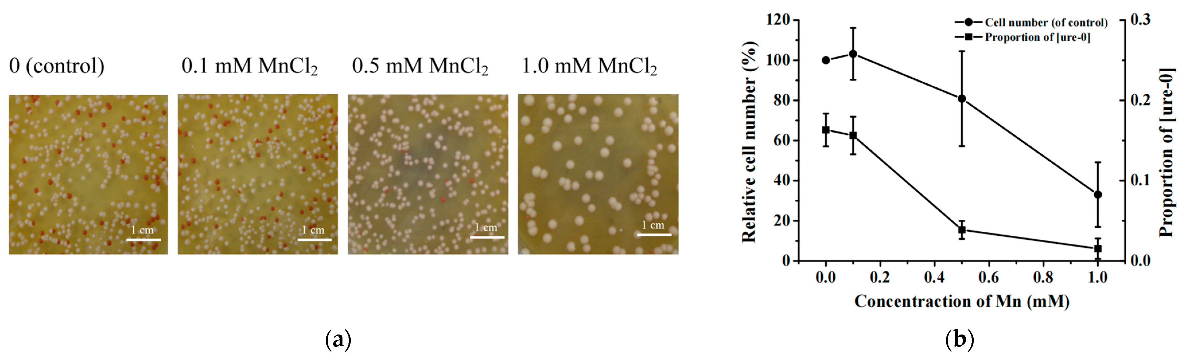
To investigate the effect of manganese on the de novo generation or propagation of prion, yeast cells were cultured to late exponential phase in YPD medium containing various concentrations of MnCl2 (0, 0.1, 0.5 and 1.0 mM). Then, the cell culture was diluted in same fold and spread onto standard YPD plates. After 120 h incubation at 30 °C, the numbers of red and white colonies on each plate were counted.
In the absence of Mn2+, NT64C yeast [ure-0] colonies were almost red with a few white colonies (Figure 1a). With the increase in Mn2+ concentration, this initial phenotype changed and trended toward the white color (Ade+ phenotype). Especially in 0.5 mM and 1.0 mM Mn2+ groups, more than 30% and 50% of the colonies changed to white color, respectively (Figure 1b). Compared with control group, the presence of Mn2+ can promote the spontaneous conversion from non-prion form to prion form in a dose-dependent manner. The group of 0.1 mM Mn2+ did not show statistically significant effect on cell number compared with control groups (0 mM Mn2+). However, 0.5 mM and 1.0 mM Mn2+ strongly reduced yeast cell number (dramatically to 20% of control groups) likely by killing cells and/or by inhibiting cells growth (Figure 1b). To rule out the possibility that other mutations resulted in the conversion from red to white cells, we treated newly formed white colonies with GdnHCl, and the colonies could be cured.

3.2. Effects of MnCl2 on [URE3] Propagation

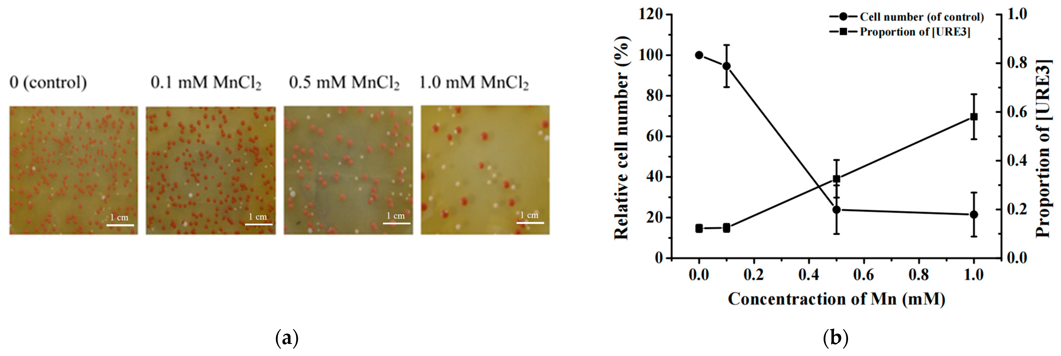
Using the non-prion [ure-0] strain as the starting material, we observed that the presence of Mn2+ can facilitate the spontaneous conversion from the non-prion form to the prion form. Additionally, the NT64C-derived [URE3] prion strain was employed as the initial strain to investigate the regulatory effect of Mn2+ on prion propagation. Under normal growth condition, [URE3] will lose its prion phenotype and reverts to normal phenotype spontaneously at a low frequency, so as [ure-0] red colonies appeared in the control groups (Figure 2a). Interestingly, along with increasing of Mn2+ concentration, the occurrence of red colonies was reduced. Especially in 0.5 mM and 1.0 mM groups, only a few or almost no red colonies appeared on the plates. If [URE3] curing frequency was defined as red color colonies/total colonies on the plate, with the increase in Mn2+ concentration, [URE3] curing frequency dropped gradually (Figure 2b). Yeast cell number did not show statistical difference between the control group (0 mM Mn2+) and 0.1 mM Mn2+ groups. However, with the increase in Mn2+ concentration, cell number gradually reduced to 20% at the concentration of 1.0 mM Mn2+.

3.3. Effects of MnCl2 on [URE3] in SB34 Strain

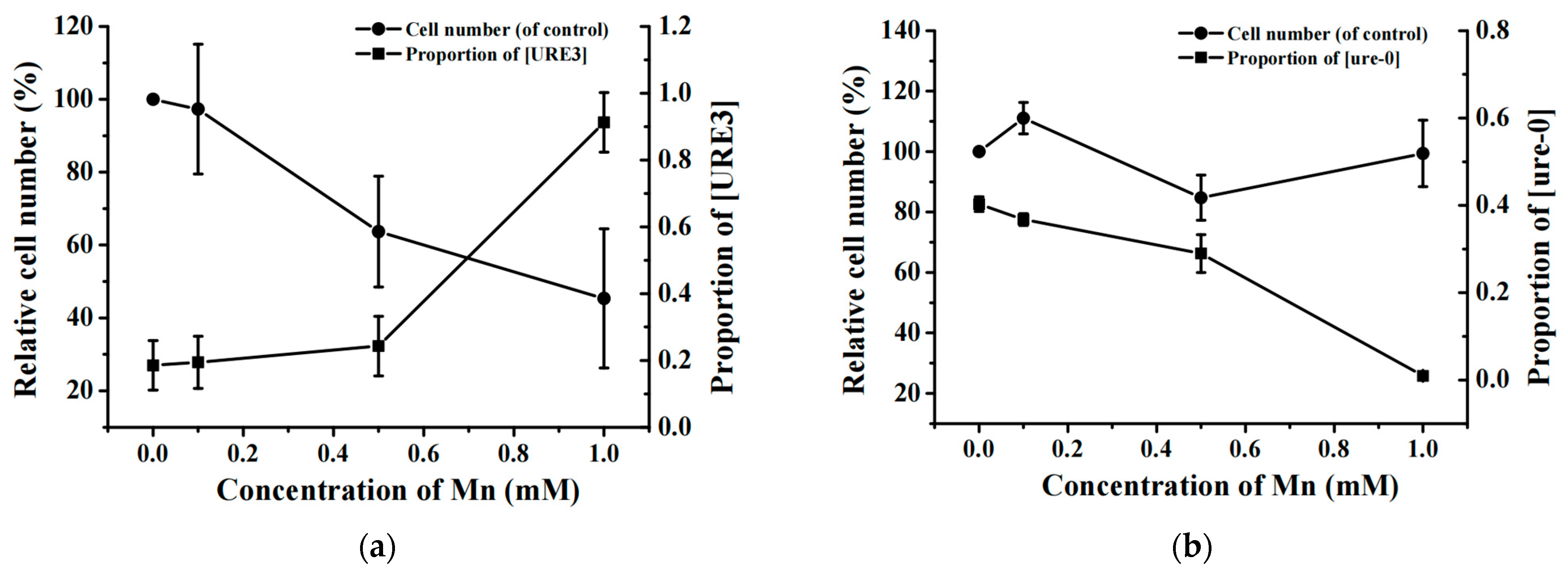
To rule out interference from yeast strain specificity, we repeated the aforementioned experiments using an alternative yeast strain SB34, which carries the [URE3] prion but exhibits a substantially different genetic background relative to NT64C. Consistent with NT64C, SB34 also utilizes the red/white screening method based on the ADE2 gene and DAL5 promoter.Given the extremely high conversion frequency between the prion and normal states of the SB34 [URE3] strain, we directly quantified the total number of white colonies (a marker for the prion state) and red colonies (a marker for the normal state). Following the inoculation and cultivation of this strain in YPD medium supplemented with different concentrations of Mn2+, we observed that the overall colony phenotype shifted progressively toward white with increasing Mn2+ concentrations in the medium, regardless of whether the strain exhibited the normal [ure-0] phenotype or the prion [URE3] phenotype at the initial stage. As clearly shown in Figure 3, high Mn2+ concentrations elevated the induction frequency of the [URE3] prion and simultaneously reduced its curing frequency—a pattern consistent with our findings in the NT64C strain. Furthermore, similar to the NT64C strain, the presence of Mn2+ also led to a decrease in the cell numbers of both the normal [ure-0] state and the prion [URE3] state in the SB34 strain.

3.4. Effect of Other Metal Ions on Cell Number and Prion Generation

In addition to Mn2+, we also studied the effect of Cu2+, Fe3+, Mg2+, K+ and Zn2+ exposure on cell number and prion generation. NT64C [ure-0] cells were incubated in YPD, containing various concentrations of CuCl2, FeCl3, MgCl2, KCl, or ZnCl2, for 60 h at 30 °C, then diluted in the same medium, and spread onto standard YPD plates. After incubation for 5 days at 30 °C, the red colony number and white colony number were counted (Table 2). Cu2+ had strong inhibition effect on the cell number, but did not show obvious effect on prion generation. The presence of Mg2+ and Zn2+ decreased the cell number and elevated prion generation, but the effects were very weak. Exposure to Fe3+ and K+ had no obvious effect on prion generation, while K+ could weakly decrease the cell number.

3.5. Expression Levels of Chaperones Under Mn2+ Stress Condition

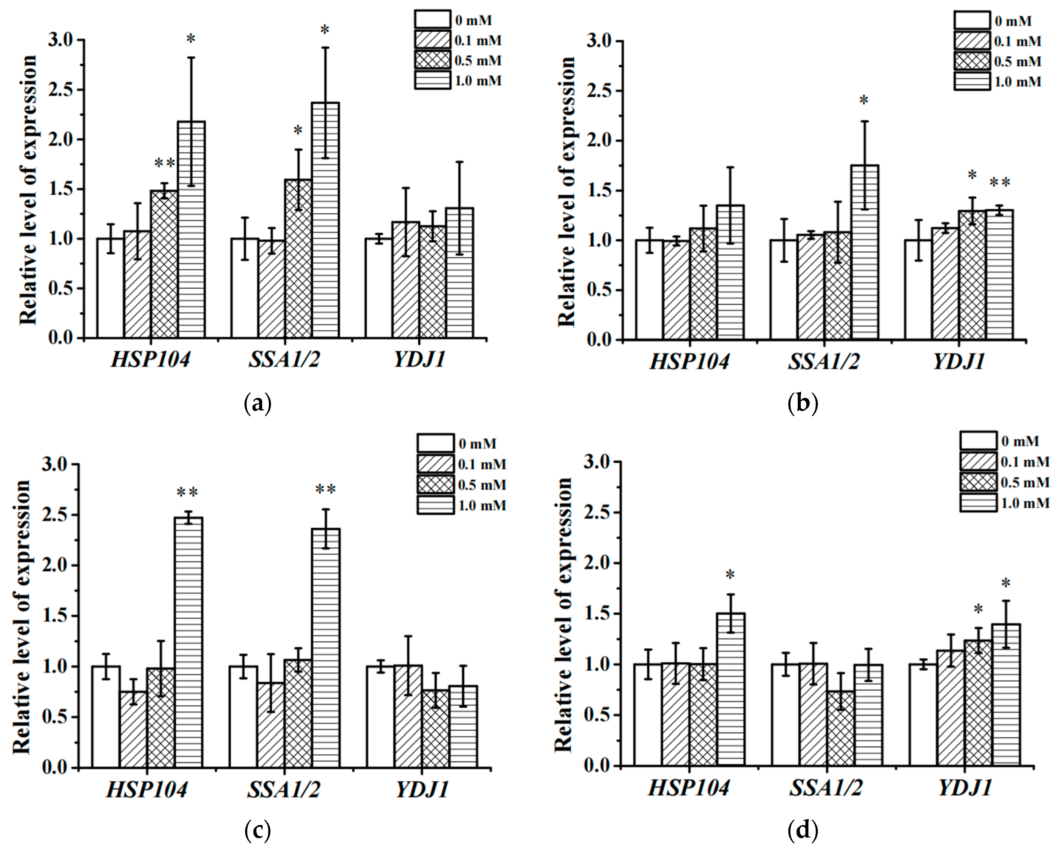
Given the indispensable roles of Hsp chaperones in yeast prion regulation, qRT-PCR was performed to investigate whether Mn2+ alters the transcriptional levels of HSP104, HSP70-SSA1/2 and HSP40-YDJ1 in yeast cells (Figure 4). Strains were grown on YPD plates containing various concentrations of MnCl2 for 4 days. Strains were collected and applied for qRT-PCR analysis. qRT-PCR analysis showed that stimulation by 1.0 mM Mn2+ for 60 h resulted in increase in the transcription level of HSP104 in NT64C [ure-0] (p < 0.05), SB34 [ure-0] (p < 0.001), SB34 [URE3] (p < 0.05) and NT64C [URE3] (p > 0.05) strains (Figure 4). With the decrease in Mn2+ concentration, the effect of Mn2+ on HSP104 transcription levels weakened. The transcription levels of HSP104 did not have any significant change in 0.1 mM groups and 0.5 mM groups, only with one exception of NT64C [ure-0]-cultured in 0.5 mM, in which the transcription of HSP104 gene was significantly up-regulated. In the presence of 1.0 mM Mn2+, transcription level of SSA1/2 was 2.3-fold, 1.7-fold and 2.3-fold higher than control groups in NT64C [ure-0] (p < 0.05), NT64C [URE3] (p < 0.05) and SB34 [ure-0] (p < 0.001), respectively, while no change was detected in SB34 [URE3] groups. Similar to HSP104, the up-regulation level decreased with the decrease in Mn2+ concentration. Mn2+ did not affect the transcription level of YDJ1 in NT64C [ure-0] and SB34 [ure-0] at all concentrations examined, while 1.0 mM Mn2+ significantly up-regulated the expression level of YDJ1 in NT64C [URE3] (p < 0.001) and SB34 [URE3] (p < 0.05). In the presence of 0.5 mM Mn2+, the up-regulation in YDJ1 transcription level was significant in NT64C [URE3] (p < 0.05) and SB34 [URE3] (p < 0.05) strains but not in NT64C [ure-0] and SB34 [ure-0]. The incubation with 0.1 mM Mn2+ did not affect YDJ1 gene transcription level in all four strains.
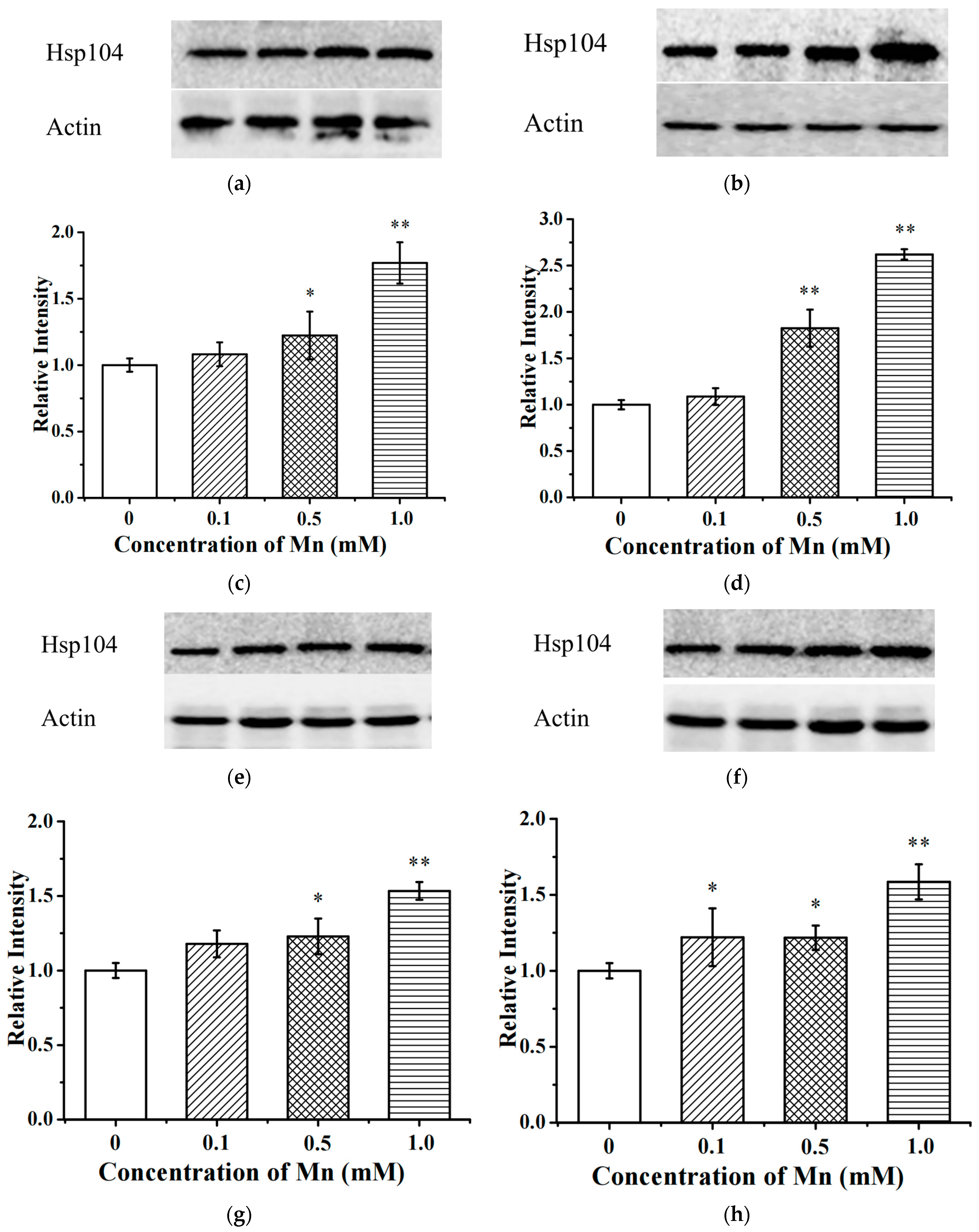
Transcription was elevated in all four strains we tested; therefore, we wondered whether the protein level is also elevated under the same conditions. We analyzed the HSP104 protein expression level by Western blotting. Consistent with the transcriptional data, the Hsp104 protein expression level was also elevated in a dose-dependent manner in all four strains (Figure 5).

3.6. The Effect of Mn2+ and Other Metal Ions on the Fibrillogenic Activity of Ure2 Protein In Vitro

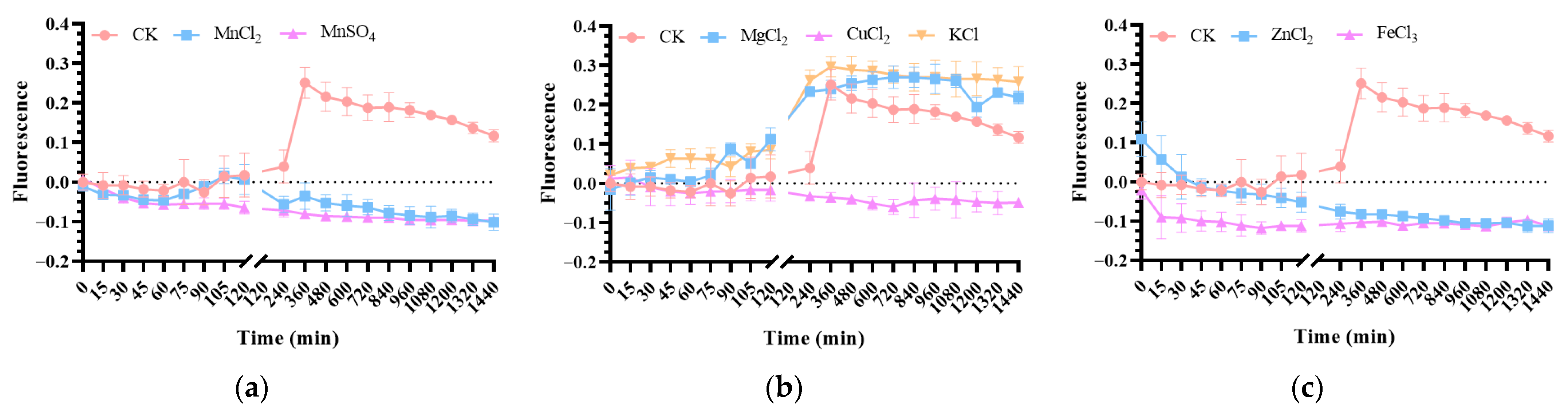
To investigate the effect of manganese ions on Ure2p fibrillogenesis, a 100 mM MnCl2 solution was used to achieve a final concentration of 2 mM, and fluorescence intensity was measured using a microplate reader. The fluorescence intensity of the blank control group increased at 240 min, while the fluorescence intensity of the group with added Mn2+ remained nearly unchanged (Figure 6a). This indicates that Mn2+ inhibits the in vitro fibrillation of the Ure2p. To investigate whether this effect is mediated by anions, we also used a 100 mM MnSO4 solution. The fluorescence values of the MnSO4 solution were comparable to those of the MnCl2 solution, ruling out the involvement of anions.
To investigate the effects of other metal ions on the in vitro fibrillogenesis of the Ure2 protein, we prepared solutions of CuCl2, MgCl2, KCl, FeCl3 and ZnCl2 at a final concentration of 2 mM and measured the fluorescence intensity using a microplate reader. The fluorescence intensity of the blank control group increased at 240 min, while the fluorescence intensity in the CuCl2 group remained almost unchanged, indicating that Cu2+ can inhibit the in vitro fibrillogenesis of Ure2p. However, the fluorescence intensity of the MgCl2 and KCl treatment groups at 120 min showed a slight increase without statistical significance compared with the blank control group (Figure 6b). In contrast, the fluorescence intensity of the FeCl3- and ZnCl2-treated groups remained relatively stable and was consistently lower than that of the control group, indicating that Fe3+ and Zn2+ exert an inhibitory effect on the amyloid aggregation of Ure2p. (Figure 6c).

4. Discussion

The knowledge of how TSEs emerge is strongly needed to eradicate this disease from both animal and human populations. Recently, studies have caught our eyes on the tight link between manganese and prion disease. Manganese is essential for human brain, and the highest levels of manganese were found in the putamen (0.7–4.5 μg/g) and the lowest were found in the medulla (0.2–1.8 μg/g) [49]. However, a tendency for an age-related increase of manganese levels was discovered in human brain [49], and manganese levels were shifted in specific anatomical region in the brains between the healthy and Alzheimer’s disease patients [50]. It was thought that the age-related increase of manganese levels in brain might disrupt metal ion homeostasis, which eventually led to neurodegeneration diseases [51,52].
In the present study, we employed Ure2p, the core constituent protein of the yeast prion [URE3], as our research object. Our data demonstrated that the induction frequency of [URE3] in all tested yeast strains was significantly elevated by approximately 2–6 fold with increasing Mn2+ concentrations. Notably, we characterized the cytotoxic effect of Mn2+ in a dose-dependent manner: yeast cells showed favorable tolerance to 0.1 mM Mn2+, while obvious cytotoxicity emerged at 0.5 mM and was drastically intensified at 1 mM. This cytotoxic profile is consistent with previous observations in cells overexpressing mammalian prion protein (PrP) [53,54]. Importantly, the enhanced induction of [URE3] was not merely a secondary outcome of impaired cell growth but was directly correlated with Mn2+ concentrations acting as a cellular stressor. This conclusion was validated by the strain SB34 with a distinct genetic background, in which elevated Mn2+ levels increased [URE3] induction by approximately 3.5-fold without causing a significant reduction in cell viability. Furthermore, incubation of cultures in water or poor medium for 24 h resulted in low cell optical densities, yet failed to trigger an increase in prion induction [55], further supporting the specific effect of Mn2+.
The effect of metal on the amyloid formation is complex and its mechanism remains unclear. Various metal ions have been found to play some roles in the amyloid diseases in vivo and in vitro. Interestingly, a certain metal has different effects on different amyloid proteins, while the effects of various metals on a certain amyloid protein are different as well. For example, Mn2+ and Zn2+ fail to bind to Sup35p-NM, and exert no significant effect on the formation of NM fibrils that promote amyloid aggregation in vitro. Cu2+ can bind to the N-region of Sup35p with an affinity comparable to that of PrP and this binding affects the nucleation steps of fibril assembly [56]. In our study, only Mn2+ showed significant effect on prion phenotype, whereas obvious effect of Cu2+, Mg2+, Fe3+, K+ or Zn2+ was not observed. It will be interesting to find out why the relationships between different metal ions and different amyloid-forming proteins are so different.
We observed that in the intracellular environment, the introduction of Mn2+ can increase the induction frequency of the yeast strain [URE3]; however, in the in vitro environment, Mn2+ does not promote the formation of amyloid aggregates by the Ure2 protein. The core contradiction stems from regulatory differences between the in vitro single-protein system and in vivo complex cellular microenvironment. In vitro, manganese directly inhibits Ure2 fibrillization, which is consistent with the regulatory pattern of metal ions on amyloid fibrillization in prion proteins: Bocharova [57] demonstrated via circular dichroism that metal ions bind the prion amyloid core to stabilize soluble α-helical conformations, suppressing misfolding and fibrillary aggregation—Mn2+ follows this mechanism albeit weaker than Cu2+. Inayathullah [58] further identified via NMR that coordinate binding of metal ions to protein histidine residues is critical for inhibition, a mode conserved in the amyloid core region (residues 1–89) of yeast Ure2p. In vivo, beyond direct prion binding, manganese significantly upregulates Hsp104p via activating cellular stress pathways. King [59] confirmed Hsp104p as a core regulator of prion seed formation and propagation in yeast, with its upregulation potently enhancing prion spread. This study detected marked intracellular Hsp104 upregulation post-manganese treatment. Given that Hsp104 is essential for prion maintenance and its stress-induced elevation has been shown to enhance prion seed propagation by severing amyloid fibrils to generate new seeds [60,61], we speculate that the Hsp104-dependent pathway may contribute to manganese-mediated prion-related effects. This Hsp104 upregulation is driven by manganese-induced proteotoxic stress, which is a known trigger of chaperone induction and prion misfolding [62]. However, direct experimental evidence is required to confirm whether this pathway exerts a dominant biological effect relative to other potential mechanisms (e.g., direct manganese-mediated inhibition). In contrast, Cu2+ significantly inhibits cell proliferation in vivo; nevertheless, Cu2+ has no significant effect on prion formation and also fails to induce amyloid aggregation of the Ure2p in vitro. This phenomenon may be attributed to the fact that the presence of Cu2+ reduces the melting temperature (TM value) of the protein [63]. Specifically, Cu2+ inhibits the conversion of α-rPrP to a stable proteinase K-resistant form, thereby preventing the transformation of this protein into amyloid fibrils that enhance amyloid aggregation [57]. Additionally, the presence of Mg2+ and Zn2+ slightly reduces cell count while exerting a weak promoting effect on prion formation. Notably, Mg2+ can also induce amyloid aggregation of the Ure2p in vitro (albeit to a weak extent). It is hypothesized that Mg2+ may bind to the protein during the protein folding process, leading to protein misfolding. In contrast, Zn2+ does not trigger such aggregation in vitro, which may also be associated with the reduction in the protein’s TM value by Zn2+ [63]. Regarding Fe3+ and K+, neither of them exerts a significant effect on prion formation in vivo. However, in the in vitro environment, K+ can weakly promote the formation of amyloid aggregates by the Ure2p, whereas Fe3+ has no such effect.
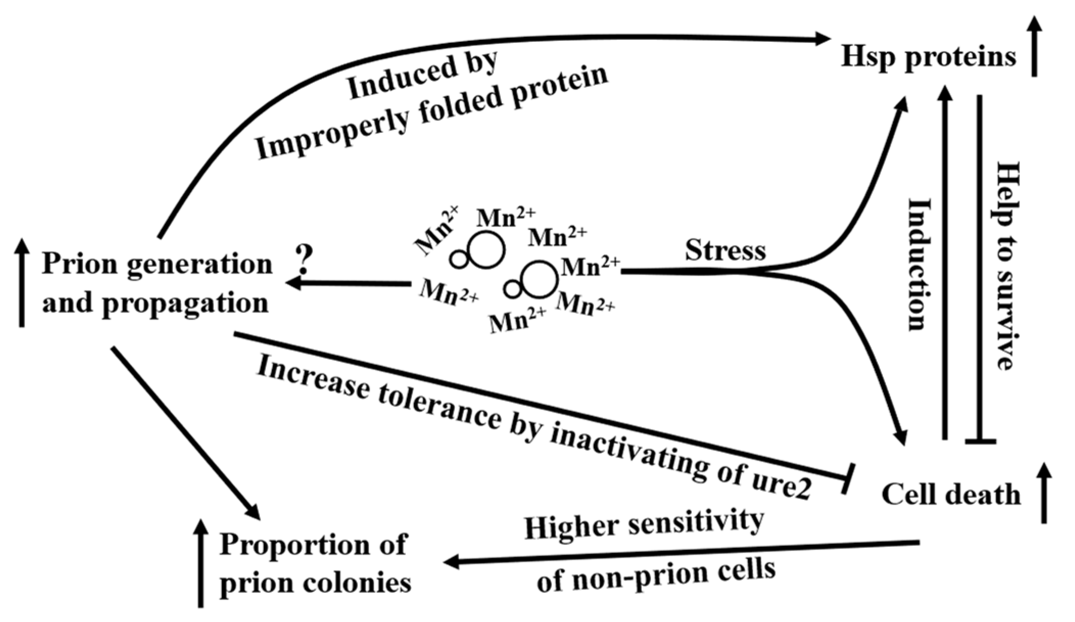
The correlation between Mn2+ levels and [URE3] induction frequency is consistent with a theoretical prediction: if [URE3] acts as a mechanism for evolvability, its emergence ought to correlate with stress [64]. [URE3] conferred a transient survival benefit across diverse conditions, thereby elevating the probability that novel traits become fixed through subsequent genetic alterations [65]. Strains carrying the prions are better adapted to many stresses, under which cells containing [URE3] had higher survival rate [64]. It has been reported that the gene disruption mutation of Ure2 could improve ion tolerance of Sacchanromyces cerevisiae lacking the Ca2+/CaM-dependent phosphatase (calcineurin) to Mn2+, Na+ and Li+ [66]. Notably, this Ure2 mutant requires the transcription factor Gln3p to confer ion tolerance, which revealed the same pathway as our prion state assay by color in this study. Although the improvement of ions tolerance was found in a calcineurin-deficient mutant, which was more sensitive to high concentrations of some ions including Mn2+, this finding could still suggest that the non-functional state of Ure2p could lead to improvement of tolerance to Mn2+. In this case, the strains in prion state will be easier than the strains in non-prion state to survive in the presence of high concentration of Mn2+, which may make some contribution to the increased proportion of prion-state colonies under Mn2+ stress. The difference between SB34 and NT64C also meets this hypothesis. The SB34 strain has stronger tendency to prion state than NT64C, which will offer SB34 higher tolerance to Mn2+ than NT64C, and eventually lead to more survivals than NT64C under Mn2+ stress. The increased proportion of prion cells could not be simply attributed solely to the slow growth and/or death of non-prion cells, which was clearly opposite to the effects of 0.5 mM and 1.0 mM Mn2+ on all the two [ure-0] strains we tested. When the concentration of Mn2+ was increased from 0.5 mM to 1.0 mM, the proportion of prion state cells increased dramatically, yet the survival numbers remained similar (Figure 1 and Figure 3). Furthermore, the numbers of white colonies in 1.0 mM groups were also more than that in 0.5 mM groups. Therefore, the improvement of prion generation and propagation definitely contributed to the increased proportion of prion colonies in the presence of high concentration Mn2+, while the slower growth and/or higher death rate of non-prion state cells might also make some contribution. Figure 7 illustrates our model of cell response to manganese stress. It is interesting that the presence of 0.5 mM and 1.0 mM Mn2+ reduced cell number to a similar extent, which is opposite to the idea that higher concentration of Mn2+ has higher impact on cells. The higher survivability of prion state cells might be a reason for this phenomenon, and this finding could be another evidence for the hypothesis that the prion is a state to adapt the environment stress.
The presence of Mn2+ could promote the prion de novo generation and propagation of Ure2p (this study) and Sup35p [67] in yeast. Previous studies have documented that Hsp104p, Hsp70p (including Ssa1p and Ssa2p) and the co-chaperone Hsp40p (such as Ydj1p and Sis1p) participate in yeast prion propagation in vivo as well as fibril assembly in vitro [23,68]. Therefore, we tried to test the expression levels of molecular chaperones induced by Mn2+ stress condition to explain how Mn2+ affected [URE3] propagation. Our results showed that the transcription levels of Hsp104p, Hsp70p and Hsp40p were elevated to various extents in a dose-dependent manner (Figure 4 and Figure 5). Other than the involvements in prion propagation, Heat shock proteins (Hsps) are also a suite of highly conserved proteins for their quick responses to environmental stresses. The expression of Hsps usually increases in response to exposure to various stressful stimuli, such as heat, toxins, metals and so on [69,70,71,72]. The elevated expressions of chaperones in this study were reasonable response to the stress in this study, considering the high death rate of cells and misfolding of prion proteins.
After Chernoff found Hsp104p was required for the maintenance of Sup35 prion aggregates [73], it was revealed that Hsp10p4 is essential for the propagation of all known yeast prions [21]. The Hsp104p chaperone disrupts the small fibers formed in the early stage of Sup35 fibrillogenesis, leading to the effective formation of fiber fragments, which in turn induces the accelerated formation of amyloid. Moreover, the accelerated formation of amyloid is dependent on ATPase activity [74]. Consistent with in vivo findings, low Hsp104 levels in vitro can accelerate assembly of prion fibers of both Sup35 and Ure2p [61,75,76]. Although overexpression of Hsp104p could cure [PSI+] [77], the stability of [URE3] was not affected by Hsp104p overproduction [73]. The explanations of why Hsp104p could cure [PSI+] while other prions are insensitive to this treatment still remain unclear. It was generally agreed that Hsp104p, as a disaggregation machinery, could act directly on prion fibrils to sever them, then generate new prion seeds from preexisting material, and is therefore required for prion replication [68]. It has also been reported that the restoration of [PSI+] by Hsp104p overexpression is not due to the asymmetric segregation of prion seeds, but rather to their dissolution by Hsp104p [77], although Hsp104p functions to break down prion fibrils, at normal levels, it splits prions into multiple smaller fragments, thereby ensuring prion propagation and accelerating prion conversion [78]. In vitro investigations have revealed that Hsp104p resolves diverse protein aggregates by translocating polypeptide chains through the central pore of its hexamer. Nevertheless, how Hsp104p-mediated processing impacts protein function in vivo remains elusive. The middle domain (MD) of Hsp104p modulates ATPase activity and its interactions with Hsp70p [79]. In the presence of manganese, the gene transcription level (Figure 4) and protein expression level (Figure 5) of Hsp104p were elevated in an obvious dose-dependent manner in all the four strains we tested, no matter if the starting strains were in prion state or non-prion state. Overexpression of Hsp104p could promote prion formation of [URE3] in the presence of [PIN+], but not in the absence of [PIN+], which suggested Hsp104p overexpression could make the Rnq1 amyloid more suitable for accelerating Ure2p amyloid formation, rather than directly promote formation of initial amyloid seeds [60]. In this study, the Hsp104p was overexpressed in the presence of manganese; however, all strains used in prion generation assay had been applied to the guanidine hydrochloride treatment to cure all prions including [PIN+]. Therefore, the promotion of [URE3] generation in the presence of manganese could not be attributed to the effect of Hsp104p overexpression mediated by [PIN+]. The overexpression Hsp104p did not have curing effect on [URE3] and probably had some improvement effect on [URE3] in this context, since the cells were suffering from severe stress. The stress from high concentration of Mn2+ and aggregation of Ure2 would induce the expression of Hsp104p, while on the other hand, the overproduced Hsp104p, together with its improvement effect on prion propagation, may help cells survive from Mn2+ stress (Figure 7).
Hsp70p family is another major class of chaperones involved in regulating prion propagation [80]. Yeast has six cytoplasmic Hsp70s: four of essential Ssa subfamily and two of Ssb subfamily. Notably, the gene products of SSA1 and SSA2 are 96 per cent identical, but have distinct function on prion propagation. Overexpression of Ssa1p but not the Ssa2p could cure [URE3] [32]. Hsp70 chaperones also exert distinct effects on the yeast prions [PSI+] and [URE3] in vivo. Notably, [PSI+] stability was maintained at a normal level in strains deficient in SSA1 or SSA2 family members [81], and overexpression of Ssa1p increased de novo [PSI+] formation [82]. Studies have shown that the binding of Ssa1p to the regions outside the amyloid core plays a key role in promoting the depolymerization and propagation of yeast prions both in vitro and in vivo [83]. In this study, exposure to Mn2+ upregulated the transcriptional levels of SSA1/SSA2 in NT64C [ure-0], NT64C [URE3] and SB34 [ure-0] strains (Figure 4a–c), while only the expression of SSA1/SSA2 in SB34 [URE3] (Figure 4d) was not affected. The reason why overproduced Ssa1p and Ssa2p did not cure the prion might have two explanations. One is that we could not distinguish whether Ssa1p or Ssa2p was overexpressed in this study, while overexpression of Ssa1p and Ssa2p had distinct effect on [URE3]. Another possible explanation could be that the soluble Ssa1/2 might be no higher-than-normal level even when Ssa1/2 was overexpressed, because the consumption of Ssa1/2 might also increase under Mn2+ stress, since the Ssa proteins are involved in a variety of cellular processes other than prion propagation, such as translation, translocation and general protein folding.
In conclusion, we investigated the effects of the manganese on prion using budding yeast prion [URE3] as model system. We found that the exposure to manganese can elevate the de novo generation and propagation of yeast prion [URE3], as well as the expression levels of chaperones Hsp104p and Hsp70p, in a dose-dependent manner.

Author Contributions

Y.-H.Z., K.-W.L. and H.-Y.L. performed all experiments. H.-Y.L. and Y.-H.Z. constructed and maintained the exposure systems. K.-W.L. and H.-Y.L. conceived the study. Y.-H.Z., H.-Y.L. and K.-W.L. wrote the manuscript. T.Z. led the project and reviewed the manuscript. All authors have read and agreed to the published version of the manuscript.

Funding

This work was supported by the Hainan Provincial Natural Science Foundation of China (No. 225MS098), Natural Science Foundation of China (No. 51967006), The Innovation Platform for Academicians of Hainan Province, Hainan Sea Food Engineering and Technology Research Center, Hainan Province Graduate Student Innovation Research Project (Qhys2023-513), and The Graduate Innovation Project of Hainan Tropical Ocean University (RHDYC-202420).

Data Availability Statement

All data generated or analyzed during this study are included in this published article.

Acknowledgments

We express our gratitude to Gary Jones (NUI Maynooth, Ireland) for kindly providing the yeast strains.

Conflicts of Interest

The authors declare no competing financial or personal interests.

Abbreviations

The following abbreviations are used in this manuscript:
IPTGIsopropyl β-D-thiogalactoside
UVUltraviolet
GdnHClGuanidine Hydrochloride

References

  1. Prusiner, S.B.; Bolton, D.C.; Groth, D.F.; Bowman, K.A.; Cochran, S.P.; McKinley, M.P. Further Purification and Characterization of Scrapie Prions. Biochemistry 1982, 21, 6942–6950. [Google Scholar] [CrossRef] [Scilit]
  2. Saitoh, Y.; Mizusawa, H. Prion Diseases, Always a Threat? J. Neurol. Sci. 2024, 463, 123119. [Google Scholar] [CrossRef] [Scilit] [PubMed]
  3. Jager, K.; Orozco-Hidalgo, M.T.; Springstein, B.L.; Joly-Smith, E.; Papazotos, F.; McDonough, E.; Fleming, E.; McCallum, G.; Yuan, A.H.; Hilfinger, A.; et al. Measuring Prion Propagation in Single Bacteria Elucidates a Mechanism of Loss. Proc. Natl. Acad. Sci. USA 2023, 120, e2221539120. [Google Scholar] [CrossRef] [Scilit]
  4. Moore, R.A.; Sturdevant, D.E.; Chesebro, B.; Priola, S.A. Proteomics Analysis of Amyloid and Nonamyloid Prion Disease Phenotypes Reveals Both Common and Divergent Mechanisms of Neuropathogenesis. J. Proteome Res. 2014, 13, 4620–4634. [Google Scholar] [CrossRef] [Scilit] [PubMed]
  5. Castellani, R.J.; Perry, G.; Smith, M.A. Prion Disease and Alzheimer’s Disease: Pathogenic Overlap. Acta Neurobiol. Exp. 2004, 64, 11–17. [Google Scholar] [CrossRef] [Scilit] [PubMed]
  6. Bhamra, S.; Arora, P.; Manka, S.W.; Schmidt, C.; Brown, C.; Rayner, M.L.D.; Klöhn, P.-C.; Clarke, A.R.; Collinge, J.; Jat, P.S. Prion Propagation Is Dependent on Key Amino Acids in Charge Cluster 2 within the Prion Protein. J. Mol. Biol. 2023, 435, 167925. [Google Scholar] [CrossRef] [Scilit]
  7. Jones, E.; Hill, E.; Linehan, J.; Nazari, T.; Caulder, A.; Codner, G.F.; Hutchison, M.; Mackenzie, M.; Farmer, M.; Coysh, T.; et al. Characterisation and Prion Transmission Study in Mice with Genetic Reduction of Sporadic Creutzfeldt-Jakob Disease Risk Gene Stx6. Neurobiol. Dis. 2024, 190, 106363. [Google Scholar] [CrossRef] [Scilit]
  8. Sakaguchi, S. Prion Pathogenesis Revealed in a Series of the Special Issues “Prions and Prion Diseases”. Int. J. Mol. Sci. 2022, 23, 6490. [Google Scholar] [CrossRef] [Scilit]
  9. Wickner, R.B.; Masison, D.C.; Edskes, H.K. [PSI] and [URE3] as Yeast Prions. Yeast 1995, 11, 1671–1685. [Google Scholar] [CrossRef] [Scilit]
  10. Aigle, M.; Lacroute, F. Genetical Aspects of [URE3], a Non-Mitochondrial, Cytoplasmically Inherited Mutation in Yeast. Mol. Gen. Genet. MGG 1975, 136, 327–335. [Google Scholar] [CrossRef] [Scilit]
  11. Wickner, R.B. [URE3] as an Altered URE2 Protein: Evidence for a Prion Analog in Saccharomyces cerevisiae. Science 1994, 264, 566–569. [Google Scholar] [CrossRef] [Scilit]
  12. Cox, K.H.; Rai, R.; Distler, M.; Daugherty, J.R.; Coffman, J.A.; Cooper, T.G. Saccharomyces cerevisiae GATA Sequences Function as TATA Elements during Nitrogen Catabolite Repression and When Gln3p Is Excluded from the Nucleus by Overproduction of Ure2p. J. Biol. Chem. 2000, 275, 17611–17618. [Google Scholar] [CrossRef] [Scilit]
  13. Martin, D.P.; Anantharam, V.; Jin, H.; Witte, T.; Houk, R.; Kanthasamy, A.; Kanthasamy, A.G. Infectious Prion Protein Alters Manganese Transport and Neurotoxicity in a Cell Culture Model of Prion Disease. NeuroToxicology 2011, 32, 554–562. [Google Scholar] [CrossRef] [Scilit]
  14. Masison, D.C.; Wickner, R.B. Prion-Inducing Domain of Yeast Ure2p and Protease Resistance of Ure2p in Prion-Containing Cells. Science 1995, 270, 93–95. [Google Scholar] [CrossRef] [Scilit] [PubMed]
  15. Hong, J.Y.; Mathur, V.; Liebman, S.W. A New Colour Assay for [URE3] Prion in a Genetic Background Used to Score for the [PSI+] Prion. Yeast 2011, 28, 555–560. [Google Scholar] [CrossRef] [Scilit]
  16. Zhouravleva, G.A.; Bondarev, S.A.; Trubitsina, N.P. How Big Is the Yeast Prion Universe? Int. J. Mol. Sci. 2023, 24, 11651. [Google Scholar] [CrossRef] [Scilit]
  17. Halfmann, R.; Alberti, S.; Lindquist, S. Prions, Protein Homeostasis, and Phenotypic Diversity. Trends Cell Biol. 2010, 20, 125–133. [Google Scholar] [CrossRef] [Scilit] [PubMed]
  18. Halfmann, R.; Jarosz, D.F.; Jones, S.K.; Chang, A.; Lancaster, A.K.; Lindquist, S. Prions Are a Common Mechanism for Phenotypic Inheritance in Wild Yeasts. Nature 2012, 482, 363–368. [Google Scholar] [CrossRef] [Scilit] [PubMed]
  19. Chernova, T.A.; Chernoff, Y.O.; Wilkinson, K.D. Yeast Models for Amyloids and Prions: Environmental Modulation and Drug Discovery. Molecules 2019, 24, 3388. [Google Scholar] [CrossRef] [Scilit]
  20. Chen, Y.-J.; Inouye, M. The Intramolecular Chaperone-Mediated Protein Folding. Curr. Opin. Struct. Biol. 2008, 18, 765–770. [Google Scholar] [CrossRef] [Scilit]
  21. Wickner, R.B.; Shewmaker, F.P.; Bateman, D.A.; Edskes, H.K.; Gorkovskiy, A.; Dayani, Y.; Bezsonov, E.E. Yeast Prions: Structure, Biology, and Prion-Handling Systems. Microbiol. Mol. Biol. Rev. 2015, 79, 1–17. [Google Scholar] [CrossRef] [Scilit] [PubMed]
  22. Wyszkowski, H.; Janta, A.; Sztangierska, W.; Obuchowski, I.; Chamera, T.; Kłosowska, A.; Liberek, K. Class-Specific Interactions between Sis1 J-Domain Protein and Hsp70 Chaperone Potentiate Disaggregation of Misfolded Proteins [Biochemistry]. Proc. Natl. Acad. Sci. USA 2021, 118, e2108163118. [Google Scholar] [CrossRef] [Scilit]
  23. Ruger-Herreros, C.; Svoboda, L.; Mogk, A.; Bukau, B. Role of J-Domain Proteins in Yeast Physiology and Protein Quality Control. J. Mol. Biol. 2024, 436, 168484. [Google Scholar] [CrossRef] [Scilit]
  24. Ness, F.; Cox, B.S.; Wongwigkarn, J.; Naeimi, W.R.; Tuite, M.F. Over-Expression of the Molecular Chaperone Hsp104 in Saccharomyces cerevisiae Results in the Malpartition of [PSI+] Propagons. Mol. Microbiol. 2017, 104, 125–143. [Google Scholar] [CrossRef] [Scilit] [PubMed]
  25. Ganser, S.J.; McNish, B.A.; Corpus, B.A.; Hines, J.K. Structural Elements of “Anti-Prion J Protein 1” (Apj1) Required for Efficient Curing of the [PSI+] Prion by Hsp104 Overexpression. FASEB J. 2022, 36. [Google Scholar] [CrossRef] [Scilit]
  26. Moriyama, H.; Edskes, H.K.; Wickner, R.B. [URE3] Prion Propagation in Saccharomyces cerevisiae: Requirement for Chaperone Hsp104 and Curing by Overexpressed Chaperone Ydj1p. Mol. Cell. Biol. 2000, 20, 8916–8922. [Google Scholar] [CrossRef] [Scilit]
  27. Glover, J.R.; Lindquist, S. Hsp104, Hsp70, and Hsp40: A Novel Chaperone System That Rescues Previously Aggregated Proteins. Cell 1998, 94, 73–82. [Google Scholar] [CrossRef] [Scilit]
  28. Kryndushkin, D.S.; Alexandrov, I.M.; Ter-Avanesyan, M.D.; Kushnirov, V.V. Yeast [PSI+] Prion Aggregates Are Formed by Small Sup35 Polymers Fragmented by Hsp104. J. Biol. Chem. 2003, 278, 49636–49643. [Google Scholar] [CrossRef] [Scilit]
  29. Kryndushkin, D.; Wickner, R.B. Nucleotide Exchange Factors for Hsp70s Are Required for [URE3] Prion Propagation in Saccharomyces Cerevisiae. Mol. Biol. Cell 2007, 18, 2149–2154. [Google Scholar] [CrossRef] [Scilit] [PubMed]
  30. Kumar, J.; Reidy, M.; Masison, D.C. Yeast J-Protein Sis1 Prevents Prion Toxicity by Moderating Depletion of Prion Protein. Genetics 2021, 219, iyab129. [Google Scholar] [CrossRef] [Scilit]
  31. Miller, S.C.; Wegrzynowicz, A.K.; Cole, S.J.; Hayward, R.E.; Ganser, S.J.; Hines, J.K. Hsp40/JDP Requirements for the Propagation of Synthetic Yeast Prions. Viruses 2022, 14, 2160. [Google Scholar] [CrossRef] [Scilit]
  32. Schwimmer, C.; Masison, D.C. Antagonistic Interactions between Yeast [PSI+] and [URE3] Prions and Curing of [URE3] by Hsp70 Protein Chaperone Ssa1p but Not by Ssa2p. Mol. Cell. Biol. 2002, 22, 3590–3598. [Google Scholar] [CrossRef] [Scilit]
  33. Roberts, B.T.; Moriyama, H.; Wickner, R.B. [URE3] Prion Propagation Is Abolished by a Mutation of the Primary Cytosolic Hsp70 of Budding Yeast. Yeast 2003, 21, 107–117. [Google Scholar] [CrossRef] [Scilit] [PubMed]
  34. Lian, H.-Y.; Zhang, H.; Zhang, Z.-R.; Loovers, H.M.; Jones, G.W.; Rowling, P.J.E.; Itzhaki, L.S.; Zhou, J.-M.; Perrett, S. Hsp40 Interacts Directly with the Native State of the Yeast Prion Protein Ure2 and Inhibits Formation of Amyloid-like Fibrils. J. Biol. Chem. 2007, 282, 11931–11940. [Google Scholar] [CrossRef] [Scilit] [PubMed]
  35. Lee, S.H.; Lee, H.; Cho, M.S.; Nam, J.-D.; Lee, Y. Morphology and Composition Control of Manganese Oxide by the Pulse Reverse Electrodeposition Technique for High Performance Supercapacitors. J. Mater. Chem. A 2013, 1, 14606. [Google Scholar] [CrossRef] [Scilit]
  36. Russo, F.; Johnson, C.J.; Johnson, C.J.; McKenzie, D.; Aiken, J.M.; Pedersen, J.A. Pathogenic Prion Protein Is Degraded by a Manganese Oxide Mineral Found in Soils. J. Gen. Virol. 2009, 90, 275–280. [Google Scholar] [CrossRef] [Scilit] [PubMed]
  37. Nyarko-Danquah, I.; Pajarillo, E.; Digman, A.; Soliman, K.F.A.; Aschner, M.; Lee, E. Manganese Accumulation in the Brain via Various Transporters and Its Neurotoxicity Mechanisms. Molecules 2020, 25, 5880. [Google Scholar] [CrossRef] [Scilit]
  38. Dayan, R.; Arkadir, D. Manganese Accumulation in the Brain. N. Engl. J. Med. 2023, 389, 1320. [Google Scholar] [CrossRef] [Scilit]
  39. Lin, M.T.; Beal, M.F. Mitochondrial Dysfunction and Oxidative Stress in Neurodegenerative Diseases. Nature 2006, 443, 787–795. [Google Scholar] [CrossRef] [Scilit]
  40. Holley, A.K.; Bakthavatchalu, V.; Velez-Roman, J.M.; Clair, D.K.S. Manganese Superoxide Dismutase: Guardian of the Powerhouse. Int. J. Mol. Sci. 2011, 12, 7114–7162. [Google Scholar] [CrossRef] [Scilit]
  41. Sant, V.; Matthes, D.; Mazal, H.; Antonschmidt, L.; Wieser, F.; Movellan, K.T.; Xue, K.; Nimerovsky, E.; Stampolaki, M.; Nathan, M.; et al. Lipidic Folding Pathway of α-Synuclein via a Toxic Oligomer. Nat. Commun. 2025, 16, 760. [Google Scholar] [CrossRef] [Scilit] [PubMed]
  42. Ferreiro, E.; Costa, R.; Marques, S.; Cardoso, S.M.; Oliveira, C.R.; Pereira, C.M.F. Involvement of Mitochondria in Endoplasmic Reticulum Stress-Induced Apoptotic Cell Death Pathway Triggered by the Prion Peptide PrP(106-126). J. Neurochem. 2007, 104, 766–776. [Google Scholar] [CrossRef] [Scilit]
  43. Mitteregger, G.; Korte, S.; Shakarami, M.; Herms, J.; Kretzschmar, H.A. Role of Copper and Manganese in Prion Disease Progression. Brain Res. 2009, 1292, 155–164. [Google Scholar] [CrossRef] [Scilit]
  44. Tong, Y.; Yang, H.; Tian, X.; Wang, H.; Zhou, T.; Zhang, S.; Yu, J.; Zhang, T.; Fan, D.; Guo, X.; et al. High Manganese, a Risk for Alzheimer’s Disease: High Manganese Induces Amyloid-β Related Cognitive Impairment. J. Alzheimer’s Dis. 2014, 42, 865–878. [Google Scholar] [CrossRef] [Scilit]
  45. Kawahara, M.; Kato-Negishi, M.; Tanaka, K. Neurometals in the Pathogenesis of Prion Diseases. Int. J. Mol. Sci. 2021, 22, 1267. [Google Scholar] [CrossRef] [Scilit]
  46. Thackray, A.M.; Knight, R.; Haswell, S.J.; Bujdoso, R.; Brown, D.R. Metal Imbalance and Compromised Antioxidant Function Are Early Changes in Prion Disease. Biochem. J. 2002, 362, 253. [Google Scholar] [CrossRef]
  47. Bach, S.; Talarek, N.; Andrieu, T.; Vierfond, J.-M.; Mettey, Y.; Galons, H.; Dormont, D.; Meijer, L.; Cullin, C.; Blondel, M. Isolation of Drugs Active against Mammalian Prions Using a Yeast-Based Screening Assay. Nat. Biotechnol. 2003, 21, 1075–1081. [Google Scholar] [CrossRef] [Scilit]
  48. Pierce, M.; Kahn, J.N.; Chiou, J.; Tumer, N.E. Development of a Quantitative RT-PCR Assay to Examine the Kinetics of Ribosome Depurination by Ribosome Inactivating Proteins Using Saccharomyces cerevisiae as a Model. RNA 2010, 17, 201–210. [Google Scholar] [CrossRef] [Scilit]
  49. Ramos, P.; Santos, A.; Pinto, N.R.; Mendes, R.; Magalhães, T.; Almeida, A. Anatomical Region Differences and Age-Related Changes in Copper, Zinc, and Manganese Levels in the Human Brain. Biol. Trace Elem. Res. 2014, 161, 190–201. [Google Scholar] [CrossRef] [Scilit] [PubMed]
  50. Andrási, E.; Farkas, E.; Scheibler, H.; Réffy, A.; Bezúr, L. Al, Zn, Cu, Mn and Fe Levels in Brain in Alzheimer’s Disease. Arch. Gerontol. Geriatr. 1995, 21, 89–97. [Google Scholar] [CrossRef] [Scilit] [PubMed]
  51. Chen, L.; Shen, Q.; Liu, Y.; Zhang, Y.; Sun, L.; Ma, X.; Song, N.; Xie, J. Homeostasis and Metabolism of Iron and Other Metal Ions in Neurodegenerative Diseases. Signal Transduct. Target. Ther. 2025, 10, 31. [Google Scholar] [CrossRef] [Scilit]
  52. Pyatha, S.; Kim, H.; Lee, D.; Kim, K. Association between Heavy Metal Exposure and Parkinson’s Disease: A Review of the Mechanisms Related to Oxidative Stress. Antioxidants 2022, 11, 2467. [Google Scholar] [CrossRef] [Scilit]
  53. Choi, C.J.; Anantharam, V.; Saetveit, N.J.; Houk, R.S.; Kanthasamy, A.; Kanthasamy, A.G. Normal Cellular Prion Protein Protects against Manganese-Induced Oxidative Stress and Apoptotic Cell Death. Toxicol. Sci. 2007, 98, 495–509. [Google Scholar] [CrossRef] [Scilit]
  54. Kitazawa, M.; Anantharam, V.; Yang, Y.; Hirata, Y.; Kanthasamy, A.; Kanthasamy, A.G. Activation of Protein Kinase C Delta by Proteolytic Cleavage Contributes to Manganese-Induced Apoptosis in Dopaminergic Cells: Protective Role of Bcl-2. Biochem. Pharmacol. 2005, 69, 133–146. [Google Scholar] [CrossRef] [Scilit]
  55. Tyedmers, J.; Madariaga, M.L.; Lindquist, S. Prion Switching in Response to Environmental Stress. PLoS Biol. 2008, 6, e294. [Google Scholar] [CrossRef] [Scilit]
  56. Suhre, M.H.; Hess, S.; Golser, A.V.; Scheibel, T. Influence of Divalent Copper, Manganese and Zinc Ions on Fibril Nucleation and Elongation of the Amyloid-like Yeast Prion Determinant Sup35p-NM. J. Inorg. Biochem. 2009, 103, 1711–1720. [Google Scholar] [CrossRef] [Scilit] [PubMed]
  57. Bocharova, O.V.; Breydo, L.; Salnikov, V.V.; Baskakov, I.V. Copper(II) Inhibits in Vitro Conversion of Prion Protein into Amyloid Fibrils. Biochemistry 2005, 44, 6776–6787. [Google Scholar] [CrossRef] [Scilit] [PubMed]
  58. Inayathullah, M.; Satheeshkumar, K.S.; Malkovskiy, A.V.; Carre, A.L.; Sivanesan, S.; Hardesty, J.O.; Rajadas, J. Solvent Microenvironments and Copper Binding Alters the Conformation and Toxicity of a Prion Fragment. PLoS ONE 2013, 8, e85160. [Google Scholar] [CrossRef] [Scilit] [PubMed]
  59. King, C.-Y. The Mutability of Yeast Prions. Viruses 2022, 14, 2337. [Google Scholar] [CrossRef] [Scilit]
  60. Kryndushkin, D.S.; Engel, A.; Edskes, H.; Wickner, R.B. Molecular Chaperone Hsp104 Can Promote Yeast Prion Generation. Genetics 2011, 188, 339–348. [Google Scholar] [CrossRef] [Scilit] [PubMed]
  61. Shorter, J. Hsp104 Catalyzes Formation and Elimination of Self-Replicating Sup35 Prion Conformers. Science 2004, 304, 1793–1797. [Google Scholar] [CrossRef] [Scilit] [PubMed]
  62. DeSantis, M.E.; Shorter, J. Hsp104 Drives “Protein-Only” Positive Selection of Sup35 Prion Strains Encoding Strong [PSI+]. Chem. Biol. 2012, 19, 1400–1410. [Google Scholar] [CrossRef] [Scilit]
  63. Samorodnitsky, D.; Nicholson, E.M. Differential Effects of Divalent Cations on Elk Prion Protein Fibril Formation and Stability. Prion 2018, 12, 63–71. [Google Scholar] [CrossRef] [Scilit]
  64. True, H.L.; Lindquist, S.L. A Yeast Prion Provides a Mechanism for Genetic Variation and Phenotypic Diversity. Nature 2000, 407, 477–483. [Google Scholar] [CrossRef] [Scilit]
  65. True, H.L.; Berlin, I.; Lindquist, S.L. Epigenetic Regulation of Translation Reveals Hidden Genetic Variation to Produce Complex Traits. Nature 2004, 431, 184–187. [Google Scholar] [CrossRef] [Scilit]
  66. Withee, J.L.; Sen, R.; Cyert, M.S. Ion Tolerance of Saccharomyces cerevisiae Lacking the Ca2+/CaM-Dependent Phosphatase (Calcineurin) Is Improved by Mutations in URE2 or PMA1. Genetics 1998, 149, 865–878. [Google Scholar] [CrossRef] [Scilit]
  67. Song, Y.; Lan, W.; Wu, X.; He, J.; Li, H.; Ben, S.; Song, Y. Quantitative Effects of Magnesium Chloride Stress on Aggregation of Sup35p in [Psi-] Yeast Cells. Protein Pept. Lett. 2010, 17, 1489–1494. [Google Scholar] [CrossRef] [Scilit] [PubMed]
  68. Reidy, M.; Masison, D.C. Modulation and Elimination of Yeast Prions by Protein Chaperones and Co-Chaperones. Prion 2011, 5, 245–249. [Google Scholar] [CrossRef] [Scilit]
  69. Qian, Z.; Liu, X.; Wang, L.; Wang, X.; Li, Y.; Xiang, J.; Wang, P. Gene Expression Profiles of Four Heat Shock Proteins in Response to Different Acute Stresses in Shrimp, Litopenaeus vannamei. Comp. Biochem. Physiol. Part C Toxicol. Pharmacol. 2012, 156, 211–220. [Google Scholar] [CrossRef] [Scilit]
  70. Zhang, B.-Y.; Chen, S.; Ye, F.-L.; Zhu, C.-C.; Zhang, H.-X.; Wang, R.-B.; Xiao, C.-F.; Wu, T.-C.; Zhang, G.-G. Effect of Manganese on Heat Stress Protein Synthesis of New-Born Rats. World J. Gastroenterol. 2002, 8, 114. [Google Scholar] [CrossRef] [Scilit] [PubMed]
  71. Zhu, Y.; Lu, X.; Wu, D.; Cai, S.; Li, S.; Teng, X. The Effect of Manganese-Induced Cytotoxicity on mRNA Expressions of HSP27, HSP40, HSP60, HSP70 and HSP90 in Chicken Spleen Lymphocytes in Vitro. Biol. Trace Elem. Res. 2013, 156, 144–152. [Google Scholar] [CrossRef] [Scilit] [PubMed]
  72. Kurop, M.K.; Huyen, C.M.; Kelly, J.H.; Blagg, B.S.J. The Heat Shock Response and Small Molecule Regulators. Eur. J. Med. Chem. 2021, 226, 113846. [Google Scholar] [CrossRef] [Scilit] [PubMed]
  73. Chernoff, Y.O.; Lindquist, S.L.; Ono, B.; Inge-Vechtomov, S.G.; Liebman, S.W. Role of the Chaperone Protein Hsp104 in Propagation of the Yeast Prion-like Factor [Psi+]. Science 1995, 268, 880–884. [Google Scholar] [CrossRef] [Scilit]
  74. Nakagawa, Y.; Shen, H.C.-H.; Komi, Y.; Sugiyama, S.; Kurinomaru, T.; Tomabechi, Y.; Krayukhina, E.; Okamoto, K.; Yokoyama, T.; Shirouzu, M.; et al. Amyloid Conformation-Dependent Disaggregation in a Reconstituted Yeast Prion System. Nat. Chem. Biol. 2022, 18, 321–331. [Google Scholar] [CrossRef] [Scilit]
  75. Shorter, J.; Lindquist, S. Destruction or Potentiation of Different Prions Catalyzed by Similar Hsp104 Remodeling Activities. Mol. Cell 2006, 23, 425–438. [Google Scholar] [CrossRef] [Scilit]
  76. Mahapatra, S.; Sarbahi, A.; Madhu, P.; Swasthi, H.M.; Sharma, A.; Singh, P.; Mukhopadhyay, S. Substoichiometric Hsp104 Regulates the Genesis and Persistence of Self-Replicable Amyloid Seeds of Sup35 Prion Domain. J. Biol. Chem. 2022, 298, 102143. [Google Scholar] [CrossRef] [Scilit] [PubMed]
  77. Stanford, K.E.; Zhao, X.; Kim, N.; Masison, D.C.; Greene, L.E. Overexpression of Hsp104 by Causing Dissolution of the Prion Seeds Cures the Yeast [PSI+] Prion. Int. J. Mol. Sci. 2023, 24, 10833. [Google Scholar] [CrossRef] [Scilit]
  78. Kushnirov, V.V.; Dergalev, A.A.; Alexandrov, A.I. Amyloid Fragmentation and Disaggregation in Yeast and Animals. Biomolecules 2021, 11, 1884. [Google Scholar] [CrossRef] [Scilit]
  79. Buchholz, H.E.; Dorweiler, J.E.; Guereca, S.; Wisniewski, B.T.; Shorter, J.; Manogaran, A.L. The Middle Domain of Hsp104 Can Ensure Substrates Are Functional after Processing. PLoS Genet. 2024, 20, e1011424. [Google Scholar] [CrossRef] [Scilit]
  80. Jones, G.W.; Tuite, M.F. Chaperoning Prions: The Cellular Machinery for Propagating an Infectious Protein? BioEssays 2005, 27, 823–832. [Google Scholar] [CrossRef] [Scilit]
  81. Jung, G.; Jones, G.; Wegrzyn, R.D.; Masison, D.C. A Role for Cytosolic Hsp70 in Yeast [PSI+] Prion Propagation and [PSI+] as a Cellular Stress. Genetics 2000, 156, 559–570. [Google Scholar] [CrossRef] [Scilit] [PubMed]
  82. Allen, K.D.; Wegrzyn, R.D.; Chernova, T.A.; Müller, S.; Newnam, G.P.; Winslett, P.A.; Wittich, K.B.; Wilkinson, K.D.; Chernoff, Y.O. Hsp70 Chaperones as Modulators of Prion Life Cycle: Novel Effects of Ssa and Ssb on the Saccharomyces cerevisiae Prion [PSI+]. Genetics 2005, 169, 1227–1242. [Google Scholar] [CrossRef] [Scilit] [PubMed]
  83. Shen, C.H.; Komi, Y.; Nakagawa, Y.; Kamatari, Y.O.; Nomura, T.; Kimura, H.; Shida, T.; Burke, J.; Tamai, S.; Ishida, Y.; et al. Exposed Hsp70-Binding Site Impacts Yeast Sup35 Prion Disaggregation and Propagation. Proc. Natl. Acad. Sci. USA 2024, 121, e2318162121. [Google Scholar] [CrossRef] [Scilit] [PubMed]
Figure 1. Effects of different manganese levels on prion de novo generation in NT64C [ure-0]. (a) NT64C [ure-0] cells were incubated in YPD containing indicated concentration of MnCl2 for 60 h at 30 °C, then diluted in the same medium, and spread onto normal YPD plates. After incubation for 5 days at 30 °C, the plates were imaged. Red color represents [ure-0], while white color represents [URE3]. (b) After 5-day incubation on plate, the red colony number and white colony number were counted. The [URE3] induction frequency was represented by the ratio of white colony number/total colony number. The experiments were repeated 3 times, with at least 3 parallel samples each time. Data are presented as the mean ± SD of three independent biological replicates.
Applsci 16 04486 g001
Figure 2. Effects of different Manganese levels on prion propagation in NT64C [URE3]. (a) NT64C [URE3] cells were incubated in YPD containing indicated concentration of MnCl2 for 60 h at 30 °C, then diluted in the same medium, and spread onto normal YPD plates. After incubation for 5 days at 30 °C, the plates were imaged. Red color represents [ure-0], while white color represents [URE3]. (b) After 5-day incubation on plate, the red colony number and white colony number were counted. The [URE3] curing frequency was represented by the ratio of red colony number/total colony number. The experiments were repeated 3 times, with at least 3 parallel samples each time. Data are presented as the mean ± SD of three independent biological replicates.
Applsci 16 04486 g002
Figure 3. Effects of different manganese levels on prion de novo generation and propagation in SB34 strains. SB34 [ure-0] (a) and SB34 [URE3] (b) cells were incubated in YPD containing indicated concentration of MnCl2 for 60 h at 30 °C, then diluted in the same medium, and spread onto normal YPD plates. After incubation for 5 days at 30 °C, the red colony number and white colony number were counted. The [URE3] induction frequency was represented by the ratio of white colony number/total colony number in (a). The [URE3] curing frequency was represented by the ratio of red colony number/total colony number in (b). The experiments were repeated 3 times, with at least 3 parallel samples each time. Data are presented as the mean ± SD of three independent biological replicates.
Applsci 16 04486 g003
Figure 4. Effects of different Mn2+ levels on the transcription level of molecular chaperones. The transcription level of HSP104, SSA1/2 and YDJ1 in (a) NT64C [ure-0], (b) NT64C [URE3], (c) SB34 [ure-0], and (d) SB34 [URE3] were detected by quantitative RT-PCR after Mn2+ exposure. ACT1, a structural protein involved in cell polarization, endocytosis, and other cytoskeletal functions, was used as control. All data represent three independent experiments. Bars represent SEM of the mean. “*” means p < 0.05, compared with the control, while “**” represents p < 0.01, with respect to the control. p-values were obtained by significance analysis using the independent-samples t-test.
Applsci 16 04486 g004
Figure 5. Effects of different Mn2+ levels exposure on the expression level of molecular chaperones. The expression level of HSP104 in (a) NT64C [ure-0], (b) NT64C [URE3], (e) SB34 [ure-0], and (f) SB34 [URE3] were detected by Western blotting after Mn2+ exposure. β-actin was used as internal control. The band density of (a,b,e,f) were quantified in (c,d,g,h) respectively. All data represent three independent experiments. Bars represent SEM of the mean. “*” means p < 0.05, compared with the control, while “**” represents p < 0.01, with respect to the control. p-values were obtained by significance analysis using the independent-samples t-test.
Applsci 16 04486 g005
Figure 6. Effects of other metal ions on the in vitro fibrillation of Ure2p. CK refers to the Ure2 protein sample supplemented with Thioflavin T (ThT) but without the addition of metal ions. Fluorescence intensities were normalized by subtracting the value of control sample at 0 time point. (a) Shows the effect of Mn2+ on the fibrillation of Ure2p in vitro. (b) Shows Cu2+, Mg2+, and K+ metal ions. It can be seen that the ThT fluorescence intensities of Mg2+ and K+ groups showed no statistically significant increase compared with the metal-free control group, indicating that Mg2+ and K+ had no significant promoting effect on the amyloid conformational aggregation of Ure2p. Cu2+ exerts an inhibitory effect on the amyloid aggregation of Ure2p. Panel (c) shows that Fe3+ and Zn2+ also exerted an inhibitory effect on the amyloid aggregation of Ure2p.
Applsci 16 04486 g006
Figure 7. Models of the cell response to Manganese stress. The stress caused by the presence of high concentration Mn2+ will induce expression of Hsp proteins including Hsp104p, Hsp70p and Hsp40p, will lead to cell death, and will improve the generation and propagation of yeast prion [URE3] by unknown mechanism. Improved prion generation and propagation will prevent cell from death by inactivating Ure2p and induce Hsp protein expression. Improved prion generation and propagation, probably together with higher sensitivity of non-prion cells, contribute to the improved proportion of [URE3] colonies.
Applsci 16 04486 g007
Table 1. Primers used in RT-PCR assay.
Gene NameForward Primer (5′-3′)Reverse Primer (5′-3′)
ACT-1GAAGTGTGATGTCGATGTCCGTACTTTCTTTCTGGAGGAGCAAT
HSP40-YDJ1ACTCGCAGACTTTGACCCAGCCGCCACCTTGTTCTTCTTCAT
HSP70-SSACGAAAAGATGGTTGCTGAAGCGGTGTCCTTGTCAGCTTGT
HSP104GGTGAGCCAGGTATCGGTAAGGCGGCCAAATCTAGACTGAAC
Note: ACT-1 (SGD: S000001855; UniProt AC: P38696); HSP40-YDJ1 (SGD: S000005008; UniProt AC: P25491); HSP70-SSA (SGD: S000000004; UniProt AC: P10591); HSP104 (SGD: S000003949; UniProt AC: P31539).
Table 2. Effect of other metal ions on cell number and prion generation.
Concentration (mM)0.10.51.0
Cell NumberProportion of [URE3]p-ValueCell NumberProportion of [URE3]p-ValueCell NumberProportion of [URE3]p-Value
Mn2+8020.10060.4611460.4022<0.011440.5543<0.01
Cu2+6990.10970.8800-00-
Fe3+8190.10140.4477330.10130.6977430.11690.427
Mg2+7280.12160.4926880.12570.1016220.13290.147
K+6840.10540.7126650.10870.9936560.09450.267
Zn2+7300.10950.9367120.11050.9147210.11580.732
Note: The cell number and proportion of [URE3] in control samples are 794 and 0.1085 respectively. The p-values were obtained by independent-samples t-test of the proportion of [URE3] compared with the control group.
Disclaimer/Publisher’s Note: The statements, opinions and data contained in all publications are solely those of the individual author(s) and contributor(s) and not of MDPI and/or the editor(s). MDPI and/or the editor(s) disclaim responsibility for any injury to people or property resulting from any ideas, methods, instructions or products referred to in the content.