Article

Lactobacillus-Fermented Aloe Vera Gel as a Source of Bioactive Phytochemicals with Enhanced Antioxidant, Cytoprotective and Anti-Aging Properties and Its Application in a Skin Gel Formulation

by
Aleksandra Ziemlewska
1,*,
Martyna Zagórska-Dziok
1,
Zofia Nizioł-Łukaszewska
1,
Aleksandra Samborska
2,
Magdalena Wójciak
2 and
Ireneusz Sowa
2
1
Department of Technology of Cosmetic and Pharmaceutical Products, Medical College, University of Information Technology and Management in Rzeszow, Sucharskiego 2, 35-225 Rzeszow, Poland
2
Department of Analytical Chemistry, Medical University of Lublin, Aleje Raclawickie 1, 20-059 Lublin, Poland
*
Author to whom correspondence should be addressed.
Appl. Sci. 2026, 16(9), 4098; https://doi.org/10.3390/app16094098
Submission received: 1 April 2026 / Revised: 19 April 2026 / Accepted: 20 April 2026 / Published: 22 April 2026

Abstract

In addition to its well-documented biological properties, Aloe barbadensis Miller (Aloe vera) can serve as an effective substrate for fermentation involving lactic acid bacteria. In this study, Aloe vera gel was fermented using Lactobacillus strains (L. plantarum, L. rhamnosus, L. fermentum, L. paracasei). The chemical composition of the gel and its fermented products were analyzed using chromatographic methods, identifying key bioactive compounds, including the predominant aloesin. Antioxidant activity was assessed using chemical methods (DPPH, ABTS, FRAP) and in an in vitro cellular model, analyzing superoxide dismutase activity and the level of reactive oxygen species in skin cells. The L. rhamnosus ferment exhibited the strongest antioxidant properties. Cytoprotective properties were evaluated in HaCaT and HDF cell lines. The tested samples showed no cytotoxicity, and the ferments often outperformed the unfermented gel. Moreover, the developed model cosmetic gel based on fermented Aloe gel stimulated keratinocyte proliferation. Additionally, it was demonstrated that the ferments effectively inhibit the activity of enzymes associated with skin aging processes (collagenase, elastase, hyaluronidase), often more effectively than the unfermented Aloe gel. The results obtained indicate the potential of fermented Aloe vera gel as an ingredient in products that protect the skin.

1. Introduction

Aloe barbadensis Miller (Aloe vera) is a succulent belonging to the Liliaceae family, widely distributed in tropical and subtropical climates. For centuries, this plant has been used in traditional medicine and modern therapeutic systems [1]. The raw material extracted from aloe leaves can occur in two basic forms—as purified pulp (gel) or pulp that also contains the epidermal layer. Differences in the extraction method determine their chemical composition and biological properties. The purified pulp, devoid of bitter anthraquinones, consists mainly of water (approx. 96%) and dry matter rich in dietary fiber, proteins, lipids, and minerals, while the unpurified pulp additionally contains aloin, which is responsible for its laxative effect [2,3]. Aloe vera is a rich source of bioactive compounds, including flavonoids, terpenoids, anthraquinones, sterols (e.g., β-sitosterol), fatty acids, enzymes, and numerous vitamins (A, C, E, and B vitamins) and minerals [4,5]. Particular importance is attributed to polysaccharides, such as glucomannan, whose content depends, among other factors, on the plant’s age. These compounds demonstrate the ability to bind to fibroblast receptors, stimulating their proliferation and accelerating regenerative processes [6,7]. The presence of lignin further enhances the penetration of active ingredients through the skin, which promotes increased collagen synthesis [8]. The biological effects of Aloe vera primarily include anti-inflammatory, healing, and regenerative properties. It has been shown that components of the gel, such as mannose-6-phosphate, can act as growth factors, accelerating tissue repair and reducing inflammation [9]. Furthermore, Aloe vera exhibits antiviral activity and the ability to reduce scarring by stimulating cell renewal and enzymatic activity in the deeper layers of the skin [10]. Moreover, this plant exhibits significant antioxidant properties, resulting from the presence of phenolic compounds, vitamins and enzymes that neutralize reactive oxygen species. As a result, Aloe vera can protect cells from oxidative stress, supporting repair processes and delaying skin aging [11,12]. Due to its wide range of biological properties, Aloe vera is widely used in both the cosmetics and pharmaceutical industries. In cosmetology, it is a valued ingredient in moisturizers, creams, shampoos, and skin care products, where it acts as a humectant and soothing agent. In pharmacy, it is used in preparations that aid in the treatment of burns, ulcers, and other skin lesions, as well as in products that accelerate wound healing [13].
The fermentation of plant-based raw materials using lactic acid bacteria (LAB) is an effective method for enhancing their nutritional and functional value. Plant-based beverages, rich in vitamins, minerals, and bioactive compounds, can also serve as carriers of probiotic microorganisms, offering an alternative to dairy products [14]. The fermentation process promotes the release and biotransformation of bioactive compounds, leading to an increase in their bioavailability. Bacteria of the genus Lactobacillus may participate in the biotransformation of complex phenolic compounds into forms with altered bioavailability and biological activity, whereby the nature of these changes depends on the structure of the compound and the fermentation conditions. Additionally, fermentation increases antioxidant activity by releasing phenolic compounds from the plant matrix [15,16]. In this context, Aloe vera represents a promising substrate for fermentation involving LAB, which may lead to the enhancement of its health-promoting properties and the expansion of its applications.
In addition to the direct use of plant extracts in cosmetic formulations, increasing importance is being attributed to fermentation products, including raw materials obtained through the action of microorganisms. Examples include fermented Aloe vera preparations, produced using various microorganisms such as fungi of the genus Aspergillus, Bacillus bacteria, and Saccharomyces yeast [17].
The aim of this study was to perform a comprehensive evaluation of Aloe vera fermentation products obtained using selected lactic acid bacteria strains (L. plantarum, L. rhamnosus, L. fermentum, and L. paracasei), with particular emphasis on their composition and biological activity. The antioxidant potential of the obtained samples was assessed using a set of complementary assays based on different mechanisms of action, alongside the evaluation of their anti-aging properties and cytotoxicity toward skin cells. Additionally, a model moisturizing cosmetic gel formulation containing Aloe vera and its ferments was developed and subjected to in vitro safety assessment using keratinocyte models.

2. Materials and Methods

2.1. Plant Material and Fermentation Procedure

The Aloe vera leaves were harvested from certified organic plantations where no synthetic fertilizers or pesticides were used. The pulp was separated from the leaves, mixed with distilled water in a 1:1 ratio (m/m; 115 g: 115 g), and then homogenized at room temperature to obtain a homogeneous Aloe vera gel. The sample concentration is expressed as fresh weight equivalent (mg FW/mL). Fermentation using lactic acid bacteria was carried out with strains of L. plantarum, L. rhamnosus, L. fermentum, and L. paracasei (Argenta, Poznań, Poland). The process was conducted in bacteriological tubes to which 4 mL of Aloe gel and 300 μL of a suspension of the appropriate bacterial strain were added. Fermentation was continued under anaerobic conditions for three days at a constant temperature of 37 °C. After this time, the samples were centrifuged (ThermoFisher Scientific, Waltham, MA, USA), and the resulting supernatant was decanted and stored for further analysis. Depending on the method used, the samples were diluted to obtain concentrations of 5, 10, 25, and/or 50 mg FW/mL. The resulting ferments were named according to the bacterial strains used in the fermentation process: L. plantarum, L. rhamnosus, L. fermentum, and L. paracasei.

2.2. Determination of Biologically Active Compounds

The analysis of bioactive constituents was performed using an Infinity II Series ultra-high-performance liquid chromatography (UHPLC) system coupled with a diode array detector (DAD) and an Agilent 6224 ESI/TOF mass spectrometer (Agilent Technologies, Santa Clara, CA, USA). Chromatographic separation was achieved on a Kinetex C18 reversed-phase column (100 Å, 150 × 2.1 mm, 1.7 µm; Phenomenex, Torrance, CA, USA). The applied chromatographic protocol was based on a previously reported procedure [18]. Calibration curves used for quantitative analysis were established over appropriate concentration ranges and are provided in the Supplementary Materials (Table S1). Deriva-tives of aloesin were quantified based on the calibration curve for the aloesin standard; derivatives of aloin were quantified based on the calibration curve for aloin A standard; p-coumaroylquinic acid derivatives were quantified based on 3-p-coumaroylquinic acid standard. UV–Vis spectra were recorded within the 200–600 nm range. The mass spectrometer was operated under the following conditions: drying gas temperature set at 325 °C, gas flow maintained at 8 L/min, nebulizer pressure of 30 psi, capillary voltage of 3500 V, fragmentor voltage of 220 V, and skimmer voltage of 65 V [19].

2.3. Determination of Antioxidant Properties

2.3.1. ABTS+ Scavenging Assay

To assess the free radical scavenging capacity using the ABTS (2,2’-azino-bis(3-ethylbenzothiazoline-6-sulfonic acid)) radical, a solution was prepared by combining 7 mM ABTS with 2.4 mM potassium persulfate in a 1:1 volume ratio. The mixture was then left at room temperature for at least 14 h. After incubation, the solution was diluted with methanol until the absorbance reached approximately 1.0 at a wavelength of 734 nm. In the next step, sample solutions were prepared at concentrations of 5, 10, 25, and 50 mg FW/mL. Each sample (1 mL) was mixed with 1 mL of the prepared ABTS solution, and the absorbance was measured at λ = 734 nm using an Aquamate Helion UV/VIS spectrophotometer (Thermo Fisher Scientific, Waltham, MA, USA). A mixture of 1 mL of ABTS solution with 1 mL of methanol was used as a control sample. Next, the ability to scavenge ABTS+ radicals was calculated using the appropriate formula below [20].
%   A B T S   s c a v e n g i n g = ( 1 ( A b s   s a m p l e A b s   c o n t r o l ) ) × 100

2.3.2. DPPH Radical Scavenging Assay

To assess the antioxidant properties using the DPPH (1,1-diphenyl-2-picrylhydrazyl) radical, samples were prepared at concentrations of 5, 10, 25, and 50 mg FW/mL and dispensed into a 96-well microplate (100 µL per well). Next, 100 µL of a 4 mM DPPH solution dissolved in methanol was added to each well and thoroughly mixed. Absorbance readings at a wavelength of 517 nm were taken using a UV-Vis spectrophotometer (Filter Max, Thermo Fisher Scientific, Waltham, MA, USA). DPPH solution (100 µL per well) and purified water (100 µL per well) served as controls. Subsequently, based on the obtained absorbance values, the DPPH radical scavenging activity was calculated using the appropriate formula below [21].
%   D P P H   s c a v e n g i n g = A b s   c o n t r o l A b s   s a m p l e A b s   c o n t r o l × 100

2.3.3. Determination of Ferric Reducing Antioxidant Power (FRAP Assay)

To assess the antioxidant capacity of the tested samples using the FRAP method, a mixture was first prepared consisting of 0.3 M acetate buffer, 0.01 M tripyridyltriazine (TPTZ, Merck KGaA, Darmstadt, Germany), and 0.02 M FeCl3 × 6H2O mixed in a 10:1:1 ratio. Next, 180 μL of the FRAP mixture and 20 μL of the test samples at various concentrations (5, 10, 25, and 50 mg FW/mL) were added to the wells of a 96-well plate. A blank control consisted of a mixture of 180 μL of the FRAP mixture and 20 μL of distilled water. The prepared plates were incubated for 20 min, after which absorbance measurements were performed at λ = 593 nm using a plate reader (BioTek Synergy SH1MG, Agilent Technologies, Santa Clara, CA, USA). A Trolox (Merck KGaA, Darmstadt, Germany) standard curve was prepared over a concentration range of 0–1000 µM. Results were calculated using the calibration curve equation and expressed in Trolox equivalents (µM TE/L) [22].

2.3.4. Determination of Superoxide Dismutase (SOD) Activity

To assess the antioxidant potential of the tested Aloe vera gel and its Lactobacillus ferments, their effect on cellular superoxide dismutase (SOD)-related antioxidant activity was assessed. HDF cells were seeded into 6-well plates and incubated for 24 h. After this time, the tested extracts and ferments were added to the wells at concentrations of 10 and 25 mg/mL and incubated again for 24 h. The culture medium was then removed from the wells and washed with sterile PBS. 100 µL of RIPA (4-nonylphenol; ethoxylated) buffer (EURx; Gdansk, Poland)was then added to each well to lyse the cells. The lysate was centrifuged at 4 °C (14,000× g for 5 min), and the supernatant was transferred to a new tube. Superoxide dismutase (SOD) activity was then assessed using the SOD Activity Kit (CS0009; Sigma-Aldrich, St. Louis, MO, USA) according to the manufacturer’s protocol. The assay is based on the ability of SOD to inhibit the reduction in WST by superoxide anions generated in the xanthine–xanthine oxidase system. The assay was performed in a 96-well plate. 20 µL of test sample was added to each well. 20 µL of Dilution Buffer was added to the control wells (No SOD), and 40 µL of this buffer was added to the blank well. 160 µL of WST working solution was then added to all wells (samples, standards, controls, and blanks). The reaction was initiated by adding 20 µL of xanthine oxidase working solution to the wells containing samples, standards, and the No SOD control (no additive to the blank wells). The plate was incubated at 20–25 °C for 30 min, after which absorbance was measured at 450 nm using a microplate reader (FilterMax F5, Thermo Fisher Scientific). Three independent experiments were performed, with each concentration tested in triplicate SOD activity was expressed as the inhibition rate (%) of WST reduction, calculated according to the manufacturer’s instructions. The results were normalized to total protein content determined by the Bradford method and presented relative to the untreated control group. The ability to SOD activity was calculated using the following equation:
S O D   a c t i v i t y [ % ] = ( A b s N o   S O D A b s B l a n k ) ( A b s S a m p l e A b s B l a n k ) ( A b s N o   S O D A b s B l a n k ) × 100

2.3.5. Detection of Intracellular Levels of Reactive Oxygen Species (ROS)

The antioxidant activity of the tested compounds was assessed by measuring the levels of reactive oxygen species (ROS) in human fibroblasts (HDF) and keratinocytes (HaCaT) using the H2DCFDA (2′,7′-dichlorodihydrofluorescein diacetate, Merck KGaA, Darmstadt, Germany) fluorescent probe. The cells were cultured, then treated with various concentrations (5, 10, 25, and 50 mg FW/mL), and subsequently subjected to oxidative stress induced by 500 µM hydrogen peroxide. ROS levels were quantified by fluorescence measurement (λ_ex = 485 nm, λ_em = 530 nm) using a microplate reader (Thermo Fisher Scientific, Waltham, MA, USA). Negative controls were not exposed to the samples or hydrogen peroxide, while positive controls were exposed to H2O2 alone. Results were expressed as a percentage fold change relative to the negative control [23].

2.4. Cytotoxicity Analysis

2.4.1. Cell Culture

Cytotoxicity assessment, intracellular level of reactive oxygen species (ROS) and SOD activity were determined using human skin fibroblast (HDF) and keratinocyte (HaCaT) cell lines. Both lines were obtained from CLS Cell Lines Service (Eppelheim, Germany). Cells were maintained in DMEM medium (Biological Industries, Cromwell, CO, USA) with high glucose content (4.5 g/L) supplemented with sodium pyruvate, L-glutamine, and 10% fetal bovine serum (FBS; Genos, Łódź, Poland). To ensure sterility of the culture, 1% of a mixture of antibiotics (penicillin 100 U/mL, streptomycin 1000 μg/mL; Thermo Fisher Scientific, Waltham, MA, USA) was added to the medium. The culture process was carried out in flasks with a surface area of 75 cm2 (Googlab Scientific, Rokocin, Poland), placed in an incubator ensuring constant conditions (37 °C and 5.0% carbon dioxide (CO2)). After achieving a degree of confluence of 70–80%, the cells were passaged using trypsin solution. Then, the cells were seeded into 96-well plates at a density of 1 × 10⁴ cells/well and incubated for 24 h for further analysis.

2.4.2. Alamar Blue Assay

The effect of Aloe vera gel and its Lactobacillus ferments on cell viability was analyzed using the Alamar Blue assay, based on the procedure described by Page et al. [24] Skin cells were seeded into 96-well plates and, after 24 h of incubation, were subjected to 24 h of incubation with test preparations at concentrations of 5, 10, 25, and 50 mg FW/mL. Next, resazurin (Merck KGaA, Darmstadt, Germany) was added to the culture medium to obtain a final concentration of 60 µM. Cells maintained in medium without the test substances served as the control group. After 2 h of incubation, fluorescence was measured at a wavelength of 570 nm using a microplate reader (Thermo Fisher Scientific). Each analysis was performed in triplicate within three independent experimental runs for each concentration.
c e l l   v i a b i l i t y   [ % ] = A b s   s a m p l e A b s   c o n t r o l × 100

2.4.3. Neutral Red Assay

The cell viability analysis was complemented by a Neutral Red uptake assay, performed according to the protocol by Borenfreund et al. [25]. HDF and HaCaT cells, previously seeded in 96-well plates, were incubated for 24 h with extracts and ferments at concentrations of 5, 10, 25, and 50 mg FW/mL. After this time, the medium was replaced with a Neutral Red solution prepared in DMEM medium (Merck KGaA, Darmstadt, Germany), and the cells were incubated for 2 h. After removing the dye and washing the pellet with PBS buffer, a desorbing solution (ethanol/acetic acid/water in a 50:1:49 volume ratio) was added to the wells. Control cells, untreated with the test substances, served as a reference. Absorbance measurements were performed at 540 nm using a microplate reader. All analyses were performed in three technical replicates, repeating the entire procedure in three independent research series.
c e l l   v i a b i l i t y   [ % ] = A b s   s a m p l e A b s   c o n t r o l × 100

2.5. Assessment of Extracellular Matrix (ECM) Degrading Enzymes Activity

The inhibitory effects of Aloe gel and Lactobacillus ferments (L. fermentum, L. rhamnosus, L. plantarum, and L. paracasei) on the protein levels of collagenase, hyaluronidase, and elastase levels were evaluated using enzyme-linked immunosorbent assay (ELISA) kits specific for human proteins (Elabscience Biotechnology Inc., Houston, TX, USA), in accordance with the manufacturer’s instructions. The ELISAs were used to quantify the protein levels of the analyzed enzymes rather than their catalytic activity. Human dermal fibroblasts (HDF) were cultured under standard conditions and treated with aloe gel and fermented aloe gel samples at concentrations of 10 and 25 mg FW/mL. Following incubation, the cells were collected and lysed, and the levels of collagenase (COL), hyaluronidase (HAase), and elastase (ELA) were determined using ELISA-based detection. The assay procedure included incubation with specific antibodies, washing steps, and detection using enzyme-conjugated reagents, followed by colorimetric measurement at 450 nm using a microplate reader (Thermo Fisher Scientific, Waltham, MA, USA). 1,10-phenanthroline (300 µM), succinyl–alanyl–alanyl–prolyl–valyl chloromethyl ketone (SPCK, 30 µM), and tannic acid (300 µM) were included as reference compounds with well-documented inhibitory activity toward collagenase, elastase, and hyaluronidase, respectively, to provide a functional benchmark for comparison. Changes in enzyme levels were interpreted as indicative of potential modulation of ECM remodeling processes. The results were expressed as fold change relative to untreated control cells (HDF not exposed to the tested samples).

2.6. Preparation of the Model Moisturizing Aloe Vera Gel

The final formulation of the model skin moisturizing gels analyzed is presented in Table 1. Raw materials commonly used in the cosmetics industry were used to prepare the samples. 100 g cosmetic gel was prepared according to the following procedure: purified water was poured into a glass beaker, and hydroxyethylcellulose (Chwaściarnia, Bieniec, Poland) was dissolved in it. After the mixture thickened, the remaining ingredients were added: glycerin (Ecospa, Warsaw, Poland), trehalose (Zrób Sobie Krem, Prochowice, Poland), D-panthenol (Ecospa, Warsaw, Poland),, and dehydroacetic acid and benzyl alcohol (Ecospa, Warsaw, Poland),. The mixture was stirred with a mechanical stirrer (Chemland OS2O, Stargard, Poland) until completely dissolved. The resulting base gel was divided into 6 equal parts. One of these parts served as a control base gel without the addition of Aloe vera gel or bacterial ferments. Aloe vera gel/Lactobacillus ferment was added to the remaining samples at a concentration of wt. % in each sample.

2.7. Statistical Analysis

All results are expressed as mean values ± standard deviation (SD) from three separate experiments. Statistical evaluation was carried out using a one-way analysis of variance (ANOVA) followed by Dunnett’s and Tukey’s post hoc tests. Differences were considered statistically significant at **** p < 0.0001, *** p < 0.001, ** p < 0.01, and * p < 0.05 compared with the control group. Data analysis was performed with GraphPad Prism software, version 8.4.3 (GraphPad Software, Inc., San Diego, CA, USA).

3. Results

3.1. Chromatographic Analysis

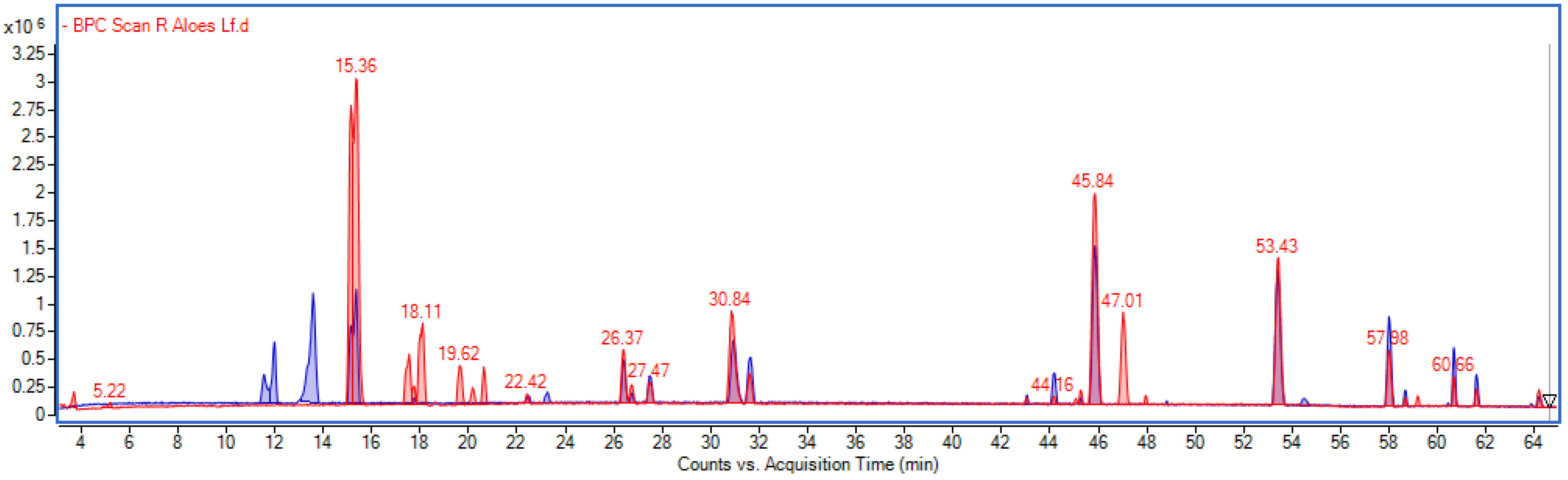
In general, the qualitative and quantitative profile of secondary metabolites in Aloe gel samples were consistent with that obtained in our previous study, with only minor differences [26]. The predominant compounds were aloesin (m/z-H = 393) and aloin (m/z-H = 417) (Figure 1). Slightly lower levels of aloesin and higher levels of its methylated derivative were observed. Similar differences were noted for the two isomers of aloin: a higher content of aloin A and a lower content of aloin B were recorded compared to the previous study, although the total amount of these compounds remained within a similar range.
In contrast to the previous results, a prominent peak at a retention time of 53.43 min, with m/z [M–H] 343.08308 (estimated formula C18H16O7), was observed and tentatively assigned to feralolide, a compound belonging to the dihydroisocoumarin class. According to the literature, this compound has been reported to occur in Aloe resin [27]. Additionally, the presence of chlorogenic acid was detected. Quantitative analysis revealed differences in the levels of major compounds between the Aloe vera gel and ferments (L. fermentum, L. rhamnosus, L. plantarum and L. paracasei) (Table 2). Chlorogenic acids were present at comparably low levels in all samples. In contrast, a pronounced increase was observed for aloesin, which showed markedly higher concentrations in all fermented samples (1168.6–1228.3 µg/mL) compared to the extract (201.4 µg/mL) (Figure S1). Minor variations were noted for 8-C-glucosyl-aloesol and hydroxyaloins, which remained at relatively similar levels across all samples. However, 7-O-methyl aloesin exhibited a substantial increase in fermented samples. In contrast, p-coumaroylquinic acids were more abundant in the extract than in the fermented samples, indicating a decrease during fermentation. Interestingly, within the isomers of p-coumaroylquinic acid, fermentation led to shifts in the distribution of individual isomers: the content of one isomer decreased, while isomer 4 increased, followed by an increase in isomer 5. Aloin B increased after fermentation, particularly in the L. rhamnosus sample. In contrast, aloin A showed a decrease in fermented samples, with the exception of L. rhamnosus. Homonataloin B remained relatively constant across all samples. Furthermore, lactic acid was detected in the fermentation products, indicating active lactic acid fermentation and the metabolism of lactic acid bacteria, which leads to acidification of the environment and the formation of metabolites of potential biological significance.

3.2. Assessment of Antioxidant Activity

3.2.1. ABTS and DPPH Radical Scavenging Assays and FRAP Reducing Power Assay

To comprehensively assess the antioxidant potential of aloe vera gel and its LAB ferments, a set of complementary analytical methods were employed, including both chemical assays and cellular models. The ABTS and DPPH assays are among the most commonly used methods for assessing free radical scavenging capacity; they are based on the reduction in the ABTS•+ cation radical and the DPPH• radical, respectively, which allows for a rapid and sensitive assessment of the antioxidant activity of samples with varying polarity [28]. Additionally, the FRAP (Ferric Reducing Antioxidant Power) method was employed, which involves assessing the ability to reduce Fe3+ ions to Fe2+ in the presence of the TPTZ (2,4,6-tripyridyl-s-triazine) complex, leading to the formation of an intensely colored Fe2+–TPTZ complex, measured spectrophotometrically. The choice of method allowed for a more comprehensive characterization of antioxidant activity by taking into account the various mechanisms of action of antioxidants [29].
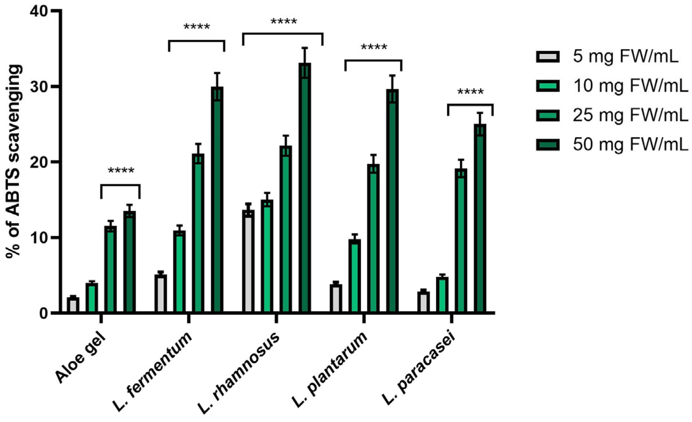
As shown in Figure 2, all tested samples (Aloe vera gel and its fermented products obtained using lactic acid bacteria of the strains Lactobacillus) exhibited antioxidant activity in the ABTS assay. A clear concentration-dependent relationship was observed. As the concentration of the tested samples increased, the degree of inhibition of the ABTS•+ radical also increased. The bacterial ferments exhibited significantly higher activity compared to the unfermented Aloe vera gel. Statistical significance was observed mainly for the two highest concentrations (25 and 50 mg FW/mL). The most beneficial antioxidant activity was demonstrated by the L. rhamnosus ferment, reaching 33.12% ± 1.21 ABTS scavenging capacity at the concentration of 50 mg FW/mL, which was approximately three times higher than that of Aloe vera gel.
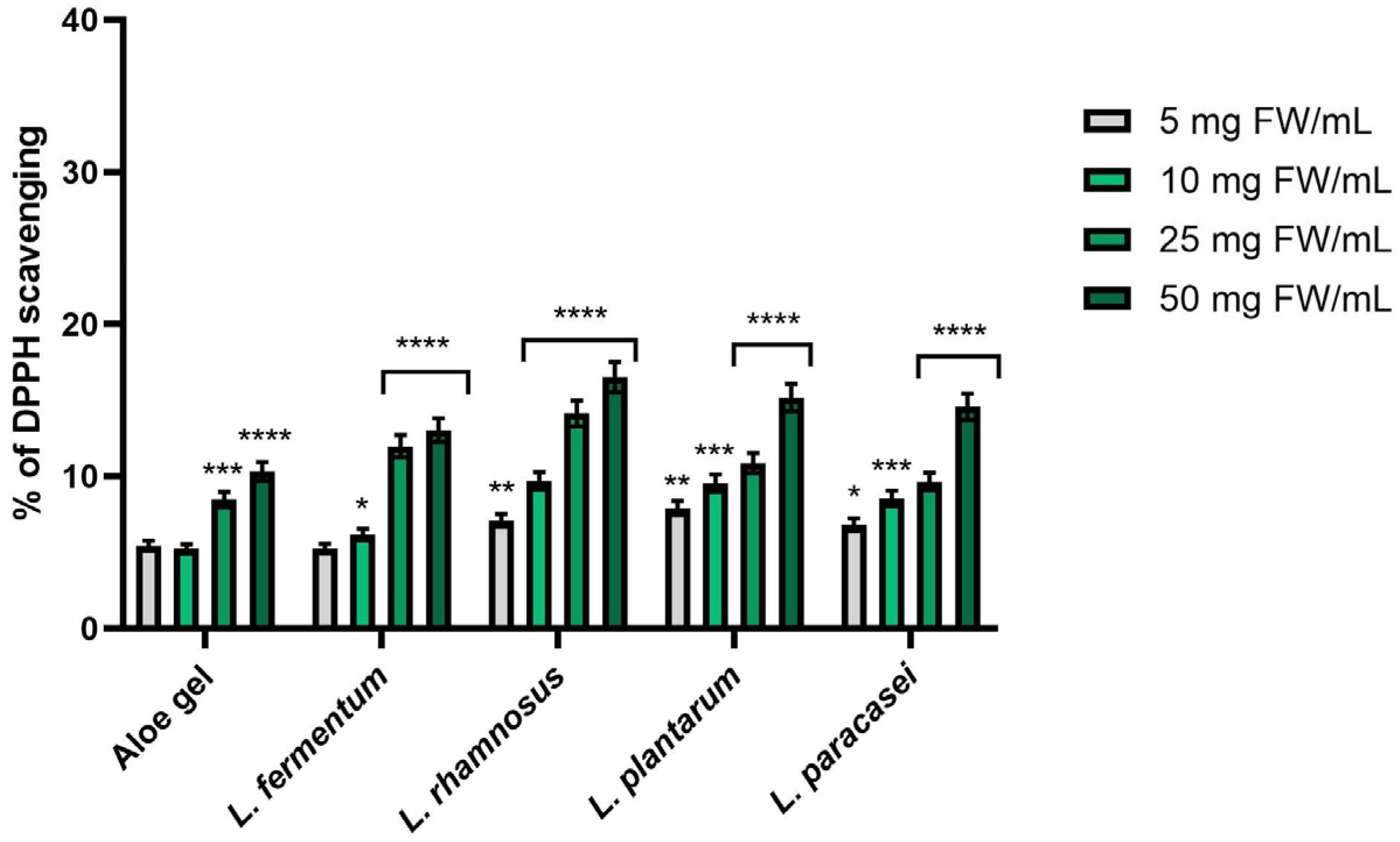
A similar trend was observed in the DPPH assay (Figure 3), where an increase in the concentration of the tested aloe samples resulted in increased antioxidant activity. It is worth noting, however, that higher inhibition values were generally obtained in the ABTS assay, which may be attributed to the hydrophilic nature of the tested samples and their greater efficacy in an aqueous environment. The differences between Aloe vera gel and its ferments were less pronounced than in the ABTS assay. However, in this case as well, the ferments, particularly those containing L. rhamnosus, exhibited the highest activity, achieving 11.97% ± 0.72 and 12.03% ± 0.78 DPPH scavenging, respectively, at concentrations of 25 and 50 mg FW/mL.
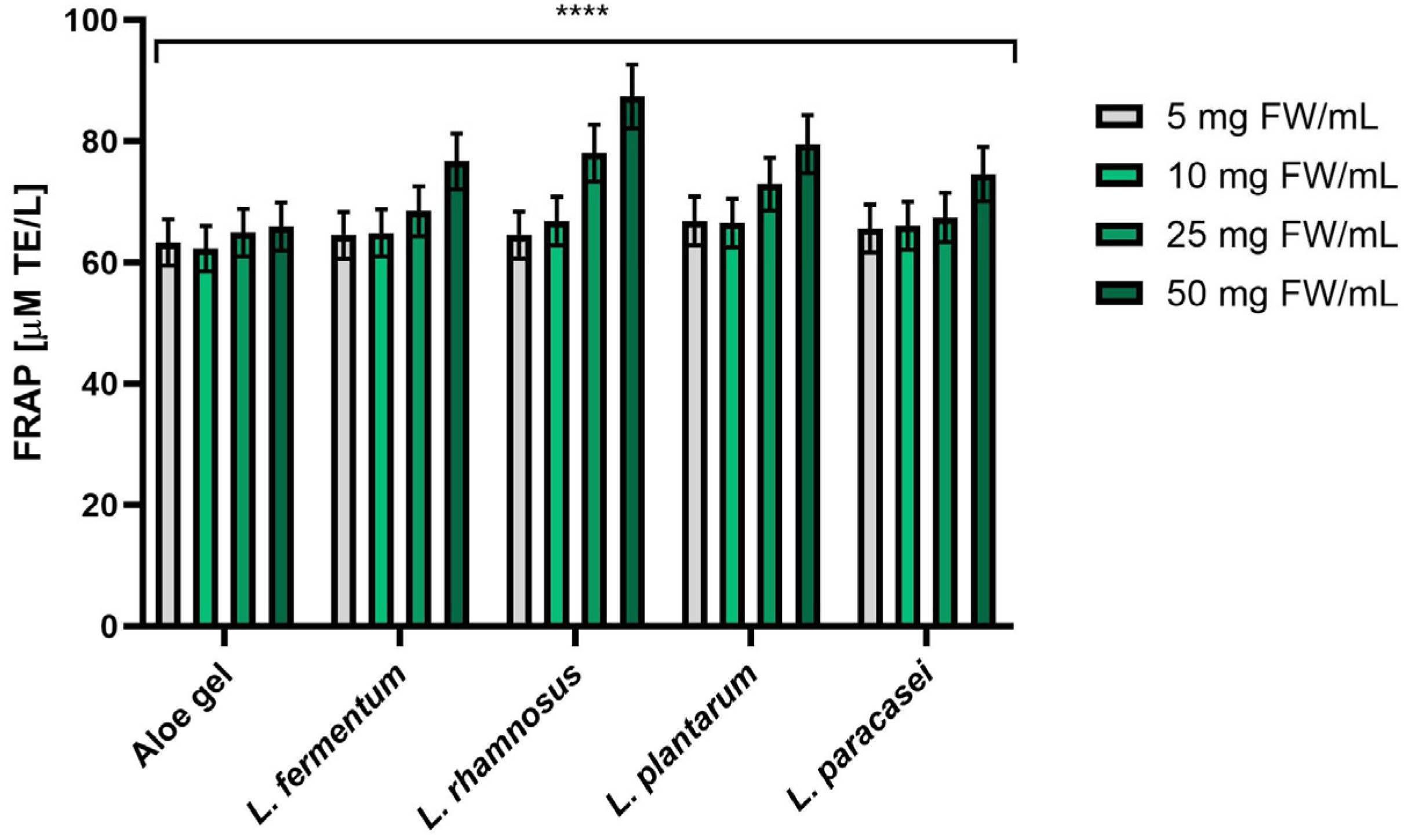
The analysis of reducing capacity using the FRAP method (Figure 4) showed that at lower concentrations (5 and 10 mg FW/mL), the values were comparable for all tested Aloe vera samples and its fermented products. Clear differences were observed only at higher concentrations, particularly at 50 mg FW/mL, where Lactobacillus ferments exhibited increased reducing potential. The highest value was obtained for the ferment containing L. rhamnosus (87.38 ± 5.87 µM TE/L), which confirms its greatest ability to reduce iron ions and indicates a significant enhancement of antioxidant properties as a result of the fermentation process.

3.2.2. Evaluation of the Effect on Superoxide Dismutase (SOD) Activity

The chemical analyses were supplemented by an assessment of the effect of aloe gel and its ferments on superoxide dismutase (SOD) activity in fibroblasts (HDF). SOD is a key enzyme of the endogenous antioxidant system, responsible for the dismutation of superoxide anion into hydrogen peroxide [30]. Measuring the activity of this enzyme in dermal cells allows us to assess whether the tested samples not only directly neutralize free radicals but also support cellular defense mechanisms, which are essential for protection against oxidative stress and skin aging processes.
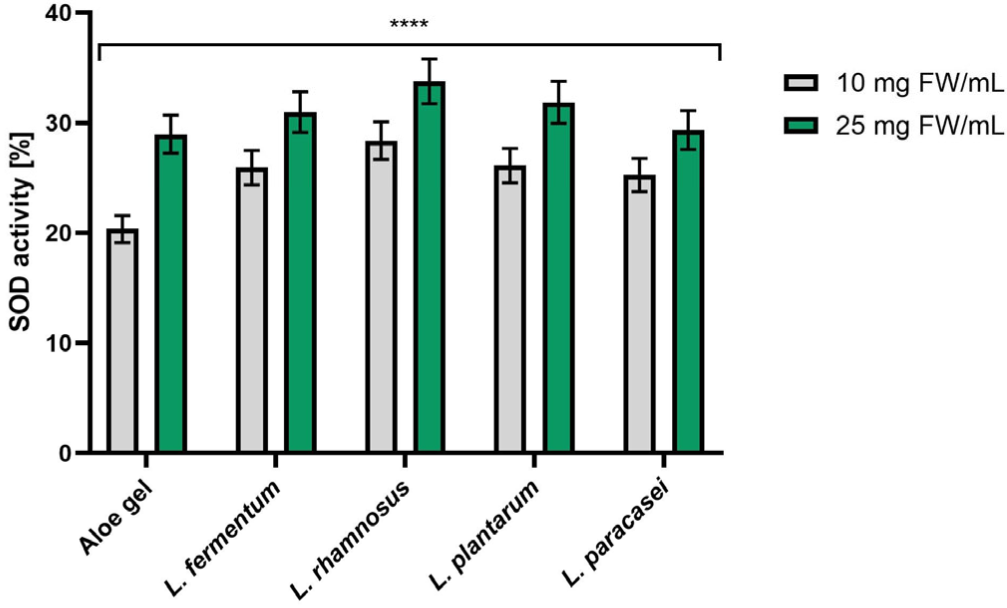
As shown in Figure 5, all tested samples of aloe vera gel and its bacterial ferments affected superoxide dismutase (SOD) activity in HDF cells. The study was conducted at two concentrations (10 and 25 mg FW/mL). A clear dose-dependent increase in SOD activity was observed. SOD activity, expressed as the inhibition rate (%) of WST reduction, increased with increasing sample concentration. This relationship is consistent with the results obtained in chemical assays (ABTS, DPPH, FRAP), which may indicate the preservation of antioxidant activity even under cellular conditions. The strongest effect was observed for ferments containing L. rhamnosus, which showed the greatest impact on SOD activity (28.38% ± 1.76 and 33.77% ± 2.28 for concentrations of 10 and 25 mg FW/mL, respectively). This may suggest that fermentation enhances the ability of the tested samples to modulate the endogenous antioxidant defense system of cells, as reflected by increased SOD-related activity.

3.2.3. Intracellular ROS Levels in Fibroblasts and Keratinocytes

In the next step, the levels of reactive oxygen species (ROS) in HaCaT and HDF cells were also measured, which allows for the assessment of antioxidant activity under biologically relevant conditions. This method relies on the use of fluorescent probes oxidized by ROS, allowing for quantitative analysis of oxidative stress at the cellular level [31]. The study utilized the 2′,7′-dichlorodihydrofluorescein diacetate (H2DCFDA) probe, which undergoes intracellular deacetylation followed by oxidation to the fluorescent DCF, enabling real-time monitoring of ROS levels. In the study, oxidative stress was induced by adding hydrogen peroxide (H2O2) to the cells, and the ability of the tested samples to reduce it was then assessed by measuring changes in ROS levels. This made it possible to evaluate the efficacy of Aloe vera gel and its Lactobacillus ferments in alleviating induced oxidative stress under physiologically relevant conditions.
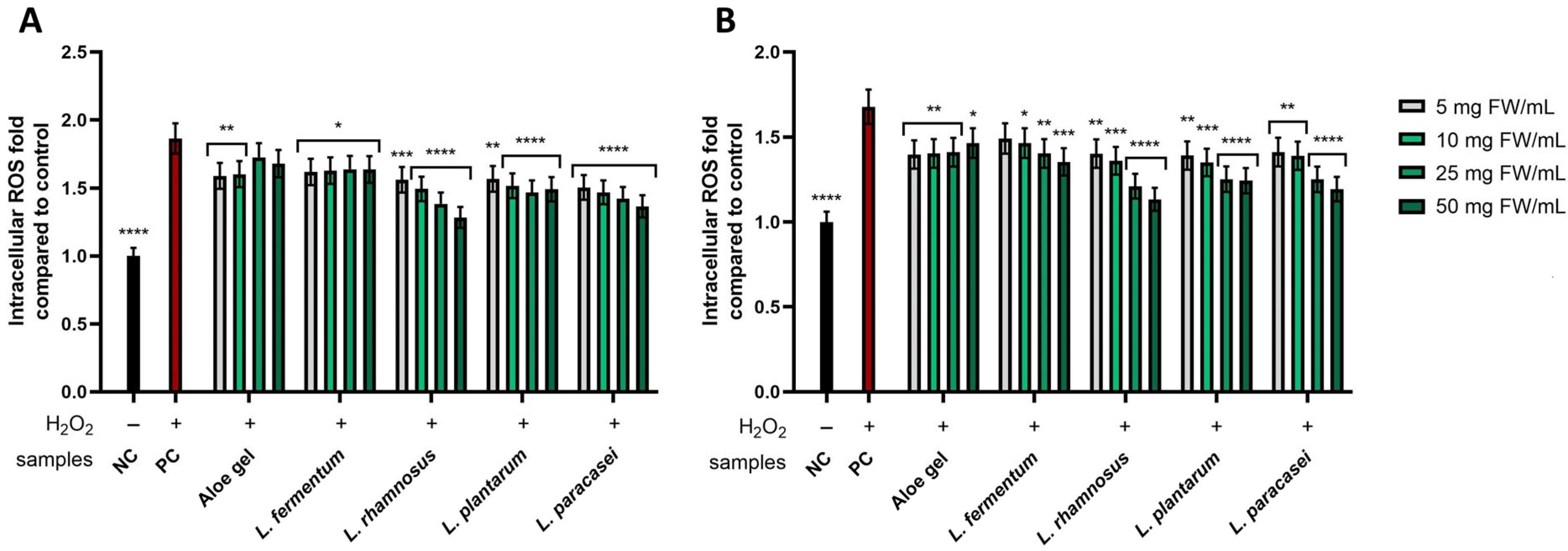
In the study of intracellular ROS levels, the results were expressed as a fold relative to the negative control (NC), which consisted of cells treated with neither H2O2 nor the tested compounds. Statistical significance was assessed relative to the positive control (PC), cells treated with H2O2 but not exposed to the tested compounds. As shown in Figure 6A, all analyzed samples reduced ROS levels in HDF cells, with this effect being dose dependent. The strongest effect was observed for the L. rhamnosus and L. paracasei ferments, which at the highest tested concentration (50 mg FW/mL) reached 1.283 ± 0.07- and 1.365 ± 0.09-fold, respectively, compared to the negative control. These results indicate an effective reduction in H2O2-induced oxidative stress and confirm the higher biological activity of the ferments compared to the non-fermented gel.
An analysis of the effect of the tested aloe samples on ROS levels in HaCaT cells (Figure 6B) revealed a similar relationship—the reduction in ROS levels was most pronounced at higher concentrations, confirming a dose-dependent effect. This phenomenon was particularly pronounced in the case of bacterial ferments, whereas no significant relationship between concentration and ROS levels was observed for Aloe vera gel. The most statistically significant effects were observed for L. rhamnosus, L. plantarum, and L. paracasei ferments at concentrations of 25 and 50 mg FW/mL, indicating their high efficacy in reducing intracellular oxidative stress in keratinocytes.

3.3. Cytotoxicity Assessment

The assessment of cytotoxicity is a key component of research into new raw materials intended for cosmetic and dermocosmetic applications. This study evaluated the cytotoxic properties of Aloe vera gel and its bacterial ferment. The analyses were conducted on two cell lines: fibroblasts (HDF) and keratinocytes (HaCaT). Two complementary tests were used to assess cell viability: Alamar Blue (AB) and Neutral Red (NR). The parallel use of both methods is particularly important for plant-based materials, such as plant-bacterial ferments, as it allows for the detection of both changes in metabolic activity and disturbances in intracellular structures. The AB test is based on the reduction in resazurin to resorufin by metabolically active cells and thus primarily reflects the state of redox metabolism, whereas the NR test assesses the ability of living cells to take up and accumulate dye in lysosomes, thereby indirectly providing information on cell membrane integrity [32,33].
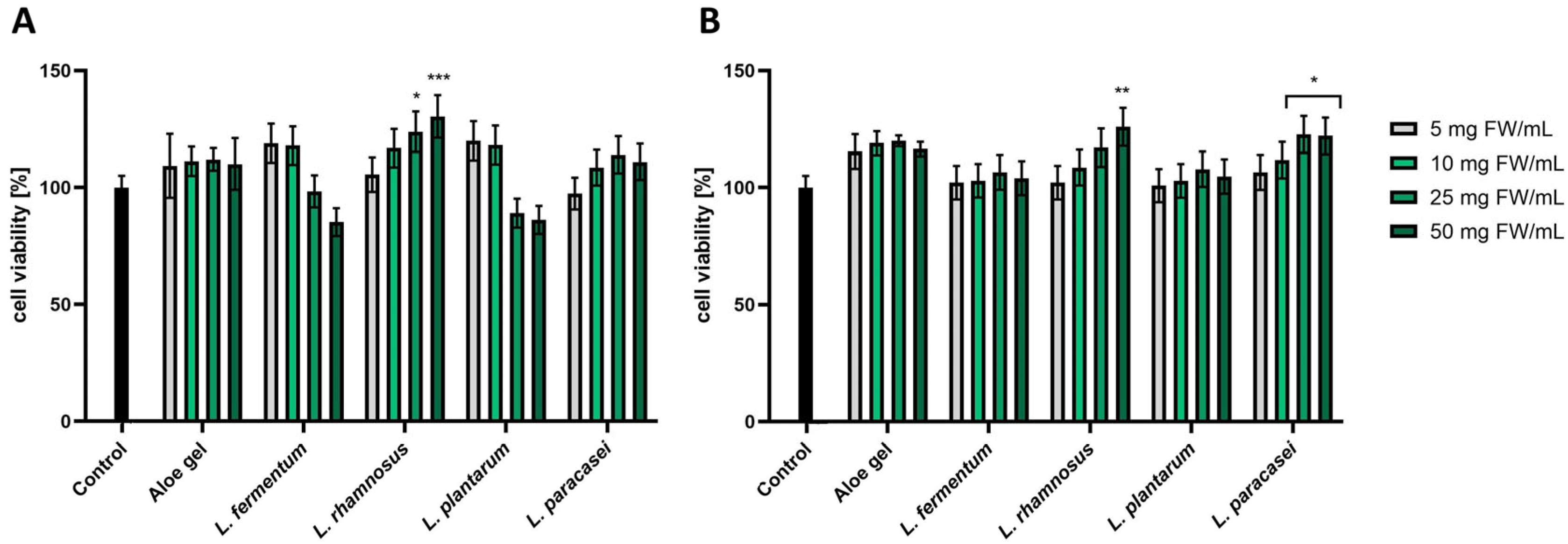
In a study evaluating the effect of the analyzed aloe samples on the viability and metabolic activity of HDF cells using the Alamar Blue assay, no cytotoxic effect was observed for any of the tested samples (Figure 7A). The most beneficial effect on fibroblast viability, statistically confirmed, was observed for L. rhamnosus at concentrations of 25 and 50 mg FW/mL, where the values were 123.84% ± 8.67 and 130.37% ± 9.13 cell viability, respectively, relative to the negative control (100%). Moreover, a downward trend was observed for the L. fermentum and L. plantarum fermentation. As the concentration increased, cell viability decreased. However, these values did not indicate a cytotoxic effect.
In the case of HaCaT cells (Figure 7B), no cytotoxicity of the tested samples was observed in the AB assay. The most beneficial, statistically significant proliferation values were obtained for L. rhamnosus at the highest tested concentration (125.98% ± 8.12) and for L. paracasei, reaching 122.71% ± 7.89 and 122.07% ± 7.85 cell viability at concentrations of 25 and 50 mg FW/mL, respectively. For most ferments, a dose–response relationship was observed, characterized by an increase in cell viability with increasing concentration. This trend was not observed for the non-fermented Aloe gel.
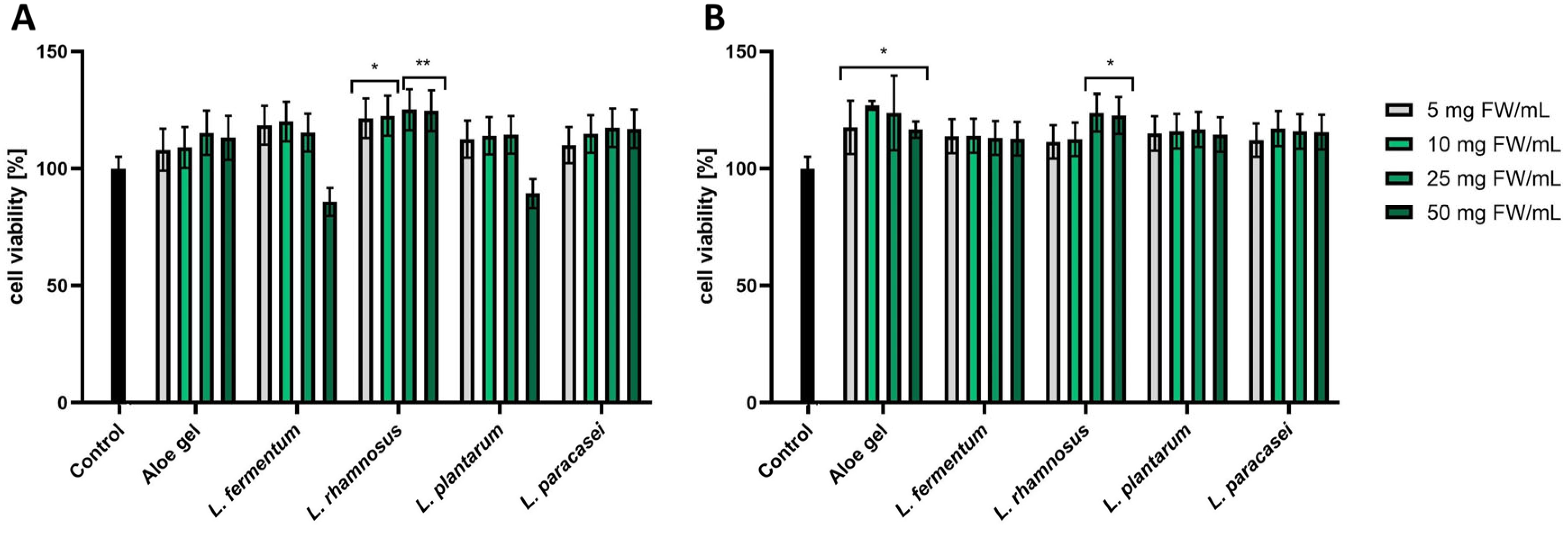
The results obtained in the Neutral Red assay were consistent with the observations from the AB test. For HDF cells (Figure 8A), the highest proliferation values, which were statistically significant, were observed for L. rhamnosus at all concentrations analyzed. Furthermore, a reduced cell viability (below the control level) was observed for L. fermentum and L. plantarum at a concentration of 50 mg FW/mL. However, this difference was not statistically significant, indicating the absence of cytotoxic activity.
In HaCaT cells (Figure 8B), no clear dose–response relationship was observed, and viability values remained at a level similar to the control, confirming the lack of cytotoxicity in the tested samples. A statistically significant increase in proliferation was observed for L. rhamnosus at concentrations of 25 and 50 mg FW/mL (123.82% ± 7.97 and 122.71% ± 7.89 cell viability, respectively). In addition, increased proliferative activity was observed at all tested concentrations for the unfermented Aloe vera gel.
Cytotoxicity studies were also conducted on model cosmetic formulations using HaCaT cells, which are a human keratinocyte cell line and are commonly used as a model for the outermost layer of the epidermis, which is directly exposed to the applied formulations. For this reason, they are considered a more appropriate model for assessing the safety of cosmetic formulations than dermal fibroblasts (HDF), which do not come into direct contact with the product.
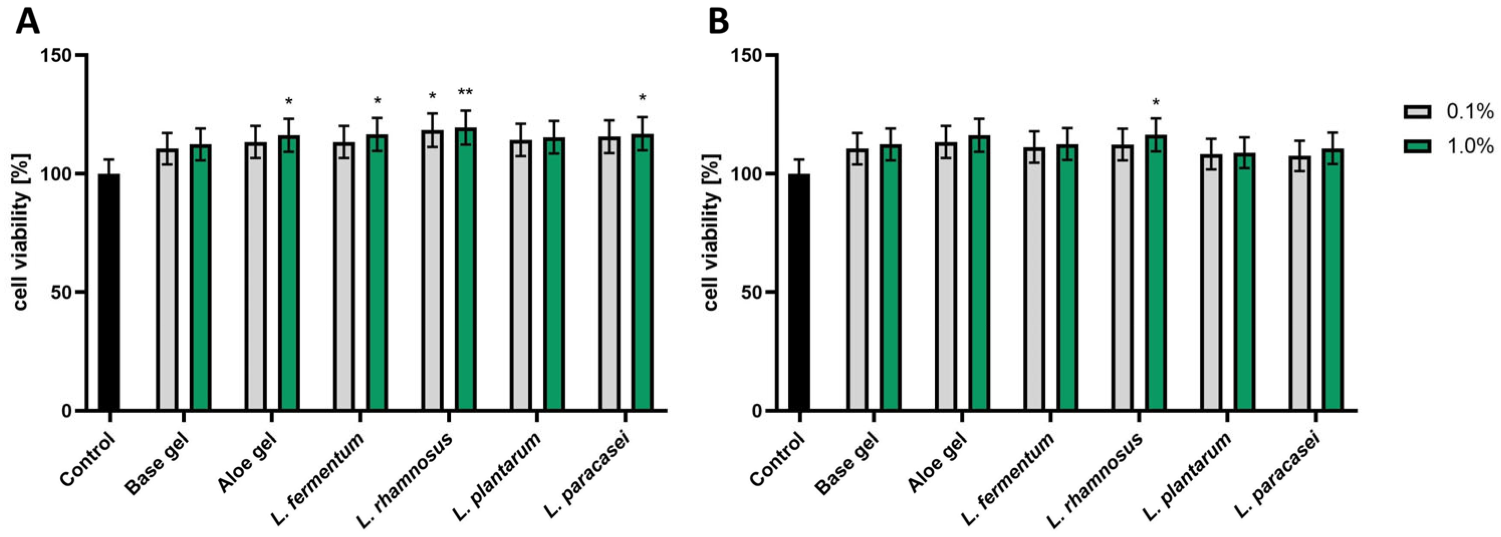
As shown in Figure 9A, in the Alamar Blue assay, none of the tested formulations (Base gel, cosmetic gel with added Aloe vera, and gels containing Lactobacillus ferments) exhibited cytotoxic effects on HaCaT cells. However, a slight increase in proliferation was observed, particularly for the Aloe vera gel and its ferments (L. fermentum, L. rhamnosus, L. paracasei), most evident at the higher concentration used (1.0%) compared to the base formulation.
A similar trend was observed in the Neutral Red test (Figure 9B), where the highest, statistically significant cell viability was recorded for the formulation containing L. rhamnosus at a gel concentration of 1.0% (116.47% ± 7.04 cell viability). The results indicate that both Aloe vera gel and its LAB ferments may have a beneficial effect on the viability and proliferation of keratinocytes. This suggests their potential use as active ingredients in cosmetic formulations that support the regeneration and barrier functions of the epidermis.

3.4. Assessment of Anti-Aging Potential via Inhibition of Extracellular Matrix (ECM)-Degrading Enzymes

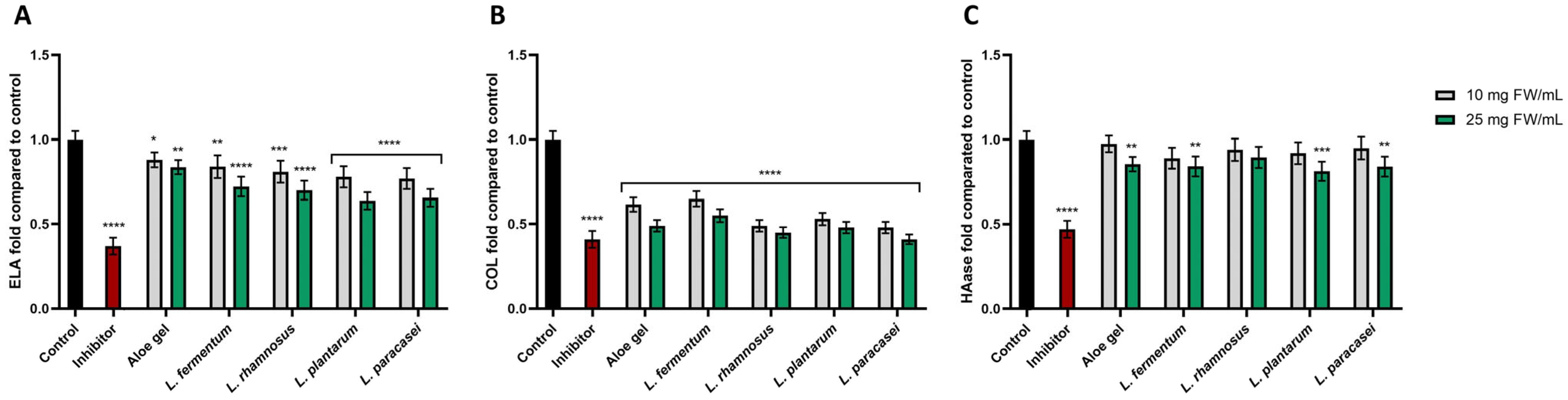
Enzymes such as elastase, collagenase, and hyaluronidase play a central role in the degradation of extracellular matrix (ECM) components, including collagen, elastin, and hyaluronic acid, which are essential for maintaining skin elasticity, firmness, and hydration. Therefore, their expression and activity are commonly associated with skin aging processes [34]. The protein levels of elastase (ELA), collagenase (COL), and hyaluronidase (HAase) in fibroblasts was assessed after treatment with Aloe gel and its ferments obtained using Lactobacillus strains (L. fermentum, L. rhamnosus, L. plantarum, and L. paracasei) at 10 and 25 mg FW/mL. Enzyme levels were expressed as fold change relative to untreated control cells (set as 1.0).
All tested samples reduced elastase levels compared to the control (Figure 10A). Aloe gel caused a moderate decrease, more pronounced at 25 mg FW/mL. Fermented samples exhibited stronger reduction, particularly at the higher concentration used. The most evident reduction was observed for ferments obtained with L. plantarum and L. paracasei at 25 mg/mL (approximately 0.6–0.7-fold of control). L. fermentum and L. rhamnosus also significantly reduced ELA levels, although the effect was slightly less pronounced. A general concentration-dependent trend was observed, although differences between strains were relatively modest and did not reach statistical significance in all cases.
Regarding collagenase levels, a marked reduction was observed in all analyzed samples compared to the control (Figure 10B). Aloe gel decreased COL activity to approximately 0.6-fold (10 mg FW/mL) and ~0.5-fold (25 mg FW/mL). Fermentation did not consistently enhance the effect relative to the non-fermented extract. While some fermented samples (e.g., L. rhamnosus and L. paracasei) showed slightly lower COL levels, the differences between aloe gel and ferments were relatively small. Importantly, all treated groups (both Aloe gel and ferments) showed significantly reduced collagenase levels compared to control, indicating a strong overall effect, with only a mild concentration-dependent trend.
The effect on hyaluronidase activity was less pronounced than for elastase and collagenase (Figure 10C). Aloe gel at 10 mg FW/mL showed minimal change relative to control, while a moderate decrease was observed at 25 mg FW/mL. Fermented samples resulted in a slight reduction in HAase levels, generally in the range of ~0.8–0.9-fold of control. The strongest decrease was observed for L. plantarum at 25 mg FW/mL, although overall differences between strains were modest. A weak concentration-dependent effect was noted, and statistically significant differences were observed mainly at the higher concentration used.

4. Discussion

Research shows that Aloe vera gel is a suitable substrate for fermentation involving lactic acid bacteria (LAB), such as L. fermentum, L. paracasei, L. rhamnosus, and L. plantarum. This is due to its polysaccharide-rich composition and susceptibility to oxidation processes, which promote natural fermentation [35].
Fermentation of plant-derived materials may lead to transformations of phenolic compounds, including their release from bound forms or structural modification, rather than simple degradation. In the present study, an increase in the concentration of selected phenolic compounds was observed after fermentation (Table 2), which may result from the enzymatic hydrolysis of conjugated forms. Spontaneous fermentation of plant-derived substrates has been widely reported to involve lactic acid bacteria (LAB); however, in the present study, no microbiological analyses (e.g., viable cell counts or metabolic profiling) were performed, and therefore no conclusions regarding microbial activity can be drawn. Polysaccharides present in Aloe vera gel, such as acemannan, have been described as potential carbon sources for microorganisms. Nevertheless, the prebiotic effect of these compounds is highly dependent on their structure and microbial selectivity [36,37]. As studies have shown, the fermentation process involves enzymatic transformations, including the hydrolysis of glycosidic bonds by β-glucosidases and proteolytic degradation of proteins, which can lead to the formation of lower molecular weight compounds with altered bioactivity [38].
A study by Zhou et al. demonstrated that the fermentation of kiwi fruit using Lactobacillus plantarum led to an increase in the content of phenolic compounds and flavonoids, which directly translated into an enhanced ability to scavenge free radicals. At the same time, changes in the metabolite profile were observed, including an increase in the concentration of lactic acid and other compounds involved in redox reactions, indicating the significant role of fermentation in modulating antioxidant activity [39]. In turn, Li et al. demonstrated that the fermentation of plant material using Lactobacillus plantarum and Lactobacillus rhamnosus led to an increase in antioxidant activity, soluble protein content, and organic acid content. This effect was attributed to the enzymatic degradation of complex compounds and the formation of more active metabolites, which confirms the validity of using these strains in the bioconversion of plant materials, including Aloe vera gel [40].
The increase in antioxidant potential is mainly associated with the increased availability of phenolic compounds and flavonoids and their conversion into more active forms [41]. Strains such as L. plantarum and L. rhamnosus demonstrate the ability to modify the metabolite profile and enhance antioxidant activity. At the same time, fermentation leads to the formation of new compounds, including organic acids and bioactive peptides, which demonstrate the ability to scavenge free radicals, chelate metal ions, and inhibit lipid peroxidation [42,43]. Fermentation products also influence properties that are important for the skin. The presence of lactic acid lowers the pH, which supports the function of the skin barrier and the microbiome [44]. The resulting metabolites have a moisturizing effect and may support skin regeneration. Additionally, fermentation increases the bioavailability of active ingredients, which may improve their penetration and efficacy. It should be noted that changes in the content of phenolic compounds are not always clear-cut. Some studies have shown reduced levels of active compounds in fermentation products, accompanied by an increase in antioxidant activity [45,46]. This indicates that not only the quantity but also the chemical form and bioavailability of these compounds are of key importance.
As studies have shown, Aloe vera gel exhibits moderate direct free radical scavenging capacity (higher IC50 in the DPPH assay); however, its antioxidant activity is multifaceted and concentration dependent. Significantly better results were obtained in the ABTS assay, which may be attributed to the gel’s hydrophilic nature and greater efficacy in an aqueous environment [47]. An important mechanism of action is the modulation of the enzymatic antioxidant system. Studies have observed an increase in SOD, catalase, and glutathione peroxidase activity, as well as a decrease in lipid peroxidation, indicating an indirect protective effect [48]. Additionally, the presence of peroxidases may support the elimination of H2O2 on the skin surface [49]. Furthermore, the gel also exhibits anti-inflammatory properties by concentration-dependently inhibiting NO production, suggesting its potential role in reducing oxidative-inflammatory stress, which is significant in skin aging processes and inflammatory dermatoses [50].
The biological activity of the gel stems primarily from the presence of polysaccharides, which constitute its dominant fraction. These compounds support skin regeneration by stimulating cell proliferation, improving hydration, and forming a protective layer [51]. Additionally, they modulate the inflammatory response, which promotes healing processes. Glycoproteins and sulfated polysaccharides also contribute to the anti-inflammatory effect [52,53].
Furthermore, it has been shown that aloe extracts contain compounds such as salicylates, lactates, and magnesium ions, which may be responsible for the analgesic effect. This mechanism may resemble the action of aspirin-like substances or result from the synergistic interaction of several components simultaneously [54].
As some studies show, the antioxidant activity of Aloe vera gel is lower than that of leaf epidermis extracts or alcoholic extracts, which contain higher amounts of phenolic compounds and flavonoids. At the same time, however, the gel has an advantage in terms of safety of use and skin compatibility. Differences in activity among aloe preparations result not only from the part of the plant but also from the extraction method used, the type of solvent, and environmental factors affecting the phytochemical composition [55,56].
It is also worth noting that aloesin, identified as the predominant phenolic compound in Aloe vera samples (Table 2), exhibited a marked increase in concentration following fermentation. This observation may be associated with enzymatic processes occurring during fermentation, such as the hydrolysis of bound or conjugated forms and the enhanced release of phenolic compounds from the plant matrix. Microbial enzymes, including glycosidases and esterases, have been reported to increase the extractability of phenolic compounds in fermented plant materials, which may lead to a higher apparent concentration in analytical determinations. The pronounced increase in aloesin (approximately sixfold) compared to other compounds may reflect its specific structural features and its susceptibility to such transformations, although this hypothesis requires further confirmation through targeted analysis of bound fractions. From a biological perspective, aloesin has been reported to exhibit significant antioxidant activity, as demonstrated in both chemical assays and cellular models. Its activity is primarily attributed to its ability to scavenge reactive oxygen species due to the presence of hydroxyl groups, as well as to inhibit lipid peroxidation, suggesting a protective role against oxidative stress at the cellular level [57].
It has also been shown that the fermentation of Aloe vera involving lactic acid bacteria can significantly modify its phytochemical profile, leading to an increase in the biological activity of the extracts. This effect is attributed to the biotransformation of phenolic compounds and the formation of new, more active metabolites, which translates into enhanced antioxidant and anti-aging properties [58].
Moreover, analyses conducted by other researchers indicate that aloe ferments and derivatives of the studied lactic acid bacteria can significantly affect the viability of skin cells, including keratinocytes and fibroblasts, as well as the integrity of cellular structures. The introduction of bacterial fermentation significantly modifies the biological profile of the tested material, which can lead to both increased bioavailability of active compounds and the formation of new metabolites with cytoprotective effects. In the case of fermented aloe preparations, the fermentation process has been shown to enhance their biological activity. Ro et al. demonstrated that fermentation of Aloe arborescens with Lactobacillus plantarum enhanced the anti-aging effect and activated fibroblasts more strongly than unfermented material [58]. Jiang et al. and Jung et al. [59,60] reported that the supernatant obtained by fermenting aloe with Lactobacillus plantarum was characterized by strong antioxidant, anti-inflammatory, and cell-protective activity. Importantly, a beneficial effect on skin cell viability was also observed with other types of fermentation. It was shown that fermented aloe gel obtained with kombucha did not reduce the viability of fibroblasts and keratinocytes, and in selected concentration ranges, it even promoted the proliferation of these cells [26]. Data indicate that fermenting aloe with different microbial strains can lead to achieving products with diverse effects on cellular metabolism, membrane integrity, and the proliferative response of skin cells [61,62].
In our previous study, kombucha-mediated fermentation of Aloe vera gel enhanced its cosmetic potential and increased the reduction in the levels of collagenase, elastase, and hyaluronidase compared with the unfermented material [26]. The present study extends those observations by showing that fermentation of Aloe gel with selected lactic acid bacteria also modifies its anti-aging-related effects, although the extent of this effect depended on the enzyme analyzed and, to a lesser degree, on the fermentation variant. These findings support the view that microbial biotransformation may represent an effective strategy for modulating the skin-related properties of aloe-derived materials.
Only a limited number of studies have examined the effects of aloe ferments on collagenase, elastase, and hyaluronidase. Most available studies on lactic acid bacteria-fermented aloe have focused instead on broader anti-wrinkle or photoprotective endpoints. In Aloe arborescens, fermentation with Lactobacillus plantarum enhanced fibroblast activity, increased collagen production, and reduced MMP-1 synthesis more effectively than the non-fermented material [58]. Similarly, Lee et al. demonstrated that L. plantarum-fermented outer leaf skin of Aloe barbadensis Miller improved the anti-photoaging response of UVB-irradiated fibroblasts, reducing ROS production, increasing collagen levels, and lowering MMP-1 secretion [63]. Taken together, these studies suggest that lactic acid fermentation can strengthen the skin-protective properties of aloe, even though direct evidence in the modulation of collagenase, elastase, and hyaluronidase levels or activity remainslimited. This is also consistent with earlier work showing that aloe fermentation supernatant obtained with L. plantarum HM218749.1 exhibited strong antioxidant activity and significantly reduced IL-1β, TNF-α, and IL-6 at both the mRNA and protein levels [59]. Since oxidative stress and chronic low-grade inflammation are closely linked to extracellular matrix degradation and skin aging, these data provide a plausible mechanistic background for the beneficial effects of fermented aloe preparations in skin-related models.
In the present study, both the non-fermented Aloe gel and the fermented samples reduced the levels of collagenase, elastase, and hyaluronidase relative to the untreated control, with the most pronounced effect observed for collagenase and a weaker response noted for hyaluronidase. This pattern suggests that the tested samples may contribute to the preservation of extracellular matrix integrity rather than exerting uniform modulation across all ECM-degrading enzymes. Such an interpretation is supported by the established role of collagenase, elastase, and hyaluronidase in the degradation of collagen, elastin, and hyaluronic acid, respectively, and thus in the progressive loss of skin firmness, elasticity, and hydration [26,64]. These results indicate that the tested samples may modulate ECM remodeling by reducing the levels of key ECM-degrading enzymes, although direct inhibition of enzymatic activity was not assessed in this study.
The enzyme-modulating effects observed here may be related, at least in part, to fermentation-induced changes in the phytochemical profile of the samples (Table 2). LC-MS analysis showed that aloesin and aloin derivatives were the dominant constituents in both the extract and fermented gels, while fermentation markedly increased the concentration of aloesin and also elevated the level of 7-O-methyl aloesin. This may be relevant because aloesin derivatives isolated from Aloe vera have previously been reported to exhibit antioxidant, free radical scavenging, and anti-inflammatory activity [57]. Although the present study does not demonstrate a direct causal relationship between individual compounds and enzyme level modulation, the marked increase in aloesin-related constituents after fermentation suggests that these metabolites may contribute to the observed biological effects. A similar cautious interpretation can be made for chlorogenic acid, which was detected in all analyzed samples, although at relatively low levels. This acid has been shown to protect human dermal fibroblasts against UVA-induced photoaging, regulate collagen metabolism and apoptosis, and reduce inflammatory and oxidative stress-related changes in UV-exposed fibroblasts [65]. Therefore, even a relatively low contribution of this compound may be relevant when considered together with other phenolics and anthraquinones present in aloe samples. Another interesting finding was the tentative identification of feralolide in the analyzed samples. This compound has been reported as a dihydroisocoumarin present in Aloe resin, and more recent data indicate that it shows antioxidant potential in in vitro assays [27]. Although its role in the anti-aging effects observed in this study is not clear, its presence suggests that the activity of aloe gels and their ferments likely results from the combined action of multiple compounds rather than a single dominant component.
In this study, we demonstrated that fermentation did not result in a uniform increase in all quantified constituents. In particular, p-coumaroylquinic acids decreased after fermentation, while aloin B increased in some variants and aloin A decreased in most fermented samples. These findings indicate that lactic acid fermentation does not simply enrich the starting material but rather remodels its chemical profile through selective biotransformation. Accordingly, the anti-aging-related effects observed in fibroblasts are more likely to reflect the overall compositional shift induced by fermentation than changes in a single metabolite alone. This finding is also in line with the broader literature on fermented aloe, where enhanced biological activity has been linked to fermentation-associated compositional changes, including modification of polysaccharide fractions and retention or transformation of bioactive constituents [58,63].
Overall, the present results, together with our previous findings on kombucha-fermented aloe gel, support the conclusion that fermentation is a useful approach for modulating the anti-aging potential of Aloe vera preparations. While kombucha fermentation previously demonstrated a clear increase in inhibitory activity toward collagenase, elastase, and hyaluronidase, the present study shows that fermentation with lactic acid bacteria also produces biologically active Aloe gels capable of reducing the levels of key ECM-degrading enzymes. Importantly, the available literature suggests that aloe ferments obtained with lactic acid bacteria have so far been studied mainly in the context of collagen synthesis, MMP-1 suppression, antioxidant effects, and photoprotection, whereas direct evaluation of collagenase, elastase, and hyaluronidase remains limited. In this context, the present work provides additional evidence that lactic acid bacteria-mediated fermentation may broaden the cosmetic applicability of aloe by influencing multiple pathways associated with skin aging.

5. Conclusions

The results confirm that Aloe vera gel is a rich source of bioactive compounds, such as aloesin and aloin, whose quality profile remains stable, whereas fermentation involving lactic acid bacteria significantly alters their content. A particularly marked increase in the concentration of aloesin and its derivatives in the ferments indicates that the fermentation process increases the availability of selected secondary metabolites of potential biological significance. Moreover, the presence of lactic acid confirms the metabolic activity of the bacteria and the formation of additional functional compounds. Fermentation significantly enhances the antioxidant properties of Aloe vera, as demonstrated in both chemical tests and cellular models. Lactobacillus ferments, particularly those involving L. rhamnosus, more effectively neutralize free radicals, increase the activity of endogenous defense mechanisms (SOD), and reduce ROS levels in skin cells. Cytotoxicity studies confirmed the safety of both the Aloe gel and its fermented products, while also demonstrating a stimulating effect on the proliferation of fibroblasts and keratinocytes. Additionally, all samples exhibited the ability to inhibit enzymes that degrade the extracellular matrix, indicating their anti-aging potential. It should be noted, however, that the quantitative comparison of samples was performed without normalization to dry weight content, which may impact the interpretation of changes in compound concentrations. Furthermore, target metabolite profiling was not performed, limiting the ability to precisely identify the mechanisms underlying the observed transformations. The biological evaluation was based on in vitro skin cell models, which do not fully reflect skin barrier function or bioavailability, and key aspects such as skin penetration and activity of the final formulation were not comprehensively assessed. Despite these limitations, the obtained results provide a solid foundation for the further development of fermented Aloe vera-based systems and indicate that fermentation with selected Lactobacillus strains may be a promising strategy for enhancing the biological activity of plant-derived cosmetic ingredients.

Supplementary Materials

The following supporting information can be downloaded at: https://www.mdpi.com/article/10.3390/app16094098/s1, Table S1. Calibration curve parameters for the analyzed compounds. Figure S1. Extracted ion chromatogram (EIC) of a mass range characteristic for aloesin, with a mass error not exceeding 5 ppm (upper panel). DAD chromatogram recorded at a wavelength of 300 nm with the aloesin peak marked (lower panel). Green line—extract; red line—fermented extract.

Author Contributions

Conceptualization, A.Z., methodology, A.Z., M.Z.-D., Z.N.-Ł., A.S., M.W. and I.S.; validation, A.Z., M.Z.-D., Z.N.-Ł., M.W. and I.S.; formal analysis, A.Z., M.Z.-D., Z.N.-Ł., M.W. and I.S.; investigation, A.Z., M.Z.-D., Z.N.-Ł., A.S., M.W. and I.S.; data curation, A.Z., M.Z.-D., Z.N.-Ł., M.W. and I.S.; writing—original draft preparation, A.Z., M.Z.-D., Z.N.-Ł., A.S., M.W. and I.S.; writing—review and editing, A.Z., M.Z.-D., Z.N.-Ł., M.W. and I.S.; visualization, A.Z.; supervision, A.Z. All authors have read and agreed to the published version of the manuscript.

Funding

This research received no external funding.

Institutional Review Board Statement

Not applicable.

Informed Consent Statement

Not applicable.

Data Availability Statement

The data presented in this study are available on request from the corresponding author.

Conflicts of Interest

The authors declare no conflicts of interest.

Abbreviations

The following abbreviations are used in this manuscript:
ABAlamar Blue Assay
ABTS2,2′-azino-bis(3-ethylbenzothiazoline-6-sulfonic acid)
CO2carbon dioxide
COLcolagenase
DADdiode array detector
DCF2′,7′-dichlorofluorescein
DMEMdulbecco’s modified eagle medium
DPPH1,1-diphenyl-2-picrylhydrazyl
ELAelastase
ELISAenzyme-linked immunosorbent assay
EMCextracellular matrix component
ESI/TOFElectrospray Ionization/Time-of-Flight
FBSfetal bovine serum
FRAPferric reducing antioxidant power
H2DCFDA2′,7′-dichlorodihydrofluorescein diacetate
H2O2hydrogen peroxide
HAasehyaluronidase
HaCaThuman keratinocyte cell line
HDFhuman dermal fibroblasts
IL-1βinterleukin-1 beta
IL-6interleukin-6
LC-MSliquid chromatography–mass spectrometry
MMP-1matrix metalloproteinase-1
mRNAmessenger RNA
NRNeutral Red Assay
PBSphosphate-buffered saline
ROSreactive oxygen species
SODsuperoxide dismutase
SPCKsuccinyl–alanyl–alanyl–prolyl–valyl chloromethyl ketone
TNF-αtumor necrosis factor alpha
TPTZ2,4,6-Tripyridyl-s-triazine
UHPLCultra-high-performance liquid chromatography
UVultraviolet radiation
UVAultraviolet A
UVBultraviolet B
UV-VISultraviolet-visible spectroscopy

References

  1. Hamman, J.H. Composition and Applications of Aloe Vera Leaf Gel. Molecules 2008, 13, 1599. [Google Scholar] [CrossRef] [Scilit]
  2. Kaur, S.; Bains, K. Aloe Barbadensis Miller (Aloe Vera) Pharmacological Activities and Clinical Evidence for Disease Prevention. Int. J. Vitam. Nutr. Res. 2024, 94, 308–321. [Google Scholar] [CrossRef] [Scilit]
  3. Ahlawat, K.S.; Khatkar, B.S. Processing, Food Applications and Safety of Aloe Vera Products: A Review. J. Food Sci. Technol. 2011, 48, 525–533. [Google Scholar] [CrossRef] [Scilit] [PubMed]
  4. Misir, J.; Brishti, F.H.; Hoque, M.M. Aloe Vera Gel as a Novel Edible Coating for Fresh Fruits: A Review. Am. J. Food Sci. Technol. 2014, 2, 93–97. [Google Scholar] [CrossRef] [Scilit]
  5. Zhang, Y.; Bao, Z.; Ye, X.Y.; Xie, Z.Y.; He, K.; Mergens, B.; Li, W.; Yatcilla, M.K.; Zheng, Q.Y. Chemical Investigation of Major Constituents in Aloe Vera Leaves and Several Commercial Aloe Juice Powders. J. AOAC Int. 2018, 101, 1741–1751. [Google Scholar] [CrossRef] [Scilit] [PubMed]
  6. Shi, X.D.; Yin, J.Y.; Huang, X.J.; Que, Z.Q.; Nie, S.P. Structural and Conformational Characterization of Linear O-Acetyl-Glucomannan Purified from Gel of Aloe Barbadensis Miller. Int. J. Biol. Macromol. 2018, 120, 2373–2380. [Google Scholar] [CrossRef] [Scilit]
  7. Chokboribal, J.; Tachaboonyakiat, W.; Sangvanich, P.; Ruangpornvisuti, V.; Jettanacheawchankit, S.; Thunyakitpisal, P. Deacetylation Affects the Physical Properties and Bioactivity of Acemannan, an Extracted Polysaccharide from Aloe Vera. Carbohydr. Polym. 2015, 133, 556–566. [Google Scholar] [CrossRef] [Scilit]
  8. Semerel, J.; John, N.; Dehaen, W.; Fardim, P. Valorization of Aloe Barbadensis Miller. (Aloe Vera) Processing Waste. J. Renew. Mater. 2022, 11, 1031–1061. [Google Scholar] [CrossRef] [Scilit]
  9. Davis, R.H.; Donato, J.J.; Hartman, G.M.; Haas, R.C. Anti-Inflammatory and Wound Healing Activity of a Growth Substance in Aloe Vera. J. Am. Podiatr. Med. Assoc. 1994, 84, 77–81. [Google Scholar] [CrossRef] [Scilit]
  10. Radha, M.H.; Laxmipriya, N.P. Evaluation of Biological Properties and Clinical Effectiveness of Aloe Vera: A Systematic Review. J. Tradit. Complement. Med. 2015, 5, 21–26. [Google Scholar] [CrossRef] [Scilit]
  11. Boudreau, M.D.; Beland, F.A. An Evaluation of the Biological and Toxicological Properties of Aloe Barbadensis (Miller), Aloe Vera. J. Environ. Sci. Health C Environ. Carcinog. Ecotoxicol. Rev. 2006, 24, 103–154. [Google Scholar] [CrossRef] [Scilit] [PubMed]
  12. Matei, C.E.; Visan, A.I.; Cristescu, R. Aloe Vera Polysaccharides as Therapeutic Agents: Benefits Versus Side Effects in Biomedical Applications. Polysaccharides 2025, 6, 36. [Google Scholar] [CrossRef] [Scilit]
  13. Saleem, A.; Naureen, I.; Naeem, M.; Murad, H.S.; Maqsood, S.; Tasleem, G. Aloe Vera Gel Effect on Skin and Pharmacological Properties. Sch. Int. J. Anat. Physiol. 2022, 5, 1–8. [Google Scholar] [CrossRef] [Scilit]
  14. Valero-Cases, E.; Cerdá-Bernad, D.; Pastor, J.J.; Frutos, M.J. Non-Dairy Fermented Beverages as Potential Carriers to Ensure Probiotics, Prebiotics, and Bioactive Compounds Arrival to the Gut and Their Health Benefits. Nutrients 2020, 12, 1666. [Google Scholar] [CrossRef] [Scilit] [PubMed]
  15. Cuvas-Limon, R.B.; Nobre, C.; Cruz, M.; Rodriguez-Jasso, R.M.; Ruíz, H.A.; Loredo-Treviño, A.; Texeira, J.A.; Belmares, R. Spontaneously Fermented Traditional Beverages as a Source of Bioactive Compounds: An Overview. Crit. Rev. Food Sci. Nutr. 2021, 61, 1–23. [Google Scholar] [CrossRef] [Scilit]
  16. Morais, S.G.G.; da Silva Campelo Borges, G.; dos Santos Lima, M.; Martín-Belloso, O.; Magnani, M. Effects of Probiotics on the Content and Bioaccessibility of Phenolic Compounds in Red Pitaya Pulp. Food Res. Int. 2019, 126, 108681. [Google Scholar] [CrossRef] [Scilit]
  17. Majchrzak, W.; Motyl, I.; Śmigielski, K. Biological and Cosmetical Importance of Fermented Raw Materials: An Overview. Molecules 2022, 27, 4854. [Google Scholar] [CrossRef] [Scilit]
  18. Wójciak, M.; Paduch, R.; Drozdowski, P.; Żuk, M.; Wójciak, W.; Tyszczuk-Rotko, K.; Feldo, M.; Sowa, I. Ultra-Performance Liquid Chromatography and Mass Spectrometry Characterization, and Antioxidant, Protective, and Anti-Inflammatory Activity, of the Polyphenolic Fraction from Ocimum Basilicum. Molecules 2024, 29, 5043. [Google Scholar] [CrossRef] [Scilit]
  19. Wójciak, M.; Paduch, R.; Drozdowski, P.; Wójciak, W.; Żuk, M.; Płachno, B.J.; Sowa, I. Antioxidant and Anti-Inflammatory Effects of Nettle Polyphenolic Extract: Impact on Human Colon Cells and Cytotoxicity Against Colorectal Adenocarcinoma. Molecules 2024, 29, 5000. [Google Scholar] [CrossRef] [Scilit] [PubMed]
  20. Miller, N.J.; Rice-Evans, C.A. Factors Influencing the Antioxidant Activity Determined by the ABTS.+ Radical Cation Assay. Free Radic. Res. 1997, 26, 195–199. [Google Scholar] [CrossRef] [Scilit]
  21. Brand-Williams, W.; Cuvelier, M.E.; Berset, C. Use of a Free Radical Method to Evaluate Antioxidant Activity. LWT-Food Sci. Technol. 1995, 28, 25–30. [Google Scholar] [CrossRef] [Scilit]
  22. Benzie, I.F.F.; Strain, J.J. The Ferric Reducing Ability of Plasma (FRAP) as a Measure of “Antioxidant Power”: The FRAP Assay. Anal. Biochem. 1996, 239, 70–76. [Google Scholar] [CrossRef] [Scilit]
  23. Grauzdytė, D.; Pukalskas, A.; Viranaicken, W.; Kalamouni, C.E.; Venskutonis, P.R. Protective Effects of Phyllanthus Phillyreifolius Extracts against Hydrogen Peroxide Induced Oxidative Stress in HEK293 Cells. PLoS ONE 2018, 13, e0207672. [Google Scholar] [CrossRef] [Scilit] [PubMed]
  24. Page, B.; Page, M.; Noel, C. A New Fluorometric Assay for Cytotoxicity Measurements in Vitro. Int. J. Oncol. 1993, 3, 473–476. [Google Scholar] [CrossRef] [Scilit]
  25. Borenfreund, E.; Puerner, J.A. Toxicity Determined in Vitro by Morphological Alterations and Neutral Red Absorption. Toxicol. Lett. 1985, 24, 119–124. [Google Scholar] [CrossRef] [Scilit] [PubMed]
  26. Ziemlewska, A.; Zagórska-Dziok, M.; Nowak, A.; Muzykiewicz-Szymańska, A.; Wójciak, M.; Sowa, I.; Szczepanek, D.; Nizioł-Łukaszewska, Z. Enhancing the Cosmetic Potential of Aloe Vera Gel by Kombucha-Mediated Fermentation: Phytochemical Analysis and Evaluation of Antioxidant, Anti-Aging and Moisturizing Properties. Molecules 2025, 30, 3192. [Google Scholar] [CrossRef] [Scilit] [PubMed]
  27. Khan, I.; Mohanta, T.K.; Ihsan, N.; Halim, S.A.; Khan, A.; Rehman, N.U.; Khan, F.; Khalid, A.; Abdalla, A.N.; Karim, N.; et al. Antiamnesic Effects of Feralolide Isolated from Aloe Vera Resin Miller against Learning Impairments Induced in Mice. Antioxidants 2023, 12, 161. [Google Scholar] [CrossRef] [Scilit]
  28. Wołosiak, R.; Drużyńska, B.; Derewiaka, D.; Piecyk, M.; Majewska, E.; Ciecierska, M.; Worobiej, E.; Pakosz, P. Verification of the Conditions for Determination of Antioxidant Activity by ABTS and DPPH Assays—A Practical Approach. Molecules 2022, 27, 50. [Google Scholar] [CrossRef] [Scilit]
  29. Wojtunik-Kulesza, K.A. Approach to Optimization of FRAP Methodology for Studies Based on Selected Monoterpenes. Molecules 2020, 25, 5267. [Google Scholar] [CrossRef] [Scilit]
  30. Zheng, M.; Liu, Y.; Zhang, G.; Yang, Z.; Xu, W.; Chen, Q. The Applications and Mechanisms of Superoxide Dismutase in Medicine, Food, and Cosmetics. Antioxidants 2023, 12, 1675. [Google Scholar] [CrossRef] [Scilit]
  31. Gomes, A.; Fernandes, E.; Lima, J.L.F.C. Fluorescence Probes Used for Detection of Reactive Oxygen Species. J. Biochem. Biophys. Methods 2005, 65, 45–80. [Google Scholar] [CrossRef] [Scilit]
  32. Rampersad, S.N. Multiple Applications of Alamar Blue as an Indicator of Metabolic Function and Cellular Health in Cell Viability Bioassays. Sensors 2012, 12, 12347–12360. [Google Scholar] [CrossRef] [Scilit]
  33. Repetto, G.; del Peso, A.; Zurita, J.L. Neutral Red Uptake Assay for the Estimation of Cell Viability/Cytotoxicity. Nat. Protoc. 2008, 3, 1125–1131. [Google Scholar] [CrossRef] [Scilit] [PubMed]
  34. Tak, R.D.; Patil, A.B.; Jaiwal, B.V.; Kale, Y.P. Exploring Medicinal Plants as Natural Inhibitors of Collagenase, Elastase, and Hyaluronidase: A Novel Approach for Cosmeceutical Innovation. Biomed. Pharmacol. J. 2025, 18, 2133–2149. [Google Scholar] [CrossRef] [Scilit]
  35. Esteban-Carrasco, A.; López-Serrano, M.; Zapata, J.M.; Sabater, B.; Martín, M. Oxidation of Phenolic Compounds from Aloe Barbadensis by Peroxidase Activity: Possible Involvement in Defence Reactions. Plant Physiol. Biochem. 2001, 39, 521–527. [Google Scholar] [CrossRef] [Scilit]
  36. Cuvas-Limón, R.B.; Julio, M.S.; Carlos, C.E.J.; Mario, C.H.; Mussatto, S.I.; Ruth, B.C. Aloe Vera and Probiotics: A New Alternative to Symbiotic Functional Foods. Annu. Res. Rev. Biol. 2016, 9, 1–11. [Google Scholar] [CrossRef] [Scilit]
  37. Zheng, Z.; Wei, L.; Zhu, M.; Qian, Z.; Liu, J.; Zhang, L.; Xu, Y. Effect of Lactic Acid Bacteria Co-Fermentation on Antioxidant Activity and Metabolomic Profiles of a Juice Made from Wolfberry and Longan. Food Res. Int. 2023, 174, 113547. [Google Scholar] [CrossRef] [Scilit]
  38. Escobar-Beiza, N.; Pérez-Correa, J.R.; Franco, W. Fermentation of Murta (Ugni molinae) Juice: Effect on Antioxidant Activity and Control of Enzymes Associated with Glucose Assimilation. Int. J. Mol. Sci. 2023, 24, 15197. [Google Scholar] [CrossRef] [Scilit]
  39. Zhou, Y.; Wang, R.; Zhang, Y.; Yang, Y.; Sun, X.; Zhang, Q.; Yang, N. Biotransformation of Phenolics and Metabolites and the Change in Antioxidant Activity in Kiwifruit Induced by Lactobacillus Plantarum Fermentation. J. Sci. Food Agric. 2020, 100, 3283–3290. [Google Scholar] [CrossRef] [Scilit]
  40. Li, X.; He, T.; Mao, J.; Sha, R. Effects of Lactic Acid Bacteria Fermentation on Physicochemical Properties, Functional Compounds and Antioxidant Activity of Edible Grass. Fermentation 2022, 8, 647. [Google Scholar] [CrossRef] [Scilit]
  41. Carciochi, R.A.; Galván-D’Alessandro, L.; Vandendriessche, P.; Chollet, S. Effect of Germination and Fermentation Process on the Antioxidant Compounds of Quinoa Seeds. Plant Foods Hum. Nutr. 2016, 71, 361–367. [Google Scholar] [CrossRef] [Scilit] [PubMed]
  42. Kober, M.M.; Bowe, W.P. The Effect of Probiotics on Immune Regulation, Acne, and Photoaging. Int. J. Womens Dermatol. 2015, 1, 85–89. [Google Scholar] [CrossRef] [Scilit]
  43. Abarshi, M.M.; Mada, S.B.; Umar, U.A.; Abdulazeez, M. Fermented Horse Milk Exhibits Antioxidant Activoty in Vitro. FUW Trends Sci. Technol. J. 2021, 6, 397–403. [Google Scholar]
  44. Hur, S.J.; Lee, S.Y.; Kim, Y.C.; Choi, I.; Kim, G.B. Effect of Fermentation on the Antioxidant Activity in Plant-Based Foods. Food Chem. 2014, 160, 346–356. [Google Scholar] [CrossRef] [Scilit] [PubMed]
  45. Li, Z.; Teng, J.; Lyu, Y.; Hu, X.; Zhao, Y.; Wang, M. Enhanced Antioxidant Activity for Apple Juice Fermented with Lactobacillus Plantarum ATCC14917. Molecules 2019, 24, 51. [Google Scholar] [CrossRef] [Scilit] [PubMed]
  46. Chen, W.; Xie, C.; He, Q.; Sun, J.; Bai, W. Improvement in Color Expression and Antioxidant Activity of Strawberry Juice Fermented with Lactic Acid Bacteria: A Phenolic-Based Research. Food Chem. X 2023, 17, 100535. [Google Scholar] [CrossRef] [Scilit]
  47. Ray, A.; Gupta, S.D.; Ghosh, S. Evaluation of Anti-Oxidative Activity and UV Absorption Potential of the Extracts of Aloe Vera L. Gel from Different Growth Periods of Plants. Ind. Crops Prod. 2013, 49, 712–719. [Google Scholar] [CrossRef] [Scilit]
  48. Nwajo, H. Antioxidant Activity of the Exudate from Aloe barbadensis Leaves in Diabetic Rats. Biokemistri 2010, 18, 77–81. [Google Scholar] [CrossRef] [Scilit]
  49. Esteban, A.; Zapata, J.M.; Casano, L.; Martin, M.; Sabater, B. Peroxidase Activity in Aloe Barbadensis Commercial Gel: Probable Role in Skin Protection. Planta Med. 2000, 66, 724–727. [Google Scholar] [CrossRef] [Scilit]
  50. Saini, D.K.; Saini, M.R. Evaluation of Radioprotective Efficacy and Possible Mechanism of Action of Aloe Gel. Environ. Toxicol. Pharmacol. 2011, 31, 427–435. [Google Scholar] [CrossRef] [Scilit]
  51. Li, D.; Wang, H.W.; Dong, A.D.; Zhang, Q.; Wang, C.; Wang, J.; Sun, J.; Zhao, C. Extraction and Purification, Structural Characteristics, Biological Activities, Applications, and Quality Control of Aloe Vera Polysaccharides: A Review. Int. J. Biol. Macromol. 2025, 329, 147927. [Google Scholar] [CrossRef] [Scilit]
  52. Yang, J.; Xiong, K.; Li, T.; Zhang, M.; Li, Z.; Wen, Z.; Jiang, Y. Anti-Inflammatory Effects of Natural Polysaccharides: Molecular Mechanisms and Nanotherapeutic Applications. Front. Immunol. 2025, 16, 1723346. [Google Scholar] [CrossRef] [Scilit] [PubMed]
  53. Choi, S.W.; Son, B.W.; Son, Y.S.; Park, Y.I.; Lee, S.K.; Chung, M.H. The Wound-Healing Effect of a Glycoprotein Fraction Isolated from Aloe Vera. Br. J. Dermatol. 2001, 145, 535–545. [Google Scholar] [CrossRef] [Scilit]
  54. Surjushe, A.; Vasani, R.; Saple, D. Aloe Vera: A Short Review. Indian J. Dermatol. 2008, 53, 163. [Google Scholar] [CrossRef] [Scilit]
  55. Moniruzzaman, M.; Rokeya, B.; Ahmed, S.; Bhowmik, A.; Khalil, M.I.; Gan, S.H. In Vitro Antioxidant Effects of Aloe Barbadensis Miller Extracts and the Potential Role of These Extracts as Antidiabetic and Antilipidemic Agents on Streptozotocin-Induced Type 2 Diabetic Model Rats. Molecules 2012, 17, 12851–12867. [Google Scholar] [CrossRef] [Scilit]
  56. Kumar, S.; Yadav, A.; Yadav, M.; Yadav, J.P. Effect of Climate Change on Phytochemical Diversity, Total Phenolic Content and in Vitro Antioxidant Activity of Aloe vera (L.) Burm.f. BMC Res. Notes 2017, 10, 60. [Google Scholar] [CrossRef] [Scilit]
  57. Yagi, A.; Kabash, A.; Okamura, N.; Haraguchi, H.; Moustafa, S.M.; Khalifa, T.I. Antioxidant, Free Radical Scavenging and Anti-Inflammatory Effects of Aloesin Derivatives in Aloe Vera. Planta Med. 2002, 68, 957–960. [Google Scholar] [CrossRef] [Scilit] [PubMed]
  58. Ro, H.S.; Jang, H.J.; Kim, G.R.; Park, S.J.; Lee, H.Y. Enhancement of the Anti-Skin Wrinkling Effects of Aloe Arborescens Miller Extracts Associated with Lactic Acid Fermentation. Evid.-Based Complement. Altern. Med. 2020, 2020, 2743594. [Google Scholar] [CrossRef] [Scilit]
  59. Jiang, M.; Deng, K.; Jiang, C.; Fu, M.; Guo, C.; Wang, X.; Wang, X.; Meng, F.; Yang, S.; Deng, K.; et al. Evaluation of the Antioxidative, Antibacterial, and Anti-Inflammatory Effects of the Aloe Fermentation Supernatant Containing Lactobacillus Plantarum HM218749.1. Mediat. Inflamm. 2016, 2016, 2945650. [Google Scholar] [CrossRef] [Scilit]
  60. Jung, Y.O.; Jeong, H.; Cho, Y.; Lee, E.O.; Jang, H.W.; Kim, J.; Nam, K.T.; Lim, K.M. Lysates of a Probiotic, Lactobacillus Rhamnosus, Can Improve Skin Barrier Function in a Reconstructed Human Epidermis Model. Int. J. Mol. Sci. 2019, 20, 4289. [Google Scholar] [CrossRef] [Scilit] [PubMed]
  61. Mohammedsaeed, W.; Cruickshank, S.; McBain, A.J.; O’Neill, C.A. Lactobacillus Rhamnosus GG Lysate Increases Re-Epithelialization of Keratinocyte Scratch Assays by Promoting Migration. Sci. Rep. 2015, 5, 16147. [Google Scholar] [CrossRef] [Scilit]
  62. Lee, K.S.; Kim, Y.; Lee, J.H.; Shon, S.; Kim, A.; Pham, A.V.Q.; Kim, C.; Kim, D.H.; Kim, Y.K.; Cho, E.G. Human Probiotic Lactobacillus Paracasei-Derived Extracellular Vesicles Improve Tumor Necrosis Factor-α-Induced Inflammatory Phenotypes in Human Skin. Cells 2023, 12, 2789. [Google Scholar] [CrossRef] [Scilit]
  63. Lee, H.; Choi, W.; Ro, H.; Kim, G.; Lee, H. Skin Antiaging Effects of the Fermented Outer Layers of Leaf Skin of Aloe Barbadensis Miller Associated with the Enhancement of Mitochondrial Activities of UVb-Irradiated Human Skin Fibroblasts. Appl. Sci. 2021, 11, 5660. [Google Scholar] [CrossRef] [Scilit]
  64. Chylińska, N.; Maciejczyk, M. Hyaluronic Acid and Skin: Its Role in Aging and Wound-Healing Processes. Gels 2025, 11, 281. [Google Scholar] [CrossRef] [Scilit]
  65. Xue, N.; Liu, Y.; Jin, J.; Ji, M.; Chen, X. Chlorogenic Acid Prevents UVA-Induced Skin Photoaging through Regulating Collagen Metabolism and Apoptosis in Human Dermal Fibroblasts. Int. J. Mol. Sci. 2022, 23, 6941. [Google Scholar] [CrossRef] [Scilit]
Figure 1. Overlapped base peak chromatograms Aloe vera gel (blue) and its Lactobacillus ferments (red).
Applsci 16 04098 g001
Figure 2. The ability of Aloe vera gel and Lactobacillus ferments (L. fermentum, L. rhamnosus, L. plantarum, L. paracasei) to scavenge ABTS free radicals at concentrations of 5, 10, 25 and 50 mg FW/mL. Data are presented as mean ± SD from three independent experiments, with each sample tested in triplicate. **** p < 0.0001.
Applsci 16 04098 g002
Figure 3. The ability of Aloe vera gel and Lactobacillus ferments (L. fermentum, L. rhamnosus, L. plantarum, L. paracasei) to scavenge DPPH free radicals at concentrations of 5, 10, 25 and 50 mg FW/mL. Data are presented as mean ± SD from three independent experiments, with each sample tested in triplicate. **** p < 0.0001, *** p < 0.001, ** p < 0.01, * p < 0.05.
Applsci 16 04098 g003
Figure 4. Antioxidant capacity in FRAP assay of Aloe vera gel and Lactobacillus ferments (L. fermentum, L. rhamnosus, L. plantarum, L. paracasei) at concentrations of 5, 10, 25 and 50 mg FW/mL. Data are presented as mean ± SD from three independent experiments, with each sample tested in triplicate. **** p < 0.001.
Applsci 16 04098 g004
Figure 5. The influence of Aloe vera gel and Lactobacillus ferments (L. fermentum, L. rhamnosus, L. plantarum, L. paracasei) on superoxide dismutase (SOD) activity in fibroblasts (HDF) at concentrations of 10 and 25 mg FW/mL. Data are presented as mean ± SD from three independent experiments, with each sample tested in triplicate. **** p < 0.0001.
Applsci 16 04098 g005
Figure 6. The influence of Aloe vera gel and Lactobacillus ferments (L. fermentum, L. rhamnosus, L. plantarum, L. paracasei) on the intracellular level of reactive oxygen species in fibroblasts (HDF) (A) and keratinocytes (HaCaT) (B) at concentrations of 5, 10, 25 and 50 mg FW/mL. Data are presented as mean ± SD from three independent experiments, with each sample tested in triplicate. **** p < 0.0001, *** p = 0.0009, ** p < 0.01, * p < 0.05.
Applsci 16 04098 g006
Figure 7. The reduction in resazurin after 24 h exposure to Aloe vera gel and Lactobacillus ferments (L. fermentum, L. rhamnosus, L. plantarum, L. paracasei) in cultured fibroblasts (HDF) (A) and keratinocytes (HaCaT) (B) at concentrations of 5, 10, 25 and 50 mg FW/mL. Data are presented as mean ± SD from three independent experiments, with each sample tested in triplicate. *** p = 0.0005, ** p = 0.004, * p < 0.05.
Applsci 16 04098 g007
Figure 8. The influence of Aloe vera gel and Lactobacillus ferments (L. fermentum, L. rhamnosus, L. plantarum, L. paracasei) on Neutral Red dye uptake in cultured fibroblasts (HDF) (A) and keratinocytes (HaCaT) (B) after 24 h of exposure at concentrations of 5, 10, 25 and 50 mg FW/mL. Data are presented as mean ± SD from three independent experiments, with each sample tested in triplicate. ** p < 0.01, * p < 0.05.
Applsci 16 04098 g008
Figure 9. The reduction in resazurin after 24 h exposure to model moisturizing cosmetic gel (A) and the influence of model moisturizing cosmetic gel on Neutral Red dye uptake (B) containing Base gel, Aloe vera gel and Lactobacillus ferments (L. fermentum, L. rhamnosus, L. plantarum, L. paracasei) in cultured keratinocytes (HaCaT) at the gel concentrations of 0.1 and 1.0%. Data are presented as mean ± SD from three independent experiments, with each sample tested in triplicate. ** p = 0.0076, * p < 0.05.
Applsci 16 04098 g009
Figure 10. Effect of Aloe vera gel and Lactobacillus ferments (L. fermentum, L. rhamnosus, L. plantarum, L. paracasei) on elastase (ELA) (A), collagenase (COL) (B) and hyaluronidase (HAase) (C) levels in fibroblasts (HDF) at concentrations of 10 and 25 mg FW/mL, expressed as fold change relative to the untreated control. Succinyl–alanyl–alanyl–prolyl–valyl chloromethyl ketone (SPCK, 30 µM) served as a positive control for elastase inhibition. Data are presented as mean ± SD from three independent experiments, with each condition analyzed in duplicate. **** p < 0.0001 *** p = 0.0003 ** p < 0.01 * p = 0.0444.
Applsci 16 04098 g010
Table 1. Formulation of the analyzed model moisturizing cosmetic gels.
INCI NameConcentration [wt.%]
Aqua89.7
Hydroxyethylcellulose0.5
Glycerin2.0
Trehalose1.0
D-Panthenol1.0
Aloe vera gel/Lactobacillus Ferment5.0
Dehydroacetic Acid and Benzyl Alcohol0.8
Table 2. Quantitative profile of selected phenolic compounds and anthraquinones in Aloe vera gel and Lactobacillus ferments (L. fermentum, L. rhamnosus, L. plantarum and L. paracasei) determined by LC-MS. Values (µg/mL) are expressed as mean ± SD. RT—retention time.
RT (min.)m/z-HComponentAloe GelL. fermentumL. rhamnosusL. plantarumL. paracasei
15.36393.12028Aloesin201.41 ± 11.001168.78 ± 58.441213.45 ± 60.671228.33 ± 61.421208.24 ± 60.41
16.15395.134638-C-glucosyl–aloesol12.23 ± 0.7013.99 ± 0.7014.88 ± 0.7414.54 ± 0.7314.37 ± 0.72
16.60353.08894Chlorogenic acids0.15 ± 0.010.18 ± 0.020.19 ± 0.010.18 ± 0.010.16 ± 0.02
18.11407.134227-O-methyl aloesin21.41 ± 0.7030.58 ± 1.5336.87 ± 1.8442.05 ± 2.1039.15 ± 1.96
11.93; 19.85; 22.11337.09334p-coumaryl quinic acids0.71 ± 0.050.21 ± 0.010.27 ± 0.010.27 ± 0.010.28 ± 0.01
26.37; 27.09447.129517-hydroxy-8-O-methylaloins8.12 ± 0.2610.56 ± 0.6811.52 ± 0.7311.98 ± 0.7012.41 ± 0.67
27.47; 28.67; 30.84433.11439Hydroxyaloins16.25 ± 0.6117.02 ± 0.7016.98 ± 0.7015.14 ± 0.7116.12 ± 0.71
43.08417.12087Aloin B5.21 ± 1.3811.21 ± 1.5118.18 ± 2.7810.01 ± 1.3514.56 ± 2.43
44.16431.13496Homonataloin B2.87 ± 0.142.58 ± 0.132.69 ± 0.132.47 ± 0.122.98 ± 0.15
45.84417.12011Aloin A91.52 ± 4.0348.98 ± 2.2597.89 ± 3.8939.78 ± 1.9975.87 ± 3.29
Disclaimer/Publisher’s Note: The statements, opinions and data contained in all publications are solely those of the individual author(s) and contributor(s) and not of MDPI and/or the editor(s). MDPI and/or the editor(s) disclaim responsibility for any injury to people or property resulting from any ideas, methods, instructions or products referred to in the content.