Article

Biological Activity of Extracts and Kombucha Ferments Obtained from Melliferous Plant Flowers (Tilia L. and Calluna vulgaris L.): Antioxidant, Cytotoxic, Anti-Inflammatory and Antibacterial Properties

by
Agnieszka Mokrzyńska
1,
Martyna Zagórska-Dziok
1,
Magdalena Wójciak
2,
Ireneusz Sowa
2,
Dariusz Szczepanek
3 and
Zofia Nizioł-Łukaszewska
1,*
1
Department of Technology of Cosmetic and Pharmaceutical Products, Medical College, University of Information Technology and Management in Rzeszow, Sucharskiego 2, 35-225 Rzeszow, Poland
2
Department of Analytical Chemistry, Medical University of Lublin, Aleje Raclawickie 1, 20-059 Lublin, Poland
3
Department of Neurosurgery and Paediatric Neurosurgery, Medical University of Lublin, 20-090 Lublin, Poland
*
Author to whom correspondence should be addressed.
Appl. Sci. 2026, 16(13), 6586; https://doi.org/10.3390/app16136586
Submission received: 15 May 2026 / Revised: 13 June 2026 / Accepted: 29 June 2026 / Published: 1 July 2026

Abstract

Flowers of melliferous plants, such as Tilia L. and Calluna vulgaris L., are rich in highly active polyphenols and flavonoids. The aim of this study was to evaluate the biological properties of extracts and kombucha ferments from Tilia L. and Calluna vulgaris L. flowers. The phytochemical profile was analyzed by LC-MS. Antioxidant potential was assessed using ABTS and DPPH assays, as well as by measuring intracellular ROS levels in skin cells. Cytotoxicity was determined using Alamar Blue assay in in vitro models. Anti-inflammatory activity was assessed based on IL-6, TNF-α, and COX-2 levels, and antibacterial properties were assessed against Gram-positive and Gram-negative bacteria. ABTS and DPPH assays showed the greatest antioxidant potential for Calluna vulgaris L. 7-day ferments, achieving 90.41 and 63.24% reductions in these radicals, respectively. Resazurin tests showed that in most cases, sample concentrations not exceeding 250 µg/mL did not cause a cytotoxic effect. In anti-inflammatory tests, the compound most strongly inhibited IL-6, reducing the level of this cytokine from 3.55-fold (for the positive control with LPS) to only 2.53-fold. The results demonstrated antimicrobial activity, with a zone of inhibition for S. aureus of 16 mm. Overall, the results indicate that Tilia L. and Calluna vulgaris L. flower extracts and kombucha ferments represent promising natural sources of bioactive compounds with antioxidant, antibacterial, and anti-inflammatory properties.

1. Introduction

The modern cosmetics and pharmaceutical industry is dynamically developing towards the search for new, more effective, and simultaneously natural sources of active substances. Active compounds present in popular plant extracts exhibit numerous health-promoting properties, including antioxidant, cytoprotective, anti-inflammatory, and antibacterial effects [1,2]. This is due to the presence of a wide range of secondary plant metabolites, such as polyphenols, flavonoids, terpenes, and mucilage compounds, which affect the skin at both the surface and cellular levels, supporting its natural defense mechanisms and regenerative processes [3,4].
Increasing attention is being paid not only to plant extracts themselves but also to methods of modifying them, which can increase their bioavailability and effectiveness. A key biotechnological process in this area is fermentation [5]. During this process, plant ingredients undergo biotransformation with the help of microorganisms—most often bacteria of the genus Lactobacillus spp. and Bifidobacterium spp., the yeast Saccharomyces cerevisiae, and a symbiotic culture of bacteria and yeast known as kombucha tea fungus (SCOBY) [5,6]. Traditional kombucha is made from tea (Camellia sinensis) and sugar, which are the substrates used in the fermentation process by the symbiotic culture of bacteria and yeast (SCOBY) [7]. While water constitutes the primary base of the beverage, its chemical profile is characterized by a significant presence of organic acids, such as acetic, gluconic, and lactic acids, which are responsible for the drink’s characteristic acidic pH. Additionally, kombucha contains B vitamins (B1, B2, B6, and B12) as well as vitamin C, minerals (e.g., iron, manganese and zinc), and polyphenolic compounds [8,9]. This allows it to exhibit antioxidant properties and potentially support the body’s microbiological balance. Today, kombucha is not only an element of ancient culinary traditions but also an inspiration for research on the fermentation of various plant materials [10]. The search for novel fermentation substrates is primarily driven by the desire to obtain products with enhanced biological activity and broader application potential.
Considering the possibility of using substrates other than tea for fermentation, increasing attention has recently been paid to plant materials rich in bioactive compounds, including extracts from dogwood (Cornus mas L.), aloe vera (Aloe barbadensis Miller) and plants from the Apiaceae family, such as Apium graveolens, Daucus carota or Petroselinum crispum in order to enhance the biological value of the resulting ferments [11,12,13]. These studies demonstrate that kombucha fermentation may effectively modify the chemical composition of plant extracts and improve their biological properties. However, to the best of our knowledge, no studies have investigated the use of Tilia L. and Calluna vulgaris L. flower extracts as substrates for kombucha fermentation. Given the rich phytochemical composition and well-documented biological activity of these melliferous plants, they appear to be promising candidates for the production of novel fermented preparations with potential cosmetic applications.
Tilia L. (linden) belongs to the mallow family (Malvaceae) and is a common deciduous tree in the temperate climate zone of Europe [14]. The flowers are yellowish, fragrant, and melliferous and are often used in the herbal industry [15]. Its flowers are rich in flavonoids, tannins, saponins, mucilage compounds, and essential oils, which means that they have been used in phytotherapy for centuries as a raw material with diaphoretic, soothing and anti-inflammatory properties [16,17]. Aqueous extract of Tilia L. is used as an antibacterial, antioxidant and anti-inflammatory agent, due to the presence of phenolic acids (such as vanillic acid, chlorogenic, and caffeic acid) and flavonoids (such as rutoside and (−)-epicatechin) [18].
Similar to Tilia L. flowers, Calluna vulgaris L. flowers are rich in bioactive phytochemicals and have been traditionally used as medicinal plant materials. Calluna vulgaris L. (heather) belongs to the Ericaceae family and is a low, evergreen shrub native to Europe, particularly in poor, acidic soils of heathlands, peat bogs, and sandy areas. This species is characterized by high resistance to harsh environmental conditions, including low mineral availability and periodic water shortages [19,20]. The herbal raw material is primarily flowering shoots, which contain numerous bioactive compounds, such as phenolic compounds, saponins, organic acids, and hydroquinone glycosides. These compounds exert anti-inflammatory, antioxidant, antiseptic, and antiviral effects, which are used in traditional herbal medicine [21,22,23,24].
When substituting traditional tea with alternative matrices, such as Tilia L. and Calluna vulgaris L. extracts, specific biochemical shifts in the metabolite profile are expected due to the distinct enzymatic capabilities of the SCOBY consortium. During this bioprocess, extracellular enzymes (predominantly β-glucosidases) secreted by the yeast and acetic acid bacteria actively target complex, glycosidically bound plant polyphenols [25]. These microbial enzymes drive the hydrolysis of major target molecules, such as rutoside, chlorogenic acid, and arbutin, converting them into their corresponding free aglycones, including quercetin, caffeic acid, and hydroquinone. This metabolic transformation systematically alters the molecular structure of the medium, yielding simpler phenolic compounds with a higher density of free, unhindered hydroxyl (-OH) groups. Consequently, these targeted biotransformations provide a strong chemical basis for the anticipated enhancements in antioxidant capacity, antibacterial efficacy, and anti-inflammatory cellular responses [25,26,27].
Therefore, the aim of the present study was to investigate, for the first time, the suitability of Tilia L. and Calluna vulgaris L. flower extracts as substrates for kombucha fermentation and to evaluate how different fermentation times influence the phytochemical profile and biological activity of the obtained ferments. To achieve this objective, the chemical composition of extracts and ferments was characterized using LC-MS analysis, and their antioxidant, intracellular ROS-scavenging, cytoprotective, anti-inflammatory, and antibacterial properties were assessed. Particular attention was paid to comparing unfermented extracts with ferments obtained after different fermentation periods in order to determine whether kombucha-mediated biotransformation enhances their bioactive potential and applicability as natural ingredients for cosmetic and functional food applications.

2. Materials and Methods

2.1. Materials

The chemical reagents and materials used in the experiments were sourced from various commercial suppliers.
2-diphenyl-1-picrylhydrazyl (DPPH), 2,2′-azino-bis(3-ethylbenzothiazoline-6-sulfonic acid) (ABTS), and potassium persulfate (K2S2O8) were obtained from Merck KGaA (Darmstadt, Germany). High-purity solvents, including MS-grade acetonitrile and formic acid, were acquired from Sigma-Aldrich (St. Louis, MO, USA). Analytical-grade ethanol (96%) and acetic acid (≥99%) were provided by Warchem (Zakręt, Poland) and Sigma-Aldrich (Poznań, Poland), respectively, while methanol was obtained from ChemPur (Piekary Śląskie, Poland). Ultrapure water was generated using a Millipore Direct-Q® 3UV-R system (Merck KGaA, Darmstadt, Germany). For the in vitro cell culture and biochemical analyses, Dulbecco’s Modified Eagle’s Medium (DMEM), RPMI-1640, fetal bovine serum (FBS), and L-glutamine were supplied by Biological Industries (Beit Haemek, Israel) and distributed via Genos (Łódź, Poland). Trypsin-EDTA, penicillin-streptomycin antibiotic cocktail, lipopolysaccharide (LPS), diclofenac, Neutral Red (NR) solution (0.33%), and resazurin sodium salt (RES) were purchased from Merck KGaA and Sigma-Aldrich (Poznań, Poland). Intracellular oxidative stress was evaluated using 2′,7′-dichlorodihydrofluorescein diacetate (H2DCFDA) from Thermo Fisher Scientific (Waltham, MA, USA). Enzyme-linked immunosorbent assay (ELISA) components, including horseradish peroxidase (HRP) conjugate, 3,3′,5,5′-tetramethylbenzidine (TMB) substrate reagent, and sulfuric acid stop solution, were obtained from Elabscience (Houston, TX, USA). Cell lysis was performed using RIPA buffer sourced from EURx (Gdańsk, Poland). Additionally, Mueller-Hinton broth (MHB) for microbiological assays was purchased from Argenta (Poznań, Poland), and hydroxyethyl cellulose (HEC) was obtained from CHMES (Poznań, Poland).

2.2. Plant Material, Extraction and Fermentation Procedure

2.2.1. Plant Material and Starter Cultures

Flowers of Tilia L. and Calluna vulgaris L. were obtained from Dary Natury (Grodzisk, Poland). Commercially available kombucha starter cultures were obtained from a supplier operating in Poland. The SCOBY applied in the fermentation process comprised a symbiotic community of acetic acid bacteria (AAB), mainly representatives of the Acetobacter and Gluconobacter genera, as well as several yeast species, including Saccharomyces, Zygosaccharomyces, Candida, and Pichia, which are responsible for the fermentation of sugars.

2.2.2. Extraction Process

To prepare extracts, 30 g of Tilia L. and 30 g of Calluna vulgaris L. flowers were separately mixed with 600 mL of distilled water. The extraction process was carried out at room temperature using a magnetic stirrer for 24 h. Subsequently, the mixtures were subjected to ultrasonic treatment in a ultrasonic bath (Digital Ultrasonic Cleaner, Thermo Fisher Scientific, Waltham, MA, USA) for 30 min under the same temperature conditions. After extraction, the solutions were filtered under vacuum with the use of a vacuum pump (Aga Labor, Warsaw, Poland). The yield of extraction was 5.87% for Tilia L. and 6.43% for Calluna vulgaris L. The extracts were evaporated and concentrated at 40 °C in a concentrator (Eppendorf Concentrator Plus, Merck KGaA, Darmstadt, Germany) to obtain stock solution with a concentration of 10 mg/mL. The samples were designated as TE (Tilia L. extract) and CE (Calluna vulgaris (L.) extract).

2.2.3. Kombucha Fermentation

The prepared extracts of Tilia L. and Calluna vulgaris L. flowers were used in the fermentation process, which was conducted according to the methodology described by Ziemlewska et al. [10]. Fermentation was carried out using a kombucha tea fungus (SCOBY), consisting mainly of acetic acid bacteria and yeast. To sterile 800 mL beakers, 150 mL of extract was added and enriched with sucrose and kombucha starter culture to obtain a final concentration of 10% (w/v). The fermentation process was carried out for 7, 14, and 21 days, respectively, in separate beakers covered with sterile gauze, at room temperature away from light sources. After fermentation, the bioferments obtained were filtered through sterile gauze and classified according to both plant material and fermentation duration; specifically, Tilia L. ferments were designated as TF7, TF14, and TF21, while Calluna vulgaris L. ferments were designated as CF7, CF14, and CF21.

2.3. Chromatographic Analysis

Qualitative profiling of the extracts was performed using an ultra-high-performance liquid chromatography (UHPLC) system Infinity Series II equipped with a diode array detector (DAD) and an Agilent 6224 electrospray ionization mass spectrometer (ESI-MS) (Agilent Technologies, Santa Clara, CA, USA). Analyte separation was performed on a reversed-phase Kinetex C18 column (150 mm × 2.1 mm, 1.7 μm; Phenomenex, Torrance, CA, USA). The MS parameters were set according to previously reported conditions. Quantitative analysis was carried out using an HPLC system VWR Hitachi Chromaster 600 with a DAD (Hitachi, Tokyo, Japan) and a Kinetex XB-C18 core-shell column (250 mm × 4.6 mm i.d., 5 μm; Phenomenex). Spectral data were recorded within the wavelength range of 210-550 nm. Chromatographic separation was conducted at a column temperature of 30 °C. The flow rate was set at 0.2 mL/min for UHPLC and 1.2 mL/min for HPLC. The mobile phase consisted of water (A) and acetonitrile (B), both containing 0.05% formic acid. The gradient elution program was as follows: 0–8 min, 98–93% A; 8–15 min, 93–88% A; 15–29 min, 88–85% A; 29–40 min, 85–80% A; and 40–80 min, 80–55% A. Target compounds were quantified via external standard calibration curves. The chromatographic separation parameters as well as the MS operating conditions were adopted from our previously published study [28]. For analytes lacking authentic standards, quantification was conducted using calibration equations of structurally analogous compounds exhibiting equivalent UV-Vis spectral profiles.

2.4. Determination of Antioxidant Properties

2.4.1. ABTS Radical Scavenging Assay

The antioxidant activity of extracts and ferments obtained from flowers of Tilia L. and Calluna vulgaris L. was determined using the ABTS Radical Scavenging Assay according to the procedure reported by Miller et al. [29]. Initially, an ABTS solution was prepared by mixing 7 mM ABTS with 2.4 mM potassium persulfate in a 1:1 volume ratio and incubating for 14 h in the dark. After incubation, the solution was diluted with phosphate-buffered saline (PBS) until an absorbance value of 1.0 ± 0.04 at 734 nm was achieved. Next, sample solutions were prepared at concentrations of 50, 100, 250, and 500 µg/mL. For the assay, 50 µL of each sample was transferred into the wells of a 96-well microplate, followed by the addition of 150 µL of the ABTS working solution. The reaction mixtures were incubated in darkness for 6 min. A mixture of ABTS solution and distilled water was used as a control. Absorbance was measured at λ = 734 nm using a plate reader (BioTek Synergy SH1MG, Agilent Technologies, Santa Clara, CA, USA). All analyses were performed in three independent experiments, with each concentration tested in triplicate. The percentage of ABTS radical scavenging activity was calculated using Equation (1).
%   A B T S   s c a v e n g i n g = 1 A b s   s a m p l e A b s   c o n t r o l × 100

2.4.2. DPPH Radical Scavenging Assay

The antioxidant activity of extracts and ferments obtained from flowers of Tilia L. and Calluna vulgaris L. was assessed using the DPPH Radical Scavenging Assay, following the methodology described by Brand-Williams et al. [30]. Initially, a 4 mM DPPH solution was first prepared in methanol and used as the working reagent. Then, test samples were diluted to concentrations of 50, 100, 250, and 500 µg/mL. Subsequently, 100 µL of each sample solution was added to the wells of a 96-well microplate, followed by 100 µL of the DPPH solution. The mixtures were incubated for 20 min before analysis. Control wells contained a mixture of DPPH solution and distilled water. After incubation, absorbance was measured at 517 nm using a BioTek Synergy SH1MG microplate reader (Agilent Technologies, Santa Clara, CA, USA). Each experiment was performed independently three times, and all concentrations were analyzed in triplicate. Three independent experiments, each with triplicate measurements for each concentration, were conducted. Then, the DPPH radical scavenging capacity was calculated using the appropriate Equation (2).
%   D P P H   s c a v e n g i n g = A b s   c o n t r o l A b s   s a m p l e A b s   c o n t r o l × 100

2.4.3. Detection of Intracellular Levels of Reactive Oxygen Species (ROS)

To assess the ability of extracts and ferments obtained from Tilia L. and Calluna vulgaris L. flowers to inhibit intracellular reactive oxygen species (ROS) generation in fibroblasts and keratinocytes (HDFs and HaCaTs), analyses were performed based on the method described by Nizioł-Łukaszewska et al. [11] using the fluorogenic probe H2DCFDA (Thermo Fisher Scientific, Waltham, MA, USA). Briefly, skin cells were seeded in 96-well plates at a density of 1 × 10⁴ cells per well and incubated for 24 h to allow attachment. The DMEM was removed and replaced with fresh medium containing the tested samples at concentrations of 50, 100, 250, and 500 μg/mL dissolved in DMEM and incubated again for 24 h. Afterwards, the DMEM (Dulbecco’s Modified Eagle Medium) was removed, and the cells were treated with 10 μM H2DCFDA (Sigma Aldrich, Sant Louis, MO, USA) dissolved in FBS-free DMEM. Oxidative stress was then induced by adding hydrogen peroxide (H2O2) to each well to reach a final concentration of 500 µM. Cells exposed only to H2O2 served as positive controls, while negative controls were neither treated with H2O2 nor with the tested samples. After 60 min of incubation, fluorescence intensity was measured at excitation/emission wavelengths of 485 nm and 530 nm, respectively, using a FilterMax F5 microplate reader (Thermo Fisher Scientific, Waltham, MA, USA). All experiments were performed in triplicate across three independent runs. Results are presented as percentage fold change relative to the negative control.

2.5. Cytotoxicity Assessment

2.5.1. Cell Culture

To analyze cell viability and intracellular reactive oxygen species (ROS) generation, we utilized human skin fibroblast (HDF) and keratinocyte (HaCaT) cell lines obtained from CLS Cell Lines Service (Eppelheim, Germany). Additionally, a monocyte cell line (THP-1) obtained from Merck (Merck KGaA, Darmstadt, Niemcy) was used to assess anti-inflammatory activity. HDFs and HaCaTs were cultured in high-glucose DMEM (VWR International, Gdańsk, Polska) supplemented with L-glutamine, sodium pyruvate (Genos, Łódź, Poland), 10% fetal bovine serum (FBS; Genos, Łódź, Poland) and 1% penicillin-streptomycin solution (penicillin 100 U/mL, streptomycin 1000 μg/mL; Thermo Fisher Scientific, Waltham, MA, USA). THP-1 were cultured in RPMI1640 supplemented with 10% FBS and 1% penicillin-streptomycin solution. Cells were maintained in 75 cm2 cell culture flasks (Googlab Scientific, Rokocin, Poland) under standard conditions (37 °C, 5% CO2). After reaching 70–80% confluence, cells were trypsinized and transferred onto 96-well culture plates at a seeding density of 1 × 10⁴ cells/well, and left to adhere for 24 h prior to testing.

2.5.2. Alamar Blue Assay

To assess the viability of cells exposed to extracts and ferments from Tilia L. and Calluna vulgaris L. flowers, the Alamar Blue assay was used, following the methodology optimized by Page et al. [31]. Fibroblasts and keratinocytes were transferred into 96-well plates and, after 24 h, were exposed to the analyzed flower extracts and ferments of Tilia L. and Calluna vulgaris L. at concentrations of 50, 100, 250 and 500 µg/mL for a subsequent 24 h. Subsequently, DMEM was aspirated from the test samples and replaced with a 60 µM resazurin solution in DMEM (Sigma Aldrich, Saint Louis, MO, USA). Control group consisted of untreated cells maintained under standard culture conditions. Following 2 h of incubation, fluorescence was recorded at 570 nm using a microplate reader (Thermo Fisher Scientific, Waltham, MA, USA). All experiments were conducted in triplicate across three independent runs for each concentration.
c e l l   v i a b i l i t y   % = A b s   s a m p l e A b s   c o n t r o l × 100

2.6. Assessment of Anti-Inflammatory Activity

To assess the anti-inflammatory potential of the tested extracts and ferments derived from Tilia L. and Calluna vulgaris L. flowers, the levels of selected inflammatory mediators, such as interleukin 6 (IL-6), cyclooxygenase 2 (COX-2), and tumor necrosis factor α (TNF-α), were determined in a monocyte cell line (THP-1). For this purpose, cells cultured in 6-well plates were treated with the investigated extracts and ferments at concentrations of 100 and 250 µg/mL. In parallel, an inflammatory response was induced by stimulation with lipopolysaccharide (LPS, 20 µg/mL) from Escherichia coli O111:B4 for 24 h to induce an inflammatory response. Diclofenac was used as a reference anti-inflammatory compound. After 24 h incubation with LPS and the tested substances, the culture medium (DMEM) was removed, and the cells were washed with phosphate-buffered saline (PBS). Cells were then lysed by adding 100 µL of RIPA buffer to each well. The resulting lysates were analyzed using commercial ELISA kits (Elabscience Biotechnology Inc., Houston, TX, USA) according to the manufacturer’s recommendations. Absorbance was measured spectrophotometrically at 450 nm using a microplate reader (FilterMax F5, Thermo Fisher Scientific, Waltham, MA, USA). Cells untreated with either LPS or the test samples served as negative controls, while cells treated with LPS alone served as positive controls. Protein concentrations were determined based on a standard calibration curve (0-10,000 µg/mL). Analyses were performed in duplicate, and the degree of inhibition of the inflammatory response was determined by comparing the levels of the assayed proteins in the control and treated samples.

2.7. Assessment of Antibacterial Activity

2.7.1. Bacterial Strains and Culture Conditions

Five reference microorganisms were selected to assess antibacterial properties, including the Gram-positive strains Staphylococcus aureus ATCC BAA-2312™, Staphylococcus capitis ATCC® 146™, and Micrococcus luteus ATCC® 10240™, as well as the Gram-negative strains Escherichia coli ATCC® 25922 and Pseudomonas aeruginosa ATCC® 35032.
Prior to each experiment, bacterial strains were cultured on tryptic soy agar (TSA, Argenta, Poznań, Poland) for 18-24 h at 37 ± 1 °C under aerobic conditions. Fresh bacterial colonies were subsequently transferred into Mueller-Hinton broth (MHB, Argenta, Poznań, Poland), and the density of the resulting suspensions was adjusted spectrophotometrically to an OD625 value of 0.080–0.100. The suspensions were then diluted with fresh MHB to obtain the bacterial inocula required for individual assays. The final bacterial concentration used in the MIC assay was approximately 1.5 × 10⁵ CFU/mL.

2.7.2. Determination of Minimal Inhibitory Concentration (MIC)

The antibacterial activity of aqueous extracts and kombucha ferments obtained from Tilia L. and Calluna vulgaris L. flowers was evaluated using a broth microdilution assay with resazurin as a metabolic indicator. The experimental protocol was adapted from procedures reported by Sarker et al. and Kowalska-Krochmal and Dudek-Wicher, with modifications introduced to accommodate the characteristics of the analyzed samples [32,33]. Samples obtained at three different stages of the fermentation process were analyzed, namely after 7, 14, and 21 days of fermentation, designated as F7, F14, and F21, respectively.
Antibacterial testing was conducted in sterile 96-well microplates (Googlab Scientific, Rokocin, Poland). Test samples were first prepared at a concentration of 10 mg/mL, and 100 µL aliquots were transferred to the first row of the plate. The remaining wells received 100 µL of Mueller-Hinton broth (MHB). Two-fold serial dilutions were performed across the plate to achieve concentrations between 5000 and 78 µg/mL. Following preparation of the dilution series, 25 µL of the bacterial inoculum was added to each well. The plates were then incubated for 20 h at 37 °C in aerobic conditions.
Following incubation, 50 µL of freshly prepared 0.05% (w/v) aqueous resazurin (Sigma Aldrich, Sant Louis, MO, USA) solution was added to each well, and the plates were incubated for an additional 30 min at 37 °C under light-protected conditions. Fluorescence intensity was determined using a BioTek Synergy microplate reader (Agilent Technologies, Santa Clara, CA, USA), with excitation and emission wavelengths set at 530 nm and 595 nm, respectively.
To ensure the reliability of the assay, several control conditions were incorporated into each experimental run. These included a growth control containing bacterial cultures in MHB without the addition of test samples, a sterility control consisting solely of MHB, a dye control composed of MHB supplemented with resazurin, and sample blank controls containing the tested preparations, MHB, and resazurin in the absence of bacterial inoculum.
The latter controls were used to eliminate the influence of sample autofluorescence and potential non-biological reduction in resazurin caused by plant-derived compounds.
Ciprofloxacin was employed as the reference antibacterial agent against the Gram-negative strains E. coli and P. aeruginosa, while gentamicin served as the positive control for the Gram-positive bacteria S. aureus, S. capitis, and M. luteus. All stock solutions of the tested samples were prepared in phosphate-buffered saline (PBS). Since the resulting solutions exhibited pH values close to neutrality, the antimicrobial activity observed was unlikely to be attributable to acidity-related effects.
The MIC value was defined as the lowest concentration of the tested sample at which no visible metabolic reduction in resazurin was detected relative to the untreated growth control. Bacterial viability was calculated relative to the untreated control and expressed as a percentage after subtraction of the corresponding blank values. Half-maximal inhibitory concentration (IC50) values were determined by nonlinear curve-fitting analysis. Each experimental condition was evaluated in three independent replicates.

2.7.3. Determination of Inhibition Zone Diameter (Disk Diffusion Assay)

The antibacterial activity of aqueous extracts and kombucha ferments obtained from Tilia L. and Calluna vulgaris L. flowers was additionally evaluated using the agar disk diffusion method. The analysis was performed against the same bacterial strains used in the MIC assay. Prior to the experiment, bacterial cultures were grown on tryptic soy agar (TSA, Argenta, Poznań, Poland) for 18-24 h at 37 ± 1 °C. Fresh bacterial colonies were suspended in sterile saline solution and adjusted to a density corresponding to approximately 5 × 10⁷ CFU/mL. Subsequently, 100 µL of each bacterial suspension was uniformly spread onto Mueller-Hinton agar plates (MHA, Argenta, Poznań, Poland). Sterile paper disks were impregnated with 10 µL of the tested extracts and kombucha ferments prepared at a concentration of 5 mg/mL and placed on the inoculated agar surface. Disks loaded with sterile PBS were used as negative controls. Gentamicin served as the positive control for Gram-positive bacteria (S. aureus, S. capitis, and M. luteus), whereas ciprofloxacin was used as the reference antibiotic for Gram-negative strains (E. coli and P. aeruginosa). The plates were incubated at 37 °C for 24 h under aerobic conditions. After incubation, the diameters of the inhibition zones surrounding the disks were measured and expressed in millimeters (mm). All experiments were performed in triplicate, and the results were presented as mean inhibition zone diameters.

2.8. Statistical Analysis

The values of the analyzed parameters are presented as mean ± standard deviation (SD). Data analysis was preceded by assessing the normality of distribution using the Shapiro-Wilk test. Statistical differences were assessed using a two-way analysis of variance (ANOVA), followed by Dunnett’s and Tukey’s post hoc multiple comparison tests. Differences were considered statistically significant at **** p < 0.0001, *** p < 0.001, ** p < 0.01 and * p < 0.05 relative to the control group. For chromatographic analysis, statistically significant differences between samples within the same row were indicated using different superscript letters (a–c) at p < 0.05. All statistical analyses were carried out using GraphPad Prism 8.4.3 (GraphPad Software, Inc., San Diego, CA, USA).

3. Results

3.1. LC-MS Phenolic Profile of the Flower Extracts

The identification of phenolic compounds was performed based on LC-MS data, including pseudomolecular ions [M − H], accurate mass measurements, and mass errors (Δ ppm), as well as characteristic fragmentation patterns. Additionally, UV-Vis spectra recorded using a diode array detector (DAD) were used to support the classification of compounds into specific phenolic groups. The tentative classification of unidentified compounds as flavonoids was based on their UV-Vis spectral profiles, showing two characteristic absorption maxima at 250-270 nm and at 340-380 nm, typical of flavonoid chromophores.
LC-MS analysis revealed a diverse phenolic composition of Calluna vulgaris L. and Tilia L. flowers extracts, including phenolic acids, and flavonoids (Tables S1 and S2 in Supplementary Materials). As can be seen fermentation did not substantially alter the qualitative profile but significantly affected the concentrations of individual compounds (Figures S1 and S2).
Among detected compounds in Calluna vulgaris L. extract, chlorogenic acids, catechin/epicatechin, and taxifolin hexoside were the most abundant, with chlorogenic acids exceeding 300 µg/mL in all samples (Table 1). Moreover, the content of chlorogenic acids increased from 303.3 µg/mL in the extract to 354.3 µg/mL after 21 days of fermentation. In contrast, gallic acid hexoside decreased, while free gallic acid increased more than twofold in fermented samples. Protocatechuic acid, p-coumaroylquinic acids, and caffeic acid remained stable throughout the process.
Flavonoids exhibited compound-dependent trends. Catechin/epicatechin and taxifolin hexosides remained relatively stable during fermentation. In contrast, quercetin 3-O-galactoside and 3-O-glucoside (isoquercitrin) decreased, along with other bound flavonoid forms (e.g., apigenin glucuronide, the compound at m/z 487.08722). Meanwhile, a clear and gradual increase was observed for flavonoid aglycones. Quercetin, luteolin, and apigenin increased significantly during fermentation, with the most pronounced change observed for quercetin, rising from 0.26 to 2.34 µg/mL (Figure S3). Additionally, a flavonoid aglycone at m/z 285.04007 was detected only in fermented samples.
In the Tilia L. extract, gallic acid hexoside, protocatechuic acid, and taxifolin hexosides were the most abundant compounds (Table 2). As observed for Calluna samples, gallic acid hexoside decreased during fermentation, accompanied by a marked increase in free gallic acid. Chlorogenic acid and p-coumaroylquinic acids also increased, while protocatechuic acid showed a slight rise and most hydroxybenzoic acid derivatives remained relatively stable. The most pronounced changes in Tilia L. extract were observed for flavonoids. The content of catechin/epicatechin increased markedly (from 2.12 to 46.30 µg/mL), and a similar trend was noted for procyanidin B (Figure S4). Taxifolin hexoside showed a gradual increase, reaching its highest level in F21. Furthermore, quercetin galactoside and kaempferol hexosides increased, whereas some quercetin derivatives exhibited a decreasing trend. In contrast to the Calluna extract, no increase in free aglycones was observed; on the contrary, kaempferol and quercetin decreased after fermentation.

3.2. Assessment of Antioxidant Activity

3.2.1. ABTS and DPPH Radical Scavenging

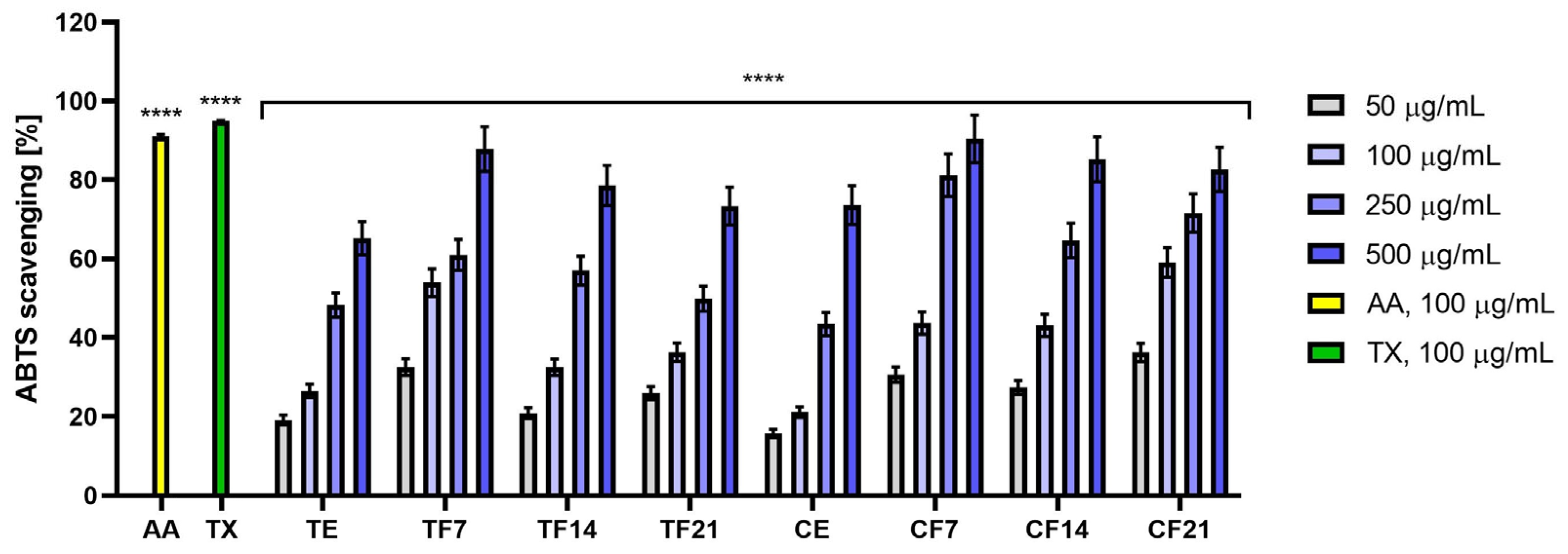
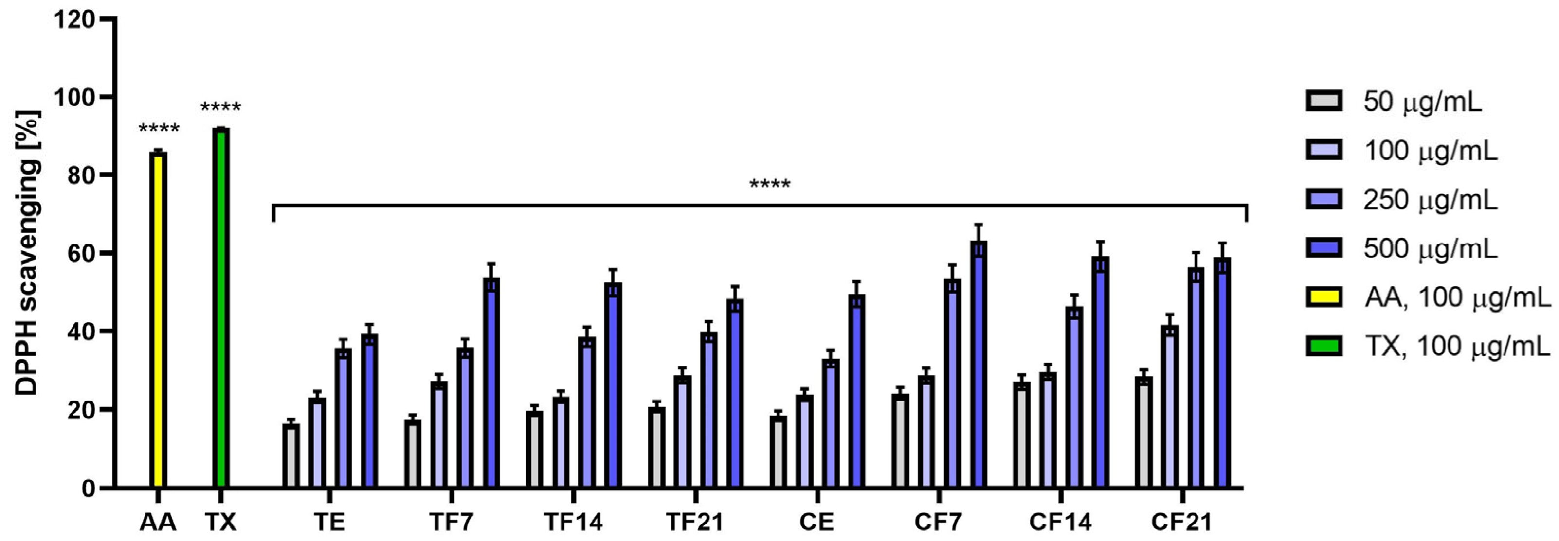
The ABTS and DPPH tests were used to evaluate the antioxidant properties of extracts and ferments obtained from flowers of Tilia L. and Calluna vulgaris L. Both tests are based on the ability to scavenge the free radical ABTS or DPPH [34].
The analysis of the antioxidant properties of extracts and ferments obtained from Tilia L. and Calluna vulgaris L. flowers covered a concentration range from 50 µg/mL to 500 µg/mL. The results were expressed as percentage free radical scavenging capacity (Figure 1 and Figure 2). The obtained results clearly demonstrated that both Tilia L. and Calluna vulgaris L. preparations exhibited antioxidant properties. In both cases, fermentation significantly enhanced the antioxidant activity of the tested raw materials. A relationship was also observed between concentration and free radical scavenging capacity—the higher the concentration, the greater the ability of the tested samples to neutralize free radicals. F7 proved to be the best among the ferments, obtaining 87.77 ± 5.66 for Tilia L. and 90.42 ± 6.02 for Calluna vulgaris L. at a concentration of 500 µg/mL in the ABTS test, and 53.81 ± 3.49 for Tilia L. and 63.24 ± 4.13 for Calluna vulgaris L. at a concentration of 500 µg/mL in the DPPH test, respectively. In both the ABTS test (Figure 1) and the DPPH test (Figure 2), Calluna vulgaris L. demonstrated the strongest antioxidant properties for both the extract (CE) and the ferments (CF7, CF14, and CF21), compared to analogous preparations obtained from Tilia L. flowers.
To further quantify and compare the antioxidant efficiency of the studied matrices, the IC50 values (the concentration required to scavenge 50% of the free radicals) were calculated for both the ABTS and DPPH assays (Table 3). In perfect agreement with the percentage scavenging data, the IC50 analysis mathematically confirmed the superior performance of the fermented samples over the raw extracts. Specifically, the lowest IC50 —indicating the highest antioxidant potency—recorded for the 7-day ferments (F7) of both plant species. Furthermore, comparing the two botanical sources, Calluna vulgaris L. preparations consistently exhibited lower IC50 values than the corresponding Tilia L. samples across all tested models. This lower concentration threshold required to achieve 50% radical neutralization highlights the exceptionally dense concentration of highly bioaccessible antioxidant phenotypes in the heather matrices, particularly after undergoing the initial stages of SCOBY biotransformation.

3.2.2. Intracellular ROS Levels in Skin Cells

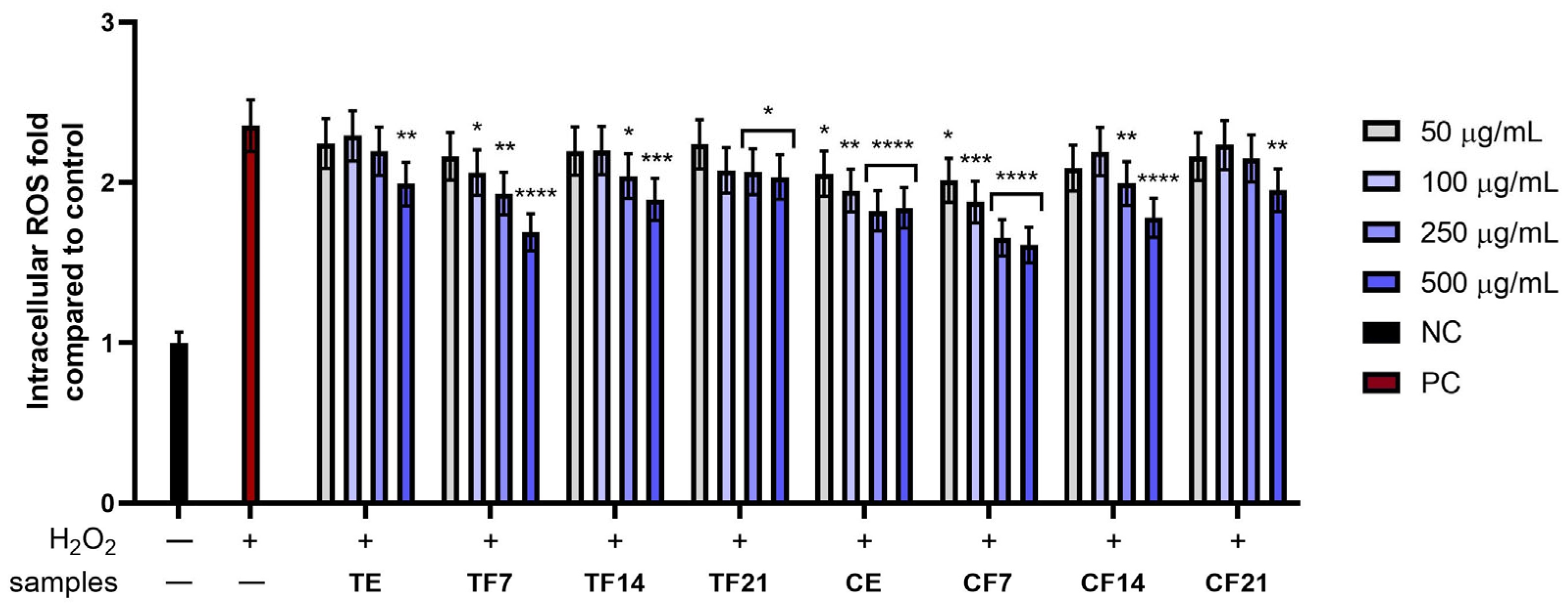
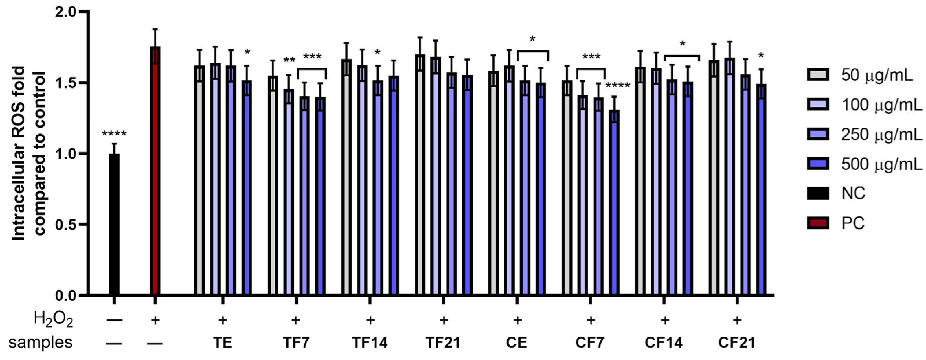
To determine the ability of extracts (E) and ferments (F7, F14 and F21) obtained from Tilia L. and Calluna vulgaris L. flowers to reduce intracellular levels of reactive oxygen species (ROS), analyses were performed using a fluorescent probe (H2DCFDA). In the presence of reactive oxygen species, H2DCFDA was oxidized and converted to DCF (2′,7′-dichlorofluorescein) [35]. The analyses were performed on two skin cell lines: fibroblasts (HDFs) and keratinocytes (HaCaTs). Hydrogen peroxide (H2O2) was used as a factor inducing oxidative stress in cells, leading to increased production of ROS. Results were expressed as a fold increase relative to the negative control (NC), which consisted of cells untreated with H2O2 and untreated with the extracts or ferments tested.
The obtained results clearly demonstrated that both Tilia L. and Calluna vulgaris L. preparations had the ability to reduce the level of ROS in skin cells. Treatment of skin cells solely with hydrogen peroxide (PC) resulted in a significant increase in ROS levels, reaching values of 2.35 ± 0.16-fold for HDF cells and 1.76 ± 0.12-fold for HaCaT cells compared to NC. Incubation of cells with the tested extracts and ferments obtained from Tilia L. flowers and Calluna vulgaris L. demonstrated a reduction in ROS levels relative to the positive control (PC) in both skin cell lines. In the case of both HDF and HaCaT cells, the Calluna vulgaris L. extracts and ferments demonstrated a greater ability to reduce ROS levels than the analogous Tilia L. preparations. Furthermore, a dose-dependency effect was demonstrated—increasing the concentration of the tested samples resulted in a stronger reduction in intracellular ROS levels. The strongest effect was observed for F7: in the case of HDF cells, reaching 1.69 ± 0.13-fold for Tilia L. and 1.61 ± 0.11-fold for Calluna vulgaris L., respectively, at a concentration of 500 µg/mL against PC, in the case of HaCaT cells, reaching 1.39 ± 0.1-fold for Tilia L. and 1.31 ± 0.09-fold for Calluna vulgaris L., respectively, at a concentration of 500 µg/mL against PC (Figure 3 and Figure 4).

3.3. Cytotoxicity Assessment

In this study, the Alamar Blue assay enabled the assessment of the metabolic activity of HaCaT keratinocytes and HDFs after exposure to the tested ferments. This method relies on the ability of living cells to reduce resazurin to resorufin; therefore, a decreased signal may indicate cytotoxic activity, disruption of mitochondrial function, or inhibition of proliferation. Maintained or increased metabolic activity, on the other hand, may indicate a beneficial effect of the tested material on cell function [36,37].
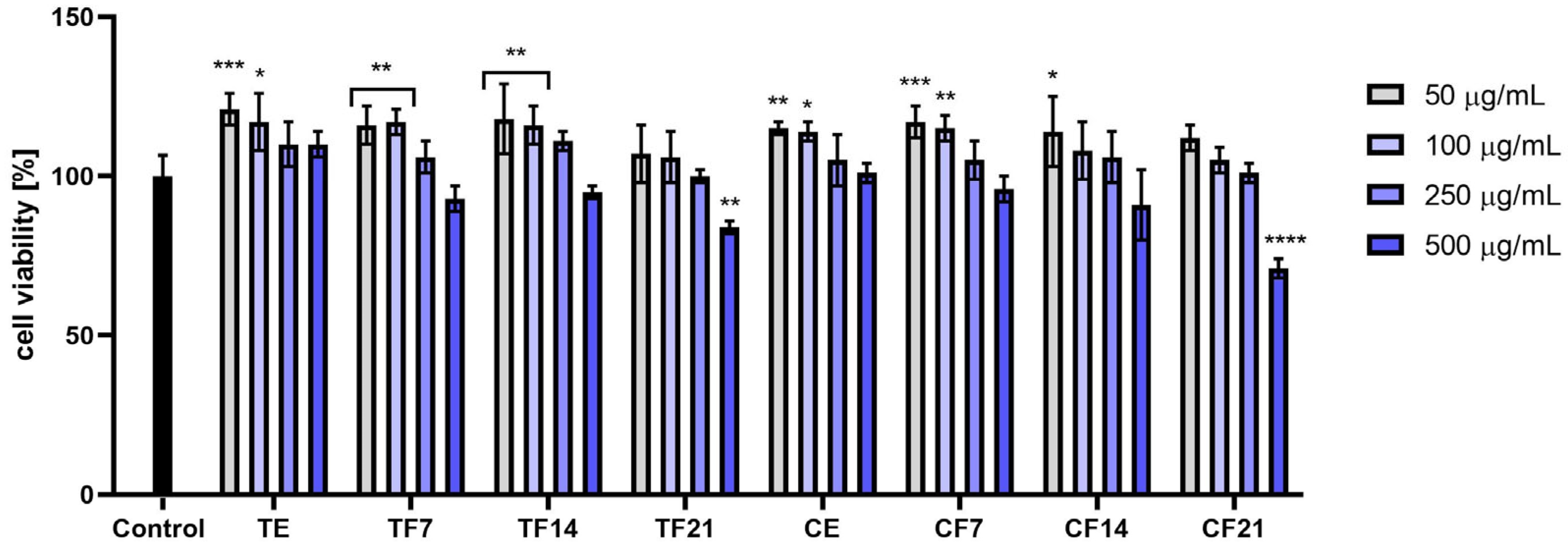
The Alamar Blue assay on HDF cells demonstrated that cell viability decreased with increasing concentration. The highest observed values were denoted for the lowest concentrations of 50 and 100 µg/mL, respectively, for each tested sample. The highest overall values were denoted for Tilia L. extract and Calluna vulgaris L. in the lowest concentration, remaining significantly above the control level. Only slightly lower values, but still remaining significantly above the control, were denoted for Tilia L. extract in a concentration of 100 µg/mL as well as Tilia L. 7-day and 14-day ferments at concentrations of 50 and 100 µg/mL. Similar results of cell viability level, also significantly higher than the control, were denoted for Calluna vulgaris L. extract at concentrations of 50 and 100 µg/mL, 7-day ferment at a concentration of 100 µg/mL and 14-day ferment at a concentration of 50 µg/mL. The worst results were denoted for 21-day ferments at the highest concentration of 500 µg/mL, remaining significantly below the control for both Tilia L. and Calluna vulgaris L. Analysis of influence of fermentation shows that at lower concentrations, cell viability remains at a similar level for all extracts, both 7-day and 14-day ferments. A very slight advantage of extract from Tilia L. over all the extracts was observed. In the case of Calluna vulgaris L., only a slight advantage of the 7-day ferment over the other cases was observed. For both Tilia L. and Calluna vulgaris L., the 21-day ferment resulted in noticeably lower values of cell viability compared to other ferments and extracts (Figure 5).
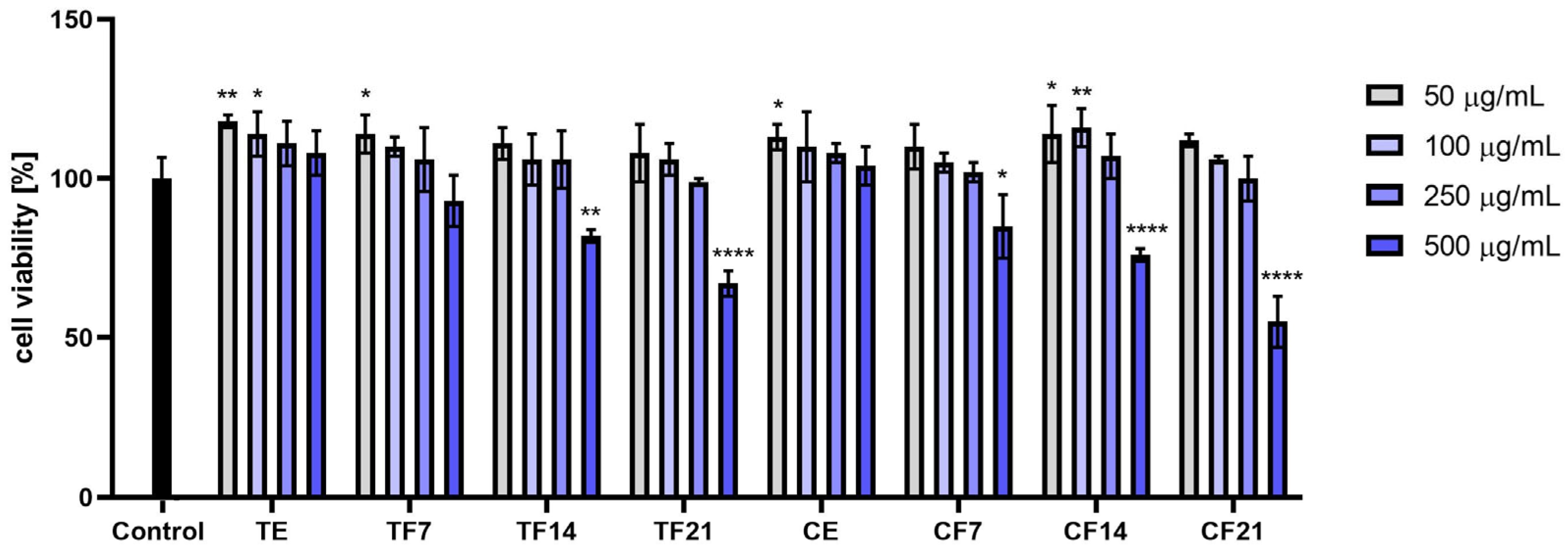
Alamar Blue assay performed using HaCaT cells showed that cell viability decreased with increasing concentration. The highest observed values were denoted for the lowest concentrations of 50 and 100 µg/mL, respectively, for each tested sample. The highest overall values were denoted for Tilia L. extract at the lowest concentration and Calluna vulgaris L. 14-day ferment at the concentration of 100 µg/mL, remaining significantly above the control level. Only slightly lower values, but still remaining significantly above the control, were denoted for Tilia L. extract at a concentration of 100 µg/mL, Tilia L. 7-day ferment at a concentration of 50 µg/mL, and Calluna vulgaris L. 14-day ferment at a concentration of 50 µg/mL. The worst results were denoted for 14- and 21-day ferments at the highest concentration of 500 µg/mL, all of which remained significantly below the control for both Tilia L. and Calluna vulgaris L. The analysis of the influence of fermentation showed that for Tilia L., cell viability values decrease slightly with increasing fermentation time at lower concentrations, and decrease significantly at the highest concentration. In the case of Calluna vulgaris L., the 14-day ferment appeared to demonstrate the most favorable activity, although this effect was observed only at the two lowest tested concentrations. The behavior at the highest concentration was similar to the one observed for Tilia L., meaning that increased fermentation time causes a significant drop in cell viability (Figure 6).
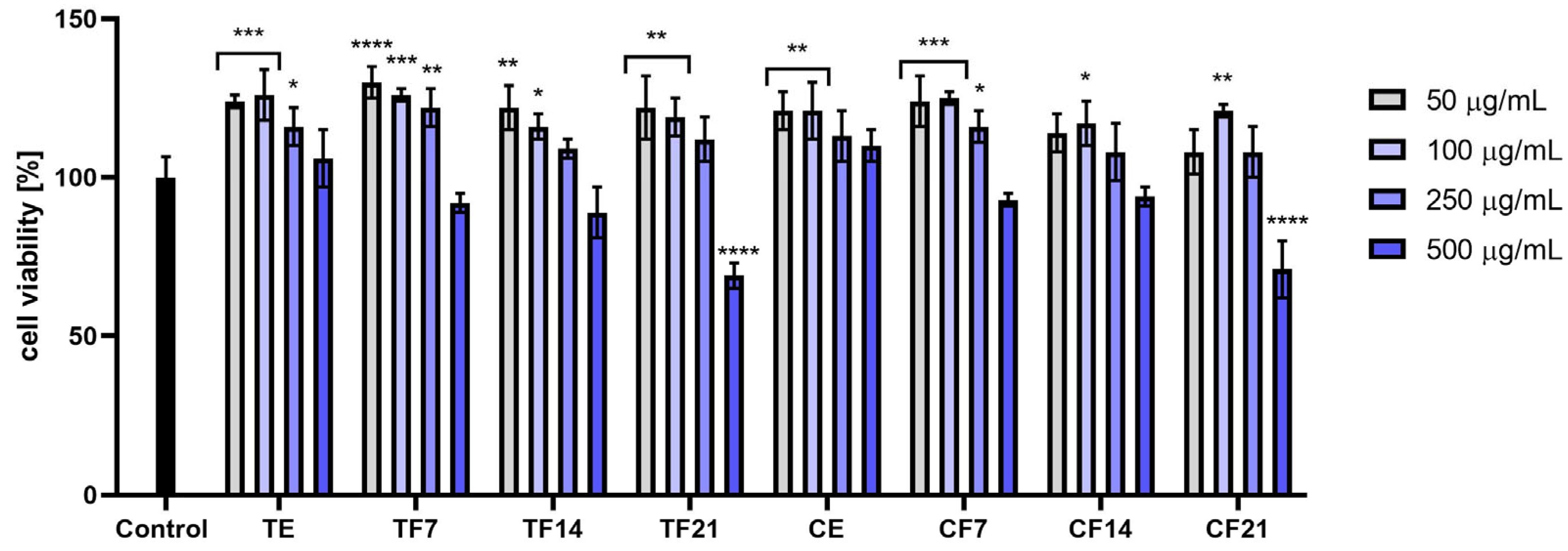
For the Alamar Blue assay performed on THP-1 cells, a trend was observed in which the two lower concentrations generally resulted in the highest cell viability, whereas a marked decrease in viability was observed at the two higher concentrations. The highest overall values were denoted for Tilia L. 7-day ferment at the lowest concentration. Only slightly lower values, but still remaining significantly above the control, were denoted for Tilia L. extract at concentrations of 50, 100 and 250 µg/mL; for the 7-day ferments at concentrations of 100 and 250 µg/mL, and for the 14-day ferments. The worst results were obtained for 21-day ferments of both Tilia L. and Calluna vulgaris L. at the highest concentration of 500 µg/mL, both remaining significantly below the control. Analysis of influence of fermentation shows that, for both Tilia L. and Calluna vulgaris L., lower concentrations of cell viability were only slightly higher for the extract and 7-day ferment compared to 14- and 21-day ferments, while for higher concentrations, cell viability markedly decreased when fermentation time was extended (Figure 7).

3.4. Assessment of Anti-Inflammatory Activity

To assess the anti-inflammatory potential of the tested extracts and ferments from Tilia L. and Calluna vulgaris L. flowers, the levels of key inflammatory mediators in THP-1 cells- proinflammatory cytokines (interleukin 6 (IL-6), tumor necrosis factor α (TNF-α)) and cyclooxygenase-2 (COX-2) enzyme—were measured using enzyme-linked immunosorbent assay (ELISA). Inflammation was induced by treating the cells with 20 µg/mL of LPS. As a reference standard for anti-inflammatory activity, diclofenac was applied at a concentration of 50 µg/mL [38]. The data regarding cytokine secretion are presented as fold-increase values over the negative control (NC = 1; cells untreated with compounds and LPS).
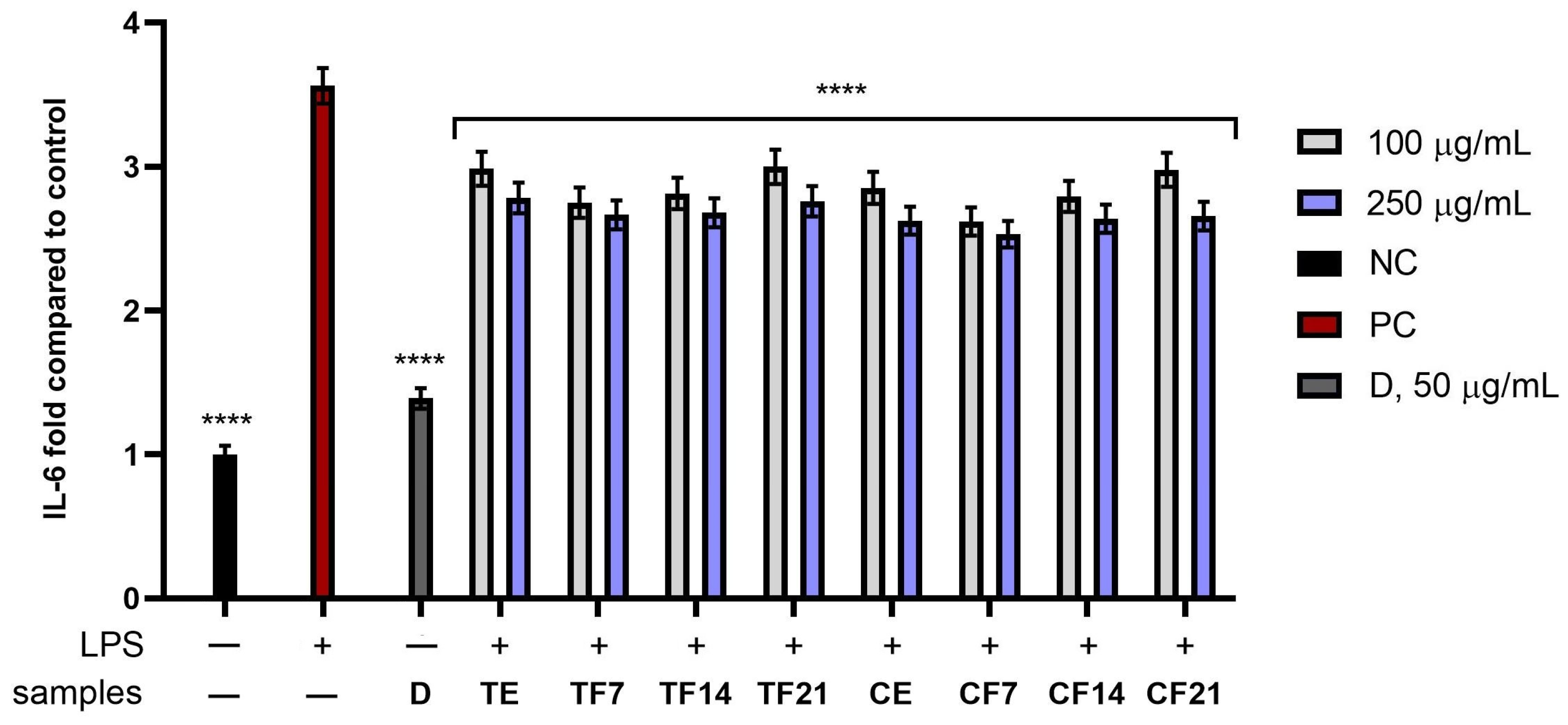
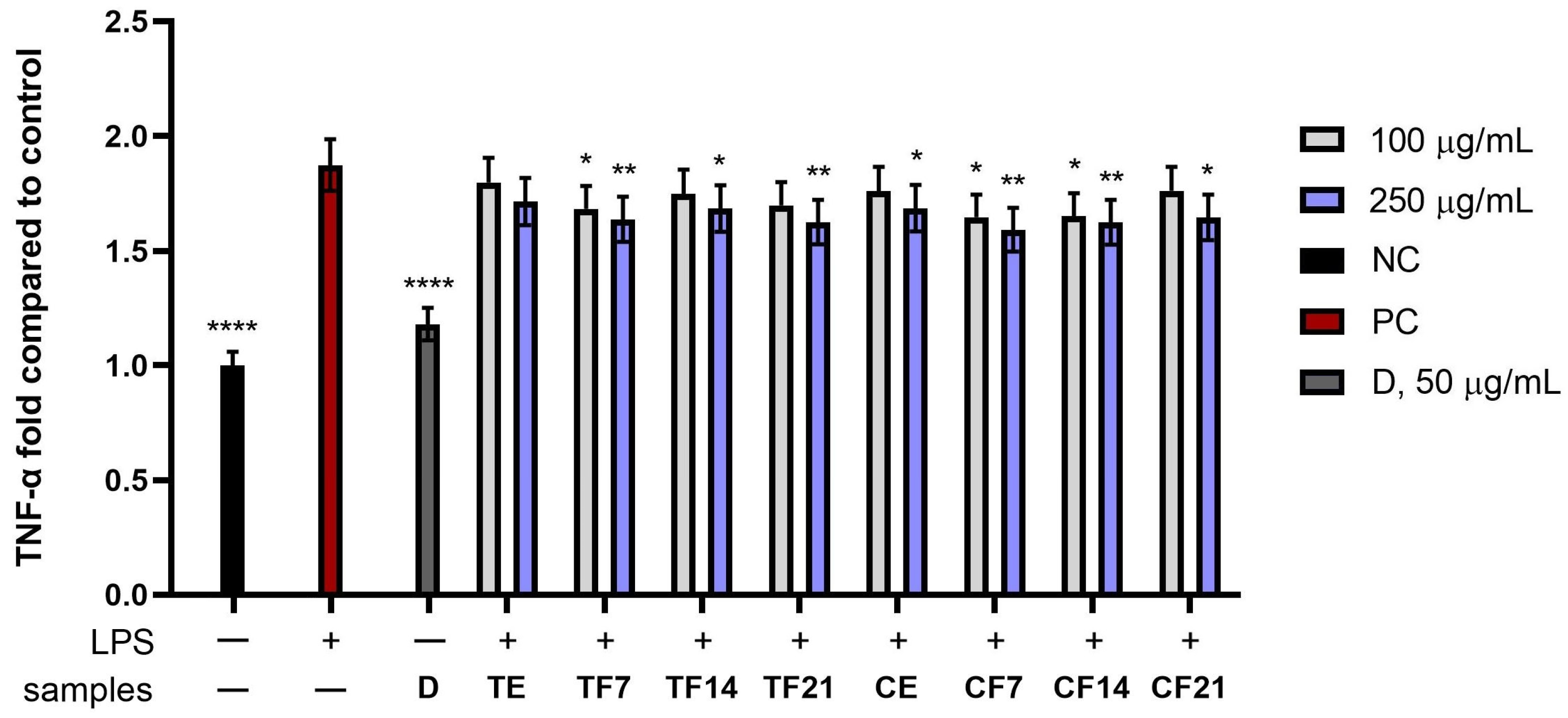
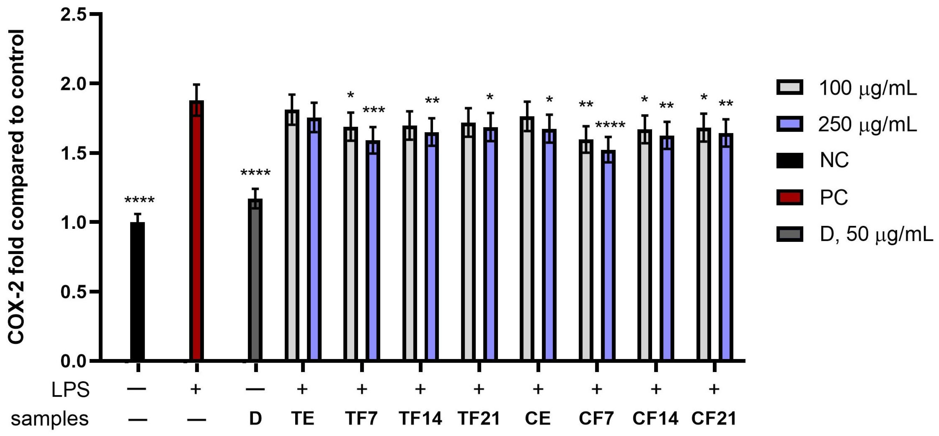
As shown in Figure 8, Figure 9 and Figure 10, both extracts and ferments obtained from Tilia L. flowers and Calluna vulgaris L. demonstrated the ability to inhibit inflammatory markers at both tested concentrations (100 and 250 μg/mL): IL-6, TNF-α, and COX-2. Stimulation of cells with LPS (PC; positive control) induced an inflammatory response, with an approximately 3.5-fold increase in IL-6 and approximately 2-fold increase in TNF-α and COX-2 compared to the negative control (NC). Significantly better effects were observed for the ferments than for their corresponding extracts, regardless of the plant material.
For IL-6 (Figure 8), all extracts and ferments from Tilia L. and Calluna vulgaris L. reduced the LPS-stimulated response compared to the positive control (PC; cells not treated with the test compounds but stimulated with LPS), with Calluna vulgaris L. demonstrating higher efficacy. Among the Calluna vulgaris L. preparations, E exhibited 2.62 ± 0.1-fold NC, while the fermented extracts F7, F14, and F21 achieved 2.53 ± 0.11-fold, 2.63 ± 0.12-fold, and 2.65 ± 0.09-fold NC at a concentration of 250 µg/mL, respectively. Higher concentrations were also observed to better inhibit the inflammatory response.
Figure 9 shows the effect of the tested samples on reducing TNF-α levels. The results indicate that both plants (Tilia L. and Calluna vulgaris L.) have the ability to inhibit TNF-α, with better effects observed for Calluna vulgaris L. The strongest inhibitory effect was observed for F7, both for Tilia L. and Calluna vulgaris L., achieving 1.64 ± 0.09-fold NC for Tilia L. and 1.59 ± 0.11-fold NC Calluna vulgaris L. at a concentration of 250 µg/mL, respectively.
Similar results were also observed in the COX-2 enzyme inhibition study (Figure 10). In this case, Calluna vulgaris L. again proved to be the raw material with stronger anti-inflammatory potential. F7 at the highest tested concentration (250 µg/mL) achieved a level of 1.52 ± 0,12-fold compared to the control, while the corresponding Tilia L. ferment achieved a result of 1.59 ± 0.13-fold.
Comparing all the obtained results, it can be observed that Calluna vulgaris L. exhibits stronger anti-inflammatory potential than Tilia L. across all analyzed parameters. With respect to both cytokines (IL-6, TNF-α) and the COX-2 enzyme, Calluna vulgaris L. preparations demonstrated a stronger inhibitory potential for proinflammatory markers.

3.5. Assessment of Antibacterial Activity

The antibacterial activity of aqueous extracts and kombucha ferments obtained from Tilia L. and Calluna vulgaris L. flowers depended on both the tested bacterial strain and the fermentation time, as demonstrated by MIC values (Table 4) and inhibition zone diameters (Table 5). In general, Gram-positive bacteria were more susceptible to the analyzed samples than Gram-negative strains, although Escherichia coli showed relatively high sensitivity to several preparations, particularly those obtained from Tilia L. flowers.
Among all tested microorganisms, Escherichia coli was the most sensitive to preparations obtained from Tilia L. flowers. The lowest MIC value was noted for the 7-day ferment (F7; 110 µg/mL), suggesting that short-term fermentation enhanced the antibacterial potential of the extract. With longer fermentation times, the activity gradually decreased, which was reflected by higher MIC values for F14 and F21 samples. A similar trend was observed for Calluna vulgaris L., although the activity against E. coli was lower compared to Tilia L. preparations. The agar diffusion assay confirmed these observations. Among all tested samples, the largest inhibition zone was recorded for the 7-day ferment of Tilia L. (21 mm), followed by the 14-day ferment (15 mm) and the aqueous extract (17 mm). Preparations obtained from Calluna vulgaris L. produced smaller inhibition zones ranging from 6 to 9 mm, indicating weaker antibacterial activity against E. coli. The consistency between MIC values and inhibition zone diameters suggests that short-term fermentation enhanced the antibacterial potential of Tilia L. extracts against this strain.
In the case of Staphylococcus aureus, the strongest effect was observed for the aqueous extract of Tilia L. flowers (335 µg/mL). Fermentation reduced antibacterial activity, particularly after 14 and 21 days. For Calluna vulgaris L., both the extract and the 7-day ferment showed relatively good antibacterial properties, whereas prolonged fermentation weakened the inhibitory effect. A similar tendency was observed in the agar diffusion assay. The largest inhibition zone was obtained for the 7-day ferment of Tilia L. (16 mm), whereas prolonged fermentation resulted in a reduction in activity, with inhibition zones decreasing to 8 mm and 3 mm for F14 and F21, respectively. Preparations derived from Calluna vulgaris L. exhibited weaker antibacterial activity, with inhibition zones ranging from 4 to 11 mm.
Specific differences were observed for Staphylococcus capitis. The aqueous extract of Tilia L. did not exhibit detectable antibacterial activity in the MIC assay, whereas all fermented samples showed inhibitory effects, with MIC values decreasing from 9452 µg/mL for F21 to 7400 µg/mL for F7. In contrast, no inhibition zones were observed for either the extract or fermented samples in the agar diffusion assay. This discrepancy may be related to the limited diffusion of antibacterial constituents present in Tilia L. ferments through the agar medium, despite their measurable activity in liquid culture conditions. Preparations obtained from Calluna vulgaris L. exhibited antibacterial activity both before and after fermentation. The strongest effect was observed for the 7-day ferment, which showed the lowest MIC value (756 µg/mL) and produced the largest inhibition zone (8 mm). Prolonged fermentation reduced antibacterial activity, as reflected by increased MIC values and smaller inhibition zones. The results suggest that short-term kombucha fermentation may enhance the activity of Calluna vulgaris L. flower preparations against S. capitis, whereas extended fermentation diminishes this effect.
For Micrococcus luteus, the lowest MIC value was recorded for the 7-day ferment prepared from Calluna vulgaris L. flowers (1155 µg/mL). This may suggest that fermentation promoted the formation or release of compounds active against Gram-positive bacteria. At the same time, extended fermentation generally resulted in lower antibacterial activity in both analyzed plant materials. The inhibition zone assay showed comparable results. For both plant species, the largest inhibition zones were observed for the 7-day ferments (6 mm for Tilia L. and 8 mm for Calluna vulgaris L.). No inhibition zone was detected for the 21-day ferment of Calluna vulgaris L., indicating a loss of antibacterial activity after prolonged fermentation.
The weakest antibacterial effect was observed against Pseudomonas aeruginosa, which is consistent with the naturally high resistance of Gram-negative bacteria. Nevertheless, the 7-day ferment obtained from Calluna vulgaris L. showed improved activity compared to the non-fermented extract. In the case of Tilia L., prolonged fermentation resulted in a complete loss of detectable antibacterial activity against this strain. The inhibition zone assay supported the findings obtained from MIC determination. No inhibition zones were observed for Tilia L. extracts and ferments, whereas Calluna vulgaris L. samples produced relatively small inhibition zones ranging from 4 to 10 mm, with the strongest effect observed for the 7-day ferment. These findings confirm the high resistance of P. aeruginosa and indicate only limited susceptibility to the tested preparations.
Overall, both MIC determination and agar diffusion assays demonstrated that kombucha fermentation influenced the antibacterial properties of the investigated extracts in a time-dependent manner. In most cases, the strongest antibacterial activity was observed after 7 days of fermentation, whereas prolonged fermentation generally reduced the inhibitory effect. The agreement between MIC values and inhibition zone diameters indicates that short-term fermentation may enhance the formation or bioavailability of compounds responsible for antibacterial activity.

4. Discussion

The search for natural bioactive substances with antioxidant and anti-inflammatory properties is currently a key development direction for the cosmetics and pharmaceutical industries. This study has shown that both extracts and ferments from Tilia L. and Calluna vulgaris L. flowers are a rich source of phytochemicals that effectively protect skin cells from oxidative stress induced by external factors, while also demonstrating anti-inflammatory and antibacterial potential.
Fermentation using a symbiotic culture of bacteria and yeast (SCOBY) is a biological process that leads to intensive changes in the chemical composition of plant materials. During fermentation, yeast and acetic acid bacteria metabolize sugars and modify the structure of polyphenols, flavonoids, and other secondary metabolites, leading to the formation of compounds with lower molecular weight and higher bioavailability. Additionally, this process can increase the content of organic acids, vitamins, and compounds with antioxidant properties, which increases the health-promoting potential of fermented products [39]. Chromatographic analyses and biological assays, which included antioxidant analyses, assessment of cytotoxicity towards skin cells (HDF and HaCaT) and studies of anti-inflammatory and antibacterial activity, showed that fermentation changes the profile of active compounds and enhances the biological activity of Tilia L. and Calluna vulgaris L. extracts [40,41].
The elevated antioxidant activity observed in ABTS and DPPH assays for both Tilia L. and Calluna vulgaris L. ferments compared to their raw counterparts highlights the profound chemical remodeling driven by the SCOBY consortium. This enhancement of radical scavenging capacity via the ABTS and DPPH pathways is fundamentally anchored in the qualitative changes documented in our chromatographic profiling (Table 1 and Table 2). The chromatographic analysis showed that the main component in Calluna vulgaris L. extracts and ferments was chlorogenic acid, which has proven antioxidant activity [42]. Similar observations were made by Kaunaite et al., who indicated that chlorogenic acid was the main phenolic component of Calluna vulgaris L. extracts [20]. Tilia L. extracts, in turn, are described as a rich source of gallic acid, catechin, and kaempferol, compounds with strong antioxidant activity [43]. The obtained results suggest that the fermentation process not only modifies the phytochemical profile of raw materials, but also significantly increases their functionality as effective free radical scavengers.
An interesting observation was that the strongest antioxidant activity was generally recorded for the 7-day ferments, whereas prolonged fermentation resulted in a gradual decline in activity. This phenomenon may be associated with the dynamic nature of kombucha fermentation. During the early stages of fermentation, microbial enzymes can promote the release of phenolic compounds from bound forms and facilitate the formation of low-molecular-weight metabolites with enhanced antioxidant potential. As a result, the antioxidant activity of the ferments may exceed that of the corresponding unfermented extracts [44,45]. However, extended fermentation may lead to further biotransformation or degradation of some antioxidant constituents. In addition, certain phenolic compounds may be utilized by microorganisms during prolonged fermentation, resulting in changes in the composition and antioxidant capacity of the final product. Therefore, the highest antioxidant activity observed after 7 days of fermentation likely reflects a balance between the formation of highly active metabolites and their subsequent transformation during later stages of the fermentation process [7,46]. The next stage of the research was to evaluate the ability of Tilia L. and Calluna vulgaris L. extracts and ferments to inhibit intracellular levels of reactive oxygen species (ROS) exposed to hydrogen peroxide (H2O2). Excessive ROS production in skin cells, induced by external factors (such as UV radiation or environmental pollution), leads to oxidative stress, which is a major cause of premature skin aging and damage to DNA and structural proteins [13,47]. These studies were conducted on two cell lines: fibroblasts (HDFs) and keratinocytes (HaCaT). The use of both cell lines allowed for a comprehensive assessment of the antioxidant properties of the ferments in different skin layers. The results of the analyses showed that the tested extracts and ferments effectively reduced ROS levels in both cell types, confirming their potential to protect the entire skin tissue profile from the negative effects of oxidative stress. It was observed that the ferments had a stronger effect than the analogous extracts, with Calluna vulgaris L. ferments demonstrating a higher capacity for ROS reduction than those obtained from Tilia L., which results from the different content of biologically active compounds in the tested samples. Chromatographic analysis of both plants revealed the presence of chlorogenic acid, gallic acid, catechin, and quercetin in various concentrations (Table 1 and Table 2). These constituents act as potent antioxidants, neutralizing reactive free radicals via electron transfer or hydrogen atom donation mechanisms [48]. Chlorogenic acid has been proven to inhibit lipid peroxidation [49]. Gallic acid works by removing harmful superoxide anions and improves the body’s antioxidant barrier, restoring the proper functioning of key protective enzymes, such as superoxide dismutase and catalase [50]. The results suggest that the fermentation process may increase the content of active compounds by transforming extended phenolic structures into smaller molecules, which directly translates into a higher antioxidant potential of the final product [51].
A key aspect of the evaluation of innovative cosmetic raw materials, apart from their biological activity, is determining their safety profile towards skin cells. The biological activity of the plant material should be considered in conjunction with its effect on the viability and metabolic activity of skin cells. According to the available literature, studies directly examining kombucha ferments obtained from Tilia L. or Calluna vulgaris L. and their cytotoxicity towards keratinocytes and fibroblasts have not been reported so far. However, there are studies on other fermented plant extracts (kombucha) in which cytotoxicity was assessed using the Alamar Blue assay. In studies on kombucha-fermented extracts from berry leaves, the authors emphasized that fermentation can improve the biological properties of plant extracts while maintaining their good tolerance to skin cells [52]. The results obtained for Tilia L. and Calluna vulgaris L. ferments can therefore be interpreted as filling an existing research gap. The lack of significant reduction in keratinocyte and fibroblast viability in the Alamar Blue assay indicates that the tested ferments do not exhibit cytotoxic effects on basic skin cell models. The observed dose-dependent decrease in cell viability at higher concentrations of both raw extracts and kombucha ferments aligns with established toxicological principles of in vitro screening. This downward trend is typically governed by a combination of altered microenvironmental osmolarity and the well-documented pro-oxidant paradox of highly concentrated polyphenols, which can induce intracellular stress rather than mitigate it [53,54]. Crucially, however, the fact that cell viability remained well within acceptable limits (or showed no significant cytotoxicity) at lower and medium concentrations (up to 250 µg/mL) demonstrates a wide and safe therapeutic window for cosmetic and topical applications. At selected concentrations, an increase in metabolic activity was observed, suggesting a potential effect supporting cellular metabolism. The potential biocompatibility of the tested ferments may result from the phytochemical composition of Tilia L. and Calluna vulgaris L. Tilia spp. flower extracts contain numerous phenolic compounds, including flavonoids and phenolic acids, which may contribute to protecting cells from oxidative stress [55,56,57]. Calluna vulgaris L., in turn, is rich in phenolic acids, flavonols, flavan-3-ols, and proanthocyanidins, which may be important for supporting the viability and protection of skin cells [20,24].
Furthermore, kombucha fermentation may influence cytotoxicity and biological activity through the hydrolysis of phenolic glycosides, the formation of organic acids, and the conversion of metabolites to more bioavailable forms. As a result, it may alter not only the total content of active compounds but also their bioavailability and interaction with cells [39]. Therefore, the good cellular tolerance after exposure to Tilia L. and Calluna vulgaris L. ferments may indicate that the fermentation process did not generate metabolites toxic to skin cells and, at the same time, may have promoted the formation of fractions with cytoprotective potential. These results are particularly important because they suggest that kombucha-fermented Tilia L. and Calluna vulgaris L. extracts could be an innovative ingredient in cosmetic products.
This study also assessed the anti-inflammatory potential of extracts and ferments obtained from Tilia L. and Calluna vulgaris L. (Calluna vulgaris L.) flowers using the THP-1 cell model. The THP-1 cell line is a commonly used human monocyte model in inflammation research, as it allows for the analysis of the immune response and expression of proinflammatory mediators in vitro [58]. To induce an immune response, cells were stimulated with lipopolysaccharide (LPS), a potent activator of proinflammatory pathways. This allowed for an examination of the extent to which the tested samples were able to inhibit induced inflammation by reducing the secretion of key mediators such as IL-6, TNF-α, and COX-2. The mechanism underlying the modulation of these proinflammatory markers is directly linked to the presence of numerous bioactive compounds characteristic of Tilia L. and Calluna vulgaris L. flowers. The literature particularly emphasizes the role of polyphenols, flavonoids, and phenolic acids, which have the ability to neutralize reactive oxygen species and modulate signaling pathways associated with the inflammatory response [59]. Tilia L. flowers are rich in, among others, quercetin, kaempferol, and chlorogenic acid derivatives [14,60], while Calluna vulgaris L. contains significant amounts of arbutin, flavonoids, and tannins [24,61]. These compounds may inhibit the activation of the transcription factor NF-κB, which plays a key role in regulating the expression of proinflammatory cytokines such as TNF-α, IL-1β, and IL-6. Hwang et al. demonstrated that chlorogenic acid exhibits anti-inflammatory effects in RAW 264.7 cells stimulated with LPS [62]. Furthermore, Shan et al. proved that chlorogenic acid reduces the expression of the COX-2 enzyme by blocking key signaling pathways, such as NF-κB and JNK/AP-1, induced by LPS [63]. Furthermore, other researchers pointed to the anti-inflammatory properties of the plants tested in this study. Yükel et al. showed that Tilia L. extracts effectively reduced inflammation by preventing the production of nitric oxide (NO) in a dose-dependent manner and demonstrated the ability to inhibit the secretion of interleukin 6 (IL-6) and prostaglandin E2 (PGE2) in cells stimulated by the inflammatory factor [64]. Villanueva-Bermejo et al. showed that Calluna vulgaris L. extracts had the ability to reduce the level of IL-1β and IL-6 in LPS-treated THP-1/M cells [65]. The obtained results are consistent with literature reports indicating that fermentation of plant materials can enhance their biological properties [7,10,66]. Similar observations were described for fermented extracts of radish, dogwood, and aloe, which demonstrated increased antioxidant activity and the ability to reduce proinflammatory cytokine levels [6,10,11,12,13]. This indicates that the fermentation process may be an effective method for increasing the biological potential of plant materials.
The evaluation of antibacterial activity is particularly important in the context of plant-derived raw materials intended for potential cosmetic and pharmaceutical applications. Skin exposure to pathogenic and opportunistic microorganisms may contribute to irritation, inflammation, impaired skin barrier function, or the development of infections [67]. For this reason, increasing attention has recently been paid to natural compounds capable of limiting the growth of microorganisms while simultaneously exhibiting antioxidant and anti-inflammatory properties. Plant extracts and fermented botanical preparations are considered especially promising due to the presence of multiple bioactive metabolites that may act synergistically and provide multifunctional biological effects [68,69].
Compared with conventional synthetic antimicrobial agents, plant extracts and fermented botanical preparations offer several additional advantages that are particularly relevant in cosmetic applications [70]. Unlike many synthetic compounds that primarily target microbial growth, botanical preparations are rich in phenolic compounds, flavonoids, and organic acids that may simultaneously exert antimicrobial, antioxidant, and anti-inflammatory effects [71]. Such multifunctional activity is desirable in cosmetic products intended for sensitive, irritated, or inflammation-prone skin, where microbial imbalance is often accompanied by oxidative stress and inflammatory processes [72]. Moreover, increasing attention has recently been paid to plant-derived antimicrobial agents as sustainable and renewable sources of bioactive compounds.
A similar trend can also be observed in the field of antimicrobial nanomaterials. Recent studies have demonstrated that plant extracts are increasingly used in the green synthesis of metallic nanoparticles, serving as natural reducing and stabilizing agents due to their rich phytochemical composition and biological activity [73]. Although nanoparticle-based systems often exhibit strong antimicrobial efficacy, concerns regarding their long-term safety, potential cytotoxicity, environmental persistence, and accumulation remain under investigation [74]. In contrast, plant extracts and kombucha ferments represent naturally derived and biodegradable materials that may provide broad-spectrum biological activity without the need for complex technological processing. Therefore, fermented botanical preparations may constitute attractive multifunctional ingredients for cosmetic formulations, combining antimicrobial activity with antioxidant and anti-inflammatory properties.
The antibacterial activity observed for the aqueous extracts and kombucha ferments obtained from Tilia L. and Calluna vulgaris L. flowers may be associated with the presence of numerous phenolic compounds identified by LC-MS analysis (Table 1 and Table 2). Both plant materials were rich in phenolic acids and flavonoids, including chlorogenic acid, gallic acid derivatives, protocatechuic acid, catechin/epicatechin, taxifolin derivatives, quercetin glycosides, apigenin derivatives, and luteolin. Many of these compounds are known to exhibit antibacterial activity, particularly against Gram-positive bacteria [24,60].
In the present study, Gram-positive strains, especially Staphylococcus aureus, Staphylococcus capitis, and Micrococcus luteus, were generally more susceptible to the analyzed samples than Gram-negative bacteria. Similar observations have been reported previously for polyphenol-rich plant extracts, including extracts from Calluna vulgaris L. and Tilia L. flowers [75,76]. The lower susceptibility of Gram-negative bacteria is usually attributed to the presence of the outer membrane, which limits the penetration of phenolic compounds into bacterial cells [77]. For some samples, particularly Tilia L. ferments tested against S. capitis, antibacterial activity was detected in the MIC assay but not in the agar diffusion test. Such differences are commonly observed for complex plant extracts and may result from the limited diffusion of high-molecular-weight phenolic compounds in agar media.
The strongest antibacterial activity among the tested samples was observed for the 7-day kombucha ferment of Tilia L. flowers against Escherichia coli. Short-term fermentation may enhance the release of low-molecular-weight compounds with antibacterial potential or increase the bioavailability of phenolic constituents already present in the extract.
At the same time, prolonged fermentation reduced antibacterial activity in many cases, despite the relatively stable or even increased concentration of selected phenolic compounds. This suggests that antibacterial activity was not determined solely by the concentration of individual metabolites, but rather by the overall composition of the extracts and possible synergistic interactions between compounds. Kombucha fermentation is a complex biotransformation process that leads not only to changes in polyphenol composition but also to the formation of organic acids, low-molecular-weight metabolites, and other microbial products that were not included in the present LC-MS analysis [26]. Moreover, antibacterial activity is likely influenced by synergistic and antagonistic interactions among multiple compounds present in the fermentation matrix [78]. Therefore, relatively small changes in the overall composition of the ferments may result in substantial differences in their biological activity, even when the concentrations of major identified phenolic compounds remain largely unchanged. Interestingly, in several cases the strongest antibacterial activity was observed for the 7-day ferments, although the concentrations of selected phenolic compounds remained stable or continued to increase during prolonged fermentation. This observation suggests that the antibacterial activity of the investigated samples may depend not only on the accumulation of bioactive compounds but also on the relative abundance of individual constituents and their potential synergistic or antagonistic interactions [78]. It is possible that prolonged fermentation alters the balance between compounds contributing positively to antibacterial activity and those exhibiting neutral or antagonistic effects. Consequently, increased concentrations of individual metabolites do not necessarily translate into enhanced antibacterial efficacy. LC-MS analysis showed that fermentation of Tilia L. extracts increased the content of several compounds previously associated with antibacterial activity, including chlorogenic acid, catechin/epicatechin, procyanidin B, taxifolin hexosides, quercetin galactoside, and kaempferol hexosides [79,80,81,82,83]. Chlorogenic acid has been reported to disrupt bacterial membrane integrity and interfere with microbial metabolism, while catechins may increase membrane permeability and induce oxidative stress in bacterial cells [79,80]. Similar antibacterial mechanisms have also been described for quercetin and kaempferol derivatives. These flavonoids may disrupt bacterial cell membrane integrity, alter membrane permeability, and interfere with energy metabolism and nucleic acid synthesis [84]. Quercetin has additionally been reported to inhibit bacterial DNA gyrase and reduce biofilm formation, while kaempferol derivatives may affect membrane-associated proteins and bacterial enzymatic systems [85]. Their antibacterial activity is particularly pronounced against Gram-positive bacteria, which are generally more susceptible to phenolic compounds due to the absence of an outer membrane barrier [86,87].
Interestingly, the 7-day ferment of Calluna vulgaris L. showed improved activity against Micrococcus luteus and Staphylococcus capitis compared to the non-fermented extract. This may indicate that kombucha fermentation promoted partial biotransformation of phenolic compounds into more biologically active forms. Similar effects have previously been described for kombucha-fermented plant materials, where microbial metabolism enhanced antibacterial activity through the formation of smaller phenolic metabolites and organic acids [88,89].
The relatively weak activity observed against Pseudomonas aeruginosa is consistent with the literature data describing the high intrinsic resistance of this bacterium to plant-derived antimicrobial compounds. P. aeruginosa possesses multiple resistance mechanisms, including low membrane permeability and efficient efflux systems, which considerably reduce susceptibility to phenolic metabolites [90,91].
Previous studies concerning Tilia L. and Calluna vulgaris L. flowers focused mainly on antioxidant and anti-inflammatory properties, while reports describing their activity against Staphylococcus capitis and Micrococcus luteus remain very limited. Therefore, the present results expand the current knowledge regarding the antibacterial potential of aqueous extracts and kombucha ferments obtained from melliferous plant flowers.
Overall, the obtained results indicate that kombucha fermentation may be beneficial for enhancing the biological properties of the investigated flower extracts. However, the effect strongly depended on the duration of the fermentation process. Among the tested samples, the 7-day ferments generally exhibited the most favorable profile, characterized by enhanced antioxidant and anti-inflammatory activity together with improved antibacterial properties against selected bacterial strains. In contrast, prolonged fermentation often resulted in a reduction in biological activity despite relatively small changes in the concentrations of major phenolic compounds. These findings suggest that short-term fermentation may represent the most effective strategy for obtaining biologically active preparations from the investigated plant materials.

5. Conclusions

The present study demonstrated for the first time that Tilia L. and Calluna vulgaris L. flowers can be successfully used as substrates for kombucha fermentation. The fermentation process significantly influenced the phytochemical profile and biological activity of the obtained preparations, confirming that kombucha-mediated biotransformation is an effective approach for modifying plant-derived materials.
Among the tested samples, ferments obtained after 7 days of fermentation generally exhibited the most favorable biological properties, including enhanced antioxidant, anti-inflammatory, and antibacterial activity, while maintaining good biocompatibility toward skin cells. These findings indicate that the duration of fermentation is a key factor determining the biological potential of the final product.
The results expand current knowledge regarding kombucha fermentation of melliferous plant flowers and demonstrate that fermentation may be used to obtain multifunctional preparations with enhanced biological activity. In particular, the study provides new data on the antibacterial properties of the investigated kombucha ferments against Staphylococcus capitis and Micrococcus luteus, which remain poorly characterized in the available literature.
Overall, kombucha ferments obtained from both investigated plant species represent promising sources of bioactive compounds with potential applications as ingredients in cosmetic and cosmeceutical formulations. Further studies should focus on the identification of compounds responsible for the observed biological effects, optimization of fermentation conditions, and evaluation of the stability and efficacy of the obtained ferments in more advanced experimental models.

Supplementary Materials

The following supporting information can be downloaded at https://www.mdpi.com/article/10.3390/app16136586/s1, Table S1: LC-MS profile of phenolic compounds identified in Calluna vulgaris L. flowers; Table S2: LC-MS identification of phenolic compounds in Tilia L. extracts and fermented samples; Figure S1: Overlaid representative LC-DAD-MS chromatograms of Calluna vulgaris L. flower extract (green line) and fermented extract (red line); Figure S2: Overlaid representative LC-DAD-MS chromatograms of Tilia L. flower extract (green line) and fermented extract (red line); Figure S3: Extracted ion chromatograms in the mass ranges characteristic of kaempferol (a), and quercetin (b), proanthocyanidins (c). Calluna vulgaris L. flower extract (green line) and fermented extract (red line); Figure S4: Extracted ion chromatograms in the mass ranges characteristic of (epi)catechin (a), chlorogenic acid (b), and proanthocyanidins (c). Tilia L. flower extract (green line) and fermented extract (red line).

Author Contributions

Conceptualization, A.M., Z.N.-Ł. and M.Z.-D.; methodology, A.M., Z.N.-Ł., M.Z.-D., M.W. and I.S.; formal analysis, A.M., Z.N.-Ł., M.Z.-D., M.W. and I.S.; investigation, A.M., Z.N.-Ł., M.Z.-D., D.S., M.W. and I.S.; data curation, A.M., M.Z.-D., Z.N.-Ł., M.W. and I.S.; writing—original draft preparation, A.M., Z.N.-Ł., M.Z.-D., D.S., M.W. and I.S.; writing—review and editing, A.M., Z.N.-Ł., M.Z.-D., M.W. and I.S.; visualization, A.M., Z.N.-Ł., M.Z.-D., M.W. and I.S.; supervision, A.M., Z.N.-Ł., M.Z.-D., M.W. and I.S. All authors have read and agreed to the published version of the manuscript.

Funding

This research received no external funding.

Institutional Review Board Statement

Not applicable.

Informed Consent Statement

Not applicable.

Data Availability Statement

The data presented in this study are available on request from the corresponding author.

Conflicts of Interest

The authors declare no conflicts of interest.

Abbreviations

The following abbreviations are used in this manuscript:
ABAlamar Blue Assay
ABTS2,2′-azino-bis(3-ethylbenzothiazoline-6-sulfonic acid)
AHAalpha hydroxy acids
CFUcolony forming units
CO2carbon dioxide
COX-2cyclooxygenase 2
DADdiode array detector
DCF2′,7′-dichlorofluorescein
DMEMDulbecco’s Modified Eagle Medium
DPPH1,1-diphenyl-2-picrylhydrazyl
Eextract
ELISAenzyme-linked immunosorbent assay
ESI/TOFElectrospray Ionization/Time-of-Flight
F77-day ferment
F1414-day ferment
F2121-day ferment
FBSfetal bovine serum
FRAPferric reducing antioxidant power
H2DCFDA2′,7′-dichlorodihydrofluorescein diacetate
H2O2hydrogen peroxide
HaCaThuman keratinocyte cell line
HDFhuman dermal fibroblasts
IL-1βinterleukin-1 beta
IL-6interleukin 6
iNOSinducible nitric oxide synthase
LPSlipopolysaccharide
MICminimum inhibitory concentration
NCnegative control
NF-κBnuclear factor kappa B
NOnitric oxide
PCpositive control
PBSphosphate-buffered saline
RAW264.7murine macrophage cell line
ROSreactive oxygen species
SCOBYsymbiotic culture of bacteria and yeast
THP-1human acute monocytic leukemia cell line
TNF-αtumor necrosis factor alpha
TPTZ2,4,6-Tripyridyl-s-triazine
UHPLCultra-high-performance liquid chromatography
UV-VISultraviolet-visible spectroscopy

References

  1. Hoang, H.T.; Moon, J.Y.; Lee, Y.C. Natural Antioxidants from Plant Extracts in Skincare Cosmetics: Recent Applications, Challenges and Perspectives. Cosmetics 2021, 8, 106. [Google Scholar] [CrossRef] [Scilit]
  2. Yahya, N.A.; Attan, N.; Wahab, R.A. An Overview of Cosmeceutically Relevant Plant Extracts and Strategies for Extraction of Plant-Based Bioactive Compounds. Food Bioprod. Process. 2018, 112, 69–85. [Google Scholar] [CrossRef] [Scilit]
  3. Matos, M.S.; Romero-Díez, R.; Álvarez, A.; Bronze, M.R.; Rodríguez-Rojo, S.; Mato, R.B.; Cocero, M.J.; Matias, A.A. Polyphenol-Rich Extracts Obtained from Winemaking Waste Streams as Natural Ingredients with Cosmeceutical Potential. Antioxidants 2019, 8, 355. [Google Scholar] [CrossRef] [Scilit] [PubMed]
  4. Michalak, M. Plant-Derived Antioxidants: Significance in Skin Health and the Ageing Process. Int. J. Mol. Sci. 2022, 23, 585. [Google Scholar] [CrossRef] [Scilit] [PubMed]
  5. Majchrzak, W.; Motyl, I.; Śmigielski, K. Biological and Cosmetical Importance of Fermented Raw Materials: An Overview. Molecules 2022, 27, 4845. [Google Scholar] [CrossRef] [Scilit] [PubMed]
  6. Ziemlewska, A.; Nizioł-Łukaszewska, Z.; Zagórska-Dziok, M.; Mokrzyńska, A.; Krupski, W.; Wójciak, M.; Sowa, I. Anti-Aging, Anti-Inflammatory, and Cytoprotective Properties of Lactobacillus- and Kombucha-Fermented C. Pepo L. Peel and Pulp Extracts with Prototype Skin Toner Development. Molecules 2025, 30, 4082. [Google Scholar] [CrossRef] [Scilit] [PubMed]
  7. Villarreal-Soto, S.A.; Beaufort, S.; Bouajila, J.; Souchard, J.P.; Taillandier, P. Understanding Kombucha Tea Fermentation: A Review. J. Food Sci. 2018, 83, 580–588. [Google Scholar] [CrossRef] [Scilit] [PubMed]
  8. Malbaša, R.V.; Lončar, E.S.; Vitas, J.S.; Čanadanović-Brunet, J.M. Influence of Starter Cultures on the Antioxidant Activity of Kombucha Beverage. Food Chem. 2011, 127, 1727–1731. [Google Scholar] [CrossRef] [Scilit]
  9. Bauer-Petrovska, B.; Petrushevska-Tozi, L. Mineral and Water Soluble Vitamin Content in the Kombucha Drink. Int. J. Food Sci. Technol. 2000, 35, 201–205. [Google Scholar] [CrossRef] [Scilit]
  10. Ziemlewska, A.; Zagórska-Dziok, M.; Mokrzyńska, A.; Nizioł-Łukaszewska, Z.; Szczepanek, D.; Sowa, I.; Wójciak, M. Comparison of Anti-Inflammatory and Antibacterial Properties of Raphanus Sativus L. Leaf and Root Kombucha-Fermented Extracts. Int. J. Mol. Sci. 2024, 25, 5622. [Google Scholar] [CrossRef] [Scilit] [PubMed]
  11. Nizioł-Łukaszewska, Z.; Ziemlewska, A.; Zagórska-Dziok, M.; Mokrzyńska, A.; Wójciak, M.; Sowa, I. Apiaceae Bioferments Obtained by Fermentation with Kombucha as an Important Source of Active Substances for Skin Care. Molecules 2025, 30, 983. [Google Scholar] [CrossRef] [Scilit] [PubMed]
  12. Zagórska-Dziok, M.; Ziemlewska, A.; Mokrzyńska, A.; Nizioł-Łukaszewska, Z.; Sowa, I.; Szczepanek, D.; Wójciak, M. Comparative Study of Cytotoxicity and Antioxidant, Anti-Aging and Antibacterial Properties of Unfermented and Fermented Extract of Cornus mas L. Int. J. Mol. Sci. 2023, 24, 13232. [Google Scholar] [CrossRef] [Scilit] [PubMed]
  13. Ziemlewska, A.; Zagórska-Dziok, M.; Nowak, A.; Muzykiewicz-Szymańska, A.; Wójciak, M.; Sowa, I.; Szczepanek, D.; Nizioł-Łukaszewska, Z. Enhancing the Cosmetic Potential of Aloe Vera Gel by Kombucha-Mediated Fermentation: Phytochemical Analysis and Evaluation of Antioxidant, Anti-Aging and Moisturizing Properties. Molecules 2025, 30, 3192. [Google Scholar] [CrossRef] [Scilit] [PubMed]
  14. Ražná, K.; Žiarovská, J.; Ivanišová, E.; Urbanová, L.; Harenčár, L.; Kováčik, A.; Kučka, M.; Hrubík, P. Flowers Characteristics of Selected Species of Lime-Tree (Tilia spp.) in Terms of MiRNA-Based Markers Activity, Mannose Expression and Biological Compounds Content. Forests 2021, 12, 1748. [Google Scholar] [CrossRef] [Scilit]
  15. Varbanova, E.; Georgieva, D.; Stefanova, V. Quantification of the Total and Extractable Content of Micro- and Trace Elements in Linden Blossom and Infusions—The Impact of Urban Pollution on Health Risk for Samples from Plovdiv, Bulgaria. Environments 2024, 11, 274. [Google Scholar] [CrossRef] [Scilit]
  16. Polak, B.; Jaglińska, K.; Boćkowska, A.; Świątek, Ł.; Salwa, K.; Boguszewska, A.; Józefczyk, A.; Jóźwiak, G. Elderberry and Linden Flowers Ethanol–Water Extracts: Extraction Type Effect, Analysis and Biological Activity Determination. Molecules 2026, 31, 764. [Google Scholar] [CrossRef] [Scilit] [PubMed]
  17. Ali, S.A.; Al-Atbi, H.S.; Ali, F.; Ali, S.A.; Al-Atbi, H.S.; Moein, F.; Ali, B.M. Antibacterial and Antioxidant Activity of Flavonoid, Glycoside and Alkaloid Extracts of Tilia Cordata. Int. J. Health Sci. 2022, 6, 3976–3983. [Google Scholar] [CrossRef] [Scilit]
  18. Kosakowska, O.K.; Baczek, K.; Przybył, J.L.; Ejdys, M.; Kuźma, P.; Obiedziński, M.; Weglarz, Z. Intraspecific Variability in the Content of Phenolic Compounds, Essential Oil and Mucilage of Small-Leaved Lime (Tilia cordata Mill.) from Poland. Ind. Crops Prod. 2015, 78, 58–65. [Google Scholar] [CrossRef] [Scilit]
  19. Chepel, V.; Lisun, V.; Skrypnik, L. Changes in the Content of Some Groups of Phenolic Compounds and Biological Activity of Extracts of Various Parts of Heather (Calluna vulgaris (L.) Hull) at Different Growth Stages. Plants 2020, 9, 926. [Google Scholar] [CrossRef] [Scilit] [PubMed]
  20. Kaunaite, V.; Vilkickyte, G.; Raudone, L. Phytochemical Diversity and Antioxidant Potential of Wild Heather (Calluna vulgaris L.) Aboveground Parts. Plants 2022, 11, 2207. [Google Scholar] [CrossRef] [Scilit] [PubMed]
  21. Orhan, I.; Küpeli, E.; Terzioǧlu, S.; Yesilada, E. Bioassay-Guided Isolation of Kaempferol-3-O-β-d-Galactoside with Anti-Inflammatory and Antinociceptive Activity from the Aerial Part of Calluna vulgaris L. J. Ethnopharmacol. 2007, 114, 32–37. [Google Scholar] [CrossRef] [Scilit] [PubMed]
  22. García-Risco, M.R.; Vázquez, E.; Sheldon, J.; Steinmann, E.; Riebesehl, N.; Fornari, T.; Reglero, G. Supercritical Fluid Extraction of Heather (Calluna vulgaris) and Evaluation of Anti-Hepatitis C Virus Activity of the Extracts. Virus Res. 2015, 198, 9–14. [Google Scholar] [CrossRef] [Scilit] [PubMed]
  23. Monschein, M.; Iglesias Neira, J.; Kunert, O.; Bucar, F. Phytochemistry of Heather (Calluna vulgaris (L.) Hull) and Its Altitudinal Alteration. Phytochem. Rev. 2010, 9, 205–215. [Google Scholar] [CrossRef] [Scilit]
  24. Cucu, A.A.; Baci, G.M.; Cucu, A.B.; Dezsi, Ş.; Lujerdean, C.; Hegeduş, I.C.; Bobiş, O.; Moise, A.R.; Dezmirean, D.S. Calluna Vulgaris as a Valuable Source of Bioactive Compounds: Exploring Its Phytochemical Profile, Biological Activities and Apitherapeutic Potential. Plants 2022, 11, 1993. [Google Scholar] [CrossRef] [Scilit] [PubMed]
  25. Wang, B.; Rutherfurd-Markwick, K.; Zhang, X.X.; Mutukumira, A.N. Kombucha: Production and Microbiological Research. Foods 2022, 11, 3456. [Google Scholar] [CrossRef] [Scilit] [PubMed]
  26. Jakubczyk, K.P.; Piotrowska, G.; Janda, K. Characteristics and Biochemical Composition of Kombucha-Fermented Tea. Med. Ogólna I Nauk. O Zdrowiu 2020, 26, 94–96. [Google Scholar] [CrossRef] [Scilit]
  27. de Miranda, J.F.; Ruiz, L.F.; Silva, C.B.; Uekane, T.M.; Silva, K.A.; Gonzalez, A.G.M.; Fernandes, F.F.; Lima, A.R. Kombucha: A Review of Substrates, Regulations, Composition, and Biological Properties. J. Food Sci. 2022, 87, 503–527. [Google Scholar] [CrossRef] [Scilit] [PubMed]
  28. Sawicki, J.; Wójciak, W.; Zuk, M.; Dresler, S.; Sowa, I.; Skalska-Kamí Nska, A.; Wójciak, M. Impact of Preservation Techniques on Polyphenols in Aronia melanocarpa Pomace and Their Recovery by Optimized Accelerated Solvent Extraction. Appl. Sci. 2026, 16, 4116. [Google Scholar] [CrossRef] [Scilit]
  29. Miller, N.J.; Rice-Evans, C.A. Factors Influencing the Antioxidant Activity Determined by the ABTS. + Radical Cation Assay. Free Radic. Res. 1997, 26, 195–199. [Google Scholar] [CrossRef] [Scilit] [PubMed]
  30. Brand-Williams, W.; Cuvelier, M.E.; Berset, C. Use of a Free Radical Method to Evaluate Antioxidant Activity. LWT-Food Sci. Technol. 1995, 28, 25–30. [Google Scholar] [CrossRef] [Scilit]
  31. Page, B.; Page, M.; Noel, C. A New Fluorometric Assay for Cytotoxicity Measurements in Vitro. Int. J. Oncol. 1993, 3, 473–476. [Google Scholar] [CrossRef] [Scilit]
  32. Sarker, S.D.; Nahar, L.; Kumarasamy, Y. Microtitre Plate-Based Antibacterial Assay Incorporating Resazurin as an Indicator of Cell Growth, and Its Application in the in Vitro Antibacterial Screening of Phytochemicals. Methods 2007, 42, 321–324. [Google Scholar] [CrossRef] [Scilit] [PubMed]
  33. Kowalska-Krochmal, B.; Dudek-Wicher, R. The Minimum Inhibitory Concentration of Antibiotics: Methods, Interpretation, Clinical Relevance. Pathogens 2021, 10, 165. [Google Scholar] [CrossRef] [Scilit] [PubMed]
  34. Wołosiak, R.; Drużyńska, B.; Derewiaka, D.; Piecyk, M.; Majewska, E.; Ciecierska, M.; Worobiej, E.; Pakosz, P. Verification of the Conditions for Determination of Antioxidant Activity by ABTS and DPPH Assays—A Practical Approach. Molecules 2022, 27, 50. [Google Scholar] [CrossRef] [Scilit] [PubMed]
  35. de Haan, L.R.; Reiniers, M.J.; Reeskamp, L.F.; Belkouz, A.; Ao, L.; Cheng, S.; Ding, B.; van Golen, R.F.; Heger, M. Experimental Conditions That Influence the Utility of 2′7′-Dichlorodihydrofluorescein Diacetate (DCFH2-DA) as a Fluorogenic Biosensor for Mitochondrial Redox Status. Antioxidants 2022, 11, 1424. [Google Scholar] [CrossRef] [Scilit] [PubMed]
  36. Rampersad, S.N. Multiple Applications of Alamar Blue as an Indicator of Metabolic Function and Cellular Health in Cell Viability Bioassays. Sensors 2012, 12, 12347–12360. [Google Scholar] [CrossRef] [Scilit] [PubMed]
  37. O’Brien, J.; Wilson, I.; Orton, T.; Pognan, F. Investigation of the Alamar Blue (Resazurin) Fluorescent Dye for the Assessment of Mammalian Cell Cytotoxicity. Eur. J. Biochem. 2000, 267, 5421–5426. [Google Scholar] [CrossRef] [Scilit] [PubMed]
  38. Alfaro, R.A.; Davis, D.D. Diclofenac. xPharm Compr. Pharmacol. Ref. 2007, 1–7. [Google Scholar] [CrossRef] [Scilit]
  39. Anantachoke, N.; Duangrat, R.; Sutthiphatkul, T.; Ochaikul, D.; Mangmool, S. Kombucha Beverages Produced from Fruits, Vegetables, and Plants: A Review on Their Pharmacological Activities and Health Benefits. Foods 2023, 12, 1818. [Google Scholar] [CrossRef] [Scilit] [PubMed]
  40. Re, R.; Pellegrini, N.; Proteggente, A.; Pannala, A.; Yang, M.; Rice-Evans, C. Antioxidant Activity Applying an Improved ABTS Radical Cation Decolorization Assay. Free Radic. Biol. Med. 1999, 26, 1231–1237. [Google Scholar] [CrossRef] [Scilit] [PubMed]
  41. Sharma, O.P.; Bhat, T.K. DPPH Antioxidant Assay Revisited. Food Chem. 2009, 113, 1202–1205. [Google Scholar] [CrossRef] [Scilit]
  42. Nguyen, V.; Taine, E.G.; Meng, D.; Cui, T.; Tan, W. Chlorogenic Acid: A Systematic Review on the Biological Functions, Mechanistic Actions, and Therapeutic Potentials. Nutrients 2024, 16, 924. [Google Scholar] [CrossRef] [Scilit] [PubMed]
  43. Kolayli, S.; Okumus Yukunc, G.; Kara, Y.; Demir Kanbur, E.; Can, Z.; Ozkok, A.; Tanugur Samanci, A.E. Bee Bread from Anatolia: Its Chemical Composition, Phenolic and Aromatic Profiles, and Antioxidant Properties. J. Apic. Res. 2025, 64, 944–958. [Google Scholar] [CrossRef] [Scilit]
  44. Hur, S.J.; Lee, S.Y.; Kim, Y.C.; Choi, I.; Kim, G.B. Effect of Fermentation on the Antioxidant Activity in Plant-Based Foods. Food Chem. 2014, 160, 346–356. [Google Scholar] [CrossRef] [Scilit] [PubMed]
  45. Jakubczyk, K.; Kałduńska, J.; Kochman, J.; Janda, K. Chemical Profile and Antioxidant Activity of the Kombucha Beverage Derived from White, Green, Black and Red Tea. Antioxidants 2020, 9, 447. [Google Scholar] [CrossRef] [Scilit] [PubMed]
  46. Pal Singh, B.; Kelebek, H.; Prangdimurti, E.; Yıkmış, S.; Althawab, S.A.; Aljobair, M.O.; Ahmed, M.; Alkan, G.; Türkol, M.; Karrar, E.; et al. Evaluation of the Antidiabetic Potential and Bioaccessibility of Propolis-Enriched Aronia Kombucha: An in Vitro Study. Front. Nutr. 2026, 13, 1819568. [Google Scholar] [CrossRef] [Scilit] [PubMed]
  47. Olteanu, D.; Baldea, I.; Clichici, S.; Bolfa, P.; Cenariu, M.; Schrepler-Perde, M.; Alupei, M.; Muresan, A.; Filip, A. In Vitro Studies on the Mechanisms Involved in Chemoprevention Using Calluna Vulgaris on Vascular Endothelial Cells Exposed to UVB. J. Photochem. Photobiol. B 2014, 136, 54–61. [Google Scholar] [CrossRef] [Scilit] [PubMed]
  48. Zeb, A. Concept, Mechanism, and Applications of Phenolic Antioxidants in Foods. J. Food Biochem. 2020, 44, e13394. [Google Scholar] [CrossRef] [Scilit] [PubMed]
  49. Jeszka-Skowron, M.; Sentkowska, A.; Pyrzyńska, K.; De Peña, M.P. Chlorogenic Acids, Caffeine Content and Antioxidant Properties of Green Coffee Extracts: Influence of Green Coffee Bean Preparation. Eur. Food Res. Technol. 2016, 242, 1403–1409. [Google Scholar] [CrossRef] [Scilit]
  50. Wianowska, D.; Olszowy-Tomczyk, M. A Concise Profile of Gallic Acid—From Its Natural Sources through Biological Properties and Chemical Methods of Determination. Molecules 2023, 28, 1186. [Google Scholar] [CrossRef] [Scilit] [PubMed]
  51. Yang, F.; Chen, C.; Ni, D.; Yang, Y.; Tian, J.; Li, Y.; Chen, S.; Ye, X.; Wang, L. Effects of Fermentation on Bioactivity and the Composition of Polyphenols Contained in Polyphenol-Rich Foods: A Review. Foods 2023, 12, 3315. [Google Scholar] [CrossRef] [Scilit] [PubMed]
  52. Ziemlewska, A.; Nizioł-łukaszewska, Z.; Zagórska-Dziok, M.; Bujak, T.; Wójciak, M.; Sowa, I. Evaluation of Cosmetic and Dermatological Properties of Kombucha-Fermented Berry Leaf Extracts Considered to Be By-Products. Molecules 2022, 27, 2345. [Google Scholar] [CrossRef] [Scilit] [PubMed]
  53. Mahmutović, L.; Sezer, A.; Bilajac, E.; Hromić-Jahjefendić, A.; Uversky, V.N.; Glamočlija, U. Polyphenol Stability and Bioavailability in Cell Culture Medium: Challenges, Limitations and Future Directions. Int. J. Biol. Macromol. 2024, 279, 135232. [Google Scholar] [CrossRef] [Scilit] [PubMed]
  54. Mu, K.; Kitts, D.D. Hydrogen Peroxide Produced from Selective Phenolic Acids in Cell Culture Underlies Caco-2 Changes in Cell Proliferation Parameters. J. Agric. Food Chem. 2023, 71, 3022–3032. [Google Scholar] [CrossRef] [Scilit] [PubMed]
  55. Działo, M.; Mierziak, J.; Korzun, U.; Preisner, M.; Szopa, J.; Kulma, A. The Potential of Plant Phenolics in Prevention and Therapy of Skin Disorders. Int. J. Mol. Sci. 2016, 17, 160. [Google Scholar] [CrossRef] [Scilit] [PubMed]
  56. Ziaja, M.; Pawłowska, K.A.; Józefczyk, K.; Pruś, A.; Stefańska, J.; Granica, S. UHPLC-DAD-MS/MS Analysis of Extracts from Linden Flowers (Tiliae flos): Differences in the Chemical Composition between Five Tilia Species Growing in Europe. Ind. Crops Prod. 2020, 154, 112691. [Google Scholar] [CrossRef] [Scilit]
  57. Czerwińska, M.E.; Dudek, M.K.; Pawłowska, K.A.; Pruś, A.; Ziaja, M.; Granica, S. The Influence of Procyanidins Isolated from Small-Leaved Lime Flowers (Tilia cordata Mill.) on Human Neutrophils. Fitoterapia 2018, 127, 115–122. [Google Scholar] [CrossRef] [Scilit] [PubMed]
  58. Chanput, W.; Mes, J.J.; Wichers, H.J. THP-1 Cell Line: An in Vitro Cell Model for Immune Modulation Approach. Int. Immunopharmacol. 2014, 23, 37–45. [Google Scholar] [CrossRef] [Scilit] [PubMed]
  59. Hussain, T.; Tan, B.; Yin, Y.; Blachier, F.; Tossou, M.C.B.; Rahu, N. Oxidative Stress and Inflammation: What Polyphenols Can Do for Us? Oxidative Med. Cell. Longev. 2016, 2016, 7432797. [Google Scholar] [CrossRef] [Scilit] [PubMed]
  60. Zhou, Y.-J.; Ren, Q.; Shen, Y.-B. Comprehensive Review of Tilia L.: Phytochemical Profiles, Edible Value, Therapeutic Potentials, and Ecological Significance. Food Med. Homol. 2025, 2, 9420035. [Google Scholar] [CrossRef] [Scilit]
  61. Starchenko, G.; Hrytsyk, A.; Raal, A.; Koshovyi, O. Phytochemical Profile and Pharmacological Activities of Water and Hydroethanolic Dry Extracts of Calluna vulgaris (L.) Hull. Herb. Plants 2020, 9, 751. [Google Scholar] [CrossRef] [Scilit] [PubMed]
  62. Hwang, S.J.; Kim, Y.W.; Park, Y.; Lee, H.J.; Kim, K.W. Anti-Inflammatory Effects of Chlorogenic Acid in Lipopolysaccharide-Stimulated RAW 264.7 Cells. Inflamm. Res. 2014, 63, 81–90. [Google Scholar] [CrossRef] [Scilit] [PubMed]
  63. Shan, J.; Fu, J.; Zhao, Z.; Kong, X.; Huang, H.; Luo, L.; Yin, Z. Chlorogenic Acid Inhibits Lipopolysaccharide-Induced Cyclooxygenase-2 Expression in RAW264.7 Cells through Suppressing NF-KappaB and JNK/AP-1 Activation. Int. Immunopharmacol. 2009, 9, 1042–1048. [Google Scholar] [CrossRef] [Scilit] [PubMed]
  64. Yüksel, G.; Özhan, Y.; Güreşçi, D.; Güzelmeriç, E.; Şen, N.B.; Bedir, İ.; Senol, O.; Emre, G.; Telci, D.; Petrikaitė, V.; et al. Tilia Species (Linden) Exert Anti-Cancer Effects on MIA PaCa-2 Cells through the Modulation of Oxidative Stress and Inflammation. Sci. Rep. 2025, 15, 2317. [Google Scholar] [CrossRef] [Scilit] [PubMed]
  65. Villanueva-Bermejo, D.; Vázquez, E.; Villalva, M.; Santoyo, S.; Fornari, T.; Reglero, G.; García-Risco, M.R. Simultaneous Supercritical Fluid Extraction of Heather (Calluna vulgaris L.) and Marigold (Calendula officinalis L.) and Anti-Inflammatory Activity of the Extracts. Appl. Sci. 2019, 9, 2245. [Google Scholar] [CrossRef] [Scilit]
  66. Jayabalan, R.; Malbaša, R.V.; Lončar, E.S.; Vitas, J.S.; Sathishkumar, M. A Review on Kombucha Tea-Microbiology, Composition, Fermentation, Beneficial Effects, Toxicity, and Tea Fungus. Compr. Rev. Food Sci. Food Saf. 2014, 13, 538–550. [Google Scholar] [CrossRef] [Scilit] [PubMed]
  67. Lee, H.J.; Kim, M. Skin Barrier Function and the Microbiome. Int. J. Mol. Sci. 2022, 23, 13071. [Google Scholar] [CrossRef] [Scilit] [PubMed]
  68. Künili, İ.E.; Akdeniz, V.; Akpınar, A.; Budak, Ş.Ö.; Curiel, J.A.; Guzel, M.; Karagözlü, C.; Berkel Kasikci, M.; Caruana, G.P.M.; Starowicz, M.; et al. Bioactive Compounds in Fermented Foods: A Systematic Narrative Review. Front. Nutr. 2025, 12, 1625816. [Google Scholar] [CrossRef] [Scilit] [PubMed]
  69. Khayatan, D.; Nouri, K.; Momtaz, S.; Roufogalis, B.D.; Alidadi, M.; Jamialahmadi, T.; Abdolghaffari, A.H.; Sahebkar, A. Plant-Derived Fermented Products: An Interesting Concept for Human Health. Curr. Dev. Nutr. 2024, 8, 102162. [Google Scholar] [CrossRef] [Scilit] [PubMed]
  70. Michalak, M. Plant Extracts as Skin Care and Therapeutic Agents. Int. J. Mol. Sci. 2023, 24, 15444. [Google Scholar] [CrossRef] [Scilit] [PubMed]
  71. Ribeiro, A.S.; Estanqueiro, M.; Oliveira, M.B.; Lobo, J.M.S. Main Benefits and Applicability of Plant Extracts in Skin Care Products. Cosmetics 2015, 2, 48–65. [Google Scholar] [CrossRef] [Scilit]
  72. Nørreslet, L.B.; Agner, T.; Clausen, M.L. The Skin Microbiome in Inflammatory Skin Diseases. Curr. Dermatol. Rep. 2020, 9, 141–151. [Google Scholar] [CrossRef] [Scilit]
  73. Huq, M.A. Extracellular Synthesis of Bioactive Silver Nanoparticles Using Brevibacillus sp. MAHUQ-41 and Their Potential Application Against Drug-Resistant Bacterial Pathogens Listeria monocytogenes and Yersinia enterocolitica. J. Funct. Biomater. 2025, 16, 241. [Google Scholar] [CrossRef] [Scilit] [PubMed]
  74. Yaqoob, A.A.; Ahmad, H.; Parveen, T.; Ahmad, A.; Oves, M.; Ismail, I.M.I.; Qari, H.A.; Umar, K.; Mohamad Ibrahim, M.N. Recent Advances in Metal Decorated Nanomaterials and Their Various Biological Applications: A Review. Front. Chem. 2020, 8, 341. [Google Scholar] [CrossRef] [Scilit] [PubMed]
  75. Vučić, D.M.; Vučić, D.M.; Petković, M.R.; Rodić-Grabovac, B.B.; Stefanović, O.D.; Vasić, S.M.; Čomić, L.R. In Vitro Activity of Heather [Calluna vulgaris (L.) Hull] Extracts on Selected Urinary Tract Pathogens. Biomol. Biomed. 2014, 14, 234–238. [Google Scholar] [CrossRef] [Scilit] [PubMed]
  76. Ismail, A.; Hneini, F.; Nawas, T. Tilia cordata: A Potent Inhibitor of Growth and Biofilm Formation of Bacterial Clinical Isolates. World J. Pharm. Res. 2019, 8, 147–158. [Google Scholar] [CrossRef] [Scilit]
  77. Bouarab-Chibane, L.; Forquet, V.; Lantéri, P.; Clément, Y.; Léonard-Akkari, L.; Oulahal, N.; Degraeve, P.; Bordes, C. Antibacterial Properties of Polyphenols: Characterization and QSAR (Quantitative Structure-Activity Relationship) Models. Front. Microbiol. 2019, 10, 829. [Google Scholar] [CrossRef] [Scilit] [PubMed]
  78. Wagner, H. Synergy Research: Approaching a New Generation of Phytopharmaceuticals. Fitoterapia 2011, 82, 34–37. [Google Scholar] [CrossRef] [Scilit] [PubMed]
  79. Lou, Z.; Wang, H.; Zhu, S.; Ma, C.; Wang, Z. Antibacterial Activity and Mechanism of Action of Chlorogenic Acid. J. Food Sci. 2011, 76, M398–M403. [Google Scholar] [CrossRef] [Scilit] [PubMed]
  80. Mita, S.R.; Muhtar, N.I.; Kusuma, S.A.F.; Sriwidodo, S.; Hendrawan, R.P. Catechins as Antimicrobial Agents and Their Contribution to Cosmetics. Cosmetics 2025, 12, 11. [Google Scholar] [CrossRef] [Scilit]
  81. Chen, H.; Wang, W.; Yu, S.; Wang, H.; Tian, Z.; Zhu, S. Procyanidins and Their Therapeutic Potential against Oral Diseases. Molecules 2022, 27, 2932. [Google Scholar] [CrossRef] [Scilit] [PubMed]
  82. Chen, K.; Peng, C.; Chi, F.; Yu, C.; Yang, Q.; Li, Z. Antibacterial and Antibiofilm Activities of Chlorogenic Acid Against Yersinia enterocolitica. Front. Microbiol. 2022, 13, 885092. [Google Scholar] [CrossRef] [Scilit] [PubMed]
  83. Jan, R.; Khan, M.; Asaf, S.; Lubna; Asif, S.; Kim, K.M. Bioactivity and Therapeutic Potential of Kaempferol and Quercetin: New Insights for Plant and Human Health. Plants 2022, 11, 2623. [Google Scholar] [CrossRef] [Scilit] [PubMed]
  84. Nguyen, T.L.A.; Bhattacharya, D. Antimicrobial Activity of Quercetin: An Approach to Its Mechanistic Principle. Molecules 2022, 27, 2494. [Google Scholar] [CrossRef] [Scilit] [PubMed]
  85. Plaper, A.; Golob, M.; Hafner, I.; Oblak, M.; Šolmajer, T.; Jerala, R. Characterization of Quercetin Binding Site on DNA Gyrase. Biochem. Biophys. Res. Commun. 2003, 306, 530–536. [Google Scholar] [CrossRef] [Scilit] [PubMed]
  86. Lobiuc, A.; Pavăl, N.E.; Mangalagiu, I.I.; Gheorghiță, R.; Teliban, G.C.; Amăriucăi-Mantu, D.; Stoleru, V. Future Antimicrobials: Natural and Functionalized Phenolics. Molecules 2023, 28, 1114. [Google Scholar] [CrossRef] [Scilit] [PubMed]
  87. Ottová, M.; Zvonařová, K.; Otta, J.; Knížková, K.; Cihlářová, P.; Michailidu, J.; Maťátková, O.; Jarošová Kolouchová, I. Antimicrobial and Antibiofilm Activity of Polyphenol-Rich Extracts from Vitis Vinifera By-Products Against Foodborne Pathogens. 2026. Available online: https://papers.ssrn.com/sol3/papers.cfm?abstract_id=6539058 (accessed on 9 May 2026).
  88. Chandran, A.; Wyka, J.; Klein, G.R.; Stefanska, B.; Kolniak-Ostek, J. Shaping the Bioactive Properties of Kombucha Drinks by Using Raw Materials Alternative to Tea. Molecules 2026, 31, 1170. [Google Scholar] [CrossRef] [Scilit] [PubMed]
  89. Alharbi, N.A. Polyphenol Metabolites in Fermented Foods: Biotransformation, Bioavailability, and Functional Roles. Front. Nutr. 2026, 13, 1767453. [Google Scholar] [CrossRef] [Scilit] [PubMed]
  90. Akter, T.; Islam, S.; Haider, A.M.; Fatema, K.; Stapleton, F.; Willcox, M. Antibiotic Resistance Mechanisms and Global Resistance Patterns of Pseudomonas aeruginosa in Microbial Keratitis. Eur. J. Clin. Microbiol. Infect. Dis. 2026, 45, 1867–1890. [Google Scholar] [CrossRef] [Scilit] [PubMed]
  91. Elfadadny, A.; Ragab, R.F.; AlHarbi, M.; Badshah, F.; Ibáñez-Arancibia, E.; Farag, A.; Hendawy, A.O.; De los Ríos-Escalante, P.R.; Aboubakr, M.; Zakai, S.A.; et al. Antimicrobial Resistance of Pseudomonas Aeruginosa: Navigating Clinical Impacts, Current Resistance Trends, and Innovations in Breaking Therapies. Front. Microbiol. 2024, 15, 1374466. [Google Scholar] [CrossRef] [Scilit] [PubMed]
Figure 1. Evaluation of ABTS radical scavenging activity of Tilia L. and Calluna vulgaris L. flower extracts (E) and their respective ferments (F7, F14, F21) tested at various concentrations (50, 100, 250, and 500 µg/mL). Ascorbic acid (AA; 100 µg/mL) and Trolox (TX; 100 µg/mL) were used as reference compounds. Data are presented as the mean ± standard deviation of three independent experiments, with each sample tested in triplicate. **** p < 0.0001.
Applsci 16 06586 g001
Figure 2. Evaluation of DPPH radical scavenging activity of Tilia L. and Calluna vulgaris L. flower extracts (E) and their respective ferments (F7, F14, F21) tested at various concentrations (50, 100, 250, and 500 µg/mL). Ascorbic acid (AA; 100 µg/mL) and Trolox (TX; 100 µg/mL) were used as reference compounds. Data are presented as the mean ± standard deviation of three independent experiments, with each sample tested in triplicate. **** p < 0.0001.
Applsci 16 06586 g002
Figure 3. Effect of Tilia L. and Calluna vulgaris L. flower extracts (E) and ferments (F7, F14, F21) on intracellular reactive oxygen species (ROS) production in human dermal fibroblasts (HDFs) at concentrations of 50, 100, 250, and 500 µg/mL. Data represent the mean ± standard deviation (SD) of three independent biological replicates, with each technical sample analyzed in triplicate. **** p < 0.0001, *** p < 0.001, ** p < 0.01, * p < 0.05.
Applsci 16 06586 g003
Figure 4. Effect of Tilia L. and Calluna vulgaris L. flower extracts (E) and ferments (F7, F14, F21) on intracellular reactive oxygen species (ROS) production in keratinocytes (HaCaT) at concentrations of 50, 100, 250, and 500 µg/mL. Data represent the mean ± standard deviation (SD) of three independent biological replicates, with each technical sample analyzed in triplicate. **** p < 0.0001, *** p < 0.001, ** p = 0.0027, * p < 0.05.
Applsci 16 06586 g004
Figure 5. Cell viability assessment using the resazurin reduction assay in human fibroblasts (HDFs) treated with extracts (E) and ferments (F7, F14, and F21) from Tilia L. and Calluna vulgaris L. flowers at concentrations of 50, 100, 250, and 500 µg/mL. Results are expressed as the mean ± standard deviation (SD) from three independent biological replicates, with each technical sample analyzed in triplicate. **** p < 0.0001, *** p < 0.001, ** p < 0.01, * p < 0.05.
Applsci 16 06586 g005
Figure 6. Cell viability assessment using the resazurin reduction assay in human keratinocytes (HaCaT) treated with extracts (E) and ferments (F7, F14, and F21) from Tilia L. and Calluna vulgaris L. flowers at concentrations of 50, 100, 250, and 500 µg/mL. Results are expressed as the mean ± standard deviation (SD) from three independent biological replicates, with each technical sample analyzed in triplicate. **** p < 0.0001, ** p < 0.01, * p < 0.05.
Applsci 16 06586 g006
Figure 7. Assessment of resazurin reduction in cultured THP-1 monocytes for extracts (E) and ferments (F7, F14, and F21) obtained from flowers of Tilia L. and Calluna vulgaris L. at concentrations of 50, 100, 250 and 500 µg/mL. Results are expressed as the mean ± standard deviation (SD) from three independent biological replicates, with each technical sample analyzed in triplicate. **** p < 0.0001, *** p < 0.001, ** p < 0.01, * p < 0.05.
Applsci 16 06586 g007
Figure 8. The effect of extracts (E) and ferments (F7, F14 and F21) obtained from flowers of Tilia L. and Calluna vulgaris L. at concentrations of 100 and 250 µg/mL after exposure to bacterial LPS (20 μg/mL) on the level of interleukin 6 (IL-6). Diclofenac (D; 50 µg/mL) was used as reference compound. Data are presented as the mean ± standard deviation of three independent experiments, with each sample tested in triplicate. **** p < 0.0001.
Applsci 16 06586 g008
Figure 9. The effect of extracts (E) and ferments (F7, F14 and F21) obtained from flowers of Tilia L. and Calluna vulgaris L. at concentrations of 100 and 250 µg/mL after exposure to bacterial LPS (20 μg/mL) on the level of tumor necrosis factor α (TNF-α). Diclofenac (D; 50 µg/mL) was used as reference compound. Data are presented as the mean ± standard deviation of three independent experiments, with each sample tested in triplicate. **** p < 0.0001, ** p < 0.01, * p < 0.05.
Applsci 16 06586 g009
Figure 10. The effect of extracts (E) and ferments (F7, F14 and F21) obtained from flowers of Tilia L. and Calluna vulgaris L. at concentrations of 100 and 250 µg/mL after exposure to bacterial LPS (20 μg/mL) on level of cyclooxygenase-2 (COX-2). Diclofenac (D; 50 µg/mL) was used as reference compound. Data are presented as the mean ± standard deviation of three independent experiments, with each sample tested in triplicate. **** p < 0.0001, *** p = 0.0008, ** p < 0.01, * p < 0.05.
Applsci 16 06586 g010
Table 1. Content of phenolic compounds (µg/mL) in extract (E) and fermented extracts (F) of Calluna vulgaris (L.) flower.
m/z-HFormulaCompoundCECF7CF14CF21
331.06686C13H15O10Galic acid hexoside56.12 ± 3.09 a48.85 ± 2.93 b43.03 ± 2.37 c43.94 ± 2.64 c
169.01442C7H6O5Galic acid4.99 ± 0.30 b12.21 ± 0.73 a11.84 ± 0.65 a12.09 ± 0.73 a
315.07193C13H16O9Dihydroxybenzoic acid hexoside2.89 ± 0.17 a2.85 ± 0.17 a2.94 ± 0.16 a3.01 ± 0.18 a
153.01907C7H6O4Protocatechuic acid5.98 ± 0.36 a6.42 ± 0.37 a6.26 ± 0.34 a6.19 ± 0.43 a
337.09155C16H18O8p-Coumaroylquinic acid3.98 ± 0.23 a3.96 ± 0.24 a4.07 ± 0.22 a4.09 ± 0.25 a
289.07139C15H14O6Catechin/Epicatechin86.26 ± 4.31 a89.67 ± 5.44 a89.70 ± 4.93 a88.48 ± 5.31 a
353.08752C16H18O9Chlorogenic acid303.3 ± 18.2 c310.5 ± 18.6 c319.8 ± 17.6 b354.3 ± 21.3 a
179.03471C9H8O4Caffeic acid13.34 ± 0.80 a12.85 ± 0.77 a12.39 ± 0.68 a13.11 ± 0.79 a
465.10387C21H22O12Taxifolin glucoside196.5 ± 11.8 a189.8 ± 11.3 a193.8 ± 10.3 a203.1 ± 12.8 a
507.11400C23H24O13Unknown flavonoid11.25 ± 0.68 c11.58 ± 0.69 bc11.52 ± 0.69 b11.46 ± 0.81 a
493.09843C22H22O13Unknown flavonoid1.60 ± 0.10 a1.23 ± 0.07 b1.58 ± 0.09 a1.61 ± 0.10 a
463.08897C21H20O12Quercetin 3-O-galactoside12.69 ± 0.76 a10.87 ± 0.65 b11.01 ± 0.61 b11.82 ± 0.71 b
463.08878C21H20O12Quercetin 3-O-glucoside (isoquercitrin)2.71 ± 0.13 b2.17 ± 0.13 a2.21 ± 0.14 a2.06 ± 0.17 a
433.07865C20H18O11Unknown flavonoids5.56 ± 0.33 a5.21 ± 0.31 a5.23 ± 0.29 a4.59 ± 0.28 b
449.10815C21H22O11Unknown flavonoid14.73 ± 0.88 a14.29 ± 0.86 a14.61 ± 0.80 a14.67 ± 0.88 a
445.07720C21H18O11Apigenin 7-glucuronide32.82 ± 1.97 a31.81 ± 1.91 a31.98 ± 1.76 a30.03 ± 1.98 b
301.03505C15H10O7Quercetin0.26 ± 0.02 c0.82 ± 0.05 b1.91 ± 0.11 a2.34 ± 0.14 a
285.04029C15H10O6Luteolin0.17 ± 0.01 c0.22 ± 0.01 b0.24 ± 0.01 b0.32 ± 0.02 a
705.16645C32H34O18Unknown flavonoid3.16 ± 0.19 a2.36 ± 0.14 b2.93 ± 0.16 a2.78 ± 0.17 a
487.08722C23H20O12Unknown flavonoid9.44 ± 0.57 a6.73 ± 0.40 b6.16 ± 0.34 b4.19 ± 0.25 c
269.04488C15H10O5Apigenin1.61 ± 0.10 c1.64 ± 0.10 c2.11 ± 0.12 b2.55 ± 0.15 a
285.04007C15H10O6Unknown flavonoidn.d.n.d.0.38 ± 0.02 b0.53 ± 0.03 a
Values are expressed as mean ± standard deviation (SD). Within each row, values sharing the same superscript letter are not significantly different (p ≥ 0.05), whereas values marked with different letters differ significantly (p < 0.05). n.d.—not detected.
Table 2. Content of phenolic compounds (µg/mL) in extract (E) and fermented extracts (F) of Tilia L. flower.
m/z [M − H]FormulaCompoundTETF7TF14TF21
331.06701C13H15O10Gallic acid hexoside49.85 ± 2.99 a36.86 ± 2.21 b37.03 ± 2.22 b37.69 ± 2.26 b
169.01446C7H6O5Gallic acid7.33 ± 0.44 c8.70 ± 0.52 b13.62 ± 0.82 a12.59 ± 0.76 a
299.07801 C13H16O8Hydroxybenzoic acid hexoside8.15 ± 0.49 a7.81 ± 0.47 a8.38 ± 0.50 a9.05 ± 0.54 a
315.07224 C13H16O9Dihydroxybenzoic acid hexoside2.40 ± 0.14 a2.05 ± 0.12 b2.07 ± 0.12 b2.52 ± 0.15 a
153.01937C7H6O4Protocatechuic acid25.21 ± 1.51 b26.82 ± 1.49 a,b27.06 ± 1.62 a27.09 ± 1.63 a
329.08806 C14H18O9Vanillic acid hexoside2.82 ± 0.17 a2.16 ± 0.13 b2.95 ± 0.18 a2.18 ± 0.13 b
325.09324C15H18O8p-Coumaric acid hexoside7.76 ± 0.47 a7.38 ± 0.44 a7.53 ± 0.45 a7.91 ± 0.47 a
337.09167 C16H18O8p-Coumaroylquinic acid (I)13.62 ± 0.82 c14.92 ± 0.90 b15.30 ± 0.92 b15.98 ± 0.96 a
353.08831C16H18O9Chlorogenic acid6.57 ± 0.39 c10.18 ± 0.61 b11.08 ± 0.66 b12.77 ± 0.77 a
289.07169C15H14O6Catechin/epicatechin2.12 ± 0.13 c36.91 ± 2.21 b37.02 ± 2.22 b46.30 ± 2.78 a
577.13546C30H26O12Procyanidin B2.19 ± 0.13 c12.38 ± 0.74 b12.91 ± 0.77 b15.93 ± 0.96 a
353.05189 C15H14O10Coumaroylhydroxycitric acid7.67 ± 0.46 b7.91 ± 0.47 b8.68 ± 0.52 a9.13 ± 0.55 a
593.15170 C27H30O15Flavonoid1.03 ± 0.06 c1.09 ± 0.07 c1.40 ± 0.08 b1.59 ± 0.10 a
465.10402C21H22O12Taxifolin hexoside19.31 ± 1.16 c21.62 ± 1.30 b21.40 ± 1.28 b22.28 ± 1.34 a
563.14148C26H28O14Unknown flavonoid2.21 ± 0.13 a2.09 ± 0.13 a1.96 ± 0.12 a2.15 ± 0.13 a
755.20528C33H40O20Flavonoid1.78 ± 0.11 a2.02 ± 0.12 a1.97 ± 0.12 a2.10 ± 0.13 a
449.10951C21H22O11Flavonoid16.22 ± 0.97 a16.11 ± 0.97 a16.44 ± 0.99 a16.54 ± 0.99 a
639.15758C28H32O17Flavonoid1.84 ± 0.11 a1.90 ± 0.11 a1.73 ± 0.10 a1.85 ± 0.11 a
609.14714C27H30O16Quercetin derivative3.28 ± 0.20 a3.27 ± 0.20 a3.34 ± 0.20 a3.46 ± 0.21 a
463.08889 C21H20O12Quercetin galactoside6.90 ± 0.41 c10.96 ± 0.66 b12.59 ± 0.76 a12.01 ± 0.72 a
593.15223C27H30O15Kaempferol derivative1.40 ± 0.08 a1.15 ± 0.07 a1.27 ± 0.08 a1.22 ± 0.07 a
433.07844C20H18O11Quercetin derivative2.40 ± 0.14 b2.59 ± 0.16 b2.71 ± 0.16 a3.03 ± 0.18 a
447.09398 C21H20O11Kaempferol hexosides3.34 ± 0.20 c3.65 ± 0.22 b3.89 ± 0.23 b4.90 ± 0.29 a
477.06830 C21H18O13Quercetin derivative4.78 ± 0.29 a3.40 ± 0.20 b3.08 ± 0.18 b2.40 ± 0.14 c
461.10940 C22H22O11Flavonoid2.46 ± 0.15 a0.46 ± 0.03 b0.38 ± 0.02 b0.40 ± 0.02 b
463.08897C21H20O12Unknown flavonoid2.34 ± 0.14 a1.90 ± 0.11 b1.94 ± 0.12 b1.96 ± 0.12 b
447.09344C21H20O11Quercetin derivative1.02 ± 0.06 a0.65 ± 0.04 b0.62 ± 0.04 b0.66 ± 0.04 b
461.07316C21H18O12Unknown flavonoid1.40 ± 0.08 a0.84 ± 0.05 b0.80 ± 0.05 b0.78 ± 0.05 b
637.17835 C29H34O16Unknown flavonoid4.40 ± 0.26 c6.96 ± 0.42 b7.24 ± 0.43 b7.71 ± 0.46 a
491.12053 C23H24O12Flavonoid14.28 ± 0.86 a1.34 ± 0.08 b1.46 ± 0.09 b1.21 ± 0.07 b
Values are expressed as mean ± SD (n = 3). Different letters (a–c) within a row indicate significant differences at p < 0.05.
Table 3. ABTS and DPPH radical scavenging IC50 values for Tilia L. and Calluna vulgaris L. flower extracts and ferments. Values are means ± standard deviation (SD) of triplicates.
IC50 [µg/mL]
TETF7TF14TF21CECF7CF14CF21
ABTS421.81 a187.37 b198.41 c 259.13 d311.99 a152.65 b167.74 c97.18 d
DPPH632.79 a481.73 b491.08 c 523.19 d510.42 a 237.73 b 399.55 c 203.45 d
Values are expressed as mean ± SD (n = 3). Different letters (a–d) within a row indicate significant differences at p < 0.05.
Table 4. Minimum inhibitory concentration (MIC, µg/mL) of aqueous extracts (E) and kombucha-fermented preparations obtained after 7, 14, and 21 days of fermentation (F7, F14, and F21, respectively) from flowers of Tilia L. and Calluna vulgaris L. against selected bacterial strains.
BacteriaTilia L.Calluna vulgaris L.
EF7F14F21EF7F14F21
Staphylococcus aureus335212980240075068516503210
Staphylococcus capitisnd740078509452275075626258250
Micrococcus luteus29502451325044502800115525505857
Escherichia coli1781102573207505129421020
Pseudomonas aeruginosa82508816ndnd2700136029547588
nd—not detected.
Table 5. Antibacterial activity of Tilia L. and Calluna vulgaris L. flower extracts (E) and kombucha-fermented preparations obtained after 7, 14, and 21 days of fermentation (F7, F14, and F21), expressed as the mean diameter of the inhibition zone (mm). The inhibition zones were determined using samples at a concentration of 5 mg/mL.
BacteriaPlant SpeciesZone of Inhibition [mm]
EF7F14F21
Staphylococcus aureusTilia L.121683
Calluna vulgaris L.9114nd
Staphylococcus capitisTilia L.ndndndnd
Calluna vulgaris L.384nd
Micrococcus luteusTilia L.464nd
Calluna vulgaris L.487nd
Escherichia coliTilia L.17211512
Calluna vulgaris L.9967
Pseudomonas aeruginosaTilia L.ndndndnd
Calluna vulgaris L.7104nd
nd—not detected.
Disclaimer/Publisher’s Note: The statements, opinions and data contained in all publications are solely those of the individual author(s) and contributor(s) and not of MDPI and/or the editor(s). MDPI and/or the editor(s) disclaim responsibility for any injury to people or property resulting from any ideas, methods, instructions or products referred to in the content.