Review

Advancements, Challenges, and Innovations in Mechanical and Animal Testing of Lumbar Spine Implants


1
College of Medicine, Florida Atlantic University, Boca Raton, FL 33431, USA
2
Department of Biomedical Engineering, Florida Atlantic University, Boca Raton, FL 33431, USA
3
Department of Neurosurgery, Marcus Neuroscience Institute, Boca Raton Regional Hospital, Boca Raton, FL 33486, USA
*
Authors to whom correspondence should be addressed.
Appl. Sci. 2026, 16(8), 3662; https://doi.org/10.3390/app16083662
Submission received: 8 March 2026 / Revised: 7 April 2026 / Accepted: 8 April 2026 / Published: 9 April 2026
(This article belongs to the Section Computing and Artificial Intelligence)

Abstract

Lumbar spine disorders often require surgical intervention using medical implants to stabilize or replace damaged structures. As the prevalence of these surgeries increases due to an aging population, rigorous preclinical evaluation is critical. This narrative review aims to summarize current testing methods, identify gaps in clinical translatability, and explore the role of emerging computational technologies. Mechanical testing protocols established by the American Society for Testing and Materials (ASTM) and the International Organization for Standardization (ISO) provide essential standardized data on structural integrity but fail to replicate the complex biological interactions of the human spine. Similarly, animal models offer insights into biological responses like osseointegration but are limited by quadrupedal biomechanics and anatomical differences. Recent advancements in Artificial Intelligence (AI) and Finite Element Analysis (FEA) enable rapid, patient-specific modeling and high-throughput screening, significantly reducing the time and cost of physical testing. Future innovations include 3D-printed personalized implants, bio-responsive materials, and genetically modified animal models to bridge existing translatability gaps. In conclusion, improving the clinical success of lumbar spine implants requires an integrated framework that combines mechanical, biological, and computational approaches. This interdisciplinary collaboration is vital for developing safer and more effective treatments for patients.

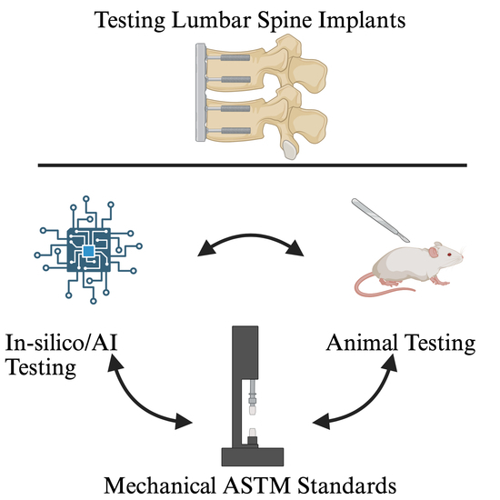
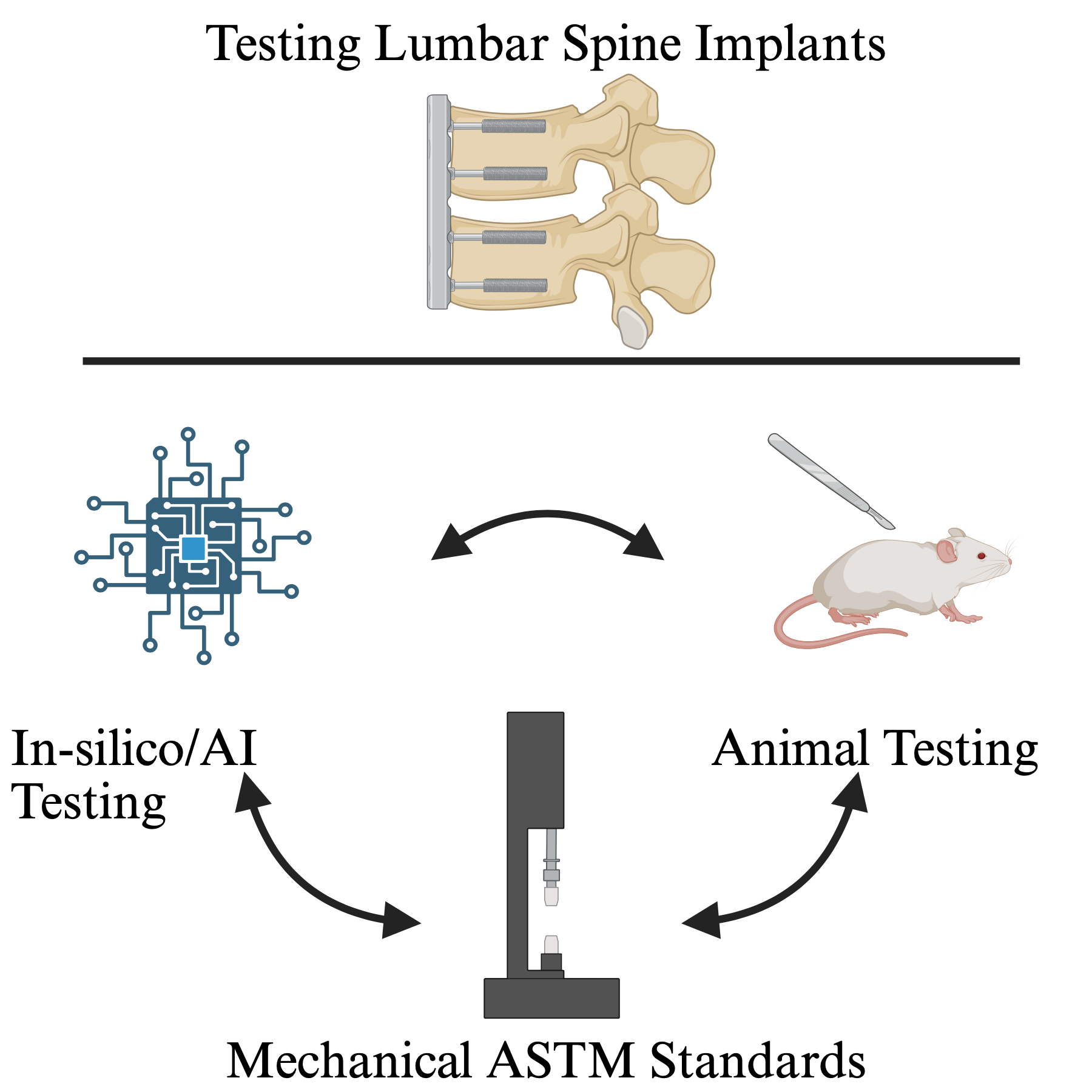
Graphical Abstract

1. Introduction

Lumbar spine implants can be defined as medical devices used to stabilize, replace, or reinforce the skeletal system at the lumbar section of the spinal cord. Lumbar spine disorders, a broad category of diseases encompassing intervertebral disk degeneration, spondylolisthesis, and spinal stenosis, often require surgical management [1,2,3]. In 2022, there were over 1.1 million spine surgeries performed, which often use spinal implants. The prevalence of spinal surgeries is increasing due to a demographic shift towards an aging population and growing rates of obesity and other metabolic disorders [3]. This has propelled advancements in implant technology with a focus on the advancements of materials science, surface technology, computational modeling including the use of AI, and the use of genetically engineered animal systems [4,5].
These implants are subject to failure, however, and spinal implant failure has a particularly high-risk fracture because of the implant’s proximity to the spine and nerve roots. Kienle et al. report a failure rate of 0.5 percent to over 60 percent depending on the procedure [6]. Furthermore, spinal implant failure carries a significant economic burden [7]. The cost of implant failures was reported to be between 2.5 and 5 billion dollars [6].
Prior to surgical implementation, these advancements need to be rigorously tested. This testing follows a sophisticated, multi-step protocol. Despite advancements made in silico and animal models, mechanical tests still serve as the cornerstone testing methods. Protocols put forward by the ASTM and ISO, such as ASTM F1717 and ISO 12189 [8,9,10], provide guidelines for assessing strength, fatigue, resistance, and durability under projected physiological loads. This allows physicians and companies to compare implant designs [8]. This mechanical testing does not capture the complexity of the in vivo spine, necessitating the next step of biological testing [11].
The Food and Drug Administration (FDA) regulates all medical devices that enter the market and categorizes them into three classes based on risk. Class 1 devices are usually non-invasive, have a well-established history of use, do not support or sustain life, and contain virtually zero risks. This class includes tongue depressors, examination gloves, bandages, and mechanical wheelchairs. Class 2 devices include a moderate amount of risk and require regulatory approval. Special controls tailored to a specific device type are necessary to ensure the safety of these devices. There is usually a history of use of a similar base device and mechanical testing methods, such as those provided by the ASTM, which are responsible for new devices to prove substantial equivalence to older models. The FDA states that nearly 43% of medical devices fall under this category, including most lumbar implant devices. Any spinal implant that contains non-organic material fits into this category as long as there is a predicate device.
Biological testing evaluates the interaction of the lumbar implant with living tissue, assessing biocompatibility, osseointegration, and long-term effects. Biological testing comes with its own challenges. Commonly used organisms such as rodents, pigs, and sheep differ from humans in their basic anatomy, spinal loading direction, bone microstructure, and immunological profiles. All the previously listed animals are quadrupedal, significantly reducing the translatability of tests done on these animals. Furthermore, most animals are young, and even if raised to an advanced age, still fail to replicate the decades of degenerative conditions common in human lumbar pathology. Ethical concerns with animal testing are present as well [12,13,14].
With 21st century advances in mathematics and computing, FEA was a further step forward, allowing researchers to test stress distribution and potential failure points before physical models are made, greatly increasing efficiency. FEA also allows high-throughput screening of many different digital models of implants [15,16].
AI has now emerged as a key player in lumbar spine implant research and testing. AI workflows include image analysis in all aspects of the surgical process, from diagnosing a candidate for surgery to surgical planning and postoperative analysis. Often, AI workflows are combined with FEA [4,15,16]. Recently, Ahmadi et al. reported a process to generate FEA of the lumbar spine from Computed Tomography (CT) and Magnetic Resonance Imaging (MRI) imaging on a scale of minutes, not hours. This model allows simulation of natural physiological movements such as bending and twisting, allowing better planning of surgical interventions [17].
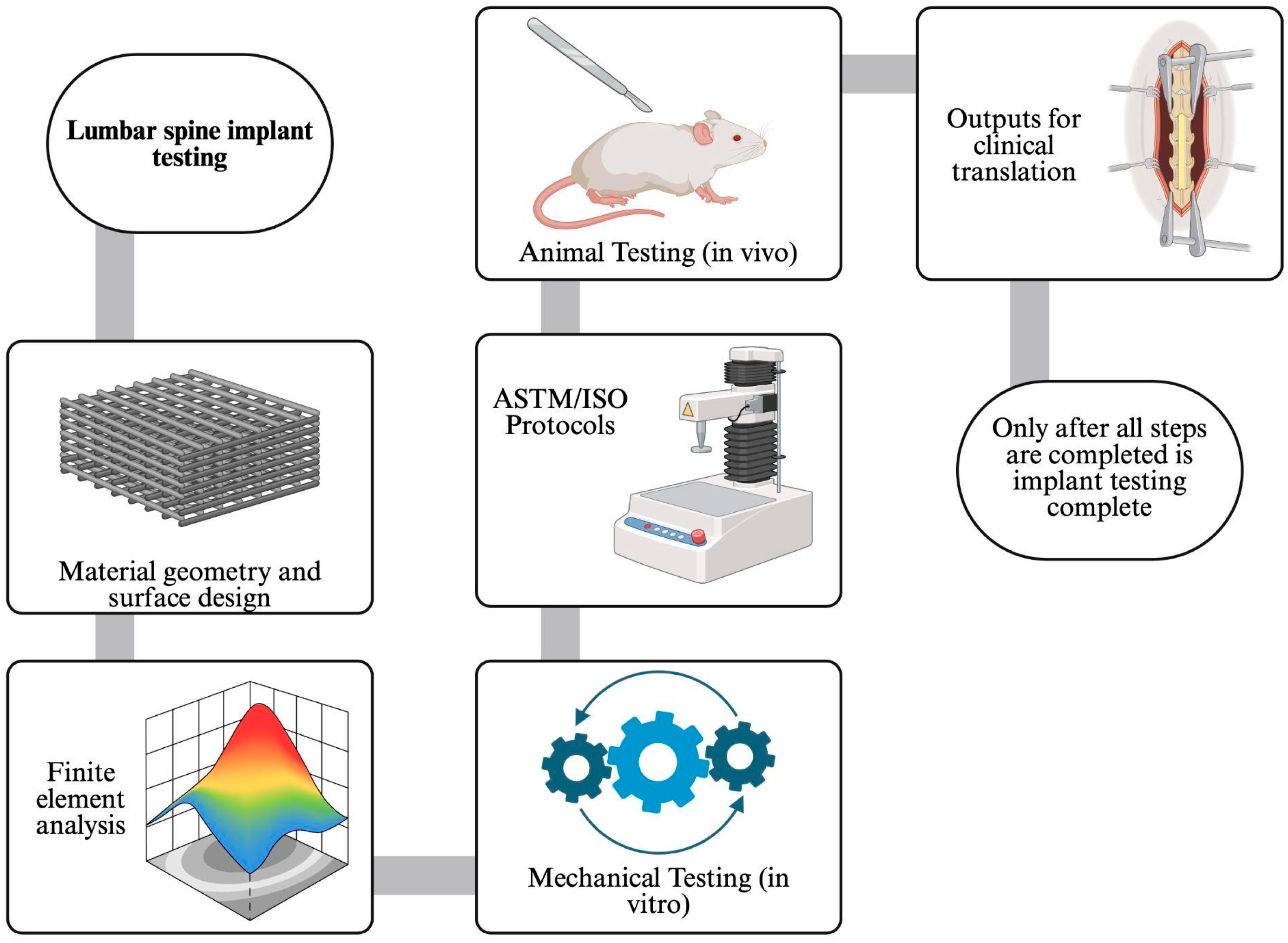
Given the increasing number of uses of lumbar implants in spine surgery [1,3,4], this review aims to describe the advancements made in their testing, shortfalls in current testing methods, and future directions for the field. The general overview of the current spinal implant testing process is summarized in Figure 1.

2. Current Testing Methods

In vitro mechanical testing methods have been developed and standardized by the ASTM or the ISO. They are particularly useful for their role in proving the substantial equivalence of novel medical devices for the FDA.
ASTM-F1717: The ASTM-F1717 is a well-established and common protocol for static and dynamic compression of posterior spinal fixation systems, such as pedicle screw–rod systems [18]. The testing method simulates the worst-case scenario, a vertebrectomy, by using two polyethylene blocks to imitate vertebral bodies with a gap representing a missing vertebra. There is no anterior support system, and the entire load must be carried by the fusion instrument. The device is subjected to high bending moments and stresses to assess bending, tensile bending, torsion, and fatigue life under cyclic loading conditions [19,20]. Many devices that are submitted to the FDA undergo this protocol to assess how they compare to other devices.
ASTM-F1798: The ASTM-F1798 looks to assess the mechanical integrity of connections between components of medical devices, like within a spinal fixation system (like a thoracolumbosacral pedicle screw system). It is therefore used in conjunction with F1717. The interconnection would be assembled per instructions and mounted on a test fixture that can apply controlled torsional and axial loads. Tests can either assess static capabilities or can have the device undergo cyclic stress for a pre-determined number or until failure of the device [18,19].
ASTM-F543: This protocol looks to assess various factors, including the torque required to insert the screw into standardized polyurethane foam blocks or bone models, reflecting ease of insertion and risk of screw head or shaft failure. Consistent driving torque is critical for optimal fixation and avoiding intraoperative complications. Furthermore, this method is designed to test the maximum force required to extract inserted pedicle screws from polyurethane. It simulates variable bone conditions by altering the bone model densities to give rise to osteopenia and osteoporotic models [8,18,21].
ASTM F2077 and F2267: Both standards are used to assess the function of lumbar intervertebral body fusion devices (IBFDs) and complement the other protocols used for arthrodesis. The protocols place the IBFD between synthetic blocks to parody vertebral end plates and evaluate static and dynamic axial compression, compression–shear, and torsion. This provides the necessary parameters to assess structural integrity over repetitive loads [21]. On the other hand, the ASTM F2267 addresses subsidence testing, or the risk of the device sinking into the vertebral body, which is amplified in osteoporotic or weakened bone. The test measures the stiffness (Kp) of the device block construct under axial loading, with higher Kp values indicating greater resistance to subsidence [22,23].
ISO 12189 (Physiological Support Model): This protocol resembles the ASTM F1717 setup, but it is closer in mimicking clinical uses by introducing a modular anterior support, typically using calibrated springs, to better mimic the load-sharing environment of the spine. The protocol allows for testing of more flexible or dynamic implants and includes a precompression step to simulate physiological preload. The assembly and loading procedures are more complex, but yield data that more closely reflect clinical conditions, including the load-sharing mechanism between anterior and posterior elements.
Biological protocols are crucial when testing lumbar spine implants. Animals are invaluable in bringing data from the tabletop to clinical trials as they provide a glimpse into the biological response to these medical devices. Inadequate preclinical testing can result in extreme risks if first implemented in human trials. Animal studies can provide understanding of issues regarding tissue–device interactions, osseointegration and biomechanical performance prior to human use.
Protocols for operating on these animals are usually detailed to ensure animal welfare and reproducibility. The surgical approach (anterior, posterior, or lateral) will then be chosen depending on the type of implant, with properly sized hardware suitable for the respective animals, and using surgical techniques resembling human surgery [24,25]. Studies often include positive controls (ideal fixation), negative controls (compromised fixation), and experimental groups (novel devices or materials) to assess relative performance [24,26].
Many of the tests that were performed using machinery are also performed post-implantation to assess if in vitro results translate to in vivo. Multi-directional loading is performed to assess the flexibility and range of motion of the implant [27,28]. Furthermore, pullout strength is measured to assess the force required to remove a screw from the vertebrae [26]. Fatigue and cyclic stress are also performed to analyze the lifetime of the device and the activity it can handle before failure [24,29].
Medical imaging techniques are also applied to gain deeper insight into the device on the bone. Radiographs are utilized to assess bridging of the bone across the pedicle screws for successful fusion or if the implant has moved or sunk into vertebral bodies [24,30]. Also, at predetermined times, the animals are euthanized, and the histology of the tissue surrounding the implants is evaluated. The biocompatibility of the material is assessed by the presence of an immune response, determined by the number of inflammatory cells, fibrosis, and osteolysis [24,30]. Studies will also evaluate whether debris due to wear of the device can lead to neurotoxicity [28,31].
Outcomes of the studies are necessary to determine the effectiveness of the device in biological subjects and whether they are safe for human use. Favorable outcomes include having a solid fusion in a large portion of animals, minimal inflammatory response, and a lack of particulate debris and neuropathy.

3. Gaps, Problems, and Challenges

There are 2 main categories that testing can be broken down into: mechanical and biological.
Mechanical testing relies on recreating the biomechanics of the spine, which is extremely difficult. The human spine includes multi-axial loads that vary across small time periods. These axes include flexion/extension, lateral bending, rotation (turning to the right/left), and shear force. All these loads vary with posture and activity. Laboratory tests naturally simplify conditions. This commonly results in loading along only 1 axis; mechanical tests can test multiple axes but do so individually. Furthermore, these tests are generally static. The lack of combined axis loading, as well as movement-based tests, reduces the validity of mechanical models to human spine conditions [8,32].
There has been an effort to implement mechanical standardized testing systems for lumbar spine implants (ASTM). These systems are a double-edged sword, however. They trade realism for repeatability. Standardized vertebral blocks inherently do not and cannot reproduce the vertebral anatomy along with the myriad of differences in vertebrae seen with decades of biological pressures. Specimen variability, which includes age, bone mineral density, and vertebral geometry, also affects spinal implant outcomes such as screw pullout and micromotion. Studies that do not control for these factors are very difficult to generalize [33,34,35].
Mechanical tests inherently ignore biological interaction. Osseointegration, bone remodeling, and debris generation influence implant stability but cannot effectively be replicated in a mechanical test. Lumbar spine implants are not static structures, and once implanted, they go through a complex process to integrate with the spine [36,37,38,39,40]. Therefore, any mechanical test performed would have to test multiple points in time along this integration pathway. At each time point the implant would have to be tested along with any combination of the many axes of the spine as previously described. Short-term laboratory mechanical tests fail to account for these factors, not because of researcher failure, but rather the innate shortcomings of testing a spinal implant in a lab. From a pragmatic perspective, mechanical testing demonstrates a persistent disconnection between lab performance and meaningful clinical outcomes. Case reports also describe the failure of lumbar spine implants. For instance, Leute et al. describe the case of a 44-year-old woman who, after transforaminal lumbar interbody fusion (TLIF), had her set screws fracture and experienced dislocation of the interbody cage left dorsally as verified by X-ray [41]. This implant failure occurred 3 months post operation. The early timing of this implant failure is suggestive of gross mechanical overload or suboptimal fusion. Revision surgery to remove the fractured hardware and replace and reposition the interbody cage was performed. To summarize, mechanical testing of lumbar implants is constrained by the physical complexity of the spine and the diversity of biological factors influencing the spine’s biomechanics.
With the limitations discussed above with mechanical testing, animal testing attempts to solve many of these problems. For one, animals are not subject to loading along only one or a few axes and inherently do not ignore implant biological interaction; these were the two major problems associated with mechanical testing. Yet, that does not mean animal testing is without problems.
Translating results from animals to humans comes with several important gaps. Most of these gaps are not unique to spinal implant testing; rather, problems translating animal research to humans plague almost all areas of biomedical research [42,43].
Biomechanics and spinal loading present a particularly tricky problem with spine implants. Large mammals such as sheep, goats, dogs, and pigs are all quadrupeds and their spinal loading differs remarkably from upright humans [44]. This difference is minimized in non-human primates due to their anatomical and physiological resemblance to humans, but not entirely; primates can walk upright, but they still spend significant amounts of time on all 4 limbs. Furthermore, they are used in rare instances because they are expensive and there are multiple ethical considerations to their use [27,45]. Additionally, all the animals mentioned above have very different gaits than humans, changing bending movement and the way implants load the vertebral bodies and interbody spaces. Gait differences can also change the way implants experience micromotion and osseointegration, factors central to spinal fusion success [26,28,45].
Aspects of spinal anatomy that differ across species include vertebral body thickness, pedicle width, pedicle angle, canal size, and disk thickness. Calf, pig, and sheep spines are commonly used for lumbar testing due to their anatomical and mechanical similarities to human lumbar spines [29]. This provides a good instrument to evaluate the effects of the instruments on a macro level in interbody fusion and pedicle screw experiments. However, it should be noted that due to differing morphology between them and humans, some aspects, most notably the range of motion and intradiscal pressure, must be considered when compared to human trials [24,29,46]. For example, sheep vertebral bodies are small with a slightly different shape than humans; additionally, their altered pedicle anatomy can prevent normal human surgical instrumentation from being used in surgery without modification. This leads to animal testing in which altered surgical instrumentation is used to implant altered implants into an animal with a spine with different microanatomical components on top of a completely different macro loading direction [32,47,48]. The porcine spine is also widely used, especially for pedicle screw evaluation, due to its comparable size and mechanical properties, but immature pigs may have physeal defects affecting biomechanical outcomes [25,46]. Furthermore, although calf spines were easier to use, the porcine spine was shown to more closely resemble human spinal flexion capabilities after device implantation [29].
Smaller animals are not helpful in measuring the effects of forces and torsion on implants due to their small vertebrae. Yet they provide a cheap and insightful method to examine histological and biocompatibility studies, especially for particulate wear debris and host response [31,49]. However, there are shortcomings in this aspect as well since small rodents have more rapid bone turnover than humans, along with a different trabecular architecture [34,50,51]. Large mammals such as sheep and goats may be a better approximate model for bone architecture; they are still imperfect as outlined above in the macro anatomical differences [52]. All these factors affect osseointegration and resorption of graft material.
Disease modeling presents another challenge. Human spine changes and degeneration take place over decades and have a multifactorial etiology. One of the main causes of this etiology is age, however. Age-related degeneration is very difficult to replicate in the lab because animals do not have the same lifespan as humans, and even if they did, the experiments would take too long to be useful. Therefore, creating animal models of degenerative disks or osteoporotic vertebra is difficult in animals. Current methods include surgical injury, enzymatic degradation, or mechanical overload to induce degeneration. While creative, these do not reliably replicate the decades-long systemic process that occurs in humans [40]. The average age of patients undergoing a spinal fusion is roughly 55 years old [53].
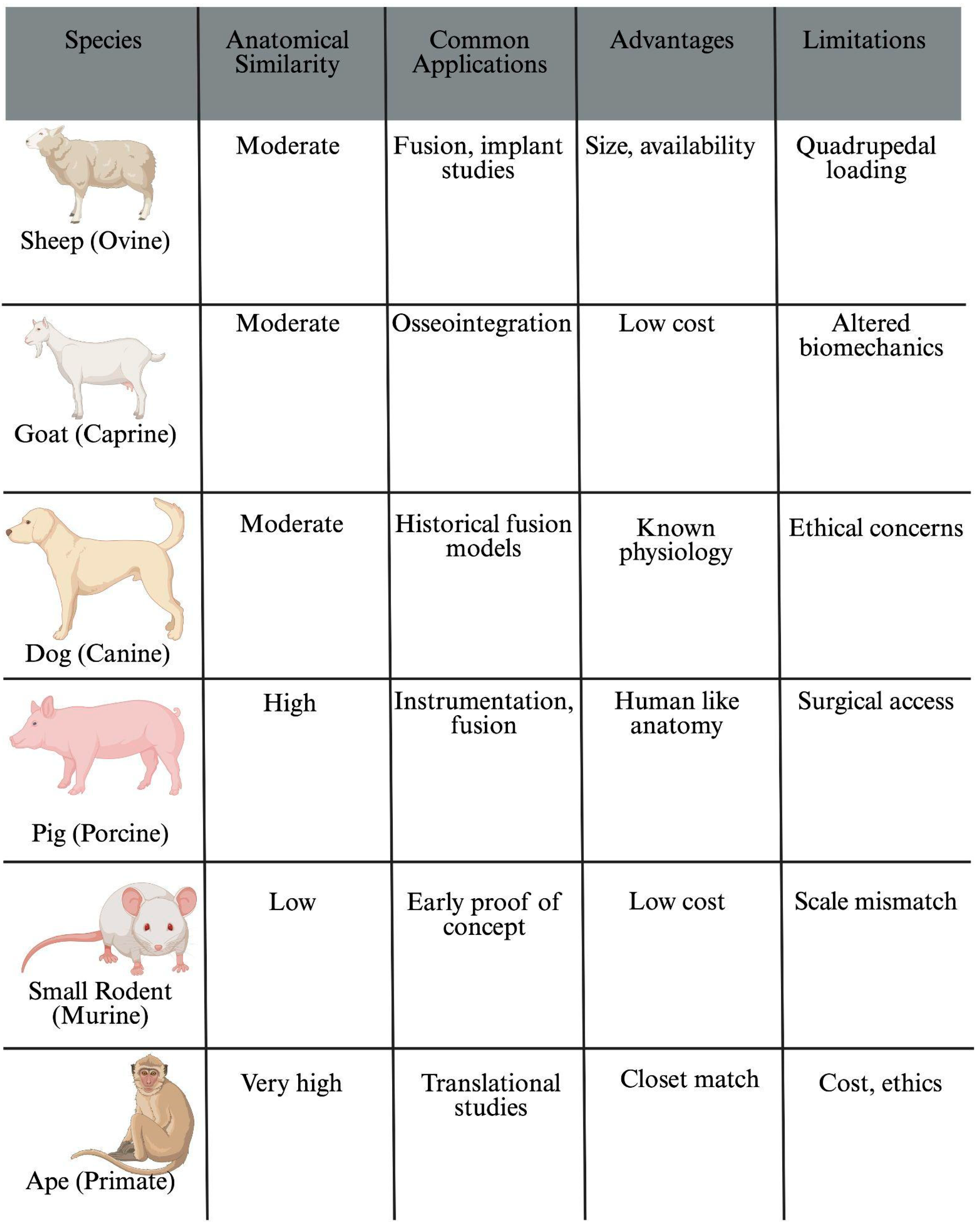
With each commonly used animal model to test spinal lumbar spine implants, there are problems. Sheep (ovine model) and goats (caprine model) are widely used because they are a manageable size, not aggressive, and have a moderate cost. Their vertebral sizes are somewhat similar to humans, although by no means a perfect representation of the human spine; the differences in anatomy are described above. Despite this, sheep and goats allow for the modification of human surgical instrumentation and implants rather than the use of completely different ones. However, both animals are quadrupeds, limiting translatability. Pigs (porcine model) vertebrae are the most like humans, although differences in pedicle widths have been noted [54,55]. Additionally, their pelvic anatomy can complicate lumbar access. Dogs (canine model) have also been historically used; however, ethical concerns have not limited their use. Small rodents (mice model, rat model, rabbit model) are the most widely used animals in biomedical research. Many such variations exist, and they are useful for many proof-of-concept studies. However, when testing spinal implants specifically, their size, fast healing, and significantly different biomechanics make them poor test subjects for lumbar implants [56]. Non-human primates are the most similar to humans, but cost, ethics, and scarcity severely limit their use.
In sum, no animal model is without limitations in testing lumbar spinal implants. Species-specific anatomical differences exist while modeling human spinal degeneration remains a persistent problem. The benefits and difficulties with each commonly used animal are summarized in Figure 2 [25,27,29,31,32,34,44,45,46,47,48,49,50,51,52,54,55].

4. AI Applications in Testing

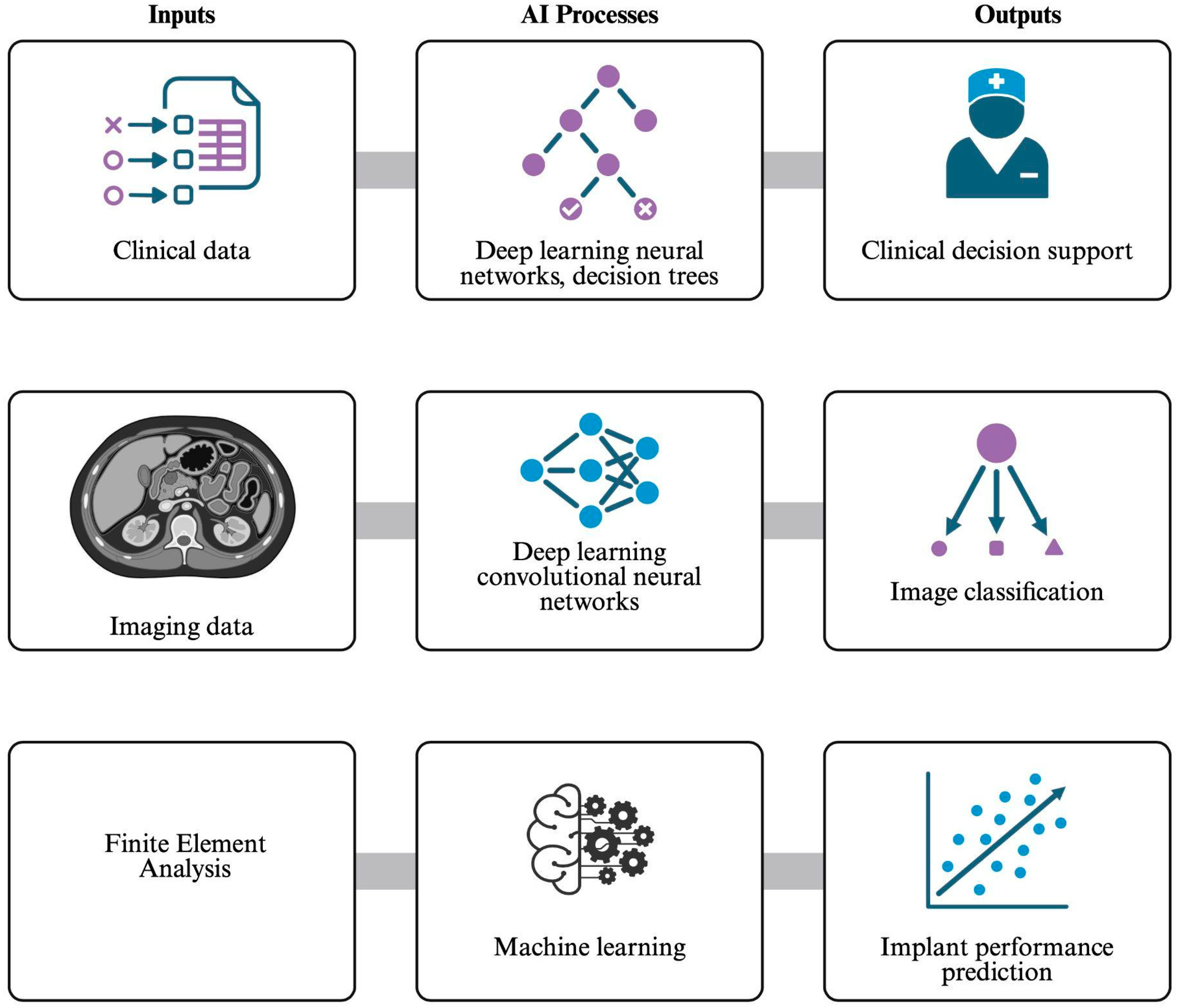
AI is becoming an increasingly valuable tool in the design and testing of spinal implants. Traditional mechanical testing methods, such as physical stress testing and FEA, remain reliable but are often time-intensive, costly, and sometimes overpredict implant durability compared with real-world outcomes. AI can help address these limitations by generating faster and more flexible models that estimate implant performance across diverse patient conditions [57,58,59]. AI also has uses in image classification, preoperative risk models, and as a clinical decision tool in addition to its uses in research [60].
In terms of applying these AI systems to spine research, they are particularly useful in FEA. While FEA has long served as the standard for simulating spinal biomechanics each simulation can take several hours or even days to complete. Recent studies have shown that machine learning (ML) models trained on FEA data can reproduce these results with remarkable accuracy in a fraction of the time [58]. These AI-based “surrogate models” enable rapid exploration of implant designs and materials, minimizing the need for repeated physical testing. In practice, FEA serves as a preclinical screening tool that informs which implant designs and materials advance to mechanical bench testing and subsequent animal models. By identifying stress distributions, failure points, and range-of-motion changes computationally, FEA reduces the number of physical prototypes and animal experiments required while refining the hypotheses tested in vivo [58].
An example of a successful integrated framework is demonstrated in the systematic evaluation of novel ceramic and polymer interbody fusion spacers, where FEA results were directly leveraged to train AI regression models for predicting performance across diverse patient profiles [58]. This combined pipeline facilitates high-throughput screening while refining the experimental designs prioritized for subsequent animal studies [56,58]. Despite these advantages, several critical limitations must be addressed, such as high data dependency, where the reliability of predictive models is strictly limited by the quality and diversity of the initial datasets [57,60]. Additionally, validation challenges persist, requiring rigorous comparison of algorithmic outputs against established mechanical benchmarks to ensure clinical translatability [58,61]. Finally, current regulatory pathways are often ill-equipped to handle rapidly evolving AI-driven design iterations, highlighting a need for updated standards that balance innovation with safety [57,60].
One study found that ML algorithms could accurately predict the mechanical behavior of ceramic and polymer implants under spinal loading conditions, achieving comparable precision to full computational models while substantially reducing computation time [58,62]. This study focused on developing implants to treat lumbar spine stenosis, which is the compression of the foramina of the spine. Typically, titanium implants are the gold standard for spine implants due to their durability, but they also cause MRI artifacts and hypersensitivity, prompting researchers to attempt to use alternative materials [5]. Polymers such as polyetheretherketone (PEEK) and polymethylmethacrylate (PMMA) have been considered due to their use in orthopedics and dentistry, but in order to use these materials, and other biocompatible materials such as ceramics, they need to be tested theoretically before being used in practice. This study made use of a two-phased model. First, using FEA, the study was able to identify implant stress, strain on adjacent vertebrae, and range of motion for the ceramic and polymer implants. Then, the study made use of AI-based regression models using FEA data sets to allow for the prediction of device performance. AI was able to take into account the material used, the bone density of the implant recipient, and the many kinds of motion the implant would be subjected to. It also created a prediction of stresses, strains, and ranges of motion beyond what FEA could have predicted, creating an overall more thorough picture of the implants created from the new polymer and ceramic materials [58].
AI also streamlines image processing and model generation. Within the broader category of AI, deep learning (DL) techniques, particularly convolutional neural networks, can convert CT or MRI scans into detailed, patient-specific three-dimensional representations of the spine [59,63]. This capability allows researchers to evaluate implant performance across a wide range of anatomical variations, including osteoporotic and deformed spines, scenarios that are difficult to reproduce with cadaveric or generic models. A simple and proven use of the technology was demonstrated in a study sampling 6988 radiographs to test AI’s ability to correctly label those radiographs. These labels included not only the X-ray view, but also rotation, position, and whether there was spinal hardware in the patient. The AI performed well and showed promise as an assistive tool in analyzing imaging. It learned quickly, but researchers deemed that more time and training for the AI is necessary before the algorithm can be used in clinical practice [64]. With further refinement, these algorithms could be used to model implants from radiographs analyzed by AI.
Studies have also shown postoperative uses for AI regarding spinal implants. ML can identify complications before they arise. One study used ML to predict the failure of a spinal epidural abscess due to the AI’s ability to be specific to the patient’s risk factors and personal traits [59]. ML can successfully predict outcomes during and after surgery due to its ability to consider many different factors that can affect patient outcomes. Multiple forms of AI have been used to create algorithms to assist in both decision-making for surgery and postoperative prediction. Decision trees, neural networks, and support vector machines all can be used for automated vertebral segmentation and fracture detection, which can help physicians decide whether they should operate and where exactly they should focus. The risk management predictive tools have also come a long way, being able to scour patients’ charts and consider things a physician may miss [60,61].
A great proof of concept for these predictive models is the Intelligence-Based Spine Care Model (IBSC) and its use in adult spinal deformity (ASD) prediction. IBSC is a protocol that integrates many data sets into a single learning system. It makes use of clinical records, radiograph imaging, biomechanical parameters, genetics, and outputs from wearable devices to create a detailed, specific profile for a given patient. AI then analyzes this data to find clinically relevant patterns, make predictions, or find optimal treatment strategies for these patients [65]. This protocol can be used to make predictive models for ASD and has proven to be very effective. By leveraging the IBSC’s multidimensional data integration pipeline, ASD predictive models utilize clinical, radiographic, biomechanical, and perioperative variables to forecast outcomes such as mechanical complications, length of stay, and achievement of minimal clinically important difference (MCID) thresholds. Through advanced ML techniques, including neural networks, random forests, and support vector machines, these models can identify nonlinear relationships among diverse predictors that are often overlooked by conventional statistical methods. The iterative feedback mechanism central to the IBSC architecture allows these models to improve continuously as new data are incorporated, refining accuracy and generalizability across patient populations. This approach supports personalized risk stratification, enhances preoperative planning, and informs intraoperative decision support, aligning with the broader IBSC goal of transforming spine surgery into a precision, outcome-driven discipline. Collectively, the integration of predictive modeling within the IBSC paradigm represents a significant step toward optimizing ASD management through intelligent, adaptive, and patient-specific care [65].
Beyond the preclinical phase, AI supports post-market evaluation by analyzing large datasets to identify materials or implant designs associated with increased complication rates. Integrating data from simulations, imaging, and patient outcomes provides a more comprehensive understanding of device safety and real-world performance and could help address the trend of overestimating the safety and durability of spinal implants [57,62,63]. Although these advances are promising, AI models must undergo rigorous validation to ensure reliability, and their limited interpretability remains a barrier to widespread regulatory approval. Overall, AI offers a faster, more personalized, and cost-effective approach to spinal implant testing and design, complementing rather than replacing traditional biomechanical methods. An integration framework of AI is shown in Figure 3.

5. Future Directions

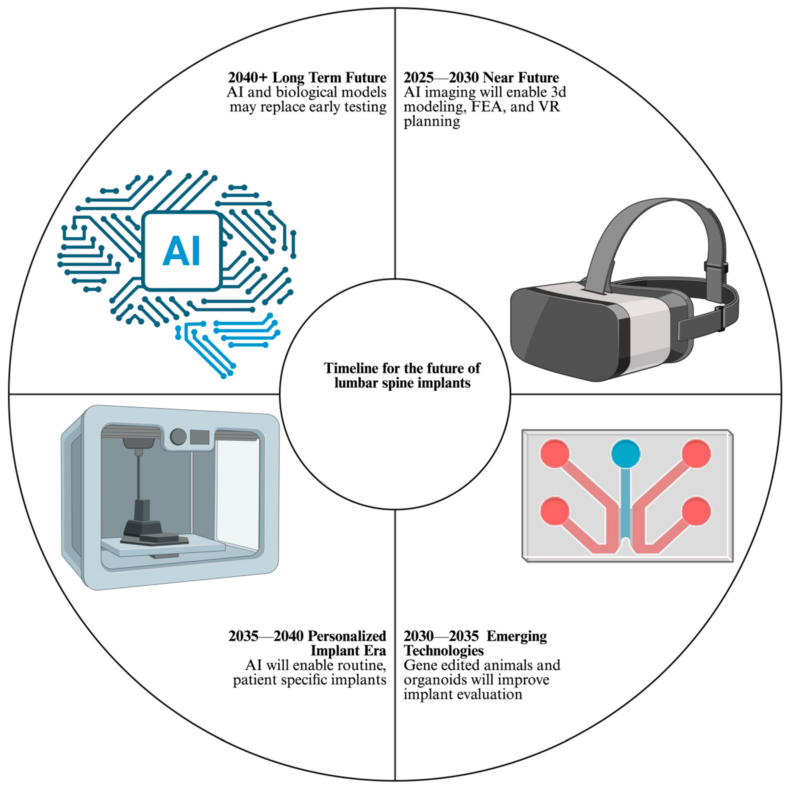
Emerging research priorities include integrating AI with imaging analysis. This can reduce the amount of radiation patients are exposed to by building multiple images from a singular CT scan. It can also take these 2D images and build a 3D environment to plan surgical approaches, in addition to allowing surgeons to practice said surgeries in virtual reality (VR). For instance, Chen et al. showed that following VR surgical simulation training, orthopedic and neurosurgical residents’ anatomical knowledge increased. Furthermore, this study was done on patient-specific 3D models [66]. Multiple disciplines are continuously improving, creating workable 3D models from 2D CT/MRI imaging to work in the aforementioned surgical practice.
Customizable spinal implants mirror the broader trend in medicine of personalization [67]. Traditional lumbar spine implants come in limited shapes and sizes, requiring the surgeon to adapt the patient’s anatomy to fit the implant. In the case of lumbar spine disk replacements, “off the shelf” implants can lead to poor endplate contact and thus distribute load unevenly, ultimately leading to subsidence and pseudoarthrosis, defined as the sinking or settling of an implant into the adjacent bone and failure of bone to fuse at the intended site, respectively [68]. As implants become increasingly customized in geometry, material composition, and surface characteristics, traditional implant testing and validation frameworks become less applicable. Each category of customization introduces distinct biomechanical, biological, and regulatory testing challenges, which are illustrated by the examples discussed below.
As early as 2016, there have been documented case reports of 3D printed, customizable implants being used. Phan et al. describe the case of a 65-year-old woman who underwent a C1/2 arthrodesis. In this case, the woman’s anatomy was so distorted by C1/2 facet arthropathy that normal screw trajectories and rods were not suitable. The medical team used a CT scan to construct a computer model of the patient and then 3D print a suitable implant. At both two and seven months post operation, the patient had decreased occipital neuralgia pain and imaging showed satisfactory implant positioning. The surgeons described the procedure as uneventful [69]. This case study serves as a useful proof of concept for the idea of customizable implants and the idea of personalized medicine.
Material science will also play a role in the future of lumbar spine implants. For context, titanium and PEEK are the two materials that are most often used for lumbar spine implants. Titanium is used because it naturally forms a TiO2 layer, which allows easy osteoblast adhesion and biocompatibility [70]. Titanium, however, because of its density, is difficult to view on CT or X-ray. PEEK, by contrast, is easy to visualize in CTs and X-rays. Additionally, its elasticity is similar to that of cortical bone. The downsides of PEEK are that it forms a fibrous surface that osteoblasts struggle to adhere to [71,72].
Immediate future directions include a lumbar spine implant with a PEEK core and titanium surface, combining the benefits and avoiding the downsides outlined above. In 2021, Tan et al. showed improved spinal fusion rates in titanium-coated PEEK cages as opposed to PEEK alone, but these results are inconsistent across meta-analyses [73]. Furthermore, some meta-analyses consider PEEK coated with titanium the same as just titanium implants in their analysis. Multiple companies are producing these systems, although their integration is not complete. The future of these combined material implants involves using 3D printing for manufacturing [74]. 3D printing allows implants to be manufactured with a lattice, porous structure throughout the entire implant, allowing deeper bone ingrowth and superior fusion rates. Non-3D-printed manufacturing methods only create superficial porous structures [71].
Future implants will also likely be bio-responsive. Bio-responsive implants actively interact with the surrounding tissue. Basic examples that are currently in use include surface coating of calcium phosphate and hydroxyapatite, which enhance fusion rates. Other applications include antibiotic-coated implants. Moving forward, “smart” systems are in clinical trial, including infection-triggered antibiotic release and pH-responsive polymers [75].
Organoids or organ-on-a-chip systems are becoming increasingly common in biomedical research. As opposed to traditional cell culture, these systems involve multiple cell types to act as better models for multiple organ systems. These systems will be used to evaluate how titanium, PEEK, lattice networks, and bio-responsive implants can interact with multiple cell types such as osteoblasts, fibroblasts, and macrophages [76,77]. For example, in 2025, Zhu et al. describe a dynamic hydrogel system that supports woven bone organoid formation by providing a mechanically adaptive, bioactive microenvironment. This platform enhances osteogenic differentiation and mineralization, further advancing bone organoid translatability [78]. Organoid systems also limit ethical concerns surrounding animal testing.
While promising and useful in many areas of biomedical research, these systems have limited use in the testing of lumbar spine implants. The testing of lumbar spine implants is inherently mechanical in nature, and miniaturized systems are plagued with problems of scaling. Because of these limitations, genetically modified animals are a more promising future alternative as opposed to organoid systems.
As outlined earlier, animal models are plagued by issues in translatability. Recent gene editing techniques such as CRISPR cas-9 and AAV vectors reviewed in recent studies [79,80,81] are promising technologies to modify these animals and help close some of the translatability gaps. Other technologies, such as increasingly sophisticated medications and surgeries, such as oophorectomies, can also aid in the creation of increasingly sophisticated animal models [82]. As an example of the feasibility of gene-edited animals, 2025 marked the year in which a man received a kidney from a genetically modified pig, marking a significant step forward in genetically engineered animals [83].
AI can be applied to all the processes outlined above. Key areas of advancement include the switch from supervised to unsupervised learning to streamline efficiency and speed up process times for FEA. In 2025, Namiranian et al. used micro-CT-based FEA to noninvasively estimate bone mechanical competence, demonstrating that high-resolution imaging coupled with µFEA can predict differences in bone strength and the effects of therapeutic interventions in animal models [84].
With the rapid advancements in AI and medicine, current policies will have to change as well. The current 510(k) substantial equivalence model will likely become outdated, and a standardized framework for 3D printed implants, genetically engineered organisms, and AI/model transparency will have to be put forth and constantly updated. Regulators may allow AI and genetically engineered animals to replace early preclinical testing.
Regulating AI-driven implant development presents several challenges that are not seen with traditional medical devices. AI systems can change rapidly as new data are incorporated, making it difficult to determine which version of an algorithm was used at the time of clinical decision-making or implant design. This raises questions surrounding validation, reproducibility, and trust, particularly when algorithmic outputs may outperform humans but lack intuitive interpretability. Regulation of 3D-printed implants is similarly complex, as patient-specific devices cannot feasibly undergo mechanical or clinical testing on an individual basis. Rather than validating each implant, future regulatory efforts will need to focus on validating the underlying design constraints, manufacturing processes, and quality-control standards that govern customization.
The future of lumbar spine implants involves using fewer imaging modalities and radiation to evaluate patients for surgery. Unsupervised neural networks can aid in determining who is a candidate for surgery and what risk factors to be wary of. Newer implants will be designed and tested in silico, with FEA performed quickly with the aid of AI. Genetically, chemically, and surgically modified animals will then be used to test the implants in vivo. Organ-on-a-chip systems will be used to aid in the biological process of osseointegration. Based on the imaging, 3D models will be created with the help of AI. Based on these models, sophisticated personalized implants will be created with a lattice network for osseointegration, PEEK core, and a titanium superficial layer. The future directions of lumbar spine implants are summarized in Figure 4.

6. Conclusions

The preclinical evaluation of lumbar spine implants has evolved into a sophisticated multi-stage process, yet it remains constrained by the inherent limitations of traditional methodologies. While advancements in ASTM and ISO protocols provide essential benchmarks for mechanical reliability, they present significant shortfalls in replicating the dynamic, multi-axial loading and long-term biological integration required in a clinical setting. Furthermore, while animal models remain invaluable for assessing biological responses, their effectiveness is often compromised by anatomical differences and quadrupedal biomechanics that limit their direct translatability to human patients.
Future directions for the field point toward a transformative, integrated framework. The synergy of AI and FEA is set to revolutionize the industry by enabling rapid, patient-specific in silico testing and high-throughput design optimization. Emerging technologies—including 3D-printed personalized implants, bio-responsive materials, organ-on-a-chip systems, and genetically modified animal models—promise to bridge the current gaps between laboratory testing and successful clinical outcomes. Ultimately, the development of safer and more effective lumbar implants will depend on an interdisciplinary synthesis of engineering, biology, and computational science tailored to the unique needs of the individual patient.

Author Contributions

Conceptualization, M.L.; methodology, Z.C., R.K., Y.A. and E.B.; software, Z.C., R.K., Y.A. and E.B.; validation, Z.C., R.K., Y.A. and E.B.; formal analysis, Z.C., R.K., Y.A. and E.B.; investigation, Z.C., R.K., Y.A. and E.B.; resources, Z.C., R.K., Y.A. and E.B.; data curation, Z.C., R.K., Y.A. and E.B.; writing—original draft preparation, Z.C., R.K., Y.A. and E.B.; writing—review and editing, Z.C., R.K., Y.A., E.B., M.L., R.S., F.D.V. and R.P.; visualization, Z.C.; supervision, M.L. and F.D.V.; project administration, M.L. and T.S.C. All authors have read and agreed to the published version of the manuscript.

Funding

This research was supported by the Marcus Neuroscience Institution (Grant No. C-24-335) and the Helene and Stephen Weicholz Foundation.

Data Availability Statement

None.

Acknowledgments

Some of the images were made with biorender.com.

Conflicts of Interest

The authors declare no conflicts of interest.

References

  1. Wellington, I.J.; Kia, C.; Coskun, E.; Torre, B.B.; Antonacci, C.L.; Mancini, M.R.; Connors, J.P.; Esmende, S.M.; Makanji, H.S. Cervical and Lumbar Disc Arthroplasty: A Review of Current Implant Design and Outcomes. Bioengineering 2022, 9, 227. [Google Scholar] [CrossRef] [Scilit]
  2. Foster, N.E.; Anema, J.R.; Cherkin, D.; Chou, R.; Cohen, S.P.; Gross, D.P.; Ferreira, P.H.; Fritz, J.M.; Koes, B.W.; Peul, W.; et al. Prevention and treatment of low back pain: Evidence, challenges, and promising directions. Lancet 2018, 391, 2368–2383. [Google Scholar] [CrossRef] [Scilit]
  3. Dykhouse, G.L.; Bratescu, R.A.; Kashlan, O.N.; McGrath, L.J.; Härtl, R.; Elsayed, G.A. Trends in spinal implant utilization and pricing. J. Craniovertebral Junction Spine 2024, 15, 404–410. [Google Scholar] [CrossRef] [Scilit]
  4. Jain, P.; Rana, M.; Biswas, J.K.; Khan, M.R. Biomechanics of spinal implants—A review. Biomed. Phys. Eng. Express 2020, 6, 042002. [Google Scholar] [CrossRef] [Scilit] [PubMed]
  5. Warburton, A.; Girdler, S.J.; Mikhail, C.M.; Ahn, A.; Cho, S.K. Biomaterials in Spinal Implants: A Review. Neurospine 2020, 17, 101–110. [Google Scholar] [CrossRef] [Scilit] [PubMed]
  6. Kienle, A.; Wilke, H.J.; Schröder, C.; Pietsch, A. How to improve the mechanical safety of a novel spinal implant while saving costs and time. JOR Spine 2024, 7, e70026. [Google Scholar] [CrossRef] [Scilit] [PubMed]
  7. Koshimizu, H.; Nakashima, H.; Ohara, T.; Tauchi, R.; Kanemura, T.; Shinjo, R.; Machino, M.; Ito, S.; Ando, K.; Imagama, S. Implant-Related Complications after Spinal Fusion: A Multicenter Study. Glob. Spine J. 2024, 14, 74–81. [Google Scholar] [CrossRef] [Scilit]
  8. Gonzalez-Blohm, S.A.; Doulgeris, J.J.; Lee, W.E.; Shea, T.M.; Aghayev, K.; Vrionis, F.D. The Current Testing Protocols for Biomechanical Evaluation of Lumbar Spinal Implants in Laboratory Setting: A Review of the Literature. BioMed Res. Int. 2015, 2015, 506181. [Google Scholar] [CrossRef] [Scilit]
  9. ASTM F1717-21; Standard Test Methods for Spinal Implant Constructs in a Vertebrectomy Model. ASTM International: West Conshohocken, PA, USA, 2021.
  10. ISO 12189:2008; Implants for Surgery—Mechanical Testing of Implantable Spinal Devices—Fatigue Test Method for Spinal Implant Assemblies Using an Anterior Support. International Organization for Standardization: Geneva, Switzerland, 2008.
  11. Goel, V.K.; Panjabi, M.M.; Patwardhan, A.G.; Dooris, A.P.; Serhan, H.; American Society for Testing and Materials. Test protocols for evaluation of spinal implants. J. Bone Jt. Surg. Am. 2006, 88, 103–109. [Google Scholar] [CrossRef] [Scilit]
  12. Levy, H.A.; Astudillo Potes, M.D.; Nassr, A.N.; Freedman, B.A.; Sebastian, A.S. Biomechanical analysis of lumbar decompression technique and the effect on spinal instability: A narrative review. AME Med. J. 2024, 9, 12. [Google Scholar] [CrossRef] [Scilit]
  13. Sundaram, V.; Infant, S.S.; Saravanan, A.; Bhavani, S.B.; Gulothungan, G.; Arora, S.; Chopra, H. Innovative animal models for surgical interventions and implant biocompatibility: A translational perspective. Ann. Med. Surg. 2025, 87, 6496. [Google Scholar] [CrossRef] [Scilit]
  14. Choudhary, O.P. Animal models for surgeries and implants: A vital tool in medical research and development. Ann. Med. Surg. 2025, 87, 4090. [Google Scholar] [CrossRef] [Scilit]
  15. Smit, T.; Aage, N.; Haschtmann, D.; Ferguson, S.J.; Helgason, B. In silico medical device testing of anatomically and mechanically conforming patient-specific spinal fusion cages designed by full-scale topology optimisation. Front. Bioeng. Biotechnol. 2024, 12, 1347961. [Google Scholar] [CrossRef] [Scilit]
  16. Wang, M.C.; Kiapour, A.; Massaad, E.; Shin, J.H.; Yoganandan, N. A guide to finite element analysis models of the spine for clinicians. J. Neurosurg. Spine 2024, 40, 38–44. [Google Scholar] [CrossRef] [Scilit]
  17. Ahmadi, M.; Zhang, X.; Lin, M.; Tang, Y.; Engeberg, E.D.; Hashemi, J.; Vrionis, F.D. Automated Finite Element Modeling of the Lumbar Spine: A Biomechanical and Clinical Approach to Spinal Load Distribution and Stress Analysis. World Neurosurg. 2025, 201, 124236. [Google Scholar] [CrossRef] [Scilit] [PubMed]
  18. Peck, J.H.; Cadel, E.; Palepu, V.; Ferrell, B.M.; Warner, C.H. Mechanical performance of thoracolumbosacral pedicle screw systems: An analysis of data submitted to the Food and Drug Administration. J. Biomech. 2021, 125, 110551. [Google Scholar] [CrossRef] [Scilit] [PubMed]
  19. La Barbera, L.; Ottardi, C.; Villa, T. Comparative analysis of international standards for the fatigue testing of posterior spinal fixation systems: The importance of preload in ISO 12189. Spine J. 2015, 15, 2290–2296. [Google Scholar] [CrossRef] [Scilit] [PubMed]
  20. La Barbera, L.; Galbusera, F.; Villa, T.; Costa, F.; Wilke, H.J. ASTM F1717 standard for the preclinical evaluation of posterior spinal fixators: Can we improve it? Proc. Inst. Mech. Eng. [H] 2014, 228, 1014–1026. [Google Scholar] [CrossRef] [Scilit]
  21. Ramaswamy, R.; Evans, S.; Kosashvili, Y. Holding power of variable pitch screws in osteoporotic, osteopenic and normal bone: Are all screws created equal? Injury 2010, 41, 179–183. [Google Scholar] [CrossRef] [Scilit]
  22. Peck, J.H.; Kavlock, K.D.; Showalter, B.L.; Ferrell, B.M.; Peck, D.G.; Dmitriev, A.E. Mechanical performance of lumbar intervertebral body fusion devices: An analysis of data submitted to the Food and Drug Administration. J. Biomech. 2018, 78, 87–93. [Google Scholar] [CrossRef] [Scilit]
  23. Fogel, G.; Martin, N.; Lynch, K.; Pelletier, M.H.; Wills, D.; Wang, T.; Walsh, W.R.; Williams, G.M.; Malik, J.; Peng, Y.; et al. Subsidence and fusion performance of a 3D-printed porous interbody cage with stress-optimized body lattice and microporous endplates—A comprehensive mechanical and biological analysis. Spine J. 2022, 22, 1028–1037. [Google Scholar] [CrossRef] [Scilit] [PubMed]
  24. Krijnen, M.R.; Mullender, M.G.; Smit, T.H.; Everts, V.; Wuisman, P.I.J.M. Radiographic, histologic, and chemical evaluation of bioresorbable 70/30 poly-L-lactide-CO-D, L-lactide interbody fusion cages in a goat model. Spine 2006, 31, 1559–1567. [Google Scholar] [CrossRef] [Scilit]
  25. Moon, Y.J.; Kim, J.K.; Oh, H.G.; Kang, J.H.; Park, G.J.; Lee, K.B. An Anesthesia, Surgery, and Harvest Method for the Evaluation of Transpedicular Screws Using an In Vivo Porcine Lumbar Spine Model. J. Vis. Exp. JoVE 2017, 55225. [Google Scholar] [CrossRef] [Scilit]
  26. Easley, J.; Puttlitz, C.M.; Seim, H.; Ramo, N.; Abjornson, C.; Cammisa, F.P.; McGilvray, K.C. Biomechanical and histologic assessment of a novel screw retention technology in an ovine lumbar fusion model. Spine J. 2018, 18, 2302–2315. [Google Scholar] [CrossRef] [Scilit]
  27. Cunningham, B.W.; Dmitriev, A.E.; Hu, N.; McAfee, P.C. General principles of total disc replacement arthroplasty: Seventeen cases in a nonhuman primate model. Spine 2003, 28, S118–S124. [Google Scholar] [CrossRef] [Scilit]
  28. Cunningham, B.W. Basic scientific considerations in total disc arthroplasty. Spine J. 2004, 4, 219S–230S. [Google Scholar] [CrossRef] [Scilit]
  29. Kettler, A.; Liakos, L.; Haegele, B.; Wilke, H.J. Are the spines of calf, pig and sheep suitable models for pre-clinical implant tests? Eur. Spine J. 2007, 16, 2186–2192. [Google Scholar] [CrossRef] [Scilit]
  30. McGilvray, K.C.; Waldorff, E.I.; Easley, J.; Seim, H.B.; Zhang, N.; Linovitz, R.J.; Ryaby, J.T.; Puttlitz, C.M. Evaluation of a polyetheretherketone (PEEK) titanium composite interbody spacer in an ovine lumbar interbody fusion model: Biomechanical, microcomputed tomographic, and histologic analyses. Spine J. 2017, 17, 1907–1916. [Google Scholar] [CrossRef] [Scilit]
  31. Cunningham, B.W.; Hallab, N.J.; Hu, N.; McAfee, P.C. Epidural application of spinal instrumentation particulate wear debris: A comprehensive evaluation of neurotoxicity using an in vivo animal model. J. Neurosurg. Spine 2013, 19, 336–350. [Google Scholar] [CrossRef] [Scilit]
  32. Costi, J.J.; Ledet, E.H.; O’Connell, G.D. Spine biomechanical testing methodologies: The controversy of consensus vs scientific evidence. JOR Spine 2021, 4, e1138. [Google Scholar] [CrossRef] [Scilit] [PubMed]
  33. Csernátony, Z.; Manó, S.; Tiba, Z.; Husi, G.; Jónás, Z.; Váradi, T.; Csámer, L.; Kovács, Á.É. Critical analysis of in vitro stability testing of spinal implants and proposal for standardization. Expert Rev. Med. Devices 2022, 19, 281–286. [Google Scholar] [CrossRef] [Scilit]
  34. Ebisch, I.; Lazaro-Pacheco, D.; Farris, D.J.; Holsgrove, T.P. Replicating spine loading during functional and daily activities: An in vivo, in silico, in vitro research pipeline. J. Biomech. 2024, 163, 111916. [Google Scholar] [CrossRef] [Scilit] [PubMed]
  35. Holsgrove, T.P.; Amin, D.B.; Pascual, S.R.; Ding, B.; Welch, W.C.; Gheduzzi, S.; Miles, A.W.; Winkelstein, B.A.; Costi, J.J. The equivalence of multi-axis spine systems: Recommended stiffness limits using a standardized testing protocol. J. Biomech. 2018, 70, 59–66. [Google Scholar] [CrossRef] [Scilit]
  36. Hallab, N.J. A review of the biologic effects of spine implant debris: Fact from fiction. SAS J. 2009, 3, 143–160. [Google Scholar] [CrossRef] [Scilit] [PubMed]
  37. Rao, P.J.; Pelletier, M.H.; Walsh, W.R.; Mobbs, R.J. Spine interbody implants: Material selection and modification, functionalization and bioactivation of surfaces to improve osseointegration. Orthop. Surg. 2014, 6, 81–89. [Google Scholar] [CrossRef] [Scilit] [PubMed]
  38. Antoniac, I.; Manescu, V.; Paltanea, G.; Antoniac, A.; Fosca, M.; Laptoiu, D.; Rau, J.V. Advancements in biomaterials and bioactive solutions for lumbar spine fusion cages: Current trends and future perspectives. Bioact. Mater. 2025, 53, 656–703. [Google Scholar] [CrossRef] [Scilit]
  39. Talukdar, R.G.; Saviour, C.M.; Tiwarekar, K.; Dhara, S.; Gupta, S. Bone Remodeling Around Solid and Porous Interbody Cages in the Lumbar Spine. J. Biomech. Eng. 2022, 144, 101011. [Google Scholar] [CrossRef] [Scilit]
  40. Hallab, N.J.; Cunningham, B.W.; Jacobs, J.J. Spinal implant debris-induced osteolysis. Spine 2003, 28, S125–S138. [Google Scholar] [CrossRef] [Scilit]
  41. Leute, P.J.H.; Hammad, A.; Hoffmann, I.; Hoppe, S.; Klinger, H.M.; Lakemeier, S. Set screw fracture with cage dislocation after two-level transforaminal lumbar interbody fusion (TLIF): A case report. J. Med. Case Rep. 2015, 9, 22. [Google Scholar] [CrossRef] [Scilit]
  42. Frangogiannis, N.G. Why animal model studies are lost in translation. J. Cardiovasc. Aging 2022, 2, 22. [Google Scholar] [CrossRef] [Scilit]
  43. Akhtar, A. The flaws and human harms of animal experimentation. Camb. Q. Healthc. Ethics 2015, 24, 407–419. [Google Scholar] [CrossRef] [Scilit]
  44. Smit, T.H. The use of a quadruped as an in vivo model for the study of the spine—Biomechanical considerations. Eur. Spine J. 2002, 11, 137–144. [Google Scholar] [CrossRef] [Scilit]
  45. Cunningham, B.W.; Dawson, J.M.; Hu, N.; Kim, S.W.; McAfee, P.C.; Griffith, S.L. Preclinical evaluation of the Dynesys posterior spinal stabilization system: A nonhuman primate model. Spine J. 2010, 10, 775–783. [Google Scholar] [CrossRef] [Scilit]
  46. McLain, R.F.; Yerby, S.A.; Moseley, T.A. Comparative morphometry of L4 vertebrae: Comparison of large animal models for the human lumbar spine. Spine 2002, 27, E200–E206. [Google Scholar] [CrossRef] [Scilit] [PubMed]
  47. Mageed, M.; Berner, D.; Jülke, H.; Hohaus, C.; Brehm, W.; Gerlach, K. Is sheep lumbar spine a suitable alternative model for human spinal researches? Morphometrical comparison study. Lab. Anim. Res. 2013, 29, 183–189. [Google Scholar] [CrossRef] [Scilit] [PubMed]
  48. Valentin, S.; Licka, T.F.; Elliott, J. MRI-determined lumbar muscle morphometry in man and sheep: Potential biomechanical implications for ovine model to human spine translation. J. Anat. 2015, 227, 506–513. [Google Scholar] [CrossRef] [Scilit] [PubMed]
  49. Barz, T.; Lange, J.; Melloh, M.; Staub, L.P.; Merk, H.R.; Klöting, I.; Follak, N. Histomorphometric and radiographical changes after lumbar implantation of the PEEK nonfusion interspinous device in the BB.4S rat model. Spine 2013, 38, E263–E269. [Google Scholar] [CrossRef] [Scilit]
  50. Jilka, R.L. The Relevance of Mouse Models for Investigating Age-Related Bone Loss in Humans. J. Gerontol. Ser. A Biol. Sci. Med. Sci. 2013, 68, 1209–1217. [Google Scholar] [CrossRef] [Scilit]
  51. Koh, N.Y.Y.; Miszkiewicz, J.J.; Fac, M.L.; Wee, N.K.Y.; Sims, N.A. Preclinical Rodent Models for Human Bone Disease, Including a Focus on Cortical Bone. Endocr. Rev. 2024, 45, 493–520. [Google Scholar] [CrossRef] [Scilit]
  52. Aerssens, J.; Boonen, S.; Lowet, G.; Dequeker, J. Interspecies Differences in Bone Composition, Density, and Quality: Potential Implications for In Vivo Bone Research. Endocrinology 1998, 139, 663–670. [Google Scholar] [CrossRef]
  53. Rajaee, S.S.; Bae, H.W.; Kanim, L.E.A.; Delamarter, R.B. Spinal fusion in the United States: Analysis of trends from 1998 to 2008. Spine 2012, 37, 67–76. [Google Scholar] [CrossRef] [Scilit] [PubMed]
  54. Cachon, T.; Pillard, P.; Odent, T.; Carozzo, C.; Viguier, E. Safe corridor for the implantation of thoracolumbar pedicle screws in growing pigs: A morphometric study. PLoS ONE 2017, 12, e0184857. [Google Scholar] [CrossRef] [Scilit] [PubMed]
  55. Säteri, T.; Nurro, J.; Hätinen, O.P.; Hakulinen, M.; Leinonen, V.; Elomaa, A.P. Ex Vivo Porcine Models Are Valid for Testing and Training Microsurgical Lumbar Decompression Techniques. World Neurosurg. 2021, 155, e64–e74. [Google Scholar] [CrossRef] [Scilit]
  56. Gelles, K.; Butylina, M.; Pietschmann, P. Animal Models for Age-Related Osteoporosis. Gerontology 2025, 71, 755–772. [Google Scholar] [CrossRef] [Scilit]
  57. Hornung, A.L.; Hornung, C.M.; Mallow, G.M.; Barajas, J.N.; Rush, A.; Sayari, A.J.; Galbusera, F.; Wilke, H.-J.; Colman, M.; Phillips, F.M.; et al. Artificial intelligence in spine care: Current applications and future utility. Eur. Spine J. 2022, 31, 2057–2081. [Google Scholar] [CrossRef] [Scilit]
  58. Shash, Y.H.; Elden, R.H. Computational analysis of L4-L5 interspinous process devices and interbody fusion spacers using ceramic and polymeric materials via finite element modeling and artificial intelligence. Sci. Rep. 2025, 15, 36142. [Google Scholar] [CrossRef] [Scilit]
  59. Bcharah, G.; Gupta, N.; Panico, N.; Winspear, S.; Bagley, A.; Turnow, M.; D’Amico, R.; Ukachukwu, A.-E.K. Innovations in Spine Surgery: A Narrative Review of Current Integrative Technologies. World Neurosurg. 2024, 184, 127–136. [Google Scholar] [CrossRef] [Scilit]
  60. Chang, M.; Canseco, J.A.; Nicholson, K.J.; Patel, N.; Vaccaro, A.R. The Role of Machine Learning in Spine Surgery: The Future Is Now. Front. Surg. 2020, 7, 54. [Google Scholar] [CrossRef] [Scilit] [PubMed]
  61. Charles, Y.P.; Lamas, V.; Ntilikina, Y. Artificial intelligence and treatment algorithms in spine surgery. Orthop. Traumatol. Surg. Res. 2023, 109, 103456. [Google Scholar] [CrossRef] [Scilit]
  62. Yagi, M.; Yamanouchi, K.; Fujita, N.; Funao, H.; Ebata, S. Revolutionizing Spinal Care: Current Applications and Future Directions of Artificial Intelligence and Machine Learning. J. Clin. Med. 2023, 12, 4188. [Google Scholar] [CrossRef] [Scilit]
  63. Adida, S.; Legarreta, A.D.; Hudson, J.S.; McCarthy, D.; Andrews, E.; Shanahan, R.; Taori, S.; Lavadi, R.S.; Buell, T.J.; Hamilton, D.K.; et al. Machine Learning in Spine Surgery: A Narrative Review. Neurosurgery 2024, 94, 53–64. [Google Scholar] [CrossRef] [Scilit]
  64. Kitamura, G. Hanging protocol optimization of lumbar spine radiographs with machine learning. Skelet. Radiol. 2021, 50, 1809–1819. [Google Scholar] [CrossRef] [Scilit]
  65. Mallow, G.M.; Siyaji, Z.K.; Galbusera, F.; Espinoza-Orías, A.A.; Giers, M.; Lundberg, H.; Ames, C.; Karppinen, J.; Louie, P.K.; Phillips, F.M.; et al. Intelligence-Based Spine Care Model: A New Era of Research and Clinical Decision-Making. Glob. Spine J. 2021, 11, 135–145. [Google Scholar] [CrossRef] [Scilit] [PubMed]
  66. Chen, T.; Zhang, Y.; Ding, C.; Ting, K.; Yoon, S.; Sahak, H.; Hope, A.; McLachlin, S.; Crawford, E.; Hardisty, M.; et al. Virtual reality as a learning tool in spinal anatomy and surgical techniques. N. Am. Spine Soc. J. 2021, 6, 100063. [Google Scholar] [CrossRef] [Scilit]
  67. Johnson, K.B.; Wei, W.; Weeraratne, D.; Frisse, M.E.; Misulis, K.; Rhee, K.; Zhao, J.; Snowdon, J.L. Precision Medicine, AI, and the Future of Personalized Health Care. Clin. Transl. Sci. 2021, 14, 86–93. [Google Scholar] [CrossRef] [Scilit] [PubMed]
  68. Bębenek, A.; Dominiak, M.; Karpiński, G.; Pawełczyk, T.; Godlewski, B. Impact of Implant Size and Position on Subsidence Degree after Anterior Cervical Discectomy and Fusion: Radiological and Clinical Analysis. J. Clin. Med. 2024, 13, 1151. [Google Scholar] [CrossRef] [Scilit]
  69. Phan, K.; Sgro, A.; Maharaj, M.M.; D’Urso, P.; Mobbs, R.J. Application of a 3D custom printed patient specific spinal implant for C1/2 arthrodesis. J. Spine Surg. 2016, 2, 314–318. [Google Scholar] [CrossRef] [Scilit]
  70. Tsou, H.K.; Hsieh, P.Y.; Chi, M.H.; Chung, C.J.; He, J.L. Improved osteoblast compatibility of medical-grade polyetheretherketone using arc ionplated rutile/anatase titanium dioxide films for spinal implants. J. Biomed. Mater. Res. Part A 2012, 100, 2787–2792. [Google Scholar] [CrossRef] [Scilit]
  71. Cheers, G.M.; Weimer, L.P.; Neuerburg, C.; Arnholdt, J.; Gilbert, F.; Thorwächter, C.; Holzapfel, B.M.; Mayer-Wagner, S.; Laubach, M. Advances in implants and bone graft types for lumbar spinal fusion surgery. Biomater. Sci. 2024, 12, 4875–4902. [Google Scholar] [CrossRef] [Scilit]
  72. Toop, N.; Gifford, C.; Motiei-Langroudi, R.; Farzadi, A.; Boulter, D.; Forghani, R.; Farhadi, H.F. Can activated titanium interbody cages accelerate or enhance spinal fusion? A review of the literature and a design for clinical trials. J. Mater. Sci. Mater. Med. 2021, 33, 1. [Google Scholar] [CrossRef] [Scilit]
  73. Tan, J.H.; Cheong, C.K.; Hey, H.W.D. Titanium (Ti) cages may be superior to polyetheretherketone (PEEK) cages in lumbar interbody fusion: A systematic review and meta-analysis of clinical and radiological outcomes of spinal interbody fusions using Ti versus PEEK cages. Eur. Spine J. 2021, 30, 1285–1295. [Google Scholar] [CrossRef] [Scilit]
  74. Sheha, E.D.; Gandhi, S.D.; Colman, M.W. 3D printing in spine surgery. Ann. Transl. Med. 2019, 7, S164. [Google Scholar] [CrossRef] [Scilit]
  75. Croft, A.J.; Chanbour, H.; Chen, J.W.; Young, M.W.; Stephens, B.F. Implant Surface Technologies to Promote Spinal Fusion: A Narrative Review. Int. J. Spine Surg. 2023, 17, S35–S43. [Google Scholar] [CrossRef] [Scilit]
  76. Zhou, G.; Pang, S.; Li, Y.; Gao, J. Progress in the generation of spinal cord organoids over the past decade and future perspectives. Neural Regen. Res. 2023, 19, 1013–1019. [Google Scholar] [CrossRef] [Scilit]
  77. Bai, L.; Zhou, D.; Li, G.; Liu, J.; Chen, X.; Su, J. Engineering bone/cartilage organoids: Strategy, progress, and application. Bone Res. 2024, 12, 66. [Google Scholar] [CrossRef] [Scilit]
  78. Zhu, M.; Zhang, H.; Zhou, Q.; Sheng, S.; Gao, Q.; Geng, Z.; Chen, X.; Lai, Y.; Xu, K.; Bai, L.; et al. Dynamic GelMA/DNA Dual-Network Hydrogels Promote Woven Bone Organoid Formation and Enhance Bone Regeneration. Adv. Mater. 2025, 37, e2501254. [Google Scholar] [CrossRef] [Scilit] [PubMed]
  79. Tavora, R.; Zhang, L.; Tran, M.H.; Li, H.; O’hAgan, D.; Pan, A.; Barrett, L.; Jablonski, J.A.; Mediouni, S.; Lopez, A.; et al. Halting Recombinant Adeno-Associated Virus Transgene Expression Using mRNA-Lipid Nanoparticle-Delivered Meganucleases. Hum. Gene Ther. 2025, 36, 870–883. [Google Scholar] [CrossRef] [Scilit]
  80. Li, T.; Yang, Y.; Qi, H.; Cui, W.; Zhang, L.; Fu, X.; He, X.; Liu, M.; Li, P.-F.; Yu, T. CRISPR/Cas9 therapeutics: Progress and prospects. Signal Transduct. Target. Ther. 2023, 8, 36. [Google Scholar] [CrossRef] [Scilit] [PubMed]
  81. Wang, J.Y.; Doudna, J.A. CRISPR technology: A decade of genome editing is only the beginning. Science 2023, 379, eadd8643. [Google Scholar] [CrossRef] [Scilit] [PubMed]
  82. Vickram, A.S.; Infant, S.S.; Saravanan, A.; Mathan, M.C.M.; Gulothungan, G.; Chopra, H. Personalized and genetically engineered animal models for next-generation surgical implant validation. Ann. Med. Surg. 2025, 87, 6451–6469. [Google Scholar] [CrossRef] [Scilit] [PubMed]
  83. Kawai, T.; Williams, W.W.; Elias, N.; Fishman, J.A.; Crisalli, K.; Longchamp, A.; Rosales, I.A.; Duggan, M.; Kimura, S.; Morena, L.; et al. Xenotransplantation of a Porcine Kidney for End-Stage Kidney Disease. N. Engl. J. Med. 2025, 392, 1933–1940. [Google Scholar] [CrossRef] [Scilit] [PubMed]
  84. Namiranian, B.; Doi, K.; Alenezi, S.; Shah, S.B.; Jerban, S.; Chang, E.Y. Bone Evaluation with Micro Finite Element Analysis in Animal Models. Tomogr. Ann. Arbor. Mich. 2025, 11, 101. [Google Scholar] [CrossRef] [Scilit] [PubMed]
Figure 1. Lumbar spine implant testing framework. Overview of the preclinical evaluation pathway, including implant design, FEA, mechanical testing, animal testing, and standardized ASTM/ISO protocols supporting clinical translation.
Applsci 16 03662 g001
Figure 2. Animal models used for lumbar spine implant evaluation. Comparison of commonly used animal models highlighting anatomical similarity, applications, advantages, and translational limitations.
Applsci 16 03662 g002
Figure 3. AI workflow for spine implant assessment. Integration of clinical data, imaging, and FEA into ML and deep learning models for implant performance prediction and decision support.
Applsci 16 03662 g003
Figure 4. Future timeline of lumbar spine implant development. Projected evolution of implant testing and design from AI-assisted modeling to patient-specific implants and advanced regulatory pathways.
Applsci 16 03662 g004
Disclaimer/Publisher’s Note: The statements, opinions and data contained in all publications are solely those of the individual author(s) and contributor(s) and not of MDPI and/or the editor(s). MDPI and/or the editor(s) disclaim responsibility for any injury to people or property resulting from any ideas, methods, instructions or products referred to in the content.