1. Introduction
The human foot is a complex anatomical and biomechanical structure with multiple degrees of freedom, contributing to both static support and dynamic movement [
1,
2]. During gait, the interactions of active (muscles, tendons), passive (bones, ligaments, fascia), and neural components allow for an effective distribution of ground reaction forces and propulsion [
3,
4]. The medial longitudinal arch, in particular, plays a key role in shock absorption and force transmission, supported by intrinsic and extrinsic musculature, as well as the plantar aponeurosis [
1,
5,
6]. Alterations in foot structure have been associated with changes in foot biomechanics and lower limb kinematics. A lowered medial longitudinal arch and excessive pronation are linked to greater foot mobility, altered force absorption, and compensatory movements throughout the kinetic chain [
7,
8,
9]. Flatfoot deformity, characterized by talar internal rotation, calcaneal eversion, and forefoot abduction, is associated with altered plantar pressure, reduced rigidity, and changes in muscle activation patterns [
10,
11,
12]. Moreover, flatfoot is often accompanied by hallux deformity, which further affects pressure distribution, strength, proprioception, and dynamic stability [
13,
14,
15].
Postural balance is essential for functional activities, and deficits in balance are risk factors for injury [
16,
17]. Dysfunction of the foot and hallux can negatively affect both static and dynamic balance [
18,
19]. Intrinsic foot muscle strengthening has been shown to improve the medial arch, redistribute plantar pressure, and enhance balance [
20,
21,
22]. Orthotic interventions, such as toe spacers, can also influence alignment, pressure distribution, pain reduction, and functional performance [
18,
23,
24]. While strength training primarily targets active stabilizers of the foot, orthotic devices provide passive support and can modify somatosensory input [
18]. Recent evidence suggests that combining exercise with orthotic support may yield additive benefits, yet findings regarding structural and functional outcomes remain inconsistent [
25,
26,
27]. Although both approaches appear effective for reducing pain and improving function, randomized trials report only small or inconsistent advantages of combined interventions over single treatments, particularly for structural measures such as arch height or navicular drop [
25,
26,
27]. This suggests that benefits may primarily reflect functional adaptation rather than consistent structural change.
The aim of this study was to investigate the effects of a silicone toe spacer combined with a foot strengthening exercise program on hallux valgus alignment, foot and ankle mobility, medial longitudinal arch height, and postural stability during single-leg stance, as well as subjective user responses. Based on previous evidence on intrinsic foot muscle strengthening and orthotic interventions, we hypothesized that the exercise program would improve hallux alignment, hallux mobility, ankle dorsiflexion, and postural stability. Furthermore, we hypothesized that performing the exercises while wearing a silicone toe spacer would provide additional benefits compared with exercise alone due to the combined influence of active muscular strengthening and passive alignment support.
Despite growing interest in combined approaches, randomized controlled trials directly comparing foot strengthening exercise alone versus exercise combined with a silicone toe spacer remain scarce, particularly in healthy adults. The present study addresses this gap by providing preliminary evidence on the short-term efficacy of such a combined intervention, with a focus on hallux alignment, foot and ankle mobility, medial longitudinal arch height, and postural stability.
2. Materials and Methods
2.1. Participants
A total of 25 healthy, moderately physically active participants participated in this study. They were allocated into two groups. The first group consisted of 13 participants (3 males and 10 females; age = 23.3 ± 0.5 years; weight = 71.3 ± 11.3 kg; height = 172.9 ± 7.9 cm), while the second group included 12 participants (3 males and 9 females; age = 24.3 ± 1.8 years; weight = 65.8 ± 10.5 kg; height = 170.1 ± 10.1 cm). Inclusion criteria were: (1) age between 18 and 30 years; (2) moderate recreational physical activity level; (3) no history of lower limb injury; and (4) no current musculoskeletal, neuromuscular, or neurological disorders affecting the lower limbs. Exclusion criteria were: (1) any prior surgical intervention on the foot or ankle; (2) use of orthotic devices or foot-related treatments during the study period; (3) presence of symptomatic hallux valgus or other foot deformities requiring clinical management; and (4) inability to perform the prescribed exercises. Participation was voluntary. Prior to their involvement, all participants were informed about the purpose and procedures of the study via email and subsequently provided written informed consent. All procedures were ethically reviewed and approved by the University of Primorska’s Commission for Ethics in Human Subjects Research (approval number: 4264-19-6/23). Participants were recruited between April 2024 and June 2024 through announcements distributed to students and staff of the University of Primorska. No formal a priori sample size calculation was performed because reliable estimates of the expected interaction effects were not available from previous studies. Therefore, the sample size was determined pragmatically based on participant availability during the recruitment period, and the study was designed as an exploratory trial intended to provide preliminary estimates to inform future adequately powered studies.
2.2. Study Design and Procedures
The study was designed as a randomized controlled experimental trial including two testing sessions (pre- and post-intervention). All measurements were conducted at the University of Primorska laboratories. Participants were randomly allocated to one of two intervention groups: (1) foot strengthening exercise program or (2) foot strengthening exercise combined with the use of a silicone toe spacer. Participants were randomly allocated (1:1) to either group using a computer-generated random sequence. Allocation was concealed using sealed opaque envelopes prepared by an investigator not involved in testing. Group assignment was revealed after baseline testing. Due to the nature of the intervention, participants were not blinded to group allocation. Outcome assessments were performed by an assessor blinded to group allocation.
The intervention lasted six weeks, and participants performed the exercises every other day. The intervention period of six weeks was selected because similar durations have been commonly used in previous studies investigating intrinsic foot muscle strengthening interventions. This timeframe was also considered feasible for maintaining participant adherence within the study context.
Participants were instructed to maintain their habitual physical activity and to refrain from initiating additional foot/ankle treatments during the study period. The exercise program consisted of four foot strengthening exercises: forward and backward toe walking, forward and backward heel walking, alternating heel-to-toe transitions in place, and lateral toe walking in a semi-squat position. Outcome assessments were performed before and immediately after the intervention. The following variables were measured: passive and active hallux flexion range of motion (ROM), active ankle dorsiflexion ROM, medial longitudinal arch height (navicular drop), hallux valgus angle, and postural stability during single-leg stance under eyes-open and eyes-closed conditions. Passive and active hallux flexion ROM were measured using a universal goniometer, with the axis aligned to the first metatarsal and proximal phalanx. Functional ankle dorsiflexion was assessed using the weight-bearing lunge test. Medial longitudinal arch height was evaluated using the navicular drop test [
28,
29]. Hallux valgus angle was determined from standardized digital photographs analyzed using Kinovea software (version 2024.1). Postural stability was assessed during 30 s single-leg stance trials using a force platform, with center of pressure (CoP) variables recorded. All measurements were performed on the dominant limb, except ankle dorsiflexion, which was assessed bilaterally. Limb dominance was defined as the limb preferentially used to kick a ball. Outcome assessments were performed by an assessor blinded to group allocation. No changes to the study design, eligibility criteria, interventions, or outcome measures were made after the commencement of the trial.
2.3. Outcome Variables
The primary outcome of the study was the hallux valgus angle. Secondary outcomes included active and passive hallux range of motion, ankle dorsiflexion range of motion, navicular drop, and postural stability variables derived from center-of-pressure measures.
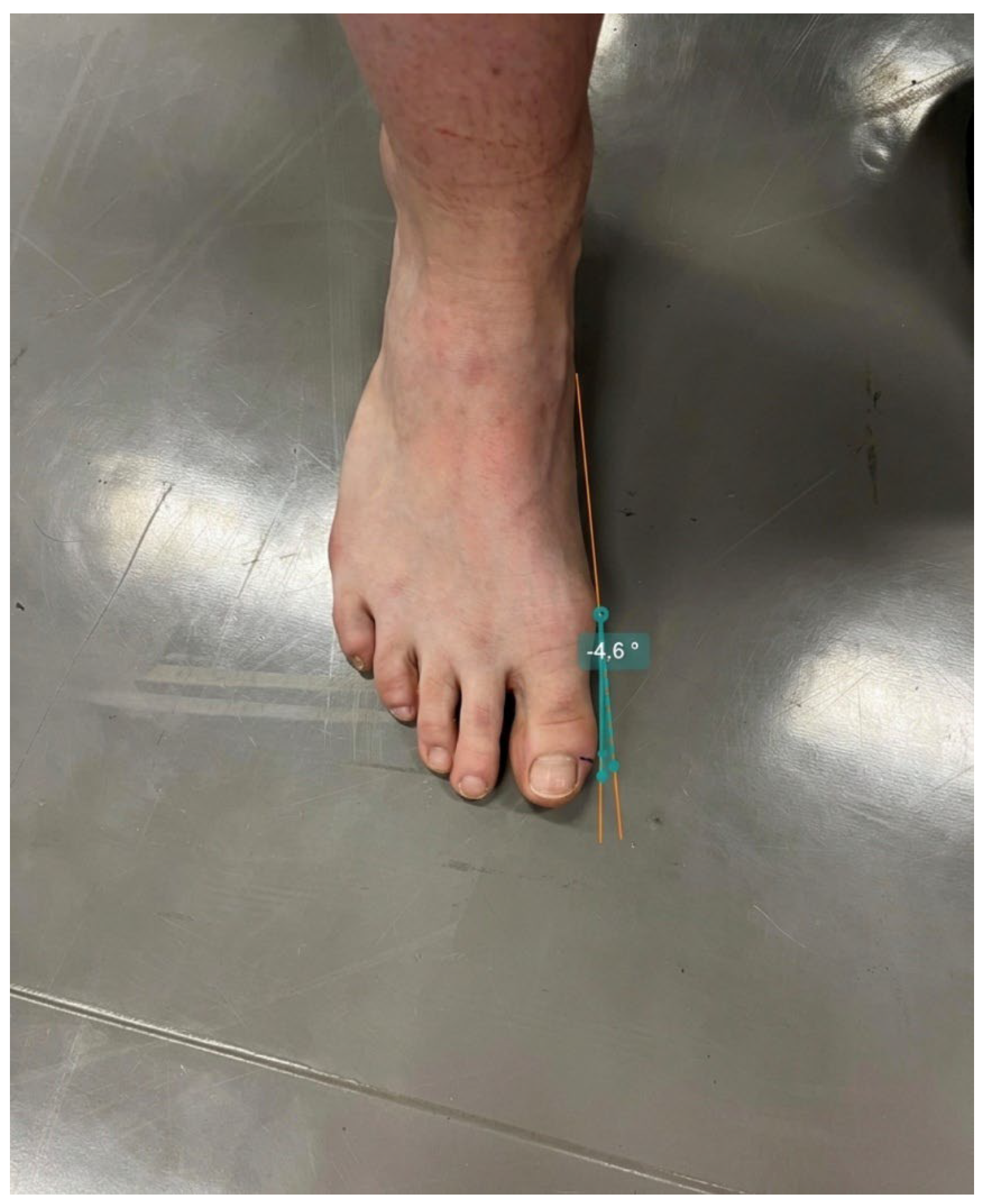
Hallux valux alignment in the frontal plane was quantified using standardized digital photography (
Figure 1). Participants stood barefoot on a flat surface with body weight evenly distributed between both feet and toes relaxed in a natural position. Photographs were taken from a superior view with the camera positioned perpendicular to the foot. Image analysis was performed using Kinovea software (version 2024.1), where lines were drawn along the longitudinal axis of the first metatarsal and the proximal phalanx of the hallux. The angle between these axes was defined as the hallux valgus angle. Image analysis was performed by an investigator blinded to group allocation and measurement time point.
Passive and active hallux flexion range of motion (ROM), ankle dorsiflexion, medial longitudinal arch height, hallux valgus angle, and postural stability were assessed using standardized procedures. All measurements were performed by the same examiner under identical laboratory conditions.
Passive flexion of the hallux was measured on the dominant limb using a universal goniometer. An anatomical reference point was marked at the head of the first metatarsal. Participants were seated with the ankle positioned at approximately 90° relative to the shank. The stationary arm of the goniometer was aligned with the longitudinal axis of the first metatarsal, while the movable arm followed the proximal phalanx of the hallux. Participants passively flexed the hallux using one hand until a point of mild resistance was reached. Care was taken to avoid lifting the foot or allowing pronation or supination during the movement. The angle between the first metatarsal and the proximal phalanx was recorded. Three trials were performed, and the mean value was used for analysis.
Active hallux flexion was assessed in the same seated position and anatomical alignment as the passive measurement. Participants actively flexed the hallux without external assistance while maintaining full foot contact with the supporting surface. The stationary arm of the goniometer was aligned with the first metatarsal axis and the movable arm with the proximal phalanx. Three trials were performed, and the mean value was retained for statistical analysis.
Functional ankle dorsiflexion ROM was assessed bilaterally using a weight-bearing lunge test. A measuring tape was fixed perpendicular to a wall. Participants performed the test barefoot, placing the tested foot along the measuring tape with the great toe positioned at the starting point. While keeping the heel in contact with the floor, participants slowly advanced the knee toward the wall until gentle contact was achieved without heel lift. The distance between the wall and the tip of the great toe was recorded in centimeters. Three trials were performed for each limb, and the highest value (greatest distance achieved while maintaining heel contact with the floor) was used for analysis.
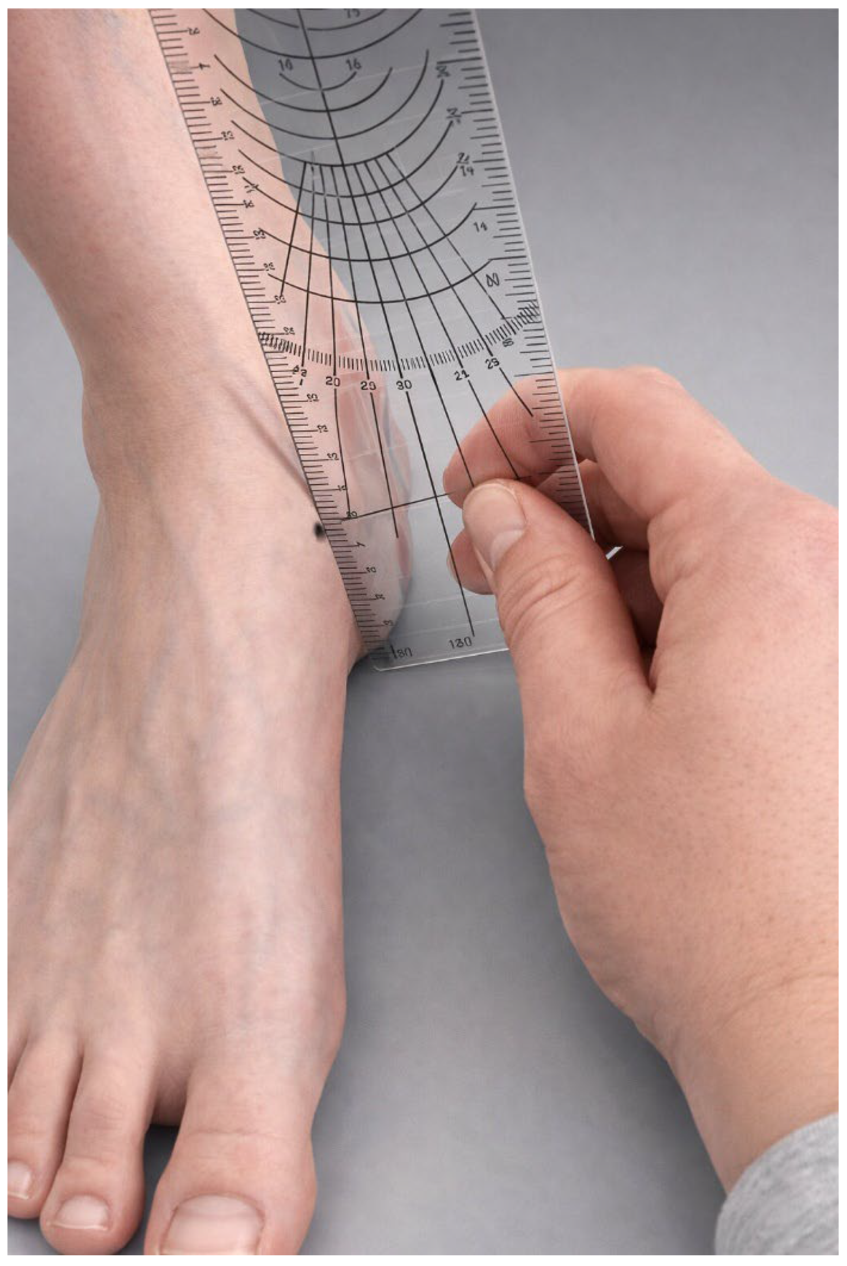
Arch height was evaluated using the navicular drop test (
Figure 2). The navicular tuberosity was palpated and marked. With the subtalar joint positioned in neutral, the vertical distance between the navicular tuberosity and the floor was measured in millimeters first in a seated position (hips and knees flexed at 90°) and subsequently during relaxed standing. Navicular drop was calculated as the difference between seated and standing measurements.
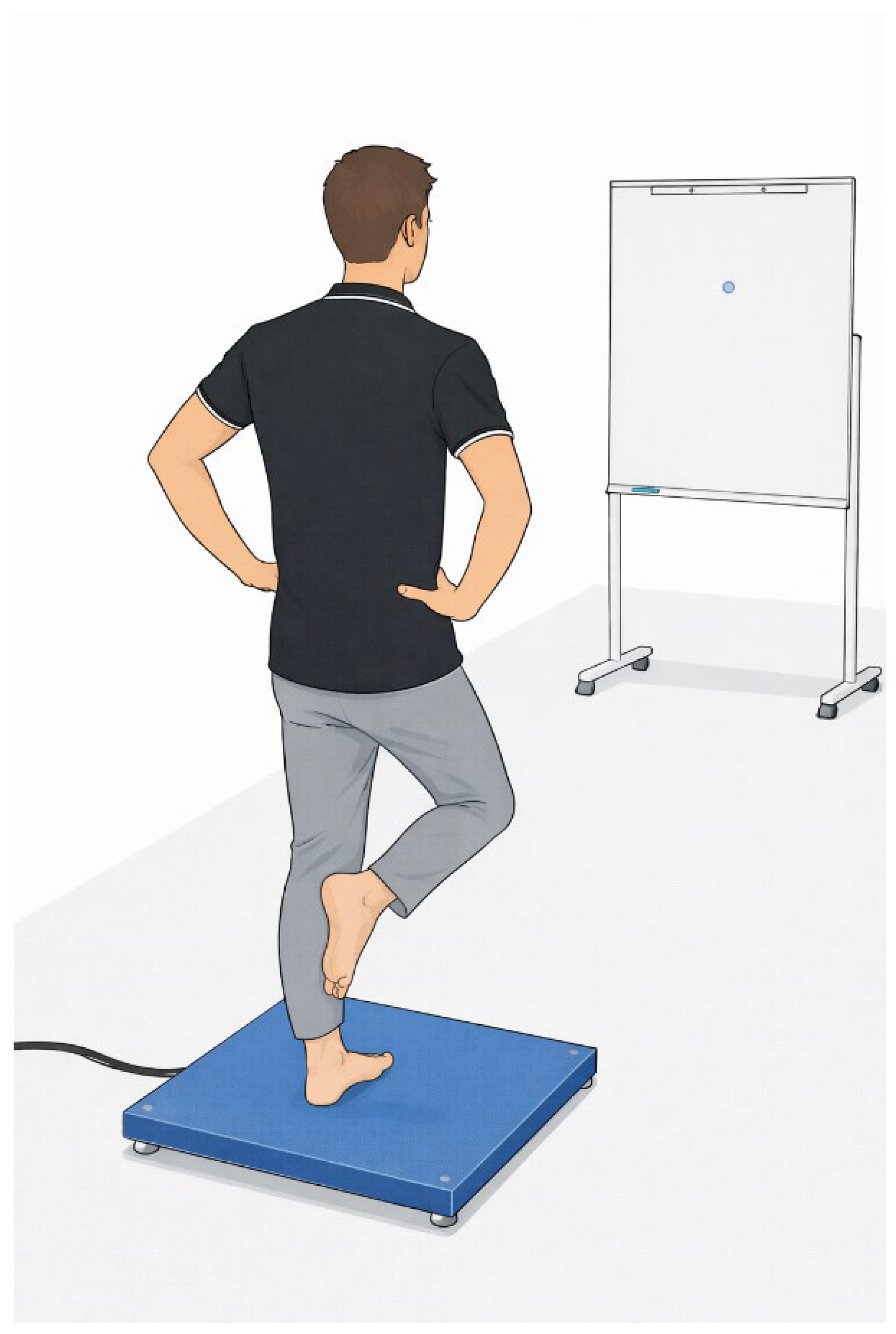
Postural stability was evaluated using a single-leg stance test performed on a force platform (Kistler, Winterthur, Switzerland) (
Figure 3). Participants stood barefoot on the dominant limb with arms positioned alongside the body. Each participant completed three 30 s trials under eyes-open conditions followed by three trials with eyes closed. During testing, the stance foot was required to remain stationary. Center-of-pressure (CoP) displacement was recorded, and the following variables were analyzed: CoP sway area (mm
2) and CoP velocity (mm/s) in the anterior–posterior and medial–lateral directions. Mean values across trials were used for statistical analysis.
2.4. Intervention
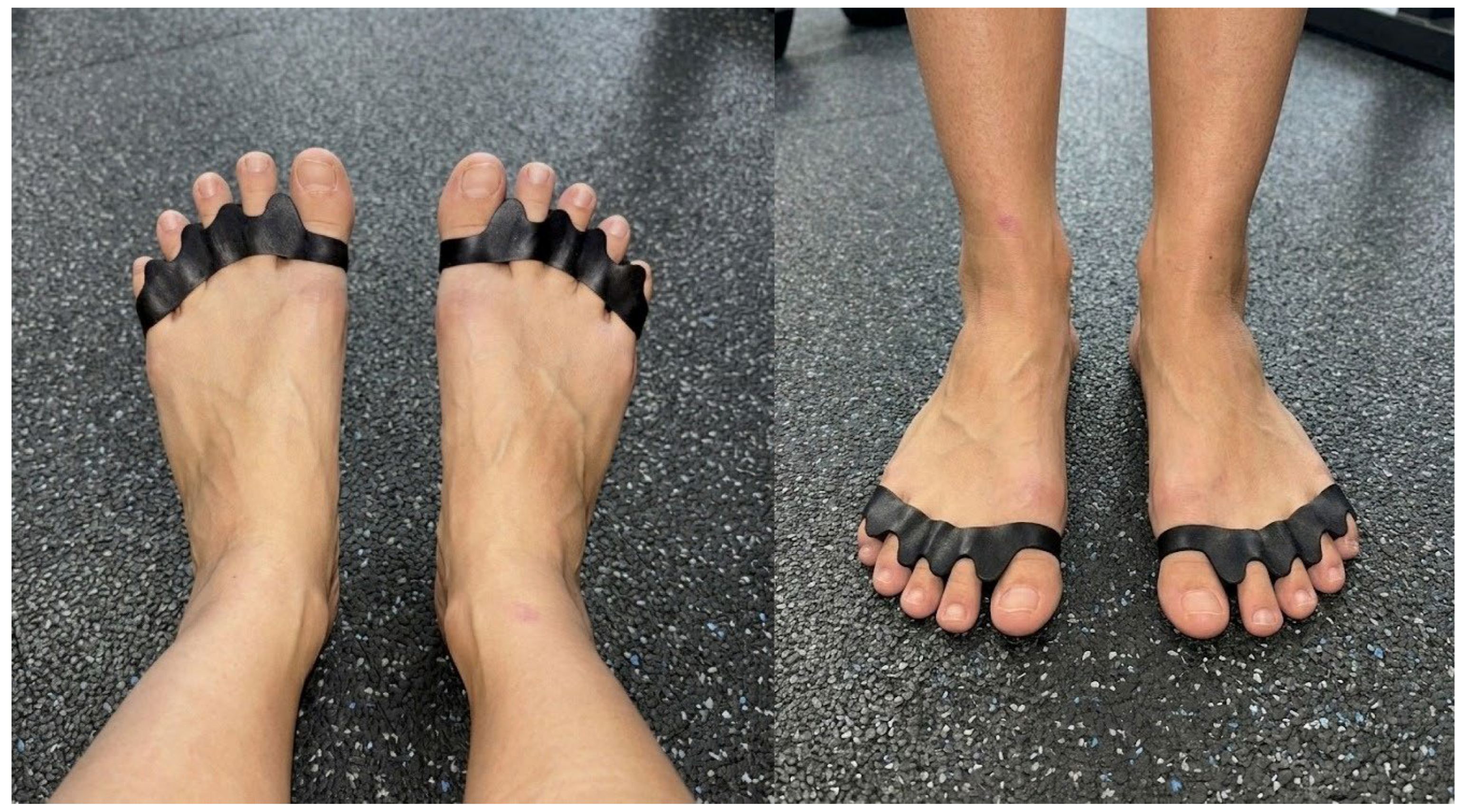
The exercise intervention was designed to strengthen intrinsic and extrinsic foot muscles and to improve ankle joint stability and foot posture control. The exercises aimed to increase active and passive hallux range of motion, improve functional ankle dorsiflexion, and enhance neuromuscular control of foot positioning during single- and double-leg support tasks. The intervention lasted six weeks, during which participants performed exercise sessions every other day (22 sessions in total). Depending on group allocation, participants completed either the strengthening program alone or the same program combined with the use of a silicone toe spacer worn during exercise execution. The exercise program consisted of four functional strengthening exercises: forward and backward toe walking, forward and backward heel walking, alternating heel-to-toe transitions performed in place, and lateral toe walking performed in a semi-squat position. All exercises were performed barefoot on a flat surface. Each exercise was completed in three sets, with approximately 60–90 s of rest between sets. Toe and heel walking exercises were performed over a distance of 10 m, repeated five times forward and backward per set. Alternating heel-to-toe transitions consisted of 10 repetitions per set. Lateral toe walking in a semi-squat position was performed over five lengths in each direction per set. No external loading was used; intensity was controlled through movement quality and postural alignment. Participants were instructed to perform all movements in a controlled manner while maintaining stable foot alignment and avoiding compensatory trunk or lower-limb movements. Participants received standardized instructions at the beginning of the intervention and were encouraged to maintain consistent exercise execution throughout the study period. Participants in the combined group performed the same strengthening program while wearing a silicone-based interdigital toe spacer (placed between the hallux and second toe) during exercise execution (
Figure 4). Participants recorded the completion of each exercise session in individual training logs to monitor adherence to the prescribed intervention.
2.5. Statistical Analysis
Analyses were performed using all available data from participants who completed both testing sessions. Because all randomized participants completed the intervention period, analyses were effectively conducted according to the intention-to-treat principle. Statistical analyses were performed using IBM SPSS Statistics (version 26.0, IBM Corp., Armonk, NY, USA). Normality of data distribution was assessed using the Shapiro–Wilk test. Descriptive statistics are presented as mean ± standard deviation (SD). To examine the effects of the interventions, a two-way repeated measures analysis of variance (ANOVA) was conducted, with time (pre-intervention vs. post-intervention) as the within-subject factor and group (exercise, exercise + toe spacer) as the between-subject factor. Interaction effects (time × group) were analyzed to determine differential intervention effects between groups. Effect sizes were calculated using partial eta squared (η2). Statistical significance was set at p < 0.05. To control for the increased risk of Type I error associated with multiple comparisons of secondary outcomes, p-values were additionally evaluated using the Holm–Bonferroni sequential correction procedure, which controls the family-wise error rate while maintaining greater statistical power than the traditional Bonferroni correction.
3. Results
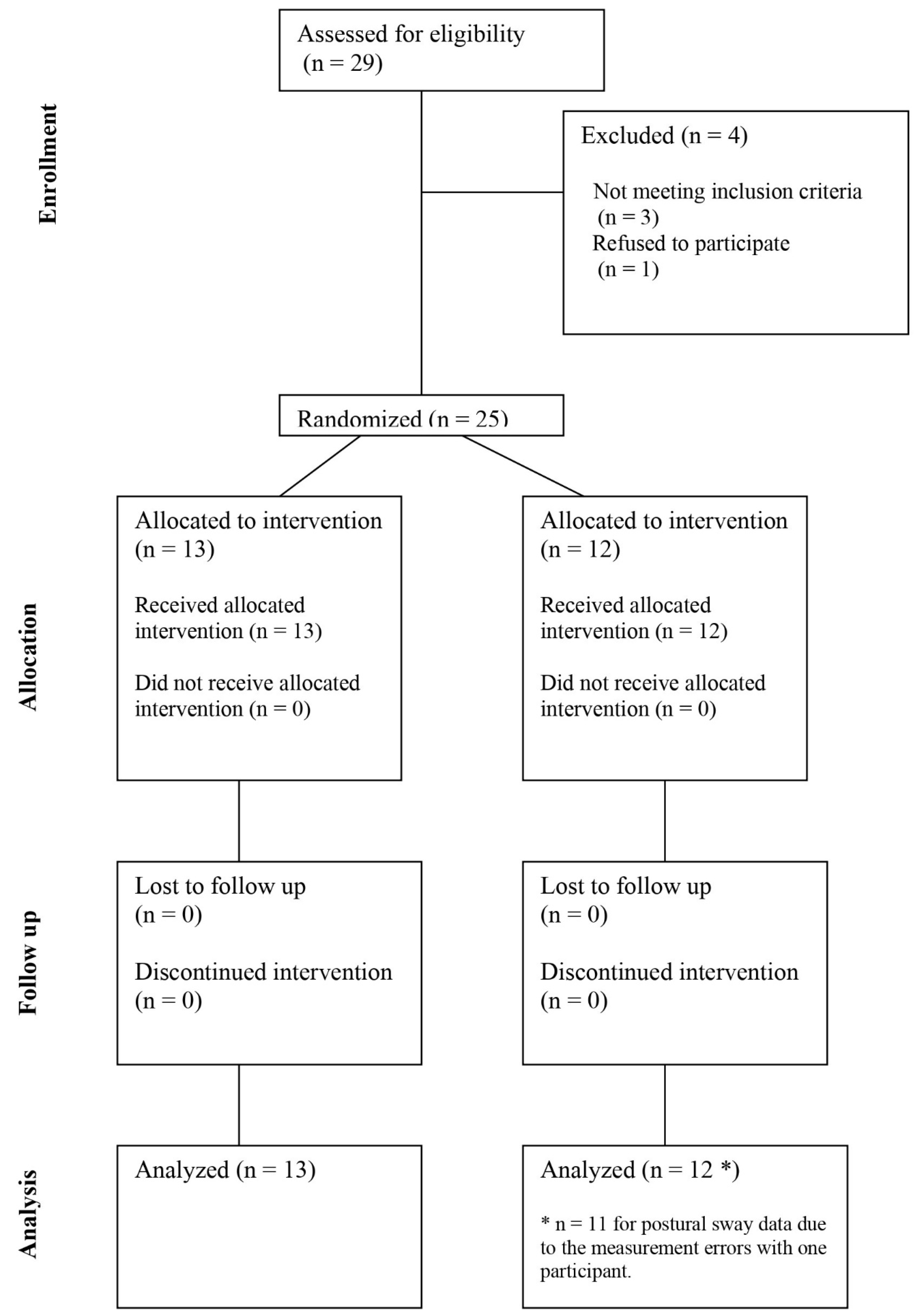
A total of 29 individuals initially volunteered to participate in the study. After screening for eligibility, 25 participants met the inclusion criteria and were included in the study, while four individuals were excluded (three did not meet the inclusion criteria due to previous injury of the ankle, and one declined to participate after detailed familiarization with the study).
Independent samples t-tests indicated that there were no statistically significant baseline differences between the groups for any of the measured variables (all p ≥ 0.105). Specifically, groups did not differ at baseline in active hallux range of motion (t(23) = 0.18, p = 0.863), passive hallux range of motion (t(23) = 0.44, p = 0.667), ankle dorsiflexion of the left (t(23) = −0.46, p = 0.651) or right limb (t(23) = −0.90, p = 0.379), or navicular drop measures (all p ≥ 0.105). Likewise, no baseline differences were observed in postural stability variables derived from center-of-pressure measures under eyes-open or eyes-closed conditions (all p ≥ 0.227). These results indicate that the groups were comparable prior to the intervention.
All participants completed the intervention period and post-intervention testing. Exercise adherence was high, with participants completing the prescribed sessions throughout the six-week intervention period, and no adverse events or injuries were reported. No participants withdrew from the study, and all available data were included in the analyses, with the exception of postural sway data for one participant (measurement error) (
Figure 5).
Table 1 presents descriptive statistics and results of the repeated-measures ANOVA analyses. Significant main effects of time were observed for several mobility-related outcomes. Hallux valgus angle decreased significantly from pre- to post-intervention (F = 13.0,
p = 0.001, η
2 = 0.361), while active hallux range of motion (ROM) increased (F = 27.5,
p < 0.001, η
2 = 0.545). Passive hallux ROM also demonstrated a significant time effect (F = 4.31,
p = 0.049, η
2 = 0.158). In addition, ankle dorsiflexion improved bilaterally, with significant effects of time for both the left (F(1,22) = 37.9,
p < 0.001, η
2 = 0.633) and right limb (F = 23.9,
p < 0.001, η
2 = 0.510). No significant main effects of group were identified for any variable (all
p ≥ 0.120).
A significant time × group interaction was observed only for passive hallux ROM (F = 4.72, p = 0.040, η2 = 0.170), indicating that the increase occurred predominantly in the exercise-only group, whereas no meaningful change was observed in the exercise plus toe spacer group. No interaction effects were found for hallux valgus angle (F = 1.65, p = 0.212, η2 = 0.067), active hallux ROM (F = 0.120, p = 0.732, η2 = 0.005), left ankle dorsiflexion (F = 0.312, p = 0.582, η2 = 0.014), or right ankle dorsiflexion (F = 0.601, p = 0.446, η2 = 0.025).
Measures of medial longitudinal arch height showed no significant changes over time, either during standing (F = 2.45, p = 0.131, η2 = 0.096) or single-leg stance (F = 2.63, p = 0.118, η2 = 0.103), and no significant interaction effects were detected (p ≥ 0.193).
Postural stability outcomes derived from center-of-pressure (CoP) measures demonstrated no statistically significant effects of time or time × group interaction under eyes-open conditions, including CoP velocity in the anterior–posterior direction (F = 0.014, p = 0.906, η2 = 0.001), medial–lateral direction (F = 0.792, p = 0.383, η2 = 0.035), or sway area (F = 0.079, p = 0.781, η2 = 0.004). Similarly, no significant effects were observed under eyes-closed conditions for CoP velocity in the anterior–posterior (F = 1.85, p = 0.188, η2 = 0.081) or medial–lateral directions (F = 0.010, p = 0.921, η2 < 0.001). CoP sway area during eyes-closed stance showed a non-significant trend toward reduction over time (F(1,19) = 3.88, p = 0.064, η2 = 0.170), without a significant interaction effect (F(1,19) = 0.513, p = 0.482, η2 = 0.026).
4. Discussion
The aim of this study was to investigate the effect of a silicone toe separator combined with exercise compared with exercise alone on hallux valgus alignment and foot function in healthy individuals. Changes in selected variables were analyzed over time between the two groups to determine whether either intervention produced superior effects. The results showed that both exercise alone and exercise combined with a silicone toe separator reduced the hallux valgus angle and increased active hallux flexion ROM, whereas only exercise alone improved passive hallux flexion ROM. Both groups demonstrated increased active ankle dorsiflexion bilaterally. No significant improvements were observed in medial longitudinal arch height, assessed as navicular drop, nor in static balance with eyes open. During single-leg stance with eyes closed, small reductions in center-of-pressure sway velocity and area were observed in both groups; however, these changes were insufficient to confirm the hypothesis.
The reduction in hallux valgus angle in both groups suggests that targeted intrinsic foot muscle strengthening alone may positively influence hallux alignment. This finding is consistent with previous research demonstrating that strengthening the intrinsic foot muscles, particularly the abductor hallucis, contributes to improved hallux alignment and foot function [
21]. The addition of a silicone toe separator did not significantly enhance the corrective effect compared with exercise alone. Mechanical devices primarily provide passive alignment without necessarily inducing functional neuromuscular adaptations, whereas strengthening intrinsic muscles directly improves medial stabilization of the hallux. In contrast, other studies have reported reductions in hallux valgus angle following the use of toe separators alone in both healthy individuals [
24] and individuals with symptomatic deformity [
30]. Kim et al. [
31] also reported improvements in resting and active hallux alignment after an eight-week combined intervention. Differences between studies may be attributable to variations in design, particularly the inclusion of a toe-separator-only group in previous research. Additionally, baseline deformity values in the present sample were relatively small (~7°), likely limiting the magnitude of change. The short intervention duration may have further contributed to the absence of significant between-group differences. Nevertheless, the trend toward greater reduction in the combined group (−0.55° vs. −0.26°) suggests that longer interventions might yield more pronounced effects, as reported in longer-term studies [
32,
33,
34].
Active hallux flexion ROM increased significantly over time (
p < 0.001) in both groups, with no group or interaction effects, indicating comparable effectiveness. This improvement likely reflects enhanced activation and strength of the flexor hallucis brevis and flexor hallucis longus, supporting previous findings on intrinsic foot muscle training [
3,
35]. Increased muscle strength and neuromuscular control may contribute to improved toe function and medial longitudinal arch support. In contrast, passive hallux flexion improved only in the exercise group, suggesting that the toe separator may have partially restricted passive motion. Similar observations were reported by Scherer et al. [
36], who found that orthotic devices can influence first metatarsophalangeal joint motion, not always in the direction of flexion. Abdalbary [
33] likewise reported alignment improvements but minimal ROM changes following combined interventions. These findings indicate that targeted exercise appears sufficient for improving both active and passive hallux mobility, whereas adjunctive passive devices may not provide additional benefit in this regard. However, this interaction effect should be interpreted cautiously. Passive hallux ROM represented the only outcome showing a significant time × group interaction among several tested variables, and therefore the possibility that this finding reflects a chance occurrence cannot be excluded.
The functional weight-bearing ankle dorsiflexion test demonstrated significant improvements following both interventions, without differences between groups. This suggests that strengthening exercises targeting the foot may indirectly enhance ankle mobility. Previous research has reported positive associations between intrinsic foot muscle strength and ankle dorsiflexion in healthy individuals, supporting this interpretation [
37]. Improved neuromuscular control, reduced stiffness, and more favorable segmental alignment may contribute to these changes.
Neither intervention significantly affected navicular drop. This finding aligns with studies reporting minimal changes in arch height following short-term strengthening programs [
20,
38,
39]. Conversely, other investigations have demonstrated improvements after longer or more comprehensive interventions [
35,
40,
41]. Kirmizi et al. [
27] reported positive effects after a 12-week intervention using orthotic insoles; however, methodological differences limit direct comparison. Given that navicular drop reflects both passive and active foot stability, longer intervention periods or more specific short-foot exercises [
3,
42,
43] may be required to induce measurable structural adaptations. Moreover, the navicular drop test may lack sensitivity to detect subtle short-term changes.
No statistically significant changes were observed in static balance under either visual condition. Although a trend toward reduced sway area was noted (
p = 0.064; η
2 = 0.17), the effect did not reach statistical significance. Previous research in individuals with flexible flatfoot demonstrated improvements in static balance following both exercise and combined interventions [
27], suggesting that baseline characteristics may influence responsiveness. Additionally, a recent study by Kozinc et al. [
44] reported improvements in postural stability parameters, including sway velocity and area, during single-leg stance in healthy individuals even without an intervention, highlighting the influence of intrinsic factors on balance outcomes. In the present study, participants were healthy individuals with near-optimal baseline values, limiting the potential for detectable improvement. Postural stability adaptations typically require longer or more demanding interventions. The observed trend toward improvement in the eyes-closed condition may reflect enhanced proprioceptive control in the absence of visual feedback, which warrants further investigation in future studies.
The reliability and sensitivity of the measurement procedures should be considered when interpreting the findings. Digital photographic assessment of hallux valgus angle is reliable but may be less sensitive to small short-term changes [
45]. Goniometric measurements of hallux ROM demonstrate moderate reliability (ICC ≈ 0.60–0.85) and may be influenced by landmark identification [
46]. The navicular drop test also shows moderate reliability (ICC ≈ 0.50–0.80) [
28,
29]. In contrast, the weight-bearing ankle dorsiflexion test (ICC > 0.90) [
47] and force-plate postural stability assessments (ICC > 0.85) [
48] demonstrate high reproducibility. Taken together, the pattern of significant findings may therefore partly reflect differences in measurement sensitivity across tools, with outcomes assessed using more reliable instruments showing more consistent intervention-related changes.
In summary, targeted intrinsic foot muscle strengthening effectively improved active and partially passive hallux mobility and ankle dorsiflexion in healthy individuals. The addition of a silicone toe separator did not provide superior short-term benefits compared with exercise alone. Although no significant changes were observed in medial longitudinal arch height or static balance, the observed trends suggest potential neuromuscular adaptations that may become more pronounced with longer or more intensive interventions, or in populations with existing functional impairments. Future research should further explore the long-term effects of combined interventions and clarify the clinical relevance of adjunctive toe separators in both healthy and pathological populations.
4.1. Clinical Implications
From a clinical perspective, the findings of the present study suggest that a structured intrinsic foot muscle strengthening program alone may be sufficient to produce meaningful short-term improvements in hallux alignment and foot and ankle mobility in healthy adults. Clinicians and rehabilitation professionals working with individuals at risk of developing hallux valgus deformity or foot dysfunction may therefore consider targeted strengthening exercises as a primary conservative intervention. The absence of additional benefit from the silicone toe spacer in the short term suggests that routine prescription of such devices alongside exercise may not be necessary in healthy populations, potentially reducing intervention costs and complexity. However, given that the present study was conducted in healthy young adults with relatively mild baseline deformity, the clinical implications for individuals with symptomatic hallux valgus or existing foot pathology remain to be established and should be explored in future research.
4.2. Limitations
Several limitations should be acknowledged. First, the trial was exploratory and not powered to detect small between-group differences, particularly for interaction effects; thus, clinically relevant additive effects of the toe spacer cannot be excluded. In addition, multiple secondary outcomes were tested without adjustment for multiplicity, so isolated significant findings (e.g., passive hallux ROM interaction) should be interpreted cautiously. The relatively small sample size may have limited the statistical power to detect small between-group differences, particularly for interaction effects. Consequently, the absence of significant between-group effects should be interpreted cautiously, and future studies with larger samples are needed to confirm the present findings. Only healthy individuals of similar age were included, which restricts the applicability of the results to broader or clinical populations. Future studies should include larger and more diverse samples and compare responses between healthy individuals and those with existing foot or ankle pathologies. Second, the intervention period was relatively short. A longer duration of exercise, either alone or combined with a toe separator, might provide clearer insight into potential between-group differences and effect magnitudes. An additional methodological limitation is the absence of a separate non-intervention control group. Without such a control condition, it is not possible to determine whether the observed outcomes (or lack thereof) are attributable solely to the interventions or to other factors, such as natural variability in foot function, participant motivation, or limited sensitivity of the measurement tools. Next, although the outcome assessor was blinded to group allocation, participant blinding was not possible due to the nature of the intervention. This may have introduced some degree of performance bias. Furthermore, a potential learning effect cannot be excluded, as repeated testing may have led to improved performance due to increased familiarity with the procedures rather than true functional changes. Inclusion of an appropriate control group in future research would allow for more robust interpretation of the findings. Although adherence to the intervention was monitored using participant training logs, the exercise sessions were not supervised. Consequently, variability in exercise execution, intensity, and movement quality cannot be excluded and may have influenced the magnitude of the observed effects. Finally, sex-related differences in foot morphology, biomechanics, and neuromuscular responses to exercise may also influence the effects of the intervention. Although both male and female participants were included in the present study, the sample size was not sufficient to allow meaningful sex-specific analyses. Future studies with larger samples should examine whether the responses to intrinsic foot muscle strengthening and toe separator use differ between sexes.