1. Introduction
High-intensity static isometric exercises are widely used in both sports training and rehabilitation, because they can effectively enhance muscle strength, joint stability, and neuromuscular coordination. However, these types of exercises are associated with substantial peripheral and cardiovascular stress [
1]. Sustained isometric muscle contractions restrict intramuscular blood flow, increase intramuscular pressure, and may result in rapid local fatigue and delayed performance recovery. These responses are closely associated with the balance between muscle oxygen delivery and utilization, autonomic nervous system modulation, and post-exercise hemodynamic adjustments [
2,
3]. Furthermore, elevated intramuscular pressure may promote anaerobic metabolism, increase lactate accumulation, and transiently alter muscle mechanical properties, such as stiffness and elastic behavior, potentially affecting subsequent exercise performance and increasing injury risk [
4,
5]. More broadly, recovery after muscular work should also be considered in the context of muscle energetics and task-specific mechanical demand [
6,
7].
Recovery strategies following high-intensity exercise play a crucial role in both the restoration of short-term performance and long-term physiological adaptation. Passive rest remains the most commonly applied recovery method, since it facilitates spontaneous recovery of energy stores and the clearance of metabolic by-products [
7]. However, recently, increasing attention has been directed toward mechanically based recovery interventions, with potential to influence peripheral circulation and cardiovascular load without additional metabolic demand. These approaches include passive movements, massage, use of compression devices, and vibratory stimuli, which can enhance venous return, local perfusion, and mechanoreceptor activation [
8,
9,
10]. Among these methods, passive limb movement represents a relatively novel and underexplored strategy. Passive movement may be used to enhance muscle blood flow, reduce intramuscular pressure, and provide sensory and mechanical stimulation to the musculature, all without requiring voluntary muscle activation [
10,
11].
Muscle oxygen saturation (StO
2) can be assessed using near-infrared spectroscopy (NIRS), to enable non-invasive evaluation of the relative balance between oxygen delivery and utilization within skeletal muscle [
12]. When combined with arterial blood pressure, heart rate variability (HRV), and muscle mechanical measurements, StO
2 can provide an integrated but indirect assessment of peripheral and systemic responses to exercise and recovery [
13]. Importantly, these parameters should be interpreted as surrogate physiological markers and not as direct measures of muscle blood flow, venous return, cardiac output, or specific metabolic mechanisms. Nevertheless, their combined assessment can be used to enable more comprehensive characterization of recovery processes following intense isometric exercise. Accordingly, recovery after repeated isometric work is likely shaped by interacting autonomic, peripheral, and task-specific factors rather than a single mechanism [
2,
3,
6].
Although previous studies have extensively investigated the effectiveness of active and passive recovery strategies after dynamic exercise; research data related to the effects of passive foot flexions following high-intensity static isometric exercise remains limited, and existing recovery research has largely focused on traditional methods rather than more mechanically driven approaches [
7,
14]. In addition, most research studies have focused on isolated physiological outcomes. Consequently, the integrative interpretation of recovery processes remains challenging. In contrast, a limited number of studies have reported integrated assessments of peripheral (muscle oxygenation, mechanical muscle properties) and systemic (hemodynamic and autonomic) responses within a single experimental protocol. Moreover, because of the multi-factorial nature of physiological responses to isometric exercise, a comprehensive research approach is warranted [
14]. Therefore, the aim of the present study was to evaluate and compare the acute effects of passive foot flexions versus passive rest on post-exercise recovery using indirect markers of autonomic modulation, peripheral tissue oxygenation, muscle mechanical properties, and subsequent isometric performance.
2. Materials and Methods
2.1. Trial Design
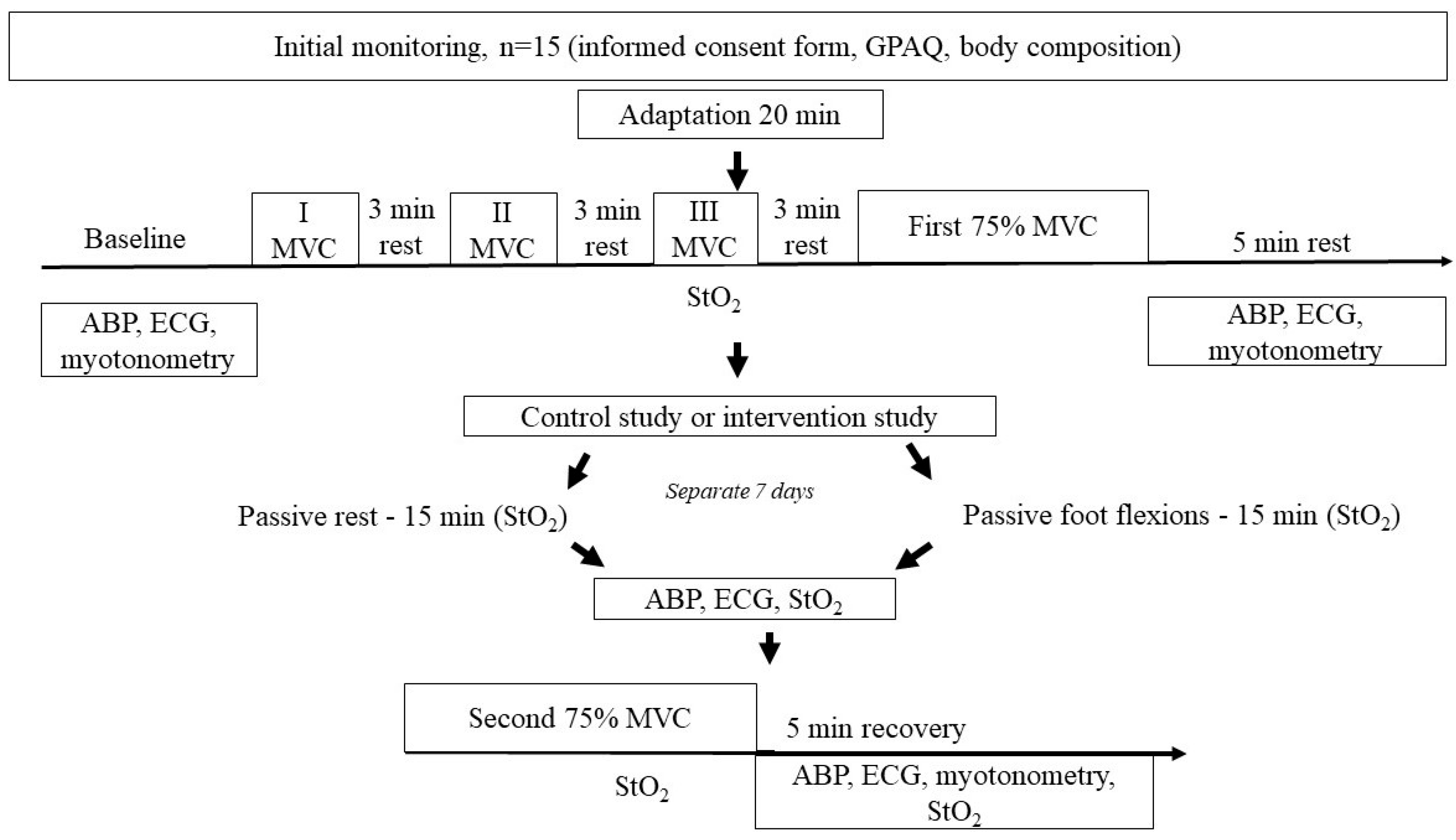
The present study employed a randomized crossover experimental trial design. Before the initial testing session, participants attended an introductory visit, during which they were familiarized with the study procedures and provided written informed consent. For each study participant, body composition was evaluated, and physical activity levels were assessed using a Global Physical Activity Questionnaire (GPAQ). Participants were instructed to refrain from strenuous physical activity and food intake for at least 3 h before each experimental session. All measurements were recorded in a standardized laboratory environment (temperature: 20–22 °C; relative humidity: 45–55%). To minimize circadian variation, each participant was tested at the same time of day in both experimental conditions, and all experimental sessions were conducted between 10:00 and 12:00. Each participant completed two experimental sessions in a randomized order, as follows: (1) a control condition involving passive rest during recovery; and (2) an intervention condition involving passive foot flexions during recovery. The two sessions were separated by a 7-day washout period to minimize any potential carry-over effects. With the exception of the recovery modality, both experimental sessions were conducted using an identical protocol (
Figure 1).
At the beginning of each session, participants underwent a 20 min adaptation period in a resting position, followed by assessment of baseline cardiovascular and muscular parameters, including: arterial blood pressure (ABP), heart rate variability (HRV), muscle oxygen saturation (StO2), and muscle tone. Maximal voluntary contraction (MVC) of the plantar flexor muscles was then assessed to facilitate individualized standardization of exercise intensities. Following MVC assessment, participants performed a sustained static isometric plantar flexion contraction at 75% of their MVC, which served as the initial fatigue-inducing task for both experimental conditions. During this task, participants were instructed to sustain the target force level as steadily as possible, with real-time visual feedback provided when applicable, and were verbally encouraged to continue the contraction for as long as possible. Immediately after this contraction, participants were given a 15 min recovery phase, during which they either remained at rest (control condition) or underwent passive foot flexions (intervention condition).
After the 15 min recovery phase, participants performed a second static isometric plantar flexion contraction at 75% of their MVC, in order to assess work capacity. Work capacity was defined as the duration of force maintenance (time to task failure), expressed in seconds. This contraction was followed by a 5 min recovery period, during which post-exercise physiological measurements were recorded. For each participant, the holding duration at 75% MVC was individually determined, and subsequently divided into four relative time intervals, corresponding to 25%, 50%, 75%, and 100% of the individual holding time. Study participants were carefully monitored throughout all maximal and submaximal contractions, in order to ensure the use of correct technique and to prevent compensatory movements. Adequate rest periods (3 min—maximal, 5 min—submaximal) were provided between contractions, in order to minimize the influence of cumulative fatigue on performance outcomes. No pharmacological agents, medical devices intended for clinical use, invasive procedures, or therapeutic interventions were administered, and no clinical outcomes were assessed during the present study. Study participation was permitted only after medical clearance was obtained from a physician. The study protocol was approved by the Lithuanian Bioethics Committee (Authorisation for Biomedical Research, 23 January 2020, No. L-20-1/1). All procedures were conducted in accordance with the principles of the Declaration of Helsinki.
2.2. Study Participants
The present study involved 14 physically active, healthy, non-smoking men (age: 22.8 ± 1.0 years; height: 179.1 ± 7.2 cm; body mass: 76.8 ± 9.8 kg; body mass index: 23.9 ± 1.6 kg/m2). All participants presented with a normal resting blood pressure and body mass index, and reported that they did not have a diagnosed medical condition, and did not use any medications or dietary supplements. Based on results from the Global Physical Activity Questionnaire, all study participants were classified as moderately physically active. Each participant had at least 1 year of regular gym-based training experience and reported at least 3 h of weekly physical activity, including aerobic and resistance exercises. Before enrolment, each participant obtained medical clearance, received detailed information regarding the study objectives and procedures, and provided written informed consent.
2.3. Maximal Voluntary Contraction Assessment
At the beginning of each experimental session, Maximal Voluntary Contraction (MVC) of the plantar flexor muscles was assessed using a dynamometer (VALD DynaMo Max, Brisbane, Queensland, Australia), in order to determine individual maximal force-generating capacities, and to standardize exercise intensity for each participant. Participants were seated in a steel dynamometer chair and were instructed to stabilize their body position by holding the support frame with both hands during exertion. To minimize compensatory muscle activity, the thigh of the exercising leg was fixed to the chair, thereby restricting hip and knee movement. The dynamometer was individually calibrated to fix the participant’s exercising leg at a knee angle of 90° and an ankle angle of 70°.
MVC was then determined by having the participant perform three maximal voluntary isometric plantar flexion contractions, each lasting approximately 3–5 s, but separated by 3 min passive rest intervals to minimize fatigue. Strong verbal encouragement was provided during each contraction to ensure maximal effort. The highest MVC value obtained during the session was used for subsequent analyses, and for normalization of exercise intensity. MVC values were fixed before the experimental protocol and used as a reference value for force normalization. The mean session-specific MVC values were 974.3 ± 177.6 N in the passive rest condition and 986.2 ± 184.4 N in the passive foot flexions condition.
2.4. Recovery Conditions and Work-Capacity Assessment
Following the initial static isometric plantar flexion task performed at 75% of the session-specific MVC, participants underwent one of two recovery conditions. Both isometric tasks were performed under the same conditions and analyzed using the same task-failure criterion. For the control condition, participants remained at rest for 15 min in a seated position, during which no voluntary or externally induced movement of the lower limbs was allowed. Participants were instructed to remain relaxed and avoid any muscle activation of the lower extremities throughout the passive rest period.
In the passive foot flexions condition, participants underwent 15 min of device-assisted passive ankle–foot flexion immediately after completion of the same static isometric plantar flexion task performed at 75% MVC. The passive foot flexion intervention was administered using a motor-driven mechanical device, and was performed without voluntary muscle activation (AnkleMotion CPM, Kerala, India). Participants’ feet were secured to the pedals, which were moved by an electric motor, inducing rhythmic passive foot flexions at a frequency of 24 cycles per minute. The total range of motion was 35°, with the ankle positioned at 90° and the movement consisted of 10° of plantar flexion and 25° of dorsiflexion. The movement was performed for 15 min.
During the 15 min recovery phase, participants in both conditions were continuously monitored for signs of discomfort or adverse responses and were instructed to remain relaxed. No adverse events were reported. Although muscle activation was not directly assessed using electromyography, participants were instructed to remain relaxed and the intervention was continuously visually monitored. Therefore, low-level voluntary muscle activation or co-contraction cannot be fully excluded. Importantly, these variables should be interpreted as indirect physiological markers rather than direct measures of muscle blood flow, venous return, cardiac output, or metabolic clearance. After the 15 min recovery phase, participants performed a second static isometric plantar flexion task at 75% MVC to assess work capacity. Work capacity was defined as time to task failure and expressed in seconds. Real-time visual feedback and standardized verbal encouragement were provided during both isometric tasks, which were performed under the same conditions and analyzed using the same task-failure criterion.
2.5. Assessments
2.5.1. Assessment of PA
PA was assessed using the Global Physical Activity Questionnaire (GPAQ), a widely validated and reliable tool developed by the World Health Organization [
15]. The GPAQ assesses PA across three domains: occupational activity, active transportation, and leisure-time or recreational activity. The GPAQ consists of 16 questions that assess the intensity, frequency, and duration of PA. In the present study, all participants reported engagement in at least moderate-intensity PA.
2.5.2. Body Composition
Height and body mass for each participant were measured using calibrated instruments; while the subjects were barefoot and wearing only underwear. Stature was measured using a SECA® 213 stadiometer (SECA GmbH & Co., Hamburg, Germany), and body mass was recorded using a TANITA® BC-545 scale (TANITA Corporation, Tokyo, Japan).
2.5.3. Oxygen Saturation
Throughout each experimental protocol, changes in muscle oxygen saturation (StO2) were assessed using non-invasive near-infrared spectroscopy (NIRS), coupled with a photosensor device (Hutchinson Technology, Hutchinson, MN, USA). The photosensor was positioned over the primary working muscle group, the plantar flexor muscles.
StO2 was then recorded continuously throughout the entire protocol, including the pre-intervention resting period, warm-up, exercise, recovery phases, and for an additional 10 min post-exercise recovery period following the final workload stage. For data analysis, all StO2 values were normalized to baseline and expressed as percentages.
Near-infrared spectroscopy provides a relative measure of skeletal muscle oxygen saturation, which monitors the balance between oxygen delivery and oxygen utilization in the working muscle. However, NIRS cannot directly assess mitochondrial oxidative phosphorylation, ventilation–perfusion matching, or lactate metabolism.
2.5.4. Passive Muscle Stiffness
Passive muscle stiffness was measured in a relaxed state using a handheld myotonometry device MyotonPRO (Myoton AS, Tallinn, Estonia). The device applies a brief mechanical impulse of 15 ms to the muscle, which induces damped oscillations that are recorded by a built-in accelerometer. Key parameters were then calculated based on this oscillatory response, including muscle tone (F, Hz) and stiffness (S, N/m). Measurements were performed on the medial gastrocnemius muscle. This muscle was selected as an accessible superficial site relevant to the plantar flexion task. Two sets of five consecutive measurements were recorded at each site, and the mean value was used for analysis. The measurement point was marked on the subject’s skin with a permanent marker, in order to ensure consistency across sessions. All measurements were conducted by the same tester.
2.5.5. ABP Measurements
Arterial blood pressure (ABP) was measured using a cuff-based auscultatory method, where systolic blood pressure (SBP) and diastolic blood pressure (DBP) were identified by monitoring Korotkoff sounds (Riester, Jungingen, Germany). Using an appropriately sized cuff, all ABP measurements were made with participants in a seated position, and on the same arm across all experimental sessions.
Baseline ABP was assessed before the intervention, after a 20 min adaptation period in a quiet seated position. ABP measurements were obtained at baseline; immediately following the first static isometric contraction at 75% MVC; during the 15 min recovery phase for both the passive rest and passive foot flexions group conditions; and during the post-exercise recovery phase at 3 and 5 min following the second static isometric contraction at 75% MVC.
2.5.6. HRV Measurements
Heart rate variability (HRV) was assessed using a standard 12-lead electrocardiogram (ECG), which was recorded using a CardioScout Multi ECG system (Medset, Hamburg, Germany). ECG recordings were obtained at rest under baseline conditions, and during predefined measurement periods before and after the interventions. RMSSD was selected as the primary HRV index because it is considered the most suitable short-term time-domain measure for the post-exercise recovery windows used in the present protocol.
In order to record a stable baseline signal, after electrode placement, participants rested quietly in a seated position for a stabilization period before ECG recordings were conducted. HRV analysis was performed using ECG segments that were recorded at rest before the first fatigue-inducing task, and during the post-intervention recovery period following either passive rest or the passive foot flexions intervention.
Respiration was spontaneous and was not externally controlled during HRV assessment.
2.6. Statistical Analysis
All statistical analyses were performed using IBM SPSS Statistics version 26.0 (IBM Corp., Armonk, NY, USA). Data are expressed as the mean ± standard deviation (SD). The normality of the data distribution was assessed using the Shapiro–Wilk test (n = 14). No a priori sample size calculation was performed. Therefore, an additional sensitivity analysis was conducted for the primary outcome (holding time) to estimate the effect size detectable with the available sample size. Because all participants completed both experimental conditions (passive rest and passive foot flexions), a two-way repeated-measures ANOVA was conducted with two within-subject factors: time (measurement time points) and condition (passive rest vs. passive foot flexions conditions). Separate analyses were performed for the following parameters: heart rate, systolic and diastolic arterial blood pressure, RMSSD, StO2, muscle tone, muscle stiffness, and time to task failure. For cases where the assumption of sphericity was violated, the Greenhouse–Geisser correction was applied. In the presence of significant main or interaction effects, post hoc pairwise comparisons were performed using a Holm–Bonferroni adjustment. Effect sizes were reported as partial eta squared (η2p) for ANOVA and Cohen’s d for pairwise comparisons. Cohen’s d values were interpreted as small (0.2), moderate (0.5), and large (0.8). Statistical significance was set at p < 0.05.
3. Results
Changes in heart rate were monitored during the first recovery phase, the passive foot flexions intervention, and the second recovery period. During the first recovery phase, heart rates were found to gradually decrease for both conditions (F(4,56) = 255.01, p < 0.05), with no significant between-condition differences or interaction effects. Immediately after exercise, values were between 142.8 ± 16.1 and 142.6 ± 14.6 bpm; and gradually declined to between 83.0 ± 7.6 and 84.0 ± 13.5 bpm by the fifth minute of the recovery phase. Throughout the 15 min passive foot flexions intervention, heart rates remained relatively stable, and displayed only minor fluctuations; ranging between 82.7 ± 7.4 and 87.7 ± 12.8 bpm during the intervention. These results suggest that after the initial post-exercise decline, a steady heart rate was maintained during the intervention phase. During the second recovery phase, heart rate again decreased over time in both conditions (F(4,56) = 235.84, p < 0.05), from 142.0 ± 12.3 and 147.0 ± 14.5 bpm immediately after the intervention to between 89.0 ± 11.5 and 91.0 ± 16.4 bpm by the fifth minute of recovery. Overall, heart rate followed a similar pattern under both conditions, with no significant between-condition differences.
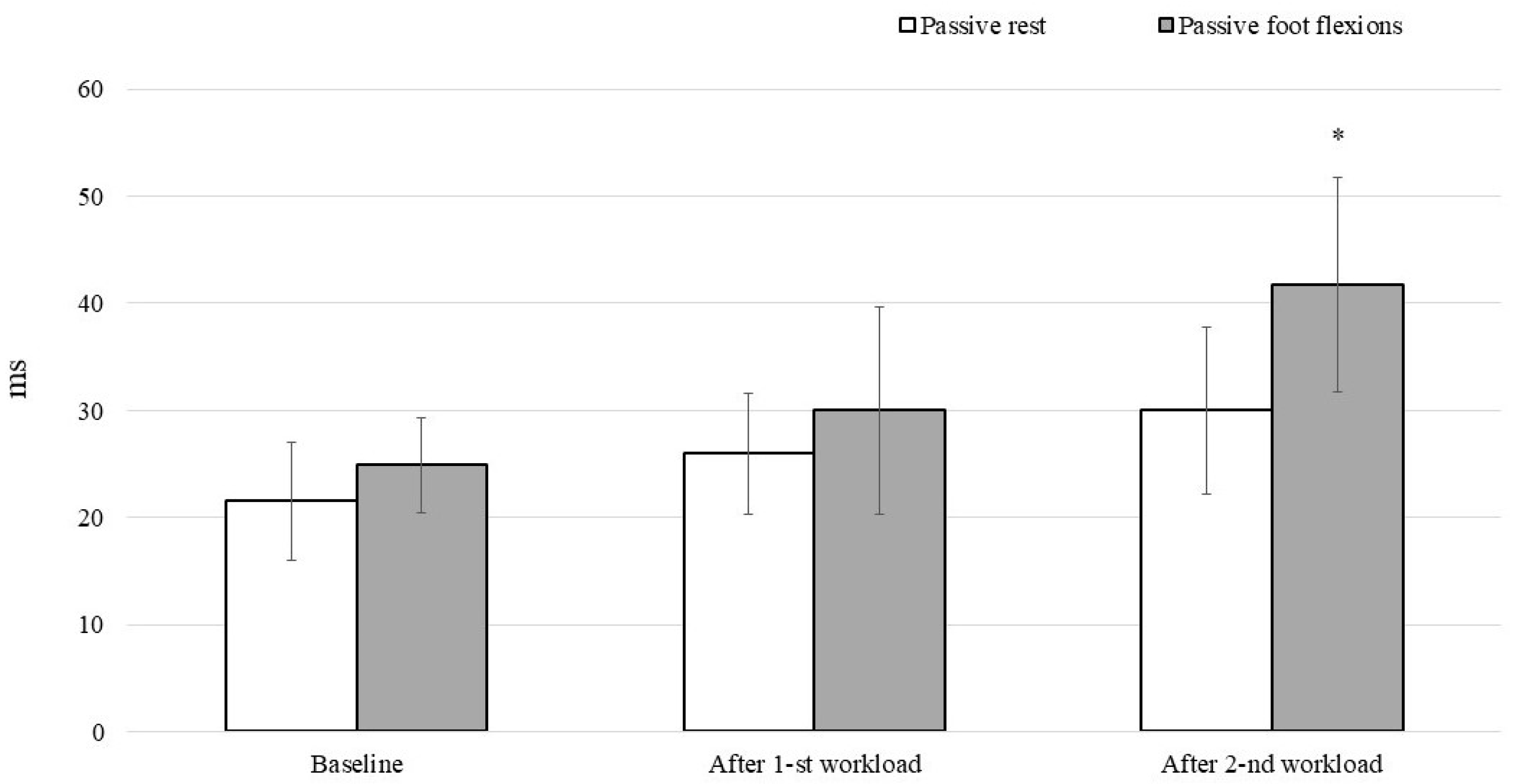
RMSSD indices were recorded at rest and after the interventions (
Figure 2). Before the intervention, RMSSD did not differ significantly between the two tested conditions: for passive rest, RMSSD was 21.55 ± 5.50 ms; for passive foot flexions, RMSSD was found to be 24.91 ± 4.43 ms. Following the first recovery period (After I), RMSSD increased for both conditions.
After the second recovery period (After II), RMSSD continued to rise in both conditions, with a significantly larger increase observed for the passive foot flexions condition: for passive rest, the RMSSD was 32.00 ± 7.78 ms; for passive foot flexions, the RMSSD was 41.79 ± 11.50 ms (p < 0.05, d = 1.50).
SBP values increased during both static workloads, and gradually declined throughout the recovery phase for both the passive rest and passive foot flexions conditions. Baseline SBP values were similar between conditions: for passive rest, SBP was 126.2 ± 8.2 mmHg; for passive foot flexions, SBP was found to be 128.3 ± 8.4 mmHg. Immediately after the first workload, SBP reached a level of 142.4 ± 17.2 mmHg under passive rest and 138.2 ± 16.1 mmHg under passive foot flexions, decreasing progressively to 120.0 ± 9.0 mmHg and 119.7 ± 8.8 mmHg, respectively, by 15 min of recovery. During the second workload, SBP was found to increase at a similar rate for both conditions and declined during recovery: immediately after the second workload, SBP for the passive rest condition was 136.3 ± 12.8 mmHg; for the passive foot flexion condition, SBP was 138.6 ± 18.5 mmHg.
DBP levels were measured at the following time points: at rest; before exercise; during two consecutive static workloads; and throughout the recovery phase under both the passive rest and passive foot flexions conditions. Baseline DBP values were comparable between the passive rest (78.5 ± 5.6 mmHg) and passive foot flexions (82.8 ± 7.2 mmHg) conditions. During the first static workload, DBP levels increased for both conditions, reaching approximately 83.6 ± 7.8 mmHg under passive rest and 85.2 ± 7.3 mmHg under the passive foot flexions condition during the recovery phase. By the end of the first 15 min recovery phase, DBP levels decreased toward baseline: for passive rest, DBP levels were 81.8 ± 6.4 mmHg; for the passive foot flexion condition, DBP levels were 82.0 ± 6.8 mmHg. During the second workload and subsequent recovery, DBP levels followed a similar pattern, with comparable values immediately after exercise (passive rest: 82.7 ± 5.9 mmHg; passive foot flexions: 82.8 ± 6.6 mmHg) and throughout the recovery period. Across all time points, no statistically significant differences in SBP or DBP levels were observed between the passive rest and passive foot flexions conditions.
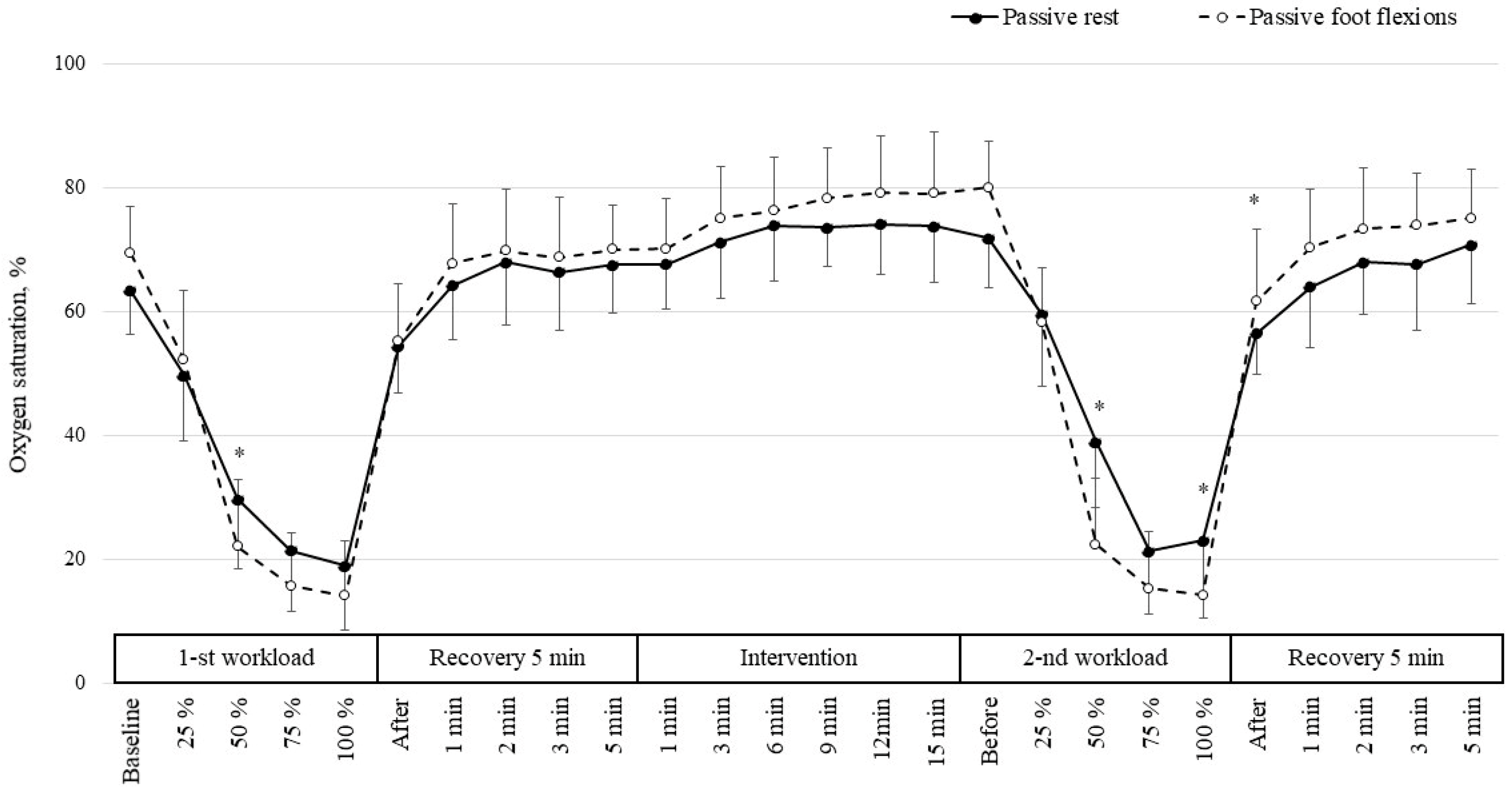
Tissue oxygen saturation dynamics were also monitored for both the passive rest and passive foot flexions conditions, during two static workloads and associated recovery periods (
Figure 3).
At baseline rest, StO2 levels were found to be higher during passive foot flexions compared to the passive rest condition. With increasing workload intensity (25–100%), a pronounced decrease in StO2 levels was observed for both interventions. At 25% workload, StO2 was significantly higher during passive foot flexions compared to the passive rest condition (p < 0.05, d = 0.75). At 75% workload, the decrease in StO2 was more pronounced during passive foot flexions. During the early recovery phase (1–5 min), StO2 was found to increase for both interventions, and no significant differences were observed between interventions. During the late recovery period (6–15 min), StO2 values were consistently higher during passive foot flexions.
Before the second static workload, no significant differences in StO2 were observed between interventions. However, at 50% workload, StO2 was significantly lower during passive foot flexions compared to the passive rest condition (p < 0.05, d = −1.54), suggesting a large effect. At 100% workload, the difference in StO2 values was also statistically significant (p < 0.05, d = −0.70). Immediately after the second workload, StO2 was significantly higher during passive foot flexions compared to the passive rest condition (p < 0.05, d = 0.76). During the early recovery phase (1–3 min), StO2 values remained higher during passive foot flexions. After 5 min of recovery, differences between the interventions decreased and were no longer significant.
Changes in muscle mechanical properties were assessed by measuring muscle tone and stiffness for both experimental conditions. At rest, muscle tone was slightly higher for the passive rest condition (13.6 ± 2.4 Hz) compared to the passive foot flexions condition (13.0 ± 0.9 Hz), while resting muscle stiffness was similar between interventions, at 231.2 ± 58.5 Nm vs. 228.0 ± 30.9 Nm. After the first static hold, both tone and stiffness increased under both tested conditions: tone reached 13.8 ± 1.3 Hz for the passive rest condition and 13.2 ± 1.6 Hz for the passive foot flexions condition. Similarly, stiffness increased to 257.4 ± 36.8 Nm for the passive rest condition and 240.4 ± 30.8 Nm for the passive foot flexions condition. Following the second static hold, tone remained slightly higher under passive rest (14.0 ± 1.7 Hz) than with the passive foot flexions condition (13.9 ± 2.1 Hz); while stiffness remained elevated under both conditions, showing comparable values of 251.2 ± 50.8 Nm vs. 248.8 ± 37.7 Nm between interventions.
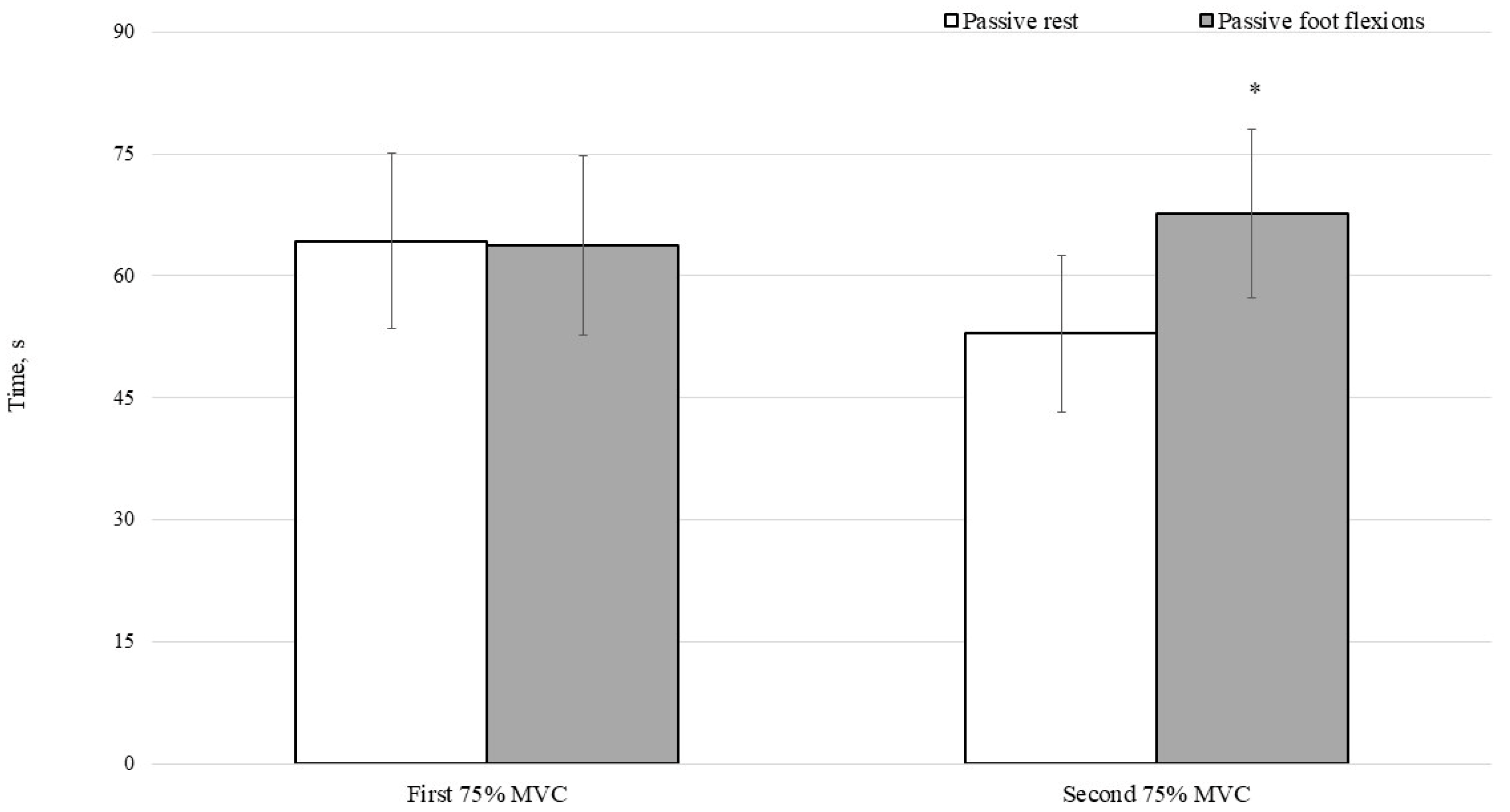
Figure 4 presents a comparison of holding times between the passive rest and passive foot flexions conditions at different measurement time points. During the first holding trial, no statistically significant difference was observed between the passive rest and passive foot flexions conditions (64.3 ± 10.8 s vs. 63.7 ± 11.0 s), indicating comparable baseline performance between the two conditions. However, a significant difference was observed during the second holding trial, with longer holding times recorded after passive foot flexions compared to the passive rest condition (67.7 ± 10.4 s vs. 52.9 ± 9.7 s, respectively;
p < 0.05). Moreover, the effect size was large (
d = 1.5). These findings suggest that the between-condition differences could be attributable to an intervention effect, rather than initial physical capacity.
4. Discussion
The present study examined the effects of passive foot flexions on isometric holding performance, heart rate, autonomic modulation, StO2, and muscle mechanical properties following repeated rounds of static exercise. Based on our results, passive foot flexions primarily influenced autonomic recovery and peripheral oxygen dynamics, whereas heart rate trajectory and mechanical properties were only modestly affected.
Heart rate increased at the onset of the exercise task, and decreased gradually during recovery under both conditions, consistent with the expected withdrawal of sympathetic activity and progressive parasympathetic reactivation after exercise. However, during the 15 min passive foot flexions intervention, heart rates remained relatively stable within a narrow range (approximately 82–87 bpm) rather than continuing to decline. This pattern indicates that the intervention did not further accelerate heart rate recovery. The relatively stable heart rate observed during the passive foot flexions phase may reflect sustained cardiovascular engagement or incomplete recovery rather than a specific stabilizing effect. Similar patterns were reported in previous studies of low-intensity or passive limb movements, suggesting that gentle peripheral movement and may be associated with maintained hemodynamic stability without imposing additional cardiac load [
10].
More distinct effects were observed with respect to autonomic modulation, which was assessed by RMSSD. Baseline RMSSD values were comparable between conditions, but after the second recovery period, RMSSD increased more markedly following passive foot flexions, with a large effect size (d = 1.50). This suggests that stronger parasympathetic reactivation could be associated with passive foot flexions compared to the passive rest condition, even in the absence of meaningful heart rate differences. This dissociation between heart rate and HRV is supported by prior observations that autonomic recovery can improve without parallel changes in mean heart rate, particularly when peripheral sensory input is manipulated. Because it is widely recognized as the most robust short-term time-domain index of cardiac vagal modulation, particularly in exercise and recovery settings, for the present study, only RMSSD was used to quantify heart rate variability [
16,
17]. Unlike frequency-domain indices, such as the LF/HF ratio, which are sensitive to breathing patterns and require longer stationary recordings [
18,
19], RMSSD is less affected by respiratory variability and remains reliable during short post-exercise assessment windows. The lack of concomitant blood pressure modulation further may suggest that the enhanced RMSSD response we observed here was more likely related to autonomic adjustments than changes in arterial pressure regulation [
20,
21].
Variations in tissue oxygen saturation (StO
2) appear to depend on the workload and recovery phase, rather than any uniform effect associated with the intervention. During the first static workload, passive foot flexions were was associated with a higher baseline StO
2 but also increased desaturation at higher intensities, suggesting altered local oxygen extraction or microvascular regulation which may reflect differences in local oxygenation dynamics, although direct conclusions regarding microvascular regulation cannot be drawn from the present measurements. Similar workload-dependent shifts in oxygenation have been described in previous studies, when peripheral circulation was modulated before exercise, indicating that baseline perfusion does not necessarily predict oxygen dynamics under load [
22]. During the late recovery phase (6–15 min), StO
2 remained consistently higher under passive foot flexions, which may reflect altered peripheral oxygenation dynamics at the measurement site. Similar oxygenation patterns have been described following exercise; however, the present measurements do not allow direct conclusions regarding local perfusion or blood flow regulation [
23,
24]. In contrast, during the second workload, StO
2 was found to be significantly lower under passive foot flexions at moderate and high intensities, particularly at the 50% workload (d = −1.54). This may suggest altered oxygenation dynamics during the subsequent exercise bout, although direct conclusions regarding oxygen utilization or local blood flow distribution cannot be drawn from the present data. Remarkably, StO
2 rebounded more rapidly immediately after the second workload for the passive foot flexions condition, indicating enhanced reoxygenation kinetics despite greater desaturation during exertion. Such a reoxygenation pattern is consistent with a modified oxygenation response [
25,
26]. Importantly, these local oxygenation responses may occur independently of marked systemic hemodynamic fluctuations. In the present study, the stability of systolic and diastolic blood pressure levels during these phases further is consistent with the possibility that peripheral factors contributed to the observed oxygenation responses, although microvascular adjustments were not directly measured.
Similar increases in muscle tone and stiffness were observed under both conditions following static holds, which is consistent with expected viscoelastic and neural responses to sustained isometric contractions [
27]. Between-condition differences were small to moderate at most, and values converged after the second hold, suggesting that passive foot flexions did not meaningfully alter muscle mechanical properties. These observations are consistent with previous data suggesting that passive or low-intensity interventions generally only induce modest and often transient changes in muscle stiffness [
28]. Consequently, the observed changes in RMSSD and StO
2 may not be due to alterations in muscle tone or stiffness, although the underlying mechanisms cannot be directly determined from the present measurements. Overall, results from the present study are consistent with the possibility that passive foot flexions influenced autonomic recovery and peripheral oxygenation responses, rather than by inducing mechanical changes in the muscle tissue. More specifically, the intervention supported parasympathetic recovery and reshaped peripheral oxygen dynamics without substantially modifying heart rate trajectory or muscle mechanical state.
Isometric holding performance exhibited condition-dependent modulation. While baseline performance was comparable across the tested conditions, repetition following passive foot flexions resulted in superior endurance compared with passive rest. This improvement may be related to more favorable recovery conditions between exercise bouts. Previous studies have suggested that passive limb movement may influence circulation- and autonomic-related responses during recovery [
29,
30,
31]; however, muscle blood flow, venous return, and metabolic markers were not directly measured in the present study, and mechanistic explanations involving enhanced perfusion, metabolite clearance, or vasodilation remain speculative. Therefore, mechanistic explanations involving enhanced perfusion, metabolite clearance, or vasodilation should be considered speculative.
From a broader physiological perspective, the observed responses may be viewed within the context of local muscle energetics and recovery from repeated isometric work. Sustained plantar flexion imposes substantial metabolic demand on the active musculature and may transiently disturb the balance between oxygen delivery and utilization, thereby contributing to fatigue development and delayed recovery [
3,
29]. In this context, changes in peripheral oxygenation patterns and subsequent holding performance may reflect recovery-related adjustments in the capacity to tolerate repeated muscular work. Although the present measurements do not allow direct conclusions regarding muscle energetics or locomotor mechanisms, the findings are consistent with the view that recovery after localized isometric exercise is shaped by the interaction of autonomic, peripheral, and task-specific factors [
2,
7,
31].
The present study has several limitations that should be considered when interpreting the findings. First, the sample was relatively small and consisted exclusively of young physically active men; therefore, the results cannot be directly generalized to women, older adults, clinical populations, or athletes of different training status. In addition, no a priori sample size calculation was performed, and the study may have been underpowered for some secondary physiological outcomes. Accordingly, the present findings should be interpreted as exploratory and should be confirmed in larger and more diverse samples. Second, only indirect physiological markers were assessed. Direct measurements of muscle blood flow, venous return, cardiac output, or metabolic by-products were not obtained, and therefore, mechanistic interpretations regarding perfusion, reperfusion, metabolite clearance, or vascular regulation should be made with caution. Third, although participants were instructed to remain relaxed and the intervention was continuously visually monitored, electromyography was not used to verify muscle quiescence during passive foot flexions; therefore, low-level voluntary activation or co-contraction cannot be fully excluded. Fourth, respiration was not externally controlled during HRV assessment, which should be considered when interpreting the RMSSD findings, as the breathing pattern may influence short-term HRV indices. Fifth, complete baseline equivalence between sessions may not have been achieved, as suggested by the higher baseline StO2 observed before the passive foot flexions condition; therefore, a possible order or carry-over effect in the crossover design cannot be fully excluded. Finally, only acute responses to a single session of passive foot flexions were examined, and the long-term effects of this intervention remain unknown.