1. Introduction
Post–core systems are widely used to restore the structural integrity of teeth that have undergone or require endodontic treatment due to excessive caries or dental trauma [
1,
2]. Intraradicular components have long been used and are generally classified as cast posts, prefabricated metal posts, and metal-free prefabricated post systems. Limitations related to the biocompatibility, corrosion, rigidity, and aesthetics associated with metal posts have promoted the use of alternative post materials, particularly glass fiber and zirconia posts [
1]. Glass fiber posts are widely accepted for their favorable elastic modulus and aesthetics, particularly in anterior restorations [
2]. Recent advances in material science have introduced high-performance polymers, such as polyether ether ketone (PEEK), as promising alternative post materials that offer a balance between mechanical performance and biocompatibility [
3].
Although metal posts are associated with unfavorable stress distribution within the root, frequently leading to oblique or horizontal coronal root fractures [
4], several studies have indicated that metal posts may exhibit superior marginal performance as compared with that of alternative post systems [
3,
5]. In addition, long-term clinical investigations, including a study with a follow-up period of up to 178 months [
6], as well as a 15-year randomized trial comparing glass-fiber and metal posts [
7], reported comparable survival outcomes between titanium and glass-fiber posts. Despite these biomechanical concerns, metal posts continue to be preferred in clinical practice, which can be attributed to the cost-effectiveness, ease of access, and lower technique sensitivity of these materials. Furthermore, in cases where an adequate ferrule is partially present or completely absent, a general recommendation is that the posts with a higher elastic modulus may be advantageous in enhancing structural support and stress distribution [
8,
9]. Therefore, selecting an appropriate post system should be based on a comprehensive evaluation of both material- and technique-related factors [
10,
11].
Following post placement, prefabricated posts require a core build-up material to support the definitive restoration [
2]. While amalgam has long been used as a core material because of its mechanical strength and moisture tolerance [
12], resin composites, known for their reliable adhesion, improved esthetic outcomes, and single-visit convenience, are now widely used in contemporary practice [
13]. A previous study demonstrated that composite resin core materials provide higher bond strength than amalgam and resin-reinforced glass ionomer materials when used with both metal and glass fiber posts [
13]. By contrast, core materials with glass ionomer components were reported to show inferior bonding performance, thereby limiting their use as core build-up materials [
14]. Dual-cure composite resins are frequently selected to ensure adequate polymerization in clinical situations where light penetration is limited. In addition, control over polymerization time and improved color stability are among the main reasons for preferring light-cure core composites [
2]. Self-cure composite materials delivered via automix systems, which enable a more homogeneous core structure and eliminate incremental technique sensitivity, may serve as an alternative for core build-up procedures [
12]. However, one study reported lower bond strength values between all-in-one adhesive systems and the core structure when such chemically cured composites were used [
15], which highlights the potential influence of reliable bonding strategies on post–core adhesion [
13].
Various surface treatment protocols, including airborne-particle abrasion using aluminum oxide (Al
2O
3) or silica particles, chemical modification strategies, and primer applications, were previously proposed and evaluated to improve bonding at the post–core interface through micromechanical and chemical mechanisms, while also demonstrating that their effectiveness varies according to the substrate characteristics of the post material [
10,
16]. Recent in vitro investigations have further demonstrated that surface treatment protocols, particularly airborne-particle abrasion with or without chemical conditioning, significantly influence bond strength in post systems [
17,
18]. This variability highlights the importance of evaluating surface treatment strategies in a substrate-specific context [
11]. Within this framework, although surface modification approaches and core composite materials have each been investigated under different experimental conditions, a more integrated evaluation of their combined influence on post–core interfacial performance may provide additional insight. In particular, clarifying how post surface condition and composite polymerization mode interact to affect both bond strength and failure behavior may contribute to a more comprehensive understanding of adhesion dynamics and support more informed material selection for prefabricated metal post systems.
Based on these considerations, the present study aimed to evaluate the effect of two clinically applied post surface treatment protocols (adhesive only vs. sandblasting + MDP primer + adhesive) in combination with different core composite polymerization modes on bond strength and failure behavior at the post–core interface of prefabricated gold-plated metal posts, while keeping post head geometry constant across all experimental groups. The null hypotheses were that surface treatment protocol and core composite polymerization mode would not significantly influence interfacial bond strength or failure patterns.
2. Materials and Methods
This in vitro study was conducted at the Department of Prosthodontics, Faculty of Dentistry, Ankara University, Ankara, Turkey. Ethical approval was not required, as no human participants or animal subjects were involved.
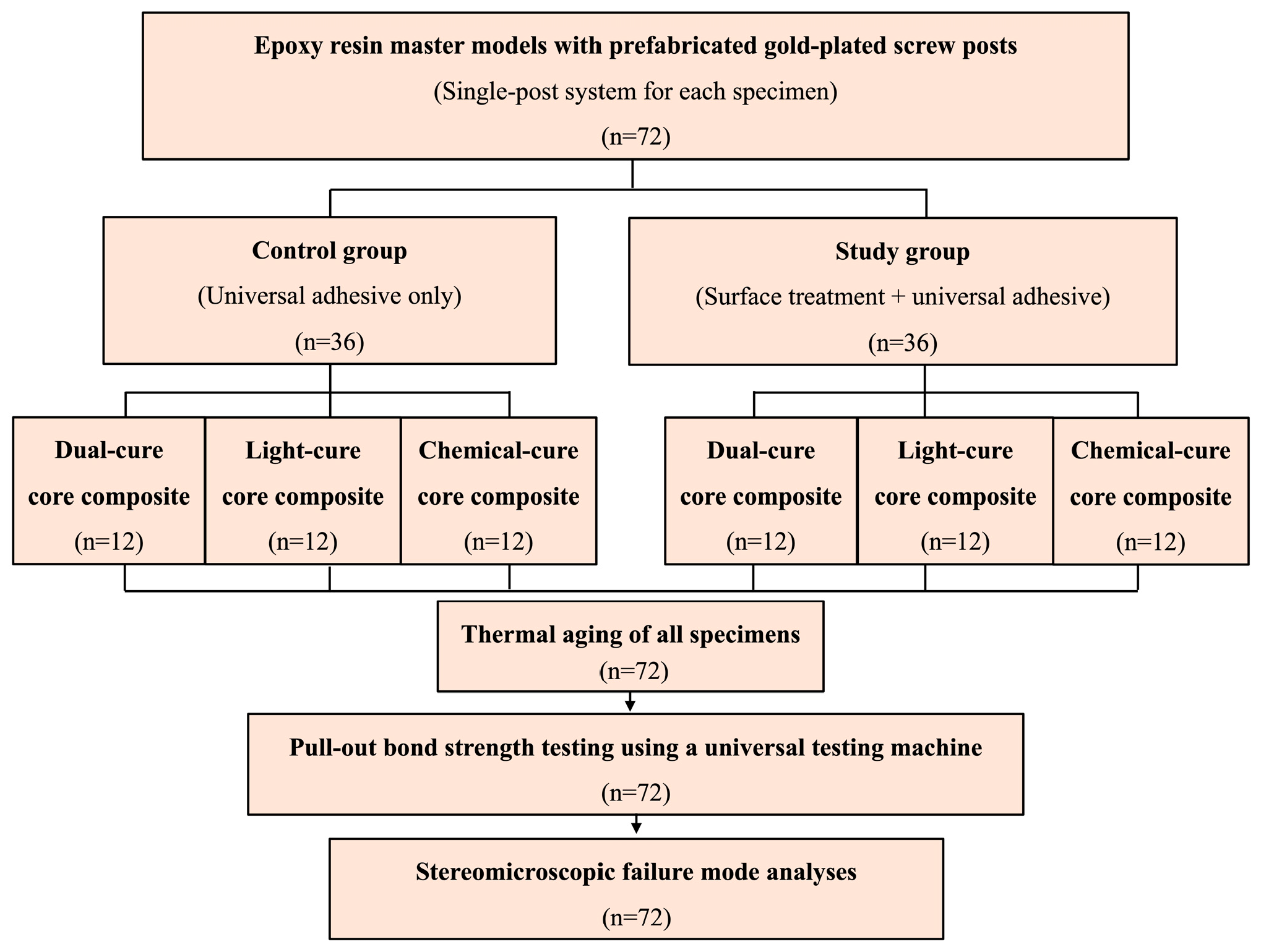
Figure 1 provides a schematic representation of the study workflow. To evaluate the effects of core material type and surface treatment protocol on the bond strength between prefabricated metal posts and resin core materials, a total of 72 specimens were prepared according to a 3 × 2 factorial experimental design. Three different resin core materials (dual-cure, light-cure, and self-cure composites) and two surface treatment approaches (universal adhesive alone and airborne-particle abrasion, followed by an MDP-containing metal primer and universal adhesive) were investigated. The specimens were randomly allocated into six experimental groups (n = 12 per group). A single prefabricated gold-plated screw post system (XL size, No. 6, Swedent Gold Plated Screw Posts, Swedish Dental Supplies AB, Åkarp, Sweden) was used to ensure standardization. According to the manufacturer, these metallic posts feature an electrolytically applied 24K gold plating. The XL size (No. 6), with a length of 14.0 mm and a diameter of 1.80 mm, was selected to enhance mechanical stabilization of the post within the epoxy resin matrix and to minimize the risk of premature displacement during pull-out testing. The geometric parameters of the post head were directly measured using a digital caliper (0–150 mm measuring range, 0.01-mm resolution, ±0.02-mm accuracy). The post head exhibited a square cross-section (1.70 mm × 1.70 mm) with a height of 3.08 mm, and the coronal portion receiving the core build-up was non-tapered.
To find the smallest sample size needed to identify statistically significant variations in bond strength between the experimental groups, an a priori power analysis was performed. Assuming a large effect size for the primary outcome measure, the analysis was conducted with a target statistical power of 0.80 and a significance level of α = 0.05. The power analysis was run using G*Power software (version 3.1.9.7, Heinrich Heine University, Düsseldorf, Germany). The results indicated that a minimum of 10 specimens per group would be sufficient to detect meaningful differences between the evaluated factors. To account for potential specimen loss and enhance the reliability of the statistical analysis, 12 specimens were included in each experimental group, resulting in a total sample size of 72.
2.1. Preparation of Test Specimens
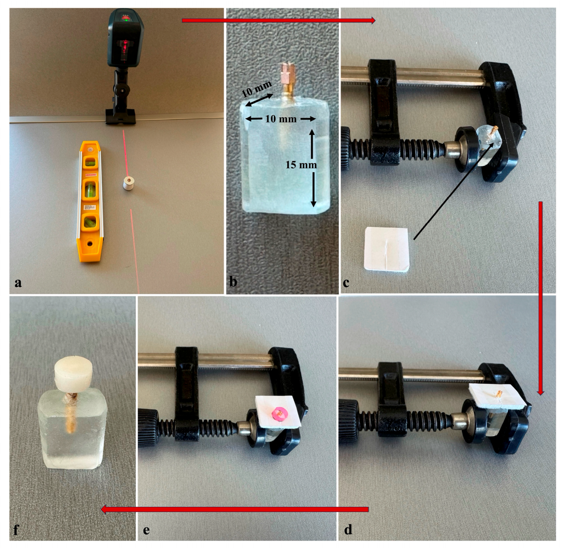
The specimens were embedded in epoxy resin model material (PL-2 and PLH-2, Vishay Precision Group Inc., Raleigh, NC, USA) for the fabrication of the master models. For the placement of the posts within the model material, individual custom-shaped plastic molds with a diameter of 1.2 cm were used, and a metal cylindrical guide was employed to support the upper part of the posts. This metal guide also enabled the posts to be positioned so that a 2 mm distance was maintained between the lower margin of the core portion and the epoxy resin base, thereby allowing complete coverage of the core by the composite material and providing sufficient relief space beneath the core for subsequent mechanical testing. A line laser level (Professional GLL 2, Robert Bosch Power Tools GmbH, 70538 Stuttgart, Germany) was used during the embedding process to ensure the precise vertical alignment of the posts relative to a flat reference surface, the horizontal orientation of which was confirmed using a spirit level (
Figure 2a). Subsequently, the epoxy resin was allowed to polymerize at room temperature for 24 h, in accordance with the manufacturer’s instructions. Following polymerization and removal from the mold, the epoxy resin blocks were standardized to dimensions of 10 mm × 10 mm × 15 mm, and two opposing surfaces were leveled to improve gripping during mechanical testing, resulting in a total sample size of 72 (
Figure 2b).
2.2. Surface Treatment Protocols
Prior to the core build-up procedures, specimens were randomly allocated into two surface treatment groups and subsequently distributed into six experimental subgroups (n = 12 per group). As all specimens were fabricated using the same prefabricated post system and prepared under strictly standardized conditions, variability among specimens was minimized, and the risk of selection bias was considered negligible. No mechanical surface modification was conducted in the control groups, and the post heads were treated only with a universal adhesive (Bond Force II, Tokuyama Dental Corp., Tokyo, Japan) using a disposable fine-tip microbrush in accordance with the manufacturer’s guidelines. The adhesive was light-cured for 20 s using a light-curing unit (Curing Pen-E, model C-004-1, Changzhou Sifary Medical Technology Co., Ltd., Changzhou, China).
For the experimental groups, the coronal portions of the posts intended to receive the core build-up were first treated by airborne-particle abrasion using 50 µm aluminum oxide particles at a controlled pressure of 2 bar for 10 s using a laboratory sandblasting device (Twin-Pen Sandblaster VI, model JG-218, Wuhan Jinguang Medical Technology Co., Ltd., Wuhan, China). The nozzle was positioned perpendicular to the post surface at a standardized distance of 10 mm. After sandblasting, the posts were thoroughly cleaned with oil-free air and alcohol to remove residual abrasive particles. An MDP-containing metal primer (Z-Prime™ Plus, BISCO Inc., Schaumburg, IL, USA) was applied to the sandblasted post surfaces in a thin layer using a disposable microbrush to ensure uniform coverage of the entire bonding surface and allowed to react for 10 s prior to air drying for a total of 10 s (5 s gentle and 5 s stronger air application), in accordance with the manufacturer’s instructions.
The selected metal primer (Z-Prime™ Plus) contains the functional phosphate monomer 10-MDP, which is known to promote adhesion to metallic substrates through interaction with surface oxide layers. Although gold is a noble metal with limited intrinsic oxide formation, airborne-particle abrasion may modify the superficial coating and expose underlying alloy components capable of forming oxide species. The use of an MDP-containing primer was therefore intended to enhance potential chemical interaction in addition to micromechanical retention.
Subsequently, the same universal adhesive (Bond Force II, Tokuyama Dental Corp., Tokyo, Japan) was applied to the post surfaces following the identical protocol to that used in the control groups.
2.3. Core Build-Up Procedure
During the core build-up procedures, a clamping fixture was used to securely stabilize the epoxy resin specimens (
Figure 2c). To preserve the predefined 2 mm relief beneath the core material, a removable spacer consisting of a compressible polymer-based material with a uniform thickness of 2 mm and a square geometry (20 × 20 mm) was positioned beneath the specimen during the build-up process. The spacer was partially slit to allow atraumatic removal after polymerization without compromising the integrity of the composite structure. To prevent unintended adhesion between the composite material and the spacer, the upper surface of the spacer was covered with a thin separator film prior to build-up (
Figure 2d). Standardized core geometries were obtained using a cylindrical plastic mold with a nominal internal diameter of 5.0 mm [
19] and a height of 3.0 mm (
Figure 2e). The mold was positioned so that the coronal head of the post was centrally located and completely surrounded by the core composite material. These dimensions provided a composite core cross-sectional area exceeding that of the post head, ensuring consistent core geometry among all specimens (
Figure 2f).
The materials used in the present study, including the core composites, primer, and universal adhesive, are summarized in
Table 1. All core build-up procedures were performed in accordance with the respective manufacturers’ instructions. The same light-curing unit used for the adhesive application was employed for all light-activated polymerization procedures.
For the dual-cure core composite groups, a dual-cure core build-up material (Clearfil™ DC Core Plus, Kuraray Noritake Dental Inc., Okayama, Japan) was applied as a single increment and then polymerized by light activation for 20 s, while retaining its dual-cure capability.
In the light-cure core composite groups, a light-cure restorative composite (Filtek™ Z250 Universal Restorative, 3M ESPE, St. Paul, MN, USA) was incrementally applied. The composite was first placed in an initial layer with an approximate thickness of 1.5 mm and light-cured for 20 s, which was followed by application of the remaining material up to the mold surface and subsequent light activation for an additional 20 s.
Polymerization in the chemical-cure core composite groups was achieved using a self-curing core build-up material (Stela™ Automix Self Cure Composite, SDI Limited, Bayswater, Victoria, Australia), which was applied as a single increment and allowed to set through chemical curing.
2.4. Measurements
All measurements were performed under standardized laboratory conditions by a single operator. Prior to testing, the specimens were visually inspected to confirm the absence of visible defects, and all data were recorded for the subsequent statistical analysis.
To simulate intraoral thermal conditions, all specimens underwent 5000 thermocycles using a thermocycling device (Thermocycler, SD Mechatronik, Feldkirchen-Westerham, Germany), following a previously reported in vitro aging protocol [
20]. Cycling was performed between 5 °C and 55 °C, with a dwell time of 30 s at each temperature and a transfer time of 10 s. Temperature control was maintained using a digital thermostatic unit (Julabo GmbH, Seelbach, Germany).
After completion of the thermal aging protocol, the specimens were subjected to an axial tensile (pull-out) bond strength test performed with a universal testing machine (LRX, Lloyd Instruments, Fareham, UK). The pull-out test configuration used in the present study was designed in accordance with similar tensile retention testing principles previously described for post–core systems [
19]. For testing, each epoxy resin block containing the post–core assembly was first securely fixed onto the lower mounting platform of the testing machine.
Because the objective of this study was to evaluate the integrity of the connection between the post head and the core complex as a single functional unit under axial tensile loading, no specimen sectioning was performed, as such an approach is not within the scope of the tensile loading protocol used in the present study. Instead, a custom-designed holding mechanism was developed to transmit axial tensile force to the core assembly, thereby allowing separation to occur at the post–core interface.
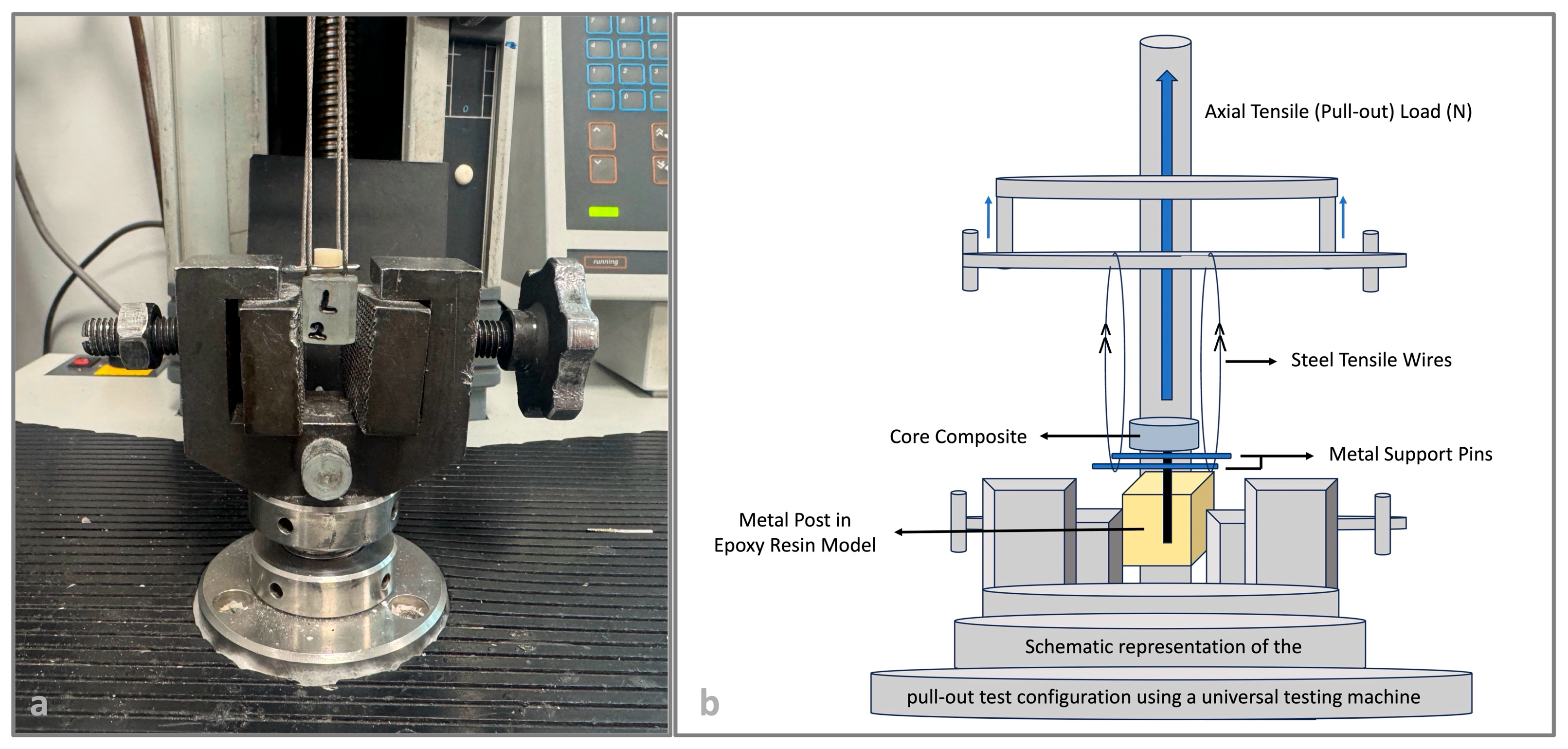
To enable controlled axial tensile loading without introducing bending moments, the holding mechanism was positioned beneath the composite core within the predefined 2 mm relief space. This mechanism consisted of two opposing metal support pins adapted symmetrically under the core to ensure stable alignment and uniform load distribution. Steel tensile wires were then routed beneath the core and securely engaged around these metal support pins, establishing a stable mechanical linkage between the upper grip of the testing machine and the support pins beneath the core (
Figure 3). Consequently, as the crosshead moved upward, the applied tensile load was transmitted from the upper grip through the steel tensile wires to the support pins and subsequently to the composite core, thereby inducing tensile stress along the post head–core interface and promoting interfacial debonding under axial loading. This configuration ensured vertical load application along the long axis of the post–core complex, thereby minimizing the oblique displacement and lateral stress components. In this setup, the core was supported from below while the post remained fixed within the epoxy block, allowing tensile stresses to be concentrated specifically at the post head–core interface.
A tensile load was then applied from the upper grip in alignment with the long axis of the post. The test was performed at a crosshead speed of 1 mm/min, and the maximum load limit was set to 2500 N. The peak load recorded at the moment of debonding was defined as the pull-out resistance and expressed in newtons (N).
Following mechanical testing, the failure modes of all specimens were examined using a stereomicroscope (Leica Microsystems, Wetzlar, Germany) at 10× magnification. The debonded surfaces were examined to determine the predominant failure behavior based on the location and characteristics of separation within the post–core complex. Failures were classified as adhesive when the separation occurred at the post–core interface and non-adhesive when the fracture involved the core composite, the post, or a combination of both. Representative images of each sample were recorded using the integrated digital imaging system of the stereomicroscope for documentation. All failure mode classifications were assigned by a calibrated examiner.
2.5. Statistical Analysis
The pull-out bond strength values were converted to megapascals (MPa) for the statistical analysis. Descriptive statistics that included mean values, standard deviations, minimum–maximum ranges, and 95% confidence intervals were calculated for each experimental group. The distribution of the data was evaluated for normality using the Shapiro–Wilk test, while the homogeneity of variances was examined with the Levene test.
To investigate the influence of the experimental variables on bond strength, a two-way analysis of variance (ANOVA) was performed, with core material type (dual-cure, light-cure, chemical-cure) and surface treatment (NON and ST) used as independent factors. The main effects of each factor, as well as their interaction effect, were evaluated separately.
Because the interaction between core material type and surface treatment was not statistically significant, post hoc comparisons for the main effects were conducted based on the estimated marginal means collapsed across the levels of the other factor. Multiple pairwise comparisons among the three core materials were performed using the Tukey honestly significant difference (HSD) test to control for type I error.
Statistical significance was established at p < 0.05, and effect sizes were expressed as eta-squared (η2) values. For the analysis of fracture patterns, failure modes were dichotomized as adhesive and non-adhesive (cohesive + mixed) to ensure adequate expected cell counts and to meet the assumptions of the chi-square test. The distribution of these categorical outcomes among the six experimental groups was analyzed using the chi-square (χ2) test. All statistical analyses were performed using SPSS software (version 22; IBM Corp., Armonk, NY, USA).
3. Results
To address the primary research question, the effects of surface treatment and core material polymerization mode on pull-out bond strength and failure behavior were analyzed in terms of their main effects and interactions. A total of 72 specimens were evaluated in the present study (n = 12 per group). As shown in
Table 2, the surface-treated groups consistently exhibited higher mean bond strength values than their non-surface-treated counterparts across all polymerization modes.
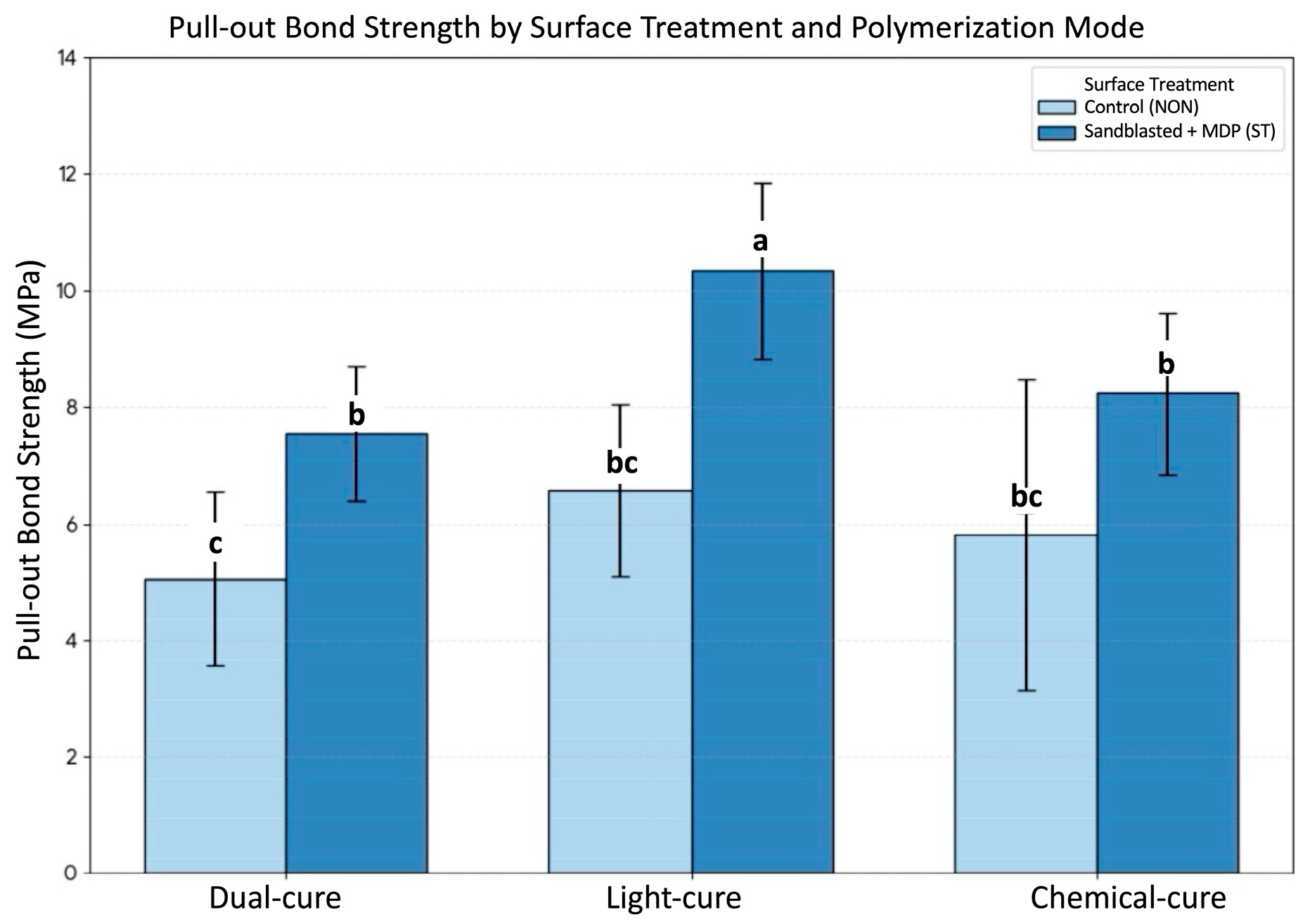
The lowest mean bond strength was observed in the NON-dual-cure group (5.06 ± 1.49 MPa; 95% confidence interval [CI]: 4.12–6.00 MPa; range: 3.94–8.14 MPa). By contrast, the highest mean bond strength was recorded in the ST-light-cure group (10.34 ± 1.51 MPa; 95% CI: 9.38–11.30 MPa; range: 7.81–13.25 MPa). The remaining groups demonstrated intermediate bond strength values, i.e., NON-light-cure (6.57 ± 1.48 MPa), NON-chemical-cure (5.81 ± 2.66 MPa), ST-dual-cure (7.55 ± 1.15 MPa), and ST-chemical-cure (8.23 ± 1.38 MPa) (
Figure 4). The comparison of mean values demonstrated higher bond strength values in the surface-treated groups across all core materials, with absolute differences of 2.49 MPa for dual-cure, 3.77 MPa for light-cure, and 2.42 MPa for chemical-cure materials, as shown in
Table 2.
Preliminary analyses confirmed normal distribution of the data (Shapiro–Wilk test, p > 0.05) and homogeneity of variances (Levene test, p > 0.05), thereby supporting the appropriateness of the two-way ANOVA model.
The results of two-way analysis of variance revealed that the core material type had a statistically significant effect on pull-out bond strength (F (2,66) = 18.7,
p < 0.001, η
2 = 0.35). Similarly, the surface treatment significantly influenced bond strength values, with higher results consistently observed in the ST groups as compared with those for the NON groups (F (1,66) = 67.8,
p < 0.001, η
2 = 0.49). The interaction between core material type and surface treatment was not statistically significant (F (2,66) = 2.05,
p = 0.138, η
2 = 0.06). The detailed ANOVA statistics, including sums of squares and effect sizes, are summarized in
Table 3. These findings demonstrate that both surface treatment and core material type had statistically significant main effects on pull-out bond strength, whereas no significant interaction was observed between the two factors.
The magnitude of the effect sizes provides additional insight into the relative contribution of each factor. Surface treatment explained a substantial proportion of the variance in bond strength (η2 = 0.49), suggesting a strong association with adhesion performance. Core material type also showed a meaningful effect (η2 = 0.35), indicating that differences in polymerization mode were associated with variations in bond strength. The absence of a significant interaction suggests that the higher bond strength values observed in the surface-treated groups were consistent across all core materials, irrespective of polymerization mode.
Post hoc multiple comparisons were performed using the Tukey honestly significant difference (HSD) test based on the estimated marginal means collapsed across surface-treatment levels. These analyses revealed significant differences among the core material types (
p < 0.05), with the light-cure material exhibiting the highest overall bond strength values. The distribution of group differences identified by the Tukey test is illustrated in
Figure 4. The graphical representation indicates that the ST-LC group had the highest mean bond strength, whereas the NON-DC group had the lowest.
Group-level comparisons showed a pattern in which surface-treated specimens exhibited higher bond strength values within each polymerization category, indicating a consistent association between surface treatment and bond strength across all core materials.
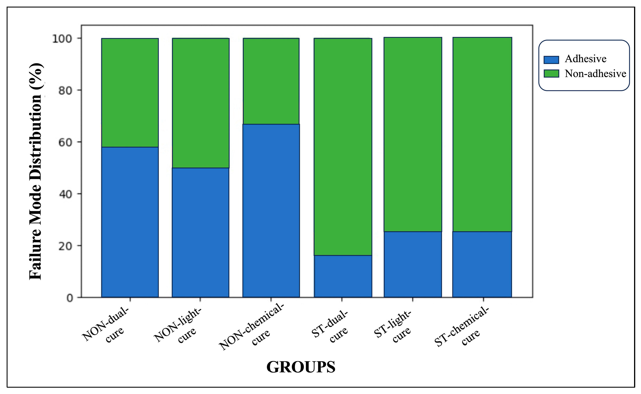
Fracture mode distributions, after dichotomization into adhesive and non-adhesive (cohesive + mixed) categories, showed a statistically significant association among the experimental groups (χ
2 = 14.8, df = 5,
p = 0.011) (
Table 4;
Figure 5).
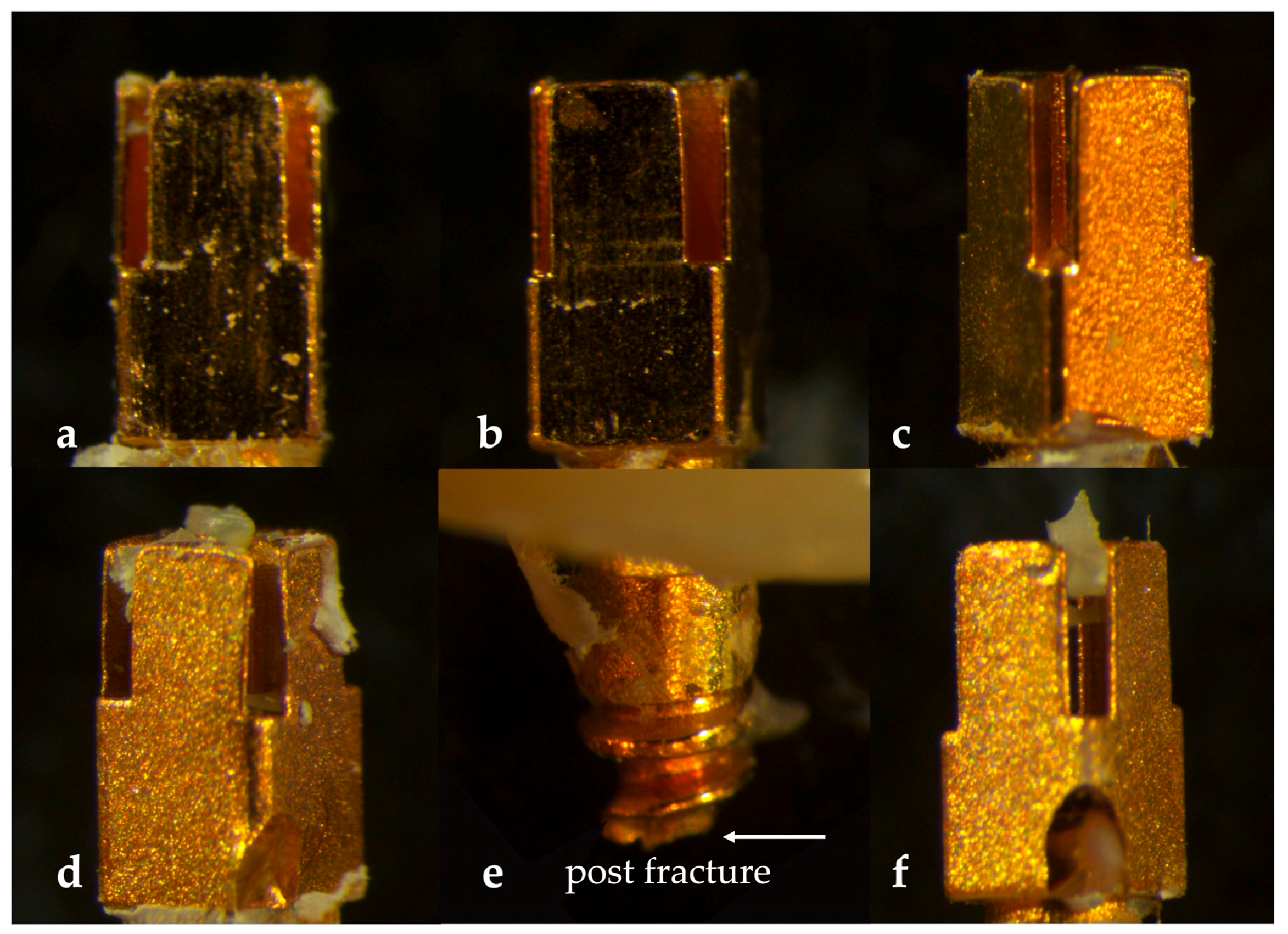
In the NON groups, adhesive failure was the predominant fracture pattern (NON-dual-cure: 58.3%; NON-light-cure: 50.0%; NON-chemical-cure: 66.7%). In contrast, the ST groups demonstrated a higher proportion of non-adhesive failures (ST-dual-cure: 83.3%; ST-light-cure: 75.0%; ST-chemical-cure: 66.7%). Representative microscopic images illustrating the fracture patterns observed in each experimental group are presented in
Figure 6.
4. Discussion
In endodontically treated teeth with extensive coronal tissue loss, post placement is essential not only to restore fracture resistance but also to provide adequate support for the core build-up and the definitive restoration [
4]. The long-term success of post–core restorations is affected by multiple post- and core-related factors, including post material properties and surface characteristics [
3,
10,
21], as well as the design and properties of the core material [
22], among which the bonding strategy at the post–core interface plays a crucial role [
16]. Accordingly, this in vitro study evaluated the effects of different core material types and surface treatment protocols on the pull-out bond strength and failure patterns at the post–core interface.
To this end, XL-sized prefabricated posts were embedded in an epoxy resin model to ensure stable fixation during mechanical testing. Consistent with this approach, a recent study using the same model material reported that epoxy resin provided sufficient resistance during bond strength tests, while enabling standardized experimental conditions by eliminating anatomical variability associated with natural teeth [
23]. To minimize geometric variability, the location and dimensions of the core build-up were standardized based on the post head diameter, thereby reducing the potential impact of uneven material distribution on mechanical outcomes. In their previous research, Aksoy et al. (2005) also reported adopting a similar dimensional standardization based on post head diameter [
19]. This approach allowed differences in bond strength and fracture behavior to be attributed mainly to the core material and surface treatment rather than to variations in core geometry.
In the present study, to simulate the oral environment and the associated degradation of adhesive bonding, the specimens were subjected to 5000 thermocycles [
20]. In addition, previous studies reported that the bond strength test used in the present study provides a more homogeneous stress distribution, causes less specimen damage, and demonstrates greater reliability than the microtensile bond strength test for evaluating post–core adhesion [
24,
25].
Across all evaluated core material types, the surface treatment was associated with higher pull-out bond strength values, suggesting that surface modification of the post may contribute substantially to post–core adhesion. These findings are consistent with those of previous studies reporting improved post–core retention by following surface treatment protocols [
26,
27]. For instance, in a study comparing airborne abrasion with Al
2O
3, intraoral sandblasting, and laser roughening for fiber posts, sandblasting with Al
2O
3 was found to exhibit the highest bond strength values, which could be attributed to the combined micromechanical and chemical effects induced by surface modification [
11]. As demonstrated in previous research on post head air abrasion [
16,
28], airborne-particle abrasion increases surface roughness and surface area, thereby enhancing mechanical interlocking between the post surface and the core material. In addition, surface treatment may improve the interaction between the post surface and functional primers, supporting more stable chemical bonding at the interface.
It should be noted, however, that the posts used in the present study were gold-plated according to the manufacturer’s instructions. Gold plating has long been used in screw post systems due to its excellent corrosion resistance, chemical stability in the oral environment, long-term biocompatibility, and its ability to reduce galvanic interactions between dissimilar metals [
29,
30]. In threaded systems, such surface characteristics may contribute to improved clinical stability and reduced surface degradation over time. Nevertheless, from a bonding perspective, gold is chemically inert and does not form stable oxide layers, which may limit the effectiveness of functional monomers such as MDP that rely on metal–oxide interactions. Although airborne-particle abrasion was performed to enhance micromechanical retention, this procedure may have modified the superficial gold coating and potentially exposed the underlying metallic substrate. Therefore, the contribution of chemical bonding via MDP may depend on whether the base alloy was partially exposed following surface treatment. Functional monomers such as 10-MDP have been widely reported to establish stable chemical interactions through their phosphate functional group, forming relatively insoluble calcium–monomer salts and contributing to long-term interfacial stability [
31]. The molecular structure of 10-MDP, characterized by a reactive phosphate group and a hydrophobic spacer chain, has been associated with enhanced chemical bonding potential and improved resistance to hydrolytic degradation. Although gold itself is chemically inert and exhibits limited intrinsic oxide formation, airborne-particle abrasion may locally disrupt the superficial gold coating and expose areas of the underlying alloy capable of forming oxide species. Under such conditions, phosphate monomers may interact with newly formed oxide layers, thereby contributing to interfacial bonding, in addition to the micromechanical retention produced by surface roughening. This consideration should be taken into account when interpreting the bonding mechanisms observed in the present study.
Th silanization of fiber posts was previously suggested to enhance chemical interaction at the post–core interface [
10], particularly as an alternative to airborne-particle abrasion, which was reported to adversely affect the fiber–resin structure of these posts [
32,
33]. Similarly, functional primers containing 10-methacryloyloxy dihydrogen phosphate (MDP) were reported in the literature to improve chemical bonding to cobalt–chromium alloy surfaces through interactions with metal oxides [
34,
35]. While these studies primarily focused on metal substrates rather than post systems, they offered a relevant mechanistic insight into the improved post–core adhesion observed following surface treatment in the present study.
Notably, surface treatment enhanced bond strength, regardless of the polymerization mode of the core material, indicating that post surface modification provides a favorable bonding substrate independent of the curing mechanism. This finding is consistent with previous evidence on surface treatment strategies in post–core systems [
26,
27]. A similar trend was reported in a previous study evaluating a universal adhesive beneath core build-up composites, where bond strength was not significantly affected by different curing modes [
36]. Another study reported that the use of one-bottle primer/adhesive systems may reduce the availability of free radicals within the core resin as a result of interactions between organic amines and non-polymerized acidic monomers, thereby compromising the self-curing polymerization mechanism [
12]. Accordingly, in the present study, a dual-cure universal adhesive was selected to ensure a standardized bonding approach compatible with different core materials and curing conditions. From a clinical perspective, this approach offers a practical advantage by allowing surface treatment protocols to be applied consistently across various core material systems, supporting predictable post–core retention.
However, previous research indicated that resin composites exhibit a weaker chemical bonding to fiber posts as compared to that of dentin and enamel [
37,
38]. This difference was previously attributed to the highly cross-linked polymer matrix of fiber posts lacking reactive functional groups [
10]. Moreover, several studies reported that aggressive surface treatments may disrupt the fiber structure and polymer matrix, potentially compromising the integrity of fiber posts [
39,
40]. By contrast, metallic posts do not contain an organic polymer matrix; accordingly, they were selected in the present study to enable a more predictable evaluation of surface treatment effects by minimizing the potential confounding influence of substrate degradation. The findings of the present study are consistent with this rationale, as surface treatment resulted in enhanced pull-out bond strength when applied to metallic posts, irrespective of the core material type.
In both the control and study groups, the highest bond strength values were observed for the light-cure core material, followed by the chemical-cure composite. These findings align with a previous study comparing light-cure and two different dual-cure core composites, in which the light-cure material exhibited the highest bonding performance [
10]. The number of studies specifically evaluating composite materials intended for use as core build-up materials remains limited [
41]. In the present study, despite being specifically designed for core build-up applications, Clearfil DC Core exhibited the lowest bond strength values in both the non-surface-treated and surface-treated groups. The superior bonding performance observed for Filtek™ Z250 Universal Restorative may be attributed to its higher viscosity and hybrid structure, which likely promote more stable adaptation around the post surface. In addition, the incremental technique associated with light-cure composites may allow for a more effective polymerization of each layer, thereby reducing polymerization shrinkage-related stresses. This interpretation is consistent with the findings of Hanabusa et al. (2016) [
42] and Suh et al. (2003) [
43], who reported that this polymerization approach limits the diffusion of acidic primers into the core material and prevents the marked deactivation of the amine component within the amine–benzoyl peroxide redox system. Within the standardized and geometrically confined core configuration surrounding the metallic post head in the present study, polymerization occurred under high C-factor conditions, where volumetric contraction is more likely to generate interfacial stress concentration. Under such circumstances, differences in polymerization kinetics and the degree of conversion among curing modes may have influenced the magnitude and distribution of shrinkage stress at the adhesive interface. In addition, potential interactions between acidic adhesive components and the amine-based initiator systems of chemically or dual-cured composites may affect polymerization efficiency at the interface. Such interfacial interactions, combined with differences in curing dynamics, may help explain the higher bond strength values observed for the light-cure composite and the comparatively lower values recorded for the dual-cure material. The higher bond strength values observed for Stela™ Automix Self-Cure Composite compared to Clearfil DC Core may be explained by the slower and more controlled polymerization characteristic of chemically cured materials. This characteristic may be advantageous at the metal post–core interface, where micromechanical interlocking predominates, in contrast to the heterogeneous polymerization and localized stress development that may occur in dual-cure systems.
When surface treatment was not applied, differences among the core materials were less pronounced. However, once surface conditions were standardized through surface modification, differences among the core materials became more evident, indicating that the intrinsic properties of the core composites started to affect bonding performance after establishment of a stable post–core interface. The present analysis is consistent with the findings reported by Yarmohamadi et al. (2023) [
44]. Although the light-cure composite exhibited the highest bond strength in the control group (6.57 ± 1.48 MPa), higher values were observed following surface treatment (10.34 ± 1.51 MPa). Under surface-treated conditions, the dual-cure and chemical-cure materials demonstrated lower mean bond strength values than did the light-cure composite. Accordingly, the influence of core material type appears to be secondary to effective surface modification.
From a clinical perspective, the present findings suggest that optimization of post surface preparation may play an important role in the retention of prefabricated metal post systems across different core composite polymerization modes. As surface treatment accounted for a substantial proportion of the variance in bond strength (η2 = 0.49), improvements in retention may be more effectively achieved through appropriate substrate conditioning, such as airborne-particle abrasion combined with an MDP-containing primer, than by solely modifying the core composite material. With respect to material and technique selection, the consistently higher bond strength values observed with the light-cure composite suggest that, when adequate access and light transmission can be ensured, incremental light-activated core build-up may represent a clinically favorable approach. However, in deeper or clinically restricted situations where optimal light delivery cannot be guaranteed, the findings suggest that effective surface modification may represent the primary contributing factor and may help reduce polymerization-related differences among core materials.
Furthermore, the results of failure mode analysis performed using a stereomicroscope provided additional insight into the interfacial behavior of the post–core complex and supported the pull-out bond strength findings of the present study. In the control groups, adhesive failures at the post–core interface predominated (NON-CC, 66.7%; NON-DC, 58.3%; NON-LC, 50.0%), suggesting relatively weaker interfacial bonding. This failure pattern demonstrated that debonding occurred mainly at the interface rather than within the post or core material. These findings are consistent with a previous study in which specimens were classified according to the presence or absence of a surface treatment in relation to bond application, and adhesive failure modes were predominantly observed in non-treated groups [
19].
By contrast, surface-treated groups demonstrated a clear shift toward non-adhesive failure patterns (ST-DC, 83.3%; ST-CC, 66.7%; ST-LC, 75.0%), reflecting the formation of a stronger and more stable post–core interface. This distribution suggests that stresses generated during pull-out testing were more effectively transferred beyond the adhesive interface after surface modification. Accordingly, the failure mode distribution observed in the surface-treated groups further supports the positive effect of surface modification on interfacial bonding quality.
When core material types were evaluated, the ST-LC group demonstrated a higher proportion of non-adhesive failures (75.0%) compared with that of the other ST groups, which is consistent with the higher pull-out bond strength values recorded for this group. This finding aligns with the results reported by Fragkouli et al. (2019) [
45]. In contrast, the ST-DC and ST-CC groups showed relatively lower proportions of non-adhesive failures compared with those of the ST-LC group, suggesting comparatively reduced resistance to dislodgement. Importantly, post fracture was observed exclusively in the ST-LC group, occurring in four specimens. The localized occurrence of post fracture suggests that, under the conditions of enhanced interfacial bonding and higher bond strength, stresses generated during pull-out testing may exceed the structural resistance of the post itself rather than result in interfacial debonding. This finding further indicates a shift in the weakest link from the post–core interface to the post structure in this specific group—a phenomenon that was also reported in previous studies [
45,
46]. Overall, the failure mode distribution confirms that surface treatment is the primary factor influencing post–core adhesion.
When the present findings are interpreted in light of the existing literature, an important distinction emerges between studies focusing on fiber posts and those evaluating metallic substrates. While many previous investigations reported that the effectiveness of surface treatment protocols depends largely on substrate composition and the integrity of the fiber–resin matrix [
32,
40,
41], the current results suggest that, in metallic post systems, surface modification may have a substantial influence across different composite polymerization modes. This distinction may partly explain variations reported in earlier studies, where differences in post substrate characteristics were shown to influence bonding performance. Therefore, the present findings provide additional perspective on metallic post–core interfaces evaluated under standardized geometric conditions.
The findings of this study rejected the null hypothesis, demonstrating that surface treatment and core composite polymerization mode influenced bond strength and failure behavior at the post–core interface. However, as an in vitro investigation, the present design cannot fully replicate the complex biological and functional conditions of the oral environment. The exclusive use of a single prefabricated metal post system and one surface treatment protocol may limit the generalizability of the findings. Because the mechanical and chemical steps were applied together, the relative contributions of surface roughening and chemical bonding could not be distinguished; thus, the improvement observed should be interpreted as the effect of the overall protocol rather than to the influence of any individual component. Future studies incorporating separate mechanical-only and chemical-only surface treatment groups would help to better clarify the individual contribution of each step. Moreover, the present study focused on mechanical performance at the post–core interface without incorporating dentin post–core interactions or cyclic mechanical loading under definitive restorative conditions, which may influence clinical behavior. Furthermore, direct morphological characterization of the treated post surfaces and fracture interfaces was not performed; therefore, potential surface alterations following airborne-particle abrasion were inferred from mechanical outcomes rather than directly observed. The integration of such surface analyses (e.g., SEM/EDS) is therefore recommended for future investigations to better understand the interfacial mechanisms involved.