Article

In Vitro Study on the Degradation Behavior of Different Antibiotic-Loaded Biomaterials for Orthopedic Applications


1
Faculty of Material Science and Engineering, National University of Science and Technology Politehnica Bucharest, 313 Splaiul Independentei, District 6, 060042 Bucharest, Romania
2
Academy of Romanian Scientists, 54 Splaiul Independentei, 050094 Bucharest, Romania
3
Faculty of Medicine, University of Medicine and Pharmacy of Craiova, 2 Petru Rares Street, 200349 Craiova, Romania
*
Author to whom correspondence should be addressed.
Appl. Sci. 2026, 16(5), 2242; https://doi.org/10.3390/app16052242
Submission received: 31 January 2026 / Revised: 16 February 2026 / Accepted: 23 February 2026 / Published: 26 February 2026

Abstract

Local antibiotic delivery systems are increasingly used in orthopedic surgery to achieve optimal drug concentrations at the site of infection while minimizing systemic side effects. Among these systems, calcium sulfate-based resorbable cements and polymethyl methacrylate-based acrylic cements are commonly used. In this study, the structure, surface properties, and in vitro degradation behavior of CaSO4-based samples loaded with gentamicin, vancomycin, or a combination thereof were investigated and compared with those of a calcium sulfate control sample and polymethyl methacrylate-based acrylic cement. The characterization of the materials was performed using Fourier Transform Infrared Spectroscopy and Scanning Electron Microscopy coupled with Energy Dispersive X-ray Spectroscopy. Surface wettability was assessed using contact angle measurements, and immersion tests were used to evaluate water absorption and mass loss. The results showed that the introduction of antibiotics into CaSO4-based samples affected surface wettability, microstructure, and degradation profiles, particularly in samples containing gentamicin. A different behavior was observed in the case of acrylic cement, for which the mass loss is mainly attributed to the release of soluble components from the composition, such as residual monomer and antibiotics. Compared with acrylic cement, calcium sulfate-based samples, particularly those loaded with gentamicin, exhibited more hydrophilic surfaces and a microstructure that favors interaction with aqueous media, thereby leading to superior performance as local antibiotic delivery systems.

1. Introduction

Hip arthroplasty is one of the most commonly performed orthopedic surgeries, offering major functional benefits to patients and a high long-term success rate [1,2]. However, infections associated with orthopedic implants, especially periprosthetic joint infections (PJI), continue to be one of the most severe and difficult to manage postoperative complications [3]. Although the incidence of PJI is relatively low, its disproportionate clinical impact, manifested by persistent pain, loss of joint function, the need for repeated surgical interventions, and high healthcare costs, highlights the critical need for improved strategies to prevent and effectively treat implant-associated infections [1,4,5,6,7,8,9].
In hip surgery, polymeric systems for local antibiotic delivery are used in combination with systemic antimicrobial therapy to prevent and treat infections at the bone-implant interface [10]. Their clinical function is predicated on the need to attain elevated local antibiotic concentrations in inadequately vascularized tissues and on implant surfaces, where systemic antibiotics frequently do not achieve bactericidal levels, particularly in the presence of biofilms [11,12].
In primary total hip arthroplasty, antibiotic-loaded polymeric bone cements, primarily polymethylmethacrylate (PMMA), are commonly used for implant fixation in patients at increased risk of infection [13,14]. These are older people and people who have other health problems, like diabetes, obesity, inflammatory disorders, or a history of joint surgery [15,16]. The cement serves as a local drug reservoir and maintains mechanical stability. Antibiotics such as gentamicin, tobramycin, or vancomycin are mixed into the polymer matrix and released primarily in the first few days after surgery, when the risk of infection and the ability of bacteria to adhere to surfaces are highest [15,17]. This approach has been linked to a lower rate of early deep infection in high-risk arthroplasty populations [13,15,16,17].
Polymeric systems are an important part of the phased surgical protocols for revision hip arthroplasty due to periprosthetic joint infection. In a two-stage revision, PMMA spacers filled with antibiotics are placed after the infected prosthesis is removed, and extensive debridement is performed [18]. These spacers keep the space between the joints open, keep the soft tissue tense, and allow for some movement while still administering high levels of antibiotics to the affected area [18,19]. The continuous local release helps systemic therapy, lowers the number of germs, and helps manage infection before reimplantation. In this case, the polymeric substance serves as both a temporary structural replacement and a means of administering medicine [19].
Resorbable polymeric carriers are increasingly used to manage dead space and bone deformities around the hip joint, alongside bulk cements and spacers. One can put biodegradable polymer beads, pellets, or pastes filled with antibiotics in peri-prosthetic cavities, medullary canals, or areas of bone loss after debridement [17,19,20]. The key clinical benefit of these materials is that they dissolve slowly, so surgery to remove them is not necessary, and antibiotics can remain in the body longer than with dense, non-resorbable polymers. This is especially important for complex revisions where there are still large, uneven gaps after the implant is removed.
Polymeric coatings on hip implants are currently under investigation in both clinical and translational contexts. These coatings incorporate antibiotics or other antimicrobial agents within a thin polymer layer on the implant’s surface [21]. Their primary objective is to prevent bacteria from adhering to surfaces and creating biofilms prematurely post-surgery. These precautions are particularly crucial for primary procedures and high-risk revision cases, where bacterial load and tissue damage are elevated [22,23]. The therapeutic application of polymeric antibiotic delivery systems in hip surgery depends on infection risk, surgical strategy, and the need for mechanical support versus resorbability [22]. Non-resorbable polymers remain the predominant materials for fixation and temporary spacers. Biodegradable polymers and hydrogel systems are increasingly utilized to enhance localized drug delivery and eliminate the necessity for subsequent removal processes [20,24,25,26]. Their shared objective is to provide substantial local antimicrobial exposure at the prosthesis site while reducing systemic toxicity, thereby directly addressing biofilm-associated infection in hip arthroplasty [20,22,23].
Diabetic foot infections, especially diabetic foot osteomyelitis (DFO), provide a considerable challenge, often leading to amputations [27]. Alongside systemic therapy, polymeric local antibiotic delivery devices are crucial in the management of diabetic foot osteomyelitis (DFO). This includes the utilization of PMMA bone cement, biocomposite injectable substances (such as those derived from calcium sulfate and hydroxyapatite), chitosan gels, and antimicrobial peptides [28,29]. The objective is to achieve elevated concentrations of antibiotics at the infection site, facilitating the eradication of the infection, reducing the likelihood of recurrence, and promoting bone repair, while minimizing adverse effects on the remainder of the body [30,31].
The local administration of antibiotics has become an essential practice in hip surgery, helping prevent infections and address IJP. Using solid or semisolid carriers simplifies the delivery of antibiotics directly to the target area. This ensures that local concentrations are higher than the levels needed to stop the infection and reduces the risk of systemic toxicity. Choosing the right carrier material is very important, as it must be compatible with the biological environment, allow controlled loading and release of antibiotics, and behave in a predictable manner when physiological fluids are present [31,32,33,34,35].
The most common methods used in orthopedic surgery to administer drugs to specific areas are bone cements made from polymethylmethacrylate (PMMA) and resorbable calcium sulfate (CaSO4) [36,37]. For decades, PMMA has been used for implant fixation and as temporary antibiotic spacers due to its mechanical properties and clinical experience. PMMA has a few problems: it does not break down naturally, it does not release antibiotics very well, and it can lead to bacterial colonization when the antibiotics run out. The exothermic polymerization reaction limits the types and amounts of antibiotics that can be added without causing thermal degradation [36,38,39,40].
Given the challenges associated with PMMA, calcium sulfate (CaSO4) is a potential alternative [36]. Calcium sulfate is a widely used resorbable carrier for the direct delivery of antibiotics to the surgical site, particularly in hip surgery. Typically, it is presented as a hemihydrate powder that can be combined with fluids and certain pharmaceuticals during surgical procedures to form a paste, beads, or pellets that solidify in situ. The setting reaction occurs at low temperatures, preserving the efficacy of heat-sensitive antibiotics and enabling selection of antimicrobial agents based on microbiological findings [41]. The total resorbability of calcium sulfate presents a significant therapeutic advantage. Upon implantation, the material gradually dissolves into physiological fluids, releasing the included antibiotic as the matrix degrades. This indicates that the carrier does not require removal through subsequent surgery, unlike non-resorbable systems [42].
Calcium sulfate (CaSO4) is widely used as a resorbable carrier for local antibiotic delivery due to its ability to generate high drug concentrations directly at the site of infection, a key requirement for targeting bacteria protected within biofilms. Unlike acrylic bone cement, CaSO4 lacks sufficient mechanical strength for implant fixation and is therefore primarily used for therapeutic purposes, such as dead-space management and localized drug delivery. In orthopedic procedures, particularly during hip surgery, calcium sulfate is commonly used to fill periprosthetic defects, medullary canals, or areas of bone loss following surgical debridement [34,42,43,44]. One of the main advantages of calcium sulfate is its bioresorbable nature and low temperature setting reaction, which allows the incorporation of a broad range of antibiotics, including gentamicin and vancomycin, either individually or in combination [45,46]. After implantation, the material gradually dissolves, enabling sustained antibiotic release while the carrier matrix is simultaneously resorbed, thus eliminating the need for a secondary surgical procedure for removal. Recent studies have demonstrated that calcium sulfate beads and granules can maintain therapeutically effective local antibiotic levels over clinically relevant periods, while exhibiting a favorable safety profile, supporting their increasing use in the management of orthopedic infections.
Vancomycin, gentamicin, and tobramycin are among the most frequently used antibiotics in local delivery systems. They can be administered either individually or in combination to provide coverage against a broader spectrum of microorganisms. Release kinetics are influenced by several factors, including bead size, antibiotic type, and loading dose. Typically, antibiotic release is characterized by an initial burst phase, followed by a prolonged elution period during which the carrier material gradually dissolves [43,44]. Vancomycin is primarily effective against Gram-positive bacteria, including methicillin-resistant strains, whereas gentamicin is active against a broader range of microorganisms, including Gram-negative bacteria [47]. The combined administration of these two antibiotics is of particular interest, as it may broaden antibacterial coverage and reduce the risk of resistance development. However, the incorporation of multiple antibiotics may also affect the carrier material’s microstructure, degradation rate, and interaction with the biological environment. These aspects, therefore, require systematic investigation [48]. An overview of representative clinical studies investigating the use of antibiotic-impregnated calcium sulfate in orthopedic applications is presented in Table 1.
The concept of combining antibiotics like vancomycin and gentamicin with calcium sulfate or orthopedic acrylic cement for antibiotic delivery is not so novel, but the previously published papers focused mainly on antibiotic elution kinetics and antimicrobial efficacy in clinical cases, as shown in Table 2. Our study intends to evaluate the structural and surface modifications of antibiotic-loaded CaSO4 and show their advantages compared to PMMA cements, from a materials science perspective.

2. Materials and Methods

Pharmaceutical-grade synthetic calcium sulfate hemihydrate (CaSO4·½H2O) is a bioresorbable material used as a carrier for local antibiotic delivery, suitable for preparing beads or pastes that can be applied directly to the wound site, both for controlling orthopedic infections and for dead-space filling followed by subsequent tissue regeneration. In this study, calcium sulfate hemihydrate (particle size in the range of 30–45 µm, Sigma–Aldrich, St. Louis, MO, USA) was loaded with two antibiotics commonly used in orthopedic practice, gentamicin (as gentamicin sulfate, Krka, Novo Mesto, Slovenia) and vancomycin (as vancomycin hydrochloride, Rompharm Company, Otopeni, Romania), resulting in three experimental compositions (gentamicin-loaded calcium sulfate, vancomycin-loaded calcium sulfate, and a combination of both antibiotics). These samples were compared with a calcium sulfate control sample, as well as with one gentamicin-loaded orthopedic acrylic cement. Unlike acrylic cements, which are non-resorbable and may require surgical removal in certain cases, calcium sulfate offers the advantage of complete resorption, making it a temporary local delivery system. In addition, calcium sulfate–based materials set at lower temperatures, allowing the incorporation of heat-sensitive antibiotics, whereas PMMA is limited to heat-stable antibiotics. The aim of this study was to evaluate the influence of antibiotics on the structure, surface properties, hydration, and degradation behavior of calcium sulfate-based materials, compared with acrylic cement, to highlight the potential of these resorbable systems for local treatment of bacterial infections. The coding of the experimental samples is presented in Table 3.
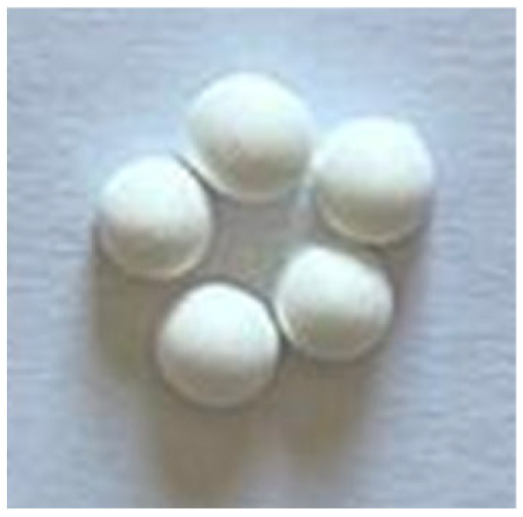
Orthopedic acrylic cement consists of two components. The powder component contains polymethyl methacrylate (PMMA), benzoyl peroxide as the polymerization initiator, barium sulfate as a radiopaque agent, and gentamicin as the antibiotic. The liquid component consists of methyl methacrylate (MMA), butyl methacrylate (BMA), N, N-dimethyl-p-toluidine as the polymerization activator, and hydroquinone as a stabilizer. The gentamicin content in the powder component consists of 3.8% (w/w), corresponding to 1 g of gentamicin base per dose of bone cement. To prepare the samples, the liquid component was added to the powder under sterile conditions and manually mixed for approximately 50–60 s, taking care to avoid air bubble formation until a homogeneous paste was obtained. The mixture was allowed to rest for approximately 1 min to reach a workable consistency, after which it was transferred into flexible silicone molds to produce spherical beads with a diameter of 6 mm. The setting time at room temperature (approximately 23 °C) was 8–10 min. The samples were allowed to polymerize completely for an additional 24 h, after which they were removed from the molds and maintained under sterile conditions until further experimental evaluation. The experimental resorbable samples, in the form of beads, were prepared by combining 6 mL of a liquid phase with 20 g of calcium sulfate as base material, with or without an antibiotic, as specified for each experimental group. The selected antibiotic addition corresponds to clinically and experimentally established concentrations reported in the literature [16,50,53,54,55]. For the control samples (SC), the calcium sulfate powder was combined with 6 mL of sterile water and manually mixed to obtain a workable paste. Gentamicin-containing samples (SC_G) were prepared by using 20 g of calcium sulfate as the base material and 6 mL of gentamicin solution (40 mg/mL) as the liquid phase, providing a total gentamicin content of 240 mg per experimental group. In the vancomycin-containing samples (SC_V), 1 g of vancomycin powder was dry-mixed with 20 g of calcium sulfate prior to the addition of 6 mL of sterile water. For the dual-antibiotic formulation (SC_VG), 1 g of vancomycin was first blended with the calcium sulfate powder, followed by the addition of 6 mL of gentamicin solution (40 mg/mL). The corresponding liquid component for each formulation was added and mixed for 30 s until a homogeneous paste formed. The material was then placed into flexible silicone molds to form spherical beads with a diameter of 6 mm. The setting time was approximately 8 min for the antibiotic-free samples and 3–5 min for the antibiotic-loaded formulations. Once hardened, the beads were removed from the molds and maintained under sterile conditions at room temperature until further experimental use. Figure 1 presents the macroscopic appearance of the prepared experimental samples.
The chemical structure of the experimental samples was analyzed by Fourier transform infrared spectroscopy (FT-IR) in attenuated total reflectance (ATR) mode. FTIR-ATR spectra were recorded in the range of 600–4000 cm−1 using a JASCO FTIR 6200 spectrometer (JASCO International Co., Tokyo, Japan) equipped with a Golden Gate ATR accessory (JASCO International Co., Tokyo, Japan). Microstructural characteristics and elemental chemical composition were examined by scanning electron microscopy with a Quattro S microscope (Thermo Fisher Scientific, Hillsboro, OR, USA) coupled with an energy-dispersive X-ray (EDS) spectrometer (Thermo Fisher Scientific, Hillsboro, OR, USA).
The surface properties of biomaterials and their interactions with physiological fluids play a crucial role in the performance of local antibiotic delivery systems, alongside their chemical composition. Surface wettability, commonly evaluated by contact angle measurements, provides valuable insight into a material’s hydrophilicity and its ability to interact with water and biological fluids. Hydrophilic surfaces promote more rapid fluid penetration, which can enhance water uptake, solubility, or degradation processes and, consequently, antibiotic release. However, excessive water absorption may induce unfavorable structural changes and accelerate material degradation, potentially compromising control over the release profile [55]. The wettability of the experimental samples was assessed by contact angle measurements using a droplet shape analysis system, KRÜSS DSA30 (A. Krüss Optronic GmbH, Hamburg, Germany), with distilled water as the test liquid. A droplet of 10 µL was gently deposited onto the sample surface at room temperature (23 ± 1 °C), and the contact angle was recorded immediately after droplet stabilization (within 5 s of deposition). For each experimental group, measurements were performed on five independent specimens (n = 5). The reported values represent the mean ± standard deviation.
Immersion tests were performed to evaluate the degree of hydration and the degradation of the experimental samples. The degree of hydration, assessed by the amount of absorbed water (WA), was determined by immersing the experimental samples in simulated body fluid (SBF) at 37 °C for 7 and 14 days. At each interval, the samples were removed from the medium, weighed, and then stored in a desiccator until a constant mass was reached. The amount of absorbed water was calculated using the following equation:
W A ( % ) = m w m f m 0 × 100
where mw represents the mass of the wet experimental samples; mf is the mass of the experimental samples after they reached a constant mass; and mo is the initial mass of the experimental samples.
The evaluation of the degradation process involved immersing the experimental samples in simulated body fluid (SBF) at 37 °C for periods of 7 and 14 days, respectively. The SBF solution, which has an ionic composition similar to that of human blood plasma, was prepared in the laboratory following the Kokubo protocol using chemicals purchased from Sigma-Aldrich (Taufkirchen, Germany). In addition, at each interval, the experimental samples were removed from the SBF solution, weighed, and stored in a desiccator until a constant mass was reached. The degradation of the experimental samples, assessed by mass loss, was determined using the following equation:
W L ( % ) = m O m f m 0 × 100
where mo represents the initial mass of the experimental samples, and mf is the mass of the experimental samples after they reached a constant mass.
Correlating FTIR and SEM–EDS techniques with contact angle measurements and gravimetric tests of water absorption and degradation allows for a more comprehensive understanding of material behavior in aqueous environments.

3. Results

The setting of calcium sulfate hemihydrate (CaSO4·½H2O) occurs through a hydration process, during which the material is converted into calcium sulfate dihydrate (CaSO4·2H2O). Upon contact with water, the hemihydrate undergoes an initial dissolution stage, followed by the formation of a supersaturated solution and the precipitation of the dihydrate phase. The progressive growth of calcium sulfate dihydrate crystals forms an interconnected network that determines the setting and hardening of the material. This exothermic reaction generates a porous microstructure consisting of acicular and plate-like crystals, whose features influence the final properties of the material, including mechanical strength, water absorption, and degradation behavior.

3.1. Fourier Transform Infrared Spectroscopy Determinations

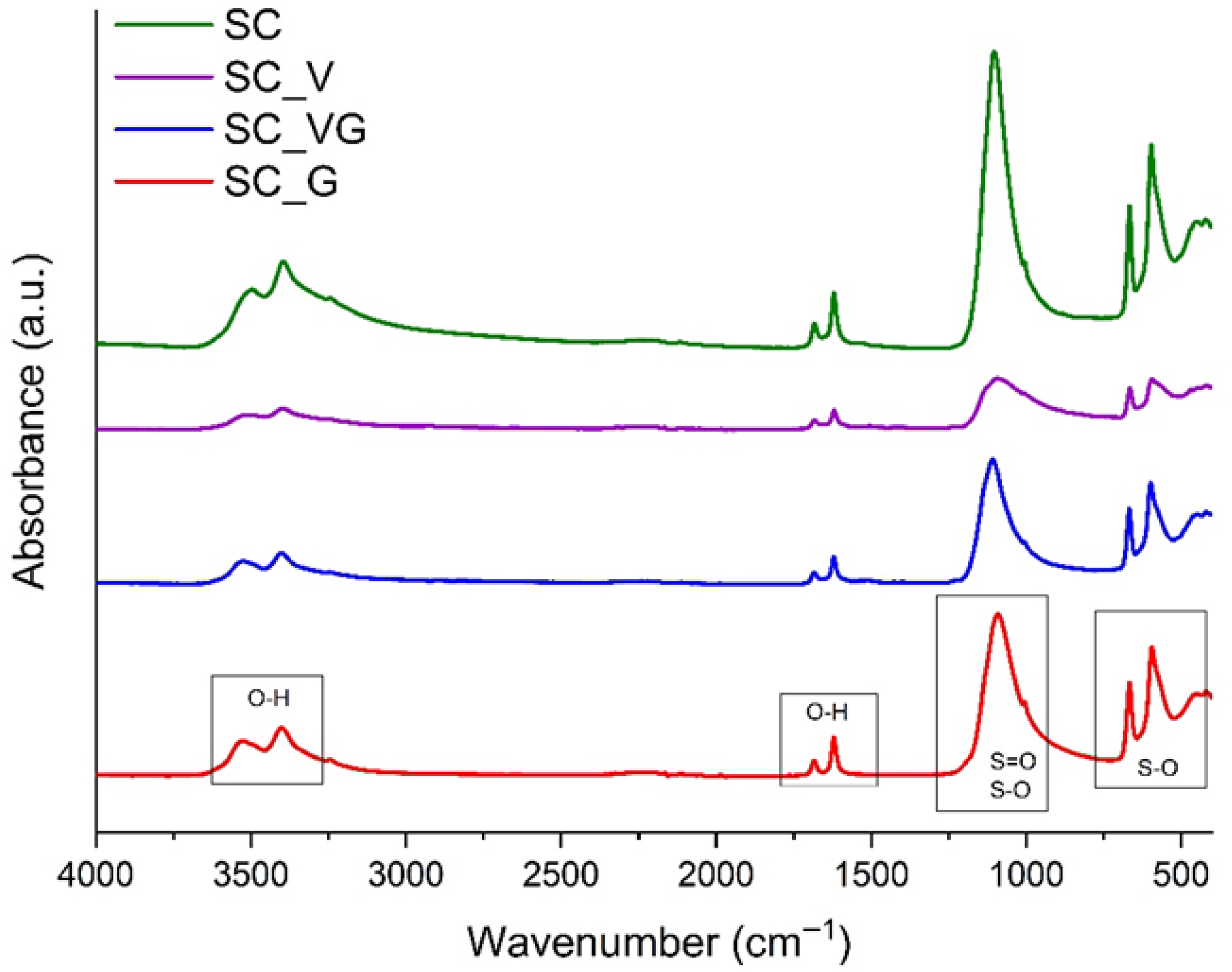
Figure 2 presents the FTIR-ATR absorption spectra of the calcium sulfate–based materials in the spectral range of 500–4000 cm−1. The absorption bands identified at 3401 cm−1 and 3526 cm−1 are attributed to O–H stretching vibrations (νOH), associated with structural water in the hydrated phase. Their presence indicates the incorporation of water into the crystalline lattice, characteristic of calcium sulfate dihydrate (CaSO4·2H2O). The bands at 1683 cm−1 and 1621 cm−1 are assigned to the O–H bending vibrations of water (δOH). In general, the band at ~1621 cm−1 is related to more strongly bound water molecules in all types of calcium sulphate (hydrated or anhydrous), whereas the band around 1683 cm−1 is associated with more weakly bound water and is frequently reported for calcium sulfate dihydrate [56,57] band at 1100 cm−1 corresponds to SO42− stretching vibrations (νS=O), the band at 1006 cm−1 corresponds to SO42− stretching vibrations (νS-O), while the bands at 667 cm−1 and 594 cm−1 are attributed to sulfate bending modes, confirming the typical calcium sulfate structure. Moreover, the absence of the band at approximately 3609 cm−1, reported as characteristic for the νOH vibration of calcium sulfate hemihydrate, together with the presence of the 1683 cm−1 band, further supports the transformation of the material toward the dihydrate phase during the sample preparation.
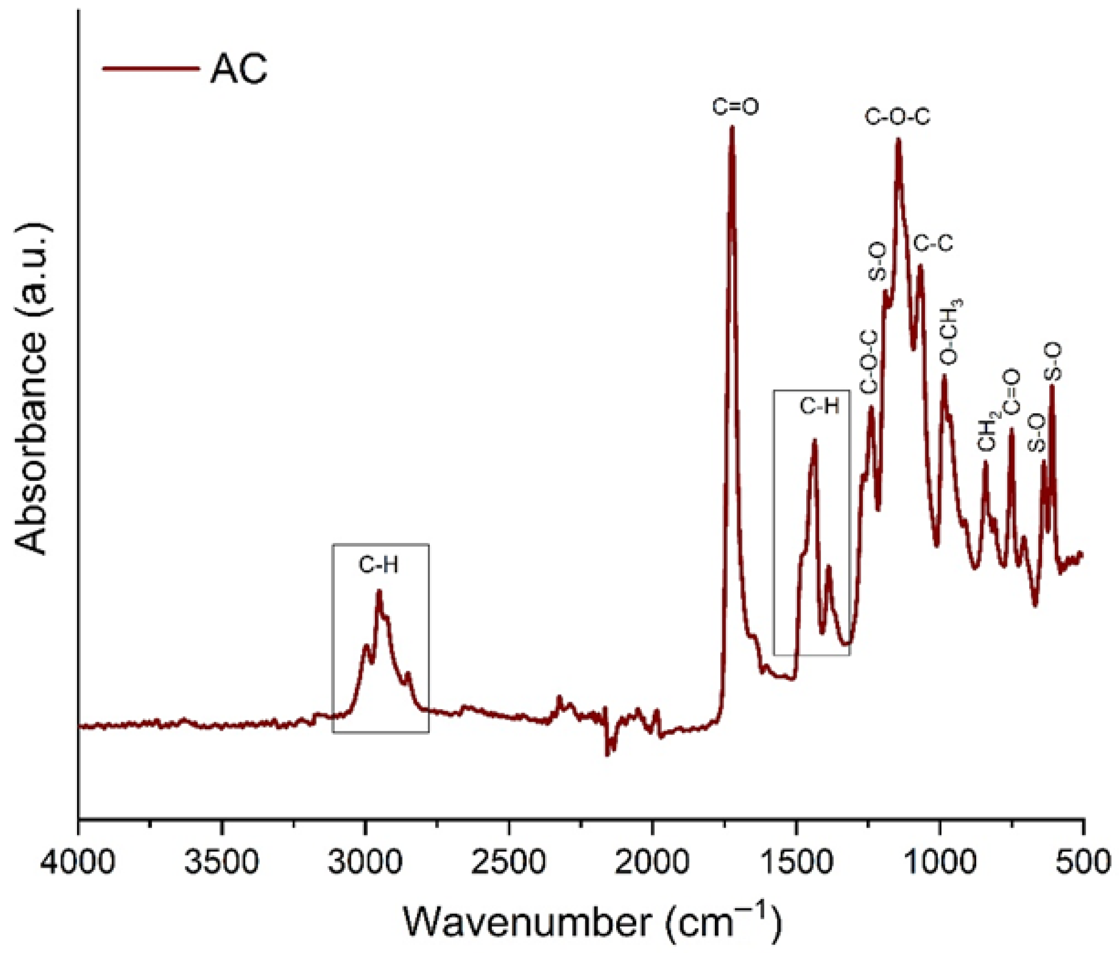
The characteristic FTIR bands of gentamicin and vancomycin could not be clearly distinguished in the spectra of the antibiotic-loaded CaSO4 samples. This is mainly due to the relatively low antibiotic content compared to the calcium sulfate matrix, whose characteristic bands dominate the spectra. Moreover, the main absorption regions of the antibiotics (e.g., O–H and N–H stretching in the 3000–3600 cm−1 range, as well as vibrations in the 1000–1150 cm−1 region) overlap with the intense bands of hydrated calcium sulfate and structural water. Therefore, antibiotic incorporation is reflected primarily by variations in band intensity and peak broadening, rather than by the appearance of new distinct peaks. The FTIR spectroscopy determination results indicate that the antibiotics are predominantly physically incorporated into the calcium sulfate structure, with no evidence of significant chemical interactions during sample preparation. The FTIR spectrum of the acrylic cement (AC), Figure 3, revealed the characteristic absorption bands of a PMMA-based orthopedic cement, confirming the typical methacrylate structure of the material [58,59,60,61].
The bands observed in the 2840–3000 cm−1 range are assigned to the asymmetric and symmetric C–H stretching vibrations of CH3 and CH2 groups. The intense peak at 1725 cm−1 corresponds to the carbonyl (C=O) stretching vibration of the ester group, a key spectral fingerprint of PMMA. Additional bands around 1440 cm−1 and 1380 cm−1 are associated with methyl/methylene bending vibrations (δCH3/δCH2). The strong absorption band at the 1144 cm−1 region is attributed to asymmetric C–O–C stretching vibrations of the ester groups, while the bands at 985 cm−1 and 841 cm−1 are related to skeletal vibrations and rocking modes of O–CH3 and CH2 groups. Moreover, the band at 1240 cm−1 is assigned to the symmetric C–O–C stretching vibration, the band observed around 1062 cm−1 corresponds to skeletal C–C stretching vibrations, and the band at 750 cm−1 can be attributed to the out-of-plane bending vibration of the C=O group. The spectrum confirms the typical FTIR profile of orthopedic acrylic cement, with no evidence of major chemical changes in the PMMA matrix. In addition to the polymer-related bands, the spectrum indicates the presence of barium sulfate (BaSO4), a radiopaque agent.
In particular, the band at 1188 cm−1 can be assigned to the symmetric stretching vibrations of the sulfate group (S–O), while the bands at 636 cm−1 and 609 cm−1 are related to O–S–O bending vibrations. The FTIR spectrum of gentamicin contains characteristic bands associated with its functional groups, including N–H bending vibrations at 1620 and 1524 cm−1, as well as sulfate-group vibrations, observed around 600 cm−1 (S–O bending) and 1040 cm−1 (S–O stretching) [62,63,64]. In the case of experimental gentamicin-loaded acrylic cement (AC), these bands could not be clearly distinguished, most likely because the relatively low antibiotic content compared to the PMMA matrix means its intense bands dominate the spectrum.

3.2. Scanning Electron Microscopy Coupled with Energy-Dispersive X-Ray Spectrometry (SEM–EDS) Determinations

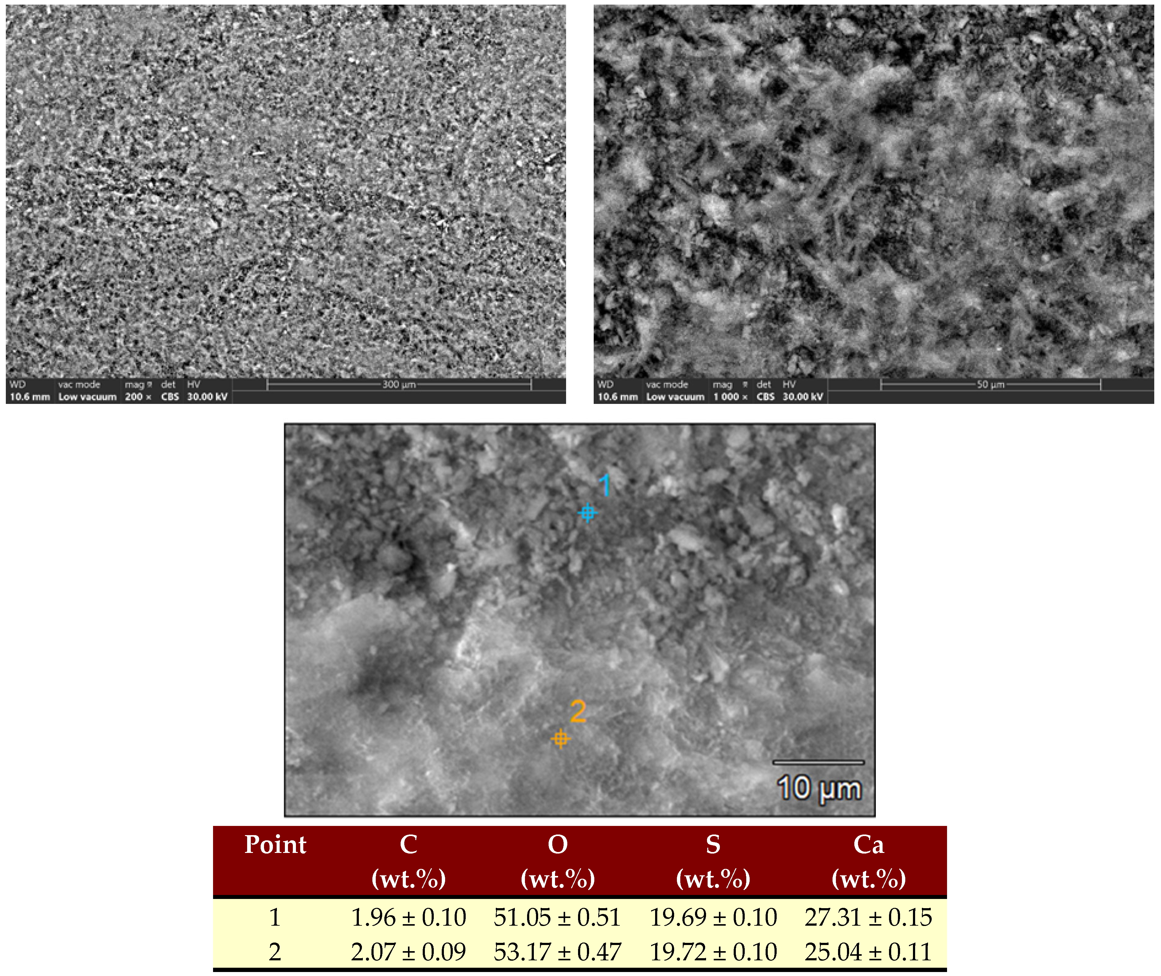
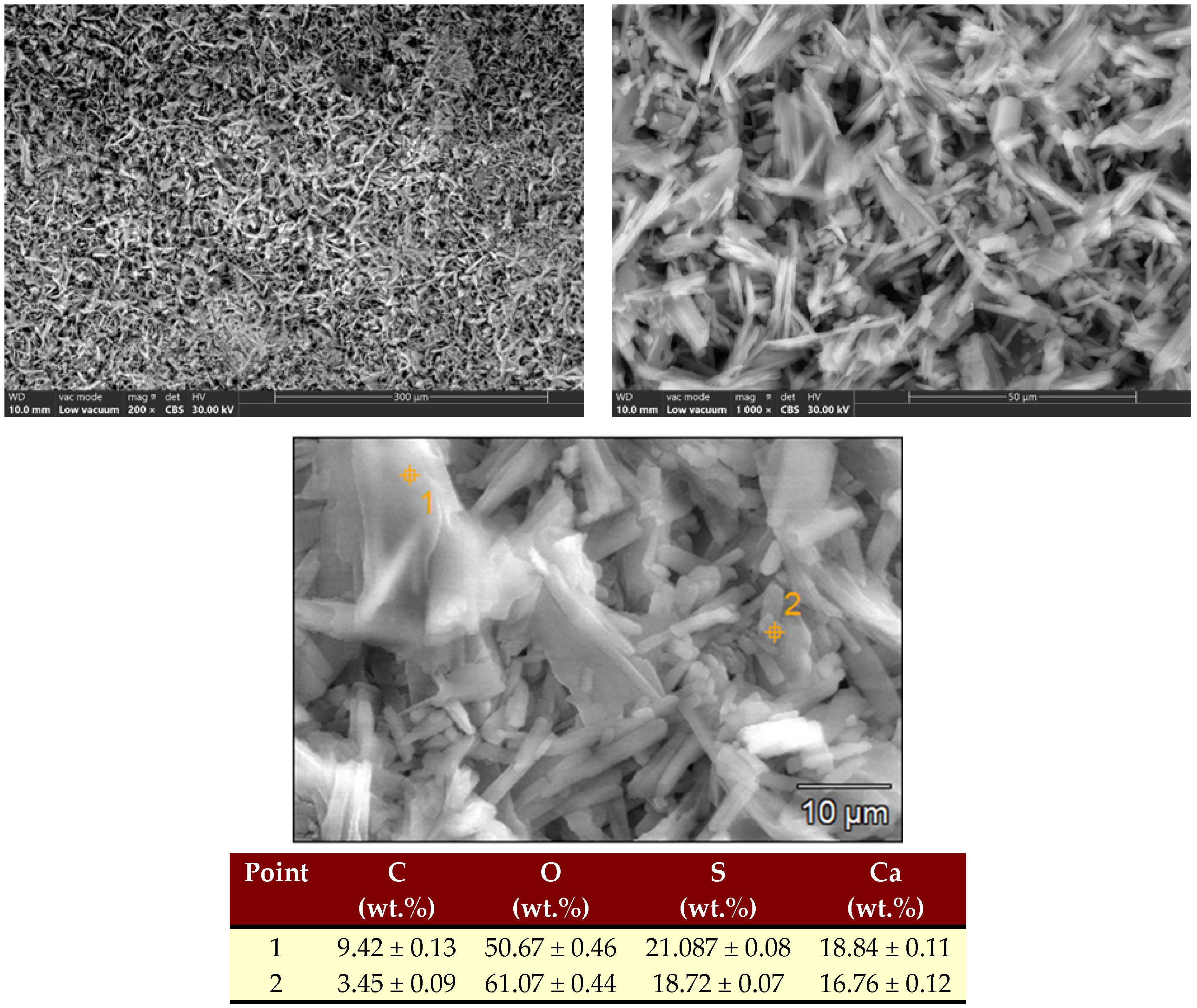
The SEM analysis was performed to investigate the morphology of the experimental samples, while the EDS analysis was used to determine their elemental composition. Figure 4 presents the SEM micrographs and the point elemental composition determined by EDS for the SC sample. The SEM images obtained for the calcium sulfate control (SC) reveal a relatively compact surface, with a fine microstructure and a uniform distribution of the inorganic phase. Point EDS analysis indicates the presence of the elements specific to calcium sulfate (O, S, and Ca), with a very low carbon (C) content, thus confirming the inorganic nature of the sample. The similar values obtained for the two analyzed points suggest a relatively homogeneous local composition.
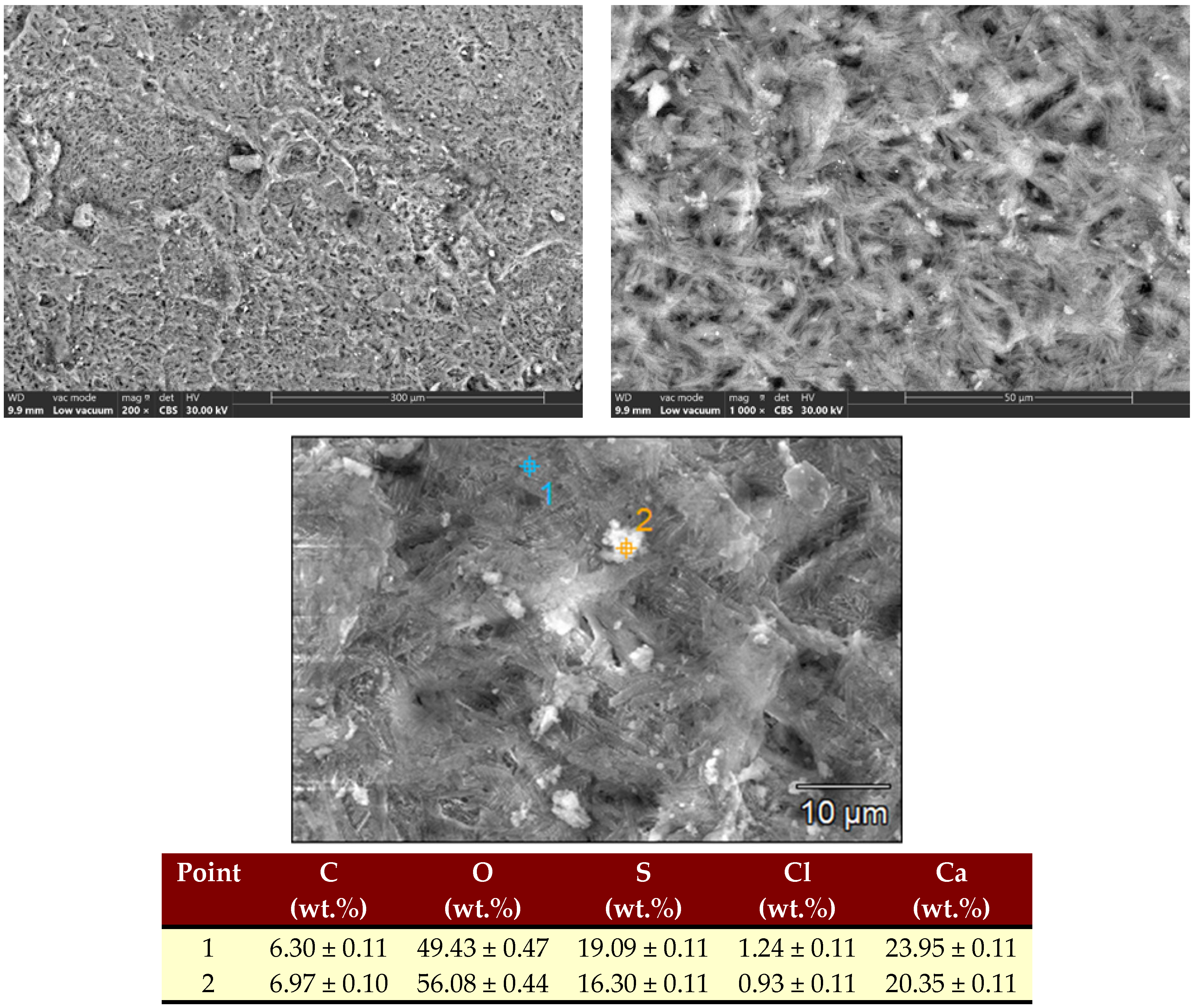
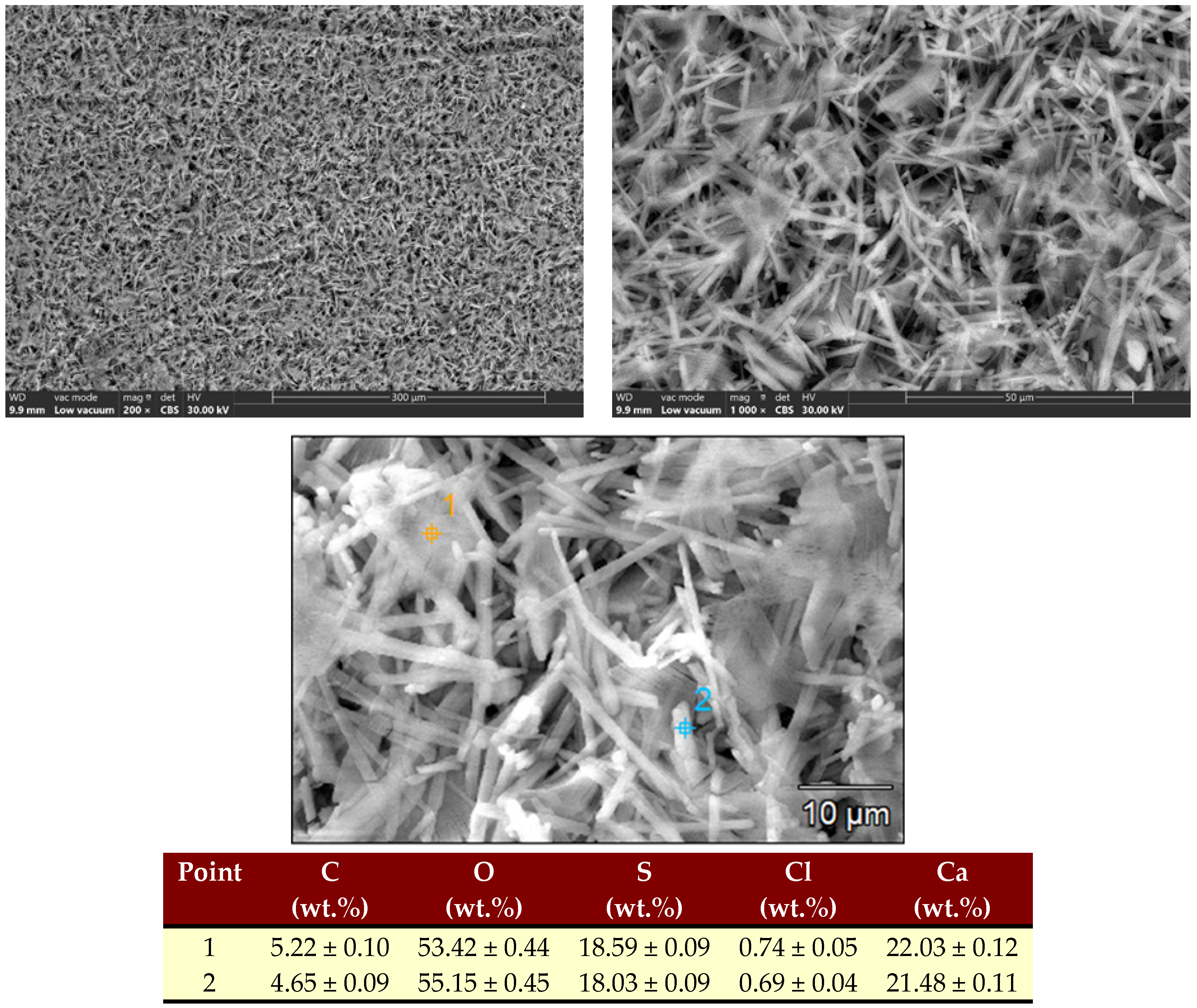
The SEM–EDS results for the antibiotic-loaded calcium sulfate samples are presented in Figure 5. In the case of the SC_G sample (calcium sulfate with gentamicin), the microstructure is characterized by a well-developed network of acicular and plate-like crystals with a porous appearance. The presence of gentamicin is reflected in the higher carbon (C) content detected by EDS compared to the control sample, suggesting its incorporation into the material matrix. The SC_V sample (calcium sulfate with vancomycin) exhibits a more compact, less uniform microstructure than SC_G, with dense areas and regions exhibiting a crystalline aspect (Figure 6). EDS analysis confirms the presence of calcium sulfate elements (O, S, Ca), as well as chlorine (Cl), associated with vancomycin, which was introduced as vancomycin hydrochloride. For the SC_VG sample, a predominantly acicular, well-developed microstructure is observed, with evident porosity and a dense network (Figure 7). EDS analysis again highlights the characteristic elements of calcium sulfate (O, S, Ca) and Cl, confirming the presence of the vancomycin-related component. Compared with the SC sample, the addition of antibiotics results in microstructural changes, particularly increased porosity and more pronounced crystals, particularly in the gentamicin-loaded sample and in the combined antibiotic composition. These microstructural modifications may result in different water absorption and degradation behaviors of the materials in the testing environment.
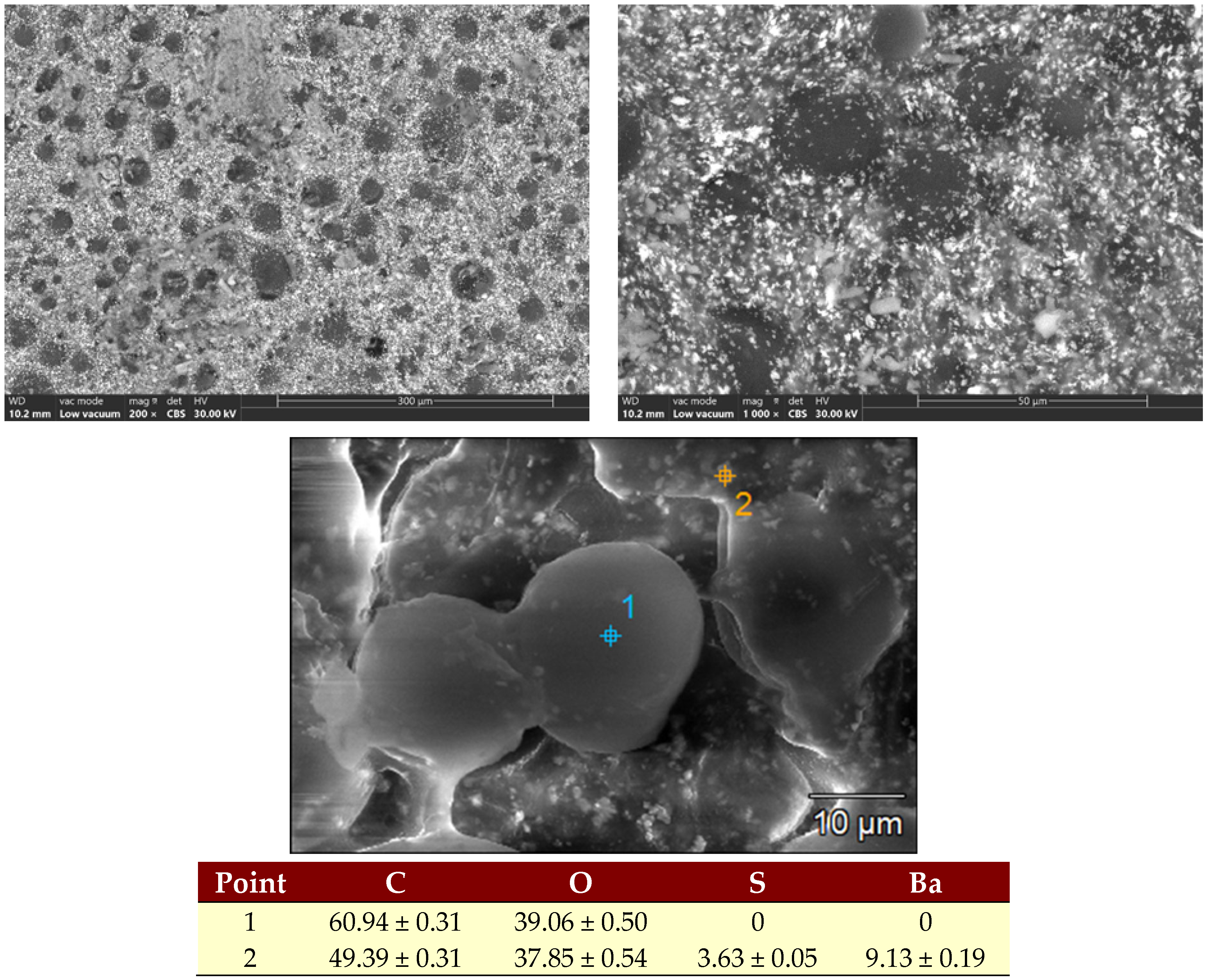
SEM analysis of the gentamicin-loaded acrylic cement highlights a microstructure typical of PMMA-based orthopedic cement, consisting of prepolymerized polymer powder granules embedded within a continuous polymeric matrix (Figure 8). Within this matrix, the radiopaque agent, barium sulfate, was identified, showing a relatively uniform dispersion together with the other additives, resulting in a more homogeneous appearance of the sample. The surface morphology indicates the presence of pores and small imperfections, which may act as diffusion pathways and facilitate antibiotic release from the material. These microstructural features, along with the PMMA particle-size distribution, can influence gentamicin elution, as surface defects such as pores, fissures, or cavities may promote fluid penetration and antibiotic transport toward the external environment.

3.3. Contact Angle Determinations

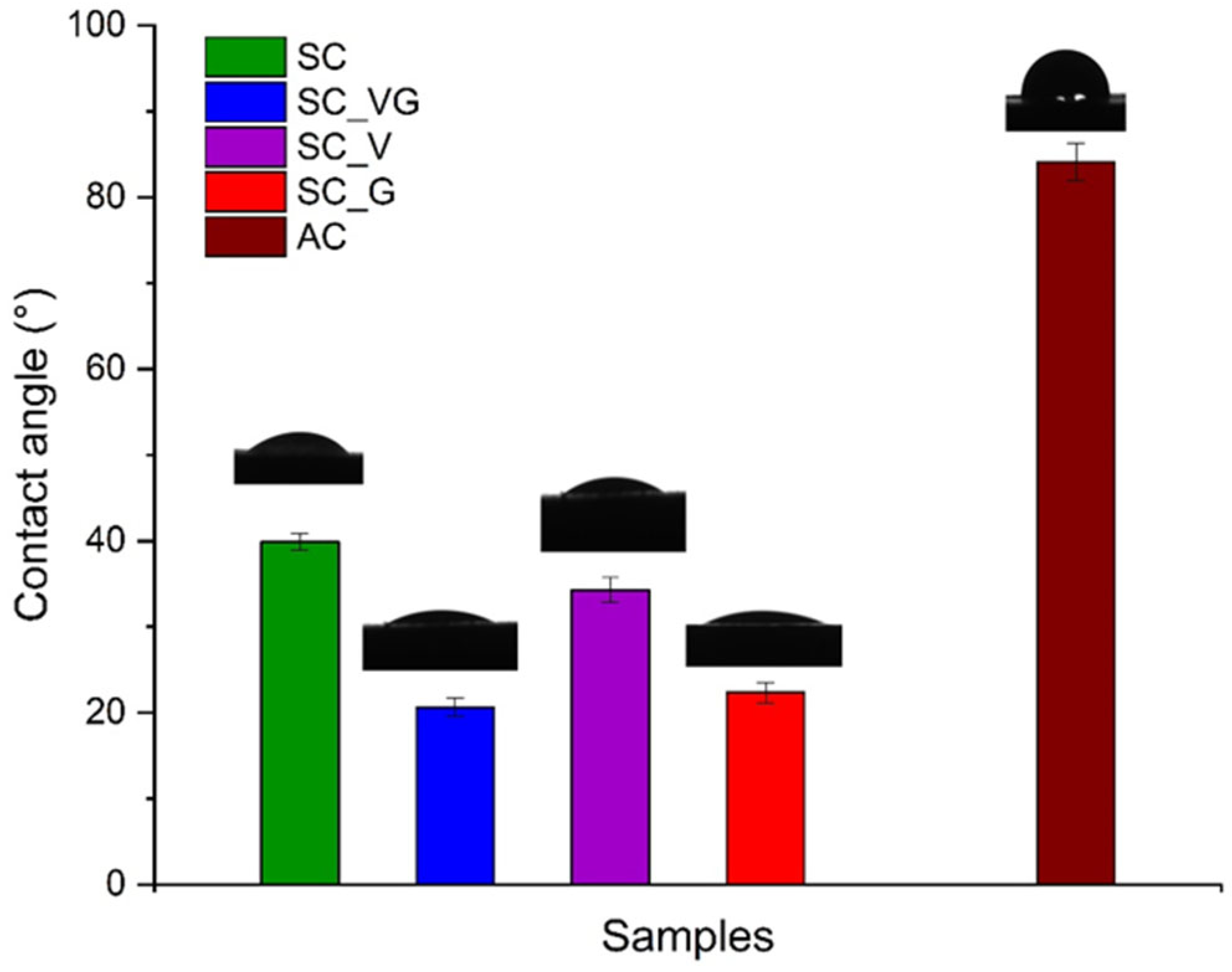
An essential factor controlling the antibiotic release process is surface wettability, as fluid penetration into the material structure is influenced by the hydrophilic/hydrophobic nature of the matrix and pore distribution [17,65,66]. Contact angle measurements highlighted clear differences in surface wettability between the PMMA-based acrylic cement and the calcium sulfate–based resorbable samples, with and without antibiotics, confirming the strong influence of composition on surface properties. The obtained results are presented in Figure 9.
The contact angle measured for the AC sample (84.09°) indicates a predominantly hydrophobic surface and may limit the spreading of physiological fluids and the initial interaction with the aqueous environment. In contrast, the calcium sulfate control sample showed a much lower contact angle (39.88°), indicating a hydrophilic surface that promotes the formation of a continuous water film, an important aspect for hydration and interaction with biological media. The incorporation of antibiotics into calcium sulfate generally enhanced surface hydrophilicity, with the most pronounced effect observed for gentamicin-containing samples: 22.34° for SC_G and 20.65° for SC_VG. These values indicate highly wettable surfaces, which may facilitate fluid penetration into the porous structure and promote mass-transport processes relevant to antibiotic elution. These results are relevant from a biological perspective, as surface wettability influences protein adsorption and cell attachment, and moderate contact angle values have often been associated in the literature with favorable cell–material interactions [67,68,69]. In addition, for calcium sulfate–based resorbable systems, increased wettability may correlate with faster hydration, greater water uptake, and altered degradation behavior, depending on the incorporated antibiotic. Compared to PMMA cement, calcium sulfate samples—particularly those loaded with gentamicin or the combined antibiotic formulation—exhibited significantly more hydrophilic surfaces, which may contribute to improved interaction with aqueous environments and enhanced performance as local antibiotic delivery systems.

3.4. Immersion Tests

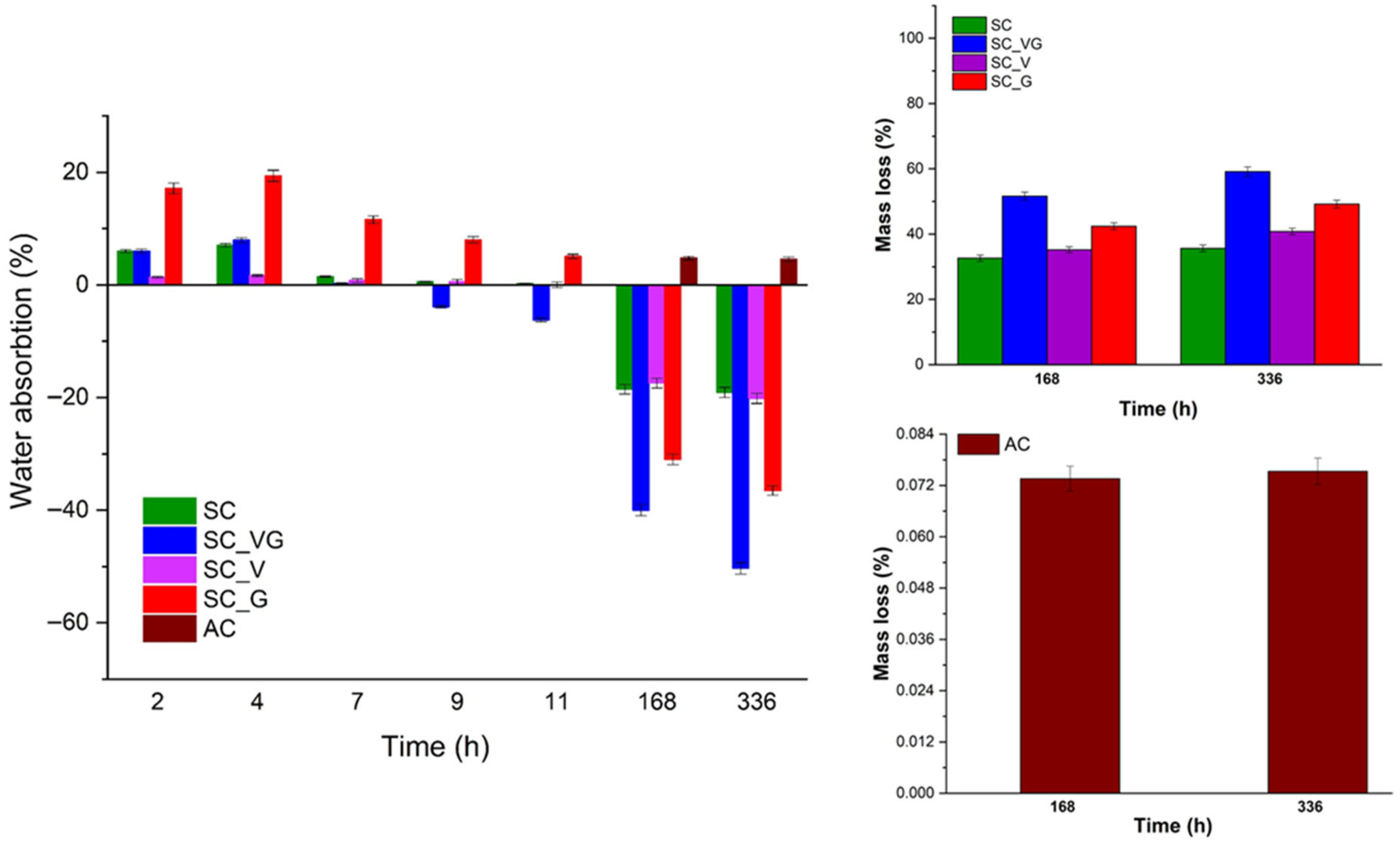
Figure 10 presents the evolution of water absorption and mass loss with immersion time for the experimental sample. Differences can be observed between the acrylic cement sample (AC) and the resorbable calcium sulfate–based materials, including both the control sample (SC) and the antibiotic-loaded compositions (SC_G, SC_V, SC_VG). During the first 11 h of immersion, the calcium sulfate samples undergo a water-uptake stage, characterized by rapid penetration of fluid into the material and hydration. In contrast, the acrylic cement (AC) exhibits a much more stable behavior, with low absorption values, suggesting limited surface wetting and restricted fluid penetration into the polymeric matrix, consistent with the hydrophobic character of PMMA-based cement.
At 168 h (7 days), all calcium sulfate-based samples exhibit negative water absorption values, indicating that the materials have left the hydration stage and reached an advanced degradation process. This behavior is further supported by SEM observations, which show that the antibiotic-loaded samples, particularly SC_G and SC_VG, exhibit well-developed acicular and plate-like structures and higher porosity, thereby enhancing fluid penetration into the material and progressive degradation. In the case of acrylic cement AC, water absorption at 168 h (7 days) and 336 h (14 days) remains low and nearly constant, confirming the material’s stability in an aqueous environment and the absence of significant degradation, with only limited surface sorption likely.
Mass loss results highlight a typical behavior for resorbable materials, in which degradation is controlled by matrix dissolution and the release of soluble components into the medium. For all calcium sulfate-based samples, higher mass loss is observed during the first 7 days of immersion, indicating a more accelerated degradation stage, followed by a slower degradation rate between 7 and 14 days. Thus, at 14 days, the mass loss values do not increase substantially compared to those at 7 days; for example, for SC increases from approximately 33% to 36%, for SC_V from ~35% to ~41%, for SC_G from ~42% to ~50%, and for SC_VG from ~52% to ~60%.
The antibiotic-loaded samples exhibit greater mass loss than the control sample, demonstrating that antibiotics accelerate degradation, most likely by altering the microstructure and enhancing interactions with the aqueous medium. SC_VG shows the highest mass loss values at both 7 and 14 days, suggesting that the gentamicin–vancomycin combination results in a more permeable structure that is more susceptible to degradation. In addition, SC_G also exhibits increased mass loss, consistent with its highly hydrophilic character and intense initial water uptake, indicating rapid fluid penetration and more pronounced degradation.
In comparison, for the AC sample, the polymer matrix is non-resorbable and exhibits low porosity; therefore, mass loss is mainly attributed to the release of soluble components from its composition, such as residual monomer or antibiotics. This process primarily occurs in the surface layer and does not involve the actual degradation of the polymeric matrix.
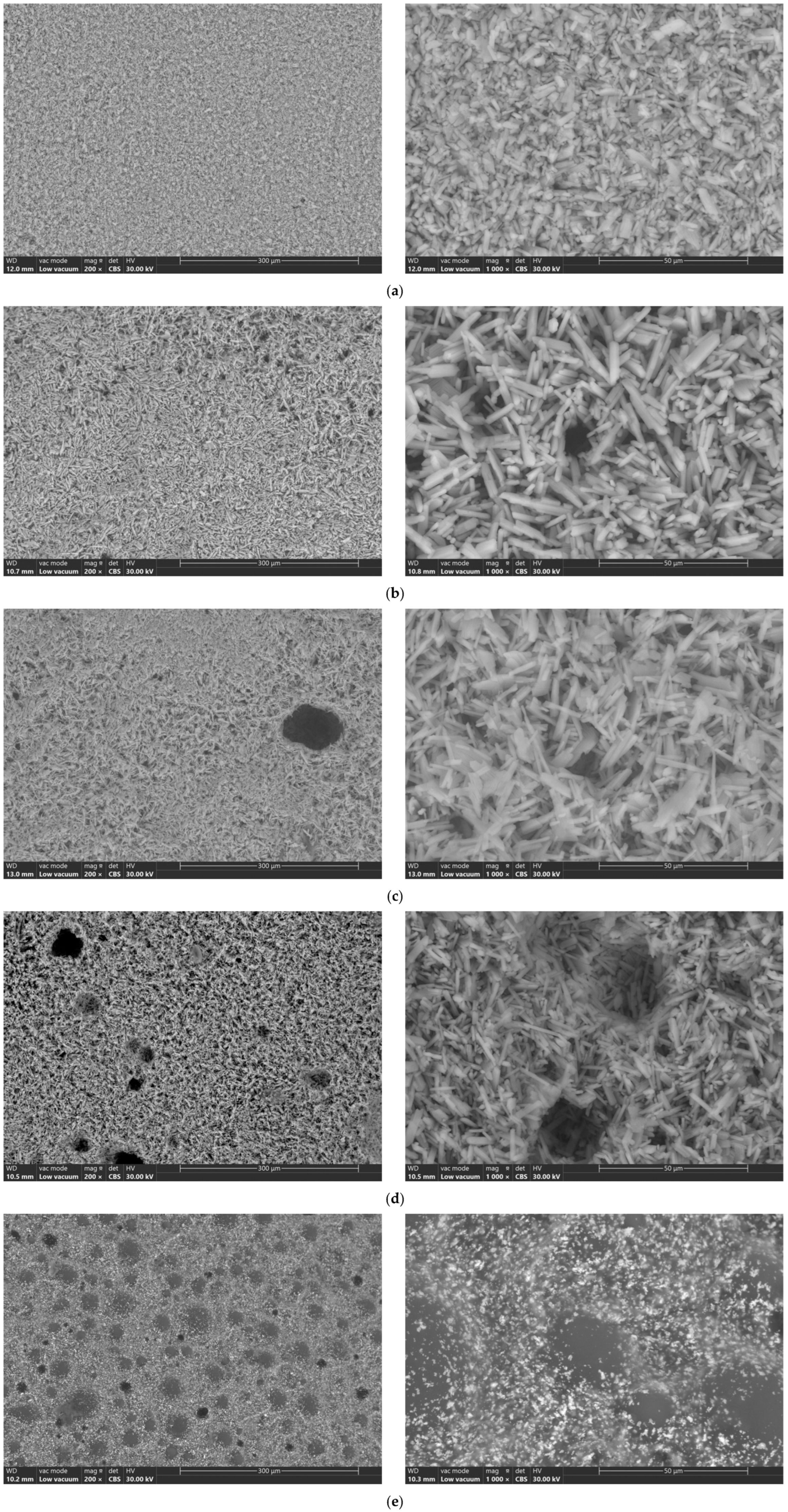
The microstructural appearance of the investigated samples after 14 days of immersion, revealed after SEM analysis, is presented in Figure 11.
After immersion in simulated body fluid (SBF), calcium sulfate-based materials exhibit progressive hydration and degradation behavior characteristic of resorbable materials. Interaction with the medium results in water absorption into the material, partial dissolution of the calcium sulfate matrix, and gradual mass loss. These processes are accompanied by microstructural changes, such as the formation of gaps on material surfaces and the evolution of acicular or plate-like calcium sulfate crystals. Depending on composition, immersion of the sample in SBF may also promote surface reactions and the precipitation of calcium-containing phases. The immersion tests provide relevant insight into the in vitro stability, degradation kinetics, and potential biological response of calcium sulfate–based materials under conditions that approximate the physiological environment. For the acrylic cement sample (AC), results after 14 days of immersion in SBF indicate high structural stability and minimal degradation. The interaction with the medium is largely limited to the surface, where slight water uptake or superficial sorption may occur without significant penetration into the polymer matrix. Microstructural observations after SBF immersion usually show the preservation of the cement’s compact morphology, with no major changes in pore structure or surface integrity.

4. Conclusions

In this study, samples based on calcium sulfate, the SC sample, as well as samples loaded with antibiotics, namely gentamicin (SC_G), vancomycin (SC_V), or their combination (SC_VG), were obtained. The incorporation of antibiotics into the calcium sulfate matrix influenced the hydration process and the morphology of the obtained material. The resulting microstructural modifications depended on the type of antibiotics, leading to variations in crystal growth, porosity, and surface characteristics. Compared with the control sample, those loaded with antibiotics exhibited altered setting behavior, with shorter hardening times. The modified setting behavior observed in the antibiotic-loaded samples relative to the control formulation may be attributed to the antibiotics’ concentration in the mixture, to the change in the effective powder-to-liquid ratio, and to the antibiotic’s influence on the hydration kinetics of calcium sulfate. The transformation of calcium sulfate hemihydrate to dihydrate proceeds via a dissolution–precipitation mechanism, and the presence of additional solid particles, such as vancomycin powder, may serve as heterogeneous nucleation sites, thereby accelerating crystal growth and hardening. In the case of gentamicin solution, changes in the ionic environment and liquid-phase composition may affect dissolution dynamics and supersaturation levels, thereby promoting faster dihydrate precipitation. These findings indicate that antibiotics do not act merely as passive fillers but may actively modify the physicochemical processes governing cement hardening. Moreover, the incorporation of gentamicin (SC_G and SC_VG samples) affected the microstructure of the samples, favoring the formation of more pronounced crystals, of the acicular and lamellar type, and an increased porosity. These structural modifications facilitated an improved interaction with aqueous media. The behavior in the simulated environment revealed a more intense initial water absorption during the hydration stage and a more pronounced mass loss during prolonged immersion. For all antibiotic-containing calcium sulfate samples, the increase in hydrophilicity, microstructural changes, and degradation behavior observed may favor their sustained release.
The investigations suggested that acrylic cement exhibited high structural stability, hydrophobicity, and low mass loss, which limit fluid penetration and lead to predominantly superficial antibiotic release without degradation of the polymer matrix. Overall, the results emphasize that the choice between acrylic and calcium sulfate-based cements should be based on specific clinical requirements, balancing the need for mechanical support with the efficiency of local antibiotic delivery. In the context of local antibiotic delivery systems, calcium sulfate may represent the more appropriate option for further biomedical application, while acrylic cement remains preferable in clinical applications that prioritize durable mechanical support.

Author Contributions

Conceptualization: I.A., A.A. and D.N.A.; methodology: A.A., A.-I.B. and I.A.; software: C.-M.C., A.S. and A.-I.B.; validation: I.A. and A.A.; formal analysis: A.A., C.-M.C., A.S. and D.N.A.; investigation: A.A., C.-M.C., D.N.A., A.S. and A.-I.B.; resources: I.A. and D.N.A.; data curation: I.A.; writing—original draft preparation: C.-M.C., A.A. and A.-I.B.; writing—review and editing: A.A., A.S. and A.-I.B.; visualization: I.A. and D.N.A.; supervision: I.A. and A.A.; funding acquisition: I.A. All authors have read and agreed to the published version of the manuscript.

Funding

This research received no external funding.

Institutional Review Board Statement

Not applicable.

Informed Consent Statement

Not applicable.

Data Availability Statement

The original contributions presented in the study are included in the article; further inquiries can be directed to the corresponding author.

Conflicts of Interest

The authors declare no conflicts of interest.

References

  1. Aggarwal, D.; Kumar, V.; Sharma, S. Drug-Loaded Biomaterials for Orthopedic Applications: A Review. J. Control. Release 2022, 344, 113–133. [Google Scholar] [CrossRef]
  2. Knight, S.R.; Aujla, R.; Biswas, S.P. Total Hip Arthroplasty—Over 100 Years of Operative History. Orthop. Rev. 2011, 3, 72–74. [Google Scholar] [CrossRef]
  3. Fuqua, A.A.; Worden, J.A.; Ayeni, A.M.; Bundschuh, K.E.; Premkumar, A.; Wilson, J.M. Extended Oral Antibiotic Prophylaxis and PJI-Free Survivorship after Primary Total Knee Arthroplasty. Knee 2025, 56, 1–10. [Google Scholar] [CrossRef]
  4. Salimi, M.; Keshtkar, A.; Abdelnaser, M.K.; Mosalamiaghili, S.A.; Saka, N.; Parvizi, J. Effect of Vancomycin Powder in Reducing Infection After Primary and Revision Hip and Knee Arthroplasty and Its Complications: An Umbrella Review and Meta-Analysis. J. Arthroplast. 2025, 1–11, in press. [Google Scholar] [CrossRef]
  5. Antoniac, I.; Negrusoiu, M.; Mardare, M.; Socoliuc, C.; Zazgyva, A.; Niculescu, M. Adverse Local Tissue Reaction after 2 Revision Hip Replacements for Ceramic Liner Fracture. Medicine 2017, 96, e6687. [Google Scholar] [CrossRef] [PubMed]
  6. Miculescu, F.; Bojin, D.; Ciocan, L.T.; Antoniac, I.V.; Miculescu, M.; Miculescu, N. Experimental Researches on Biomaterial-Tissue Interface Interactions. J. Optoelectron. Adv. Mater. 2007, 9, 3303–3306. [Google Scholar]
  7. Marinescu, R.; Antoniac, I.; Laptoiu, D.; Antoniac, A.; Grecu, D. Complications Related to Biocomposite Screw Fixation in ACL Reconstruction Based on Clinical Experience and Retrieval Analysis. Mater. Plast. 2015, 52, 340–344. [Google Scholar]
  8. Pascu, A.; Oleksik, V.; Pirvu, B.; Fratila, A.; Ionas, M.; Boitor, C. Modern Methods of Study and Research in Mechanical Engineering Applied to Medicine. In Proceedings of the Balkan Regional Conference on Engineering and Business Education & ICEBE, Sibiu, Romania, 15–17 October 2009. [Google Scholar]
  9. Cardoso, G.C.; Correa, D.R.N.; Fosca, M.; Pometun, E.V.; Antoniac, I.V.; Grandini, C.R.; Rau, J.V. Current Strategies in Developing Antibacterial Surfaces for Joint Arthroplasty Implant Applications. Materials 2025, 18, 173. [Google Scholar] [CrossRef]
  10. Berberich, C.E. Current Concepts of Local Antibiotic Delivery in Bone and Joint Infections—A Narrative Review of Techniques and Clinical Experiences. Microorganisms 2025, 13, 2276. [Google Scholar] [CrossRef] [PubMed]
  11. Hoveidaei, A.H.; Mosalamiaghili, S.; Sabaghian, A.; Hajiaghajani, S.; Farsani, A.S.; Sahebi, M.; Poursalehian, M.; Nwankwo, B.O.; Conway, J.D. Local Antibiotic Delivery: Recent Basic and Translational Science Insights in Orthopedics. Bone 2025, 193, 117416. [Google Scholar] [CrossRef]
  12. Aparicio-Blanco, J.; López-Torres, I.I.; Alonso-Berenguel, M.; Torres-Suárez, A.I.; Martín-Sabroso, C. Local Antimicrobial Delivery Systems for Prophylaxis and Treatment of Periprosthetic Traumatological Infections. Eur. J. Pharm. Sci. 2025, 204, 106940. [Google Scholar] [CrossRef]
  13. Berliner, Z.P.; Freccero, D.; Cooper, H.J.; Smith, E.L.; Kain, M.S. Antibiotic Loaded Bone Cement in Primary Total Hip and Knee Arthroplasty. Montefiore Einstein J. Musculoskelet. Med. Surg. 2025, 4, 1–6. [Google Scholar] [CrossRef]
  14. Golubchikov, D.O.; Fadeeva, I.V.; Trofimchuk, E.S.; Barbaro, K.; Yankova, V.G.; Antoniac, I.V.; Putlayev, V.I.; Rau, J.V.; Saceleanu, V. Methylcellulose-Encapsulated Magnesium-Substituted Biphasic Calcium Phosphate Granules for Local Drug Delivery in Bone Tissue Engineering: Modification for Prolonged Release and Antibacterial Behavior. Polymers 2025, 17, 2422. [Google Scholar] [CrossRef]
  15. Anagnostakos, K. Therapeutic Use of Antibiotic-Loaded Bone Cement in the Treatment of Hip and Knee Joint Infections. J. Bone Jt. Infect. 2017, 2, 29–37. [Google Scholar] [CrossRef] [PubMed]
  16. Naoum, S.; Koutserimpas, C.; Pantekidis, I.; Giovanoulis, V.; Veizi, E.; Piagkou, M.; Ioannou, P.; Samonis, G.; Domouchtsidou, A.; Tsantes, A.G.; et al. Antimicrobial Regimens in Cement Spacers for Periprosthetic Joint Infections: A Critical Review. Antibiotics 2024, 13, 772. [Google Scholar] [CrossRef] [PubMed]
  17. Fink, B.; Tetsworth, K.D. Antibiotic Elution from Cement Spacers and Its Influencing Factors. Antibiotics 2025, 14, 705. [Google Scholar] [CrossRef] [PubMed]
  18. Shi, X.; Wu, Y.; Ni, H.; Li, M.; Zhang, C.; Qi, B.; Wei, M.; Wang, T.; Xu, Y. Antibiotic-Loaded Calcium Sulfate in Clinical Treatment of Chronic Osteomyelitis: A Systematic Review and Meta-Analysis. J. Orthop. Surg. Res. 2022, 17, 104. [Google Scholar] [CrossRef]
  19. Cheng, S.; Meng, X.-H.; Li, Z.; Han, H.-H.; Zhang, Y.-F. Nanomaterial-Mediated Antibiotic Delivery: A Novel Strategy for Osteomyelitis Therapy. Front. Bioeng. Biotechnol. 2025, 13, 1671151. [Google Scholar] [CrossRef]
  20. Yong, Y.; Jia, L.; Wu, H.; Xiang, T.; Zhou, S. Gradient Hydrogels: From Fabrication Strategies, Reinforcement Mechanisms, to Biomedical Applications. Chem. Eng. J. 2025, 522, 167390. [Google Scholar] [CrossRef]
  21. ter Boo, G.-J.A.; Grijpma, D.W.; Moriarty, T.F.; Richards, R.G.; Eglin, D. Antimicrobial Delivery Systems for Local Infection Prophylaxis in Orthopedic- and Trauma Surgery. Biomaterials 2015, 52, 113–125. [Google Scholar] [CrossRef]
  22. Ershad-Langroudi, A.; Babazadeh, N.; Alizadegan, F.; Mehdi Mousaei, S.; Moradi, G. Polymers for Implantable Devices. J. Ind. Eng. Chem. 2024, 137, 61–86. [Google Scholar] [CrossRef]
  23. Rajesh, K.; Khatua, C.; Singh, P.; Roy, P.; Keshri, A.K.; Lahiri, D. Microsphere Embedded Hydroxyapatite Coating on Metallic Implant for Sustained Drug Release in Orthopedic Applications. J. Drug Deliv. Sci. Technol. 2024, 98, 105840. [Google Scholar] [CrossRef]
  24. Voicu, S.I.; Pandele, A.M.; Nicoara, A.I.; Antoniac, I.V.; Oprea, M.; Bica, C. Chitosan–Hydroxyapatite Composite Membranes for the Controlled Release of Clindamycin Phosphate to Prevent Infections at the Implantation Site. Ceramics 2025, 8, 138. [Google Scholar] [CrossRef]
  25. Oprea, M.; Pandele, A.M.; Enachescu, C.I.; Antoniac, I.V.; Voicu, S.I.; Fratila, A.M. Crown Ether-Functionalized Polyethersulfone Membranes with Potential Applications in Hemodialysis. Polymers 2025, 17, 2184. [Google Scholar] [CrossRef]
  26. Oprea, M.; Pandele, A.M.; Nechifor, A.C.; Nicoara, A.I.; Antoniac, I.V.; Semenescu, A.; Voicu, S.I.; Enachescu, C.I.; Fratila, A.M. Improved Biomineralization Using Cellulose Acetate/Magnetic Nanoparticles Composite Membranes. Polymers 2025, 17, 209. [Google Scholar] [CrossRef] [PubMed]
  27. Ren, J.; Gao, H.; Luo, W.; Lu, S.; Fu, C.; Wang, H.; Wang, G.; Zhu, Z.; Zhang, Y.; Zhang, Y. Risk Factors, Microbiology, and Prognosis of Diabetic Foot Osteomyelitis: A Retrospective Cohort Study. Endocr. Pract. 2025, 31, 578–584. [Google Scholar] [CrossRef]
  28. Metaoy, S.; Rusu, I.; Pillai, A. Adjuvant Local Antibiotic Therapy in the Management of Diabetic Foot Osteomyelitis. Clin. Diabetes Endocrinol. 2024, 10, 51. [Google Scholar] [CrossRef]
  29. Motaganahalli, S.; Reynolds, G.; Vogrin, S.; Haikerwal, S.; Richards, T.; Peel, T.; Trubiano, J. Impact of Antibiotic Duration in Management of Diabetic Foot Osteomyelitis: A Systematic Review and Meta-Analysis. CMI Commun. 2025, 2, 105095. [Google Scholar] [CrossRef]
  30. Kavarthapu, V.; Giddie, J.; Kommalapati, V.; Casey, J.; Bates, M.; Vas, P. Evaluation of Adjuvant Antibiotic Loaded Injectable Bio-Composite Material in Diabetic Foot Osteomyelitis and Charcot Foot Reconstruction. J. Clin. Med. 2023, 12, 3239. [Google Scholar] [CrossRef] [PubMed]
  31. Zhong, C.; Wu, Y.; Lin, H.; Liu, R. Advances in the Antimicrobial Treatment of Osteomyelitis. Compos. B Eng. 2023, 249, 110428. [Google Scholar] [CrossRef]
  32. Eltorai, A.E.; Haglin, J.; Perera, S.; Brea, B.A.; Ruttiman, R.; Garcia, D.R.; Born, C.T.; Daniels, A.H. Antimicrobial Technology in Orthopedic and Spinal Implants. World J. Orthop. 2016, 7, 361–369. [Google Scholar] [CrossRef]
  33. Nayak, A.K.; Maity, M.; Barik, H.; Behera, S.S.; Dhara, A.K.; Hasnain, M.S. Bioceramic Materials in Bone-Implantable Drug Delivery Systems: A Review. J. Drug Deliv. Sci. Technol. 2024, 95, 105524. [Google Scholar] [CrossRef]
  34. Aronson, J.R.; Justo, J.A.; Amanatullah, D.F. Local Antibiotic Delivery in Orthopedics. Infect. Dis. Clin. N. Am. 2025, 39, 529–547. [Google Scholar] [CrossRef] [PubMed]
  35. Liu, Q.; Zhu, B.; Yang, H.; Liu, C.; Chen, Y.; Wu, X.; Duan, W.; Feng, L.; Wang, B.; Shao, L.; et al. Engineering Injectable Bone/Bioadhesive Grafts Delivery System with Self-Healing Properties for Bone Regeneration. Bioact. Mater. 2025, 54, 47–70. [Google Scholar] [CrossRef]
  36. Luo, S.; Jiang, T.; Long, L.; Yang, Y.; Yang, X.; Luo, L.; Li, J.; Chen, Z.; Zou, C.; Luo, S. A Dual PMMA/Calcium Sulfate Carrier of Vancomycin Is More Effective than PMMA-vancomycin at Inhibiting Staphylococcus aureus Growth In Vitro. FEBS Open Bio 2020, 10, 552–560. [Google Scholar] [CrossRef]
  37. Mistry, S.; Roy, R.; Jha, A.K.; Pandit, N.; Das, S.; Burman, S.; Joy, M. Treatment of Long Bone Infection by a Biodegradable Bone Cement Releasing Antibiotics in Human. J. Control. Release 2022, 346, 180–192. [Google Scholar] [CrossRef] [PubMed]
  38. Kreve, S.; Cândido dos Reis, A. Antibiofilm Capacity of PMMA Surfaces: A Review of Current Knowledge. Microb. Pathog. 2025, 202, 107426. [Google Scholar] [CrossRef] [PubMed]
  39. Mofazali, P.; Atapour, M.; Nakamura, M.; Galati, M.; Saboori, A. Evaluation of Layer-by-Layer Assembly Systems for Drug Delivery and Antimicrobial Properties in Orthopaedic Application. Int. J. Pharm. 2024, 657, 124148. [Google Scholar] [CrossRef]
  40. Wu, S.; Qin, F.; Meng, Y. The Application and Challenges of Antimicrobial Drug-Loaded Scaffold Materials for the Treatment of Bone Infections. Nanoscale Horiz. 2025, 10, 1905–1931. [Google Scholar] [CrossRef]
  41. McPherson, E.J.; Dipane, M.V.; Chowdhry, M.; Wassef, A.J. Fabrication of Antibiotic-Loaded Dissolvable Calcium Sulfate Beads: An in Vitro Mixing Lab Utilizing Various Antibiotic Mixing Formulas. J. Bone Jt. Infect. 2021, 6, 405–412. [Google Scholar] [CrossRef]
  42. Fletcher, J.; Porter, R.; Boulton, Z.; Brown, L.; Knight, B.; Romanczuk, L.; Aiken, S.; Delury, C.; Michell, S. In Vitro Efficacy of Antibiotic Loaded Calcium Sulfate Beads (Stimulan Rapid Cure) against Polymicrobial Communities and Individual Bacterial Strains Derived from Diabetic Foot Infections. J. Med. Microbiol. 2022, 71, 001517. [Google Scholar] [CrossRef]
  43. Crowther, G.S.; Callaghan, N.; Bayliss, M.; Noel, A.; Morley, R.; Price, B. Efficacy of Topical Vancomycin- and Gentamicin-Loaded Calcium Sulfate Beads or Systemic Antibiotics in Eradicating Polymicrobial Biofilms Isolated from Diabetic Foot Infections within an In Vitro Wound Model. Antimicrob. Agents Chemother. 2021, 65, e02012-20. [Google Scholar] [CrossRef]
  44. Nandi, S.K.; Mukherjee, P.; Roy, S.; Kundu, B.; De, D.K.; Basu, D. Local Antibiotic Delivery Systems for the Treatment of Osteomyelitis—A Review. Mater. Sci. Eng. C 2009, 29, 2478–2485. [Google Scholar] [CrossRef]
  45. Hake, M.E.; Young, H.; Hak, D.J.; Stahel, P.F.; Hammerberg, E.M.; Mauffrey, C. Local Antibiotic Therapy Strategies in Orthopaedic Trauma: Practical Tips and Tricks and Review of the Literature. Injury 2015, 46, 1447–1456. [Google Scholar] [CrossRef] [PubMed]
  46. Sigmund, I.K.; Palmer, A.J.R.; Hotchen, A.J.; McNally, M.A.; Young, B.C.; Alvand, A.; Taylor, A.; Kendrick, B.J.L. The Use of Antibiotic-Loaded Calcium Sulphate Beads in Debridement, Antibiotics, and Implant Retention (DAIR) for Periprosthetic Infections: A Retrospective Comparative Cohort on Outcome. Acta Orthop. 2024, 95, 707–714. [Google Scholar] [CrossRef]
  47. Xiong, G.X.; Hammoor, B.T.; Simpson, A.K.; Hershman, S.H. Use of Vancomycin and Gentamicin-Impregnated Calcium Sulfate Beads for the Prevention of Surgical Site Infection in Adult Spinal Deformity. Spine J. 2024, 24, 330–332. [Google Scholar] [CrossRef]
  48. Pedroni, M.A.; Ribeiro, V.S.T.; Cieslinski, J.; Lopes, A.P.d.A.; Kraft, L.; Suss, P.H.; Tuon, F.F. Different Concentrations of Vancomycin with Gentamicin Loaded PMMA to Inhibit Biofilm Formation of Staphylococcus Aureus and Their Implications. J. Orthop. Sci. 2024, 29, 334–340. [Google Scholar] [CrossRef]
  49. Aiken, S.S.; Cooper, J.J.; Florance, H.; Robinson, M.T.; Michell, S. Local Release of Antibiotics for Surgical Site Infection Management Using High-Purity Calcium Sulfate: An In Vitro Elution Study. Surg. Infect. 2015, 16, 54–61. [Google Scholar] [CrossRef]
  50. McConoughey, S.J.; Howlin, R.P.; Wiseman, J.; Stoodley, P.; Calhoun, J.H. Comparing PMMA and Calcium Sulfate as Carriers for the Local Delivery of Antibiotics to Infected Surgical Sites. J. Biomed. Mater. Res. B Appl. Biomater. 2015, 103, 870–877. [Google Scholar] [CrossRef] [PubMed]
  51. Thitiyanaporn, C.; Thengchaisri, N.; Udomkusonsri, P. Comparison of Polymethylmethacrylate (PMMA), Native Calcium Sulfate, and High Porous Calcium Sulfate Beads as Gentamicin Carriers and Osteoblast Attachment. Songklanakarin J. Sci. Technol 2013, 35, 293–301. [Google Scholar]
  52. van de Belt, H.; Neut, D.; Uges, D.R.A.; Schenk, W.; van Horn, J.R.; van der Mei, H.C.; Busscher, H.J. Surface Roughness, Porosity and Wettability of Gentamicin-Loaded Bone Cements and Their Antibiotic Release. Biomaterials 2000, 21, 1981–1987. [Google Scholar] [CrossRef] [PubMed]
  53. Dusane, D.H.; Diamond, S.M.; Knecht, C.S.; Farrar, N.R.; Peters, C.W.; Howlin, R.P.; Swearingen, M.C.; Calhoun, J.H.; Plaut, R.D.; Nocera, T.M.; et al. Effects of Loading Concentration, Blood and Synovial Fluid on Antibiotic Release and Anti-Biofilm Activity of Bone Cement Beads. J. Control. Release 2017, 248, 24–32. [Google Scholar] [CrossRef] [PubMed]
  54. Hanssen, A.D. Prophylactic Use of Antibiotic Bone Cement. J. Arthroplast. 2004, 19, 73–77. [Google Scholar] [CrossRef] [PubMed]
  55. Palo, N.; Ray, B.; Lakhanpal, M.; Jeyaraman, M.; Choudhary, G.N.; Singh, A. Role of STIMULAN in Chronic Osteomyelitis-A Randomised Blinded Study on 95 Patients Comparing 3 Antibiotic Compositions, Bead Quality, Forming & Absorption Time. J. Clin. Orthop. Trauma 2024, 52, 102426. [Google Scholar] [CrossRef]
  56. Mandal, P.K.; Mandal, T.K. Anion Water in Gypsum (CaSO4·2H2O) and Hemihydrate (CaSO4·1/2H2O). Cem. Concr. Res. 2002, 32, 313–316. [Google Scholar] [CrossRef]
  57. Melo, H.P.; Cruz, A.J.; Candeias, A.; Mirão, J.; Cardoso, A.M.; Oliveira, M.J.; Valadas, S. Problems of Analysis by FTIR of Calcium Sulphate–Based Preparatory Layers: The Case of a Group of 16th-Century Portuguese Paintings. Archaeometry 2014, 56, 513–526. [Google Scholar] [CrossRef]
  58. Dias, A.M.; do Nascimento Canhas, I.; Bruziquesi, C.G.O.; Speziali, M.G.; Sinisterra, R.D.; Cortés, M.E. Magnesium (Mg2 +), Strontium (Sr2 +), and Zinc (Zn2 +) Co-Substituted Bone Cements Based on Nano-Hydroxyapatite/Monetite for Bone Regeneration. Biol. Trace Elem. Res. 2023, 201, 2963–2981. [Google Scholar] [CrossRef]
  59. Sifontes, Á.B.; Cañizales, E.; Toro-Mendoza, J.; Ávila, E.; Hernández, P.; Delgado, B.A.; Gutiérrez, G.B.; Díaz, Y.; Cruz-Barrios, E. Obtaining Highly Crystalline Barium Sulphate Nanoparticles via Chemical Precipitation and Quenching in Absence of Polymer Stabilizers. J. Nanomater. 2015, 2015, 510376. [Google Scholar] [CrossRef]
  60. Yuan, L.; Wang, Y.; Pan, M.; Rempel, G.L.; Pan, Q. Synthesis of Poly(Methyl Methacrylate) Nanoparticles via Differential Microemulsion Polymerization. Eur. Polym. J. 2013, 49, 41–48. [Google Scholar] [CrossRef]
  61. Chaurasiya, S.P.; Ghosh, R. Low Viscosity versus High Viscosity PMMA Bone Cement for Total Joint Arthroplasty: Influence of Glass Transition Temperature, Residual Monomer Content, Transmittance of Chemical Functional Groups, and Crystallinity Index on Quasi-Static Flexural Strength. Forces Mech. 2023, 10, 100176. [Google Scholar] [CrossRef]
  62. Petráková, M.; Gorejová, R.; Shepa, J.; Macko, J.; Kupková, M.; Mičušík, M.; Baláž, M.; Hajdučková, V.; Hudecová, P.; Kožár, M.; et al. Effect of Gentamicin Sulfate and Polymeric Polyethylene Glycol Coating on the Degradation and Cytotoxicity of Iron-Based Biomaterials. ACS Omega 2024, 9, 27113–27126. [Google Scholar] [CrossRef] [PubMed]
  63. Cordero García-Galán, E.; Rezusta, F.J.M.; Sarnago, H.; Burdio, J.M.; Lucía, Ó.; Esteban, J.; Gómez-Barrena, E. Electromagnetic Induction Disinfection Applied to Cemented Knee Arthroplasty Implants: Safety Evaluation of Potential Changes in the Bone Cement. J. Mater. Sci. Mater. Med. 2025, 36, 25. [Google Scholar] [CrossRef] [PubMed]
  64. Arakkal, A.; Sirajunnisa, P.; Sailaja, G.S. Natural Rubber Latex Films with Effective Growth Inhibition against S. Aureus via Surface Conjugated Gentamicin. J. Bioact. Compat. Polym. 2023, 38, 220–233. [Google Scholar] [CrossRef]
  65. Masri, B.A.; Duncan, C.P.; Beauchamp, C.P.; Paris, N.J.; Arntorp, J. Effect of Varying Surface Patterns on Antibiotic Elution from Antibiotic-Loaded Bone Cement. J. Arthroplast. 1995, 10, 453–459. [Google Scholar] [CrossRef]
  66. Bunetel, L.; Segui, A.; Cormier, M.; Percheron, E.; Langlais, F. Release of Gentamicin from Acrylic Bone Cement. Clin. Pharmacokinet. 1989, 17, 291–297. [Google Scholar] [CrossRef]
  67. Das, K.; Bose, S.; Bandyopadhyay, A. TiO2 Nanotubes on Ti: Influence of Nanoscale Morphology on Bone Cell–Materials Interaction. J. Biomed. Mater. Res. A 2009, 90A, 225–237. [Google Scholar] [CrossRef] [PubMed]
  68. Chen, S.; Guo, Y.; Liu, R.; Wu, S.; Fang, J.; Huang, B.; Li, Z.; Chen, Z.; Chen, Z. Tuning Surface Properties of Bone Biomaterials to Manipulate Osteoblastic Cell Adhesion and the Signaling Pathways for the Enhancement of Early Osseointegration. Colloids Surf. B Biointerfaces 2018, 164, 58–69. [Google Scholar] [CrossRef]
  69. Menzies, K.L.; Jones, L. The Impact of Contact Angle on the Biocompatibility of Biomaterials. Optom. Vis. Sci. 2010, 87, 387–399. [Google Scholar] [CrossRef]
Figure 1. Representative macroscopic aspects of the obtained experimental samples.
Applsci 16 02242 g001
Figure 2. The FTIR spectra of the calcium sulfate-based samples.
Applsci 16 02242 g002
Figure 3. The FTIR spectrum of the acrylic orthopedic cement.
Applsci 16 02242 g003
Figure 4. The results of the SEM–EDS investigations for the SC.
Applsci 16 02242 g004
Figure 5. The results of the SEM–EDS investigations for the SC_G sample.
Applsci 16 02242 g005
Figure 6. The results of the SEM–EDS investigations for the SC_V sample.
Applsci 16 02242 g006
Figure 7. The results of the SEM–EDS investigations for the SC_VG sample.
Applsci 16 02242 g007
Figure 8. The results of the SEM–EDS investigations for the AC sample.
Applsci 16 02242 g008
Figure 9. The contact angle values for all the experimental samples investigated.
Applsci 16 02242 g009
Figure 10. The results of the immersion tests performed on all the investigated experimental samples.
Applsci 16 02242 g010
Figure 11. Microstructural aspect of the investigated samples after 14 days of immersion in SBF: (a) SC sample; (b) SC_G sample; (c) SC_V sample; (d) SC_VG sample; (e) AC sample, at different magnifications (200×, 1000×).
Applsci 16 02242 g011
Table 1. Clinical studies regarding the use of antibiotic-impregnated calcium sulfate (adapted after reference [18]).
Patients
(I/C)
Sex (M/F)Age (Years)Follow-Up (Months)LocationCulture ResultsLocal ComplicationsOther
Adverse Events
21/NA18/349 (28–88)16 (6–25)21 tibias4 Staphylococcus aureus, 4 Coagulase-negative Staphylococci, 4 Polymicrobial, 3 Negative, 6 Other organisms7 aseptic wound leakage, 5 pin-tract infectionA transient acute kidney injury
12/NA8/410.3 (2–15)24–723 tibia, 4 femur, 2 humerus, 1 clavicle, 1 radius, 1 IV Metatarsal3 Methicillin-resistant Staphylococcus aureus, 7 Negative (other NS)NoneNone
193 (195 locations)/NA150/4346.1 (16.1–82)44.44 (15.6–85.2)88 tibia, 73 femur, 10 humerus, 6 ankles, 5 radius, 4 knee fusion, 4 pelvis, 3 calcaneum, 1 ulna, 1 forefoot49 Methicillin-sensitive Staphylococcus aureus, 10 Coagulase-negative staphylococci, 7 Methicillin-resistant Staphylococcus aureus, 4 Escherichia coli, 4 Enterobacter cloacae (other NS)30 aseptic wound leakage, 9 collection of fluid, and 9 refracture7 deaths (other reason)
25/NA15/1043 (27–69)28 (20–38)8 tibia, 6 femur, 3 ulna, 1 humerus9 Staphylococcus aureus, 4 Staphylococcus epidermidis, 4 Pseudomonas aeruginosa, 2 Enterobacter cloacae, other polymicrobial infections8 aseptic wound leakage, 3 refracture, 2 persistent nonunion, 1 superficial wound necrosis, 1 hypertrophic nonunionNR
6/NA3/350 (26–85)28 (18–40)3 tibia, 3 femur5 Staphylococcus aureus, 1 Polymicrobial infectionsNoneNR
33/NA26/744.5 (17–67)35.9 (12–75)33 calcaneum8 Staphylococcus aureus, 6 Pseudomonas aeruginosa, 2 Enterococcus faecalis, 2 Proteus mirabilis, 2 Enterobacter cloacae, 10 negative (other NS)13 aseptic wound leakage1 death (cardiovascular disease)
13/1209/448 (17–67)22 (16–29)6 tibia, 4 calcaneum, 2 femur, 1 humerus5 methicillin-sensitive Staphylococcus aureus, 2 Methicillin-resistant Staphylococcus aureus, 1 Pseudomonas aeruginosa, 1 Enterobacter cloacae, 1 Streptococcus agalactis, 1 Escherichia coli, 1 Polymicrobial infections1 hematoma, 1 mild seromaNone
30/NA25/526.2 (17–53) >1214 tibia, 11 femur, 2 radius, 2 humerus, 1 ulna15 S. aureus, 3 methicillin-resistant Staphylococcus aureus, 2 Klebsiella pneumoniae, 2 Escherichia coli, 2 Proteus mirabilis, 1 Salmonella, 1 Streptococcus, 2 polymicrobial infections, 2 negative1 refractureNR
34/NA27/741 (3–67)26 (12–68)34 calcaneum5 Pseudomonas aeruginosa, 2 Enterobacter cloacae, 2 Staphylococcus aureus (other NS)11 aseptic wound leakage1 death (other reason)
42 (43 locations)/NA24/1843.7 (23–74)42.8 (12.8–77.5)24 left tibia, 19 right tibia11 Staphylococcus aureus, 3 Pseudomonas aeruginosa, 1 Polymicrobial infections13 aseptic wound leakage, 4 slight pain after a long-distance walk, 4 limb weakness or discomfort, 1 slight claudicationNR
35/NA26938 (18–60)33.7 (25 ~ 41)35 tibia15 Staphylococcus aureus, 5 Escherichia coli, 3 Pseudomonas aeruginosa, 2 Serratia marcescens, 2 Acinetobacter baumannii, 2 Klebsiella pneumoniae, other negative8 pin-tract infection, 3 knee stiffnessNone
93/NA59/3462 (11–84)11 (6–22)35 femur, 28 tibia, 7 fibula, 5 humerus, 5 hip joint, 4 Radius, 3 talus, 3 pelvis (other NS)27 Staphylococcus aureus, 19 Staphylococcus epidermidis, 8 Pseudomonas aeruginosa, 5 Escherichia coli, 3 Klebsiella pneumoniae (other NS)NRNR
35/NA251054 (34–82)24–6035 tibia4 Staphylococcus aureus, 2 Klebsiella pneumoniae, 2 Streptococcus5 anterolateral numbness of the iliac thigh, 2 relapses, 2 hematocele in the iliac bone area, 1 nonunion, 1 aseptic exudateNR
323/NANRNR60Lower-Extremity (NS)NRNRNR
12/NA7/554 (16–72)10.8 (6–18)12 jaw3 Staphylococcus aureus, 2 β-hemolytic streptococcus, 1 Escherichia coli, 1 Streptococcus viridans (other NS)2 aseptic wound leakageNone
10/2110/048 (28.98–67.42)21.7 (15.8–27.6)5 femur, 5 tibia4 Staphylococcus aureus, 4 Negative (other NS)3 aseptic wound leakageNone
NA—Not available; NS—Not specified; NR—Not reported.
Table 2. Comparative analysis on antibiotic-loaded CaSO4 and PMMA systems.
ReferenceCarrier MaterialAntibiotics
Investigated
Main Focus and Comparison with the Current Study
[49]CaSO4 beadsVancomycin GentamicinFocused on drug elution and antimicrobial activity; no structural or wettability analysis.
[50]PMMA and CaSO4 beadsGentamicin VancomycinFocused on antimicrobial efficacy and drug release profiles; limited microstructural discussion; no quantitative wettability data
[36]PMMA/CaSO4 hybrid systemVancomycinAntibacterial effectiveness comparison; no wettability assessment; limited morphology analysis.
[51]PMMA vs. porous CaSO4GentamicinFocused on drug release kinetics vs. porosity; no contact angle measurements; no dual-antibiotic structural interaction analysis.
[52]PMMAGentamicinFocused on surface roughness and antibiotic release; analyze just hydrophobic matrix PMMA only; no comparison with resorbable CaSO4 systems.
Table 3. The coding of the experimental samples.
Samples CodeMaterialType of Material
SCcalcium sulfateresorbable
SC_VGcalcium sulfate + vancomycin + gentamicin resorbable
SC_Vcalcium sulfate + vancomycinresorbable
SC_Gcalcium sulfate + gentamicinresorbable
ACorthopedic acrylic cement + gentamicinnon-resorbable
Disclaimer/Publisher’s Note: The statements, opinions and data contained in all publications are solely those of the individual author(s) and contributor(s) and not of MDPI and/or the editor(s). MDPI and/or the editor(s) disclaim responsibility for any injury to people or property resulting from any ideas, methods, instructions or products referred to in the content.