


Abstract
Background: Rehabilitation of the severely atrophic maxilla remains a major challenge in implant dentistry, particularly when conventional endosseous implants and regenerative procedures are contraindicated due to extensive bone loss, sinus pathology, or patient-related factors. Advances in digital planning and additive manufacturing have enabled the reintroduction of juxta-osseous subperiosteal implants as a graftless, patient-specific treatment option. This case report aimed to describe the complete digital workflow, surgical placement, and immediate prosthetic rehabilitation of a customized juxta-osseous subperiosteal implant in a patient with severe posterior maxillary atrophy and a history of failed sinus augmentation procedures. Case Presentation: A 75-year-old male patient presenting with left severe posterior maxillary atrophy and previous unsuccessful sinus lift surgeries was rehabilitated using a digitally designed, additively manufactured titanium subperiosteal implant. Cone-beam computed tomography–based planning and CAD–CAM technology were used to design a patient-specific framework, which was rigidly fixed to stable maxillofacial support and immediately loaded with a screw-retained provisional prosthesis. Results: Clinical and radiographic follow-up demonstrated stable implant fixation, soft tissue healing, absence of biological or mechanical complications, and satisfactory functional and aesthetic outcomes. The patient reported high levels of comfort and satisfaction throughout the treatment period. Conclusions: Digitally manufactured juxta-osseous subperiosteal implants may represent a predictable and minimally invasive graftless alternative for selected patients with severe maxillary atrophy, particularly when conventional implant placement or extensive bone augmentation is not feasible. Accurate digital planning, rigid fixation, and appropriate patient selection appear to be key factors for clinical success.
1. Introduction
The rehabilitation of patients with severe maxillary atrophy remains one of the most challenging problems in implant dentistry. Advanced vertical and horizontal bone loss, sinus pneumatization, and poor bone quality often prevent the placement of conventional endosseous implants. As a result, complex reconstructive procedures are frequently required, leading to increased morbidity, longer treatment times, and a higher risk of complications [1,2,3].
To overcome these limitations, graftless rehabilitation strategies have gained increasing attention. Among these, subperiosteal implants have re-emerged thanks to digitally designed and additively manufactured patient-specific frameworks. In 2018, Cerea and Dolcini reported one of the first large clinical series using custom-made titanium subperiosteal implants produced by direct metal laser sintering. Their results showed improved anatomical adaptation, rigid fixation, and favorable short- to mid-term clinical outcomes compared with historical subperiosteal systems [4]. This work represented an important step in redefining subperiosteal implants as a modern and reliable therapeutic option rather than an obsolete technique.
Subsequent clinical reports confirmed the feasibility of digitally manufactured subperiosteal implants in cases of severe atrophy. Nedelcu et al. described the successful rehabilitation of a severely atrophic jaw using a customized 3D-printed subperiosteal implant. Their study emphasized the importance of accurate digital planning and patient-specific design when conventional implant placement and bone grafting are contraindicated [5]. These early experiences further confirmed that modern subperiosteal implants differ fundamentally from historical subperiosteal systems.
As clinical use expanded, Pellegrino et al. clarified the indications, limitations, and complication profiles through a literature review and case series. They proposed digitally designed subperiosteal implants as a distinct therapeutic category in contemporary implant dentistry [6]. Additional case-based studies further supported graftless rehabilitation strategies in patients with severe maxillary atrophy, especially in those with previous surgical failures or systemic conditions that limit regenerative procedures [7].
Beyond clinical applicability, finite element analysis studies have provided biomechanical validation for additively manufactured subperiosteal jaw implants. These studies demonstrated favorable stress distribution and mechanical stability when rigid fixation to stable maxillofacial skeletal structures was achieved [8]. Such findings support the use of immediate or early loading protocols in selected cases and identify digitally manufactured juxta-osseous subperiosteal implants as a viable option for patients with severe maxillary atrophy when bone augmentation or zygomatic implants are contraindicated or declined [1,2,9,10].
In contrast to conventional endosseous implant approaches, subperiosteal implants, also referred to as juxta-osseous implants, represent an alternative option for implant-supported prosthetic rehabilitation in cases with limited bone availability. These implants are custom-designed titanium 3D structures composed of a basal framework and fixation wings with screw holes for cortical anchorage. The framework is positioned directly on the jawbone beneath the periosteum and includes transmucosal connections to support the prosthetic restoration. Depending on the residual anatomy and clinical indication, the implant may be fabricated as a unilateral component or as a single interconnected structure.
Recent advances in digital planning and additive manufacturing have renewed interest in subperiosteal implants, allowing accurate patient-specific design, improved anatomical adaptation, and rigid fixation to stable maxillofacial support structures [6,7,11]. These technological developments have transformed subperiosteal implants from historical solutions into contemporary treatment options for selected patients with severe jaw atrophy.
The novelty of the present study lies in the use of a localized, digitally designed juxta-osseous subperiosteal implant for the rehabilitation of a posterior maxillary defect in a patient with severe atrophy and a history of failed sinus augmentation procedures. In contrast to most published studies, which primarily describe full-arch maxillary or mandibular reconstructions using extensive subperiosteal frameworks, this case demonstrates the feasibility of a partial, anatomically targeted approach. Limiting the framework to the affected posterior region allows effective rehabilitation while reducing surgical invasiveness and framework extension.
In addition, this case integrates a fully digital Computer-Aided Design (CAD)–Computer-Aided Manufacturing (CAM) workflow, rigid fixation to basal maxillofacial supports, and immediate prosthetic rehabilitation in a patient with chronic sinus pathology and multiple failed regenerative procedures. The combination of these elements distinguishes the present case from previously published cases and expands the potential clinical indications of digitally designed juxta-osseous subperiosteal implants.
Within the evolving clinical context, the present study describes the digital planning, surgical placement, and immediate prosthetic rehabilitation of a customized juxta-osseous subperiosteal implant used to restore a posterior maxillary defect in a patient with severe maxillary atrophy and a history of failed sinus augmentation procedures.
2. Materials and Methods
2.1. Case Design and Ethical Considerations
This manuscript presents a descriptive single-patient case conducted at the Surgery School Specialization of Aldent University, Tirana, Albania, in accordance with the CARE guidelines to ensure transparency and reproducibility in clinical reporting [12]. Relevant literature is integrated to inform diagnostic decision-making, digital planning, surgical execution, and prosthetic rehabilitation involving a patient-specific juxta-osseous subperiosteal implant.
Written informed consent was obtained from the patient before the surgical intervention for all diagnostic, surgical, and prosthetic procedures, as well as for the anonymous use of clinical data and imaging for scientific publication. All procedures were conducted in accordance with the Declaration of Helsinki and current standards of good clinical practice [13]. Ethical approval was provided by the Ethics Committee of Aldent University (Protocol No. 39/5, 12 September 2025) for the retrospective use of anonymized clinical data and imaging for publication purposes.
2.2. Patient Characteristics and Clinical Indication
A 75-year-old male patient was referred for implant–prosthetic rehabilitation of the left posterior maxilla, presenting with partial edentulism, impaired masticatory function, and a consequent reduction in quality of life. The right posterior maxilla had already been rehabilitated with a stable implant-supported prosthesis at the time of presentation.
The patient had previously undergone two maxillary sinus floor elevation procedures in the posterior maxillary regions, performed using different techniques by another clinician. Both procedures failed over time. Radiographic documentation from these previous interventions was unavailable because the patient was referred from another private practice, and earlier imaging records could not be retrieved.
Based on the patient’s medical history, clinical findings, and current cone-beam computed tomography (CBCT) examination, the failures were most likely related to persistent chronic maxillary sinusitis, altered sinus physiology, inadequate graft integration, and progressive resorption of the augmented bone. Follow-up imaging demonstrated recurrent sinus pneumatization and an inability to maintain stable vertical and horizontal bone dimensions, making further sinus augmentation biologically unpredictable.
The medical history was significant for chronic maxillary sinusitis associated with respiratory comorbidities, which strongly influenced treatment planning. Because of the increased risk of postoperative complications and disease exacerbation, surgical strategies involving direct sinus manipulation were deliberately avoided. Consequently, rehabilitation options requiring trans-sinus implant trajectories or additional sinus augmentation were excluded. The selected juxta-osseous subperiosteal implant does not penetrate the sinus cavity or residual alveolar bone, thereby minimizing the risk of sinus-related postoperative complications.
At presentation, the left posterior maxilla showed severe vertical and horizontal bone deficiency, rendering conventional endosseous implant placement and repeat regenerative procedures unreliable. In addition, the patient clearly expressed a preference for a fixed prosthetic solution and refused further extensive bone grafting procedures.
Considering the patient’s advanced age, systemic condition, history of failed sinus augmentation, chronic sinus pathology, and unfavorable local anatomy, a graftless rehabilitation strategy avoiding direct sinus involvement was considered the most appropriate therapeutic option.
2.3. Clinical and Radiographic Assessment
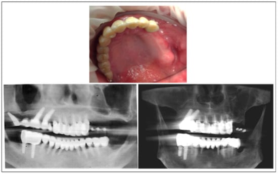
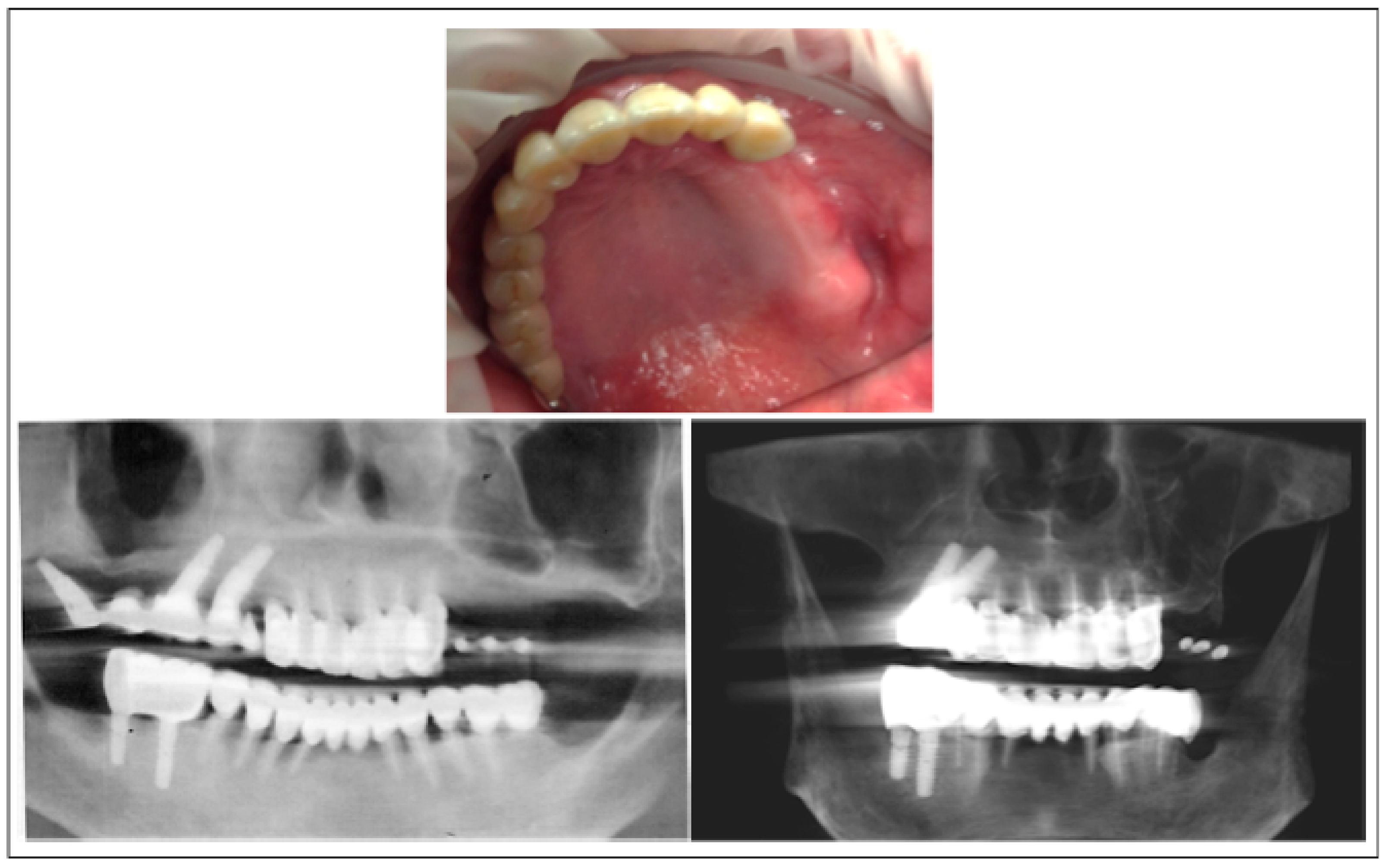
Intraoral examination revealed partial edentulism of the left posterior maxilla (Figure 1, upper panel). The overlying soft tissues showed adequate thickness and good quality, with no signs of inflammation, scarring, or mucosal pathology. These conditions were considered favorable for coverage of a juxta-osseous subperiosteal implant.
Figure 1.
Preoperative clinical and radiographic findings. Upper panel: Intraoral preoperative view of the maxillary arch showing partial edentulism of the left posterior maxilla. Lower panel: CBCT images demonstrating severe posterior maxillary atrophy, characterized by advanced vertical and horizontal alveolar bone resorption and marked maxillary sinus pneumatization, following previous failed sinus floor elevation procedures.
Despite the acceptable soft-tissue conditions, clinical palpation indicated a severe deficiency of the underlying alveolar bone. CBCT confirmed advanced vertical and horizontal bone resorption, pronounced maxillary sinus pneumatization, and irregular sinus contours consistent with previous failed sinus augmentation procedures (Figure 1, lower panel).
Overall, the above clinical and radiographic findings indicated a severely compromised posterior maxilla.
Therapeutic Decision-Making
The documented history of failed sinus augmentation procedures, together with chronic sinus pathology and recurrent sinus pneumatization, played a decisive role in excluding further regenerative approaches involving sinus manipulation.
Following a comprehensive clinical and radiographic evaluation, several therapeutic options were systematically considered for the rehabilitation of the left posterior maxilla, taking into account the condition of the contralateral side, which had already been rehabilitated with endosseous implants. The evaluated options included short implants, tilted implants, zygomatic implants, and additional regenerative procedures such as maxillary sinus floor elevation with or without guided bone regeneration. Each option was assessed based on anatomical feasibility, biological predictability, biomechanical reliability, surgical invasiveness, and patient-specific risk factors.
Short and tilted implants were excluded due to the critically reduced residual alveolar bone height and width. Residual bone dimensions were consistently below 5 mm at multiple sites, which would not have allowed predictable primary stability or long-term biomechanical reliability. In addition, compromised bone quality related to severe maxillary atrophy and previous surgical interventions further limited the feasibility of these approaches.
Zygomatic implants were also considered as a graftless alternative; however, this option was judged to be suboptimal for several reasons. Zygomatic implants are typically 42–60 mm in length, resulting in a high third-class lever arm and increased biomechanical stress. Their trans-sinus or extrasinus trajectory places them close to critical anatomical structures, including the orbit, thereby increasing surgical risk. In this case, the patient’s advanced age, chronic maxillary sinusitis, and associated respiratory comorbidities further increased the risk profile. Moreover, the patient explicitly declined highly invasive surgical procedures requiring prolonged operative time and extended postoperative recovery.
Additional regenerative approaches involving sinus augmentation were definitively excluded. The patient had previously undergone two sinus floor elevation procedures using different techniques, both of which failed over time due to persistent sinus inflammation, inadequate graft integration, and progressive graft resorption. Radiographic evaluation showed recurrent sinus pneumatization and the inability to maintain stable vertical and horizontal bone dimensions, making further regenerative attempts biologically unpredictable and clinically unjustifiable.
Considering the severe residual bone deficiency, the history of failed sinus grafting, chronic sinus pathology, and the patient’s preference for a fixed prosthetic solution with reduced invasiveness and treatment time, a digitally designed juxta-osseous subperiosteal implant was selected as the most appropriate therapeutic option. This graftless approach allows rigid fixation to stable basal and maxillofacial supports, avoids direct sinus involvement, and does not depend on residual alveolar bone volume for primary stability. In addition, it enables immediate prosthetic rehabilitation, aligning clinical indications with patient expectations while minimizing biological and surgical risks.
2.4. Diagnostic Imaging and Digital Workflow
A fully digital diagnostic and planning workflow was adopted to achieve accurate anatomical adaptation and biomechanical reliability of the customized juxta-osseous subperiosteal implant [11,14,15]. Given the critical importance of surface congruence and positional accuracy, additionally, manufactured subperiosteal implants, standardized imaging, and data acquisition protocols were applied from the initial diagnostic phase.
CBCT was performed using standardized parameters to obtain high-resolution images while minimizing artifacts. A customized digital transfer prosthesis (scan prosthesis/transfer mask) was fabricated and worn by the patient during CBCT acquisition to stabilize the mandibular position in maximum intercuspation and to transfer occlusal and prosthetic reference points. The imaging data were exported in Digital Imaging and Communications in Medicine (DICOM) format and imported into dedicated medical–dental planning software for 3D reconstruction and segmentation of the maxillary anatomy.
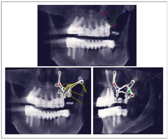
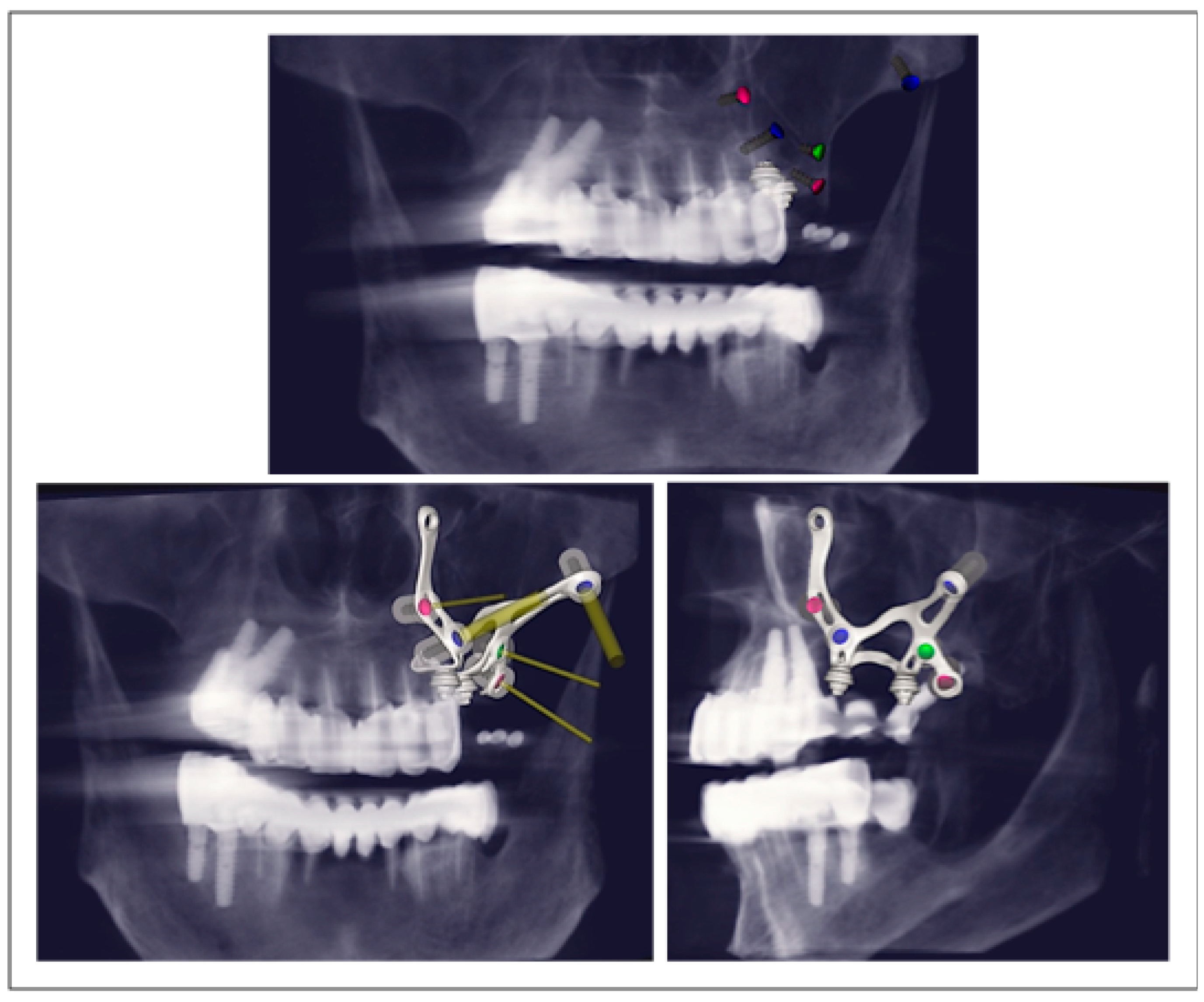
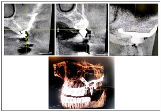
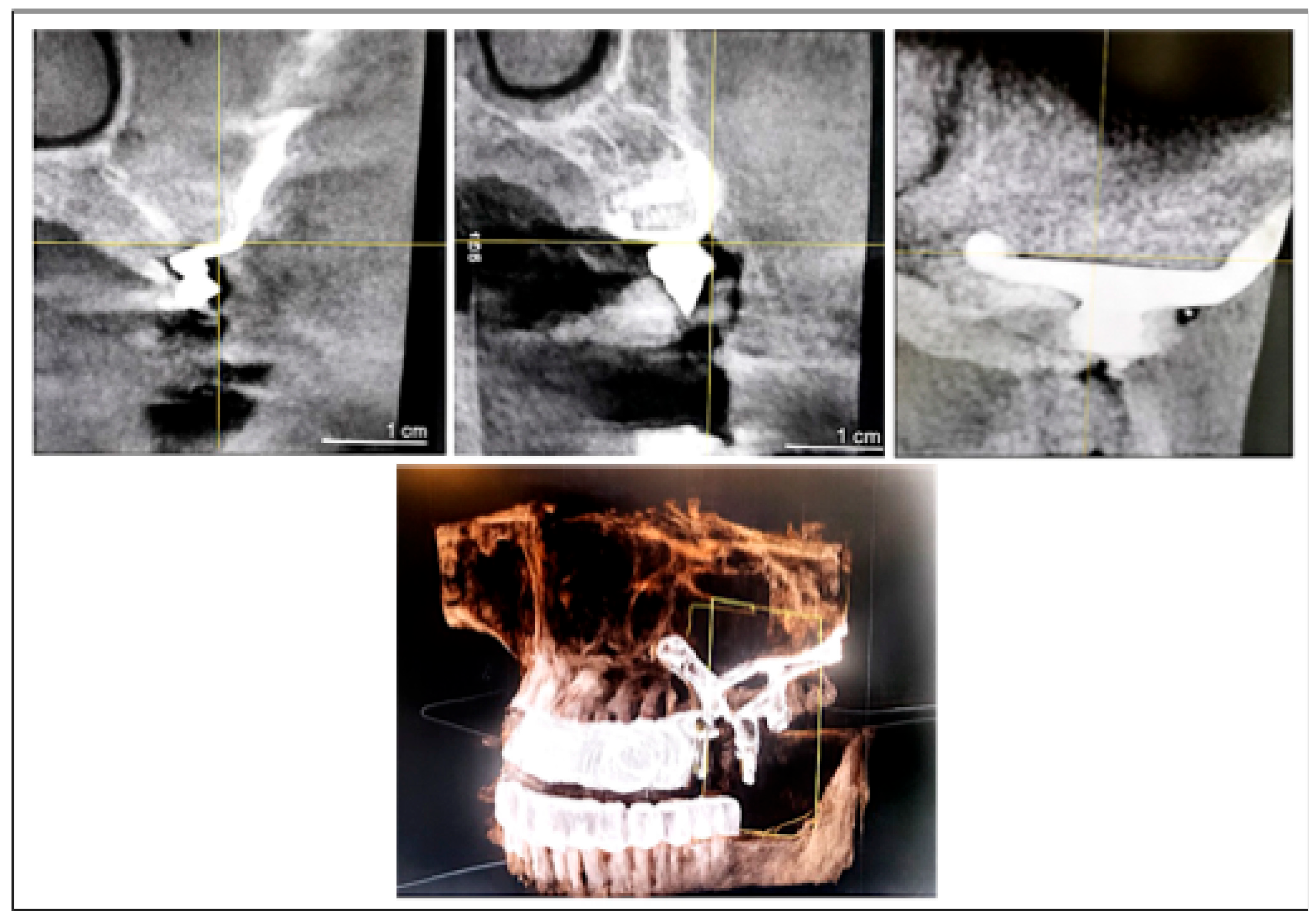
CBCT analysis confirmed severe atrophy of the left posterior maxilla, characterized by critically reduced vertical bone height, marked horizontal ridge narrowing, and advanced maxillary sinus pneumatization (Figure 2). Sequential cross-sectional evaluation at multiple offsets demonstrated insufficient residual bone volume for conventional endosseous implant placement. Digital segmentation allowed detailed visualization of the maxillary bone, sinus anatomy, and adjacent anatomical structures, supporting the indication for a graftless, subperiosteal rehabilitation approach.
Figure 2.
Virtual surgical planning of the left posterior maxilla. Upper panel: CBCT-based 3D planning showing the proposed implant framework and fixation screws in relation to the residual alveolar bone and maxillary sinus. Lower left panel: 3D reconstruction showing the planned implant-supported framework with color-coded implant sites and angulation within the atrophic posterior maxilla. Lower right panel: Post-planning CBCT view demonstrating the final virtual positioning, demonstrating prosthetically driven implant alignment and spatial distribution optimized for primary stability and biomechanical load distribution. Color-coded Butterfly implants correspond to the following dimensions: red (Ø 6.0 mm × 7.5 mm); blue (Ø 8.0 mm × 9.5 mm); green (Ø 4.0 mm × 5.5 mm); light blue (Ø 8.0 mm × 9.5 mm); pink (Ø 6.0 mm × 7.5 mm).
Segmentation was further used to identify stable cortical anchorage zones, including the zygomatic buttress, piriform rim, and residual alveolar crest. These regions were selected as fixation sites for the juxta-osseous framework to ensure rigid anchorage and optimal load transfer.
CAD of the patient-specific juxta-osseous subperiosteal implant was performed using dedicated dental CAD software (Exocad GmbH 2023, Darmstadt, Germany), Version 1.6.15. The framework was designed based on patient-specific anatomy, prosthetic emergence requirements, and biomechanical considerations. Particular attention was given to framework thickness and geometry to ensure structural rigidity while maintaining a low-profile configuration compatible with soft-tissue closure. The number, position, and orientation of osteosynthetic screws were digitally planned to achieve rigid fixation to stable maxillofacial support and uniform stress distribution under functional loading.
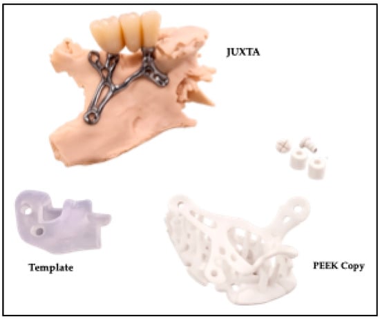
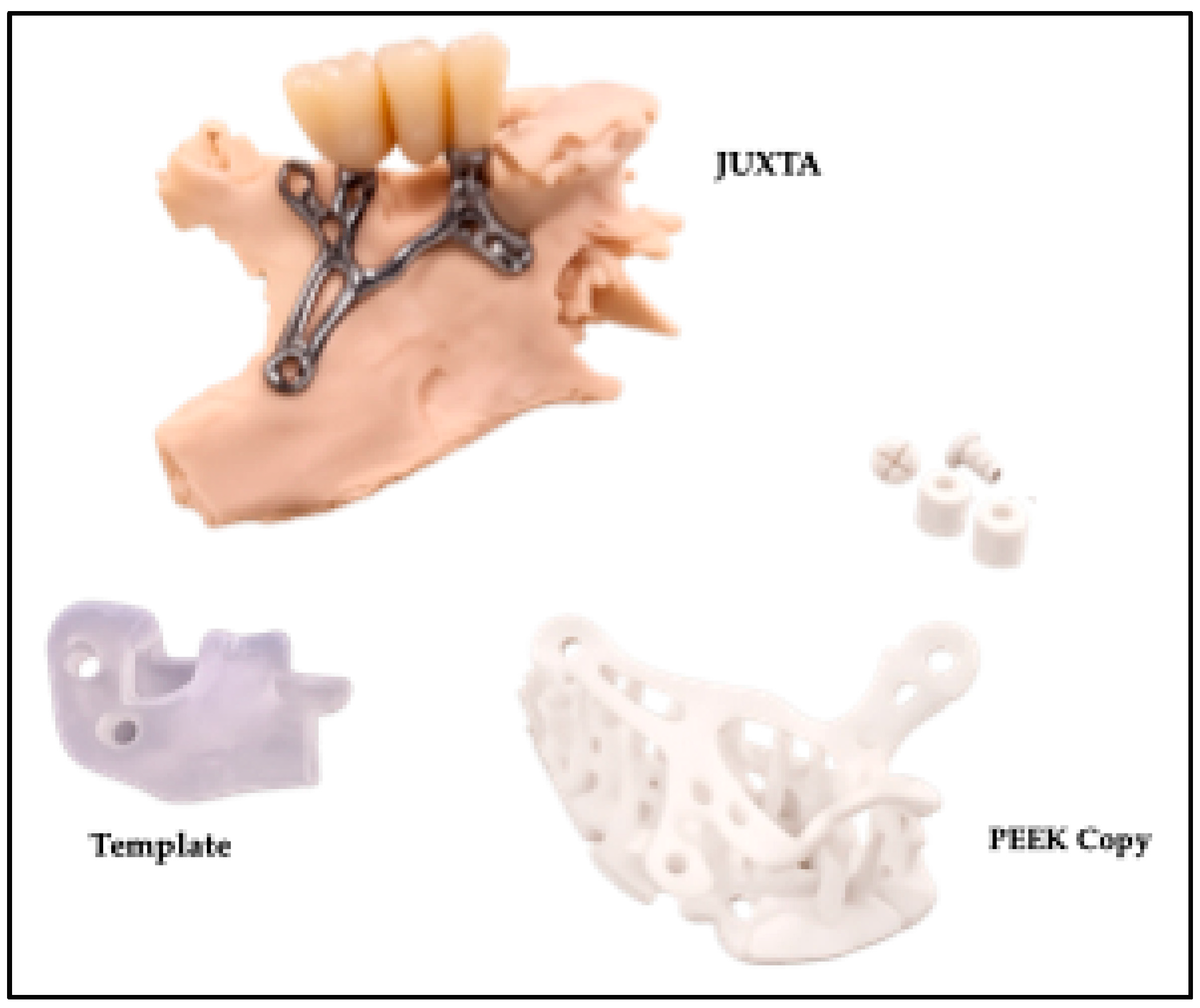
Based on these data, a patient-specific Additively Manufactured Subperiosteal Jaw Implant–Juxta-Osseous Digitally Regenerated (AMSJI–JODR, Genymplant, Biotech s.r.l, Legnano, Milano, Italy) was designed. The implant consisted of bilateral mainframes connected by stabilizing wings, allowing rigid fixation to the maxillofacial skeleton via titanium osteosynthesis screws (Figure 3). The digital design process was carried out collaboratively by the treating clinician and biomedical engineers to ensure consistency between surgical and prosthetic requirements.
Figure 3.
CAD–CAM workflow and components of the AMSJI–JODR. The figure illustrates the customized unilateral AMSJI–JODR framework designed for the rehabilitation of the left posterior maxilla (upper panel). Although the implant includes multiple stabilizing extensions projecting toward available maxillofacial supports, these elements are integral parts of a single patient-specific framework. The stabilizing extensions are designed according to the patient’s anatomy to allow rigid fixation to cortical bone and optimized load distribution. The lower panels show the diagnostic/template model used for design verification (lower left) and the patient-specific polyether–ether–ketone (PEEK) surgical guide used for accurate intraoperative positioning and screw-site preparation (lower right).
The finalized CAD file was transferred to a proprietary CAM environment for production of the implant framework using additive manufacturing technology, specifically direct metal laser sintering/selective laser melting (DMLS/SLM) of medical-grade titanium alloy (Ti-6Al-4V). This manufacturing process ensured high-dimensional accuracy, appropriate surface characteristics, and mechanical strength suitable for rigid fixation. Post-processing procedures included surface finishing, dimensional verification, quality control, sterilization, and preparation for clinical use.
Nevertheless, the described workflow reflects current standards for digitally designed, patient-specific subperiosteal implant fabrication and can be replicated using equivalent digital planning and manufacturing systems.
In contrast to conventional AMSJI designs, the JODR concept incorporated a regenerative surface strategy, in which the titanium framework was fully coated with β-tricalcium phosphate (EthOss® Regeneration Ltd., Silsden, UK), combined with autologous platelet-rich plasma (PRP). This approach was intended to promote bone–implant contact and enhance peri-implant tissue integration.
2.5. CBCT Acquisition Parameters
CBCT imaging was performed using standardized acquisition parameters to support accurate digital planning and optimal adaptation of the customized juxta-osseous subperiosteal implant. Images were acquired with a 512 × 512-pixel matrix. Tube voltage was set within a range of 90–120 kVp in accordance with device specifications and patient-related factors. Tube current was set at 10 mA. A voxel size of approximately 0.150 mm was selected to ensure high spatial resolution.
Slice thickness was maintained between 0.5 and 0.7 mm, with uniform inter-slice spacing throughout the entire scan to ensure accurate three-dimensional reconstruction and segmentation. All reconstructed images shared the same field of view, orientation, reconstruction center, and axial height to maintain consistency during digital processing. Image reconstruction was performed using a high-resolution bone algorithm to optimize visualization of cortical structures.
Whenever possible, gantry inclination was maintained at 0°, and axial slice overlap was allowed to improve the quality of multiplanar reconstructions. All imaging data were exported exclusively in native, uncompressed DICOM format, and only raw axial images were accepted for processing. Lossy compression formats were strictly excluded. A permanent Picture Archiving and Communication System (PACS) archive of the raw imaging data and acquisition parameters was maintained to ensure traceability, data integrity, and reproducibility.
3. Surgical and Prosthetic Procedures
3.1. Preoperative Protocol
Before surgery, a standardized preoperative protocol was implemented to reduce the risk of intraoperative and postoperative complications and to ensure optimal surgical conditions. Given the patient’s advanced age and history of chronic sinusitis with associated respiratory comorbidities, particular attention was directed toward infection control, patient comfort, and overall surgical safety.
Antibiotic prophylaxis was administered according to established clinical guidelines. The patient received oral amoxicillin–clavulanic acid (Augmentin®, GlaxoSmithKline, Brentford, UK) preoperatively to minimize the risk of surgical site infection, considering the extent of flap elevation and the placement of a customized juxta-osseous subperiosteal implant.
Immediately before surgery, antiseptic oral rinsing was performed using 0.20% chlorhexidine digluconate (Corsodyl®, GlaxoSmithKline, Brentford, UK) for 60 s. This measure was adopted to reduce intraoral bacterial load and to promote favorable postoperative soft-tissue healing.
All surgical procedures were carried out under local anesthesia, with the option of conscious sedation to ensure patient comfort and adequate surgical control. Local anesthesia was achieved using articaine hydrochloride 4% with epinephrine 1:100,000 (Septanest®, Septodont, Saint-Maur-des-Fossés, France), providing effective analgesia and hemostasis throughout the intervention.
When clinically indicated, conscious sedation was administered using intravenous midazolam (Versed®, Roche, Basel, Switzerland) under monitored conditions. This approach allowed effective anxiety control and improved patient cooperation while preserving protective reflexes and maintaining stable vital parameters.
This preoperative protocol ensured a controlled surgical environment, minimized patient discomfort, and supported the predictable placement of the customized AMSJI-JODR implant in a medically compromised patient.
3.2. Surgical Phase
After adequate anesthesia was achieved, the surgical field was disinfected and isolated according to standard clinical protocols. A full-thickness mucoperiosteal flap was carefully designed and elevated to expose the left maxillary bone surface. Incisions were planned to provide sufficient access while preserving soft-tissue integrity.
Periosteal detachment was intentionally limited to the minimum extent required to accommodate the customized juxta-osseous framework. This conservative approach aimed to preserve local vascular supply, reduce surgical trauma, and minimize the risk of postoperative complications such as soft-tissue dehiscence or delayed healing. Particular attention was paid to flap passivation to allow tension-free repositioning at the end of the procedure.
Once the bone surface was exposed, the custom-made titanium juxta-osseous subperiosteal implant (AMSJI–JODR) was positioned onto the maxillary bone. Intraoperative assessment confirmed passive and intimate adaptation of the framework to the underlying osseous anatomy, as planned during the digital workflow. No significant intraoperative modifications were required, reflecting the accuracy of the CAD–CAM planning process.
Regenerative augmentation of the juxta-osseous framework was then performed using β-tricalcium phosphate (EthOss® Regeneration Ltd., Silsden, UK), applied over the titanium structure, and covered with autologous PRP. This technique created a regenerative “sandwich effect” between the basal bone surface and the implant framework, promoting rapid bone formation and secondary biological stabilization. Before fixation, the osteosynthesis screws were immersed in β-tricalcium phosphate to enhance early bone–screw contact, support rapid osteointegration, and improve long-term mechanical stability.
No manipulation of the maxillary sinus or Schneiderian membrane was required, thereby avoiding direct sinus involvement and reducing the risk of postoperative sinus or respiratory complications. Screws were placed sequentially to ensure uniform stabilization and to prevent micromovements of the framework. Primary mechanical stability was verified manually, confirming the absence of implant mobility.
Following fixation, the surgical site was carefully inspected to confirm correct framework positioning, adequate clearance for prosthetic components, and the absence of sharp edges or areas of soft-tissue compression. The flap was then repositioned and sutured under tension-free conditions, completing the surgical phase.
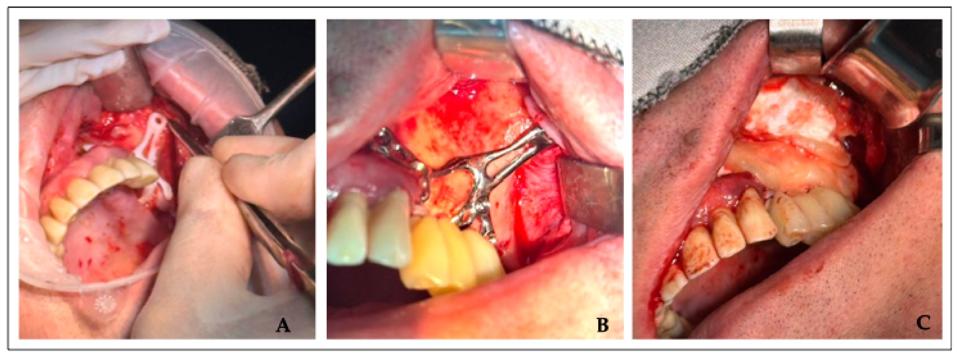
This surgical approach ensured accurate implant positioning, rigid stabilization, and optimal soft-tissue management, which are considered critical factors for the success of juxta-osseous subperiosteal implant rehabilitation in severely atrophic maxillae (Figure 4).
Figure 4.
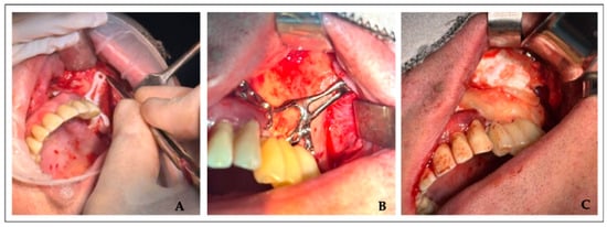
Intraoperative surgical phase. (A) Patient-specific PEEK surgical guide used to verify accurate positioning and to prepare the osteosynthetic screw sites. (B) AMSJI–JODR-structure positioned in situ and stabilized. (C) Titanium framework was augmented with β-tricalcium phosphate graft material and covered with an autologous PRP membrane obtained from venous blood collection, creating a “sandwich” regenerative effect.
Intraoperative assessment and 3D reconstruction confirmed accurate positioning and rigid fixation of the customized AMSJI–JODR to stable cortical structures of the maxilla, with titanium osteosynthesis screws placed according to the preoperative digital plan (Figure 5).
Figure 5.
Postoperative 3D reconstructed views of the customized AMSJI–JODR, indicating the locations of titanium fixation screws anchored to stable maxillary cortical structures. Yellow lines represent the selected regions used for magnification during image acquisition from the planning software interface.
3.3. Immediate Prosthetic Rehabilitation
Immediately after surgical placement and rigid fixation of the customized juxta-osseous subperiosteal implant, a screw-retained fixed provisional prosthesis, previously designed during the digital planning phase, was delivered on the day of surgery. This approach allowed a direct transition from the surgical to the prosthetic phase without increasing operative time.
The provisional prosthesis was connected to the implant framework using prosthetic screws, ensuring secure retention and full retrievability. Occlusal contacts were carefully evaluated and selectively adjusted to achieve a controlled functional occlusion, with particular attention to minimizing non-axial forces and excessive loading in the treated posterior maxillary region during the early healing period.
Immediate loading was considered appropriate due to the high primary mechanical stability provided by rigid fixation of the AMSJI–JODR framework to stable cortical support. This protocol enabled early functional rehabilitation while maintaining biomechanical protection of both the implant framework and the fixation screws.
Postoperative radiographic evaluation confirmed stable positioning of the customized AMSJI–JODR and fixation screws (Figure 5). The immediate prosthetic rehabilitation avoided postoperative edentulism and allowed restoration of masticatory function on the day of surgery, contributing to improved patient comfort and satisfaction in a clinical scenario characterized by severe maxillary atrophy and previous surgical failures.
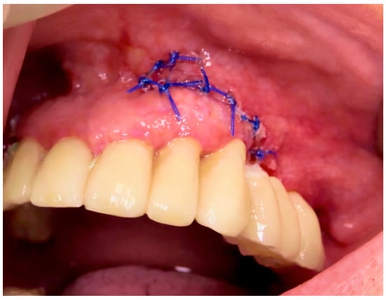
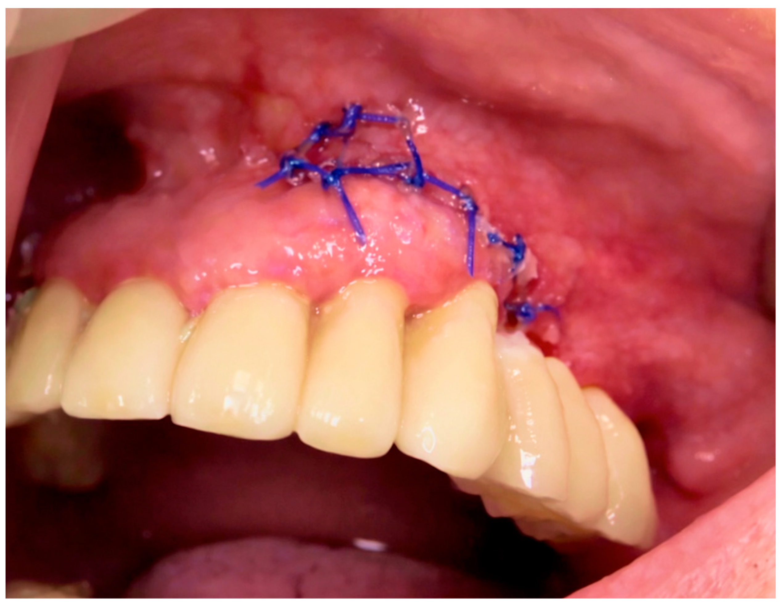
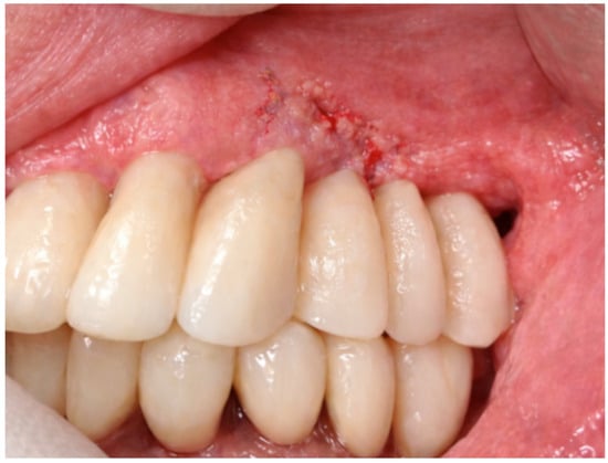
In addition to providing functional and aesthetic restoration, the provisional prosthesis acted as a transitional device, guiding soft-tissue adaptation and facilitating the subsequent fabrication of the definitive prosthesis. Intraoral postoperative evaluation demonstrated satisfactory soft-tissue healing and adaptation around the provisional restoration (Figure 6).
Figure 6.
Postoperative intraoral view. Intraoral photograph showing the screw-retained fixed provisional prosthesis in function and satisfactory soft-tissue adaptation following closure of the surgical site in the left posterior maxilla. The provisional prosthesis was delivered on the day of surgery, providing immediate functional and aesthetic rehabilitation and avoiding postoperative edentulism.
4. Follow-Up and Outcomes
A structured postoperative maintenance and follow-up protocol was implemented to support implant stability, peri-implant soft-tissue health, and long-term prosthetic function. Maintenance procedures were comparable to those used for conventional implant-supported rehabilitations, with specific considerations related to the presence of a juxta-osseous subperiosteal framework.
The prosthetic design included removable multi-unit abutments (MUAs), allowing periodic disassembly of the prosthetic superstructure. This feature enabled direct clinical inspection of the peri-implant soft tissues and facilitated professional hygiene procedures when required. This approach represents a relevant advantage compared with historical analog subperiosteal implants, which were characterized by fixed abutments and limited access to the underlying tissues.
Postoperative management followed a standardized protocol aimed at promoting uneventful healing. Antibiotic and anti-inflammatory therapy was prescribed according to clinical indications, and local cryotherapy was recommended during the first postoperative hours to reduce edema and discomfort. Detailed oral hygiene instructions were provided, including the use of chlorhexidine-based mouth rinses to support soft-tissue healing.
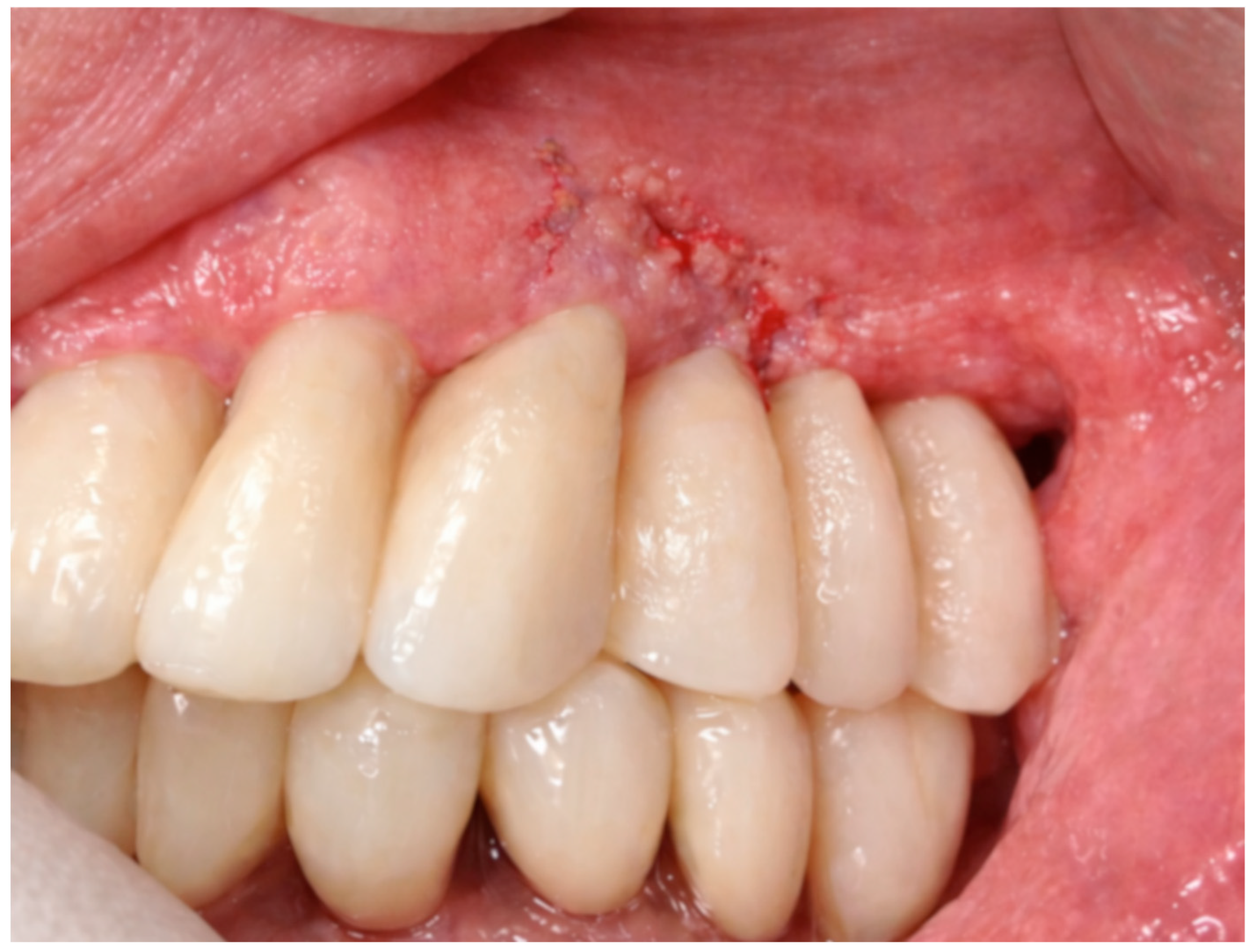
Clinical follow-up visits were scheduled at 7 days, 30 days, and 3 months, and subsequently at regular intervals. At the 7-day follow-up, soft-tissue healing was evaluated, and sutures were removed when non-resorbable materials had been used. No signs of infection, wound dehiscence, or adverse inflammatory reactions were observed, and the patient reported minimal postoperative discomfort and good adaptation to the provisional prosthesis (Figure 7).
Figure 7.
Early postoperative follow-up (7 days). Postoperative intraoral clinical view demonstrating satisfactory soft-tissue healing and stable adaptation around the provisional screw-retained prosthetic restoration.
During the follow-up period, particular attention was directed toward monitoring sinus-related symptoms and respiratory status, given the patient’s history of chronic sinusitis. The patient was evaluated for facial pain, nasal congestion, purulent discharge, or respiratory deterioration. No sinus-related complications or respiratory exacerbations were recorded, confirming the benefit of avoiding direct sinus involvement during the surgical and prosthetic phases.
At the 30-day and 3-month follow-up visits, both clinical and radiographic evaluations were performed. These assessments demonstrated stable positioning and fixation of the customized juxta-osseous subperiosteal implant, with no evidence of micromovement or mechanical complications. Radiographic controls confirmed accurate adaptation of the framework to the underlying basal bone and stable fixation of the osteosynthetic screws. Peri-implant soft tissues appeared healthy, with adequate coverage of the framework and absence of mucosal dehiscence or inflammatory signs.
Between 3 and 5 months postoperatively, following confirmation of clinical stability and favorable radiographic findings, the definitive prosthetic restoration was delivered. The final prosthesis provided satisfactory functional performance and aesthetic integration. The patient reported a high level of satisfaction, particularly appreciating the immediate fixed rehabilitation, rapid restoration of masticatory function, and avoidance of prolonged edentulism.
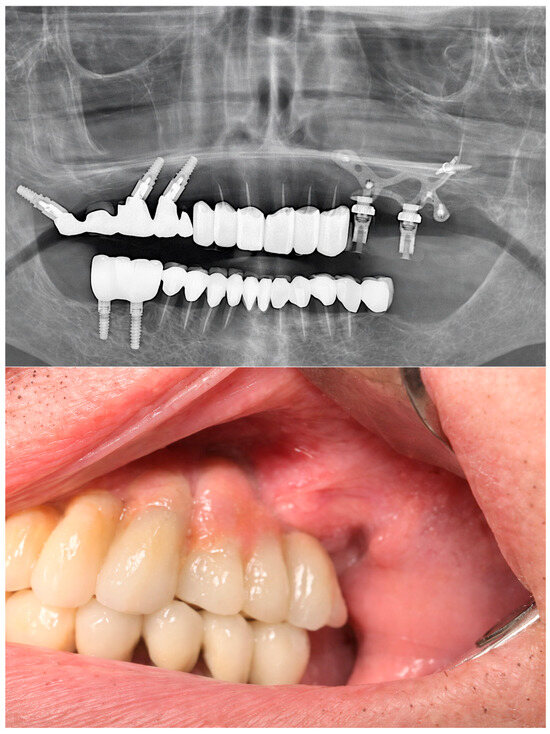
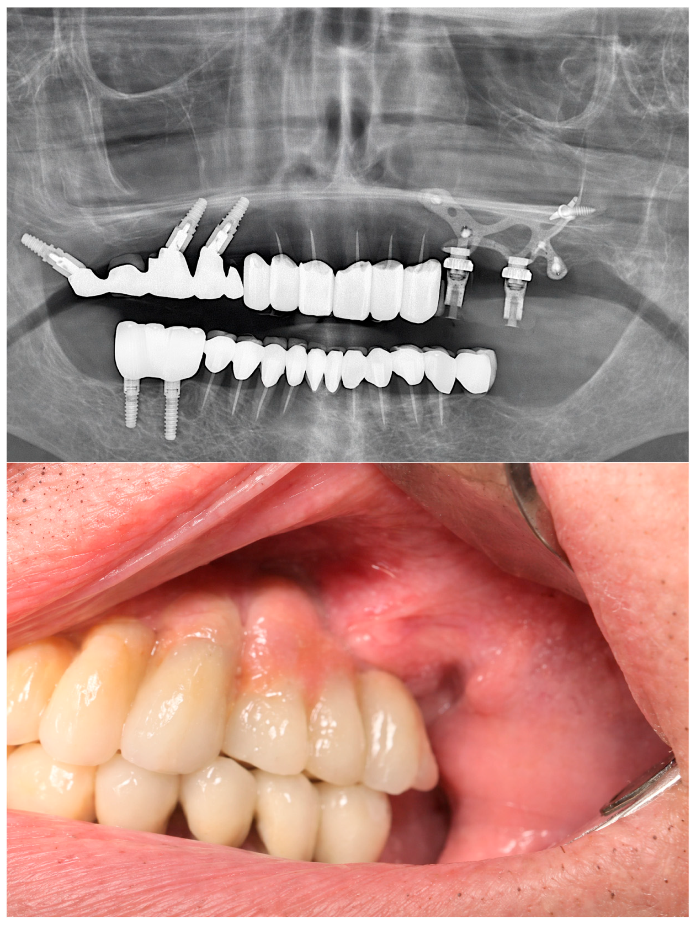
At the 1-year follow-up, clinical and radiographic evaluation confirmed a stable and predictable outcome, with healthy peri-implant soft tissues, maintained prosthetic function, and no biological or mechanical complications (Figure 8). Despite these favorable findings, it must be emphasized that the present follow-up represents a short- to mid-term evaluation. Therefore, the outcomes should be interpreted as evidence of early clinical success and feasibility rather than definitive proof of long-term predictability.
Figure 8.
One-year follow-up. Upper panel: Postoperative radiographic evaluation after 1 year, confirming stable fixation of the customized AMSJI–JODR implant. Lower panel: Intraoral clinical view showing stable peri-implant soft-tissue adaptation and maintained function of the screw-retained fixed prosthetic restoration in the left posterior maxilla.
The patient remains enrolled in a structured clinical and radiographic follow-up program, with ongoing monitoring to evaluate long-term implant integrity, peri-implant tissue health, and prosthetic stability.
5. Discussion
The present case contributes to the growing body of evidence indicating that digitally manufactured juxta-osseous subperiosteal implants may represent a reliable graftless rehabilitation option for patients with severe maxillary atrophy in whom conventional endosseous implants or regenerative procedures are contraindicated. The adoption of fully digital CAD–CAM workflows combined with additive manufacturing enables accurate transfer of virtual planning into patient-specific frameworks, which is particularly critical for juxta-osseous implants requiring passive adaptation to complex bone surfaces.
Historically, analog press-fit subperiosteal implant systems demonstrated variable and often unpredictable outcomes, with reported success rates of approximately 65–75%. These limitations were primarily related to inadequate anatomical adaptation, absence of rigid fixation, and unfavorable biomechanical load transfer. Such shortcomings are consistent with biomechanical observations reported in the literature, where non-optimized implant geometry and uncontrolled stress distribution were identified as major contributors to mechanical and biological complications in early subperiosteal designs [16]. In contrast, contemporary digitally designed juxta-osseous subperiosteal implants, produced through CAD–CAM workflows and additive manufacturing and frequently combined with adjunctive regenerative strategies such as β-tricalcium phosphate augmentation, have demonstrated substantially improved clinical performance, with reported success rates of approximately 90–95%, in agreement with recent biomechanical and experimental evidence [17].
From a biomechanical perspective, finite-element analyses support the design rationale of modern juxta-osseous subperiosteal implants. Patient-specific frameworks, when rigidly fixed to stable maxillofacial structures, achieve favorable stress distribution and reduced peak stress concentrations at fixation sites and surrounding bone. Optimization of framework thickness, geometry, and screw positioning contributes to improved mechanical stability under functional loading, explaining the feasibility of immediate or early loading protocols in selected cases [8]. The favorable mechanical properties of calcium-phosphate-based biomaterials, including hydroxyapatite and related ceramics, further support their use by enhancing bone–implant interaction and long-term stability [17].
Clinical studies corroborate these biomechanical principles. Retrospective analyses, such as those reported by Van den Borre et al., demonstrated favorable outcomes in mandibular AMSJI rehabilitations, emphasizing the importance of rigid cortical anchorage and structured postoperative monitoring [18]. Although anatomical differences exist between the mandible and maxilla, these findings indirectly support the biomechanical validity of the AMSJI concept in severely resorbed jaws. Similarly, Mommaerts reported that most adverse events following maxillary AMSJI placement were related to surgical inaccuracies, inadequate soft-tissue handling, or insufficient framework adaptation rather than intrinsic implant failure, underscoring the importance of meticulous surgical execution and passive framework adaptation [19].
The versatility of digitally designed subperiosteal implants has further expanded through alternative fixation strategies. Diss et al. described pterygoid anchorage as an adjunctive option in anatomically challenging cases, illustrating how customized frameworks can be adapted to extreme bone deficiencies through digital design and planning [20]. These developments reinforce the role of patient-specific CAD–CAM workflows in tailoring implant geometry and fixation strategies to individual anatomical constraints.
Postoperative maintenance and structured follow-up remain essential components of treatment success [1]. The possibility of prosthetic disassembly through removable abutments allows periodic inspection of peri-implant soft tissues, clearly distinguishing modern digital systems from historical analog subperiosteal implants with fixed abutments and limited tissue access. This feature contributes to improved long-term clinical control and early detection of soft-tissue complications.
When compared with other graftless rehabilitation strategies, particularly zygomatic implants, juxta-osseous subperiosteal implants may represent a less invasive alternative in selected patients. Although zygomatic implants demonstrate high survival rates, they are associated with greater surgical complexity and a higher incidence of sinus-related, neurological, and prosthetic complications [9,10]. In elderly patients or in those with chronic sinus pathology, avoiding trans-sinus trajectories may therefore offer a clinical advantage, as observed in the present case [2].
Advances in digital planning and execution play a central role in improving the predictability of complex implant rehabilitations. Emerging technologies, including artificial intelligence, augmented reality, and robotic-assisted surgery, have the potential to further enhance precision and reproducibility in patient-specific implant placement and framework adaptation [14,21]. When sufficient primary mechanical stability is achieved, immediate prosthetic rehabilitation can be safely performed, reducing overall treatment time and improving patient comfort without compromising clinical outcomes [22]. In the context of subperiosteal implant therapy, immediate loading is particularly advantageous for patients with long-standing edentulism or previous surgical failures, as it avoids prolonged postoperative edentulism.
Long-term success, however, depends not only on mechanical stability but also on peri-implant soft-tissue conditions and systemic health. Adequate soft-tissue thickness and the presence of keratinized mucosa reduce the risk of framework exposure and inflammatory complications [23,24], while systemic comorbidities may influence healing capacity and must be carefully considered during treatment planning and follow-up [25].
Recent finite-element analyses confirm that optimized digitally designed subperiosteal frameworks provide favorable biomechanical behavior under functional loading when framework thickness, geometry, and screw positioning are appropriately planned [26]. These biomechanical advantages support the clinical feasibility of subperiosteal implants as a graftless solution in anatomically compromised cases. Alternative graftless approaches, such as pterygoid implants, have demonstrated favorable short- to mid-term survival rates, ranging from approximately 85% to over 90% when digitally planned protocols are applied [27,28,29]. However, their placement is technically demanding and highly sensitive to non-axial loading and distal cantilever forces, which may increase biomechanical complexity and the risk of mechanical or prosthetic complications, particularly in the presence of adjacent teeth or existing restorations [30].
In parallel, growing experimental and clinical evidence supports the biological and technological rationale underlying modern digitally manufactured juxta-osseous subperiosteal implants. Calcium phosphate-based biomaterials, including hydroxyapatite and related ceramics, have demonstrated osteoinductive and osteoconductive properties that enhance bone–implant interaction and contribute to long-term stability in complex reconstructions [31]. Systematic reviews focusing on virtual surgical planning and customized subperiosteal implants have highlighted the critical role of digital workflows in improving accuracy, passive fit, and reproducibility, while reducing surgical and prosthetic complications [32]. Recent systematic analyses of additively manufactured subperiosteal implants further confirm favorable short-term clinical performance, with high survival rates and acceptable complication profiles when rigid fixation and appropriate case selection are applied [33]. Multicenter retrospective and cohort studies have reported encouraging outcomes in both full-arch maxillary and posterior mandibular rehabilitations using patient-specific subperiosteal implants, reinforcing the clinical reliability of contemporary CAD–CAM–based systems in severely atrophic jaws [34,35].
Partial juxta-osseous reconstructions, as used in the present case, may reduce surgical invasiveness and simplify soft-tissue management compared with full-arch designs. When combined with rigid fixation and controlled occlusion, they can achieve favorable short-term outcomes.
Mechanical complications may include loosening or fracture of fixation screws, particularly in cases of inadequate cortical anchorage or unfavorable load distribution. In the present case, no mechanical instability was observed, possibly due to rigid fixation to basal buttresses and immersion of osteosynthesis screws in β-tricalcium phosphate, which may promote rapid bone–screw integration. Prosthetic complications, such as screw loosening or occlusal wear, may occur in the presence of non-axial loading or inadequate occlusal control, highlighting the importance of careful prosthetic planning, controlled occlusal schemes, and structured maintenance.
Overall, current evidence suggests that many complications associated with modern juxta-osseous subperiosteal implants are primarily technique-related rather than implant-related, emphasizing the importance of accurate digital planning, rigid fixation, meticulous soft-tissue management, and structured postoperative follow-up.
Despite the favorable clinical and radiographic outcomes observed in the present case, the duration of follow-up remains a limitation. The reported results reflect short- to mid-term performance and do not allow definitive conclusions regarding long-term predictability, a consideration that remains particularly relevant given the historically controversial outcomes of analog subperiosteal implant systems. In patients with chronic sinusitis or respiratory comorbidities, graftless rehabilitation strategies that avoid sinus manipulation may offer a clinical advantage. In the present case, rehabilitation was achieved without sinus penetration, and no sinus-related complications occurred during follow-up. Nevertheless, longer observation periods are required to confirm long-term stability.
It is essential to distinguish historical analog subperiosteal implants from contemporary digitally designed systems. Earlier failures were primarily associated with press-fit retention, fixed abutments, lack of rigid fixation, and reliance on bone undercuts. Then, modern systems rely on patient-specific CAD–CAM planning, rigid fixation using osteosynthetic screws, and controlled load distribution along basal bone structures. Despite these advances, long-term prospective validation remains limited.
For these reasons, the present case should be considered hypothesis-generating, demonstrating feasibility and early clinical reliability rather than definitive long-term success. Continued follow-up and larger prospective comparative studies are required to define survival rates, complication profiles, and maintenance requirements of digitally designed juxta-osseous subperiosteal implants.
6. Conclusions
Digitally designed juxta-osseous subperiosteal implants appear to be a feasible graftless option for selected patients with severe maxillary atrophy, especially when conventional implant placement or regenerative procedures are contraindicated or have failed. In the present case, accurate digital planning, appropriate patient selection, and controlled surgical and prosthetic protocols enabled immediate functional and aesthetic rehabilitation with reduced morbidity and high patient satisfaction.
However, the findings of this report must be interpreted within the limitations of a single-case design and short- to mid-term follow-up. Therefore, the present outcome supports early clinical feasibility rather than definitive long-term predictability. Further clinical investigation is needed to confirm durability, define standardized indications, and establish evidence-based protocols for complication prevention, surgical execution, and long-term maintenance. Prospective cohort studies and controlled comparative trials with extended follow-up are required to evaluate long-term survival and complication rates and to better define the role of digitally designed juxta-osseous subperiosteal implants in the rehabilitation of severely atrophic maxillae.
Author Contributions
Conceptualization, E.M. and A.M. (Aida Meto); methodology, E.M. and A.M. (Agron Meto); software, A.I.; validation, A.M. (Aida Meto) and A.I.; formal analysis, E.M.; writing—original draft preparation, E.M. and A.M. (Agron Meto); writing—review and editing, A.M. (Aida Meto); visualization, A.I.; supervision, A.M. (Agron Meto). All authors have read and agreed to the published version of the manuscript.
Funding
This research received no external funding.
Institutional Review Board Statement
Ethical approval was obtained from the Ethics Committee of Aldent University (Protocol No. 39/5, 12 September 2025).
Informed Consent Statement
Written informed consent has been obtained from the patient to publish this paper.
Data Availability Statement
The data presented in this study are available on request from the corresponding author.
Conflicts of Interest
The authors declare no conflicts of interest.
References
- Sánchez-Labrador, L.; Bazal-Bonelli, S.; Pérez-González, F.; Beca-Campoy, T.; Cobo-Vázquez, C.M.; Cortés-Bretón Brinkmann, J.; Martínez-González, J.M. Clinical Performance of Subperiosteal Implants in the Full-Arch Rehabilitation of Severely Resorbed Edentulous Jaws: A Systematic Review and Metanalysis. Dent. J. 2025, 13, 240. [Google Scholar] [CrossRef] [Scilit]
- Sudhir, M.V.S.; Prasad, R.B.; Krothapalli, N.; Kumar, P. Graftless Solutions for Rehabilitation of Atrophied Maxilla-Zygomatic Versus Subperiosteal Implants—A Systematic Review. J. Pharm. Bioallied Sci. 2025, 17, S207–S210. [Google Scholar] [CrossRef] [Scilit]
- Alsharekh, M.S.; Almutairi, A.A.; Jahlan, A.S.; Alhazani, A.S.; Almohaimeed, S.M.; Aljnoubi, L.A.; AlGhamdi, G.A.; AlBenyan, T.T.; Alduhyaman, S.F.; Alnaffaie, N.M.; et al. Evolving Techniques and Trends in Maxillary Sinus Lift Procedures in Implant Dentistry: A Review of Contemporary Advances. Cureus 2024, 16, e71424. [Google Scholar] [CrossRef] [Scilit]
- Cerea, M.; Dolcini, G.A. Custom-Made Direct Metal Laser Sintering Titanium Subperiosteal Implants: A Retrospective Clinical Study on 70 Patients. BioMed Res. Int. 2018, 2018, 5420391. [Google Scholar] [CrossRef] [Scilit]
- Nedelcu, L.; Sirbu, I.; Sirbu, V.D.; Custura, A.M.; Radu, A.; Nastasie, V. Custom-made 3D printed subperiosteal implant for restoration of severe atrophic jaw: A case report. Clin. Case Rep. 2024, 12, e9515. [Google Scholar] [CrossRef] [Scilit]
- Pellegrino, G.; Karaban, M.; Barausse, C.; Giudice, A.; Antonelli, A.; Pistilli, R.; Felice, P. Indications and Complications of Subperiosteal Implants: Literature Review and Case Series. Dent. J. 2025, 13, 337. [Google Scholar] [CrossRef] [Scilit]
- Arshad, M.; Khoramshahi, N.; Shirani, G. Additively custom-made 3D-printed subperiosteal implants for the rehabilitation of the severely atrophic maxilla (a case report). Clin. Case Rep. 2023, 11, e8135. [Google Scholar] [CrossRef] [Scilit] [PubMed]
- De Moor, E.; Huys, S.E.F.; van Lenthe, G.H.; Mommaerts, M.Y.; Vander Sloten, J. Mechanical evaluation of a patient-specific additively manufactured subperiosteal jaw implant (AMSJI) using finite-element analysis. Int. J. Oral. Maxillofac. Surg. 2022, 51, 405–411. [Google Scholar] [CrossRef] [Scilit] [PubMed]
- Chrcanovic, B.R.; Albrektsson, T.; Wennerberg, A. Survival and Complications of Zygomatic Implants: An Updated Systematic Review. J. Oral. Maxillofac. Surg. 2016, 74, 1949–1964. [Google Scholar] [CrossRef] [Scilit]
- Kämmerer, P.W.; Fan, S.; Aparicio, C.; Bedrossian, E.; Davó, R.; Morton, D.; Raghoebar, G.M.; Zarrine, S.; Al-Nawas, B. Evaluation of surgical techniques in survival rate and complications of zygomatic implants for the rehabilitation of the atrophic edentulous maxilla: A systematic review. Int. J. Implant. Dent. 2023, 9, 11. [Google Scholar] [CrossRef] [Scilit] [PubMed]
- Massuda, C.K.M.; de Carvalho, M.R.; de Moraes, J.B.; Pallos, D.; Kim, Y.J. Accuracy of guided dental implant surgery using a fully digital workflow: A case series. J. Prosthet. Dent. 2024, 132, 973–980. [Google Scholar] [CrossRef] [Scilit]
- Riley, D.S.; Barber, M.S.; Kienle, G.S.; Aronson, J.K.; von Schoen-Angerer, T.; Tugwell, P.; Kiene, H.; Helfand, M.; Altman, D.G.; Sox, H.; et al. CARE guidelines for case reports: Explanation and elaboration document. J. Clin. Epidemiol. 2017, 89, 218–235. [Google Scholar] [CrossRef] [Scilit]
- World Medical Association World Medical Association Declaration of Helsinki: Ethical principles for medical research involving human subjects. JAMA 2013, 310, 2191–2194. [CrossRef] [Scilit] [PubMed]
- Meto, A.; Halilaj, G. The Integration of Cone Beam Computed Tomography, Artificial Intelligence, Augmented Reality, and Virtual Reality in Dental Diagnostics, Surgical Planning, and Education: A Narrative Review. Appl. Sci. 2025, 15, 6308. [Google Scholar] [CrossRef] [Scilit]
- D’haese, J.; Ackhurst, J.; Wismeijer, D.; De Bruyn, H.; Tahmaseb, A. Current state of the art of computer-guided implant surgery. Periodontol. 2000 2017, 73, 121–133. [Google Scholar] [CrossRef] [Scilit]
- Lisiak-Myszke, M.; Marciniak, D.; Bieliński, M.; Sobczak, H.; Garbacewicz, Ł.; Drogoszewska, B. Application of Finite Element Analysis in Oral and Maxillofacial Surgery—A Literature Review. Materials 2020, 13, 3063. [Google Scholar] [CrossRef] [Scilit] [PubMed]
- Scarano, A.; Lorusso, F.; Santos de Oliveira, P.; Kunjalukkal Padmanabhan, S.; Licciulli, A. Hydroxyapatite Block Produced by Sponge Replica Method: Mechanical, Clinical and Histologic Observations. Materials 2019, 12, 3079. [Google Scholar] [CrossRef] [Scilit] [PubMed]
- Van den Borre, C.; Otero, J.J.; Loomans, N.; Samama, M.; Lempert, J.; Baltensperger, M.; Termont, A.; Castro, R.; Mommaerts, M.Y. Retrospective analyses of additively manufactured subperiosteal jaw implants in the mandible. J. Craniomaxillofac. Surg. 2025, 53, 905–910. [Google Scholar] [CrossRef] [Scilit]
- Mommaerts, M.Y. Management of adverse effects following additively manufactured subperiosteal jaw implantation in the maxilla. J. Stomatol. Oral. Maxillofac. Surg. 2025, 126, 102206. [Google Scholar] [CrossRef] [Scilit]
- Diss, A.; Lerebours, A.; Grébonval, C.; Birault, L. Pterygoid Anchorage of Subperiosteal Implants: An Overview and Case Report. Cureus 2025, 17, e85175. [Google Scholar] [CrossRef] [Scilit]
- Pozzi, A.; Carosi, P.; Lorenzi, C.; Chow, J.; Wang, H.L.; Gallucci, G.O. In Vivo Accuracy of Autonomous Dental Implant Robotic Surgery: Systematic Review and Meta-analysis. Int. J. Oral. Maxillofac. Implants 2025, 40, 683–690. [Google Scholar] [CrossRef] [Scilit]
- Anto, A.; Sharma, S.; Sharma, A.K.; Sharma, V.; Bisharwal, H.; Sharma, R. Immediate and conventional loading of anterior dental implants: Aesthetic considerations and outcomes. Bioinformation 2024, 20, 980–985. [Google Scholar] [CrossRef] [Scilit]
- Zucchelli, G.; Tavelli, L.; McGuire, M.K.; Rasperini, G.; Feinberg, S.E.; Wang, H.L.; Giannobile, W.V. Autogenous soft tissue grafting for periodontal and peri-implant plastic surgical reconstruction. J. Periodontol. 2020, 91, 9–16. [Google Scholar] [CrossRef] [Scilit]
- Monje, A.; Roccuzzo, A.; Buser, D.; Wang, H.L. Influence of buccal bone wall thickness on the peri-implant hard and soft tissue dimensional changes: A systematic review. Clin. Oral. Implants Res. 2023, 34, 8–27. [Google Scholar] [CrossRef] [Scilit]
- Sbricoli, L.; Bazzi, E.; Stellini, E.; Bacci, C. Systemic Diseases and Biological Dental Implant Complications: A Narrative Review. Dent. J. 2022, 11, 10. [Google Scholar] [CrossRef] [Scilit] [PubMed]
- Pellegrino, G.; Karaban, M.; Scalchi, V.; Urbani, M.; Giudice, A.; Barausse, C.; Felice, P. Finite Element Analysis of Functionally Loaded Subperiosteal Implants Evaluated on a Realistic Model Reproducing Severe Atrophic Jaws. Methods Protoc. 2025, 8, 8. [Google Scholar] [CrossRef] [Scilit]
- Broumand, V.; Kirchhofer, J. Pterygoid implants as alternative to bone augmentation in implant dentistry. Br. Dent. J. 2025, 238, 99–109. [Google Scholar] [CrossRef] [Scilit] [PubMed]
- Araujo, R.Z.; Santiago Júnior, J.F.; Cardoso, C.L.; Benites Condezo, A.F.; Moreira Júnior, R.; Curi, M.M. Clinical outcomes of pterygoid implants: Systematic review and meta-analysis. J. Craniomaxillofac. Surg. 2019, 47, 651–660. [Google Scholar] [CrossRef] [Scilit] [PubMed]
- D’Amario, M.; Orsijena, A.; Franco, R.; Chiacchia, M.; Jahjah, A.; Capogreco, M. Clinical achievements of implantology in the pterygoid region: A systematic review and meta-analysis of the literature. J. Stomatol. Oral. Maxillofac. Surg. 2024, 125, 101951. [Google Scholar] [CrossRef] [Scilit]
- Raouf, K.; Chrcanovic, B.R. Clinical Outcomes of Pterygoid and Maxillary Tuberosity Implants: A Systematic Review. J. Clin. Med. 2024, 13, 4544. [Google Scholar] [CrossRef] [Scilit]
- LeGeros, R.Z. Calcium phosphate-based osteoinductive materials. Chem. Rev. 2008, 108, 4742–4753. [Google Scholar] [CrossRef] [Scilit]
- Al-Nawas, B.; Bär, A.K. Virtual surgical planning and customized subperiosteal implants: A systematic review. Int. J. Oral. Maxillofac. Surg. 2025, 54, 979–994. [Google Scholar] [CrossRef] [Scilit] [PubMed]
- Anitua, E.; Eguia, A.; Staudigl, C.; Alkhraisat, M.H. Clinical performance of additively manufactured subperiosteal implants: A systematic review. Int. J. Implant. Dent. 2024, 10, 4. [Google Scholar] [CrossRef] [Scilit] [PubMed]
- Vaira, L.A.; Biglio, A.; Roy, M.; Salzano, G.; Troise, S.; Abbate, V.; Mayo-Yanez, M.; Lechien, J.R.; Piombino, P.; De Riu, G. Full-arch rehabilitation of severely atrophic maxilla with additively manufactured custom-made subperiosteal implants: A multicenter retrospective study. J. Craniomaxillofac. Surg. 2024, 52, 991–998. [Google Scholar] [CrossRef] [Scilit] [PubMed]
- Vaira, L.A.; Biglio, A.; Favro, A.; Salzano, G.; Abbate, V.; Lechien, J.R.; De Riu, G. Implant-prosthetic rehabilitation of the atrophic posterior mandible with additively manufactured custom-made subperiosteal implants: A cohort study. Int. J. Oral. Maxillofac. Surg. 2024, 53, 533–540. [Google Scholar] [CrossRef] [Scilit]
Disclaimer/Publisher’s Note: The statements, opinions and data contained in all publications are solely those of the individual author(s) and contributor(s) and not of MDPI and/or the editor(s). MDPI and/or the editor(s) disclaim responsibility for any injury to people or property resulting from any ideas, methods, instructions or products referred to in the content. |
© 2026 by the authors. Licensee MDPI, Basel, Switzerland. This article is an open access article distributed under the terms and conditions of the Creative Commons Attribution (CC BY) license.