Article

Biomechanical Changes Among Different Walker Dependency Levels During Walker-Assisted Gait


Department of Physical Therapy, Korea National University of Transportation, Jeungpyeong 27909, Chungcheongbuk-do, Republic of Korea
*
Author to whom correspondence should be addressed.
Appl. Sci. 2026, 16(15), 7727; https://doi.org/10.3390/app16157727
Submission received: 18 June 2026 / Revised: 23 July 2026 / Accepted: 28 July 2026 / Published: 4 August 2026

Abstract

This study investigated the biomechanical changes associated with different levels of walker dependence during walker-assisted gait in older adults. Ten community-dwelling older adults participated in this cross-sectional study and performed 10-m walking trials under two walker-dependence conditions: average walker dependence (AWD) and half-average walker dependence (HAWD). Walker dependence was quantified using a weight-feedback walker. In addition, ground reaction forces (GRFs), joint angular velocities, and gait parameters were assessed using a three-dimensional motion analysis system and force plates. Lower walker dependence was associated with greater GRFs (except propulsion force), increased joint angular velocities (except the hip), faster walking velocity and cadence, and shorter stride, stance, and swing times, without significant changes in spatial gait parameters. These findings suggest that the observed biomechanical responses were associated with differences in walker dependence but should be interpreted with caution because walking velocity and the characteristic whole-body biomechanics of walker-assisted gait may also have influenced these responses. Quantitative regulation of walker dependence may influence biomechanical responses during walker-assisted gait.

1. Introduction

Walking-assistive devices are classified into walking frames, rollators, and walking tables [1,2,3,4]. These devices enhance mobility and walking safety across various populations, including older adults with impaired muscle strength and balance, and individuals with weight-bearing restrictions after lower extremity surgery [5,6].
Walker-assisted gait involves forward trunk inclination and upper-extremity support, resulting in biomechanical characteristics distinct from those of unassisted gait. Alkjær et al. compared the biomechanical characteristics of unassisted and rollator-assisted walking and reported that rollator-assisted gait was characterized by a more flexed hip posture and altered lower-limb joint kinetics. In particular, the increased contribution of the hip extensors appeared to partly compensate for reduced mechanical demands at the knee and ankle and contribute to the forward progression of the rollator [7]. Another important characteristic of walker-assisted gait is the redistribution of body weight through upper-extremity support [8].
However, the use of the upper extremities for weight support during walker-assisted gait presents several challenges. Substantial mechanical loads may be imposed on the upper-extremity joints, raising concerns regarding the risk of secondary musculoskeletal problems with prolonged walker use [9,10,11]. In addition, accurately controlling lower-limb weight bearing during gait with assistive devices remains challenging, and discrepancies between prescribed and actual weight-bearing levels have been reported [8].
To facilitate quantitative regulation of weight bearing during walker-assisted gait, recent studies have attempted to regulate walker dependence using real-time feedback. Oh et al. compared muscle activation and gait performance across different levels of walker dependence in individuals with spinal cord injury and reported that reduced walker dependence was associated with increased lower-extremity muscle activation, faster walking speed, and higher cadence [11]. Similarly, Park et al. reported that progressive weight-bearing training using a walker increased muscle activation and improved gait performance [12]. These studies demonstrated the potential advantages of quantitatively regulating walker dependence during gait training.
However, despite these promising findings, the biomechanical responses associated with different levels of walker dependence remain incompletely understood. To better understand these biomechanical responses, a comprehensive analysis integrating kinetic, kinematic, and spatiotemporal measures is needed [13]. Ground reaction forces reflect external loading as well as the braking and propulsive demands generated during stance [14,15]. Joint angular velocity captures the rate and timing of lower-limb movement and, together with temporal gait variables, provides insight into changes in movement timing across the gait cycle [16]. Spatiotemporal parameters, including walking velocity, cadence, gait-phase distribution, and stride length, further characterize the overall gait strategy [14,15]. Therefore, the combined assessment of these variables may provide a more comprehensive understanding of how gait biomechanics differ across walker-dependence conditions [17].
Accordingly, this study aimed to investigate differences in kinetic, kinematic, and spatiotemporal gait characteristics between two levels of walker dependence using a weight-feedback walker capable of real-time quantification of walker dependence and auditory feedback. The findings of this study may contribute to a better biomechanical understanding of walker-assisted gait and provide a biomechanical basis for quantitative regulation of weight bearing during gait training.

2. Materials and Methods

2.1. Participants

This study included ten community-dwelling older adults who used walkers for ambulation.
The required sample size was calculated using the G*Power software (version 3.1.9.7; Heinrich Heine University, Düsseldorf, Germany). Based on a statistical power of 0.80, a significance level of 0.05, and an effect size of 0.87 derived from a knee joint movement variable reported in a previous biomechanical study of walker-assisted gait [18], the required sample size was calculated to be 10 participants. Considering an anticipated dropout rate of approximately 10%, 11 participants were recruited. One participant withdrew during data collection for personal reasons. Therefore, data from 10 participants were included in the final analysis (Table 1).
All participants received a detailed explanation of the study purpose and procedures and provided written informed consent prior to participation.
The inclusion criteria were as follows: (1) age ≥ 65 years and (2) ability to ambulate independently using a walker. The exclusion criteria were as follows: (1) a Korean Mini-Mental State Examination (K-MMSE) score of <24; (2) wrist conditions that may cause pain or discomfort during weight-bearing through the walker, such as carpal tunnel syndrome or osteoarthritis; (3) body weight exceeding the maximum allowable walker load (100 kg); and (4) neurological, musculoskeletal, visual, or vestibular disorders that could affect gait performance, including stroke, Parkinson’s disease, and fractures.
Ethical approval was obtained from the Institutional Review Board of Korea National University of Transportation (KNUT-2026-HR-34-04).

2.2. Procedure

The participants were allowed to practice with a weight-feedback walker until they could ambulate comfortably and safely. Subsequently, a 10-m walking trial was performed to determine walker dependence. Two experimental conditions were established based on the measured walker dependence: average walker dependence (AWD), defined as 100% of the measured walker dependence, and half-average walker dependence (HAWD), defined as 50% of the measured walker dependence. The target-walker dependence was monitored using an auditory feedback function of the weight-feedback walker. Real-time auditory feedback was provided to help the participants maintain the prescribed walker-dependence level.
For gait analysis, a single experienced examiner attached 22 reflective markers to the anatomical landmarks of all participants according to the Davis protocol. Static standing calibration was then performed before gait measurement. The participants then performed walking trials under the AWD and HAWD conditions, with the order randomized. Sufficient rest was allowed between the trials to minimize fatigue. During the 10-m walk, ground reaction forces (GRFs) were recorded using a force plate embedded in the center of the walkway. Each condition was repeated until successful force plate contact was obtained without adjusting the step length to target the force plate. The gait cycle data were extracted to include one complete gait cycle, defined as the interval from initial contact to the subsequent initial contact of the ipsilateral limb. All measurements were performed barefoot, and the participants wore tight-fitting shorts to allow accurate placement of the reflective markers.

2.3. Measurement

2.3.1. Walker Load

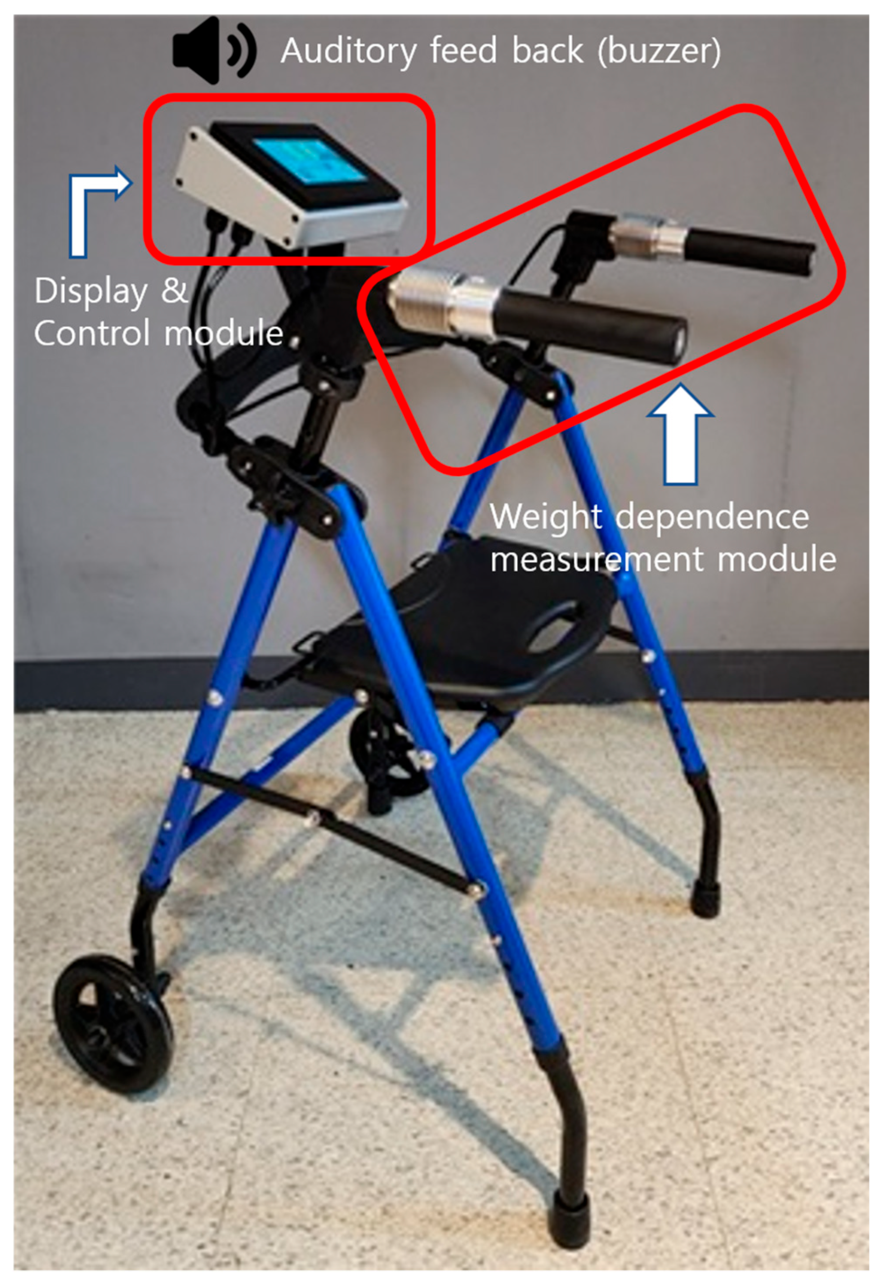
Walker load was measured using a weight-feedback walker (RehabSolution, Ltd., Chungcheongbuk-do, Republic of Korea). The device quantitatively measures the walker load applied by the user via load cells positioned between the frame and handles of the walker. In this study, the term walker load refers to the absolute load measured by these load cells and displayed in kilograms (kg). Load cells integrated into both handles measure the forces transmitted through the upper extremities during the walker-assisted gait. The total walker load is calculated as the sum of the loads recorded by the left and right load cells, and is displayed on the control module in kilograms (kg). The mean walker load is calculated automatically during walking and enables the user to set a target walker dependence between 0% and 100% in 10% increments based on the measured value. During walker-assisted gait, auditory feedback is provided when the measured walker load matches the preset target level [Figure 1].

2.3.2. Gait Analysis

Gait analysis was performed using the SMART-EVO stereophotogrammetric system (BTS Bioengineering, Milan, Italy). The system consisted of eight infrared cameras (BTS Bioengineering, Milan, Italy), a force plate (P6000, BTS Bioengineering, Milan, Italy), and a data acquisition controller. The system provides a spatial resolution of 0.3 mm and a sampling frequency of 200 Hz. Previous studies have reported the high reliability and accuracy of the system, with variability in lower extremity joint angle measurements of less than 3° [19].
The Davis protocol was used for the gait analysis [20]. A total of 22 reflective markers were attached according to the Davis Heel marker set and secured using elastic bands and hypoallergenic tape. Reflective markers were placed on the seventh cervical vertebra (C7), second sacral vertebra (S2), and both acromion processes: the anterior–superior iliac spines, greater trochanters, lateral femoral epicondyles, fibular heads, lateral malleoli, fifth metatarsal heads, and heels. To improve the accuracy of biomechanical modeling, the body segments defined by the markers were scaled according to each participant’s anthropometric measurements.
GRFs were recorded while walking using a force plate embedded in the center of the walkway. The force plate data were synchronized with the motion capture system and collected at a sampling frequency of 400 Hz. Force plate signals were low-pass-filtered using a fourth-order Butterworth filter with a cutoff frequency of 25 Hz.

2.4. Data Analysis

2.4.1. Walker Dependence

Walker dependence was defined as the walker load normalized to the participant’s body weight to account for differences in body size. During the 10-m walking trial, the loads recorded by the left and right modules were summed, and the mean walker load over the trial was calculated. Walker dependence was then calculated by dividing the mean walker load by the participant’s body weight and expressing the result as a percentage (%BW). The equations used to calculate walker dependence and define the AWD and HAWD conditions were as follows:
W a l k e r   d e p e n d e n c e   ( % B W ) = M e a n   w a l k e r   l o a d   d u r i n g   t h e   10   m   t r i a l   ( k g ) B o d y   w e i g h t   ( k g ) × 100
AWD (%BW) = Mean walker dependence (%BW)
HAWD (%BW) = 0.5 × Mean walker dependence (%BW)

2.4.2. Ground Reaction Force

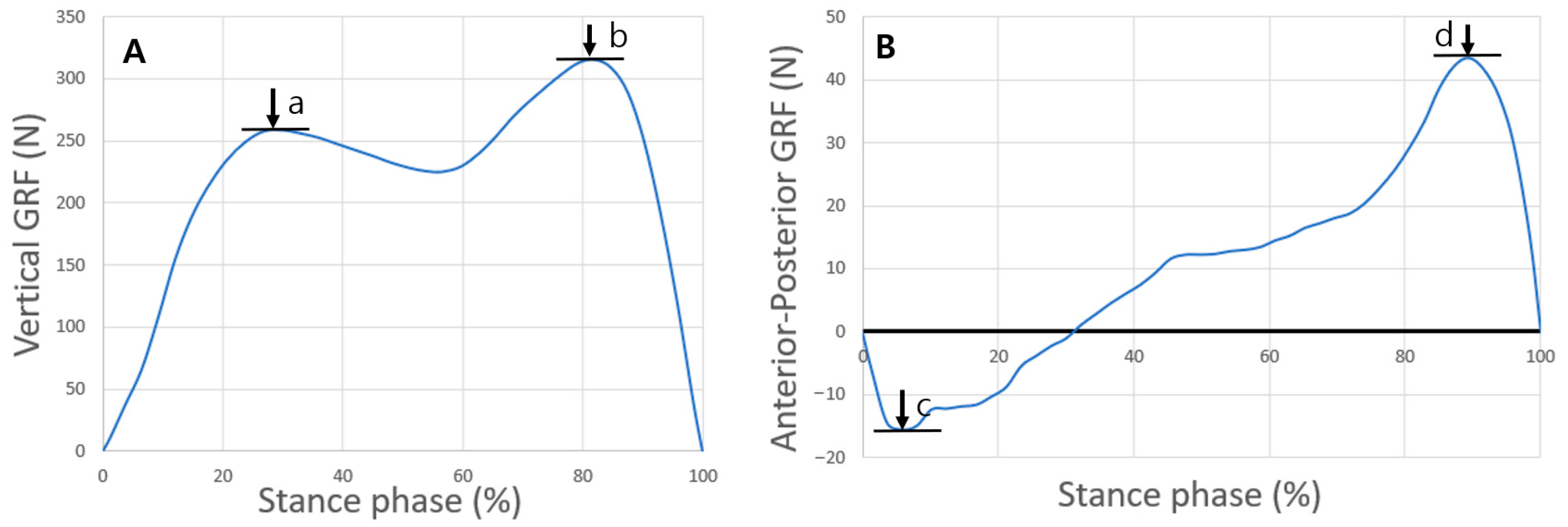
GRF data obtained during the stance phase were normalized to each participant’s body weight, and time-normalized to 0–100% of the stance phase. For the vertical GRF, the first (weight acceptance) and second (push-off) peaks were identified and extracted [Figure 2A]. For the anterior–posterior GRF, the first (braking force) and second (propulsion force) peaks were extracted [21] [Figure 2B].

2.4.3. Angular Velocity

Joint angular velocities of the hip, knee, and ankle were calculated in the sagittal plane using the SMART Analyzer software (version 1.10.470.0; BTS Bioengineering, Milan, Italy). Mean joint angular velocity was calculated as the change in joint angle divided by the corresponding change in time, as follows:
M e a n   a n g u l a r   v e l o c i t y   ( d e g r e e / s ) = Δ θ Δ t
The gait phases and intervals for angular velocity events were selected based on previous gait analysis studies [16]. The predefined gait events were identified using the SMART Analyzer software, and the mean angular velocity during each selected interval was calculated.
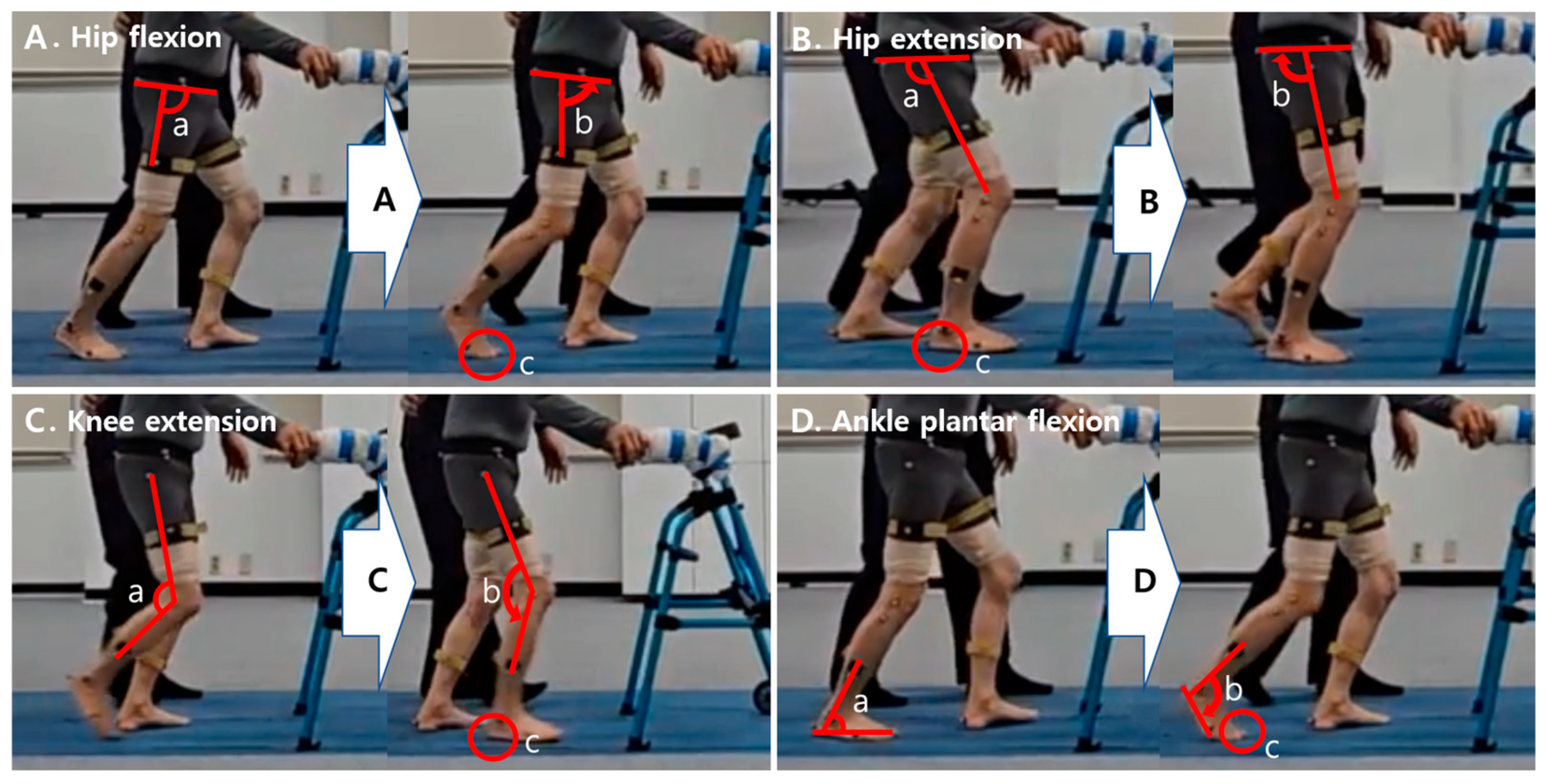
Hip flexion angular velocity during pre-swing (the period from maximum hip extension to toe-off; 40–80% of the gait cycle) and hip extension angular velocity during loading response (period 0–25% of the gait cycle) were calculated. Knee extension angular velocity during terminal swing (the period immediately preceding initial contact; 80–100% of the gait cycle) and ankle plantarflexion angular velocity during pre-swing (the period immediately preceding toe-off; 50–80% of the gait cycle) were also calculated [Figure 3].

2.4.4. Spatiotemporal Gait Parameters

Spatiotemporal gait parameters were analyzed using the SMART Clinic software (version 1.10; BTS Bioengineering, Milan, Italy). A gait cycle was defined as the interval between two consecutive initial contacts of the same limb. The variables analyzed included walking velocity, cadence, stride time, stance time, swing time, swing phase, stride length, step length, and step width. Temporal and spatial gait parameters were calculated from the gait cycles identified in each valid walking trial. When multiple gait cycles were included in a trial, the mean value across the gait cycles was used for analysis.

2.5. Statistics

Statistical analyses were performed using SPSS Statistics (version 29.0; IBM Corp., Armonk, NY, USA). Participant characteristics were summarized using descriptive statistics and presented as means and standard deviations (SD) or frequencies and percentages, as appropriate. Data normality was assessed using the Shapiro–Wilk test. Paired t-tests were used to compare kinetic and kinematic variables between the AWD and HAWD conditions. Statistical significance was set at p < 0.05.

3. Results

3.1. Walker Dependence

The mean walker load during walking was 6.73 ± 2.05 kg (Table 1), corresponding to a walker dependence of 12.83 ± 4.89%BW. Under the HAWD condition, walker dependence was reduced to 6.40 ± 2.44%BW, representing approximately 50% of the AWD condition (Table 2).

3.2. Ground Reaction Force

The weight-acceptance and push-off peaks of the vertical GRF were significantly higher under the HAWD condition than under the AWD condition (d = −1.46 and −1.45, respectively). For the anterior–posterior GRF, the braking force was significantly greater under the HAWD condition (d = −0.89), whereas the propulsion force did not differ significantly between the conditions (Table 2).

3.3. Angular Velocity

The knee extension angular velocity during the terminal swing and ankle plantarflexion angular velocity during pre-swing were significantly higher under the HAWD condition than under the AWD condition (d = −1.36 and −1.11, respectively). In contrast, no significant differences were observed for the hip angular velocity variables (Table 3).

3.4. Spatiotemporal Gait Parameters

Significant differences were observed in several spatiotemporal gait parameters between the HAWD and AWD conditions (Table 3). The stride, stance, and swing times were significantly shorter under the HAWD condition than under the AWD condition (d = 1.52, 1.44, and 1.24, respectively). In contrast, the swing phase, walking velocity, and cadence were significantly higher under the HAWD condition (d = −0.58, −1.20, and −1.62, respectively).
No significant differences were observed in spatial gait parameters, including stride length, step length, and step width (Table 3).

4. Discussion

This study aimed to investigate the biomechanical effects of varying levels of walker dependence during walker-assisted gait. Walker dependence was quantitatively measured using a weight-feedback walker, and gait characteristics were compared between the AWD and HAWD conditions. The main finding of this study was that external lower-limb loading and kinetic, kinematic, and spatiotemporal gait parameters differed across levels of walker dependence.
In this study, the weight acceptance and push-off forces were significantly greater in the HAWD condition than in the AWD condition. Vertical GRF is widely recognized as an indicator of the magnitude of load transmitted through the lower limbs during walking [14,15]. These findings are consistent with those of previous studies reporting increased vertical GRFs with greater lower limb loading and reduced body weight support [21,22]. Although previous studies used body-weight support systems different from the walker-based partial weight-bearing approach used in the present study, similar changes in vertical GRF were observed in response to changes in weight-bearing conditions. Therefore, these findings may indicate greater external loading of the lower limbs under the HAWD condition. However, this interpretation should be made with caution because walking velocity was also significantly greater under the HAWD condition. Given that vertical GRF is influenced by multiple gait-related factors, particularly walking velocity [14,21,23], the observed increase in vertical GRF cannot be attributed solely to reduced walker dependence, but may instead have been influenced by the combined effects of reduced walker dependence and increased walking velocity.
Despite the increase in walking velocity observed in the present study, no significant differences were found in spatial gait parameters, including stride length, step length, and step width. In contrast, cadence increased, whereas temporal gait parameters, including stride, stance, and swing times, decreased. In addition, knee extension and ankle plantarflexion angular velocities increased. Walking velocity can be increased through changes in spatial and temporal gait characteristics, primarily by increasing step length, cadence, or both [23]. Therefore, these findings suggest that the increase in walking velocity under the HAWD condition was characterized primarily by faster temporal progression and increased angular velocities at selected lower-extremity joints rather than by changes in spatial gait parameters.
Interestingly, despite the difference in walking velocity between the conditions, no significant difference in hip angular velocity was observed. In addition, only the braking force increased significantly under the HAWD condition, whereas the propulsive force did not differ between conditions. The anterior–posterior GRF reflects acceleration and deceleration during walking [14,15], and walking speed has been reported to influence the magnitude of GRF peaks [24,25]. In particular, propulsive force is closely associated with forward progression and ankle plantarflexor function during terminal stance and pre-swing [26]. Although ankle plantarflexion angular velocity and walking velocity increased under the HAWD condition, these changes were not accompanied by an increase in propulsive GRF. These findings may be related to the characteristic biomechanical features of walker-assisted gait. Previous biomechanical research on rollator-assisted gait, which shares biomechanical characteristics with wheeled-walker gait, reported reduced mechanical demands at the knee and ankle together with increased hip extensor contribution, which was suggested to contribute to the forward progression of the rollator and maintenance of walking velocity [7]. These characteristics may provide one possible explanation for the joint-specific angular velocity responses and the unchanged propulsive GRF observed in the present study. However, because joint kinetics and whole-body biomechanical variables were not assessed, the mechanisms underlying these findings remain unclear.
The strength of the present study is that walker dependence was quantitatively measured rather than estimated, allowing the biomechanical characteristics associated with different levels of walker dependence to be investigated.
However, this study has several limitations. First, although the sample size was determined based on an a priori power analysis, the relatively small sample size and only including community-dwelling older women may have limited the statistical power to detect differences in some biomechanical outcomes and the generalizability of the findings. Second, the independent effects of walker dependence and walking velocity on gait biomechanics could not be distinguished because walking velocity was not controlled. Future studies should independently manipulate walker dependence and walking velocity across multiple levels to clarify their respective and interactive biomechanical effects. Third, walker use is associated with upper-extremity weight support and forward trunk posture, and these characteristics may lead to whole-body biomechanical changes during gait [7,27,28]. However, whole-body biomechanical variables, including trunk and pelvic kinematics and center-of-mass displacement, were not assessed in the present study. Therefore, the characteristic whole-body adaptations associated with walker-assisted gait could not be considered when interpreting the present findings. Fourth, gait is controlled through the interaction of feedback and feedforward mechanisms [12]. In the present study, auditory feedback was provided to help participants maintain the target level of walker dependence. This external feedback may have influenced gait control and the observed gait patterns. Therefore, the observed gait characteristics may reflect gait performed under auditory feedback guidance rather than habitual walker-assisted gait, which may limit the ecological validity of the findings. Future studies should examine whether similar gait responses are observed without external feedback. Finally, paired t-tests were performed to compare biomechanical outcomes between conditions. However, because no adjustment for multiple testing was applied, the probability of Type I error may have been increased. Therefore, the statistical significance of the findings should be interpreted with caution. Future studies should consider statistical approaches that account for multiple comparisons and correlated biomechanical outcomes, such as repeated-measures multivariate analysis or linear mixed-effects models.
Future studies should include larger and more diverse populations and integrate whole-body kinematics, joint kinetics, and electromyographic measurements to clarify the biomechanical and neuromuscular mechanisms underlying gait changes associated with walker dependence. In addition, biomechanical responses across multiple levels of walker dependence should be investigated to determine whether clinically applicable ranges or safety margins for walker dependence can be established. Further intervention studies are needed to investigate the effects of quantitatively regulated walker dependence during gait training on gait function in individuals with gait impairments.

5. Conclusions

This study investigated the biomechanical effects of different levels of walker dependence during walker-assisted gait using a weight-feedback walker. Lower walker dependence was associated with greater external lower-limb loading, faster walking velocity and higher cadence, shorter temporal gait parameters, and increased knee extension and ankle plantarflexion angular velocities, without significant changes in spatial gait parameters. These findings suggest that the observed biomechanical responses cannot be attributed solely to differences in walker dependence but rather reflect the combined biomechanical influence of changes in walker dependence and walking velocity. Accordingly, quantitative regulation of walker dependence may influence biomechanical responses during walker-assisted gait and may represent a potential strategy for promoting lower-limb loading.

Author Contributions

Conceptualization, K.H.C.; methodology, K.H.C.; formal analysis, E.P.C.; investigation, E.P.C.; writing—original draft preparation, E.P.C. All authors have read and agreed to the published version of the manuscript.

Funding

This research was supported by the Regional Innovation System & Education (RISE) program through the (Chungbuk Regional Innovation System & Education Center), which is funded by the Ministry of Education (MOE) and the (Chungcheongbuk-do), Republic of Korea (2026-RISE-11-004-02). In addition, this research was supported by the U-LAB Program of the National University Development Project (2025) funded by Korea National University of Transportation.

Institutional Review Board Statement

This study was approved by the Institutional Review Board of the Korea National University of Transportation (Approval No. KNUT-2026-HR-34-04).

Informed Consent Statement

Written informed consent was obtained from the participants for the publication of this paper.

Data Availability Statement

The original contributions presented in the study are included in the article, and further inquiries can be directed to the corresponding author.

Conflicts of Interest

The authors declare no conflicts of interest.

Abbreviations

The following abbreviations are used in this manuscript:
AWDAverage walker dependence
A-PAnterior–posterior
BBSBerg balance scale
BMIBody mass index
EMGElectromyography
GRFGround reaction force
HAWDHalf-average walker dependence
MMSE-KMini-mental state examination—Korean version
VVertical

References

  1. ISO 9999:2022; Assistive Products—Classification and Terminology. International Organization for Standardization: Geneva, Switzerland, 2022.
  2. ISO 11199-1:2021; Assistive Products for Walking Manipulated by Both Arms–Requirements and Test Methods–Part 1: Walking Frames. International Organization for Standardization: Geneva, Switzerland, 2021.
  3. ISO 11199-2:2021; Assistive Products for Walking Manipulated by Both Arms–Requirements and Test Methods–Part 2: Rollators. International Organization for Standardization: Geneva, Switzerland, 2021.
  4. ISO 11199-3:2005; Walking Aids Manipulated by Both Arms–Requirements and Test Methods–Part 3: Walking Tables. International Organization for Standardization: Geneva, Switzerland, 2005.
  5. Sehgal, M.; Jacobs, J.; Biggs, W.S. Mobility assistive device use in older adults. Am. Fam. Physician 2021, 103, 737–744. [Google Scholar] [PubMed]
  6. Brandt, Å.; Iwarsson, S.; Ståhl, A. Satisfaction with rollators among community-living users: A follow-up study. Disabil. Rehabil. 2003, 25, 343–353. [Google Scholar] [CrossRef] [Scilit] [PubMed]
  7. Alkjær, T.; Larsen, P.K.; Pedersen, G.; Nielsen, L.H.; Simonsen, E.B. Biomechanical analysis of rollator walking. Biomed. Eng. Online 2006, 5, 2. [Google Scholar] [CrossRef] [Scilit] [PubMed]
  8. Youdas, J.W.; Kotajarvi, B.J.; Padgett, D.J.; Kaufman, K.R. Partial weight-bearing gait using conventional assistive devices. Arch. Phys. Med. Rehabil. 2005, 86, 394–398. [Google Scholar] [CrossRef] [Scilit] [PubMed]
  9. Mcquade, K.J.; Finley, M.; Oliveira, A.S. Upper extremity joint stresses during walkerassisted ambulation in post-surgical patients. Braz. J. Phys. Ther. 2011, 15, 332–337. [Google Scholar] [CrossRef] [Scilit] [PubMed]
  10. Choi, E.P.; Yang, S.J.; Jung, A.H.; Na, H.S.; Kim, Y.O.; Cho, K.H. Changes in Lower Limb Muscle Activation and Degree of Weight Support according to Types of Cane-Supported Gait in Hemiparetic Stroke Patients. BioMed Res. Int. 2020, 2020, 9127610. [Google Scholar] [CrossRef] [Scilit] [PubMed]
  11. Oh, G.B.; Park, C.S.; Cho, K.H. Immediate effect of weight load on lower limb muscle activity and gait ability in patients with incomplete spinal cord injury during walker gait training. J. Spinal Cord. Med. 2023, 46, 818–824. [Google Scholar] [PubMed]
  12. Park, C.S.; Oh, G.B.; Cho, K.H. Effects of gait training with weight support feedback walker on walker dependence, lower limb muscle activation, and gait ability in patients with incomplete spinal cord injury: A pilot randomized controlled trial. J. Spinal Cord. Med. 2025, 48, 103–111. [Google Scholar] [CrossRef] [Scilit] [PubMed]
  13. Spallone, G.; Mancini, L.; Carnevale, A.; Campi, S.; De Sire, A.; Schena, E.; D’Hooghe, P.; Hirschmann, M.T.; Papalia, R.; Longo, U. Step-by-step insight into gait analysis: A narrative review unlocking knee biomechanics. Knee Surg. Sports Traumatol. Arthrosc. 2025, 34, 2796–2813. [Google Scholar] [CrossRef] [Scilit] [PubMed]
  14. Perry, J.; Burnfield, J. Gait Analysis: Normal and Pathological Function; CRC Press: Boca Raton, FL, USA, 2024. [Google Scholar]
  15. Winter, D.A. Biomechanics and Motor Control of Human Movement; John Wiley & Sons: Hoboken, NJ, USA, 2009. [Google Scholar]
  16. Mentiplay, B.F.; Banky, M.; Clark, R.A.; Kahn, M.B.; Williams, G. Lower limb angular velocity during walking at various speeds. Gait Posture 2018, 65, 190–196. [Google Scholar] [CrossRef] [Scilit] [PubMed]
  17. Elias, A.; Loureiro, M.; Machado, F.; Frizera, A. Gait adaptations to walker-assisted locomotion in elderly: A principal component analysis of spatiotemporal and kinematic parameters. Sci. Rep. 2026, 16, 6872. [Google Scholar] [CrossRef] [Scilit] [PubMed]
  18. Kwon, H.; Choi, S.; Cho, Y.; Lee, H.; Tae, K. Biomechanical Analysis in the Lower Extremities During Walking with a Four Wheeled Walker (Rollator) Using the Motion Analysis System. J. Rehabil. Welf. Eng. Assist. Technol. 2018, 12, 241–248. [Google Scholar] [CrossRef] [Scilit]
  19. Huang, D.; Zhang, C.; Song, W.; Gao, J.; Tian, H.; Li, H.; Ke, X.; Jiang, C.; Lin, Z. Gait variability and biomechanical distinctions in individuals with functional ankle instability: A case–control study based on three-dimensional motion analysis. Eur. J. Med. Res. 2025, 30, 493. [Google Scholar] [CrossRef] [Scilit] [PubMed]
  20. Davis, R.B., III; Ounpuu, S.; Tyburski, D.; Gage, J.R. A gait analysis data collection and reduction technique. Hum. Mov. Sci. 1991, 10, 575–587. [Google Scholar] [CrossRef] [Scilit]
  21. Barela, A.M.; Freitas, P.B.d.; Celestino, M.L.; Camargo, M.R.; Barela, J.A. Ground reaction forces during level ground walking with body weight unloading. Braz. J. Phys. Ther. 2014, 18, 572–579. [Google Scholar] [CrossRef] [Scilit] [PubMed]
  22. Lewek, M.D. The influence of body weight support on ankle mechanics during treadmill walking. J. Biomech. 2011, 44, 128–133. [Google Scholar] [CrossRef] [Scilit] [PubMed]
  23. Fukuchi, C.A.; Fukuchi, R.K.; Duarte, M. Effects of walking speed on gait biomechanics in healthy participants: A systematic review and meta-analysis. Syst. Rev. 2019, 8, 153. [Google Scholar] [CrossRef] [Scilit] [PubMed]
  24. Hsiang, S.M.; Chang, C. The effect of gait speed and load carrying on the reliability of ground reaction forces. Saf. Sci. 2002, 40, 639–657. [Google Scholar] [CrossRef] [Scilit]
  25. Andriacchi, T.; Ogle, J.; Galante, J. Walking speed as a basis for normal and abnormal gait measurements. J. Biomech. 1977, 10, 261–268. [Google Scholar] [CrossRef] [Scilit] [PubMed]
  26. Elhafez, S.M.; Ashour, A.A.; Elhafez, N.M.; Elhafez, G.M.; Abdelmohsen, A.M. Percentage contribution of lower limb moments to vertical ground reaction force in normal gait. J. Chiropr. Med. 2019, 18, 90–96. [Google Scholar] [CrossRef] [Scilit] [PubMed]
  27. Bachschmidt, R.A.; Harris, G.F.; Simoneau, G.G. Walker-assisted gait in rehabilitation: A study of biomechanics and instrumentation. IEEE Trans. Neural Syst. Rehabil. Eng. 2001, 9, 96–105. [Google Scholar] [CrossRef] [Scilit] [PubMed]
  28. Bateni, H.; Maki, B.E. Assistive devices for balance and mobility: Benefits, demands, and adverse consequences. Arch. Phys. Med. Rehabil. 2005, 86, 134–145. [Google Scholar] [CrossRef] [Scilit] [PubMed]
Figure 1. Weight-feedback walker.
Applsci 16 07727 g001
Figure 2. Ground reaction force (GRF) variables during the stance phase. (A) Vertical GRF, including weight acceptance (a) and push-off (b). (B) Anterior–posterior GRF, including braking force (c) and propulsion force (d).
Applsci 16 07727 g002
Figure 3. Definition of joint angular velocity events. (A) Hip flexion angular velocity during pre-swing, calculated from peak hip extension to toe-off. (B) Hip extension angular velocity during loading response, calculated from peak hip flexion at heel strike (initial contact) to contralateral toe-off. (C) Knee extension angular velocity during terminal swing, calculated from peak knee flexion during the swing phase to heel strike (initial contact). (D) Ankle plantarflexion angular velocity during pre-swing, calculated from peak dorsiflexion during the stance phase to plantar flexion before toe-off. (a) Starting point; (b) End point and angular velocity direction; (c) Toe-off or heel strike (initial contact).
Applsci 16 07727 g003
Table 1. General characteristics of participants (N = 10).
ParametersMean ± SD or Frequency (%)
Sex (Male/Female)0/10 (0/100)
Age (years)81.78 ± 3.16
Height (cm)152.67 ± 3.82
Weight (kg)54.11 ± 8.85
BMI (kg/m2)23.19 ± 3.41
Duration of walking aid use (month)7.67 ± 8.01
Average walker load (kg)6.73 ± 2.05
BBS (score)46.2 ± 4.93
K-MMSE (score)24.22 ± 3.62
Note. BBS, Berg balance scale; BMI, body mass index; K-MMSE, Korean version of the mini-mental state examination.
Table 2. Walker dependence and ground reaction forces during walking with a weight-feedback walker (N = 10).
ParametersAWDHAWDΔ Values (%)tpCohen’s d95% CI
(Lower–Upper)
Walker dependence (%BW)12.83 ± 4.896.40 ± 2.446.42 ± 2.46 (50.00)7.85<0.001 *2.624.54–8.31
GRF (N)VWeight
acceptance
418.05 ± 85.92473.56 ± 95.39−48.67 ± 31.36 (−11.64)−5.46<0.001 *−1.46−77.46–−33.57
Push off434.71 ± 67.12467.68 ± 71.68−28.41 ± 26.68 (−6.54)−5.43<0.001 *−1.45−46.07–−19.86
A-PBraking17.96 ± 12.2632.19 ± 19.31−16.26 ± 17.08 (−90.53)−3.330.005 *−0.89−23.48–−5.00
Propulsion44.81 ± 14.2741.43 ± 16.212.53 ± 12.06 (5.65)0.890.3890.24−4.81–11.57
Note. Values are presented as mean ± SD. Δ values represent the mean difference between the AWD and HAWD (AWD–HAWD); values in parentheses indicate the relative percentage difference with respect to the AWD. CI, confidence interval; AWD, average walker dependence; HAWD, half-average walker dependence; BW, body weight; GRF, ground reaction force; V, vertical; A-P, anterior–posterior; * p < 0.05.
Table 3. Angular velocity and spatiotemporal gait parameters during walking with a weight-feedback walker (N = 10).
ParametersAWDHAWDΔ Values (%)tpCohen’s d95% CI
(Lower–Upper)
Angular
Velocity
(degree/s)
HE-LR 35.13 ± 12.7033.59 ± 16.113.22 ± 10.24 (9.17)0.750.4650.19−2.83–5.91
HF-Psw 41.71 ± 9.1142.51 ± 10.830.62 ± 8.43 (1.49)−0.360.723−0.10−5.52–3.93
KE-Tsw124.24 ± 34.97161.84 ± 34.56−32.36 ± 29.04 (−26.05)−5.09<0.001 *−1.36−53.58–−21.63
APF-Psw93.90 ± 32.26110.09 ± 35.22−16.69 ± 17.94 (−17.77)−4.160.001 *−1.11−24.60–−7.79
GaitVelocity (m/s)0.64 ± 0.120.76 ± 0.19−0.11 ± 0.09 (−17.19)−3.590.007 *−1.20−0.18–−0.04
Cadence (steps/min)101.67 ± 10.19114.73 ± 14.86−13.07 ± 8.08 (−12.86)−4.850.001 *−1.62−19.28–−6.85
Stride time (s)1.19 ± 0.121.07 ± 0.160.13 ± 0.08 (10.92)6.46<0.001 *1.520.08–0.17
Stance time (s)0.77 ± 0.080.68 ± 0.100.09 ± 0.06 (11.69)6.12<0.001 *1.440.06–0.12
Swing time (s)0.42 ± 0.050.38 ± 0.050.04 ± 0.03 (9.52)5.26<0.001 *1.240.02–0.05
Stance phase (%)64.34 ± 1.8163.55 ± 1.750.79 ± 1.95 (1.23)1.730.1020.41−0.18–1.76
Swing phase (%)35.20 ± 1.3836.28 ± 1.99−1.08 ± 1.88 (−3.07)−2.440.026 *−0.58−2.01–−0.15
Single support (%)35.20 ± 1.4736.27 ± 1.89−1.07 ± 2.15 (−3.04)−2.110.050−0.50−2.14–−0.00
Double support (%)14.70 ± 1.4113.69 ± 1.571.01 ± 2.14 (6.87)2.000.0620.47−0.06–2.08
Stride length (m)0.75 ± 0.110.78 ± 0.15−0.03 ± 0.08 (4.00)−1.820.086−0.43−0.07–0.01
Step length (m)0.37 ± 0.070.39 ± 0.09−0.02 ± 0.05 (5.41)−1.530.144−0.36−0.04–0.01
Step width (m)0.16 ± 0.110.15 ± 0.120.01 ± 0.02 (6.25)1.810.1080.60−0.00–0.02
Note. Values are presented as mean ± SD. Δ values represent the mean difference between the AWD and HAWD (AWD–HAWD); values in parentheses indicate the relative percentage difference with respect to the AWD. CI, confidence interval; AWD, average walker dependence; HAWD, half-average walker dependence; HE, hip extension; HF, hip flexion; KE, knee extension; APF, ankle plantarflexion; LR, loading response; Psw, pre-swing; Tsw, terminal swing; * p < 0.05.
Disclaimer/Publisher’s Note: The statements, opinions and data contained in all publications are solely those of the individual author(s) and contributor(s) and not of MDPI and/or the editor(s). MDPI and/or the editor(s) disclaim responsibility for any injury to people or property resulting from any ideas, methods, instructions or products referred to in the content.