Article

Bioactive Rocket (Eruca vesicaria subsp. sativa (Mill.) Hegi) Sprout Extracts: LC-MS Profiling and Skin-Protective Antioxidant and Enzyme Inhibitory Activities


by
Dorota Kasprzak
1,*,
Katarzyna Wojciechowska
2,
Aleksandra Nurzyńska
3,
Sebastian Granica
4,
Grażyna Ginalska
1,
Ewa Poleszak
2 and
Katarzyna Dos Santos Szewczyk
5,*
1
Department of Beauty Sciences, Faculty of Health Sciences, Vincent Pol University, Choiny 2, 20-816 Lublin, Poland
2
Chair and Department of Applied Pharmacy, Medical University of Lublin, Chodźki 1, 20-093 Lublin, Poland
3
Department of Biochemistry and Biotechnology, Medical University of Lublin, 20-093 Lublin, Poland
4
Department of Pharmaceutical Biology, Medical University of Warsaw, 02-097 Warsaw, Poland
5
Department of Pharmaceutical Botany, Medical University of Lublin, Chodźki 1, 20-093 Lublin, Poland
*
Authors to whom correspondence should be addressed.
Appl. Sci. 2026, 16(14), 7330; https://doi.org/10.3390/app16147330
Submission received: 14 May 2026 / Revised: 12 July 2026 / Accepted: 13 July 2026 / Published: 22 July 2026
(This article belongs to the Special Issue Analysis of Bioactive Natural Compounds)

Abstract

Eruca vesicaria subsp. sativa (rocket) sprouts are a rich source of bioactive compounds used in cosmetics. Phytochemical profiling (LC-PDA-MS/MS) of 5-day-old sprouts identified 26 metabolites, mainly glucosinolates (e.g., glucoraphanin and glucoerucin) and flavonol glycosides of quercetin and kaempferol. The extracts exhibited strong antioxidant activity in DPPH, ABTS•+, and ferric-reducing assays and moderate, dose-dependent inhibition of skin-degrading enzymes: tyrosinase, elastase, and collagenase, suggesting anti-wrinkle and skin-brightening effects. Cytotoxicity assays on human BJ skin fibroblasts demonstrated that the rocket sprout extracts were non-cytotoxic at concentrations up to 125 µg/mL, with low-dose treatments enhancing the cell viability. Hemocompatibility tests confirmed safety, with <2% hemolysis observed. The incorporation of these compounds into model emulsions yielded stable formulations over 90 days. These findings support the novelty of rocket sprout extracts as functional/nutricosmetic ingredients with relevance to both food and skin health.

1. Introduction

Plant sprout extracts have recently gained significant attention in the cosmetic industry because of their elevated levels of bioactive compounds formed during germination and their enhanced enzymatic activity. Germination activates metabolic pathways and promotes the de novo synthesis of phenolics, flavonoids, and antioxidants, leading to markedly higher total phenolic content and antioxidant capacity in sprouts than in ungerminated seeds [1,2]. These properties are crucial for protecting the skin from oxidative stress, pollution, and UV-induced damage, thereby delaying visible aging. Indeed, sprouts and microgreens can offer greater concentrations of vitamins and antioxidants than mature plants. For example, five- to seven-day-old sprouts often contain significantly more vitamin C, B vitamins, and antioxidant compounds than their fully grown counterparts [3]. Such nutrient boosts contribute to skin health—vitamin C brightens the skin and stimulates collagen synthesis, B-complex vitamins provide moisturizing and soothing effects, and carotenoids confer photoprotective benefits [4]. Sprouting also increases the content of free amino acids and small peptides, which may enhance epidermal penetration and support skin barrier function. Additionally, the bioavailability of minerals such as zinc and selenium improves during germination, further aiding skin healing and sebum regulation [4,5].
Sprout-derived extracts exhibit strong antioxidant, anti-inflammatory, and extracellular matrix-protective activities, making them promising active ingredients in skincare formulations for mature skin. Recent research has highlighted the positive effects of these compounds on skin and hair parameters. For instance, treatment with pea (Lathyrus oleraceus Lam.) sprout extract significantly improved hair density in volunteers (reducing hair loss and promoting growth) [6,7]. Similarly, sunflower sprout serum has been reported to restore photo-damaged skin, improve skin texture, and reduce wrinkle depth over a short-term treatment [8]. The documented capacity of sprout extracts to mitigate oxidative stress supports their use in photoprotective and anti-aging cosmetic products. Several commercial formulations have incorporated germinated-plant extracts. For example, germinated soybean (Glycine max (L.) Merr.) extract is used for its anti-aging and skin-brightening properties [9], and a micelle extract obtained from the sprouts of Phaseolus vulgaris L. was shown to enhance skin elasticity in a pilot clinical study [10]. These examples highlight the growing potential of sprout-derived phytochemicals as multifunctional cosmetic agents.
Eruca vesicaria subsp. sativa (Mill.) Hegi, a Brassicaceae family member popularly referred to as arugula or rocket, is widely consumed as a salad green. Like other cruciferous vegetables, rocket is rich in glucosinolates and polyphenols, which are recognized for their beneficial effects on human health. Notably, rocket leaves and seeds have a history of medicinal and culinary use; traditional remedies in the Mediterranean have used Eruca sativa (whole plant) preparations for skin wounds [11]. This underscores its relevance to dermatology. Phytochemical analyses have shown that rocket contains abundant amounts of vitamin C, carotenoids, and phenolic antioxidants [12]. Its seed oil (sometimes called taramira oil) and extracts contain flavonoids (e.g., quercetin and isorhamnetin) that exhibit anti-inflammatory activity and can improve skin barrier function by activating peroxisome proliferator-activated receptors in keratinocytes [4]. Rocket extracts have also demonstrated inhibitory activity against elastase, the enzyme responsible for elastin degradation, suggesting their potential to prevent skin sagging and wrinkle formation via keratinocytes [4]. Moreover, a recent formulation study reported that E. sativa seed oil provides a measurable sun protection factor (~SPF 5.6) and contributes to antimicrobial defense, highlighting its multifaceted potential in skin care [13]. Despite these promising attributes, E. vesicaria sprouts have remained relatively unexplored for cosmetic or nutraceutical applications, even though the sprouts of other Brassicaceae (such as broccoli) and legumes have been actively researched.
Given the rich phytochemistry of rocket and the general enhancements conferred by germination, we hypothesized that E. vesicaria sprouts are an excellent source of functional ingredients. for improving skin health. Therefore, in this study, we performed a comprehensive chemical and bioactivity evaluation of rocket sprout extracts. Specifically, we (i) profiled the phytochemical composition of sprouts using LC-PDA-MS/MS to identify key polyphenols and glucosinolates and quantified their total phenolics, flavonoids, and phenolic acids; (ii) assessed the antioxidant capacity of sprout extracts using multiple in vitro assays; (iii) evaluated their inhibitory effects on skin aging-related enzymes (collagenase and elastase) and a hyperpigmentation-related enzyme (tyrosinase); (iv) examined anti-inflammatory and other relevant bioactivities; (v) tested cytotoxicity on human dermal fibroblasts and hemolytic activity to ensure biocompatibility; and (vi) incorporated the sprout extracts into model cosmetic cream formulations to investigate their stability and performance. By correlating the chemical profile with the observed bioactivities, we aim to clarify how the unique composition of rocket sprouts contributes to skin-related effects on human skin cells. This study offers a comprehensive perspective on the potential of E. vesicaria sprouts as novel cosmetic ingredients and functional food resources, bridging the gap between „farm to face”—from salad bowl to skin.

2. Materials and Methods

2.1. Eruca vesicaria subsp. sativa Sprouts Sample Preparation

Germination of E. vesicaria subsp. sativa was carried out using a two-level Tribest TRIFL2000 sprouting unit (Tribest Corporation, Anaheim, CA, USA) equipped with an automated watering system. The irrigation interval was adapted to the seasonal conditions. Before sprouting, the seeds were hydrated by soaking in water for 6 h, rinsed thoroughly under running water using a fine mesh sieve, and distributed across both rays of the sprouting chamber. The spring mode (30 min intervals) was selected. Seeds of arugula were germinated at room temperature, 23 °C, under a 16 h light/8 h dark photoperiod. Approximately 50% of the germinating seeds reached the first leaf primordium stage after about 96 h of germination.
The material was harvested in two ways: the entire sprout from one tray (including the root and seed coat) and only the aerial parts from the second tray, cut just above the seed. Weighed portions of each harvest were transferred into capped glass bottles. Bioactive compounds were isolated from the plant material using 70% ethanol as the extraction solvent at a 1:3 material-to-solvent ratio (w/w). Extraction involved mechanical agitation for 2 h at 300 rpm, followed by sonication for 20 min under controlled temperature conditions of 40 °C [10]. Following extraction, the liquid phase was separated from the plant material and passed through a Whatman filter paper. The remaining solid fraction underwent a second extraction cycle using the same extraction parameters. The filtrates obtained from both extraction steps were combined and concentrated by evaporating the solvent under vacuum using an IKA RV8 Flex rotary evaporator (IKA-Werke GmbH & Co. KG, Staufen, Germany) equipped with an IKA HB10 temperature-controlled water bath and an IKA Vacstar Control vacuum system. The concentrated extracts were subsequently frozen and freeze-dried in a Labconco lyophilizer (Kansas City, MI, USA) until a stable, dry mass was achieved. Extraction was performed on 250 g batches of E. vesicaria subsp. sativa sprouts. The procedure was identical in both cases, with the only variable being the plant material selected for processing. The first preparation (ES1) was obtained from intact sprouts containing cotyledons, roots, and seed-coat remnants. This material produced approximately 10.6 g of dry extract, representing a 4.23% recovery relative to the initial fresh weight. The second preparation (ES2), generated exclusively from excised aerial parts consisting of stems and young leaves, yielded 4.3 g of dry matter, corresponding to an extraction efficiency of 1.72%. The substantially greater recovery observed for ES1 suggests that the inclusion of below-ground tissues and seed-derived structures contributed to additional extractable constituents. This effect is likely associated with the enhanced extraction of non-polar compounds, including phospholipids, phytosterols, and various fatty acids, which are naturally accumulated in these plant fractions. Prior to formulation studies, both lyophilized extracts were dispersed in a vehicle composed of water and 1,3-propanediol (1:1 (m/m)) to obtain preparations containing 2% (w/w) of the extract. Specifically, 2 g of dry material was dissolved in 98 g of the solvent mixture. The resulting stock dispersions, designated ES1 and ES2, were subsequently used as the active ingredients during cream preparation.

2.2. Quantification of Total Phenolic (TPC), Flavonoid (TFC), and Phenolic Acid (TPAC) in E. vesicaria Sprout

The phenolic compound and flavonoid concentrations in the extracts were assessed using spectrophotometric assays based on previously published protocols [14]. Optical density readings were recorded using a Pro 200F microplate reader (Tecan Group Ltd., Männedorf, Switzerland) at wavelengths of 680 nm for phenolic determination and 430 nm for flavonoid analysis.
The total phenolic content (TPC) was estimated against a standard curve prepared with gallic acid at concentrations spanning 0.002–0.16 mg/mL (R2 = 0.96). The quantitative results were reported as milligrams of gallic acid equivalents per gram of dry extract (mg GAE/g DE). The total flavonoid content (TFC) was determined using quercetin as the calibration standard. The calibration plot covered concentrations between 0.004 and 0.2 mg/mL and exhibited a correlation coefficient of R2 = 0.99. Flavonoid concentrations were reported in milligrams of quercetin equivalents per gram of dry extract (mg QE/g DE).
The phenolic acid content was quantified using the Arnov colorimetric method, in accordance with the recommendations of [15]. Absorbance values were measured at 490 nm and converted to quantitative results using a caffeic acid calibration curve built across a working range of 3.36–23.52 µg/mL (R2 = 0.99). The final values were calculated as milligrams of caffeic acid equivalents per gram of dry extract (mg CAE/g of DE).
To support the reliability of the quantitative results, the calibration curves (with regression equations and R2 values) and the raw optical density values recorded for each extract replicate in the TPC, TPAC, and TFC assays are provided in the Supplementary Materials (File S1).

2.3. LC-PDA-MS/MS Analysis

Phytochemical profiling of the extracts was performed using liquid chromatography coupled with photodiode array detection and tandem mass spectrometry (LC-PDA-MS/MS). Analyses were conducted using an Ultimate 3000 UHPLC platform connected to a PDA detector and an Amazon SL ion trap mass spectrometer (Bruker Daltonik GmbH, Bremen, Germany). The separation of constituents was achieved on a Kinetex XB-C18 analytical column (150 × 2.1 mm, particle size 1.7 µm; Phenomenex, Torrance, CA, USA), with the column compartment maintained at 25 °C throughout the analysis. The chromatographic system employed a binary solvent mixture composed of acidified water (0.1% formic acid, v/v) as eluent A and acidified acetonitrile (0.1% formic acid, v/v) as eluent B. Gradient elution was applied, starting with 1% solvent B and gradually reaching 26% B after 60 min. The mobile phase was delivered at a constant rate of 0.3 mL/min, and sample aliquots of 2 µL were introduced into the system. Before each analytical run, the stationary phase was conditioned for 7 min under initial gradient conditions. Spectral information from the PDA detector was collected between 200 and 450 nm, whereas chromatographic traces used for data evaluation were extracted at 254 nm wavelength. Mass spectrometric measurements were performed in negative electrospray ionization mode. The structural assignment of the observed constituents was proposed by comparing their chromatographic and mass-spectral characteristics with literature reports available through the Reaxys database. The search strategy focused on publications related to members of the Brassicaceae family, particularly species classified within the genera Brassica and Eruca [16,17,18,19,20].

2.4. Antioxidant Activity

2.4.1. DPPH Radical Scavenging Assay

The antioxidant capacity of E. vesicaria subsp. sativa sprout extracts was assessed using the stable free radical 2,2-diphenyl-1-picrylhydrazyl (DPPH). The assay was conducted following the procedure reported by Olech et al. [21], with slight adjustments to the original protocol. The ability of the extracts to neutralize DPPH radicals was determined spectrophotometrically by monitoring the decrease in absorbance at 517 nm. Measurements were taken after a 30 min reaction period at 28 °C. Antioxidant effectiveness was determined by reference to Trolox, with results reported in mg TE/g DE.

2.4.2. ABTS•+ Radical Cation Decolorization Assay

The free radical quenching potential of the sprout extracts was additionally investigated using the ABTS•+ decolorization method. In this assay, the antioxidant response toward the 2,2′-azinobis(3-ethylbenzothiazoline-6-sulfonic acid) radical cation was determined according to the methodology published by Pieczykolan and co-workers [22]. After allowing the reaction to proceed for 6 min, absorbance readings were collected at 734 nm. Trolox was used as the reference standard for antioxidant capacity estimation, with results reported as mg TE/g DE.

2.4.3. Metal Chelating Activity (CHEL)

The metal-chelating properties of the investigated extracts were evaluated based on their ability to complex Fe2+ ions. The assay was performed according to the modified protocol described by Olech et al. [21]. Initially, 50 µL of 0.4 mM FeCl2 solution was combined with 200 µL of the sample. Subsequently, 100 µL of 1 mM ferrozine solution was added, and the reaction was allowed to proceed for 10 min before spectrophotometric analysis at 562 nm. Control measurements included a reagent blank containing water as a substitute for the tested extract, alongside a positive reference represented by Na2EDTA. The chelating activity of the samples was calculated relative to the standard and expressed as µg Na2EDTA equivalents per gram of dry extract.

2.5. Enzyme Inhibitory Activity

2.5.1. Anti-Elastase Activity

Elastase inhibition assays were evaluated to assess the anti-aging properties of the sprout extracts. The procedure followed a modified version of the method reported by Chiocchio et al. [23]. Porcine pancreatic elastase was first combined with extract solutions prepared at concentrations ranging from 5 to 100 μg/mL in Tris–HCl buffer (1.6 mM, pH 8.0), and the mixtures were equilibrated for 15 min at 29 °C. Subsequently, N-succinyl-Ala-Ala-Ala-p-nitroanilide (AAApNA) was added to a final concentration of 0.8 mM to initiate the enzymatic reaction. The final assay volume was adjusted to 200 μL and contained 25 μL of extract solution, elastase (1 μg/mL final concentration), and the appropriate amount of buffer. Following incubation for 20 min, absorbance measurements were performed at 410 nm to determine enzyme activity. Epigallocatechin gallate (100 μg/mL) was used as a positive reference compound.

2.5.2. Anti-Collagenase Activity

The collagenase inhibitory properties of the sprout-derived extracts were evaluated using the chromogenic substrate FALGPA (N-[3-(2-furyl)acryloyl]-Leu-Gly-Pro-Ala). The experiment was conducted following the protocol reported by [24]. Collagenase obtained from Clostridium histolyticum was prepared in 50 mM Tricine buffer at pH 7.5, yielding a stock solution with an enzyme activity of 0.8 U/mL. The enzyme preparation was pre-incubated with various concentrations of the extracts for 15 min at 35 °C. Following incubation, the reaction system was adjusted to a final volume of 150 μL containing 0.1 U collagenase, 0.8 mM FALGPA, buffer, and the extract tested. The hydrolytic activity of the enzyme was determined by measuring the changes in absorbance at 345 nm. Epigallocatechin gallate (100 μg/mL) was used as a positive control.

2.5.3. Anti-Tyrosinase Activity

Tyrosinase inhibitory activity was evaluated to assess the depigmenting potential of the extracts. The procedure was adapted from the methodology reported by Kanak et al. [25]. For each determination, the extract samples (20 μL) were combined with mushroom tyrosinase solution (40 μL, 200 U/mL) along with 100 μL of phosphate buffer (50 mM, pH 6.5). The mixtures were pre-incubated for 15 min at 25 °C before substrate addition. The catalytic reaction was initiated upon the addition of 40 μL of L-DOPA solution (0.5 mM), after which the plates were incubated for a further 15 min. The measurements were recorded at 475 nm wavelength. The control wells contained phosphate buffer instead of the tested extract and represented full enzymatic activity. The inhibitory effects were calculated based on the difference between the activity observed in the control and that measured upon the addition of the extracts. Kojic acid (100 μg/mL) was used as a reference inhibitor.

2.6. Cell Culture

The potential cytotoxic effects of the ES1 and ES2 extracts derived from E. vesicaria subsp. sativa sprouts were assessed using BJ human skin fibroblasts (ATCC CRL-2522) as a cellular model for this study. Cell cultures were maintained in EMEM supplied by ATCC-LGC Standards (Teddington, UK). The growth medium contained 10% fetal bovine serum (Pan-Biotech GmbH, Aidenbach, Bavaria, Germany) and was fortified with penicillin and streptomycin at final concentrations of 100 U/mL and 100 µg/mL, respectively. The cell cultures were incubated in a Heraeus Cytoperm 2 chamber (Heraeus Cytoperm 2, Thermo Scientific, Waltham, MA, USA). under controlled environmental conditions. The temperature was maintained at 37 °C, and the atmosphere consisted of 5% carbon dioxide and high relative humidity (95%), in accordance with standard ATCC cultivation guidelines.

2.7. MTT Assay

The influence of E. vesicaria subsp. sativa extracts on fibroblast viability was determined using the colorimetric MTT reduction assay, as previously reported by Sugier et al. [26]. BJ fibroblasts were seeded in 96-well culture plates at a concentration of 1 × 10⁵ cells/mL, with 100 µL of cell suspension dispensed into each well. Cells were allowed to attach and proliferate over a 24 h period at 37 °C in a 5% CO2 atmosphere. Lyophilized extracts obtained from both whole and cut sprouts were initially dissolved in dimethyl sulfoxide (DMSO) to produce stock solutions at a concentration of 100 mg/mL. These stocks were subsequently diluted with EMEM supplemented with 2% fetal bovine serum to generate a concentration range of 0.97–500 µg/mL. At the highest extract concentration, the amount of DMSO present in the culture medium was maintained below 0.5% (v/v), with progressively lower solvent concentrations in serial dilutions. To verify that any observed effects were attributable to the extracts rather than the solvent, a vehicle control containing the corresponding concentration of DMSO without the extract was included. Cells maintained in EMEM supplemented with 2% FBS were used as the negative control. After 24 h of exposure to the tested samples, MTT reagent solution (1 mg/mL in culture medium, 100 µL per well) was introduced into each well. Subsequently, the plates were incubated at 37 °C for 3 h, during which viable cells converted the tetrazolium salt into insoluble purple formazan crystals via mitochondrial enzymatic activity. The resulting crystals were dissolved by adding 100 µL of 10% SDS prepared in 0.01 M HCl, followed by overnight incubation (12 h). The generated formazan was quantified by measuring the absorbance at 570 nm using a microplate reader (BioTek Synergy HTX, Agilent Technologies, Santa Clara, CA, USA). Cell viability was presented as a percentage of the value obtained for untreated control cultures.

2.8. Live/Dead Cell Staining

Morphological assessment of extract-treated fibroblasts was carried out employing a Live/Dead fluorescence staining assay (Sigma-Aldrich Chemicals, Warsaw, Poland). This method was employed to distinguish living cells from those that had lost membrane integrity after exposure to the tested samples. BJ fibroblasts were cultured in 96-well plates by seeding 100 µL of a cell suspension containing 1 × 10⁵ cells/mL into each well. After 24 h of cultivation, the medium was replaced with the extract solutions. Microscopic observations were restricted to three representative concentrations (0.97, 31.25, and 500 µg/mL), selected to reflect the lowest, intermediate, and highest treatment levels investigated in the cytotoxicity study. After another 24 h of incubation, the staining procedure was performed according to the manufacturer’s recommendations. This staining system simultaneously labels viable and nonviable cells with distinct fluorescent signals. Living cells emitted green fluorescence due to intact cellular membranes, whereas cells with membrane damage were identified by red fluorescence. Fluorescence imaging was performed using an confocal laser scanning microscope (CLSM; Olympus Fluoview FV1000, Tokyo, Japan). to provide visual evidence of the effects of the extracts on cell viability and membrane integrity.

2.9. Interaction of E. Vesicaria Subsp. Sativa Sprout Extracts with Human Blood

For hemolysis studies, venous blood was obtained from a healthy donor following ethical approval granted by the Bioethics Committee of the Medical University of Lublin (approval number KB-0024/19/02/2025). The procedure was based on the methodology reported by Michalicha et al. [27], with several minor modifications. Citrate was used as an anticoagulant to maintain sample stability. Hemoglobin levels in whole blood and plasma were assessed using Drabkin’s reagent (Chempur, Piekary Śląskie, Poland) in conjunction with spectrophotometric measurements conducted in 96-well plates. A standard curve prepared using commercially available human hemoglobin (Sigma-Aldrich Chemicals, Warsaw, Poland) was used for quantification. The concentration of total hemoglobin measured in the collected blood specimen was 129.68 mg%.

Hemolysis Assay

Erythrocyte membrane damage induced by the investigated extracts was assessed using a hemolysis test. The extract preparations were diluted in calcium- and magnesium-free PBS to obtain final concentrations of 500, 31.25, and 0.97 μg/mL. These concentrations were chosen to reflect the upper, middle, and lower limits of the exposure range examined in biological assays. For each determination, 1 mL of the extract solution was combined with an equal volume of diluted citrated blood. The mixtures were incubated at 37 °C for 3 h with gentle agitation. After centrifugation, the supernatant fractions were carefully collected and mixed with Drabkin’s reagent. Following a 15 min reaction period at 37 °C, absorbance measurements were carried out at 540 nm. The control samples consisted of blood suspended in PBS, representing spontaneous hemolysis, and blood treated with 0.1% Triton X-100, representing total erythrocyte lysis. Hemolytic activity of the extracts was expressed as a percentage relative to the Triton X-100 control, which was assigned a value of 100%.

2.10. Antibacterial Activity

The inhibitory potential of the analyzed samples toward Gram-positive bacteria was investigated using standard strains of S. aureus (ATCC 25923) and S. epidermidis (ATCC 12228). The experimental procedures were performed according to a protocol adapted from Sugier et al. [26]. Both bacterial isolates were obtained from the microbial collection of the Department of Biochemistry and Biotechnology at the Medical University of Lublin (Lublin, Poland). Mueller–Hinton broth was supplied by Oxoid Ltd. (Basingstoke, UK) was used as the nutrient medium for all microbiological assays.

Determination of the Minimum Inhibitory Concentration (MIC) of E. vesicaria subsp. sativa Extracts

Susceptibility testing was carried out using the broth dilution technique in microplate format to establish the minimum inhibitory concentration (MIC) of the investigated extracts. The MIC was defined as the lowest concentration resulting in complete suppression of visible bacterial growth after incubation. Lyophilized material was dissolved in dimethyl sulfoxide to yield extract stock solutions at 100 mg/mL. Working solutions covering the range of 0.97–500 µg/mL were then obtained by serial dilution in Mueller–Hinton broth (Oxoid Ltd., Basingstoke, UK). Bacterial suspensions calibrated to the 0.5 McFarland standard were transferred to the assay wells in a volume of 2 µL per well. The experimental design included appropriate controls for culture growth, medium sterility, and potential interference caused by the natural coloration of the extracts. Microplates were incubated aerobically at 37 °C for 24 h. The absence of visible turbidity after incubation was interpreted as inhibition of bacterial growth, and the corresponding extract concentration was recorded as the MIC value.

2.11. Composition and Preparation of Cosmetic Creams

To investigate the applicability of the extracts in cosmetic preparations, two semisolid cream formulations were manufactured based on an in-house developed formulation protocol. Each formulation was prepared as a 500 g batch. The only variable between the two products was the incorporated plant extract: one cream contained ES1 and the other ES2. The qualitative and quantitative composition of the formulations is provided in Table 1.

2.12. Cream Preparation

The creams were formulated in an IKA LR 1000 control reactor. The weighed oil phase was heated in the reactor vessel to 70 °C under gentle stirring at 50 rpm. The aqueous phase was prepared separately, preheated to 50 °C, added to the oil phase, and the mixture was heated to 70 °C.
Vacuum was then applied, with stirring increased to 150 rpm and simultaneous homogenization at 15,000 rpm for 15 min. The heating and homogenization steps were then discontinued, and the emulsion was stirred at 50 rpm during cooling. After the temperature had fallen to 40 °C, active ingredients phase was added, followed by a further 15 min of mixing and homogenization under vacuum. The creams were portioned into four containers and stored overnight at ambient temperature.

2.13. Assessment of the Physicochemical and Sensory Properties of the Creams

The emulsions were subjected to physicochemical analysis following protocols available in the literature [10,28,29,30,31,32,33,34,35,36].

2.14. Centrifugal Stability Test

For the test, 1 g of cream was weighed into an Eppendorf-type tube. The samples were centrifuged using a laboratory centrifuge (Centrifuge 5418 R, Eppendorf SE, Hamburg, Germany). The centrifugation test was performed at 9000 rpm (revolutions per minute), corresponding to a relative centrifugal force of approximately 7000× g (RCF) (relative centrifugal force), for 10 min. After centrifugation, a visual assessment was conducted to check for phase separation [10,36]. The analyses were conducted according to a fixed schedule on days 1, 7, 14, 30, 60, and 90 days for samples stored at 4 °C and 20 °C.

2.15. Organoleptic Assessment of the Preparations

The sensory characteristics of the creams were evaluated by the authors using a self-assessment approach. The assessment included color, odor, consistency, and spreadability after application to the skin. Odor was classified as pleasant, neutral/odorless, or unpleasant, including chemical-like notes. Texture uniformity was evaluated by checking for visible or perceptible particles during application.
The ease of spreading was evaluated by rubbing a small amount of the formulation onto the volar wrist area. Samples with good spreadability formed an even layer and absorbed readily, without streaking, pilling, tackiness, or a greasy after-feel. In contrast, formulations with poor spreadability produced uneven texture, clumping and/or left a persistent heavy on the skin surface [10,36].

2.16. Particle Size Evaluation

The particle size of the oil phase was monitored over time by bright-field optical microscopy with a Leica ICC 50 HD microscope fitted with a 40× air objective. Samples were prepared by applying a thin and uniform layer of cream onto a clean microscope slide, followed by placement of a coverslip. Three random microscopic fields were analyzed for each formulation at each time point. Oil droplet diameters were measured for 100 randomly selected droplets using LAS EZ software (Leica Las 4.5.0 [build : 418]; Switzerland), and representative micrographs were obtained with a 50 µm scale bar. To assess physical stability during storage, mean droplet diameters were quantified at days 1, 7, 14, 30, 60, and 90 and subjected to longitudinal statistical analysis. This approach enabled detection of any progressive increase in droplet size that could indicate droplet fusion and subsequent emulsion destabilization.

Statistical Analysis of Droplet Size

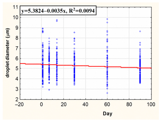
To determine whether the mean oil-droplet diameter changed during storage, the data was subjected to statistical analysis. The Shapiro–Wilk test was initially used to verify the normality of distribution. For each analyzed dataset, the obtained p-values were <0.05, demonstrating that the data significantly departed from a normal distribution. Consequently, non-parametric tests were selected for further analysis. Therefore, the Friedman ANOVA test was used to compare repeated measurements over time, and Kendall’s coefficient of concordance (W) was calculated to assess the strength and direction of changes between subsequent measurements. For better visualization of potential variations, a linear regression analysis was additionally conducted for the sample of cream with ES1 stored at 20 °C, where the Friedman test indicated statistically significant but very weak changes; linear regression was additionally performed to visualize and quantify the trend.

2.17. Spreadability Test

Spreadability was quantified with an extensometer which evaluates the sample’s lateral expansion under defined loads placed between two plexiglas disks [10,30,33]. The measurements were performed at room temperature (20–22 °C). A 1.00 g cream sample was compressed on a circular test plate with a flat piston under stepwise loads of 200–1000 g, each applied for 1 min. After each load, the radius of the spread film was determined in four directions spaced 45° apart and average. The sequence was performed three times for each formulation. From the mean diameters spread areas were calculated for every load and a spread ability factor (Sf) was derived as the area under the curve relating spread area to applied load [10]. The Sf values (g·cm2) allowed quantitative comparison of the spreadability properties of the creams. Each sample was analyzed in triplicates.

Statistical Analysis—Spreadability Factor (Sf)

The suitability of the dataset for parametric evaluation was verified before hypothesis testing. Data normality was assessed separately for each time–temperature treatment using the Shapiro–Wilk, while homogeneity of variance was examined with Levene’s test. Because no violations of these assumptions were detected, one-way ANOVA was employed for further analysis. The grouping factor corresponded to the storage condition, defined by the specific combination of temperature and storage period. Statistical outputs are presented as F-values accompanied by their respective numerator and denominator degrees of freedom and p-values. A significance level of 0.05 was adopted for all analyses.

2.18. Rheological Testing

Rheological measurements were carried out using an Anton Paar RheolabQC rotational rheometer (Anton Paar, Graz, Austria) fitted with a CC27/S concentric-cylinder geometry and a Peltier-controlled temperature unit. Rheological measurements of the creams, including flow behavior and consistency, were carried out with a RheolabQC rotational rheometer (Anton Paar) equipped with a CC27/S concentric-cylinder geometry and Peltier temperature control. The obtained data were processed using RheoCompass™ software version 1.30.1164 (Anton Paar, Graz, Austria). For rheological testing, each sample was transferred to the measuring cup and kept under measurement conditions for 10 min before analysis. The flow curves were obtained in controlled shear-rate mode by rising the shear rate from 0.1 to 300 s−1 and then reducing it back to 0.1 s−1. The degree of thixotropy was expressed as the hysteresis loop area calculated between the ascending and descending curves. Apparent viscosity was read at 5 s−1, while the consistency coefficient K [Pa·sn] and flow behavior index n [–] were derived by applying to the Ostwald-de Waele model to the experimental data. Variations in K and n over the storage period were interpreted as indicators of aging-related changes in cream structure and flow behavior.
Experimental values were expressed as mean values accompanied by standard deviation estimates. Flow characteristics of the formulations were described according to the Ostwald–de Waele model, enabling calculation of the consistency index (K) and the flow behavior exponent (n). The degree of thixotropy was estimated from the hysteresis area (A) obtained from the rheological flow curves.

2.18.1. Statistical Treatment of Rheological Measurements

The appropriateness of parametric statistical methods was verified before performing comparisons. Distribution of the rheological datasets was examined with the Shapiro–Wilk test within each storage-time and temperature combination, whereas variance uniformity was evaluated by Levene’s test. After confirming compliance with these assumptions, one-way ANOVA was employed to investigate differences among experimental conditions. All outcomes are presented as F-statistics accompanied by the corresponding degrees of freedom and p-values.

2.18.2. pH Measurement

For pH determination, each cream sample was first diluted tenfold by dispersing 5 g of formulation in 50 g of distilled water. After thorough mixing, the pH was measured potentiometrically with a calibrated Elmetron pH meter (ELMETRON CP-411, Zabrze, Poland). The electrode was inserted into the prepared dispersion, and the final value was recorded.

3. Results and Discussion

The present study expands current knowledge regarding the chemical composition and biological potential of Eruca vesicaria sprouts, highlighting their relevance as promising germinated plant materials for nutraceutical, dermatological, and cosmetic applications.

3.1. Characterization of Phenolic Compounds

As an initial step, the extracts obtained from E. vesicaria subsp. sativa sprouts were subjected to spectrophotometric evaluation to estimate the levels of major groups of phenolic metabolites. The analysis included determination of total phenolics, total phenolic acids, and total flavonoids, providing an overall assessment of the polyphenolic profile of the investigated extracts. Quantitative results were expressed relative to the corresponding reference standards, namely gallic acid, caffeic acid, and quercetin, and are reported as milligrams of equivalent compound per gram of dry extract. The measured values are presented in Table 2. Extracts were evaluated across three concentrations (1, 5, and 10 mg/mL) for all three parameters (TPC, TPAC, and TFC). Table 2 reports the results obtained at 5 mg/mL for TPC and TFC, and at 10 mg/mL for TPAC. The corresponding calibration curves and raw absorbance readings for TPC, TPAC, and TFC across all three tested concentrations (1, 5, and 10 mg/mL) are provided in the Supplementary Materials (File S1) to allow independent evaluation of assay performance.
To further elucidate the phytochemical profile of the investigated material, the extracts were subjected to LC-PDA-MS characterization. The obtained chromatograms indicated the presence of a broad spectrum of secondary metabolites characteristic of Brassicaceae plants. Overall, 26 chromatographic peaks were recorded and tentatively assigned to specific constituents using a combination of retention behavior, ultraviolet absorption features, and MS/MS fragmentation data collected in the negative-ion mode. Structural proposals were supported by comparison with previously reported LC-MS datasets describing compounds identified in Eruca and closely related taxa. The resulting chromatographic profiles together with the corresponding spectral information and proposed compound identities are presented in Figure 1 and Table 3. The raw LC-PDA-MS data files (mzXML format) for both extracts (whole-seedling extract, ES1; aerial-part extract, ES2) are provided in the Supplementary Materials (File S2) to allow independent inspection and reprocessing of the acquired spectra.
The phytochemical profile of E. vesicaria subsp. sativa sprout extracts was dominated by two major classes of compounds, namely glucosinolates and polyphenolic constituents, in agreement with previous LC-MS studies on rocket species [16]. This dual phytochemical profile mirrors previous reports on rocket and other cruciferous sprouts, which noted glucosinolates (such as glucoraphanin and glucoerucin) and flavonoid glycosides as characteristic constituents. Glucoraphanin and glucoerucin are precursors of isothiocyanates (such as sulforaphane and erucin, respectively) that are well-known for their antioxidative and anti-inflammatory effects in biological systems. The identified quercetin and kaempferol derivatives (e.g., quercetin-3,3′,4′-O-triglucoside and various sinapoylated flavonoids) exhibited potent antioxidant capacity owing to their polyphenolic structures. The prominence of these flavonoids in rocket sprouts aligns with the literature on sprouted legumes and grains, which often show enhanced phenolic profiles after germination [1]. However, unlike legume sprouts (such as soybean or common bean), which may contain exceedingly high total phenolic levels (e.g., a recent study reported ~193 mg GAE/g DE in a micellar fraction derived from common bean sprouts) [37], rocket sprouts devote a considerable portion of their metabolome to glucosinolates, which are not accounted for in Folin–Ciocalteu phenolic assays. Indeed, the total phenolic content we measured for rocket sprout extracts (on the order of 1–2 mg GAE/g) appears lower than that of phenolic-rich legume sprout extracts [37], but this is balanced by the presence of glucosinolates and other sulfur-containing antioxidants unique to the Brassicaceae family. In practical terms, the cocktail of antioxidants presents in rocket sprouts, including vitamins, flavonoids, and isothiocyanate precursors, likely interact synergistically to confer broad protective effects.

3.1.1. Glucosinolates

Several aliphatic and indole glucosinolates were detected, characterized by early elution, UV absorption maxima at approximately 220–230 nm, and characteristic deprotonated molecular ions in the negative ion mode. Compound 1 was identified as glucoraphanin, based on its [M–H] ion at m/z 436 and diagnostic MS/MS fragment ions at m/z 372 and 194, which are consistent with previously reported LC-MS data for rocket and other Brassicaceae matrices [16]. Compound 7, showing a molecular ion at m/z 420 and fragment ions at m/z 339, 275, and 258, was assigned to glucoerucin, in accordance with literature reports describing aliphatic glucosinolates in Eruca sativa [16]. Indole glucosinolates were detected at a longer retention time. Compound 17 was identified as 4-methoxyglucobrassicin, based on its [M–H] ion at m/z 477 and characteristic fragmentation pattern involving methoxy-substituted indole moieties [20]. Compound 24, with a molecular ion at m/z 506, was assigned as 1,4-dimethoxyglucobrassicin, in agreement with previously published LC-MS data for methoxylated indole glucosinolates in Brassicaceae species [20]. Additionally, compound 23 was identified as glucoalyssin, based on its [M–H] ion at m/z 450 and diagnostic MS/MS fragmentation, consistent with literature reports on rocket glucosinolate profiles [16].

3.1.2. Polyphenolic Compounds

The second major group of metabolites comprised polyphenolic compounds, predominantly flavonol glycosides, and sinapoyl-substituted derivatives. These compounds exhibited characteristic UV-Vis absorption maxima at approximately 255–265 nm and 320–350 nm, which are typical of flavonol chromophores. Several quercetin glycosides were identified by the occurrence of a characteristic aglycone fragment ion at m/z 301 in the MS/MS spectra. Compound 9 was identified as quercetin 3,3′,4′-O-triglucoside, displaying a parent ion at m/z 787 and sequential neutral losses of hexose units, consistent with LC-MS data reported for rocket species [18]. Additional quercetin derivatives (compounds 18, 20, and 25) were detected, including higher-molecular-weight conjugates bearing sinapoyl moieties, as indicated by the characteristic fragment ions at m/z 223 corresponding to sinapic acid residues. The occurrence of sinapoylated quercetin glycosides in Eruca has been previously documented [18].
Multiple kaempferol glycosides were also detected and identified on the basis of the aglycone ion detected at m/z 285 and UV absorption maxima of approximately 260 and 320–325 nm. Compound 21 was tentatively identified as a sinapoylated kaempferol sophoroside derivative, with a molecular ion at m/z 977 and fragmentation consistent with sequential losses of sugar and sinapoyl units, in agreement with published LC-MS data for sinapoylated flavonols in rocket [18]. Similar sinapoylated kaempferol derivatives were detected as compounds 22 and 26, further supporting the prevalence of acylated flavonols in E. vesicaria subsp. sativa. Compound 12 was identified as 1-O-sinapoylglucose, based on its [M–H] ion at m/z 385 and characteristic MS/MS fragmentation, consistent with earlier reports on phenylpropanoid conjugates in Brassicaceae plants [18].

3.1.3. Other Constituents

In addition to glucosinolates and polyphenols, tryptophan (compound 3) was detected as a minor constituent, identified by its [M–H] ion at m/z 203 and a characteristic fragment at m/z 116, consistent with the LC-MS data reported for amino acids in plant matrices [17]. Several additional minor peaks remained unassigned owing to insufficient structural information or lack of suitable reference spectra. However, their UV-Vis and MS features suggest that they may represent further conjugated phenolics or glucosinolate-related metabolites.
Both rocket sprout extracts showed measurable levels of polyphenols, though their total phenolic content (TPC) was modest compared to some other sprouted plants. The whole-seedling extract (ES1) and aerial-part extract (ES2) contained in the order of 1–2 mg GAE/g DE. These values are lower than phenolic-rich legume sprouts (e.g., common bean sprouts can reach ~193 mg GAE/g), reflecting the unique metabolite profile of Brassicaceae sprouts. In rocket, a significant portion of the phytochemical repertoire consists of glucosinolates (sulfur-containing compounds) which are not detected by Folin–Ciocalteu assays. This means that a relatively low TPC in rocket does not equate to a lack of bioactives. Rather, it highlights that rocket devotes much of its metabolism to glucosinolates alongside phenolics. Indeed, the two extracts contained a “cocktail” of antioxidants—including vitamins, polyphenols, and isothiocyanate precursors—that likely act in concert to confer protection. This synergistic effect can compensate for a moderate TPC, as multiple antioxidant species may work together to enhance overall activity.
In terms of phenolic classes, the total flavonoid content (TFC) differed markedly between the extracts. ES1 had a low flavonoid level (on the order of tens of µg quercetin equivalents per g), whereas ES2 was about five-fold richer in flavonoids (hundreds of µg QE/g). This indicates that phenolic compounds became more concentrated in the above-ground parts of the sprouts. Consistently, chromatographic profiling revealed rocket sprouts to be particularly enriched in flavonol glycosides [38]. Quercetin derivatives were prominent (e.g., quercetin-3,3′,4′-O-triglucoside was identified), along with various kaempferol glycosides and acylated conjugates [38]. These flavonoids, often decorated with sugar and sinapoyl (hydroxycinnamic acid) moieties, align with known profiles in rocket and other Brassica vegetables [39]. The presence of sinapate fragments in some identified compounds suggests that phenolic acids in rocket sprouts are largely incorporated as conjugates (such as sinapoyl-glucose and sinapoylated flavonols) rather than free acids. Accordingly, the measured “total phenolic acid content” was negligible in both extracts, implying that free phenolic acids (caffeic, ferulic, sinapic, etc.) are only trace-level or bound within larger molecules. This observation is consistent with Brassicaceae sprouts generally containing hydroxycinnamic derivatives (like sinapic acid) esterified to other compounds. Overall, ES2 had a higher load of polyphenols—especially flavonoids—than ES1, which can be attributed to the concentration of these compounds in leaves and cotyledons as opposed to the root or seed remnant.

3.2. Antioxidant Activity

Antioxidant activity of the E. vesicaria sprout extracts was characterized using three analytical approaches designed to assess different mechanisms of antioxidant action: DPPH radical reduction, ABTS•+ radical decolorization, and metal ion chelation (Table 4). Extracts were evaluated across three concentrations (1, 5, and 10 mg/mL) for all three assays (DPPH, ABTS, and CHEL). Table 4 reports the results obtained at 1 mg/mL. In the DPPH assay, both ES1 and ES2 exhibited strong radical-scavenging effects, with values corresponding to approximately 480–510 µg Trolox equivalents per gram of dry extract. No statistically meaningful differences were observed between the extracts (p > 0.05). The comparable performance of ES1 and ES2 suggests that the compounds responsible for neutralizing relatively stable organic radicals are present at similar functional levels in both extract types.
Results obtained from the ABTS•+ assay demonstrated a particularly strong antioxidant response, with activities corresponding to approximately 2.5–3.4 mg Trolox equivalents per gram of dry extract. Such values place the investigated rocket sprout extracts among plant materials characterized by considerable radical-quenching capacity and are comparable with, or in some cases higher than, those reported for numerous edible sprouts and microgreens. Previous studies have shown that germinated materials such as radish, broccoli, lentil, and alfalfa exhibit markedly greater antioxidant activity than their respective seeds, as measured by both DPPH and ABTS•+ assays [1]. This phenomenon is commonly associated with metabolic changes occurring during germination, including increased biosynthesis of phenolic metabolites. The findings obtained in the present study support this observation, indicating that sprouting substantially enhances the antioxidant properties of E. vesicaria. Despite similar performance in the DPPH test, the two extracts differed noticeably in their ability to neutralize ABTS•+ radicals. The ES2 preparation exhibited approximately 40% greater activity than ES1, which may be related to its substantially higher flavonoid content. Since the ABTS•+ method is particularly sensitive to water-soluble antioxidant constituents, the elevated abundance of polar flavonoid derivatives in ES2 likely contributed to its improved effectiveness in this assay.
It should be emphasized that although the DPPH and ABTS•+ results are both expressed in Trolox equivalents, this common unit does not imply that the two assays are mechanistically interchangeable. DPPH is a stable, sterically hindered radical dissolved in an organic (methanolic) medium and reacts preferentially with moderately lipophilic, hydrogen-atom-donating phenolics, whereas ABTS•+ is generated and measured in an aqueous/mixed-phase buffer and reacts mainly via single-electron transfer with polar, hydrophilic antioxidants. Because the two radicals probe different, only partially overlapping subsets of the antioxidant pool, extracts with a similar overall Trolox-equivalent capacity can be ranked differently by each assay if their phenolic profiles differ in polarity, as observed here for the comparatively less polar ES1 and the flavonoid-enriched, more polar ES2 [40,41,42]. This behavior is a well-recognized feature of Trolox-equivalent reporting rather than an inconsistency in the present dataset. In this context, Trolox and Na2EDTA already function as the reference/calibration standards against which every DPPH, ABTS•+ and CHEL value in Table 4 is expressed against; the corresponding calibration curves and raw absorbance readings for these standards are provided in the Supplementary Materials (File S3) to allow independent evaluation of assay performance.
Assessment of metal-binding properties further confirmed the antioxidant potential of both extracts. Each preparation demonstrated the capacity to interact with transition metal ions, indicating an ability to limit metal-catalyzed oxidative reactions. The pronounced chelating performance suggests that phenolic constituents present in the extracts may contribute not only through direct radical scavenging but also by reducing the pro-oxidant activity of metal ions. Our data show the extracts could chelate iron in amounts corresponding to several mg EDTA per gram. This significant chelating/reducing capacity can be attributed to the polyphenols (which readily undergo redox reactions) and other redox-active constituents like ascorbate present in the sprouts. Literature on sprout antioxidants supports these findings, noting that sprouts generally develop enhanced reducing power and radical scavenging ability compared to ungerminated seeds [39,42]. The slight differences between ES1 and ES2 across the assays likely reflect variation in antioxidant profiles: ES2, being richer in polar phenolics and vitamins, excelled in the ABTS•+ and CHEL tests (which operate in aqueous or mixed-phase environments), whereas the lipid-soluble antioxidant capacity (DPPH in organic medium) was similar for both.
Importantly, the broad-spectrum antioxidant activity of rocket sprout extracts is a key outcome of germination. Germination is known to mobilize diverse antioxidant pathways, yielding a spectrum of compounds that can scavenge radicals in both hydrophilic and lipophilic contexts. In practical terms, this means the rocket extracts can protect different cellular compartments from oxidative stress. For example, in a skin application scenario, antioxidants that operate in aqueous phases (cytosol, extracellular fluid) as well as those that integrate into lipid phases (cell membranes) are desirable. The combination of phenolic antioxidants (largely hydrophilic) with isothiocyanates and tocopherols (more lipophilic) in these extracts ensures that reactive oxygen species can be neutralized in virtually all environments. This comprehensive antioxidant defense is beneficial not only nutritionally but also for potential cosmetic or dermatological use, as it may help counteract oxidative damage in the skin’s multiple layers.

3.3. Enzyme Inhibitory Activities

In addition to scavenging free radicals, the rocket sprout extracts showed the capacity to suppress enzymes associated with hyperpigmentation and skin aging. We assessed their effects on collagenase, elastase, and tyrosinase in vitro (Table 5). Both extracts were evaluated at a single concentration (5 mg/mL), and inhibited all three enzymes to varying degrees, though the potency was moderate and varied by enzyme. The pattern of activity was not uniform across enzymes: at the tested concentration (5 mg/mL), the whole-seedling extract (ES1) showed marginally stronger inhibition of collagenase and elastase than ES2, whereas the aerial-part extract (ES2) was distinctly more effective against tyrosinase (Table 5). This enzyme-dependent pattern most likely reflects the different phytochemical composition of the two extracts (Section 3.1) together with the distinct catalytic mechanisms and active-site chemistries of the three target enzymes, rather than an overall superiority of either extract.
The investigated extracts were also capable of reducing collagenase activity, although the magnitude of inhibition was moderate. At the tested concentration, enzyme activity decreased by several dozen percent compared with the untreated control. Among the two preparations, ES1 exhibited a marginally stronger inhibitory effect than ES2 (42.6% vs. 39.8% inhibition at 5 mg/mL, Table 5), consistent with the comparatively higher recovery of non-polar constituents (e.g., phytosterols and fatty acids) in the whole-seedling extract. However, neither extract achieved 50% inhibition within the concentration range examined. Similar levels of activity have been reported for other polyphenol-rich botanical extracts. For example, extracts derived from argan fruits, despite their substantial phenolic content (>50 mg/g), were likewise found to inhibit collagenase and elastase to an extent below 50% [43]. The observed effects may be associated with the presence of phenolic constituents and glucosinolate-derived metabolites naturally occurring in rocket sprouts. Numerous studies have demonstrated that polyphenols can interfere with matrix metalloproteinases (MMPs), including collagen-degrading enzymes, thereby contributing to the preservation of extracellular matrix integrity [43]. In addition, isothiocyanates characteristic of Brassicaceae plants have been reported to modulate pathways involved in MMP regulation. Sulforaphane, for instance, has been shown to suppress MMP-1 expression triggered by ultraviolet radiation or pro-inflammatory signals [44]. Although the degree of inhibition recorded in the present study was not pronounced, even partial suppression of collagenase activity may be biologically relevant. Sustained reductions in enzyme activity, particularly when combined with decreased MMP expression and antioxidant protection, could contribute to limiting collagen degradation and supporting long-term maintenance of skin structure.
A similar trend was observed with elastase, the enzyme responsible for degrading elastin in the skin. Both extracts achieved partial elastase inhibition (again under roughly 50% at the tested concentration), with ES1 and ES2 showing comparable inhibitory activity (33.7% vs. 33.0% at 5 mg/mL, respectively, Table 5). Nevertheless, any level of elastase inhibition can be beneficial in an anti-aging context, as it may slow the breakdown of elastic fibers. The polyphenols in rocket sprouts, especially flavonols, are plausible contributors to the anti-elastase effect—flavonoid-rich extracts from other plants are known to inhibit elastase by binding to the enzyme or competing with the substrate [43]. Our findings concur with other reports that phenolic antioxidants can serve dual roles by both scavenging ROS and directly protecting extracellular matrix proteins (collagen, elastin) from enzymatic degradation [43].
The most pronounced tyrosinase-related activity was observed for the aerial extract (ES2). Tyrosinase-inhibitory activity, expressed as kojic acid equivalents, reached 54.9 ± 11.0 mg KAE/g dry extract for the aerial extract (ES2) at the tested concentration (5 mg/mL), whereas the whole-seedling extract (ES1) produced markedly weaker activity (18.9 ± 3.8 mg KAE/g dry extract; Table 5). This kojic-acid-equivalent activity indicates a measurable capacity of ES2 in particular to interfere with tyrosinase function, suggesting potential relevance to skin-brightening or anti-hyperpigmentation applications, although percentage-inhibition data for the extracts themselves were not generated in this study. Literature data on other plant extracts have reported tyrosinase inhibition expressed as true percentage values; for example, a recent study on common bean sprouts reported ~58% inhibition at 100 µg/mL, comparable to kojic acid’s 65% under the same conditions [37]. Because that comparison uses a different metric (percentage inhibition) and a different concentration than the present study, it is presented here for context only and is not a direct comparison with the ES1/ES2 results. We did not determine tyrosinase inhibition as a percentage or formal IC50 values for the extracts. The kojic-acid-equivalent activity nonetheless indicates a meaningful effect, particularly for ES2. Polyphenols are likely responsible for much of this anti-tyrosinase effect, as they can chelate the copper in tyrosinase’s active site or otherwise hinder its action [37]. The high flavonoid content of ES2 (especially quercetin derivatives) may be pivotal, given that quercetin and its glycosides are known tyrosinase inhibitors. Additionally, isothiocyanates or other sulfur compounds in rocket could contribute indirectly by antioxidative mechanisms that suppress melanogenesis.
From a broader perspective, the bioactivities observed (antioxidant and enzyme inhibition) underscore the multi-functional potential of rocket sprout extracts. Germination has enhanced the sprouts’ phytochemical profile to the point where they can address several key pathways of skin aging: oxidative stress, collagen/elastin breakdown, and melanogenesis. While the magnitudes of enzyme inhibition are moderate, they are comparable to those found in many plant-derived cosmetic ingredients and could have cumulative benefits. The fact that the cut sprout (ES2) extract was distinctly more potent specifically for tyrosinase inhibition, while the whole-seedling extract (ES1) performed marginally better against collagenase and elastase, suggests that the two extracts are enriched in partially different sets of bioactive constituents rather than one extract being uniformly superior. This could be because the aerial tissues (captured in ES2) are richer in polar flavonoids that are particularly effective copper-chelating tyrosinase inhibitors, whereas the seed and root remnants retained in ES1 contribute additional lipophilic constituents (e.g., phytosterols and fatty acids) that may favor interaction with collagenase and elastase.
Overall, our results align well with recent literature on sprout-derived cosmeceuticals. For example, in a 2024 study, micellar extracts of Phaseolus vulgaris sprouts (rich in polyphenols) were shown to exhibit broad anti-aging properties including antioxidant, collagenase/elastase inhibition, and stimulation of collagen synthesis in skin cells [37]. Similarly, other Brassicaceae sprouts and greens have been reported to contain flavonoids and isothiocyanates that protect the skin’s structural proteins and reduce inflammation [44]. Our work is the first to report such activities for Eruca vesicaria sprouts, highlighting them as a promising natural ingredient for health and cosmetic applications. The dual presence of polyphenols and glucosinolates in rocket sprouts is particularly noteworthy—this combination is somewhat unique and may offer synergistic protective effects. In practical terms, rocket sprout extracts could be used as food-derived antioxidants or functional additives in nutraceutical formulations, or as active botanical extracts in topical products aimed at anti-aging and skin brightening. The subsequent formulation tests (described elsewhere in the manuscript) further demonstrate that these extracts can be incorporated into a model emulsion without stability issues, reinforcing their suitability for product development.

3.4. Cytotoxicity

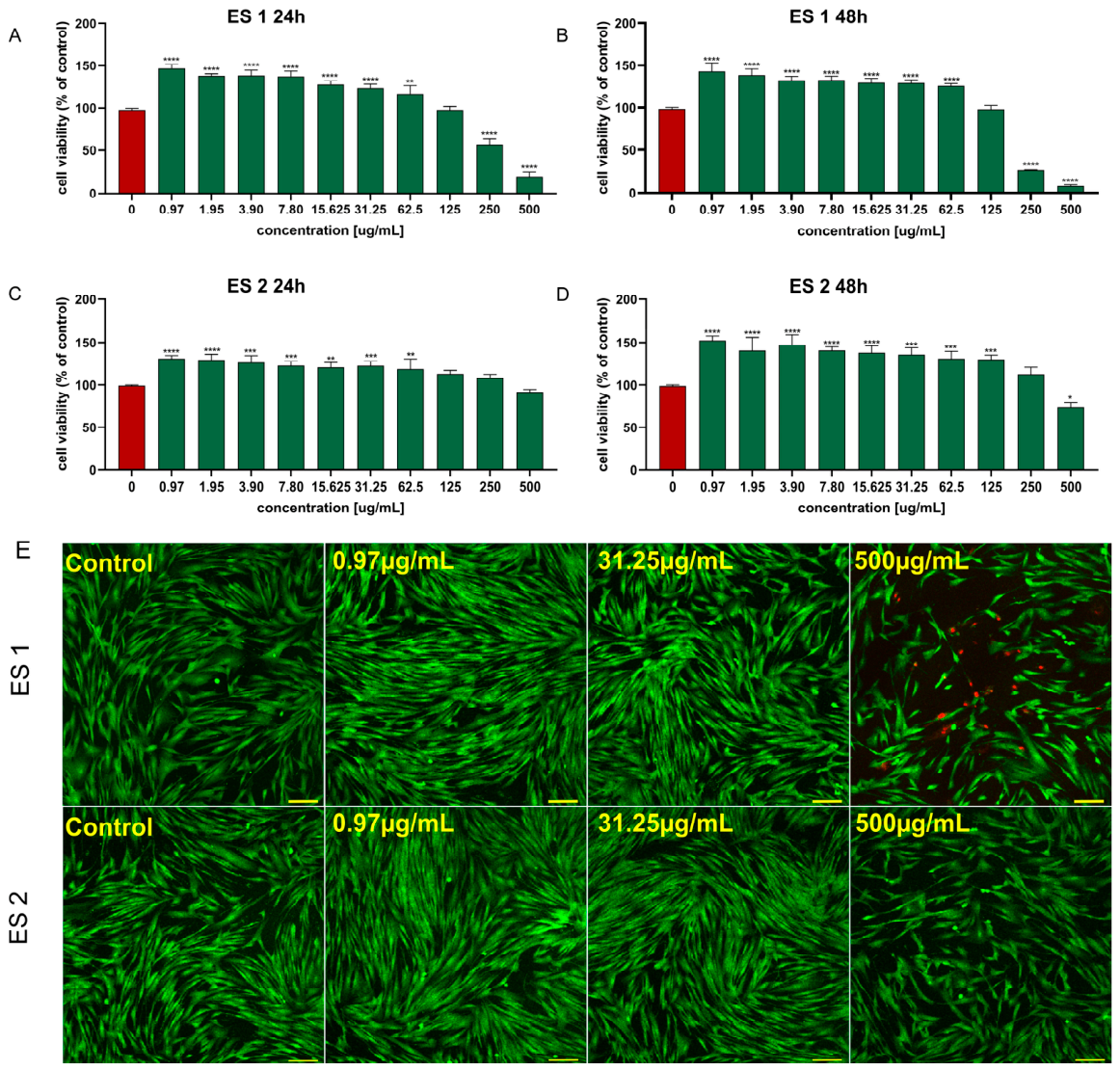
Evaluation of BJ fibroblast metabolic activity using the MTT assay revealed distinct biological responses for the two E. vesicaria subsp. sativa extracts (Figure 2). The preparation obtained from whole seedlings exhibited a favorable safety profile across the investigated concentration range. A slight reduction in viability was detected only after 48 h exposure to the highest tested dose (500 µg/mL), reaching 74.0% ± 4.95 of the control value. Despite this decrease, the extract remained within the range generally regarded as non-cytotoxic. Moreover, concentrations between 0.97 and 31.25 µg/mL significantly enhanced fibroblast viability relative to untreated cells, suggesting a stimulatory influence on cellular metabolic activity. This effect became more evident after prolonged incubation, as viability values recorded at 48 h exceeded those measured after 24 h treatment.
A different pattern was observed for the extract prepared exclusively from sprout tissues. In this case, cell viability declined progressively with increasing extract concentration, indicating a concentration-dependent cytotoxic response. The strongest effect was recorded at 500 µg/mL, where fibroblast viability decreased to 20.0% ± 5.13 following 24 h of treatment and further declined to 8.0% ± 1.5 after 48 h. Substantial growth inhibition was also evident at 250 µg/mL, reducing viability to 57.0% ± 6.4 and 26.9% ± 0.3 after 24 h and 48 h, respectively. The highest concentration that did not produce cytotoxic effects was identified as 125 µg/mL, according to the cytotoxicity criterion defined in ISO 10993-5 [45] for the biological evaluation of medical devices. Although this standard was developed for medical devices, the threshold of 70% cell viability is widely accepted and commonly applied for the interpretation of in vitro cytotoxicity assays, with cell viability below 70% being considered indicative of cytotoxicity [45,46]. Nevertheless, similarly to the whole-seedling extract, the lowest tested concentrations (0.97–31.25 µg/mL) significantly increased fibroblast viability, indicating a potential growth-promoting or metabolic activation effect under low-dose exposure conditions.
The CLSM Live/Dead observations were consistent with the MTT results. At the lowest (0.97 µg/mL) and intermediate (31.25 µg/mL) concentrations, no dead cells were observed in either whole-seedling or sprout extracts, confirming the non-cytotoxic nature of these exposure levels. In contrast, the 500 µg/mL sprout extract induced numerous dead cells, consistent with the strong cytotoxicity detected in the MTT assay. Importantly, no alterations in the morphology of viable cells were observed across all non-cytotoxic concentrations, further supporting the biocompatibility of Eruca vesicaria subsp. sativa sprout extracts at low and moderate doses.
Such a biphasic (hormetic) effect—growth stimulation at low doses and inhibition at high doses—has been described for many plant extracts rich in polyphenols or glucosinolates. For example, a micellar extract from sprouts of P. vulgaris. significantly increased the proliferation of human fibroblasts at selected concentrations [37]. Similarly, broccoli sprout extracts containing sulforaphane reduced fibroblast viability at high concentrations (e.g., ~50% at 5 mg/mL), which supports their strong cytotoxic activity against cells [47].
Thus, our results are consistent with the literature: batches of Brassicaceae sprouts (including broccoli) rich in glucosinolate hydrolysis products exhibit pronounced cytotoxic effects in vitro [47], whereas at low levels of bioactive compounds or at low doses, a proliferative effect is observed [37]. This phenomenon is likely driven by differences in phytochemical composition: sprouts are rich in volatile isothiocyanates and other metabolites (e.g., sulforaphane, phenethyl isothiocyanate), which at high concentrations damage cellular mitochondria, whereas lower doses activate cellular defense mechanisms (e.g., via Nrf2) and stimulate cell growth [37,47].
Moreover, comparison with extracts from thermally processed broccoli sprouts shows that inhibition of myrosinase activity (e.g., heating at 100 °C) markedly reduces cytotoxicity (cell viability ~99%) [47], indicating that bioactivity depends on the extent of isothiocyanate release. Likewise, our whole Eruca seedlings (including root tissue and seed remnants) may have exhibited lower enzymatic release of these compounds or a different glucosinolate profile, which could explain the milder observed effect.

3.5. Hemolysis Assay

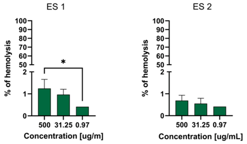
The hemolysis assay was performed using three representative concentrations of both extracts of E. vesicaria subsp. sativa sprout (500, 31.25, and 0.97 µg/mL). The results are presented in Figure 3. In all cases, a clear trend was observed: hemolysis decreased with decreasing extract concentrations.
For extracts derived from whole seedlings, the percentage of hemolysis remained very low across the tested range, decreasing from 0.69% ± 0.23 at 500 µg/mL to 0.55% ± 0.20 at 31.25 µg/mL and 0.41% at 0.97 µg/mL (identical values in all replicates). Extracts from the sprouts alone exhibited slightly higher hemolytic activity, consistent with the stronger cytotoxicity observed in the MTT assay. Hemolysis reached 1.24% ± 0.40 at 500 µg/mL and 0.96% ± 0.20 at 31.25 µg/mL, while the lowest concentration (0.97 µg/mL) again resulted in 0.41% hemolysis, with no variability between replicates. None of the measured values exceeded 2%, which is far below the commonly accepted toxicity threshold (30%) [48].
The literature contains no reports of significant hemolytic activity of extracts from the sprouts discussed here. On the contrary, similar studies report their biocompatibility. For example, aqueous extracts from mung bean sprouts were non-toxic to erythrocytes (no hemolysis) and were considered safe for skin applications [49]. In general, most plant-derived substances are regarded as “safe” for erythrocytes at hemolysis levels below 5 –10% (and clearly safe below 30%) [48].
Therefore, the results of our assays (hemolysis ≤ 2%) are fully consistent with the literature, indicating that sprout extracts do not damage red blood cells and can be considered hemocompatible [49]. Differences between Eruca sprouts and other plants included in this analysis are negligible, as none of the commonly studied microgreens (e.g., radish or broccoli sprouts) have shown significant hemolysis at concentrations typical for extracts in profiled studies (no evidence of a relevant effect).

3.6. Antibacterial Activity

The antibacterial potential of sprout-derived extracts obtained from E. vesicaria subsp. sativa was investigated against two Gram-positive reference strains namely S. epidermidis ATCC 12228 and Staphylococcus aureus ATCC 25923. Susceptibility testing was performed using a standard broth microdilution assay, and the obtained data are presented. Neither extract affected bacterial proliferation at any of the concentrations examined (0.97–500 µg/mL). Both staphylococcal strains remained viable throughout the entire concentration range, including the highest dose tested, demonstrating the absence of detectable antibacterial activity under the applied experimental conditions. Accordingly, the minimum inhibitory concentration (MIC) for each extract exceeded 500 µg/mL. This contrasts with several previous reports of antibacterial activity in Eruca extracts, and the possible reasons for this discrepancy are discussed in detail below.
The literature contains findings confirming that plant-derived sprout extracts often exhibit very moderate or no activity against Gram-positive cocci at the concentrations tested. Large-scale screening studies have shown that only a limited number of vegetable sprouts (e.g., certain cultivars of radish, white cabbage, or kohlrabi) produced inhibition zones against pathogenic bacteria [50]. In such assays, as many as 29 out of 55 tested sprout types (including many commonly consumed species) showed no detectable antibacterial activity [50]. Therefore, the lack of inhibition observed for Eruca extracts is consistent with this general trend—only some Brassicaceae species display strong antibacterial effects, whereas many others show little or no activity.
However, some reports indicate that selected Eruca and other Brassicaceae extracts may exhibit anti-staphylococcal activity when applied in an appropriate form. For instance, Eruca seed oil (rich in free fatty acids and phenethyl and erucyl isothiocyanates) showed MIC values of approximately 60–70 µg/mL against S. aureus [51], indicating high antiseptic activity. Similarly, aqueous extracts from arugula leaves inhibited the growth of S. aureus (inhibition zone of ~12 mm) [52]. In contrast, our sprout extracts were obtained from very young plants and were tested over a relatively low concentration range (up to 0.5 mg/mL), which may have been insufficient to reveal weak antibacterial effects. By comparison, phenolic extracts from 5-day-old wild mustard sprouts achieved MIC values of approximately 0.45 mg/mL against Staphylococcus [42], indicating measurable activity within a similar concentration range.
Possible explanations for the lack of activity of Eruca extracts in our study include differences in the profile of bioactive compounds: Eruca sprouts may contain a distinct set of glucosinolates and lower levels of precursors of phenethyl isothiocyanates compared with mature seeds or other Brassicaceae species. The extraction method is also likely to play a role—our extracts were hydroalcoholic and may have contained limited amounts of volatile isothiocyanates, whereas other studies employed seed esterification or more lipophilic solvents, enhancing the recovery of antibacterial compounds [51]. Likewise, radish and cabbage sprouts in one study produced inhibition zones attributed to sulfur-containing compounds and phenolics [50].
Overall, the absence of antibacterial activity of Eruca sprout extracts is not unexpected and aligns with frequent observations that plant sprout extracts are not effective antibacterial agents against S. aureus. Differences among studies may result from extract composition (variable ITC and fatty acid content), sprout cultivation conditions (germination stage), as well as the microbiological methods applied.

3.7. Evaluation of Physicochemical and Organoleptic Properties

Each formulation fulfilled the required physicochemical and organoleptic criteria. Literature-based physicochemical tests [10,36] confirmed that the creams were homogeneous, stable, and had suitable consistency. The centrifugation test showed no evidence of phase separation in any sample at any of the tested storage intervals, confirming the emulsions’ physical stability.
The formulations demonstrated satisfactory organoleptic properties, including neutral odor, characteristic greenish-gray color, smooth texture, and good spreadability. No particles, crystallization, greasiness, stickiness, or unpleasant odor were detected, confirming their homogeneity, stability, and acceptable application profile.
Oil droplet size is an important factor affecting the stability and performance of cosmetic emulsions [31]. Smaller and uniformly distributed droplets enhance kinetic stability by reducing gravitational separation, coalescence, and creaming, which in turn improves the visual appearance, smoothness, and sensory profile of the product [10,31].
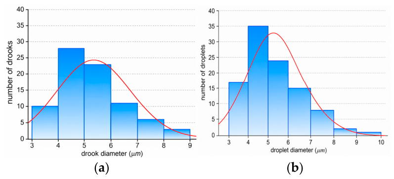
Microscopic analysis showed that the mean oil-droplet diameter remained within a narrow range during storage. For the ES1-containing cream, the mean droplet diameter ranged from 5.10 to 5.45 µm, while for the ES2-containing cream it ranged from 5.30 to 5.40 µm, depending on storage temperature and time (Table 6 and Table 7). The Friedman test confirmed no significant changes in droplet diameter for ES1 stored at 4 °C and for ES2 stored at both temperatures (Table 8). For ES1 stored at 20 °C, statistically significant differences were detected; however, Kendall’s coefficient was very low (W = 0.038), indicating that the observed effect was weak and of limited practical relevance.
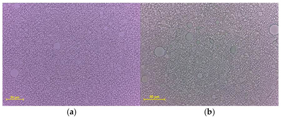
Microscopic photographs representative of the analyzed systems are presented in Figure 4a,b (50 µm scale bar), while the distribution profiles of droplet populations are summarized in Figure 5a,b.
For this formulation, linear regression (Figure 6) yielded the equation y = 5.3824 − 0.0035x, where y represents the mean oil-droplet diameter expressed in µm and x represents storage time in days. The very low coefficient of determination (R2 = 0.0094) indicates that storage time explained less than 1% of the variability in droplet diameter. Therefore, although the trend was statistically significant, it should be interpreted as practically negligible.
Sf values of approximately 71 × 103–78 × 103 g·cm2 confirmed good spreadability and acceptable application properties of the creams [10,30,33].
Normality of the Sf data was verified through the Shapiro–Wilk test (p > 0.05). Therefore, a one-way ANOVA was performed. For cream with ES1, no statistically significant variation was detected throughout the storage period (F(10.22) = 1.90, p = 0.10). Similarly, for the cream with ES2, no significant effect of storage time was observed (F(10.22) = 0.47, p = 0.89).
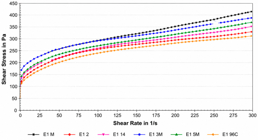
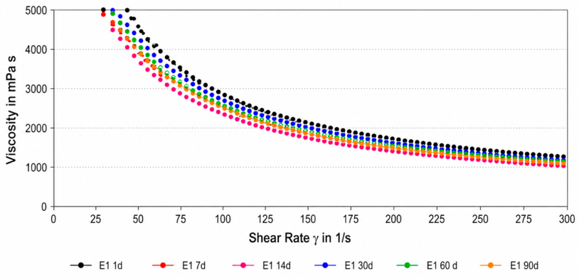
The rheological parameters remained stable regardless of storage temperature (4 °C vs. 20 °C) and time (1, 7, 14, 30, 60, and 90 days). Both creams exhibited shear-thinning behavior, as indicated by flow behavior index values below 1. Representative flow curves and viscosity curves are shown in Figure 7 and Figure 8, respectively. The ES1-containing cream showed slightly higher viscosity and consistency index values, suggesting a more compact structure, whereas the ES2-containing cream had a lighter consistency and slightly better spreadability. However, these differences did not compromise the stability or application properties of either formulation.
One-way ANOVA revealed no statistically significant variation across time points (F(10,22) = 0.40; p = 0.93); K ranged from ~124–130 (F(10,22) = 0.21; p = 0.99). The flow behavior index for cream with ES1 was essentially constant, confirming shear-thinning behavior (n < 1), (~29.9–32.8; F(10,22) = 0.84; p = 0.595) and a lower K (~111–121; F(10,22) = 1.04; p = 0.444) with n < 1 (≈0.166–0.201) and a stable thixotropy. In summary, both formulations met the criteria for rheological stability; the cream with ES1 had a more compact consistency, whereas the cream with ES2 was lighter and somewhat easier to spread, without compromising stability.
The pH values of both formulations remained stable throughout the 90-day storage period at both 4 °C and 20 °C. The pH of the cream with ES1 ranged from 5.83 to 5.91, whereas that of the cream with ES2 ranged from 5.80 to 5.86. Stable pH values throughout storage confirmed the chemical stability of both formulations.
The extraction yield was strongly dependent on the composition of the plant material. The higher efficiency of extract ES1, obtained from whole sprouts, can be attributed to the presence of lipid-rich seed and root tissues, which were efficiently solubilized in 70% (v/v) ethanol. These tissues contain storage lipids and nonpolar metabolites that increase the overall mass of dry extract. Conversely, extract ES2, derived solely from the aerial parts, represents a more hydrophilic fraction rich in phenolic and sulfur-containing compounds typical of E. sativa leaves. This compositional difference may influence both the physicochemical and sensory properties of the resulting cosmetic formulation.
Visual assessment after centrifugation showed that all cream samples remained unchanged during storage. This behavior indicates that the formulations were physically stable and did not show a tendency toward emulsion breakdown under the test conditions [10].
Spreadability is an important indicator of the application properties of semi-solid systems and an indirect measure of their rheological behavior. High Sf values correspond to a soft, plastic consistency and suitable viscosity that facilitate even application on the skin [30].
In our research, the mean Sf values for both creams ranged from 71 to 78 × 103 g·cm2, classifying the tested creams as formulations with good usability and easy application [10]. The absence of statistically significant differences during storage confirmed the rheological stability and maintenance of emulsion structure integrity. Minor decreases in Sf after 30–60 days could be related to the gradual thickening of the matrix or reduced mobility of dispersed droplets, which is typical for O/W emulsions as small amounts of water evaporate or weak intermolecular networks are rebuilt.
When comparing both formulations, the cream with ES1 exhibited slightly higher Sf values, indicating a softer and more flexible texture, whereas the cream with ES2 showed more uniform results with smaller fluctuations over time, confirming its higher physical stability. These differences may arise from the compositional differences between extracts ES1 and ES2 (e.g., a higher content of lipophilic constituents in ES1), which may subtly affect the microstructure of the emulsion rather than the oil phase or emulsifier composition, which was identical in both formulations.
The stability of Sf throughout storage suggests that the emulsions retained their internal structure and interfacial integrity owing to the optimal ratio of the oil and aqueous phases and appropriate emulsifier selection. These findings align with earlier observations indicating that well-balanced O/W emulsions maintain constant spread ability over time when no phase separation or coalescence occurs [10,30].
Although both formulations showed no statistically significant changes in Sf during storage (p > 0.05), the cream with ES2 demonstrated slightly higher stability, as evidenced by a lower F value (F = 0.47 vs. 1.90) and higher p-value (p = 0.89 vs. 0.10), suggesting lower variability overtime.
Both creams exhibited a pseudo plastic flow profile combined with consistent, moderate thixotropy (stable hysteresis loop area, A)—a textbook rheological model for topical semi-solids [10,53,54]. Under shear during application, the viscosity decreases rapidly, facilitating spreading, whereas after shear cessation, the structure rebuilds sufficiently to prevent run-off and maintain a continuous film on the skin. The absence of significant changes in viscosity at Dr 5s−1 and K overtime and between 4 °C and 20 °C indicates a stable interfacial network and appropriately balanced phase ratios and emulsifier/thickener system, with no evidence of coalescence or network densification that would drive an undesirable increase in viscosity. The differences between the two formulations were qualitative: the higher viscosity and K of the cream with ES1 translated into a creamier texture, whereas the cream with ES2, with lower values of these parameters, was easier to spread. In both cases, a constant (<1) and an essentially unchanged thixotropy index A confirmed that the chosen texture did not compromise durability, and both formulations preserved desirable application properties throughout the 90-day storage period [30,53,54].
The stability of pH observed in both formulations over the 90-day testing period indicates that neither extract induced acidification or alkalinization during storage. A stable pH profile is a key parameter that ensures the chemical integrity of active compounds and maintains user safety and skin compatibility. The measured pH values (5.80–5.91) fall within the range commonly considered to be physiologically appropriate for healthy human skin. The natural skin surface typically exhibits an acidic pH between 4.7 and 5.9, depending on the age, skin type, and anatomical site [29,34]. Maintaining this acidic environment, known as the acid mantle, plays a crucial role in upholding barrier integrity, regulating the skin microbiome, and supporting enzymatic processes involved in lipid metabolism [34].
The stable pH profile of both formulations supports their chemical stability and appropriateness for skin application.
From a nutritional standpoint, our study results reinforce that E. vesicaria sprouts are not only cosmetically active but also nutritionally valuable. The significant levels of vitamin C, polyphenols, and potassium reported in rocket [55], combined with our observed antioxidant capacity, indicate that consuming these sprouts (e.g., as microgreens or juiced ingredients) could confer health benefits, such as enhanced antioxidant defenses and reduced inflammation in the body.
Sprouting is known to improve the bioavailability of minerals and break down antinutrients like phytates [56], meaning that rocket sprouts could provide more accessible micronutrients (e.g., iron, zinc) compared to seeds. There is increasing interest in “edible cosmetics” and the idea that dietary intake of bioactives can complement topical application for skin health. In this context, rocket sprouts play a promising dual role: as a functional food, they may help combat oxidative stress and inflammation systemically, and as a cosmeceutical ingredient, they directly protect and rejuvenate the skin. The safety profile we established (non-cytotoxic and non-hemolytic) further supports their use in both domains. Unlike some synthetic antioxidants or exfoliating agents that can irritate the skin or have systemic side effects, the rocket sprout extract, derived from a common salad vegetable, demonstrated gentle behavior in our tests. This biocompatibility is a crucial advantage for translating our findings into products or dietary recommendations.
In summary, Eruca vesicaria sprouts represent a compelling example of how germination can unlock a plant’s potential for both nutritional and dermatological therapy. Our study is among the first to thoroughly document the phytochemical composition of rocket sprouts and link it to multiple bioactivities relevant to anti-aging skin care. The results not only validate the traditional knowledge of rocket as a skin remedy [11] but also expanded it by quantifying modern endpoints (antioxidant metrics, enzyme inhibition rates, cell viability influences, formulation stability).
Compared to other sprout species, rockets emerge as a multifaceted contender. Although it may not have the highest absolute phenolic content, the balance of glucosinolates and flavonoids yields a broad-spectrum effect that is highly desirable. These insights pave the way for future research, such as clinical trials of rocket sprout-based formulations on skin outcomes or investigations into optimizing sprouting conditions to maximize specific bioactives (e.g., elicitation techniques) [57].
Furthermore, the “food-derived” nature of rocket sprout extract could appeal to consumers seeking natural and sustainable skincare options. By incorporating a salad ingredient into a skin cream, we align with the clean beauty movement that favors recognizable, plant-based components. Overall, the integration of E. vesicaria sprouts into cosmetics exemplifies the productive convergence of food science and dermatology, offering a holistic approach to wellness by nourishing the body and skin with the same botanical source.

4. Conclusions

This study demonstrated that Eruca vesicaria subsp. sativa (rocket) sprouts, commonly known as nutritious salad greens, also hold significant promise as an ingredient for cosmetic and dermatological applications. Germination of rocket seeds for 5 days led to a sprout extract rich in polyphenolic antioxidants and glucosinolates, which we showed to correlate with strong antioxidant activity and the ability to inhibit key skin-aging enzymes (collagenase and elastase) and melanogenesis-related tyrosinase. The unique phytochemical profile of rocket sprouts, which combines flavonoid glycosides, vitamins, and isothiocyanate precursors, underpins these bioactivities and helps protect skin biomolecules from oxidative and enzymatic degradation. Importantly, the rocket sprout extracts were found to be safe and biocompatible; they caused no significant cytotoxicity to human skin cells at relevant concentrations and induced minimal hemolysis, meeting safety criteria for topical use. Low doses even stimulate fibroblast viability, hinting at regenerative or cell-energizing properties. When formulated into a model cream, the sprout extracts did not compromise product stability or sensory quality, indicating their potential for use in cosmetic formulations. Taken together, our findings highlight E. vesicaria sprouts as a valuable dual-purpose resource: a natural anti-aging active for skincare products and a nutrient-dense food that can contribute antioxidants and metabolic benefits to the diet. The convergence of cosmetic efficacy and nutritional richness in rocket sprouts exemplifies the concept of “food-derived” cosmeceuticals and suggests that incorporating such sprout extracts could benefit both skin health and appearance. Further research and development could pave the way for rocket sprout-based serums, creams, and supplements that harness these synergistic advantages for consumer wellness.

Supplementary Materials

The following supporting information can be downloaded at: https://www.mdpi.com/article/10.3390/app16147330/s1, File S1: Calibration curves (regression equations, R2) and raw optical density values used for the determination of total phenolic and total flavonoid content (Table 2); File S2: The raw LC-PDA-MS data files (mzXML format) for both extracts; File S3: Calibration curves and raw absorbance values of the Trolox and Na2EDTA reference standards used in the DPPH, ABTS•+, and metal-chelating assays (Table 4).

Author Contributions

Conceptualization, D.K. and K.W.; methodology, K.W., A.N., S.G. and K.D.S.S.; formal analysis, D.K., K.W., A.N., S.G. and K.D.S.S.; investigation, K.W., A.N., S.G. and K.D.S.S.; resources, A.N., S.G., G.G., E.P. and K.D.S.S.; data curation, D.K., K.W., A.N., S.G. and K.D.S.S.; writing—original draft preparation, D.K., K.W., A.N., S.G. and K.D.S.S.; writing—review and editing, K.D.S.S. and G.G.; visualization, K.W., A.N., S.G. and K.D.S.S.; supervision, K.D.S.S. All authors have read and agreed to the published version of the manuscript.

Funding

The research was partially financed by the Vincent Pol University of Lublin. Antibacterial, cytotoxicity, and hemolysis studies were supported by the Ministry of Education and Science of Poland as part of the statutory activities of the Medical University of Lublin (project Nos. DS6/2025 and Pbmb190/2025).

Institutional Review Board Statement

Not applicable.

Informed Consent Statement

Not applicable.

Data Availability Statement

The data presented in this study are available on request.

Acknowledgments

The authors would like to express their sincere gratitude to Sebastian Kanak, for his valuable assistance with the experimental analyses conducted in this study.

Conflicts of Interest

The authors declare no conflicts of interest.

References

  1. Liu, H.Y.; Liu, Y.; Li, M.Y.; Ge, Y.Y.; Geng, F.; He, X.Q.; Xia, Y.; Guo, B.L.; Gan, R.Y. Antioxidant capacity, phytochemical profiles, and phenolic metabolomics of selected edible seeds and their sprouts. Front. Nutr. 2022, 9, 1067597. [Google Scholar] [CrossRef] [PubMed]
  2. Santana, T.D.; Dantas, T.; de Souza, M.I.; Costa, E.; da Silva Binotti, F.F.; Vendruscolo, E.P.; da Costa Vieira, G.H.; da Silva, G.P.V.; Melo, P.H.R.; Scalon, S.D.P.Q. Coloured cultivation benches increase the production and bioactive compounds in arugula. S. Afr. J. Bot. 2025, 183, 136–146. [Google Scholar] [CrossRef]
  3. Zych-Wężyk, I.; Krzepiłko, A. Comparison of the antioxidant properties of selected edible sprouts from the Cruciferae family. Ecol. Chem. Eng. A. 2012, 19, 921–929. [Google Scholar] [CrossRef] [PubMed]
  4. Eid, A.M.; Jaradat, N.A.; Al-Masri, M.; Issa, L.; Zubidat, F.; Asrawi, H.; Ahmad, S. Development and antimicrobial evaluation of Eruca sativa oil nanoemulgel with determination of the oil antioxidant, sun protection factor and elastase inhibition. Curr. Pharm. Biotechnol. 2020, 21, 244–255. [Google Scholar] [CrossRef] [PubMed]
  5. Xu, Y.; Song, J.; Huang, Q.; Wei, X.; Deng, Z.; Song, Z.; Huang, H.; Luo, C.; Zhang, D.; Han, L. Functional foods and nutraceuticals with anti-aging effects: Focus on modifying the enteral microbiome. J. Funct. Foods 2025, 128, 106786. [Google Scholar] [CrossRef]
  6. Schmid, D.; Belser, E.; Zülli, F. Hair growth stimulated by pea sprout extract. Pers. Care 2013, 73–75. Available online: https://mibellebiochemistry.com/media/444/download (accessed on 20 April 2026).
  7. Grothe, T.; Wandrey, F.; Schuerch, C. Short communication: Clinical evaluation of pea sprout extract in the treatment of hair loss. Phytother. Res. 2020, 34, 428–431. [Google Scholar] [CrossRef] [PubMed]
  8. Guo, S.; Ge, Y.; Jom, K.N. A review of phytochemistry, metabolite changes, and medicinal uses of the common sunflower seed and sprouts (Helianthus annuus L.). Chem. Cent. J. 2017, 11, 95. [Google Scholar] [CrossRef] [PubMed]
  9. Harwansh, R.K.; Deshmukh, R. Recent insight into UV-induced oxidative stress and role of herbal bioactives in the management of skin aging. Curr. Pharm. Biotechnol. 2024, 25, 16–41. [Google Scholar] [CrossRef] [PubMed]
  10. Wojciechowska, K.; Rostkowska, E.; Ginalska, G.; Zimmer, Ł.; Poleszak, E. New insights into common bean (Phaseolus vulgaris L.) sprouts: Pilot studies on the formulation of a cosmeceutical based on micellar extracts bean sprouts. Appl. Sci. 2025, 15, 1831. [Google Scholar] [CrossRef]
  11. Bouissane, L.; Elfardi, Y.; Khatib, S.; Fatimi, A.; Pereira, C.; Cruz-Martins, N. Medicinal plants and their derivatives for skin and hair: A Mediterranean perspective of women care. Arch. Dermatol. Res. 2025, 317, 710. [Google Scholar] [CrossRef] [PubMed]
  12. Jaafar, N.S.; Jaafar, I.S. Eruca sativa Linn.: Pharmacognostical and pharmacological properties and pharmaceutical preparations. Asian J. Pharm. Clin. Res. 2019, 12, 39–45. [Google Scholar] [CrossRef]
  13. Kim, B.; Choi, Y.E.; Kim, H.S. Eruca sativa and its flavonoid components, quercetin and isorhamnetin, improve skin barrier function by activation of peroxisome proliferator-activated receptor (PPAR)-α and suppression of inflammatory cytokines. Phytother. Res. 2014, 28, 1359–1366. [Google Scholar] [CrossRef] [PubMed]
  14. Szewczyk, K.; Bogucka-Kocka, A.; Vorobets, N.; Grzywa-Celińska, A.; Granica, S. Phenolic composition of the leaves of Pyrola rotundifolia L. and their antioxidant and cytotoxic activity. Molecules 2020, 25, 1749. [Google Scholar] [CrossRef] [PubMed]
  15. Polish Pharmacopoeia XIII; Office for Registration of Medicinal Products, Medical Devices and Biocidal Products: Warsaw, Poland, 2023; pp. 1569–1570.
  16. Olech, M.; Łyko, L.; Nowak, R. Influence of accelerated solvent extraction conditions on the LC-ESI-MS/MS polyphenolic profile, triterpenoid content, and antioxidant and anti-lipoxygenase activity of Rhododendron luteum Sweet leaves. Antioxidants 2020, 9, 822. [Google Scholar] [CrossRef] [PubMed]
  17. Pieczykolan, A.; Pietrzak, W.; Dos Santos Szewczyk, K.; Gawlik-Dziki, U.; Nowak, R. LC-ESI-MS/MS polyphenolic profile and in vitro study of cosmetic potential of Aerva lanata (L.) Juss. herb extracts. Molecules 2022, 27, 1259. [Google Scholar] [CrossRef] [PubMed]
  18. Chiocchio, I.; Mandrone, M.; Sanna, C.; Maxia, A.; Tacchini, M.; Poli, F. Screening of a hundred plant extracts as tyrosinase and elastase inhibitors, two enzymatic targets of cosmetic interest. Ind. Crops Prod. 2018, 122, 498–505. [Google Scholar] [CrossRef]
  19. Mandrone, M.; Lorenzi, B.; Venditti, A.; Guarcini, L.; Bianco, A.; Sanna, C.; Ballero, M.; Poli, F.; Antognoni, F. Antioxidant and anti-collagenase activity of Hypericum hircinum L. Ind. Crops Prod. 2015, 76, 402–408. [Google Scholar] [CrossRef]
  20. Kanak, S.; Klimek, K.; Miazga-Karska, M.; Dybowski, M.P.; Typek, R.; Olech, M.; Dos Santos Szewczyk, K. Unveiling the ethnomedicinal potential of Alchemilla speciosa Buser: An underexplored source of bioactive compounds for skin health. J. Ethnopharm. 2025, 351, 120068. [Google Scholar] [CrossRef] [PubMed]
  21. Sugier, P.; Sugier, D.; Miazga-Karska, M.; Nurzyńska, A.; Król, B.; Sęczyk, Ł.; Kowalski, R. Effect of different Arnica montana L. plant parts on the essential oil composition, antimicrobial activity, and synergistic interactions with antibiotics. Molecules 2025, 30, 3812. [Google Scholar] [CrossRef] [PubMed]
  22. Michalicha, A.; Tomaszewska, A.; Vivcharenko, V.; Budzyńska, B.; Kulpa-Greszta, M.; Fila, D.; Pązik, R.; Belcarz, A. Poly(levodopa)-functionalized polysaccharide hydrogel enriched in Fe3O4 particles for multiple-purpose biomedical applications. Int. J. Mol. Sci. 2023, 24, 8002. [Google Scholar] [CrossRef] [PubMed]
  23. Adejokun, D.A.; Dodou, K. A novel method for the evaluation of the long-term stability of cream formulations containing natural oils. Cosmetics 2020, 7, 86. [Google Scholar] [CrossRef]
  24. Ali, S.M.; Yosipovitch, G. Skin pH: From basic science to basic skin care. Acta Derm.-Venereol. 2013, 93, 261–267. [Google Scholar] [CrossRef] [PubMed]
  25. Bakhrushina, E.O.; Anurova, M.N.; Zavalniy, M.S.; Demina, N.B.; Bardakov, A.I.; Krasnyuk, I.I. Dermatologic gels spreadability measuring methods comparative study. Int. J. Appl. Pharm. 2022, 14, 164–168. [Google Scholar] [CrossRef]
  26. Dapčević Hadnađev, T.; Dokić, P.; Krstonošić, V.; Hadnađev, M. Influence of oil phase concentration on droplet size distribution and stability of oil-in-water emulsions. Eur. J. Lipid Sci. Technol. 2013, 115, 313–321. [Google Scholar] [CrossRef]
  27. Deuschle, V.C.K.N.; Deuschle, R.A.N.; Bortoluzzi, M.R.; Athayde, M.L. Physical chemistry evaluation of stability, spreadability, in vitro antioxidant, and photo-protective capacities of topical formulations containing Calendula officinalis L. leaf extract. Braz. J. Pharm. Sci. 2015, 51, 63–76. [Google Scholar] [CrossRef]
  28. Garg, A.; Aggarwal, D.; Garg, S.; Singla, A.K. Spreading of semisolid formulations: An update. Pharm. Technol. 2002, 26, 84–105. Available online: https://www.pharmtech.com/view/spreading-semisolid-formulations-update (accessed on 20 April 2026).
  29. Lambers, H.; Piessens, S.; Bloem, A.; Pronk, H.; Finkel, P. Natural skin surface pH is on average below 5, which is beneficial for its resident flora. Int. J. Cosmet. Sci. 2006, 28, 359–370. [Google Scholar] [CrossRef] [PubMed]
  30. Moravkova, T.; Filip, P. The influence of thickeners on the rheological and sensory properties of cosmetic lotions. Acta Polytech. Hung. 2014, 11, 173–186. [Google Scholar] [CrossRef]
  31. Wojciechowska, K.; Walczak, A.; Rostowska, E.; Poleszak, E. Comparison of sensory and rheological properties of green cosmetic creams prepared on different natural, ECOCERT and BDIH certificated self-emulsifying bases. Curr. Issues Pharm. Med. Sci. 2021, 34, 218–223. [Google Scholar] [CrossRef]
  32. Bell, L.; Oruna-Concha, M.J.; Wagstaff, C. Identification and quantification of glucosinolate and flavonol compounds in rocket salad (Eruca sativa, Eruca vesicaria and Diplotaxis tenuifolia) by LC-MS: Highlighting the potential for improving nutritional value of rocket crops. Food Chem. 2015, 172, 852–861. [Google Scholar] [CrossRef] [PubMed]
  33. Tong, Q.; Chang, Y.; Shang, G.; Yin, J.; Zhou, X.; Wang, S.; Yan, X.; Zhang, F.; Wang, S.; Yao, W. Integrated chemical characterization, metabolite profiling, and pharmacokinetics analysis of Zhijun Tangshen decoction by UPLC-Q/TOF-MS. Front. Pharmacol. 2024, 15, 1363678. [Google Scholar] [CrossRef] [PubMed]
  34. Weckerle, B.; Michel, K.; Balázs, B.; Schreier, P.; Tóth, G. Quercetin 3,3′,4′-tri-O-beta-D-glucopyranosides from leaves of Eruca sativa (Mill.). Phytochemistry 2001, 57, 547–551. [Google Scholar] [CrossRef] [PubMed]
  35. Kim, J.K.; Chu, S.M.; Kim, S.J.; Lee, D.J.; Lee, S.Y.; Lim, S.H.; Ha, S.-H.; Kweon, S.J.; Cho, H.S. Variation of glucosinolates in vegetable crops of Brassica rapa L. ssp. pekinensis. Food Chem. 2010, 119, 423–428. [Google Scholar] [CrossRef]
  36. Agerbirk, N.; Petersen, B.L.; Olsen, C.E.; Halkier, B.A.; Nielsen, J.K. 1,4-Dimethoxyglucobrassicin in Barbarea and 4-hydroxyglucobrassicin in Arabidopsis and Brassica. J. Agric. Food Chem. 2001, 49, 1502–1507. [Google Scholar] [CrossRef] [PubMed]
  37. Rostkowska, E.; Poleszak, E.; Przekora, A.; Wójcik, M.; Typek, R.; Wojciechowska, K.; Dos Santos Szewczyk, K. Novel insights into Phaseolus vulgaris L. sprouts: Phytochemical analysis and anti-aging properties. Molecules 2024, 29, 3058. [Google Scholar] [CrossRef] [PubMed]
  38. Martínez-Zamora, L.; Castillejo, N.; Artés-Hernández, F. Ultrasounds and a postharvest photoperiod to enhance the synthesis of sulforaphane and antioxidants in rocket sprouts. Antioxidants 2022, 11, 1490. [Google Scholar] [CrossRef] [PubMed]
  39. Deepa, P.; Sowndhararajan, K.; Park, S.J. Polyphenolic contents and antioxidant activity of Brassicaceae sprouts cultivated in the plant factory system. J. Agric. Life Environ. Sci. 2020, 32, 321–331. [Google Scholar] [CrossRef]
  40. Abramovič, H.; Grobin, B.; Poklar Ulrih, N.; Cigić, B. Relevance and standardization of in vitro antioxidant assays: ABTS, DPPH, and Folin–Ciocalteu. J. Chem. 2018, 2018, 4608405. [Google Scholar] [CrossRef]
  41. Platzer, M.; Kiese, S.; Herfellner, T.; Schweiggert-Weisz, U.; Miesbauer, O.; Eisner, P. Common trends and differences in antioxidant activity analysis of phenolic substances using single electron transfer based assays. Molecules 2021, 26, 1244. [Google Scholar] [CrossRef] [PubMed]
  42. Salah, H.A.; Elsayed, A.M.; Bassuiny, R.I.; Abdel-Aty, A.M.; Mohamed, S.A. Improvement of phenolic profile and biological activities of wild mustard sprouts. Sci. Rep. 2024, 14, 10528. [Google Scholar] [CrossRef] [PubMed]
  43. Mechqoq, H.; Hourfane, S.; El Yaagoubi, M.; El Hamdaoui, A.; da Silva Almeida, J.R.G.; Rocha, J.M.; El Aouad, N. Molecular docking, tyrosinase, collagenase, and elastase inhibition activities of argan by-products. Cosmetics 2022, 9, 24. [Google Scholar] [CrossRef]
  44. Carpenter, E.L.; Le, M.N.; Miranda, C.L.; Reed, R.L.; Stevens, J.F.; Indra, A.K.; Ganguli-Indra, G. Photoprotective properties of isothiocyanate and nitrile glucosinolate derivatives from meadowfoam (Limnanthes alba) against UVB irradiation in human skin equivalent. Front. Pharmacol. 2018, 9, 477. [Google Scholar] [CrossRef] [PubMed]
  45. ISO 10993-5:2009; Biological Evaluation of Medical Devices—Part 5: Tests for In Vitro Cytotoxicity. ISO: Geneva, Switzerland, 2009.
  46. Sugier, D.; Nurzyńska, A.; Miazga-Karska, M.; Sęczyk, Ł.; Sugier, P. Chemical characteristics and antimicrobial activity of Arctostaphylos uva-ursi (L.) Spreng. extracts against skin-associated bacteria. Molecules 2026, 31, 1267. [Google Scholar] [CrossRef] [PubMed]
  47. Ligor, M.; Szultka-Młyńska, M.; Rafińska, K.; Cwudzińska, A. Comparative studies of extracts obtained from Brassica oleracea L. plants at different stages of growth by isolation and determination of isothiocyanates: An assessment of chemopreventive properties of broccoli. Molecules 2024, 29, 519. [Google Scholar] [CrossRef] [PubMed]
  48. Atif, M.; Shahzad, M.I.; Khan, M.Y.; Maqbool, T.; Aslam, J.; Hadi, F.; Rivera, G.; Rauf, A. Exploring in vitro antioxidant, cytotoxicity, hemolytic, thrombolytic and anticancer potentials of Ochthochloa compressa (Forssk.) Hilu. PLoS ONE 2025, 20, e0332194. [Google Scholar] [CrossRef] [PubMed]
  49. Tang, D.; Dong, Y.; Li, L.; Liu, Y.; He, C.; Lai, J. Antioxidant activity in mung bean sprouts and safety of extracts for cosmetic use. J. Cosmet. Sci. 2014, 65, 207–216. [Google Scholar] [PubMed]
  50. Szabó, S.; Németh, Z.; Polyák, É.; Bátai, I.; Kerényi, M.; Figler, M. Antibacterial effect of sprouts against human pathogens in vitro. Acta Aliment. 2014, 43, 501–508. [Google Scholar] [CrossRef][Green Version]
  51. Khoobchandani, M.; Ojeswi, B.K.; Ganesh, N.; Srivastava, M.M.; Gabbanini, S.; Matera, R.; Iori, R.; Valgimigli, L. Antimicrobial properties and analytical profile of traditional Eruca sativa seed oil: Comparison with various aerial and root plant extracts. Food Chem. 2010, 120, 217–224. [Google Scholar] [CrossRef]
  52. Qaddoumi, S.; El-Banna, N. Antimicrobial activity of arugula (Eruca sativa) leaves on some pathogenic bacteria. Int. J. Biol. 2019, 11, 10–15. [Google Scholar] [CrossRef]
  53. Barnes, H.A. Thixotropy-A review. J. Non-Newton. Fluid Mech. 1997, 70, 1–33. [Google Scholar] [CrossRef]
  54. Ghica, M.V.; Hîrjău, M.; Lupuleasa, D.; Dinu-Pîrvu, C.-E. Flow and thixotropic parameters for rheological characterization of hydrogels. Molecules 2016, 21, 786. [Google Scholar] [CrossRef] [PubMed]
  55. McCormick, D.A. Fundamental Neuroscience, 4th ed.; Academic Press: New York, NY, USA, 2013. [Google Scholar]
  56. American Heart Association. Tiny Sprouts Provide Big Nutrition. American Heart Association News, 7 June 2022. Available online: https://www.heart.org/en/news/2022/06/07/tiny-sprouts-provide-big-nutrition (accessed on 20 April 2026).
  57. Chávez-García, S.N.; Rodríguez-Herrera, R.; Nery-Flores, S.; Silva-Belmares, S.Y.; Esparza-González, S.C.; Ascacio-Valdés, J.A.; Flores-Gallegos, A.C. Sprouts as probiotic carriers: A new trend to improve consumer nutrition. Food Chem. Mol. Sci. 2023, 7, 100185. [Google Scholar] [CrossRef]
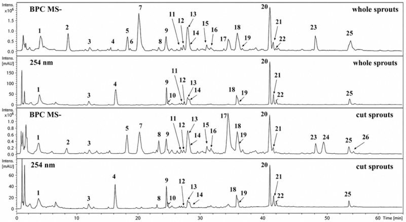
Figure 1. LC-PDA-MS fingerprint of extracts obtained from E. vesicaria subsp. sativa sprouts. Chromatographic traces were generated from PDA detection at 254 nm and from base peak intensity data recorded under negative electrospray ionization conditions. Peaks corresponding to the most abundant constituents are highlighted, and their characterization is presented in Table 3.
Applsci 16 07330 g001
Figure 2. Cytotoxic and morphological responses of BJ human fibroblasts exposed to E. vesicaria subsp. sativa extracts. Cell viability was quantified by MTT reduction after incubation with increasing concentrations of the extracts (0.97–500 µg/mL) for 24 h or 48 h. Graphs correspond to: (A) sprout extract after 24 h; (B) sprout extract after 48 h; (C) whole-seedling extract after 24 h; and (D) whole-seedling extract after 48 h. Results are expressed as mean ± SD from three independent experiments. Differences relative to untreated cells were analyzed using one-way ANOVA with Dunnett’s post hoc correction. Symbols indicate statistical significance versus control (* p < 0.05, ** p < 0.01, *** p < 0.001, **** p < 0.0001). (E) Representative Live/Dead fluorescence images obtained by CLSM following 24 h treatment with selected extract concentrations. Green fluorescence indicates viable cells with intact membranes, whereas red fluorescence identifies non-viable cells. Magnification: 100×; scale bar: 100 µm.
Applsci 16 07330 g002
Figure 3. Effect of ES1 and ES2 extracts derived from E. vesicaria subsp. sativa on erythrocyte membrane integrity. Hemolysis was determined after exposure to extract concentrations of 500, 31.25, and 0.97 µg/mL and is reported as a percentage of the response obtained for the positive control representing complete red blood cell lysis. Values represent mean ± SD of three independent replicates (n = 3). Between-group differences were evaluated by one-way analysis of variance with Tukey’s post hoc procedure. Statistically significant differences across experimental groups (p < 0.05) are indicated by asterisks.
Applsci 16 07330 g003
Figure 4. Representative micrograph of the cream formulation with ES1 [day 1 (a) and day 90 at 20 °C (b); 40× magnification]; scale bar = 50 µm.
Applsci 16 07330 g004
Figure 5. Droplet size distribution histogram for the cream containing ES1 [day 1 (a) and day 90 at 20 °C (b)].
Applsci 16 07330 g005
Figure 6. Scatter plot illustrates the relationship between the mean oil-phase droplet size and storage time for the cream with ES1 at 20 °C, with a fitted linear regression line (red).
Applsci 16 07330 g006
Figure 7. Flow behavior of the ES1 cream formulation at 20 °C after 1, 7, 14, 30, 60, and 90 days of storage.
Applsci 16 07330 g007
Figure 8. Viscosity curves of the ES1 cream formulation during storage at 20 °C, assessed after 1, 7, 14, 30, 60, and 90 days.
Applsci 16 07330 g008
Table 1. Composition of cosmetic creams.
NameAmount [%]
Oil phase
Glyceryl Stearate Citrate (GSC)5.0
Cetyl alcohol1.5
Shea butter1.0
Caprylic/Capric Triglyceride5.0
Isopropyl Myristate5.0
Isopropyl Palmitate3.0
Sodium polyacrylate1.0
Water phase
Propanediol2.0
WaterTo 100.0
Active ingredients phase
Eruca vesicaria subsp. sativa sprout extract (ES1 or ES2)4.0
Sweet Almond Oil2.0
Sodium Phytate0.1
Panthenol1.0
Vitamin E0.5
Phenoxyethanol/Ethylhexylglycerine1.0
Table 2. Concentrations of total phenolic compounds (TPC), total flavonoids (TFC), and total phenolic acids (TPAC) determined in E. vesicaria subsp. sativa sprout extracts (ES). Results are expressed as mg GAE/g DE, mg QE/g DE, and mg CAE/g DE, respectively, where GAE denotes gallic acid equivalents, QE quercetin equivalents, CAE caffeic acid equivalents, and DE dry extract. TPC and TFC values correspond to an extract concentration of 5 mg/mL; TPAC values correspond to 10 mg/mL. Data are presented as mean ± SD (n = 3).
SampleTotal Phenolic Content
[μg GAE/g DE]
Total Phenolic Acids
[μg CAE/g DE]
Total Flavonoid Content
[μg QE/g DE]
ES11046.5 ± 0.32483.8 ± 6.542.6 ± 0.3
ES21700.0 ± 0.31819.2 ± 3.8222.0 ± 0.4
Table 3. Analytical data used for the characterization of compounds present in E. vesicaria subsp. sativa sprout extracts, including UV absorption maxima, mass-to-charge ratios, fragmentation patterns, and tentative structural assignments obtained by LC-PDA-MS analysis.
Peak NumberProposed Identification of Detected CompoundRetention Time [min]UV-Vis [nm][M–H], m/zMS2 Ions, m/zClassificationRef.
1Glucoraphanin t4.2223, 268, 300436372b, 258, 194glucosinolate[16]
2undefined compound8.5227525445, 438b, 283, 242--
3Tryptophane t11.9216, 275203116bamino acid[17]
4undefined compound16.0250, 290290247, 226, 193b--
5undefined compound17.9226461445, 315b, 207, 152--
6undefined compound18.4230711693, 631b, 435, 306--
7Glucoerucin t19.8226420339, 275, 258bglucosinolate[16]
8undefined compound22.7228509375, 259b, 162--
9quercetin3,3′,4′-O-triglucoside t24.0255, 342787625b, 463, 301polyphenol[18]
10undefined compound24.2227419401, 272b, 254--
11undefined compound26.3226389343, 181b--
121-O-sinaponylglucose t26.7225, 328385247, 223b, 205polyphenol[18]
13undefined compound27.3270, 321456382, 247, 223b--
14undefined compound27.7267, 325625463b, 301--
15undefined compound30.2260, 326581419b, 371, 193--
16undefined compound30.9256, 320623548, 426, 389b, 341, 193--
174-methoxyglucobrassicin t33.5228477433, 299, 275, 259bglucosinolate[19]
18quercetin derivative34.9254, 353625463b, 301polyphenol-
19undefined compound35.2-307227b--
20quercetin derivative39.8261, 327933831b, 669, 463, 301polyphenol-
21kaempferol 3-[2′′-O-(E)-sinapoylsophoroside]-7-O-β-glucopyranoside or kaempferol 3-O-sophoroside-7-O-[2-O-(E)-sinapoyl-β-glucopyranoside] or
kaempferol 4′-[6-O-(E)-sinapoyl-β-glucopyranoside]-3,7-di-O-β-glucopyranosidet
40.4263, 321977815b, 653, 285polyphenol[18]
22flavonoid glycoside with sinapinic acid moiety40.8262, 3231007847b, 683, 477, 315, 223polyphenol-
23glucoalyssin46.9223450433, 399, 351, 286, 272b, 254glucosinolate[16]
241,4-dimetoxyglucobrassicin t48.1225506476bglucosinolate[20]
25quercetin glycoside sinapinic acid moiety52.1266, 324831669b, 463, 301, 223polyphenol-
26kaempferol 3-O-β-glucopyranoside-4′-O-(6′′′′′-O-sinapoyl-β-glucopyranoside) or kaempferol 3-O-(2′′′-O-sinapoyl-β-sophoroside) t52.7266, 323815653b, 447, 353, 285polyphenol[18]
t tentative identification based on cited literature; b—base peak (the most abundant ion in recorded spectrum); [M–H], deprotonated molecular ion; MS2, tandem mass spectrometry (product ion spectrum obtained by fragmentation of the precursor ion); m/z, mass-to-charge ratio; UV-Vis, ultraviolet-visible absorption spectrum.
Table 4. Antioxidant activities of E. vesicaria subsp. sativa sprout extracts (ES1 and ES2). Data were expressed as mean values ± SD.
SampleDPPH [μg Trolox/g DE]ABTS•+ [μg Trolox/g DE]CHEL
[μgNa2EDTA/g DE]
ES1505.7 ± 93.92454.6 ± 205.87589.8 ± 240.3
ES2477.8 ± 141.73440.4 ± 510.99572.4 ± 248.7
Table 5. Inhibitory effects of E. vesicaria sprout extracts on collagenase, elastase, and tyrosinase.
SampleCollagenase
Inhibition [%]
Elastase
Inhibition [%]
Tyrosinase
Inhibition
[mg KAE/g DE]
ES142.6 ± 1.133.7 ± 0.718.9 ± 0.0
ES239.8 ± 1.033.0 ± 0.654.9 ± 0.0
EGCG88.3 ± 2.267.1 ± 0.8nt
EGCG, epigallocatechin gallate; nt, not tested. Inhibition percentages correspond to the single extract concentration tested (5 mg/mL). Tyrosinase values are expressed as kojic acid equivalents (mg KAE/g dry extract).
Table 6. Selected physical parameters of the ES1-containing cream monitored during storage at 4 °C and 20 °C over a 90-day period.
Day 1 Day7 Day14 Day30 Day60 Day90 Day
Temp.20 °C4 °C20 °C4 °C20 °C4 °C20 °C4 °C20 °C4 °C20 °C
ɸ5.36
± 1.27
5.32
± 1.07
5.45
± 0.92
5.14
± 1.10
5.36
± 0.92
5.13
± 1.44
5.14
± 1.08
5.10
± 1.11
5.13
± 1.40
5.31
± 1.13
5.13
± 0.98
ɳ5s−133.45
± 0.57
32.97
± 0.95
33.45
± 0.95
33.40
± 1.16
33.57
± 1.14
32.78
± 1.13
34.79
± 2.22
33.78
± 1.18
33.98
± 0.89
33.11
± 0.17
33.04
± 1.00
K126.03
± 2.35
124.30
± 3.18
126.79
± 3.58
125.04
± 6.34
127.13
± 4.33
125.88
± 10.25
129.51
± 10.61
126.91
± 6.08
128.47
± 2.46
125.22
± 0.50
125.65
± 3.80
n [–]0.17
± 0.006
0.17
± 0.006
0.17
± 0.006
0.17
± 0.01
0.17
± 0.00
0.16
± 0.006
0.17
± 0.01
0.17
± 0.01
0.17
± 0.005
0.17
± 0.005
0.17
± 0.00
A 8939
± 232
8883
± 343
8850
± 659
8920
± 253
8563
± 565
9335
± 975
8572
± 546
9035
± 74
9383
± 613
8381
± 541
8486
± 570
Sf [×103]76.95
± 1.36
77.85
± 1.51
73.33
± 4.30
74.82
± 4.09
72.072
± 1.89
72.29
± 1.67
72.29
± 0.74
75.23
± 0.93
73.25
± 2.25
74.20
± 4.19
71.58
± 1.84
pH5.88
± 0.02
5.90
± 0.01
5.85
± 0.01
5.87
± 0.00
5.85
± 0.02
5.87
± 0.02
5.85
± 0.00
5.91
± 0.02
5.84
± 0.02
5.89
± 0.01
5.90
± 0.01
ɸ, mean oil droplet diameter [µm]; η5, viscosity at 5 s−1 [Pa·s]; K, Ostwald–de Waele consistency index [Pa·sn]; n, flow behavior index [–]; A, thixotropy area [Pa·s−1]; Sf, spreadability factor determined under a 1000 g load [g·cm2].
Table 7. Selected physical parameters of the ES2-containing cream monitored during storage at 4 °C and 20 °C over a 90-day period.
Day1 Day7 Day14 Day30 Day60 Day90 Day
Temp.20 °C4 °C20 °C4 °C20 °C4 °C20 °C4 °C20 °C4 °C20 °C
ɸ5.38
± 1.24
5.32
± 0.72
5.40
± 0.85
5.34
± 1.05
5.38
± 1.29
5.36
± 1.05
5.31
± 1.06
5.31
± 1.11
5.31
± 0.73
5.30
± 0.90
5.34
± 0.72
ɳ5s−130.96
± 0.31
30.21
± 1.80
30.65
± 1.21
30.79
± 0.43
31.45
± 2.25
30.98
± 2.06
32.77
± 1.71
29.89
± 1,13
30.87
± 1.03
30.99
± 1.32
30.67
± 1.66
K117.77
± 0.88
113.49
± 5.93
115.75
± 2.70
112.30
± 5.86
118.74
± 5.94
111.07
± 10.45
121.18
± 6.74
112.08
± 1.36
115.63
± 2.81
117.48
± 2.46
116.18
± 5.51
n [–]0.170
± 0.010
0.173
± 0.005
0.173
± 0.005
0.196
± 0.015
0.176
± 0.011
0.201
± 0.035
0.18
± 0.036
0.166
± 0.025
0.173
± 0.006
0.170
± 0.02
0.166
± 0.005
A8594
± 570
8699
± 545
9016
± 199
8624
± 40
8663
± 337
8811
± 162
8572
± 366
8786
± 383
8564
± 209
8407
± 53
8690
± 240
Sf [×103]73.27
± 0.47
72.27
± 1.86
74.68
± 1.93
72.93
± 2.13
72.18
± 2.68
72.38
± 0.96
71.59
± 5.18
71.66
± 1.58
71.78
± 1.34
72.89
± 2.38
73.14
± 0.82
pH5.85
± 0.02
5.80
± 0.03
5.84
± 0.00
5.82
± 0.00
5.85
± 0.03
5.82
± 0.02
5.85
± 0.02
5.86
± 0.02
5.84
± 0.02
5.83
± 0.03
5.80
± 0.02
ɸ, mean oil droplet diameter [µm]; η5, viscosity at 5 s−1 [Pa·s]; K, Ostwald–de Waele consistency index [Pa·sn]; n, flow behavior index [–]; A, thixotropy area [Pa·s−1]; Sf, spreadability factor determined under a 1000 g load [g·cm2].
Table 8. Results of the Friedman test and Kendall’s coefficient of concordance (W) for cream with ES1 and cream with ES2 stored at different temperatures.
SampleStorage Temp. [°C]χ2 (Chi-Square)dfp-ValueW
(Kendall)
Interpretation
ES147.09150.21390.014No significant differences; emulsion stable
ES12019.00050.00190.038Significant but weak differences; slight droplet
ES241.33850.93100.003No significant differences; highest stability
ES2201.97350.85290.004No significant differences; emulsion stable
Disclaimer/Publisher’s Note: The statements, opinions and data contained in all publications are solely those of the individual author(s) and contributor(s) and not of MDPI and/or the editor(s). MDPI and/or the editor(s) disclaim responsibility for any injury to people or property resulting from any ideas, methods, instructions or products referred to in the content.