Systematic Review

Antimicrobial Effect of Plasma-Treated Liquids on Skin and Oral Biofilms: A Systematic Review of In Vitro Studies


1
Department of Pharmacy, University “G. d’Annunzio” Chieti-Pescara, Via dei Vestini 31, 66100 Chieti, Italy
2
Department of Innovative Technologies in Medicine & Dentistry, University of Chieti-Pescara, Via dei Vestini 31, 66100 Chieti, Italy
3
Department of Clinical and Experimental Medicine, University of Foggia, 71122 Foggia, Italy
4
Industrial Safety Engineering, Faculty of Materials Science and Engineering, Gheorghe Asachi Technical University of Iași, 700050 Iași, Romania
5
Head & Neck Department, Fondazione Policlinico Universitario A. Gemelli IRCCS, Università Cattolica del Sacro Cuore, 00168 Rome, Italy
6
Philips Oral Healthcare, High Tech Campus 34, 5656 AE Eindhoven, The Netherlands
7
Division of Oral Biosciences, Dublin Dental University Hospital, School of Dental Science, Trinity College Dublin, D02 F859 Dublin, Ireland
8
Department of Life Sciences, Health and Health Professions, Link Campus University, Via del Casale Di San Pio V 44, 00165 Rome, Italy
9
UdA-TechLab, Research Center, University “G. d’Annunzio” of Chieti-Pescara, 66100 Chieti, Italy
*
Author to whom correspondence should be addressed.
Appl. Sci. 2026, 16(14), 7187; https://doi.org/10.3390/app16147187
Submission received: 10 June 2026 / Revised: 7 July 2026 / Accepted: 10 July 2026 / Published: 17 July 2026

Featured Application

This review highlights the potential of indirect cold atmospheric plasma as an antimicrobial approach against oral and skin biofilms and provides a framework for the standardization of experimental protocols and the translation of plasma-based technologies into clinical practice.

Abstract

This systematic review aimed to evaluate the effectiveness of indirect cold atmospheric plasma (CAP) in disrupting oral and skin microbial biofilms and to investigate whether the reported effects are mainly attributable to acidic pH, plasma-activated liquids, or specific devices and treatment settings. The review followed PRISMA guidelines and was registered on the Open Science Framework. A comprehensive literature search was conducted across major electronic databases. Studies evaluating indirect CAP on in vitro oral and skin microbial biofilms were included. Comparisons were made with untreated controls, non-activated liquids, and conventional antimicrobials. Outcomes were evaluated based on reductions in biofilm viability or biomass using quantitative assays (e.g., CFU, XTT, and Crystal Violet). Thirteen in vitro studies were included. CFU-based outcomes were reported in 11 of 13 studies (84.61%), and all of these studies showed reductions exceeding 5–6 log under specific conditions. Antimicrobial efficacy varied according to exposure time, microbial species, plasma parameters, and treatment settings, while physicochemical analyses indicated that acidic conditions and plasma-activated liquids play a key role. Indirect CAP appears to be a promising non-invasive antimicrobial strategy for managing oral and skin biofilms. However, variability among devices and settings, together with the absence of clinical data, underscores the need for further standardized studies and clinical investigations.

1. Introduction

Plasma, defined as the fourth state of matter, is an electrically neutral ionized gas that acts on the target as a multimodal therapy through the generation of a complex mixture of reactive oxygen and nitrogen species (ROS and RONS), excited molecules, charged particles, chemically reactive neutral particles, and ultraviolet (UV) radiation [1]. The composition of plasma reactive components depends on the type of source employed and the specific application, as well as on the applied operational conditions and parameters. Cold atmospheric plasma (CAP) is also referred to as non-thermal plasma (NTP), as it releases only heavy particles at near-ambient temperature, with an application temperature below 40 °C, a characteristic that enables its safe use on biological tissues [2]. Regarding these properties, CAP has attracted increasing attention for biomedical applications, particularly for its antimicrobial activity and suitability for use on human tissues without inducing thermal damage [3,4]. In recent years, CAP has also demonstrated strong potential in disrupting microbial biofilms (structured communities known for their enhanced resistance to conventional antimicrobial treatments [3,4,5] including antibiotics) supporting its application in skin and oral healthcare. A summary of the main acronyms used throughout this manuscript is provided in Table 1.
The use of plasma can be approached in two main ways: (i) direct application, where the plasma is in contact with or remotely activated on the target; and (ii) indirect application, where plasma effects are delivered through intermediaries such as plasma-treated liquids (PTLs), hydrogels, or gases [6]. For example, one of the liquids that can be used for activation through an indirect plasma methodology is water, known as plasma-activated water (PAW), which can be applied in various fields such as surface disinfection, enhancement of seed germination, and food decontamination [7]. For disinfection applications, the ROS generated in PAW, including hydrogen peroxide, hydroxyl radicals, and ozone, act as strong oxidizing agents capable of inducing oxidative stress on microbial cell membranes. In the present review, the term PTLs is used as the umbrella term for plasma-activated liquid media. However, when describing individual studies, the original terminology adopted by the respective authors (e.g., plasma-activated water (PAW) or plasma-treated water (PTW)) has been retained. The ROS produced can disrupt the intramolecular bonds of peptidoglycan by removing a hydrogen atom from peptide bonds within the peptidoglycan layer located outside the plasma membrane of most bacteria. This can lead to cell wall disruption, metabolic dysfunction, structural damage, and cell death [8].
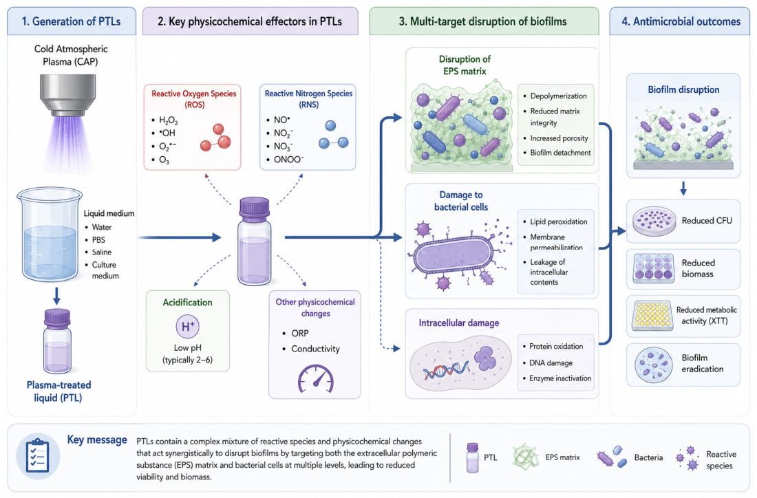
The indirect method, especially the use of PTLs, has significantly advanced biomedical applications by enabling gentler and minimally invasive treatments. It allows the targeting of internal tissues via injection or catheter, avoiding surgical procedures and reducing patient risk [6]. Among indirect methods, PTLs have shown strong promise as a tool for bacterial inactivation, as they have demonstrated effectiveness against a wide range of pathogens, including bacteria, fungi, and viruses [9,10]. They are generated by exposing water solutions to plasma, producing RONS that contribute to their antimicrobial properties. PTLs have attracted growing interest in fields such skin and oral medicine [6]. Given that plasma readily interacts with culture media in vitro and with body fluids, such as the moisture or exudate present in wounded tissues, during in vivo conditions, these plasma–liquid interactions are believed to play a significant role in shaping the observed biological effects [4]. Plasma-based bacterial inactivation presents several advantages over conventional disinfection methods, including operation at low temperatures, reduced toxicity, high efficiency, and cost-effectiveness [11]. As a result, various newly developed plasma systems have been extensively studied for their antimicrobial potential. Indeed, PTLs become enriched with a cocktail of RONS such as hydrogen peroxide, hydroxyl radicals, nitrites, and nitrates. When applied to microbial cells, these reactive species cause oxidative damage to vital biomolecules, including lipids, proteins, and nucleic acids, leading to membrane disruption, enzyme inactivation, and genetic damage. This multi-target attack overwhelms microbial defense systems and significantly lowers the likelihood of resistance development. Schnabel et al. [12] highlighted that the reactive species generated during plasma exposure play a central role in bacterial inactivation. Among these, ROS are particularly important, as they can attack critical structural components such as membrane lipids, leading to lipid peroxidation, as well as peptidoglycan layers in bacterial cell walls, and can oxidize membrane-bound proteins, disrupting ion transport and enzymatic activity. Moreover, RONS can penetrate the cell and cause direct damage to nucleic acids, leading to DNA strand breaks [10]. Unlike conventional antimicrobial methods, plasma treatments offer the advantage of effective sterilization and can be used on sensitive surfaces or biological tissues [13]. Moreover, plasma’s multi-targeted mode of action reduces the likelihood of microbial resistance making it a promising alternative to traditional chemical disinfectant and showing effectiveness to antibiotic-resistant strains [14]. These properties have led to plasma being explored for applications of dental care and medical device sterilization [9]. Despite promising results from these studies, the current body of research remains insufficient to fully define practical application of plasma-based antibacterial treatments, particularly in dental and skin settings. Figure 1 summarizes the current evidence regarding PTLs generation, the main physicochemical effectors, and their proposed multi-target mechanisms involved in biofilm disruption.
Therefore, further investigation is essential. In this study, we systematically reviewed the literature to critically assess the effectiveness of CAP in disruption of in vitro oral and skin microbial biofilms, and to explore whether specific plasma devices and operational settings are associated with greater antimicrobial efficacy.

2. Materials and Methods

This systematic review was performed according to the guidelines of the Preferred Reporting Items for Systematic Reviews and Meta-analyses (PRISMA) statement [15]. The PRISMA checklist can be found in Supplementary Material File S1. The protocol was registered on Open Science Framework (OSF) at the following link: https://doi.org/10.17605/OSF.IO/G67MF.

2.1. Search Methods

An electronic search on PubMed, Scopus, EMBASE and Web of Science was performed to identify suitable studies, using the following terms and keywords alone or in combination: (“Plasma Gases”[Mesh] OR “plasma medicine” OR “cold plasma” OR “cold atmospheric plasma” OR “cold atmospheric-pressure plasma” OR “cold physical plasma” OR “cap” OR “cold argon plasma” OR “air plasma” OR “non-equilibrium plasma” OR “non-thermal plasma” OR “non-thermal atmospheric pressure plasma” OR “low-temperature plasma” OR “dielectric-barrier discharges” OR “barrier discharges” OR “glow discharge” OR “plasma jet” OR “kinpen med” OR “plasma gases” OR pam OR ptws OR paw OR ptl OR “plasma activated liquid*” OR “plasma activated water” OR “plasma activated medi*” OR “plasma treated liquid*” OR “plasma treated water*” OR “plasma treated medi*”) AND (biofilm* OR “oral biofilm*” OR “dental biofilm” OR microrganism* OR “skin microorganism*” OR “skin bacteria” OR “oral microorganism*” OR “Dental Deposits”[Mesh] OR “dental microorganism*” OR “biofilm colonization*” OR “dental plaque” OR “dental deposit*” OR “materia alba” OR “oral microbio*” OR “oral bacteria” OR “dental bacteria”). In addition to database searches, the gray literature was searched via Bielefeld Academic Search Engine (BASE) and the Gray Literature Report (The New York Academy of Medicine) to capture potentially relevant unpublished or non-peer-reviewed material. The first search was performed on 10 June 2024. The last electronic search was performed on 8 August 2025. In addition, the reference lists of the studies included after full-text assessment were manually screened to identify potentially eligible studies not retrieved through the electronic database searches. A reference manager software program (Mendeley Desktop version 1.19.8) was used, and the duplicates were discarded first electronically, then by checking the resulting list manually. The search strategy, adapted for PubMed, Web of Science, Embase and Scopus is shown in Supplemental Material Table S1.

2.2. Focused PICO Question

A focused research question was formulated: what are the microbiological, physico-chemical and in vitro biological effects of indirect CAP application on in vitro bacterial cultures of skin and oral species? P (Population): In vitro bacterial cultures of skin and oral species. I (Intervention): Indirect CAP treatment. C (Comparison): Control groups (untreated bacteria, non-activated liquids, conventional antimicrobials). O (Outcome): Reductions in bacterial viability and biofilm biomass (e.g., CFU counts, XTT assay, CV assay), physicochemical changes (e.g., pH and reactive species) and reported cytotoxicity effects where available.

2.3. Selection Criteria

2.3.1. Inclusion Criteria

Studies were included if they met the following conditions: in vitro studies investigating the indirect application of CAP (PTLs such as saline, phosphate-buffered saline, culture media) on skin and/or oral microorganism forming biofilms; studies including control groups (e.g., untreated microorganisms, microorganisms exposed to non-activated liquids or conventional antimicrobial agents); studies assessing microbiological effects (e.g., bacterial viability, biofilm reduction, metabolic activity) and physicochemical properties (e.g., pH, feeding gas, frequency, voltage). All relevant studies published in English were included without any time restrictions.

2.3.2. Exclusion Criteria

Studies were excluded if they met any of the following conditions: studies on microbial species not relevant to skin or oral microbiomes; proceedings, abstracts, short communications, systematic reviews and meta-analyses, ecologic studies, ex vivo studies, or in vivo studies; studies investigating only direct CAP application (i.e., plasma directly applied to bacterial cultures); studies on plasma indirect application for agricultural use or other industrial applications; studies lacking a control group or comparative analysis or quantitative data; studies not measuring antimicrobial effects, physicochemical properties, or safety aspects.

2.3.3. Selection of Studies

Regarding data extraction, the reviewers (G.S. and G.M.) first conducted a calibration exercise on a subset of studies to standardize the criteria for data inclusion and exclusion and to ensure consistency. Retrieved citations were independently screened by two authors (G.S. and G.M.), and relevant studies were identified based on titles and abstracts. When these did not provide sufficient information regarding the inclusion criteria, full texts were assessed to determine eligibility. Missing or unclear data were handled by contacting the corresponding authors or, when unavailable, by reporting the data as missing. Any disagreement was solved by discussion, and a third reviewer was consulted to make final decisions (V.P.). This author also calculated a Cohen’s kappa to ascertain the level of reviewers’ agreement.

2.3.4. Data Collection and Data of Analysis

Two reviewers (G.S. and G.M.) independently extracted data from all the included studies using a predesigned extraction form (Microsoft Excel 2020, Microsoft Corporation, Redmond, WA, USA) for data collection and descriptive analysis. Extraction sheets were organized to collect:
Microbiological Outcomes: Viability (in vitro survival rates after exposure to indirect CAP); biofilm formation and reduction (quantification of biofilm biomass and metabolic activity); susceptibility differences between Gram-positive and Gram-negative bacteria. Physicochemical Outcomes: Chemical composition of PTLs; pH variations before and after exposure to indirect CAP application; temperature of PTLs.
A quantitative synthesis or meta-analysis was not attempted due to substantial methodological heterogeneity across studies, including differences in CAP devices, PTL types, exposure times, outcome measures and biofilm quantification methods.

2.4. Risk-of-Bias Assessment

Two reviewers (G.B. and V.P.) assessed the methodological quality of the included studies using a risk-of-bias assessment adapted from Perrotti et al. [16]. Six items were considered: condition of cell culture before experimentation; condition of cell culture during treatment; description of methodology to evaluate outcomes; case–control description; multiple experiments performed; descriptions of plasma settings and devices. Each item was scored as “✓” when the criterion was fulfilled, “–” when the criterion was not fulfilled, “U” when the information reported in the article was unclear, and “N/A” when the item was not applicable. Studies fulfilling at least five of the six criteria were classified as having a low risk of bias, studies fulfilling three or four criteria were classified as having a moderate risk of bias, and studies fulfilling two or fewer criteria were classified as having a high risk of bias. The methodological quality assessment is reported in Supplementary Table S2.

3. Results

3.1. Study Selection

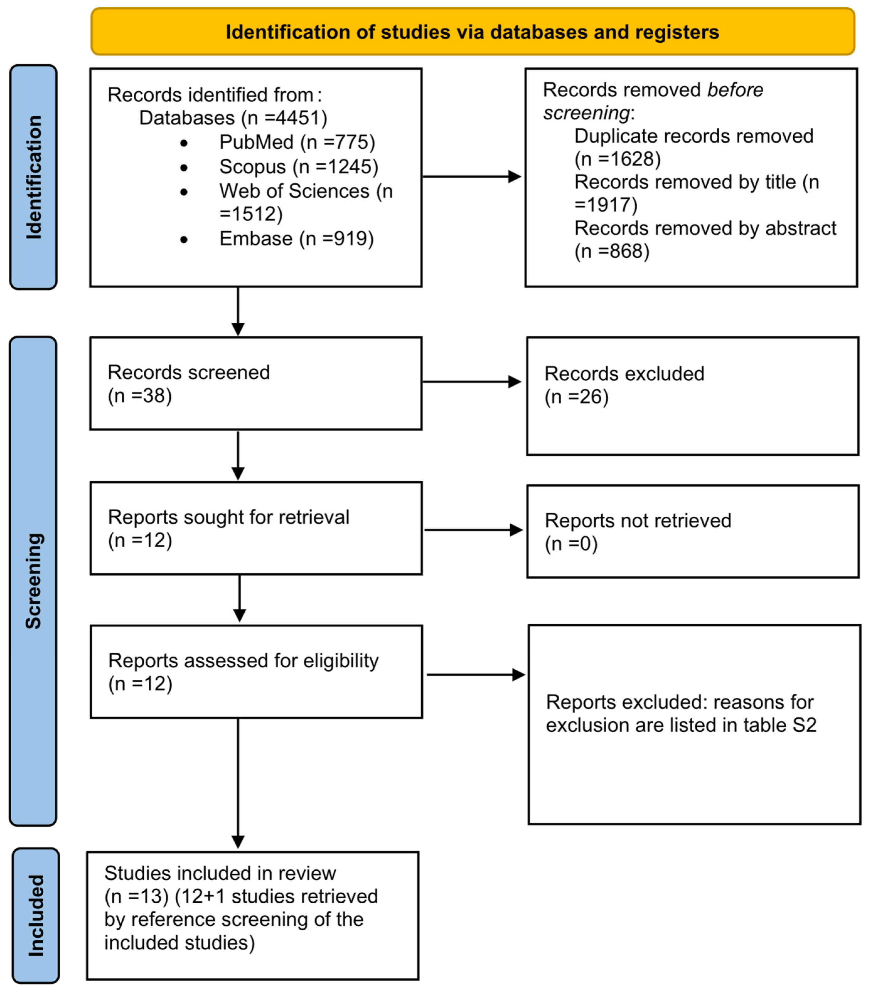
For this review 4451 potentially relevant records from the databases were analyzed. After the removal of duplicates, the articles were screened based on their titles and abstracts, and 4413 articles were excluded. A total of 38 full-text articles were evaluated, and 26 [17,18,19,20,21,22,23,24,25,26,27,28,29,30,31,32,33,34,35,36,37,38,39,40,41,42] were subsequently excluded for the reasons listed in Supplemental Table S3. One additional study was identified by screening the reference lists of the included studies and was subsequently included in the present review [43]. Therefore, a total of 13 studies [3,9,12,43,44,45,46,47,48,49,50,51,52] fulfilled the selection criteria of the present review and were included in qualitative analyses (Figure 2). The value of the k-statistic was 0.95 which indicated an excellent agreement between the two reviewers.

3.2. Descriptions of Included Studies

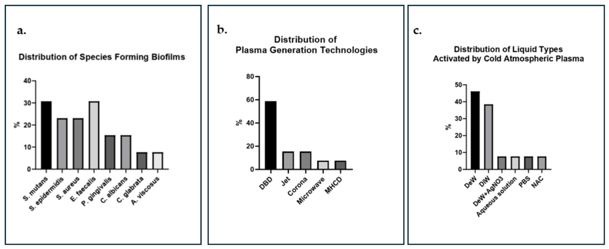
Thirteen [3,9,12,43,44,45,46,47,48,49,50,51,52] in vitro studies published between 2013 and 2025, which investigated the use of indirect cold plasma application for treatment of liquids targeting skin- and oral-associated bacterial and fungal species were included in the present review. The targeted biofilms encompassed a diverse microbiological range, as shown in Figure 3a. Streptococcus mutans was examined in four studies [43,45,46,47], Staphylococcus aureus in three [3,51,52] and Enterococcus faecalis in four [3,9,49,50]. Porphyromonas gingivalis was assessed in two studies [45,47], Staphylococcus epidermidis in three [3,44,48] and Candida albicans in two [3,12]. In contrast, only one study each focused on Actinomyces viscosus [45], and Candida glabrata [3]. Biofilm age at the time of treatment varied considerably ranging from 12 h [43,45,51] to 3 weeks [49]. Plasma exposure durations also differed significantly between studies, from a few seconds to several hours, depending on the specific experimental design and the microorganisms.

3.3. Results of Analysis

3.3.1. Plasma Device Types and Application Settings

The plasma-generating technologies varied across studies. Table 2 summarizes the operational settings of plasma devices employed across the included studies. Specifically, seven studies [3,9,43,46,47,49,50] used dielectric barrier discharge (DBD) devices; two [45,48] utilized jet system (micro-jet plasma); two studies [44,51] applied corona discharge-based sources, including a corona discharge air plasma source and a capillary-guided corona discharge (CGCD); one study [12] employed a microwave-driven discharge device; and finally one study [52] used a micro-hollow cathode discharge (MHCD) system. The distribution of plasma device types employed in the included studies is illustrated in Figure 3b.
Concerning the feeding gases, four studies [3,12,44,51] utilized atmospheric air, two [47,52] employed atmospheric air/oxygen, three [9,43,50] applied compressed air, one [21] used pure argon, one [45] employed a mixture of argon/oxygen, another used a helium/nitrogen mixture [46], and one study [49] utilized pure helium. Plasma protocols varied significantly across studies, including differences in setup characteristics. The pulse frequency ranged from 500 Hz [3] to 2.45 GHz [12]. The treatment distance ranged from 2 mm [3] to 10 mm [9,43,51], while exposure times varied from 6 s [50] to 30 min [44,52]. Voltage application was reported in 10 studies [3,9,44,45,46,47,48,50,51,52], with values ranging from 12 V [50] to 25 kV [9]. Power values were provided in seven studies [3,9,12,47,48,49,52], ranging from 0.2 W [3] to 10 kW [48]. Gas flow rates were reported in eight articles [3,9,12,43,45,47,49,52] and ranged from 0.5 L/min [49] to 260 L/h [9,43].

3.3.2. Characteristics of Plasma-Treated Liquids

Across the 13 included studies [3,9,12,43,44,45,46,47,48,49,50,51,52], various types of PTLs were employed, with notable heterogeneity regarding liquid composition, treated volume, and resulting chemical species. Deionized water was employed in six articles [3,12,44,47,51,52], with activation volumes ranging from 200 µL [47] to 50 mL [48,52]. Sterile distilled water was used in five studies [9,43,45,46,49], with reported volumes between 1 mL [3,44,46] and 20 mL [43]. In the remaining four articles, the following five liquids were used: silver nitrate aqueous solution (composed of deionized water and AgNO3) [48], a mixed aqueous solution (containing PBS, NaCl, Span 60, Tween 80, and PEG-4000) [50], PBS [3], and N-acetylcysteine solution [3]. The physicochemical characteristics of the plasma-treated liquids, including pH and reactive species composition, are detailed in Table 3.
Time of liquids activation by CAP varied between 6 s [50] and 30 min, [44,52] depending on the experimental design and energy input. In all reviewed articles, the chemical composition of the reactive species generated in the liquid phase following plasma activation was reported. The most detected species included hydrogen peroxide (H2O2), nitrate (NO3), and nitrite (NO2). Specifically, H2O2 was described in 12 studies [3,12,43,44,45,46,47,48,49,50,51,52], NO2 and NO3 in at least five studies [9,43,47,51,52], hydroxyl radicals (·OH) in five studies [43,45,48,51,52], ozone (O3) in five studies [44,48,49,51,52], and singlet oxygen (1O2) [48,49,50], nitric oxide (NO) [12,46,48,49,50,51,52], superoxide (O2) [9,43,46,51,52], and peroxynitrite (ONOO) [43,51] were variably reported across studies. An overview is presented in Figure 3c. All studies that conducted chemical analysis reported the presence of both ROS and RONS.

3.3.3. Characteristics of Biofilms

The biofilms developed to test the antibacterial properties of the PTLs greatly differed among studies. Several investigations have focused on the formation and treatment of monomicrobial biofilms using various bacterial and fungal species, with differences in exposure time and biofilm age as shown in Table 4a,b. Jia et al. [47] studied biofilms of Streptococcus mutans ATCC 25175 and Porphyromonas gingivalis ATCC 33277 after 24 h of growth, exposing them to treatments for 10 min. Similarly, Tawfeeq et al. [48] evaluated Staphylococcus epidermidis biofilms grown for 24 h and treated for the same duration. Arguello-Sànchez et al. [49] and Zhu et al. [50] focused on Enterococcus faecalis ATCC 29212 biofilms matured over 3 weeks and 7 days, respectively, applying short treatments of 5 min. Other studies, such as Qiao et al. [43] and Schnabel et al. [12] examined biofilms of Streptococcus mutans UA159 and Candida albicans SC5314, respectively, with biofilm ages ranging from 12 to 48 h and treatments lasting 5 to 20 min. Xu et al. [51] and Chen et al. [52] evaluated Staphylococcus aureus biofilms with exposure times from 0 to 180 min, whereas Hozàk et al. [44] studied Staphylococcus epidermidis DBM 3179 biofilms treated for 60 min. Li et al. [45] investigated single-species biofilms of Streptococcus mutans UA159, Porphyromonas gingivalis ATCC 33277, and Actinomyces viscosus ATCC 19246, formed between 12 and 72 h and subjected to short treatments of 10 to 120 s. Pan et al. [9] and Tasaki et al. [46] treated Enterococcus faecalis ATCC 29212 and Streptococcus mutans ATCC 25175 biofilms matured for 5 and 7 days, respectively, with exposures ranging from seconds to minutes. Ercan et al. [3] evaluated several microorganisms including Staphylococcus aureus ATCC 25923, Staphylococcus epidermidis ATCC 12228, Candida albicans, and Candida glabrata, all with 24 h biofilms subjected to treatments lasting 1 to 15 min. Regarding the tested species forming biofilms Streptococcus mutans [43,45,46,47], Staphylococcus epidermidis [3,44,48], Enterococcus faecalis [3,9,49,50], and Candida albicans [3,12] were among the most frequently studied microorganisms. Biofilm growth times also differed: PTLs were applied to Streptococcus mutans [43,47], Staphylococcus epidermidis [3,44], and Candida albicans [3,12] after 24 h of biofilm growth, whereas PTLs were applied to Enterococcus faecalis [49,50] biofilms formed over 3 weeks and 7 days, respectively. Therefore, PTLs were applied to biofilms of varying ages, from early formation to mature states, with the aim of observing changes in biomass and cell morphology. Exposure times varied widely among studies, ranging from a few seconds [45,46] to as long as 24 h [48].

3.3.4. Colony Forming Units

The results reported in 11 [3,9,12,43,44,45,47,49,51,52] out of 13 [3,9,12,43,44,45,46,47,48,49,50,51,52] studies (84.61%) indicated a reduction in terms of CFU/mL, in biofilms treated with indirect plasma. However, no standardized threshold for “effectiveness” (e.g., a 6-log reduction) could be defined due to variability in experimental conditions. In many cases, reductions above 6 log were observed, as evidenced by Jia et al. [47] where the biofilms of Streptococcus mutans and Porphyromonas gingivalis, developed on different substrates (silicone, dental veneers), showed a reduction of up to 6.3 log in the samples exposed to PAW activated by O2 -DBD. Focusing on Steptococcus mutans biofilms, several studies have shown a direct correlation between exposure time and the extent of reduction in terms of CFU. Specifically, Qiao et al. [43] reported a drastic reduction of about 7–8 log after only 10 min of treatment on mature biofilms aged 48 h. Similarly, Li et al. [45] observed a reduction of 5 log already with an exposure of 60 s. Tasaki et al. [46] on the other hand, obtained a reduction of up to 6 log with even shorter treatment times, between 10 and 30 s, on 7-day mature biofilms, confirming a rapid and marked effect even in conditions of greater structural complexity. These results are consistent with those reported by Jia et al. [47] where different conditions of exposure to PAW (both Air-DBD and O2 -DBD) on surfaces such as silicone and dental veneers resulted in reductions between 3.6 and 6.3 log. Similarly, Enterococcus faecalis showed a reduction of up to 6 log after 5 min of treatment [9,49]. Studies on Staphylococcus aureus and Staphylococcus epidermidis [44,51] confirm this trend, with reductions ranging from 2.24 to 6 log, depending on the duration and type of plasma activation. Overall, substantial reductions in CFU were consistently observed; however, the level of reduction varied depending on factors such as exposure time, microbial species, and plasma generation mode. No consensus on the exposure time required to achieve a specific level of CFU reduction was identified, as comparable reductions were reported across a broad range of treatment durations. It is important to emphasize, as shown in Table 3, that the pH values of PTLs are often quite low, ranging from 2.2 [47] to 3.8 [51]. Therefore, biofilm treatment with PTLs generally occurred in a strongly acidic environment, except in two studies, Arguello-Sánchez et al. [49] and Tasaki et al. [46], which reported pH values of 4.3 and 6.5, respectively.

3.3.5. Metabolic Activity

Of the 13 studies [3,9,12,43,44,45,46,47,48,49,50,51,52] included in the review, only three articles [3,44,51] evaluated the metabolic activity of plasma-activated fluid-treated biofilms using colorimetric assays. Specifically, the study by Ercan et al. [3] employed the XTT assay [2,3-bis(2-methoxy-4-nitro-5-sulfophenyl)-2H-tetrazolium-5-carboxanilide], while Hozàk et al. [43] and Xu et al. [51] used assays based on the reduction of fluorescent dyes such as resazurin and MTT. Results showed a reduction in metabolic activity ranging from 20% to 100% compared with untreated controls, depending on the microorganism, type of activated fluid and the biofilm treatment time. In the study by Ercan et al. [3] where biofilms of different microbial species were analyzed, it was reported that 24 h biofilms of Staphylococcus epidermidis treated for 1, 2 and 3 min exhibited reductions in metabolic activity of 40%, 98% and 100%, respectively, while for Enterococcus faecalis, the observed reduction ranged from 45% to 100%. In this study, therefore, it can be inferred that greater efficacy correlates with increased treatment time, although this dependency was not detected when treating Candida albicans and Candida glabrata biofilms where the reduction ranged from 40% to 80%. In contrast, Hozàk et al. [44] reported a 20% increase in metabolic activity in Staphylococcus epidermidis (considered not statistically significant), indicating possible stimulant effects or limitations of the adopted protocol. Since the values of metabolic activity were expressed in a heterogeneous manner (optical density or percentage) across the different studies, the data was standardized by converting optical densities into percentages to enable comparison among studies. These results underline the importance of adopting standardized protocols, including the use of uniform methods for measuring metabolic activity, comparable exposure times and the specification of the type of activated fluid.

3.3.6. Biomass

Only three studies [44,48,51] included in the review evaluated the effect of plasma-activated liquids on microbial biofilm biomass and all of them used CV staining. However, the biomass reduction values varied widely among studies, with reported decreases ranging between 21% and 68% compared to the control. This variability seems to depend on several factors, such as the microbial species involved when monospecies biofilms were evaluated or multi-species composition of the biofilms, the parameters of plasma activation, the type of liquid used, the surface colonized, the biofilm age and the media used—which affect the concentration of reactive oxygen and nitrogen species generated—and the duration of exposure to activated liquid. Tawfeeq et al. [48] reported a biomass reduction of 68.69% after 24 h treatment of 24 h-aged Staphylococcus epidermidis biofilm. In contrast, Xu et al. [51] highlighted more marked and variable effects depending on biofilm treatment time, reporting that DeW activated for 30 min and used with an exposure time of 60 min induced a reduction of 21.81%, while DeW activated for 60 min and exposed for 30 min resulted in a greater reduction of Staphylococcus aureus biofilm biomass (30.90%). Conversely, Hozàk et al. [44] reported that a 24 h Staphylococcus epidermidis biofilm treated for 60 min time showed an increase in biomass of about 60%. In conclusion, it can be stated that a greater number of studies using homogeneous methodologies is necessary to precisely define the efficacy of plasma-activated liquids in reducing biofilm biomass, to support their potential clinical or industrial application.

3.4. Methodological Quality Assessment

Overall, the methodological quality of the included in vitro studies was generally high. Nine of the 13 studies (69.2%) fulfilled all six predefined internal validity criteria (IT1–IT6), corresponding to a 100% “yes” response rate and indicating a low risk of bias [12,43,44,45,46,49,50,51,52]. Three studies (23.1%) fulfilled five of the six criteria, corresponding to an 83.3% “yes” response rate, largely due to unclear or missing information on a single item, and were therefore classified as having a low risk of bias according to the predefined threshold [3,9,47]. Tawfeeq et al. [48] fulfilled four of the six criteria, corresponding to a 66.7% “yes” response rate, and was therefore classified as having a moderate risk of bias. Overall, 12 of the 13 studies (92.3%) were classified as having a low risk of bias, while one study (7.7%) was classified as having a moderate risk of bias. The updated risk-of-bias assessment is reported in Supplementary Table S2.

4. Discussion

In relation to the PICO questions, this review synthesizes evidence on the microbiological, physicochemical and safety effects of indirect CAP application through PTLs in in vitro models of skin- and oral-associated bacterial cultures. According to the included studies, PTLs consistently demonstrated antimicrobial activity and biofilm reduction compared with untreated controls, non-activated liquids or conventional antimicrobial agents, while inducing measurable physicochemical changes.
Among the physicochemical effects reported, acidification emerged as a consistently observed outcome following plasma activation. Four studies did not report pH values [12,44,48,50]. Amond the studies reporting pH values, seven [3,9,43,45,47,51,52] out of 10 [3,9,43,44,45,46,47,49,51,52] (70%) exhibited low pH (approximately pH 2–4). Overall, several studies documented a marked decrease in pH ranging from 2.2 [9] to 4.3 [49], supporting a potential role of low pH in enhancing antimicrobial efficacy in vitro. Experimental evidence indicates that lower pH conditions improved bacterial inactivation and biofilm reduction compared with neutral or buffered conditions [9]. However, acidification alone was not sufficient to fully account for microbial inactivation, as two [46,49] out of 10 studies (20%) reporting pH values demonstrated antimicrobial effects under non-acidic or less acidic conditions. This suggests that pH acts as a contributing factor rather than a sole determinant of PTL efficacy. Tasaki et al. [46] buffered PTW samples to pH 3.5 and pH 6.5 using a 200 mM citrate-Na buffer and showed that lower pH (3.5) improved PAW efficacy against Streptococcus mutans, highlighting a pH-dependent effect. With respect to safety and translational relevance, the acidic conditions associated with PTLs raise important considerations, particularly for oral applications [53,54]. While enhanced antimicrobial effects were observed at low pH values, several studies highlighted potential limitations related to biocompatibility.
Given that saliva maintains a neutral pH [55], the clinical applicability of PTLs may depend on achieving an appropriate balance between antimicrobial activity and tissue compatibility. These findings underscore the importance of optimizing PTLs formulations and exposure conditions to ensure both efficacy and safety.
An additional translational issue concerns the delivery mode of PTLs. Although most studies have investigated PTLs as free liquids, direct topical application may be limited in clinically dynamic environments, particularly in exudative wounds and in the oral cavity, where saliva, crevicular fluid, swallowing and mechanical forces may reduce local residence time and dilute the active liquid phase.
Biomaterial-based delivery platforms, including hydrogels, flexible bio-dressings, mucoadhesive films, and polymeric scaffolds, may help address these limitations by improving local retention and enabling more sustained or spatially controlled exposure of biofilm-colonized tissues to PTL-derived or plasma-generated RONS. Plasma-treated alginate hydrogels have been shown to generate, confine, and release H2O2, NO2, and short-lived RONS, supporting their potential as local RONS delivery vehicles [56]. Similarly, thermosensitive methylcellulose hydrogels and plasma-activated hydrogels based on hydroxyethyl cellulose, carbomer, or acryloyldimethylammonium taurate/vinylpyrrolidone copolymer have demonstrated the ability to store and gradually release plasma-generated reactive species, thereby addressing some of the limitations associated with conventional plasma-treated liquids [57,58].
Flexible hydrogel-based bio-dressings are particularly relevant for wound-related applications, where conformability, hydration, and interaction with wound exudate may be advantageous. CAP-activated poly-vinyl alcohol (PVA)/poly-acrylic acid (PAA) composite hydrogels have been proposed as platforms for concomitant delivery of antimicrobial agents and CAP-generated molecules, with activity against planktonic bacteria and biofilm models Plasma-activated PVA/PAA hydrogel dressings have also shown enhanced generation of H2O2 and reactive nitrogen species and antimicrobial activity in the context of wound decontamination [59].
Recent evidence from chronic wound models further supports the potential of hydrogel-based systems as clinically relevant carriers for plasma-derived reactive species. In particular, sprayable thermosensitive hydrogels loaded with low-temperature plasma-generated oxidizing species have demonstrated broad-spectrum antibacterial activity and accelerated wound healing in infected and diabetic wound models. These effects were associated with enhanced extracellular matrix remodeling, collagen regeneration, angiogenesis and epithelialization. Although these findings derive from skin-wound models rather than oral biofilms, they reinforce the concept that biomaterial-assisted delivery may improve the local retention, handling, and therapeutic performance of plasma-treated formulations on irregular biological surfaces [60].
Moreover, polymeric scaffolds may offer an additional strategy when antimicrobial activity must be combined with structural support for tissue repair. In this regard, gelatin-based scaffolds designed for controlled CAP-derived activity have been reported to accelerate wound healing through modulation of the local immune microenvironment [61].
For oral, periodontal, and peri-implant applications, mucoadhesive or thermoresponsive systems may be especially relevant because liquid formulations are rapidly displaced in the moist oral environment. Hydrogel-based carriers are already recognized as local drug-delivery and regenerative platforms for oral mucosal diseases, wounds, periodontitis, and oral tissue engineering [62]. In this context, the recent combination of CAP with nanogel-based pharmaceutical platforms in oral ulcer models further supports the translational relevance of biomaterial-assisted plasma delivery within the oral cavity. CAP integrated with gelatin–chitosan nanoparticles has been shown to enhance oral ulcer repair by modulating inflammatory responses, activating EGFR-related regenerative pathways, and promoting fibroblast-mediated tissue regeneration, while maintaining a favorable biocompatibility profile in both cellular and animal models [63]. Nevertheless, the translation of PTL-loaded or plasma-activated hydrogels, flexible bio-dressings, mucoadhesive films, and polymeric scaffolds remains at an early stage. Future studies should define polymer compatibility, RONS stability, pH and osmolarity, release kinetics, cytocompatibility, and antibiofilm efficacy under clinically relevant saliva- or exudate-rich multi-species biofilm conditions.
A major challenge in synthesizing the available evidence arises from the substantial methodological heterogeneity among the included studies. Differences were observed in the microbial species investigated, biofilm maturity, PTL composition, pH values, exposure times, and experimental endpoints. This heterogeneity limits direct comparison across studies and precludes quantitative synthesis of outcomes. Moreover, the reliance on simplified in vitro models reduces external validity, particularly in relation to complex multi-species biofilms and clinically relevant oral environments. Consequently, the strength of the conclusions that can be drawn from the current evidence remains constrained.
Recently, Stornelli et al. [64] highlighted, through a systematic review of in vivo studies in human and animal models, the potential and challenges of applying cold atmospheric plasma in periodontology and implantology, emphasizing the need for additional research to enable effective clinical translation.
Similarly, recent advances in ROS-based therapeutic systems have further highlighted the potential of non-antibiotic strategies for biofilm eradication, reinforcing the growing interest in innovative approaches targeting biofilm-associated infections beyond conventional antimicrobial therapies [65].

Minimum Reporting Standards for Future In Vitro Studies

Given the substantial methodological heterogeneity identified across the included studies, we propose the following minimum reporting items for future in vitro studies evaluating plasma-treated liquids against biofilms:
(i)
plasma generation parameters, including device type, feeding gas, voltage, frequency, power, activation time, and plasma-to-liquid distance;
(ii)
plasma-treated liquid characteristics, including liquid type, volume subjected to plasma activation, pH before and after plasma treatment, conductivity, ORP, and quantification of major RONS such as H2O2, NO2, and NO3, together with the analytical assays used and the timing of measurements;
(iii)
biofilm model, including microbial species and strain, single- or multi-species biofilm, substrate, maturation time, and culture conditions;
(iv)
treatment protocol, including the volume of plasma-treated liquid applied to the biofilm, exposure time, temperature, interval between plasma activation and biofilm treatment, and storage conditions when aged or stored plasma-treated liquids are used;
(v)
outcome assessment including CFU counts, biomass, metabolic activity, and microscopy when available;
(vi)
appropriate control groups, including untreated controls, non-plasma-treated liquids, and biocompatibility controls.
Although these recommendations are based on the methodological heterogeneity identified in the present review, future international Delphi consensus studies involving experts in plasma medicine, microbiology, and biofilm research will be essential to validate and refine these proposed Minimum Reporting Standards.

5. Conclusions

Our synthesis of the available evidence indicates that PTLs represent a promising antimicrobial strategy against both oral and skin-associated biofilms. However, the available evidence is currently limited to heterogeneous in vitro studies and therefore does not allow definitive conclusions regarding their clinical safety or superiority over conventional antimicrobial agents. However, the review highlights a gap in the literature regarding the effects of acidic pH conditions on PTL efficacy, which should be investigated in future studies to support the translational potential of these strategies in clinical practice. Future studies should focus on the optimization of PTLs formulations, exposure protocols, and physicochemical properties, as well as on the validation of their efficacy and safety in clinically relevant models involving both oral and skin microbial communities [65,66].

Supplementary Materials

The following supporting information can be downloaded at: https://www.mdpi.com/article/10.3390/app16147187/s1, File S1: PRISMA checklist; Table S1: Search strategy; Table S2: Risk of bias assessment of the included studies using a tool adapted from Perrotti et al. [16]; Table S3: Reasons for exclusion.

Author Contributions

G.S.: Conceptualization, Data Curation, Formal Analysis, Investigation, Visualization, Writing—Original Draft. G.M.: Data Curation, Formal Analysis, Investigation, Writing—Original Draft. G.B.: Data Curation, Formal Analysis, Investigation, Writing—Original Draft. D.M.: Visualization, Writing—Original Draft. G.G.: Methodology, Writing—Review and Editing. C.L.: Visualization, Writing—Review and Editing. M.S.: Visualization, Writing—Review and Editing. G.P.M.: Visualization, Writing—Review and Editing. R.G.: Conceptualization, Methodology, Writing—Review and Editing. V.C.A.C.: Conceptualization, Data Curation, Investigation, Writing—Original Draft. V.P.: Conceptualization, Data Curation, Formal Analysis, Investigation, Visualization, Writing—Original Draft, Writing—Review and Editing. All authors have read and agreed to the published version of the manuscript.

Funding

This research received no external funding.

Institutional Review Board Statement

Not applicable.

Informed Consent Statement

Not applicable.

Data Availability Statement

The original contributions presented in this study are included in the article/Supplementary Material. Further inquiries can be directed to the corresponding author.

Acknowledgments

We thank COST Actions CA20114 (Therapeutical Applications of Cold Plasmas) for the stimulating environment provided. Giorgia Stornelli has been supported with a scholarship within the PhD program in Innovative Technologies in Medicine & Dentistry at the University of Chieti-Pescara, Cycle XXXIX, financed by the Ministerial Decree no. 117 of 2 March 2023, based on the NRRP—funded by the European Union—NextGenerationEU—Mission 4 “Education and Research”, Component 1 “Enhancement of the offer of educational services: from nurseries to universities”—Investment 3.3 and by Philips Oral Healthcare. Giuseppe Balice has been supported with a scholarship within the PhD program in Innovative Technologies in Medicine & Dentistry at the University of Chieti-Pescara, Cycle XXXIX, financed by the Ministerial Decree no. 117 of 2 March 2023, for the allocation of PNRR-funded scholarships under Mission 4 “Education and Research”.

Conflicts of Interest

Author Monique Stoffels was employed by Philips Oral Healthcare. The remaining authors declare that the research was conducted in the absence of any commercial or financial relationships that could be construed as a potential conflict of interest.

References

  1. Hui, W.L.; Perrotti, V.; Iaculli, F.; Piattelli, A.; Quaranta, A. The emerging role of cold atmospheric plasma in implantology: A review of the literature. Nanomaterials 2020, 10, 1505. [Google Scholar] [CrossRef] [PubMed]
  2. Hoffmann, C.; Berganza, C.; Zhang, J. Cold Atmospheric Plasma: Methods of production and application in dentistry and oncology. Med. Gas Res. 2013, 3, 21. [Google Scholar] [CrossRef] [PubMed]
  3. Ercan, U.K.; Wang, H.; Ji, H.; Fridman, G.; Brooks, A.D.; Joshi, S.G. Nonequilibrium plasma-activated antimicrobial solutions are broad-spectrum and retain their efficacies for extended period of time. Plasma Processes Polym. 2013, 10, 544–555. [Google Scholar] [CrossRef]
  4. Kim, S.; Kim, C.H. Applications of plasma-activated liquid in the medical field. Biomedicines 2021, 9, 1700. [Google Scholar] [CrossRef] [PubMed]
  5. Puca, V.; Marinacci, B.; Pinti, M.; Di Cintio, F.; Sinjari, B.; Di Marcantonio, M.C.; Mincione, G.; Acharya, T.R.; Kaushik, N.K.; Choi, E.H.; et al. Antimicrobial efficacy of direct air gas soft jet plasma for the in vitro reduction of oral bacterial biofilms. Sci. Rep. 2024, 14, 10882. [Google Scholar] [CrossRef] [PubMed]
  6. Tampieri, F.; Gorbanev, Y.; Sardella, E. Plasma-treated liquids in medicine: Let’s get chemical. Plasma Processes Polym. 2023, 20, e2300077. [Google Scholar] [CrossRef]
  7. Wong, K.S.; Chew, N.S.L.; Low, M.; Tan, M.K. Plasma-Activated Water: Physicochemical Properties, Generation Techniques, and Applications. Processes 2023, 11, 2213. [Google Scholar] [CrossRef]
  8. Joshi, S.G.; Cooper, M.; Yost, A.; Paff, M.; Ercan, U.K.; Fridman, G.; Friedman, G.; Fridman, A.; Brooks, A.D. Nonthermal dielectric-barrier discharge plasma-induced inactivation involves oxidative DNA damage and membrane lipid peroxidation in Escherichia coli. Antimicrob. Agents Chemother. 2011, 55, 1053–1062. [Google Scholar] [CrossRef] [PubMed]
  9. Pan, J.; Li, Y.L.; Liu, C.M.; Tian, Y.; Yu, S.; Wang, K.L.; Zhang, J.; Fang, J. Investigation of Cold Atmospheric Plasma-Activated Water for the Dental Unit Waterline System Contamination and Safety Evaluation in Vitro. Plasma Chem. Plasma Process. 2017, 37, 1091–1103. [Google Scholar] [CrossRef]
  10. Takamatsu, T.; Uehara, K.; Sasaki, Y.; Hidekazu, M.; Matsumura, Y.; Iwasawa, A.; Ito, N.; Kohno, M.; Azuma, T.; Okino, A. Microbial inactivation in the liquid phase induced by multigas plasma jet. PLoS ONE 2015, 10, e0132381. [Google Scholar] [CrossRef] [PubMed]
  11. Ziuzina, D.; Patil, S.; Cullen, P.J.; Keener, K.M.; Bourke, P. Atmospheric cold plasma inactivation of Escherichia coli in liquid media inside a sealed package. J. Appl. Microbiol. 2013, 114, 778–787. [Google Scholar] [CrossRef] [PubMed]
  12. Schnabel, U.; Yarova, K.; Zessin, B.; Stachowiak, J.; Ehlbeck, J. The Combination of Plasma-Processed Air (PPA) and Plasma-Treated Water (PTW) Causes Synergistic Inactivation of Candida albicans SC5314. Appl. Sci. 2020, 10, 3303. [Google Scholar] [CrossRef]
  13. Graves, D.B. The emerging role of reactive oxygen and nitrogen species in redox biology and some implications for plasma applications to medicine and biology. J. Phys. D Appl. Phys. 2012, 45, 263001. [Google Scholar] [CrossRef]
  14. Mai-Prochnow, A.; Murphy, A.B.; McLean, K.M.; Kong, M.G.; Ostrikov, K.K. Atmospheric pressure plasmas: Infection control and bacterial responses. Int. J. Antimicrob. Agents 2014, 43, 508–517. [Google Scholar] [CrossRef] [PubMed]
  15. Page, M.J.; McKenzie, J.E.; Bossuyt, P.M.; Boutron, I.; Hoffmann, T.C.; Mulrow, C.D.; Shamseer, L.; Tetzlaff, J.M.; Akl, E.A.; Brennan, S.E.; et al. The PRISMA 2020 statement: An updated guideline for reporting systematic reviews. Syst. Rev. 2021, 10, 89. [Google Scholar] [CrossRef] [PubMed]
  16. Perrotti, V.; Caponio, V.C.; Muzio, L.L.; Choi, E.H.; Marcantonio, M.C.; Mazzone, M.; Kaushik, N.K.; Mincione, G. Open Questions in Cold Atmospheric Plasma Treatment in Head and Neck Cancer: A Systematic Review. Int. J. Mol. Sci. 2022, 23, 10238. [Google Scholar] [CrossRef] [PubMed]
  17. Lim, L.K.; Ngoi, S.T.; Tan, S.; Bin Shah Bana, M.A.F.; Yap, S.L. Inactivation of S. mutans dental biofilm by using Cold Atmospheric Plasma. Phys. Scr. 2025, 100, 025004. [Google Scholar] [CrossRef]
  18. Abdo, A.; McWhorter, A.; Hasse, D.; Schmitt-John, T.; Richter, K. Efficacy of Plasma-Treated Water against Salmonella Typhimurium: Antibacterial Activity, Inhibition of Invasion, and Biofilm Disruption. Antibiotics 2023, 12, 1371. [Google Scholar] [CrossRef] [PubMed]
  19. Basiri, N.; Zarei, M.; Kargar, M.; Kafilzadeh, F. Effect of plasma-activated water on the biofilm-forming ability of Salmonella enterica serovar Enteritidis and expression of the related genes. Int. J. Food. Microbiol. 2023, 406, 110419. [Google Scholar] [CrossRef] [PubMed]
  20. Belov, S.V.; Danileiko, Y.K.; Gudkov, S.V.; Egorov, A.B.; Lukanin, V.I.; Semenova, A.A.; Yushina, Y.K.; Nasyrov, N.A.; Tolordava, É.R. A Device for Antibacterial Treatment of Hard Surfaces with Cold Mist Based on a Plasma-Activated Aqueous NaCl Solution. Biomed. Eng. 2023, 57, 153–158. [Google Scholar] [CrossRef]
  21. Cui, H.; Wang, Q.; Rai, R.; Salvi, D.; Nitin, N. DNA-based surrogates for the validation of microbial inactivation using cold atmospheric pressure plasma and plasma-activated water processing. J. Food Eng. 2023, 339, 111267. [Google Scholar] [CrossRef]
  22. da Silva Bullmann, M.; de Castro, V.V.; Coutinho, D.A.; Lopes, F.C.; Maurmann, N.; Pereira, M.B.; Rodrigues, M.; Pranke, P.; Ferraz, M.P.; Lopes, M.A.; et al. Eucalyptus globulus essential oil thin film polymerized by cold plasma on Ti6Al4V: Sterilization effect, antibacterial activity, adhesion, and viability of mesenchymal stem cells. Plasma Processes Polym. 2023, 20, e2300075. [Google Scholar] [CrossRef]
  23. Fernández-Gómez, P.; Oliveira, M.; Cobo-Díaz, J.F.; González-Raurich, M.; Múgica-Vidal, R.; Alba-Elías, F.; Prieto, M.; Alvarez-Ordóñez, A.; López, M. The background microbiota and sanitization agent drive the fate of Listeria monocytogenes in multispecies biofilms formed on a plasma-polymerized coating applied on stainless steel. Int. J. Food Microbiol. 2023, 386, 110017. [Google Scholar] [CrossRef] [PubMed]
  24. Liew, K.J.; Zhang, X.; Cai, X.; Ren, D.; Chen, J.; Chang, Z.; Chong, K.L.; Tan, M.C.; Chong, C.S. The Biological Responses of Staphylococcus aureus to Cold Plasma Treatment. Processes 2023, 11, 1188. [Google Scholar] [CrossRef]
  25. Xia, B.; Vyas, H.K.; Zhou, R.; Zhang, T.; Hong, J.; Rothwell, J.G.; Rice, S.A.; Carter, D.; Ostrikov, K.K.; Cullen, P.J.; et al. importance of superoxide anion for Escherichia coli biofilm removal using plasma-activated water. J. Environ. Chem. Eng. 2023, 11, 109977. [Google Scholar] [CrossRef]
  26. Handorf, O.; Pauker, V.I.; Weihe, T.; Schäfer, J.; Freund, E.; Schnabel, U.; Bekeschus, S.; Riedel, K.; Ehlbeck, J. Plasma-Treated Water Affects Listeria monocytogenes Vitality and Biofilm Structure. Front. Microbiol. 2021, 12, 652481. [Google Scholar] [CrossRef] [PubMed]
  27. Hui, W.L.; Ipe, D.; Perrotti, V.; Piattelli, A.; Fang, Z.; Ostrikov, K.; Quaranta, A. Novel technique using cold atmospheric plasma coupled with air-polishing for the treatment of titanium discs grown with biofilm: An in-vitro study. Dent. Mater. 2021, 37, 359–369. [Google Scholar] [CrossRef] [PubMed]
  28. Yang, L.; Niyazi, G.; Qi, Y.; Yao, Z.; Huang, L.; Wang, Z.; Guo, L.; Liu, D. Plasma-activated saline promotes antibiotic treatment of systemic methicillin-resistant staphylococcus aureus infection. Antibiotics 2021, 10, 1018. [Google Scholar] [CrossRef] [PubMed]
  29. Zhao, J.; Qian, J.; Luo, J.; Huang, M.; Yan, W.; Zhang, J. Morphophysiological changes in staphylococcus aureus biofilms treated with plasma-activated hydrogen peroxide solution. Appl. Sci. 2021, 11, 11597. [Google Scholar] [CrossRef]
  30. Los, A.; Ziuzina, D.; Boehm, D.; Cullen, P.J.; Bourke, P. Inactivation efficacies and mechanisms of gas plasma and plasma-activated water against Aspergillus flavus spores and biofilms: A comparative study. Appl. Environ. Microbiol. 2020, 86, e02619-19. [Google Scholar] [CrossRef] [PubMed]
  31. Sysolyatina, E.V.; Lavrikova, A.Y.; Loleyt, R.A.; Vasilieva, E.V.; Abdulkadieva, M.A.; Ermolaeva, S.A.; Sofronov, A.V. Bidirectional mass transfer-based generation of plasma-activated water mist with antibacterial properties. Plasma Processes Polym. 2020, 17, 2000058. [Google Scholar] [CrossRef]
  32. Smet, C.; Govaert, M.; Kyrylenko, A.; Easdani, M.; Walsh, J.L.; Van Impe, J.F. Inactivation of single strains of listeria monocytogenes and salmonella typhimurium planktonic cells biofilms with plasma activated liquids. Front. Microbiol. 2019, 10, 1539. [Google Scholar] [CrossRef] [PubMed]
  33. Ghafil, J.A. Assessment the effect of non-thermal plasma on Escherichia coli and Staphylococcus aureus biofilm formtion in vitro. Iraqi J. Sci. 2018, 59, 25–29. [Google Scholar] [CrossRef]
  34. Modic, M.; McLeod, N.P.; Sutton, J.M.; Walsh, J.L. Cold atmospheric pressure plasma elimination of clinically important single- and mixed-species biofilms. Int. J. Antimicrob. Agents 2017, 49, 375–378. [Google Scholar] [CrossRef] [PubMed]
  35. Pawłat, J.; Kwiatkowski, M.; Terebun, P.; Chudzik, B.; Gagoś, M. Candida Albicans Inactivation with DBD He/O2 Plasma Jet. In Proceedings of the 2017 International Conference on Electromagnetic Devices and Processes in Environment Protection with Seminar Applications of Superconductors (ELMECO & AoS); IEEE: New York, NY, USA, 2017. [Google Scholar]
  36. Ercan, U.K.; Smith, J.; Ji, H.F.; Brooks, A.D.; Joshi, S.G. Chemical Changes in Nonthermal Plasma-Treated N-Acetylcysteine (NAC) Solution and Their Contribution to Bacterial Inactivation. Sci. Rep. 2016, 6, 20365. [Google Scholar] [CrossRef] [PubMed]
  37. Pierdzioch, P.; Hartwig, S.; Herbst, S.R.; Raguse, J.D.; Dommisch, H.; Abu-Sirhan, S.; Wirtz, H.C.; Hertel, M.; Paris, S.; Preissner, S. Cold plasma: A novel approach to treat infected dentin-a combined ex vivo and in vitro study. Clin. Oral Investig. 2016, 20, 2429–2435. [Google Scholar] [CrossRef] [PubMed]
  38. Ziuzina, D.; Patil, S.; Cullen, P.J.; Boehm, D.; Bourke, P. Dielectric barrier discharge atmospheric cold plasma for inactivation of Pseudomonas aeruginosa biofilms. Plasma Med. 2014, 4, 137–152. [Google Scholar] [CrossRef]
  39. Yost, A.D.; Joshi, S.G. Atmospheric nonthermal plasma-treated PBS inactivates Escherichia coli by oxidative DNA damage. PLoS ONE 2015, 10, e0139903. [Google Scholar] [CrossRef] [PubMed]
  40. Yamazaki, H.; Ohshima, T.; Tsubota, Y.; Yamaguchi, H.; Jayawardena, J.A.; Nishimura, Y. Microbicidal activities of low frequency atmospheric pressure plasma jets on oral pathogens. Dent. Mater. J. 2011, 30, 384–391. [Google Scholar] [CrossRef] [PubMed]
  41. Naïtali, M.; Kamgang-Youbi, G.; Herry, J.M.; Bellon-Fontaine, M.N.; Brisset, J.L. Combined effects of long-living chemical species during microbialinactivation using atmospheric plasma-treated water. Appl. Environ. Microbiol. 2010, 76, 7662–7664. [Google Scholar] [CrossRef] [PubMed]
  42. Kamgang-Youbi, G.; Herry, J.M.; Brisset, J.L.; Bellon-Fontaine, M.N.; Doubla, A.; Naïtali, M. Impact on disinfection efficiency of cell load and of planktonic/adherent/ detached state: Case of Hafnia alvei inactivation by Plasma Activated Water. Appl. Microbiol. Biotechnol. 2008, 81, 449–457. [Google Scholar] [CrossRef] [PubMed]
  43. Qiao, D.; Li, Y.; Pan, J.; Zhang, J.; Tian, Y.; Wang, K. Effect of Plasma Activated Water in Caries Prevention: The Caries Related Biofilm Inhibition Effects and Mechanisms. Plasma Chem. Plasma Process. 2022, 42, 801–814. [Google Scholar] [CrossRef]
  44. Hozák, P.; Scholtz, V.; Khun, J.; Mertová, D.; Vaňková, E.; Julák, J. Further Contribution to the Chemistry of Plasma-Activated Water: Influence on Bacteria in Planktonic and Biofilm Forms. Plasma Phys. Rep. 2018, 44, 799–804. [Google Scholar] [CrossRef]
  45. Li, Y.; Pan, J.; Ye, G.; Zhang, Q.; Wang, J.; Zhang, J.; Fang, J. In vitro studies of the antimicrobial effect of non-thermal plasma-activated water as a novel mouthwash. Eur. J. Oral Sci. 2017, 125, 463–470. [Google Scholar] [CrossRef] [PubMed]
  46. Tasaki, T.; Ohshima, T.; Usui, E.; Ikawa, S.; Kitano, K.; Maeda, N.; Momoi, Y. Plasma-treated water eliminates Streptococcus mutans in infected dentin model. Dent. Mater. J. 2017, 36, 422–428. [Google Scholar] [CrossRef] [PubMed]
  47. Jia, Y.; Song, T.; Dong, Y.; Wang, X.; Zhang, R.; Zhao, P.; Ma, S.; Li, K.; Liu, J.; Zeng, G.; et al. Efficient inactivation effect of plasma-activated water on oral pathogens Streptococcus mutans and Porphyromonas gingivalis. Plasma Processes Polym. 2024, 21, e2400048. [Google Scholar] [CrossRef]
  48. Tawfeeq, H.K.; Abdalameer, N.K.; Jassim, R.H.; Shehab, M.M. Silver Nanoparticles Synthesized by Cold Plasma as an Antibiofilm Agent against Staphylococcus epidermidis Isolated from Acne. Nano Biomed. Eng. 2024, 16, 101–109. [Google Scholar] [CrossRef]
  49. Arguello-Sánchez, R.; López-Callejas, R.; Rodríguez-Méndez, B.G.; Scougall-Vilchis, R.; Velázquez-Enríquez, U.; Mercado-Cabrera, A.; Peña-Eguiluz, R.; Valencia-Alvarado, R.; Medina-Solís, C.E. Innovative Curved-Tip Reactor for Non-Thermal Plasma and Plasma-Treated Water Generation: Synergistic Impact Comparison with Sodium Hypochlorite in Dental Root Canal Disinfection. Materials 2023, 16, 7204. [Google Scholar] [CrossRef] [PubMed]
  50. Zhu, M.; Dang, J.; Dong, F.; Zhong, R.; Zhang, J.; Pan, J.; Li, Y. Antimicrobial and cleaning effects of ultrasonic-mediated plasma-loaded microbubbles on Enterococcus faecalis biofilm: An in vitro study. BMC Oral Health 2023, 23, 133. [Google Scholar] [CrossRef] [PubMed]
  51. Xu, Z.; Zhou, X.; Yang, W.; Zhang, Y.; Ye, Z.; Hu, S.; Ye, C.; Li, Y.; Lan, Y.; Shen, J.; et al. In vitro antimicrobial effects and mechanism of air plasma-activated water on Staphylococcus aureus biofilm. Plasma Processes Polym. 2020, 17, 1900270. [Google Scholar] [CrossRef]
  52. Chen, T.-P.; Liang, J.; Su, T.-L. Plasma-activated water: Antibacterial activity and artifacts? Environ. Sci. Pollut. Res. 2018, 25, 26699–26706. [Google Scholar] [CrossRef] [PubMed]
  53. Brookes, Z.L.; Belfield, L.A.; Ashworth, A.; Casas-Agustench, P.; Raja, M.; Pollard, A.J.; Bescos, R. Effects of chlorhexidine mouthwash on the oral microbiome. J. Dent. 2021, 113, 103768. [Google Scholar] [CrossRef] [PubMed]
  54. Zanatta, R.F.; Caneppele, T.M.F.; Scaramucci, T.; El Dib, R.; Maia, L.C.; Ferreira, D.M.T.P.; Borges, A.B. Protective effect of fluorides on erosion and erosion/abrasion in enamel: A systematic review and meta-analysis of randomized in situ trials. Arch. Oral Biol. 2020, 120, 104945. [Google Scholar] [CrossRef] [PubMed]
  55. Gao, X.; Jiang, S.; Koh, D.; Hsu, C.-Y.S. Salivary Biomarkers for Dental Caries; Wiley: Hoboken, NJ, USA, 2015. [Google Scholar]
  56. Labay, C.; Hamouda, I.; Tampieri, F.; Ginebra, M.P.; Canal, C. Production of reactive species in alginate hydrogels for cold atmospheric plasma-based therapies. Sci. Rep. 2019, 9, 16160. [Google Scholar] [CrossRef] [PubMed]
  57. Solé-Martí, X.; Vilella, T.; Labay, C.; Tampieri, F.; Ginebra, M.P.; Canal, C. Thermosensitive hydrogels to deliver reactive species generated by cold atmospheric plasma: A case study with methylcellulose. Biomater. Sci. 2022, 10, 3845–3855. [Google Scholar] [CrossRef] [PubMed]
  58. Chen, J.; Wang, Z.; Sun, J.; Zhou, R.; Guo, L.; Zhang, H.; Liu, D.; Rong, M.; Ostrikov, K. Plasma-activated hydrogels for microbial disinfection. Adv. Sci. 2023, 10, 2207407, Erratum in Adv. Sci. 2025, 12, e13989. https://doi.org/10.1002/advs.202513989. [Google Scholar] [CrossRef] [PubMed]
  59. Gaur, N.; Patenall, B.L.; Ghimire, B.; Thet, N.T.; Gardiner, J.E.; Le Doare, K.E.; Ramage, G.; Short, B.; Heylen, R.A.; Williams, C.; et al. Cold Atmospheric Plasma-Activated Composite Hydrogel for an Enhanced and On-Demand Delivery of Antimicrobials. ACS Appl. Mater. Interfaces 2023, 15, 19989–19996. [Google Scholar] [CrossRef] [PubMed]
  60. Chen, M.; Fang, Q.; Shen, B.; Deng, Y.; Ning, T.; Huang, Y.; Cao, F.; Chen, Z.; Chen, Z.; Chen, G. Sprayed low-temperature plasma-infused hydrogel dressing for skin-wound management. Med 2026, 7, 100935. [Google Scholar] [CrossRef]
  61. Zhang, N.; Yang, G.; Wu, Y.; Hu, L.; Zhao, C.; Liu, H.H.; Wu, L.; Pan, J.; Liu, X. Controlled Release of Cold Atmospheric Plasma by Gelatin Scaffold Enhances Wound Healing via Macrophage Modulation. ACS Appl. Mater. Interfaces 2025, 17, 15050–15066. [Google Scholar] [CrossRef] [PubMed]
  62. Huang, M.; Huang, Y.; Liu, H.; Tang, Z.; Chen, Y.; Huang, Z.; Xu, S.; Du, J.; Jia, B. Hydrogels for the treatment of oral and maxillofacial diseases: Current research, challenges, and future directions. Biomater. Sci. 2022, 10, 6413–6446. [Google Scholar] [CrossRef] [PubMed]
  63. Zheng, Y.; Zhuang, Z.; Zhou, R.; Zheng, L.; Li, C.; Zhou, R.; Gao, Y.; Zhang, L.; Zheng, Y.; Zhao, L.; et al. Next-Generation Oral Ulcer Management: Integrating Cold Atmospheric Plasma (CAP) with Nanogel-Based Pharmaceuticals for Inflammation Regulation. Adv. Healthc. Mater. 2025, 14, 2403223. [Google Scholar] [CrossRef] [PubMed]
  64. Stornelli, G.; Petrucci, G.; Caponio, V.C.; Sardella, E.; Balice, G.; Femminella, B.; Paolantonio, M.; Cela, I.; Acharya, T.R.; Jha, N.; et al. Translational application of cold atmospheric plasma in periodontology and implantology: Where are we? A systematic review of in vivo studies in human and animal models. J. Evid. Based. Dent. Pract. 2025, 25. [Google Scholar] [CrossRef] [PubMed]
  65. Wan, P.; Guo, W.; Wang, Y.; Deng, M.; Xiao, C.; Chen, X. Photosensitizer-Polypeptide Conjugate for Effective Elimination of Candida albicans Biofilm. Adv. Healthc. Mater. 2022, 11, 2200268. [Google Scholar] [CrossRef] [PubMed]
  66. Mai-Prochnow, A.; Clauson, M.; Hong, J.; Murphy, A.B. Gram positive and Gram negative bacteria differ in their sensitivity to cold plasma. Sci. Rep. 2016, 6, 38610. [Google Scholar] [CrossRef] [PubMed]
Figure 1. Proposed schematic overview of the generation of PTLs and their antimicrobial effects on microbial biofilms.
Applsci 16 07187 g001
Figure 2. PRISMA 2020 Flow Diagram of the screening process. In total, 13 studies [3,9,12,43,44,45,46,47,48,49,50,51,52] were included in the present systematic review.
Applsci 16 07187 g002
Figure 3. (a) Histogram showing the distribution of species forming biofilms in the studies included in this review. Streptococcus mutans [43,45,46,47]; Staphylococcus epidermidis [3,44,48]; Staphylococcus aureus [3,51,52]; Enterococcus faecalis [3,9,49,50]; Porphyromonas gingivalis [44,51]; Candida albicans [3,12]; Candida glabrata [3]; Actinomyces viscosus [51]. (b) Distribution of plasma generation technologies among the devices used in the studies included in the present review. The histogram shows the percentage of studies employing each type of cold plasma source, including: dielectric barrier (DBD) [3,9,43,46,47,49]; plasma jet (Jet) [45,48]; corona discharge (CD) [44,51]; micro-hollow cathode discharge (MHCD) [50]; microwave [12]; and not provided (N/P) [47]. (c) Histogram showing the distribution of liquid types activated by cold atmospheric plasma in the studies included in this review. Each category reflects the nature of the liquid subjected to plasma exposure, regardless of the plasma source used, including: deionized water (DeW) [3,12,44,47,51,52]; sterile distilled water (DiW) [9,43,45,46,49]; deionized water and AgNO3 (DeW + AgNO3) [45]; aqueous solution: PBS, NaCl, Span 60, Tween 80, PEG-4000 (Aqueous solution) [47]; phosphate-buffered saline (PBS) [3]; N-acetylcysteine solution (NAC) [3].
Applsci 16 07187 g003
Table 1. List of acronyms used in the manuscript.
AcronymFull TermDescription
CAPCold Atmospheric PlasmaNon-thermal ionized gas operating at near-ambient temperature, used for biomedical applications
NTPNon-Thermal PlasmaPlasma characterized by low temperature, suitable for biological tissues
ROSReactive Oxygen SpeciesHighly reactive molecules (e.g., H2O2, OH•, O3) involved in oxidative stress and antimicrobial activity
RNSReactive Nitrogen SpeciesReactive nitrogen-based molecules contributing to antimicrobial effects
RONSReactive Oxygen and Nitrogen SpeciesCombined group of reactive species responsible for plasma-induced biological effects
PTLsPlasma-Treated LiquidsLiquids exposed to plasma that retain reactive species for indirect application
PAWPlasma-Activated WaterWater treated with plasma, enriched with reactive species for antimicrobial use
PTWPlasma-Treated WaterWater exposed to plasma and used as an indirect application
UVUltraviolet RadiationElectromagnetic radiation emitted by plasma contributing to antimicrobial effects
XTT[2,3-bis(2-methoxy-4-nitro-5-sulfophenyl)-2H-tetrazolium-5-carboxanilide]The XTT assay is a colorimetric method used in microbiology to quantify microbial cell viability, proliferation, and metabolic activity
CVCrystal VioletThe assay measures total biomass, including both bacterial cells and the sticky extracellular polymeric substance (EPS) matrix
Table 2. Operational parameters of plasma devices used for the treatments of liquid. N/P stands for information not provided by the authors. The data illustrates the heterogenicity of plasma sources and conditions employed to achieve specific physicochemical effects in liquid-phase plasma applications.
AuthorsDevicePlasma Devices Operational Settings
Power Voltage Frequency Gas Gas Flow Rate Distance
Jia et al. [47] DBD150 W200 VN/PAtmospheric Air/Oxygen4.5 L/minN/A
Tawfeeq et al. [48]Micro-jet plasma10 kWHigh voltageN/PArgonN/PN/P
Arguello Sanchez et al. [49] DBD20 WN/P13.56 MHzHelium0.5 L/min3 mm
Zhu et al. [50]HEDBSN/P12 V12.5 kHzCompressed AirN/PN/A
Qiao et al. [43]HEDBSN/PN/P20 kHzCompressed Air260 L/h10 mm
Schnabel et al. [12]Microwave1.1 kWN/P2.45 GHzAtmospheric Air18 L/min N/P
Xu et al. [51]CDN/P10 KVN/PAtmospheric AirN/P10 mm
Chen et al. [52]MHCD40 W1 KVN/PAtmospheric Air/Oxygen4 L/min5 mm
Hozàk et al. [44]CGCDN/P9 kVN/PAtmospheric AirN/PN/P
Li et al. [45]Micro-Jet plasmaN/P18 kV10 kHzArgon/Oxygen5 L/minN/P
Pan et al. [9]HEDBS25 W25 KV20 kHzCompressed Air260 L/h10 mm
Tasaki et al. [46]DBDN/P20 KV10 kHzHelium/NitrogenN/AN/A
Ercan et al. [3]DBD 2.12 ± 0.02 W15 KV500 HzAtmospheric Air2 L/min2 mm
CGCD: capillary-guided corona discharge; DBD: dielectric barrier discharge; GHz: gigahertz; HEDBS: hollow electrode dielectric barrier structure; kHz: kilohertz; kV: kilovolt; L/h: liters per hour; L/min: liters per min; MHCD: micro-hollow cathode discharge; mm: millimeters; N/A: not applicable; N/P: not provided; W: watt.
Table 3. Physicochemical characteristics of plasma-treated liquids.
AuthorspH ValuePlasma-Treated Liquids CharacteristicsTreatment TimeTemperature
Liquid TypeLiquid VolumeChemical Composition
Jia et al. [47]2.81–2.20DeW200 µLH2O2; NO2/310 min23.5 °C
Tawfeeq et al. [48] N/PAqueous Solution (DeW and AgNO3)50 mLAgNO3; OH; NO; 1O2; O2; H2O2; O3; NO; OH3 minN/P
Arguello-Sánchez et al. [49] 4.3DiW5 mLOH; NO; 1O2; O2; H2O2; O3; O; N2+; NOγ (A-X)10 min31–20 °C
Zhu et al. [50]N/PAqueous solution: PBS, NaCl, Span 60, Tween80, PEG-400018 mLNO; H2O2; N+; O+6 sN/P
Qiao et al. [43]2.40DiW20 mLO2; H2O2; NO2; NO3; ONOO; OH; O10 minN/P
Schnabel et al. [12] N/PDeW1.3 mLNO; NO2; HNO2; HNO3; H2O25 minN/P
Xu et al. [51]3.86 ± 0.03DeW3 mLO2; H2O2; NO2; NO3; ONOO; OH; O; O3; O2+; (O and O(1D)); 1O2; NO; ONOO15 min293.3 ± 0.1 K—293.9 ± 0.6 K
Chen et al. [52]-Air-10 min: 2.6;
-O2-10 min: 2.8.
DeW50 mLO2; H2O2; NO2; NO3; OH; O; O3; O2+; NO; 10, 15 and 30 minN/P
Hozàk et al. [44]N/PDeW1 mLH2O2; HNO3; O330 minN/P
Li et al. [45]3DiW10 mLOH; O; H2O2;20 min29 °C
Pan et al. [9] ≈3DiW10 mLH+; O2; HOO; NO3; NO2; NOFe2+20 minN/P
Tasaki et al. [46]3.5
6.5
DiW1 mLO2; HOO; H2O2; HNO3; NON/PN/P
Ercan et al. [3]DeW pH = 2
PBS pH = 2.35
NAC pH = 2.58
DeW, PBS, NAC1 mLH2O2; NO31, 2, 3 min23–26 °C
°C: degrees Celsius; DeW: deionized water; DiW: distilled water; K: Kelvin; mL: milliliter; min: minute; µL: microliter; NAC: N-acetylcysteine solution; N/P: not provided; PBS: phosphate-buffered saline; PEG: polyethylene glycol; SPAN: sorbitan monostearate; s: sec; Tween: polyethylene glycol.
Table 4. (a) Characteristics of oral biofilms. (b) Characteristics non-oral of biofilms.
(a)
AuthorsBiofilm CharacteristicsExposure TimeCFU Counts (CFU/mL)Biomass (OD)Metabolic Activity (%)
SpeciesAge at Treatment
Jia et al. [47]Streptococcus mutans ATCC 25175
Porphyromonas gingivalis ATCC 33277
24 h10 minPLANKTONIC BACTERIA:
S. mutans:
-
Air-DBD PAW (60 s): 0.5 log10;
-
Air-DBD PAW (150 s): ↓ 6 log10;
-
O2-DBD PAW (10 s): 1.4 log10;
-
O2-DBD PAW (20 s): 4.5 log10;
-
O2-DBD PAW (30 s): 6.1 log10.
P. gingivalis:
-
Air-DBD PAW (60 s): 2.4 log10;
-
Air-DBD PAW (100 s): 7.4 log10;
-
O2-DBD PAW (30 s): ↓ 7 log10.
BIOFILMS ON SILICONE
S. mutans:
-
Air-DBD PAW (10 min): ± 4.1-log10;
-
O2-DBD PAW (6 min): 3.9-log10;
-
O2-DBD PAW (8 min): 6.3- log10;
BIOFILMS ON VENEERS:
S. mutans:
-
Air-DBD PAW (8 min): 3.6- log10;
-
O2-DBD PAW (3 min): ↓ 6 log10.
PAW TREATED PLANKTONIC S. mutans DEVELOPED BIOFILM ON VENEERS:
-
Air-DBD PAW (60 s): 5.8-log10;
-
O2-DBD PAW (10 s): 4.5-log10;
Air-DBD PAW (90 s) and O2-DBD PAW (20 s): ↓ 6 log10.
N/PN/P
Tawfeeq et al. [48]Staphylococcus epidermidis24 h24 hN/P↓ 68.69%N/P
Arguello-Sànchez et al. [49]Enterococcus faecalis ATCC 292123 weeks5 min; 60 min
-
5 min activated-PTW/5 min incubated: ↓ 6 log10 (on agar).
-
5 min PTW incubated for 5 min: 1.08 log10.
-
5 min PTW + 60 min PTW: 1.30 log10.
N/PN/P
Zhu et al. [50]Enterococcus faecalis ATCC 292127 days5 minN/PN/PN/P
Qiao et al. [43]Streptococcus mutans UA15912 or 48 h10 min48 h biofilm: ↓ ≈7–8 log10N/PN/P
Schnabel et al. [12]Candida albicans SC531424 h5, 15, 20 min1.1 log10N/PN/P
Li et al. [45]Streptococcus mutans UA159
Porphyromonas gingivalis ATCC 33277
Actinomyces viscosus ATCC 19246
12 h
48 h
72 h
10, 20, 40, 60, 120 sS. mutans: ↓ 5 log10 (60 s)
A. viscosus: ↓ 5 log10 (40 s)
P. gingivalis: ↓ 5 log10 (40 s)
N/PN/P
Pan et al. [9]Enterococcus faecalis ATCC 292125 days1,2,3,4,5 min
-
1 min: 1 log10
-
2 min: 1.5 log10
-
3 min: 2.5 log10
-
4 min: 4 log10
-
5 min: 6 log10
-
N/P
N/P
Tasaki et al. [46]Streptococcus mutans ATCC 251757 days10, 20, 30 s↓ 6 log10N/PN/P
*Ercan et al. [3]Staphylococcus aureus ATCC 25923; and Staphylococcus epidermidis ATCC 12228
Enterococcus faecalis
Candida albicans (from Dr. Thomas Edlind, Drexel University College Medicine)
Candida glabrata (from Dr. Thomas Edlind, Drexel University College Medicine)
24 h1, 2, 3, 5, 15 min
-
1 min: ↓ 1 log10 (NAC)
-
1 min: 0.2 log10 (PBS)
-
1 min: 0.3 log10 (DeW)
-
2 min: ↓ 2 log10 (NAC)
-
2 min: 1.2 log10 (PBS)
-
2 min: 0.5 log10 (DeW)
-
3 min: ↓ 7 log10 (DeW, NAC, PBS)
N/PS. aureus: 1 min: ↓ ≈40%
2 min: ↓ ≈90%
3 min: ↓ 100%
S. epidermidis:
1 min: ↓ ≈40%
2 min: ↓ ≈98%
3 min: ↓ 100%
E. faecalis:
1 min: ↓ ≈45%
2 min: ↓ ≈85%
3 min: ↓ 100%
C. albicans:
1 min: ↓ ≈40%
2 min: ↓ ≈60%
3 min: ↓ ≈75%
C. glabrata:
1 min: ↓ ≈60%
2 min: ↓ ≈70%
3 min: ↓ ≈80%
(b)
AuthorsBiofilm CharacteristicsExposure TimeCFU Counts (CFU/mL)Biomass (OD)Metabolic Activity (%)
SpeciesAge at Treatment
Xu et al. [51] Staphylococcus aureus NCTC-832512 h0, 15, 30, 45, 60 min
-
30 min activated-PAW 30 min incubated: 2.24 ± 0.25 log10
-
30 min activated-PAW 60 min incubated: 5.52 ± 0.23 log10
-
60 min activated-PAW 30 min incubated: 4.74 ± 0.26 log10
-
30 min activated/60 min treated: ↓ 21.81%
-
60 min activated/30 min treated: ↓ 30.90%
After:
-
30 min activated/30 min treated: 44.25 ± 3.09%
-
30 min activated/60 min treated: 27.41 ± 3.79
-
60 min activated/30 min treated:30.97% ± 3.10
Chen et al. [52] Staphylococcus aureus24 h 30 min
-
30 min activated-air-PAW/30 min incubated: ↓;
-
30 min activated-O2-PAW-30 min incubated: ↓.
unchanged
-
N/P
Hozàk et al. [44] Staphylococcus epidermidis DBM 317924 h60 min
-
S. epidermidis wild: ↓6 log10 (2 min)
-
V. streptococci ↓6 log10
(4 min);
-
S. epidermidis DBM ↓6 log10 (6 min);
S. epidermidis:
↑ ≈60%
S. epidermidis:
↑ ≈20%
ATCC: American Type Culture Collection; DBD: dielectric barrier discharge; DeW: deionized water; h: hour; min: minute; NAC: N-acetylcysteine; NCTC: National Collection of Type Cultures; N/P: not provided; PAW: plasma-activated water; PBS: phosphate-buffered saline; PTW: plasma-treated water; s—second; ↑ indicates an increase; ↓ indicates a decrease. * Ercan et al. [3] evaluated both oral (Enterococcus faecalis, Candida albicans) and non-oral (Staphylococcus aureus, Staphylococcus epidermidis, Candida glabrata) microorganisms.
Disclaimer/Publisher’s Note: The statements, opinions and data contained in all publications are solely those of the individual author(s) and contributor(s) and not of MDPI and/or the editor(s). MDPI and/or the editor(s) disclaim responsibility for any injury to people or property resulting from any ideas, methods, instructions or products referred to in the content.