Article

Influence of Adhesive Application Modes and Dentin Depth on the Micro-Shear Bond Strength of Single Shade Resin Composites


by
Fehime Alkan Aygor
*,
Ozlem Seckin Kelten
and
Hasibe Sevilay Bahadir
Department of Restorative Dentistry, Faculty of Dentistry, Ankara Yıldırım Beyazıt University, Ankara 06220, Turkey
*
Author to whom correspondence should be addressed.
Appl. Sci. 2026, 16(14), 7097; https://doi.org/10.3390/app16147097
Submission received: 17 June 2026 / Revised: 6 July 2026 / Accepted: 11 July 2026 / Published: 15 July 2026

Abstract

Because superficial and deep dentin differ in their structural characteristics and bonding behavior, optimizing adhesive strategies for both substrates is clinically important. The aim of this in vitro study was to evaluate the effects of different application strategies of a universal adhesive system and single-shade resin composites on the micro-shear bond strength (μSBS) to superficial and deep dentin. A total of 240 extracted human third molars were prepared to obtain superficial or deep dentin specimens. A universal adhesive system (Gluma Bond Universal) was applied using etch-and-rinse (E&R), self-etch (SE), and selective dentin etch (SDE) strategies. Four commercially available single-shade resin composites (Omnichroma, Zenchroma, Charisma Diamond One, and Vittra APS Unique) were evaluated. After 24 h of water storage, μSBS was evaluated and failure modes were analyzed. Dentin depth, adhesive strategy, and composite type significantly affected μSBS values (p < 0.05). Superficial dentin exhibited significantly higher bond strength than deep dentin (p < 0.001). The effect of adhesive strategy depended on dentin depth; in deep dentin, SDE provided significantly higher bond strength than both E&R and SE applications (p < 0.001). Significant interactions were observed among dentin depth, adhesive strategy, and composite type (p < 0.05). The predominant mode of failure across most experimental groups was adhesive failure. Within the limitations of this in vitro study, dentin depth, adhesive strategy, and single-shade composite type significantly influenced dentin bond strength. Selective dentin etching was associated with increased bond strength in deep dentin under the experimental conditions of this study.

1. Introduction

Because of their favorable mechanical behavior and esthetic characteristics, resin composites are routinely used for direct restorative treatments [1]. In contemporary restorative dentistry, increasing patient expectations have further emphasized the importance of accurately matching the color of restorative materials with the adjacent natural tooth structure to achieve clinically acceptable esthetic outcomes [2].
The layering technique, developed to mimic the polychromatic structure of natural teeth, has traditionally been regarded as the preferred approach for obtaining highly esthetic restorations. However, because this technique relies on the use of composite materials with different opacities and chromaticities, it increases clinical complexity, prolongs treatment time, and demands advanced clinical expertise [3]. To simplify shade selection and restorative procedures, single-shade resin composites have recently been introduced. Owing to their high translucency and homogeneous light diffusion, these materials can blend with a wide range of tooth shades, producing the so-called “chameleon effect” and reducing the need for multiple shade selections [4,5,6]. Despite these optical advantages, the clinical success of single-shade resin composites depends not only on their aesthetic performance but also on their durable adhesion to dental tissues. Because these materials differ in terms of resin matrix composition and filler properties, their interaction with adhesive systems and the resulting bond strength may differ from those of traditional resin composites.
The long-term clinical success of resin composite restorations largely depends on the effectiveness of the adhesive interface. Universal adhesives offer versatile clinical application and may be used in etch-and-rinse (E&R), self-etch (SE), or selective etching modes [7]. Among these strategies, selective (short) dentin etching (SDE), which involves brief phosphoric acid application to dentin, has recently attracted considerable attention. This approach aims to provide controlled demineralization, thereby limiting excessive collagen exposure and potentially promoting the formation of a more stable hybrid layer. In addition, short-term acid etching has been reported to preserve hydroxyapatite crystals within the collagen matrix, which may facilitate chemical interaction with universal adhesives [8,9,10]. Nevertheless, the bonding performance of universal adhesive systems remains controversial, as several studies have reported that their effectiveness may vary according to the application strategy employed [11,12].
However, differences in dentin morphology between superficial and deep dentin may affect the effectiveness of adhesive bonding. In contrast to superficial dentin, deep dentin exhibits a higher density and larger diameter of dentinal tubules, which reduce the available intertubular dentin surface for bonding and may compromise the stability of the hybrid layer. In addition to these structural differences, dentin depth also affects water content. Since most of the water within dentin is located in dentinal tubules, the greater tubule density of deep dentin results in higher water content than that of superficial dentin [13]. Consequently, adhesive infiltration and bonding performance may be affected, particularly when hydrophilic adhesive monomers are involved [14,15,16].
Although the aesthetic performance of single-shade resin composites has been widely investigated, limited information is available regarding the influence of different adhesive application strategies and dentin depth on their bond strength.
The aim of this in vitro study was to evaluate the effects of different application modes (etch-and-rinse, self-etch, and selective dentin etch) of a universal adhesive and four single-shade resin composites on the micro-shear bond strength to superficial and deep dentin.
The following null hypotheses were tested:
H1. 
The adhesive application mode (etch-and-rinse, self-etch, or selective dentin etch) does not significantly affect the micro-shear bond strength (μSBS).
H2. 
Dentin depth (superficial or deep dentin) does not significantly influence the micro-shear bond strength (μSBS).
H3. 
The type of single-shade resin composite does not significantly affect the micro-shear bond strength (μSBS).
H4. 
No significant interaction exists among adhesive application strategy, dentin depth, and composite type with respect to micro-shear bond strength (µSBS).

2. Materials and Methods

2.1. Sample Size Calculation and Ethical Approval

The study protocol was approved by the Ethics Committee of Ankara Yildirim Beyazit University (Approval No. 2025-04/1219). Sample size estimation was performed using G*Power 3.1 software (Heinrich Heine University, Düsseldorf, Germany), assuming a statistical power of 85% and a significance level of 5% [17]. According to this analysis, 240 specimens were required, corresponding to 10 specimens for each experimental subgroup.
A total of 240 extracted sound permanent third molars were included in this study. After extraction, the teeth were cleaned, disinfected in 0.1% thymol solution, and kept in distilled water at 4 °C until specimen preparation. Each tooth was then mounted in a cylindrical silicone mold with autopolymerizing acrylic resin (Imicryl, Konya, Türkiye).
The teeth were randomly allocated to two groups according to dentin depth by simple randomization (n = 120). For the superficial dentin group, the occlusal one-third of the crown was removed under water cooling using a low-speed diamond saw (Microcut 125, Metkon, Bursa, Türkiye). For the deep dentin group, the occlusal two-thirds of the crown was removed to expose deep coronal dentin. All exposed dentin surfaces were finished with 1200-grit silicon carbide paper under running water to produce a uniform smear layer.

2.2. Bonding Procedure

The specimens were then allocated into three subgroups according to the adhesive application mode used (n = 40). Gluma Bond Universal; Kulzer, Hanau, Germany was applied in three application modes according to the manufacturer’s instructions (Table 1).
For the etch-and-rinse (E&R) mode, the dentin was conditioned with 35% phosphoric acid for 15 s, thoroughly rinsed with water for approximately 20 s, and gently air-dried. A single coat of the adhesive was then actively rubbed onto the dentin surface for 20 s using a microbrush, followed by gentle air thinning for at least 5 s to evaporate the solvent before light curing for 10 s. For the self-etch (SE) mode, the adhesive was applied directly to the dentin surface using the same application and light-curing procedures without prior phosphoric acid etching. For the selective dentin etch (SDE) mode, the dentin was conditioned with 35% phosphoric acid for 3 s, rinsed thoroughly with water and gently air-dried before adhesive application. The adhesive was subsequently applied following the same protocol used for the E&R mode. Polymerization was performed using an LED light-curing unit (Elipar S10, 3M ESPE, St. Paul, MN, USA) with an irradiance of approximately 1200 mW/cm2 and a wavelength range of 430–480 nm. The output intensity of the light-curing unit was verified before each application using the radiometer integrated into the device.
Each adhesive subgroup was further randomly divided into four composite subgroups (n = 10) according to the restorative material used. The single-shade resin composites (Omnichroma, Tokuyama, Japan; Zenchroma, President Dental, Germany; Charisma Diamond One, Kulzer, Germany; and Vittra APS Unique, FGM, Joinville, SC, Brazil) were evaluated in this study. A Tygon tube (internal diameter: 0.8 mm; height: 1 mm) was carefully positioned over the bonded dentin surface, filled with assigned composite resin and light-cured for 10 s according to the manufacturer’s instructions (Table 1). The tip of the LED light-curing unit was positioned as close as possible to the composite surface to maximize irradiance.
Following specimen preparation, all specimens were stored in distilled water at 37 °C for 24 h. The Tygon tubes were then carefully removed using a sharp surgical blade. Specimens presenting bubbles, interfacial voids, or other visible defects were excluded from the study. All specimen preparation procedures, μSBS testing, and failure mode analysis were performed by a single calibrated examiner using a standardized protocol to minimize operator-related variability.

2.3. Micro-Shear Bond Strength (μSBS) Evaluation

The micro-shear bond strength was determined using a universal testing machine (Z010, Zwick, Ulm, Germany). A blade with a thickness of 0.4 mm was carefully positioned as close as possible to the resin–dentin interface, and a shear load was applied at a crosshead speed of 1.0 mm/min until failure occurred. The maximum load at failure (N) was recorded and converted to μSBS values (MPa) by dividing the failure load by the bonded surface area (mm2), which was calculated according to the internal diameter of the Tygon tube (0.8 mm). Specimens exhibiting pretest failures were assigned a bond strength value of 0 MPa and included in the statistical analysis.

2.4. Failure Mode Assessment

After completion of the μSBS test, the fractured specimens were examined under a stereomicroscope (Euromex, Germany) at 20× magnification to determine the mode of failure. Failure modes were categorized as: adhesive failure, occurring at the resin–dentin interface; (II) cohesive failure, occurring within either the dentin or the resin composite; and (III) mixed failure, involving a combination of adhesive and cohesive failures. All specimens, regardless of failure mode, were included in the statistical analysis of μSBS values. Failure mode classification was performed by the same calibrated examiner using the predefined classification criteria.

2.5. Statistical Analysis

Data were analyzed using IBM SPSS Statistics version 22.0 (IBM Corp., Armonk, NY, USA). The distribution of the μSBS data was checked with the Shapiro–Wilk test, and variance homogeneity was examined using Levene’s test. A three-way ANOVA was performed to determine the effects of dentin depth, adhesive application mode, and resin composite type on μSBS values, including both main effects and interaction terms in the model. When statistically significant differences were identified, Bonferroni-adjusted pairwise comparisons were conducted. Failure mode data were summarized descriptively. The level of statistical significance was set at 0.05.

3. Results

3.1. Micro-Shear Bond Strength

Table 2 summarizes the mean μSBS values with their corresponding standard deviations for each experimental group, while the results of the three-way ANOVA are provided in Table 3.
Three-way ANOVA demonstrated significant effects of dentin type, resin composite type, and adhesive application strategy on μSBS (p < 0.05). Significant interactions were observed between dentin type and adhesive system, composite resin type and adhesive system, as well as among dentin type, composite resin type, and adhesive system (p < 0.05). In contrast, the interaction between dentin type and composite resin type was not statistically significant (p > 0.05). The model yielded an R2 value of 0.601 and an adjusted R2 value of 0.559.
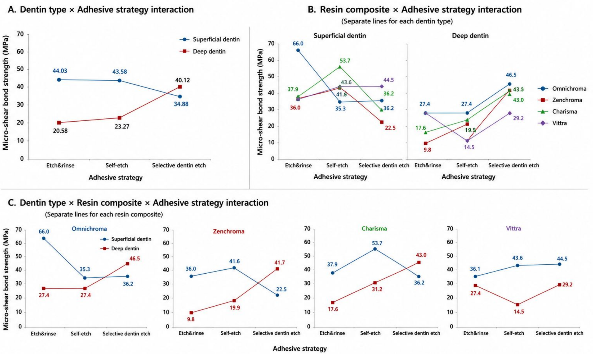
When evaluated according to dentin depth, µSBS was significantly higher in superficial dentin (40.83 ± 0.98 MPa) than in deep dentin (27.99 ± 0.98 MPa) (p < 0.001).
The effect of adhesive application strategy varied according to dentin depth. In superficial dentin, the E&R (44.03 ± 1.86 MPa) and SE (43.58 ± 1.86 MPa) strategies showed similar bond strength values (p > 0.05), whereas the SDE strategy (34.88 ± 1.86 MPa) resulted in significantly lower values (p < 0.01). In contrast, in deep dentin, the SDE strategy (40.12 ± 1.50 MPa) yielded significantly higher bond strength than both the E&R (20.58 ± 1.50 MPa) and SE (23.27 ± 1.50 MPa) strategies (p < 0.001).
When the resin composites were compared, Omnichroma (45.88 ± 2.15 MPa) and Charisma (42.61 ± 2.15 MPa) exhibited significantly higher bond strength values than Zenchroma in superficial dentin (p < 0.05). In deep dentin, Omnichroma (33.79 ± 1.73 MPa) and Charisma (30.62 ± 1.73 MPa) also showed significantly higher bond strength than Zenchroma and Vittra (p < 0.05).
Regarding the interaction between resin composite type and adhesive application strategy, the E&R strategy resulted in the highest bond strength values for Omnichroma in superficial dentin (p < 0.001). In the Charisma group, the SE strategy yielded significantly higher bond strength than the other adhesive strategies (p < 0.05). In the Vittra group, the selective dentin etch strategy produced significantly higher bond strength than the E&R strategy (p < 0.05).
In deep dentin, the highest μSBS values in the Omnichroma and Zenchroma groups were obtained with the SDE strategy (p < 0.001). Similarly, in the Charisma group, SDE resulted in significantly higher bond strength than the E&R strategy (p < 0.01). For Vittra, the SDE and E&R strategies produced similar bond strength values, and both were significantly higher than those obtained with the SE strategy (p < 0.05).
To facilitate the interpretation of the significant two-way and three-way interactions identified by the three-way ANOVA, interaction plots are presented in Figure 1.

3.2. Failure Mode Analysis

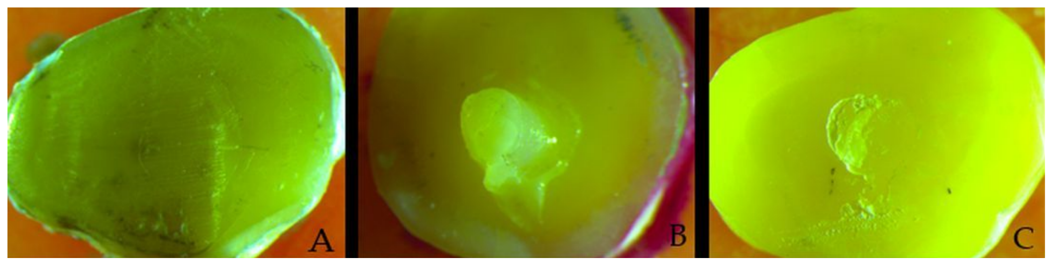
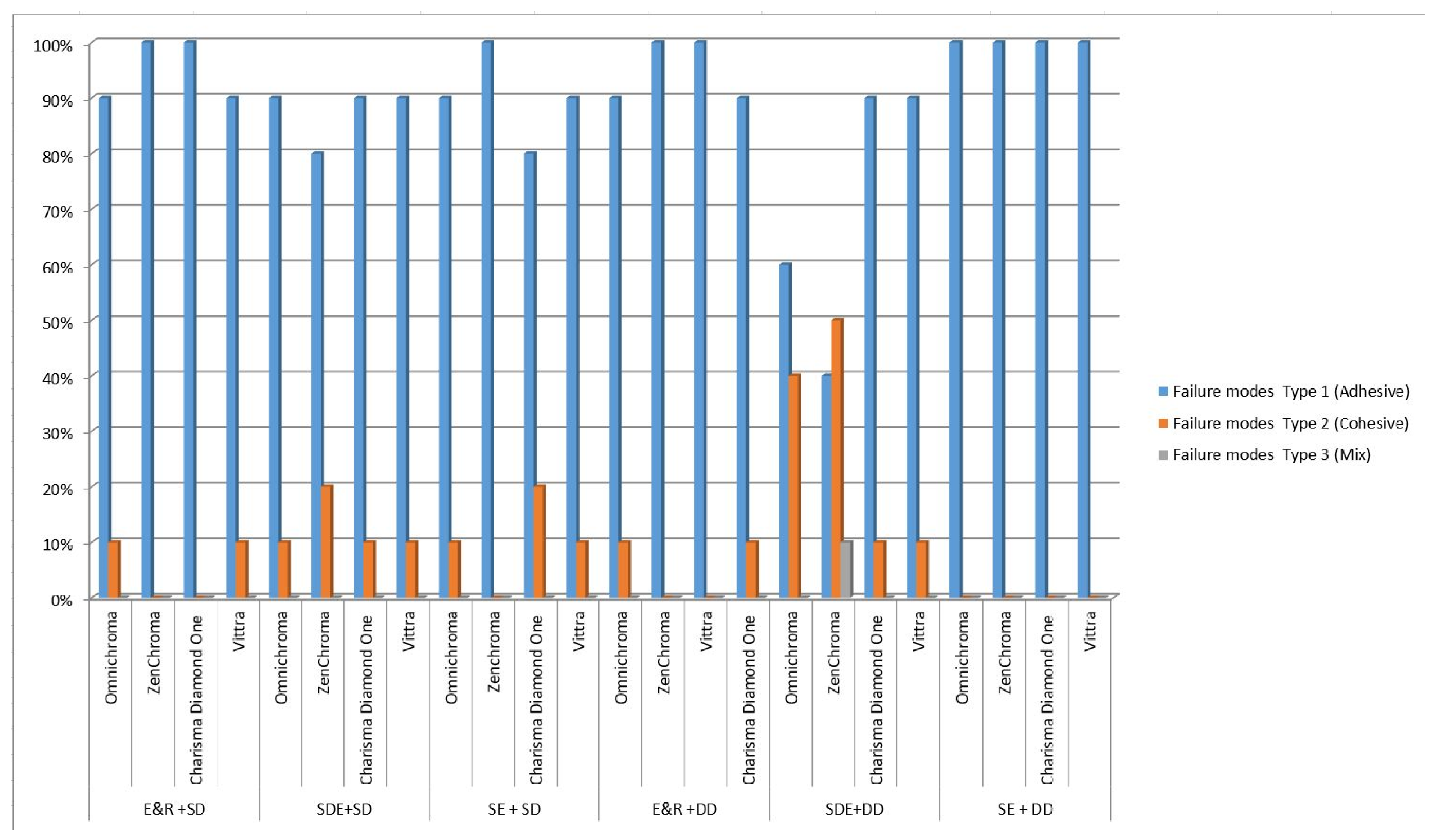
Evaluation of the failure modes demonstrated that adhesive failure occurred more frequently than the other failure types across all experimental groups. Cohesive failure was observed in varying frequencies, with the highest proportions detected in the Omnichroma group (40%) and the Zenchroma group (50%) when the SDE strategy was applied to deep dentin. Mixed failure was observed only in the Zenchroma group treated with the SDE strategy in deep dentin (10%). The distribution of failure modes is presented in Figure 2, and representative stereomicroscopic images of each failure mode are shown in Figure 3. A total of nine pretest failures occurred across seven experimental groups: one in the SE-SD/Vittra APS Unique group, one in the E&R-DD/Zenchroma group, two in the E&R-DD/Charisma Diamond One group, one in the SDE-DD/Vittra APS Unique group, one in the SE-DD/Omnichroma group, two in the SE-DD/Vittra APS Unique group, and one in the SE-DD/Charisma Diamond One group. These specimens were assigned a bond strength value of 0 MPa and included in the statistical analysis.

4. Discussion

In this study, the effects of different adhesive application strategies and single-shade resin composites on the micro-shear bond strength of superficial and deep dentin were evaluated. The results demonstrated that dentin depth, adhesive application strategy, and composite type significantly influenced bond strength. In addition, the significant interactions among these variables indicated that the bonding performance of the universal adhesive system was influenced not only by the adhesive strategy employed but also by the dentin substrate and the type of composite resin used. While the etch-and-rinse and self-etch strategies generally produced higher bond strength values in superficial dentin than in deep dentin, the selective dentin etch strategy resulted in higher bond strength values in deep dentin. These findings suggest that differences in dentin substrate characteristics may influence the bonding performance of universal adhesives. Accordingly, all four null hypotheses (H1–H4) were rejected.
Previous studies have suggested that dentin bonding is influenced by factors such as resin tag formation, the penetration of the adhesive into the intertubular dentin, and the available bonding area [18]. However, the effect of dentin depth on bond strength remains controversial. While some studies have reported lower bond strength values in deep dentin [19], others have suggested that the relationship between dentin depth and bond strength may be affected by factors such as the adhesive system used, dentin substrate characteristics, and experimental methodology [13,14,18]. Furthermore, the greater dentinal tubule density and diameter of deep dentin are associated with increased dentin permeability and water content, which may adversely affect adhesive infiltration and contribute to the differences in bond strength reported among studies [18].
In the present study, the SDE strategy was associated with higher bond strength values in deep dentin. Although the resin–dentin interface was not morphologically evaluated, previous studies have suggested that short-term phosphoric acid application may promote controlled demineralization while preserving hydroxyapatite crystals within the collagen matrix, thereby potentially facilitating the formation of a more favorable bonding substrate [20]. Furthermore, it has been reported that reducing the phosphoric acid etching time may prevent excessive demineralization and preserve higher calcium levels within demineralized dentin, which may support functional interaction between 10-MDP and residual hydroxyapatite [8,20]. Similarly, Ismail and Soliman [9] suggested that short-duration dentin etching could improve resin–dentin bonding by optimizing adhesive infiltration while minimizing the adverse effects associated with excessive demineralization. Therefore, the improved bond strength observed with the SDE strategy in deep dentin may be associated with these mechanisms. Furthermore, a recent systematic review and meta-analysis reported that selective dentin etching significantly improves the dentin bond strength of universal adhesives, which is consistent with the higher bond strength values observed in the present study [21].
The higher bond strength values obtained with the E&R strategy in the Omnichroma group on superficial dentin may be related to the structural characteristics of superficial dentin. It has been reported that the lower dentinal tubule density and higher proportion of intertubular dentin in superficial dentin facilitate resin monomer infiltration into the demineralized dentin substrate following phosphoric acid etching [14]. Furthermore, the lower permeability of superficial dentin may reduce the adverse effects of excessive moisture during the bonding procedure. It has also been reported that dentin water content, which varies according to dentin depth, may influence adhesive performance and that solvent type plays an important role in this process [13,18]. In particular, acetone-based solvent systems have been suggested to act as a “water chaser,” thereby enhancing adhesive infiltration, especially in substrates with higher water content [18]. Since Gluma Bond Universal contains an acetone-based solvent, this characteristic may have contributed to bonding performance observed in the present study. However, the superior bond strength observed with the SDE strategy in deep dentin suggests that, in addition to solvent-related factors, controlled dentin demineralization may also have contributed to the bonding performance.
The bonding performance of universal adhesives has been reported to be influenced by their pH values. With a pH of 1.6, Gluma Bond Universal can be classified as an “intermediately strong” SE adhesive [22]. Mild and intermediately strong self-etch adhesives have been reported to preserve part of the smear layer rather than completely dissolving it, which may restrict the penetration of adhesive monomers into the underlying dentin substrate [8]. Consistent with this concept, the SE application mode in the present study generally resulted in lower bond strength values in all deep dentin groups and in all superficial dentin groups, with the exception of the Charisma group. Although the resin–dentin interface was not morphologically evaluated in the present study, these findings may be associated with less effective smear layer modification and reduced resin infiltration, as suggested in previous studies [8]. These effects may be more pronounced in deep dentin because of its greater dentinal tubule density and higher water content. Interestingly, Karadaş et al. [23] in a recent systematic review and meta-analysis evaluating mild and ultra-mild universal adhesives, reported that phosphoric acid pretreatment did not significantly improve the bond strength of these adhesives. This finding suggests that the effectiveness of different adhesive application strategies may depend not only on phosphoric acid application but also on adhesive composition and dentin substrate characteristics. This interpretation is further supported by the findings of Balaban and Arısu [24], who reported that the bond strength of universal adhesives is influenced by adhesive composition, pH, and etching mode, emphasizing that universal adhesives do not exhibit uniform behavior across different application strategies.
Conversely, over-etching with phosphoric acid may negatively influence the resin–dentin interface by reducing the residual hydroxyapatite required for 10-MDP-mediated chemical bonding and by increasing the susceptibility of the demineralized collagen matrix to collapse during drying [25,26,27]. However, the presence of 10-MDP in universal adhesives has been associated with improved bond durability owing to the formation of stable and poorly soluble MDP–calcium salts at the resin–dentin interface [28]. While bonding achieved with SE strategy is considered to rely primarily on chemical interactions between functional monomers and residual hydroxyapatite, the etch-and-rinse strategy may provide additional micromechanical retention following phosphoric acid etching [29]. Although the resin–dentin interface was not evaluated morphologically in the present study, these mechanisms may have contributed to the higher bond strength values observed with the etch-and-rinse strategy in certain composite groups, particularly in superficial dentin.
Furthermore, the significant three-way interaction indicated that the effect of adhesive application strategy on bond strength was influenced not only by dentin depth but also by the type of single-shade resin composite used. This finding was reflected in the different adhesive strategy–composite combinations that yielded the highest bond strength values. For example, the etch-and-rinse strategy produced the highest bond strength values in superficial dentin for Omnichroma, whereas the self-etch strategy performed best for Charisma. In contrast, the selective dentin etch strategy generally resulted in superior performance in deep dentin, particularly for Omnichroma and Zenchroma. These findings suggest that the observed differences in bonding performance may be associated by a complex interaction among dentin morphology, adhesive infiltration, and the compositional characteristics of the resin composites.
The clinical performance of resin composites is influenced not only by their esthetic properties but also by their resin matrix composition, filler characteristics, and polymerization behavior. Recent morphological and thermogravimetric analyses have demonstrated that universal-shade resin composites exhibit material-specific differences in filler particle morphology, distribution, and filler weight fraction, highlighting the compositional diversity among these materials [30]. Similarly, Meniawi et al. [31] and Shim et al. [32] reported material-dependent differences in the optical, surface, and mechanical properties of universal-shade resin composites, including flexural strength and depth of cure. Consistent with these observations, the present study demonstrated a significant effect of composite type on bond strength. This finding may be associated with differences in the material composition and physicochemical characteristics of the tested single-shade resin composites. Previous studies have shown that variations in resin formulation, including differences in Bis-GMA, UDMA, and TEGDMA content, may influence polymerization behavior, degree of conversion, polymerization shrinkage, water sorption, cross-link density, and other mechanical properties [33,34,35,36,37]. Collectively, these compositional differences may contribute to the variation in bond strength observed among the tested composite groups. However, because resin composition, degree of conversion, filler characteristics, and polymerization behavior were not directly evaluated in the present study, these interpretations should be considered with caution.
Among the tested composites, Omnichroma exhibited higher bond strength values under certain adhesive application strategies. Omnichroma was developed to eliminate the need for shade selection and utilizes structural color technology based on 260-nm spherical fillers [38]. A recent SEM-based morphological analysis reported that Omnichroma exhibits a predominantly monomodal filler particle distribution, differing from several other commercially available universal-shade resin composites, although filler characteristics remain material specific [30]. Previous studies have suggested that variations in resin matrix composition, filler characteristics, and polymerization behavior may influence the mechanical properties of universal-shade resin composites [35,37,39]. In addition, Lopez et al. [40] demonstrated that filler content is significantly correlated with several mechanical and physical properties of resin composites, including strength, elastic modulus, polymerization shrinkage stress, translucency, and depth of cure, further supporting the importance of material composition in determining composite performance. Taken together, these observations suggest that the variation in bond strength observed among the tested single-shade resin composites may be associated with differences in their resin matrix composition, filler characteristics, and polymerization behavior. However, because these parameters were not directly evaluated in the present study, this interpretation should be considered with caution and requires further investigation.
Adhesive failure was identified as the most common failure mode in the majority of the experimental groups. Similar findings have been reported in previous studies evaluating adhesive systems on superficial and deep dentin, where adhesive failure was identified as the most common failure mode [13]. Similarly, studies on universal adhesives have shown that groups exhibiting higher bond strength values may demonstrate a higher proportion of cohesive or mixed failures, while adhesive failures are more frequently associated with weaker resin-dentin interfaces [41]. In the present study, when the SDE strategy was applied to deep dentin, cohesive failures were observed more frequently in the Omnichroma and Zenchroma groups, whereas mixed failure was detected only in the Zenchroma group under the same conditions. These observations are consistent with the concept that the integrity of the adhesive interface may influence the failure mode during bond strength testing.
The significant interactions observed in the present study indicate that the performance of adhesive application strategies is material- and substrate-dependent. Consequently, both dentin characteristics and composite-related factors should be considered when selecting an adhesive approach.
One of the strengths of the present study is the simultaneous evaluation of different adhesive application strategies, dentin depths, and single-shade resin composites within the same experimental design. Although single-shade composites have been extensively investigated with respect to their optical properties and color adjustment potential [2,4,5,6], as well as their clinical performance [42,43], information regarding their bonding behavior to dentin remains limited. Furthermore, little is known about how these materials interact with different adhesive strategies under varying dentin conditions. Therefore, the present findings provide additional insight into the bond strength performance of single-shade resin composites and may contribute to a better understanding of the interactions among adhesive strategy, dentin substrate, and restorative material.
Several limitations should be considered when interpreting the findings of the present study. First, the investigation was performed under in vitro conditions and therefore cannot fully reproduce the thermal fluctuations, mechanical loading, moisture, and other biological factors present in the oral environment. Accordingly, the findings should be interpreted within the experimental conditions of the present study and should not be generalized directly to clinical situations. In addition, bond strength was evaluated only after 24 h of water storage, without the application of artificial aging protocols. Therefore, the present findings reflect only the immediate bonding performance and should not be extrapolated to long-term clinical durability. The study also evaluated only sound superficial and deep dentin surfaces, whereas more clinically relevant substrates, such as caries-affected dentin, were not investigated. Furthermore, the absence of a conventional resin composite control group limited direct comparisons between single-shade and conventional restorative materials. The lack of SEM analysis prevented direct evaluation of the resin–dentin interface and hybrid layer morphology, limiting confirmation of the mechanisms proposed to explain the bonding performance observed in this study. Future studies incorporating long-term artificial aging protocols (e.g., thermocycling and prolonged water storage), biodegradation models, SEM-based interfacial analyses, and different dentin substrates, as well as long-term clinical studies are required to validate the present findings under clinical conditions.

5. Conclusions

Within the limitations of this in vitro study, dentin depth, adhesive application strategy, and single-shade resin composite type significantly influenced micro-shear bond strength. Higher bond strength values were generally observed in superficial dentin, whereas the selective dentin etch strategy was associated with higher bond strength values in deep dentin under the experimental conditions of this study. Significant interactions among dentin depth, adhesive strategy, and composite type further indicate that the bonding performance of universal adhesive system is both substrate and material used. These findings suggest that adhesive application strategy and restorative material selection may influence immediate bonding performance; however, further long-term and clinical studies are required before broader clinical recommendations can be made.

Author Contributions

Conceptualization, F.A.A., H.S.B. and O.S.K.; methodology F.A.A., H.S.B. and O.S.K.; validation, F.A.A. and H.S.B.; formal analysis, H.S.B.; investigation, F.A.A., O.S.K. and H.S.B.; resources, F.A.A.; data curation, F.A.A.; writing—original draft preparation, F.A.A.; writing—review and editing, H.S.B.; visualization, F.A.A.; supervision, H.S.B.; project administration, F.A.A.; funding acquisition, F.A.A., H.S.B. and O.S.K. All authors have read and agreed to the published version of the manuscript.

Funding

This research received no external funding.

Institutional Review Board Statement

The study was conducted in accordance with the Declaration of Helsinki and approved by the Ethics Committee of Ankara Yildirim Beyazit University (protocol code 2025-04/1219; date of approval: 10 April 2025).

Informed Consent Statement

Informed consent was obtained from all subjects involved in the study.

Data Availability Statement

The data presented in this study are available from the corresponding author upon reasonable request.

Conflicts of Interest

The authors declare no conflicts of interest.

Abbreviations

The following abbreviations are used in this manuscript:
μSBSMicro-shear bond strength
E&REtch-and-rinse
SESelf-etch
SDESelective dentin etch
SDSuperficial dentin
DDDeep dentine
Bis-GMABisphenol A-glycidyl methacrylate
UDMAUrethane dimethacrylate
TEGDMATriethylene glycol dimethacrylate
10-MDP10-Methacryloyloxydecyl dihydrogen phosphate
SEMScanning electron microscopy

References

  1. Josic, U.; D’Alessandro, C.; Miletic, V.; Maravic, T.; Mazzitelli, C.; Jacimovic, J.; Sorrentino, R.; Zarone, F.; Delgado, A.H.S.; Breschi, L.; et al. Clinical longevity of direct and indirect posterior resin composite restorations: An updated systematic review and meta-analysis. Dent. Mater. 2023, 39, 1085–1094. [Google Scholar] [CrossRef]
  2. Chen, F.; Toida, Y.; Islam, R.; Alam, A.; Chowdhury, A.F.M.A.; Yamauti, M.; Sano, H. Evaluation of shade matching of a novel supra-nano-filled esthetic resin composite employing structural color using simplified simulated clinical cavities. J. Esthet. Restor. Dent. 2021, 33, 874–883. [Google Scholar] [CrossRef] [PubMed]
  3. Dietschi, D.; Fahl, N., Jr. Shading concepts and layering techniques to master direct anterior composite restorations: An update. Br. Dent. J. 2016, 221, 765–771. [Google Scholar] [CrossRef] [PubMed]
  4. de Abreu, J.L.B.; Sampaio, C.S.; Benalcázar Jalkh, E.B.; Hirata, R. Analysis of the color matching of universal resin composites in anterior restorations. J. Esthet. Restor. Dent. 2021, 33, 269–276. [Google Scholar] [CrossRef] [PubMed]
  5. Zhu, J.; Xu, Y.; Li, M.; Huang, C. Instrumental and visual evaluation of the color adjustment potential of a recently introduced single shade composite resin versus multishade composite resins. J. Prosthet. Dent. 2023, 134, 832–839. [Google Scholar] [CrossRef] [PubMed]
  6. Forabosco, E.; Generali, L.; Mancuso, E.; Kaleci, S.; Consolo, U.; Checchi, V. Color match of single-shade restorations after professional dental bleaching: An in vitro study. J. Conserv. Dent. Endod. 2024, 27, 280–285. [Google Scholar] [CrossRef] [PubMed]
  7. Loguercio, A.D.; de Paula, E.A.; Hass, V.; Luque-Martinez, I.; Reis, A.; Perdigão, J. A new universal simplified adhesive: 36-Month randomized double-blind clinical trial. J. Dent. 2015, 43, 1083–1092. [Google Scholar] [CrossRef] [PubMed]
  8. Stape, T.H.S.; Wik, P.; Mutluay, M.M.; Al-Ani, A.A.S.; Tezvergil-Mutluay, A. Selective dentin etching: A potential method to improve bonding effectiveness of universal adhesives. J. Mech. Behav. Biomed. Mater. 2018, 86, 14–22. [Google Scholar] [CrossRef] [PubMed]
  9. Ismail, H.S.; Soliman, H.A.N. Short dentin etching with universal adhesives: Effect on bond strength and gingival margin adaptation. BMC Oral Health 2025, 25, 128. [Google Scholar] [CrossRef] [PubMed]
  10. Bourgi, R.; Breschi, L.; Cuevas-Suárez, C.E.; Cabanillas, J.C.H.; Kharouf, N.; Hardan, L. Three-second short dentin etching: Systematic review of in vitro evidence in adhesive dentistry. J. Esthet. Restor. Dent. 2026, 38, 1353–1366. [Google Scholar] [CrossRef] [PubMed]
  11. Atalay, C.; Ozgunaltay, G.; Yazici, A.R. Thirty-six-month clinical evaluation of different adhesive strategies of a universal adhesive. Clin. Oral Investig. 2020, 24, 1569–1578. [Google Scholar] [CrossRef] [PubMed]
  12. Barceleiro, M.O.; Lopes, L.S.; Tardem, C.; Calazans, F.S.; Matos, T.P.; Reis, A.; Calixto, A.L.; Loguercio, A.D. Thirty-six-month follow-up of cervical composite restorations placed with an MDP-free universal adhesive system using different adhesive protocols: A randomized clinical trial. Clin. Oral Investig. 2022, 26, 4337–4350. [Google Scholar] [CrossRef] [PubMed]
  13. Gökce, D.; Usumez, A.; Polat, Z.S.; Ayna, E. Evaluation of Four Different Adhesive Systems’ Bonding Strength Between Superficial and Deep Dentin. Materials 2025, 18, 3107. [Google Scholar] [CrossRef] [PubMed]
  14. Giannini, M.; Carvalho, R.M.; Martins, L.R.; Dias, C.T.; Pashley, D.H. The influence of tubule density and area of solid dentin on bond strength of two adhesive systems to dentin. J. Adhes. Dent. 2001, 3, 315–324. [Google Scholar] [PubMed]
  15. Zhang, L.; Wang, D.Y.; Fan, J.; Li, F.; Chen, Y.J.; Chen, J.H. Stability of bonds made to superficial vs. deep dentin, before and after thermocycling. Dent. Mater. 2014, 30, 1245–1251. [Google Scholar] [CrossRef] [PubMed]
  16. Simmer, F.S.; da Silva, E.M.; Bezerra, R.D.S.G.; Gomes, M.E.D.S.N.; Noronha Filho, J.D.; Amaral, C.M. Bond stability of conventional adhesive system with MMP inhibitors to superficial and deep dentin. J. Mech. Behav. Biomed. Mater. 2019, 100, 103402. [Google Scholar] [CrossRef] [PubMed]
  17. Bahadır, H.S.; Tuğutlu, E.C. Micro-shear Bond Strength of an Alternative Self-etch Application of “No Wait” Universal Adhesive to Caries-affected and Sound Dentin in Permanent and Primary Teeth. Oper. Dent. 2024, 49, 273–281. [Google Scholar] [CrossRef] [PubMed]
  18. Toledano, M.; Osorio, R.; Ceballos, L.; Fuentes, M.V.; Fernandes, C.A.; Tay, F.R.; Carvalho, R.M. Microtensile bond strength of several adhesive systems to different dentin depths. Am. J. Dent. 2003, 16, 292–298. [Google Scholar] [PubMed]
  19. Pegado, R.E.F.; do Amaral, F.L.B.; Flório, F.M.; Basting, R.T. Effect of different bonding strategies on adhesion to deep and superficial permanent dentin. Eur. J. Dent. 2010, 4, 110–117. [Google Scholar] [CrossRef] [PubMed]
  20. Cadenaro, M.; Josic, U.; Maravić, T.; Mazzitelli, C.; Marchesi, G.; Mancuso, E.; Breschi, L.; Mazzoni, A. Progress in dental adhesive materials. J. Dent. Res. 2023, 102, 254–262. [Google Scholar] [CrossRef] [PubMed]
  21. Hardan, L.; Bourgi, R.; Kharouf, N.; Mancino, D.; Zarow, M.; Jakubowicz, N.; Haikel, Y.; Cuevas-Suárez, C.E. Bond Strength of Universal Adhesives to Dentin: A Systematic Review and Meta-Analysis. Polymers 2021, 13, 814. [Google Scholar] [CrossRef] [PubMed]
  22. Arandi, N.Z. The Classification and Selection of Adhesive Agents; an Overview for the General Dentist. Clin. Cosmet. Investig. Dent. 2023, 4, 165–180. [Google Scholar] [CrossRef] [PubMed]
  23. Karadas, M.; Bedir, F.; Demirbuga, S. The role of etching protocols on bond strength of universal adhesives applied to caries affected dentin: A systematic review and meta-analysis. Clin. Oral Investig. 2024, 28, 683. [Google Scholar] [CrossRef] [PubMed]
  24. Balaban, S.; Arısu, H.D. Effect of Etching Mode on the Dentin Bond Durability of Universal Adhesives. J. Adhes. Dent. 2025, 27, 297–305. [Google Scholar] [CrossRef] [PubMed]
  25. Ikeda, M.; Tsubota, K.; Takamizawa, T.; Yoshida, T.; Miyazaki, M.; Platt, J.A. Bonding durability of single-step adhesives to previously acid-etched dentin. Oper. Dent. 2008, 33, 702–709. [Google Scholar] [CrossRef] [PubMed]
  26. Hashimoto, M.; Nagano, F.; Endo, K.; Ohno, H. A review: Biodegradation of resin-dentin bonds. Jpn. Dent. Sci. Rev. 2011, 47, 5–12. [Google Scholar] [CrossRef]
  27. Pereira, G.D.; Paulillo, L.A.; de Goes, M.F.; Dias, C.T. How wet should dentin be? Comparison of methods to remove excess water during moist bonding. J. Adhes. Dent. 2001, 3, 257–264. [Google Scholar] [PubMed]
  28. Cuevas-Suarez, C.E.; da Rosa, W.L.O.; Lund, R.G.; da Silva, A.F.; Piva, E. Bonding performance of universal adhesives: An updated systematic review and meta-analysis. J. Adhes. Dent. 2019, 21, 7–26. [Google Scholar] [CrossRef] [PubMed]
  29. Zhou, L.; Wang, Y.; Yang, H.; Guo, J.; Tay, F.R.; Huang, C. Effect of chemical interaction on the bonding strengths of self-etching adhesives to deproteinised dentine. J. Dent. 2015, 43, 973–980. [Google Scholar] [CrossRef] [PubMed]
  30. Perdigão, J.; Zatt, F.P.; Lopes, G.C.; Caon, N.B.; Chen, R. Characterization of universal composite resin filler particles. J. Dent. Res. 2025, 37, 2472–2480. [Google Scholar] [CrossRef] [PubMed]
  31. Meniawi, M.; Şirinsükan, N.; Can, E. Color stability, surface roughness, and surface morphology of universal composites. Odontology 2026, 114, 149–160. [Google Scholar] [CrossRef] [PubMed]
  32. Shim, J.E.; Kim, H.J.; Oh, S.; Jang, J.H. Optical and mechanical characteristics of one-shade composite resins. J. Funct. Biomater. 2025, 16, 419. [Google Scholar] [CrossRef] [PubMed]
  33. Vasudeva, G. Monomer systems for dental composites and their future: A review. J. Calif. Dent. Assoc. 2009, 37, 389–398. [Google Scholar] [CrossRef] [PubMed]
  34. Barutcigil, Ç.; Barutcigil, K.; Özarslan, M.M.; Dündar, A.; Yilmaz, B. Color of bulk-fill composite resin restorative materials. J. Esthet. Restor. Dent. 2018, 30, E3–E8. [Google Scholar] [CrossRef] [PubMed]
  35. Floyd, C.J.; Dickens, S.H. Network structure of Bis-GMA and UDMA-based resin systems. Dent. Mater. 2006, 22, 1143–1149. [Google Scholar] [CrossRef] [PubMed]
  36. Tarumi, H.; Imazato, S.; Ehara, A.; Kato, S.; Ebi, N.; Ebisu, S. Post-irradiation polymerization of composites containing Bis-GMA and TEGDMA. Dent. Mater. 1999, 15, 238–242. [Google Scholar] [CrossRef] [PubMed]
  37. Ling, L.; Lai, T.; Chung, P.-T.; Malyala, R. Shrinkage, degree of conversion, water sorption and solubility, and mechanical properties of novel one-shade universal composite. Polymers 2025, 17, 2728. [Google Scholar] [CrossRef] [PubMed]
  38. AlHamdan, E.M.; Bashiri, A.; Alnashmi, F.; Al-Saleh, S.; Al-shahrani, K.; Al-shahrani, S.; Alsharani, A.; Alzahrani, K.M.; Alqarawi, F.K.; Vohra, F.; et al. Evaluation of Smart Chromatic Technology for a Single-Shade Dental Polymer Resin: An In Vitro Study. Appl. Sci. 2021, 11, 10108. [Google Scholar] [CrossRef]
  39. Yılmaz Atalı, P.; Doğu Kaya, B.; Manav Özen, A.; Tarçın, B.; Şenol, A.A.; Tüter Bayraktar, E.; Korkut, B.; Bilgin Göçmen, G.; Tağtekin, D.; Türkmen, C. Assessment of Micro-Hardness, Degree of Conversion, and Flexural Strength for Single-Shade Universal Resin Composites. Polymers 2022, 14, 4987. [Google Scholar] [CrossRef] [PubMed]
  40. Lopez, C.; Nizami, B.; Robles, A.; Gummadi, S.; Lawson, N.C. Correlation between dental composite filler percentage and strength, modulus, shrinkage stress, translucency, depth of cure and radiopacity. Materials 2024, 17, 3901. [Google Scholar] [CrossRef] [PubMed]
  41. Santander-Rengifo, F.; Carreras-Presas, C.M.; Aroste-Andía, R.; Hernández-Huamaní, E.; Gavilán-Chávez, P.; Cervantes-Ganoza, L.; Cayo-Rojas, C. Microtensile bond strength and failure mode of different universal adhesives on human dentin. Int. Dent. J. 2024, 74, 1239–1247. [Google Scholar] [CrossRef] [PubMed]
  42. Anwar, R.S.; Hussein, Y.F.; Riad, M. The clinical performance of monoshade resin composite as posterior restoration: A randomized controlled clinical trial. Sci. Rep. 2025, 15, 25279. [Google Scholar] [CrossRef] [PubMed]
  43. Çadırcı, M.; Özakar, N. Clinical performance of single shade universal resin composites: A randomized controlled one year follow-up study. BMC Oral Health 2026, 26, 154. [Google Scholar] [CrossRef] [PubMed]
Figure 1. Grafical interaction plots for micro-shear bond strenght (MPa). Interaction plots illustrating the effects of dentin depth, adhesive application strat-egy, and single-shade resin composite type on μSBS. (A) Interaction between dentin type (superfi-cial and deep dentin) and adhesive strategy (E&R, SE, and SDE). (B) Interaction between resin composite type and adhesive strategy in superficial and deep dentin. (C) Three-way interaction among dentin type, resin composite type, and adhesive strategy. Data points represent the mean μSBS values (MPa) obtained for each experimental group. The plots are presented to facilitate visualization of the significant two-way and three-way interactions identified by the three-way ANOVA.
Applsci 16 07097 g001
Figure 2. Failure mode analysis (percentage).
Applsci 16 07097 g002
Figure 3. Types of failure. (A): adhesive failure; (B): cohesive failure (C): mixed failure.
Applsci 16 07097 g003
Table 1. Materials used in the study, their manufacturers, chemical components, filler contents and application methods.
Material ManufacturerMain Components Filler Content
By Weight
(%)
By Volume
(%)
Omnichroma
Nanofill
Tokuyama,
Tokyo,
Japan
UDMA, TEGDMA (260 nm spherical SiO2–ZrO2)79 68
ZenChroma
Micro hybrid
President Dental,
Allershausen, Germany
UDMA, Bis-GMA, TEGDMA, Silicon dioxide, Glass
filler
7553
Charisma Diamond One
Nanohybrid
Kulzer, Hanau,
Germany
TCD (Tricyclodecane Dimethanol Diacrylate),
UDMA, TEGDMA, Barium Aluminum Fluoride
Glass Particles
8265
Vittra APS Unique
Nanohybrid
FGM,
Joinville,
SC, Brazil
UDMA, TEGDMA, APS photoinitiator system, barium glass, spherical zirconia silicate, spherical and amorphous silica fillers76 NR (not reported by manufacturer)
Gluma Bond Universal BondKulzer, Hanau,
Germany
4-META, 10-MDP, phosphoric acid ester monomer, dimethacrylate monomers, distilled water, acetone, silicon dioxide, photoinitiator, silane
pH: 1.6
Application mode
One or two drops of the adhesive were dispensed into a clean mixing well. The adhesive was actively applied to the prepared dentin surface with a microbrush for 20 s, gently air-thinned for approximately 10 s to evaporate the solvent and obtain a uniform adhesive film, and subsequently light-cured for 10 s.
Table 2. Micro-shear bond strength (MPa) of different composite resins according to adhesive application modes.
Dentin TypeResin CompositeEtch & RinseSelf-EtchSelective Dentin Etch
Superficial dentinOmnichroma66.0 ± 5.0 a35.3 ± 4.2 b36.2 ± 6.9 b
Zenchroma36.0 ± 4.3 a41.6 ± 3.0 a22.5 ± 2.8 b
Charisma37.9 ± 4.5 b53.7 ± 6.4 a36.2 ± 4.4 b
Vittra36.1 ± 7.9 b43.6 ± 5.2 ab44.5 ± 4.2 a
Deep dentinOmnichroma27.4 ± 3.4 b27.4 ± 3.2 b46.5 ± 2.3 a
Zenchroma9.8 ± 4.5 c19.9 ± 2.3 b41.7 ± 3.8 a
Charisma17.6 ± 2.1 b31.2 ± 3.7 b43.0 ± 5.8 a
Vittra27.4 ± 1.7 a14.5 ± 1.7 b29.2 ± 3.6 a
Within each row, values identified by different superscript letters indicate statistically significant differences among the adhesive application modes (p < 0.05).
Table 3. Results of three-way ANOVA.
dfFpPartial η2
Dentin type186.45<0.0010.286
Resin composite312.46<0.0010.148
Adhesive25.230.0060.046
Dentin type × composite resin31.560.1990.021
Dentin type × Adhesive243.30<0.0010.286
Resin composite × Adhesive67.69<0.0010.176
Dentin type× Resin composite × Adhesive69.04<0.0010.201
Statistical significance was set at p < 0.05. The model demonstrated an R2 value of 0.601 and an adjusted R2 value of 0.559.
Disclaimer/Publisher’s Note: The statements, opinions and data contained in all publications are solely those of the individual author(s) and contributor(s) and not of MDPI and/or the editor(s). MDPI and/or the editor(s) disclaim responsibility for any injury to people or property resulting from any ideas, methods, instructions or products referred to in the content.