1. Introduction
Traumatic dental injuries most frequently affect the maxillary permanent incisors and may lead to loss of pulp vitality and interruption of root development [
1]. In the management of immature necrotic permanent teeth, apexification and revitalization are commonly applied treatment approaches. However, due to the lack of continued root development and the increased risk of root fracture associated with apexification [
2], revitalization therapy is currently recommended according to contemporary guidelines [
3]. This treatment aims to stimulate the regeneration of pulp-like tissue through stem cells of the apical papilla, thereby promoting continued root development, increased dentin thickness, and apical closure [
3]. Calcium silicate-based biomaterials such as mineral trioxide aggregate and Biodentine have been shown to support these processes due to their bioactive properties [
4]. In addition, Biodentine has a shorter setting time and does not induce tooth discoloration compared with mineral trioxide aggregate [
5].
Despite these biological advantages, teeth treated with revitalization remain structurally compromised and susceptible to fracture. Although continued root development may occur, these teeth frequently retain structural characteristics such as incomplete root maturation, thin dentinal walls, enlarged root canals, and an unfavorable crown-to-root ratio, all of which reduce their resistance to functional and traumatic loading [
6,
7]. Furthermore, finite element analyses have demonstrated that immature maxillary anterior teeth are subjected to high tensile stress concentrations, particularly in the cervical and coronal root regions, during oblique loading, making these areas especially vulnerable to fracture initiation [
7,
8]. In addition, loss of pulpal sensory feedback may increase functional loading through altered proprioception [
9]. Therefore, the long-term prognosis of revitalized teeth depends not only on successful biological healing but also on an appropriate restorative strategy that preserves the remaining tooth structure and provides adequate biomechanical reinforcement [
10,
11].
Direct composite restorations are commonly used; however, in extensive defects they are associated with polymerization shrinkage, marginal degradation, and microleakage, which may compromise long-term success [
12]. In contrast, indirect restorations have gained increasing attention due to their ability to preserve tooth structure and improve mechanical performance, particularly with advancements in adhesive systems and CAD/CAM technologies. Among these, endocrowns have been proposed as a conservative alternative to conventional crown restorations. Endocrowns rely on both macroretention from the pulp chamber and micromechanical retention through adhesive bonding, forming a monoblock structure [
13]. Previous studies have reported that anterior endocrowns may exhibit higher fracture resistance than direct or conventional post–core restorations [
14]. Although some clinical studies have reported encouraging outcomes when appropriate materials are selected [
15], their clinical application remains controversial because anterior teeth differ from posterior teeth in terms of crown-to-root ratio and the direction of occlusal forces [
16,
17]. Unlike posterior teeth, anterior teeth are subjected to greater lateral and shear forces during functional and parafunctional movements, which generate higher tensile stresses at the adhesive interface and cervical region [
17]. Furthermore, the relatively smaller pulp chamber and reduced available bonding surface in anterior teeth may compromise retention compared with posterior endocrowns. Consequently, although encouraging clinical outcomes have been reported, further clinical evidence is required to establish the long-term predictability of anterior endocrowns.
More recently, indirect composite materials such as Gradia Plus, breCAM.HIPC, and Trinia fiber-reinforced composites have been introduced as contemporary restorative options because of their favorable mechanical properties, including enhanced toughness, reduced polymerization shrinkage, and stress-absorbing capacity, which may influence fracture resistance outcomes [
18,
19,
20]. In the present study, Gradia Plus was selected as an indirect nano-hybrid composite because its filler-containing resin matrix has been associated with favorable wear resistance and mechanical performance in previous laboratory studies [
21,
22]. In contrast, breCAM.HIPC was selected as a CAD/CAM high-impact polymer composite because its highly cross-linked polymer network and relatively low elastic modulus compared with conventional ceramic materials may contribute to stress absorption and more favorable stress distribution within the remaining tooth structure [
23]. These materials were selected to compare two indirect composite systems with different manufacturing processes and microstructural characteristics, while sharing polymer-based mechanical behavior that may be advantageous for structurally compromised immature anterior teeth.
From a materials perspective, indirect composite systems have been increasingly used due to their favorable mechanical properties and stress distribution characteristics. Contemporary materials such as nano-hybrid composites, high-impact polymer-based CAD/CAM blocks, and fiber-reinforced composites offer different elastic moduli and fracture behaviors, which may influence the performance of restorations under functional loading. Nevertheless, the relative contribution of the tested restorative assembly and restorative material type to the fracture resistance of revitalized immature teeth remains unclear.
Although a limited number of studies have evaluated the fracture resistance of immature teeth following revitalization therapy, these investigations have primarily focused on direct restorations [
24,
25]. To date, no studies have specifically examined the influence of different restorative assemblies, such as anterior endocrown and crown configurations, in revitalized immature anterior teeth. In addition, the performance of different indirect composite materials in this specific substrate has not been sufficiently investigated, while only a few studies have evaluated fiber-reinforced composites [
26,
27]. Therefore, the aim of this study was to evaluate the effect of two different restorative assemblies and restorative material type on the fracture resistance of indirect composite restorations in simulated revitalized immature maxillary central incisors.
The null hypotheses were that restorative assembly would not affect fracture resistance and that material type would not influence fracture resistance in simulated revitalized immature maxillary central incisors.
2. Materials and Methods
2.1. Ethical Approval and Sample Size Calculation
The study protocol was approved by the Local Ethics Committee for Medical Research (25-T6/9). Written informed consent was obtained from all participants or their legal guardians before collection and use of the extracted teeth.
Sample size was determined based on the study by Kınıkoğlu et al. [
25] using G*Power version 3.1.9.7 (Heinrich-Heine University, Düsseldorf, Germany). Because effect-size estimates derived from a single previous study may overestimate statistical power, a conservative standardized large effect size (f = 0.60) was selected for the a priori power analysis. A 2 × 2 factorial ANOVA model (ANOVA: Fixed effects, special, main effects and interactions) was used with a significance level of α = 0.05, a statistical power of 95% (1 − β), and a numerator degree of freedom of 1. The minimum required sample size was calculated as 39 specimens (approximately 10 specimens per experimental group), corresponding to an actual statistical power of 95.4%. To compensate for potential specimen loss, a total of 48 teeth were included.
2.2. Specimen Selection and Standardization
Forty-eight extracted human maxillary central incisors were selected. To minimize anatomical variability, crown-to-root length and mesiodistal and buccolingual dimensions were measured using a digital caliper, and teeth deviating more than ±20% from the mean values were excluded. Crown and root lengths were standardized to 9 ± 1 mm and 12 ± 1 mm, respectively, measured from the midpoint of the buccal cemento-enamel junction (CEJ). Teeth were examined under magnification (×3.5), and those with caries, cracks, fractures, or previous restorations were excluded. Specimens were stored in 0.1% thymol solution until use.
2.3. Simulation of Immature Teeth and Fracture Model
To simulate extensive coronal tissue loss, a standardized oblique fracture was created extending from the mesial midpoint of the crown toward the distal surface, leaving approximately one-third of the coronal tooth structure below the CEJ. Following fracture simulation, the remaining coronal tooth structure was standardized to approximately 4 mm on the mesial aspect and 3 mm on the distal aspect.
Access cavities were prepared, and root canals were initially instrumented using a 30.04 rotary file (Fanta Dental Co., Ltd., Istanbul, Turkey), followed by sequential enlargement with Peeso reamers (sizes 1–4). Distilled water was used as the irrigant during this phase. Canal enlargement was continued until a standardized apical diameter of approximately 1.3 mm was obtained to simulate an immature tooth with an open apex.
The same instrumentation sequence was applied to all specimens, and the preparation was standardized by achieving the predefined apical diameter and radiographically verified remaining dentin thickness. To verify the standardization of the simulated immature tooth model, the remaining dentin thickness was measured on standardized radiographic images using ImageJ software version 1.54g (National Institutes of Health, Bethesda, MD, USA). The mean remaining dentin thicknesses in the coronal, middle, and apical thirds were 2.26 ± 0.24 mm, 1.61 ± 0.24 mm, and 0.82 ± 0.16 mm, respectively, confirming the consistency of the simulated immature tooth model among the experimental specimens.
2.4. Revitalization Protocol
The revitalization procedure was performed according to the European Society of Endodontology guidelines. Root canals were irrigated with 20 mL of 2.5% sodium hypochlorite using a 30-gauge side-vented needle positioned 2 mm short of the working length, followed by 20 mL of 17% EDTA for 5 min. Calcium hydroxide was placed as an intracanal medicament for 2 weeks.
At the second session, the canals were irrigated with 17% EDTA and distilled water, dried, and a collagen matrix (Collaplug) was placed 3 mm below the CEJ. Biodentine was prepared according to the manufacturer’s instructions and placed over the matrix with a standardized thickness of 3 mm. Radiographs were taken to confirm placement. Internal cavity walls were rounded to eliminate undercuts.
2.5. Restorative Procedures
Following the revitalization protocol, forty-eight teeth were randomly assigned into four groups (n = 12) using a computer-generated randomization system (
https://www.random.org; accessed on 10 April 2025). Cement thickness was standardized at approximately 0.06 mm based on the results of a preliminary study. The chemical compositions of the materials used in this study are presented in
Table 1.
Digital scanning was performed using an intraoral scanner (3Shape TRIOS 5, 3Shape A/S, Copenhagen, Denmark), and the obtained STL data were transferred to CAD software (DentalCAD version 3.2 Elefsina, exocad GmbH, Darmstadt, Germany). For all digitally designed restorations, the external contours of the fracture line were marked, and a standard tooth form was selected from the software library and adapted to each specimen. The crown length and width were standardized to 11 mm and 9 mm, respectively.
The experimental groups were defined according to restorative assemblies (endocrown or fiber-reinforced crown) and material type (breCAM.HIPC or Gradia Plus) as follows:
Group 1 (breCAM.HIPC Endocrown): Endocrown restorations were fabricated using CAD/CAM technology from breCAM.HIPC discs without core build-up.
Group 2 (Gradia Plus Endocrown): Endocrown restorations were fabricated using the layering technique with Gradia Plus indirect composite without core build-up.
Group 3 (breCAM.HIPC Crown): A fiber-reinforced composite core (Trinia) was fabricated using CAD/CAM, extending from the pulp chamber beyond the oblique fracture line to provide structural support. Following core fabrication, tooth preparation was performed for full crown restorations according to the manufacturer’s recommendations. Crown restorations were then fabricated using CAD/CAM from breCAM.HIPC discs.
For the endocrown groups, a standardized intracoronal extension of 3.5 mm was prepared. For the crown groups, standard crown preparation was performed with 3 mm of incisal reduction, a 1-mm shoulder finish line, and 1.3 mm of buccal axial reduction. The Trinia fiber-reinforced composite core was standardized to a height of 8 mm measured from the cemento-enamel junction toward the incisal edge. Because the fracture simulation determined the amount of remaining coronal tooth structure, no additional ferrule preparation was performed; therefore, a standardized ferrule height was not defined in the experimental design.
Group 4 (Gradia Plus Crown): Similarly to Group 3, a fiber-reinforced composite core (Trinia) was fabricated, and tooth preparation was performed. Crown restorations were fabricated using the layering technique with Gradia Plus indirect composite.
For CAD/CAM-fabricated restorations, milling was performed using a milling unit (vhf camfacture AG, Ammerbuch, Germany). The internal surfaces of these restorations were cleaned with pressurized water and air-abraded with 50 µm aluminium oxide particles under 2 bar pressure.
For restorations fabricated using the layering technique, silicone impressions were taken and type IV dental stone models were prepared. A separator (Gradia Plus Separator, GC Corp., Tokyo, Japan) was applied to the dies, and restorations were fabricated using Gradia Plus indirect composite resin. After shaping, the restorations were coated with an oxygen-inhibiting gel (GC Gradia Plus Air Barrier) and polymerized using a laboratory curing unit (Labolight Duo, GC Corp., Tokyo, Japan) for 3 min.
All restorative and cementation procedures were performed by a single operator using a standardized adhesive protocol to reduce procedural variability. Thirty-five percent orthophosphoric acid was applied to the tooth surfaces for 15 s, followed by rinsing for 60 s and air-drying. A universal adhesive (G-Premio Bond, GC Corp., Tokyo, Japan) was applied for 10 s, followed by gentle air-drying. The internal surfaces of the restorations were treated with a primer (G-Multi Primer, GC Corp., Tokyo, Japan). A self-adhesive resin cement (G-CEM One, GC Corp., Tokyo, Japan) was used for luting. Excess cement was removed after 1 s of light exposure, and final polymerization was performed for 20 s using an LED curing unit.
The use of a fiber-reinforced composite core in the crown groups was intended to simulate clinical conditions in extensively damaged teeth; however, this approach may have introduced a confounding effect when comparing the tested restorative assemblies and was therefore considered during interpretation of the results.
2.6. Fracture Testing
The specimens were subjected to 10,000 thermocycles, a protocol widely used for artificial aging in laboratory studies and generally considered to approximate one year of clinical thermal aging [
28]. A compressive load was applied using a universal testing machine at a crosshead speed of 1 mm/min, with a 4 mm stainless steel ball positioned on the palatal surface at an angle of 135° to the long axis of the tooth. The maximum fracture load was recorded in Newtons (N). Fracture patterns were classified according to their location as crown fractures or root fractures. All fracture patterns were evaluated by the same examiner under magnification to reduce assessment variability. Fractures terminating above the cemento-enamel junction (CEJ) were recorded as crown fractures, whereas fractures extending below the CEJ were recorded as root fractures. Root fractures were further classified according to their location as cervical, middle, or apical thirds, with each third corresponding to approximately one-third of the standardized root length (approximately 4 mm). Fracture modes were recorded according to the direction of the fracture line as vertical, horizontal, or oblique.
2.7. Statistical Analyses
Statistical analysis was performed using GraphPad Prism software version 10.5.0 (GraphPad Software Inc., San Diego, CA, USA). The normality of the data distribution was assessed using the D’Agostino–Pearson test. Since the fracture resistance data showed normal distribution, a two-way analysis of variance (ANOVA) was used to evaluate the effects of the tested restorative assemblies (endocrown vs. fiber-reinforced crown) and material type (breCAM.HIPC vs. Gradia Plus), as well as their interaction, on fracture resistance values. Post hoc multiple comparisons were performed using Tukey’s test. The distribution of fracture types and their localization among groups were analyzed using Fisher’s exact test. The level of statistical significance was set at p < 0.05.
3. Results
The mean maximum fracture load values (N) for each experimental group are presented in
Table 2. Two-way ANOVA revealed that the tested restorative assembly had a significant effect on fracture resistance [F (1,44) = 33.69,
p = 6.54 × 10
−7, partial η
2 = 0.434], whereas material type did not show a statistically significant effect [F (1,44) = 1.074,
p = 0.3057, partial η
2 = 0.024]. No significant interaction was observed between the tested restorative assembly and material type [F (1,44) = 0.861,
p = 0.3585, partial η
2 = 0.019], indicating that the effect of the tested restorative assembly on fracture resistance did not significantly differ according to the restorative material evaluated.
When restorations fabricated with the same material were compared, endocrown groups demonstrated significantly higher fracture resistance values than crown groups for both breCAM.HIPC (p = 0.006, 95% CI: 47.01–420.4) and Gradia Plus (p = 0.0001, 95% CI: 136–509.4).
Similarly, no statistically significant material-related differences were detected within the same restorative assembly. The fracture resistance values of breCAM.HIPC and Gradia Plus endocrowns did not differ significantly (p = 0.512), nor did those of the corresponding crown groups (p = 0.999).
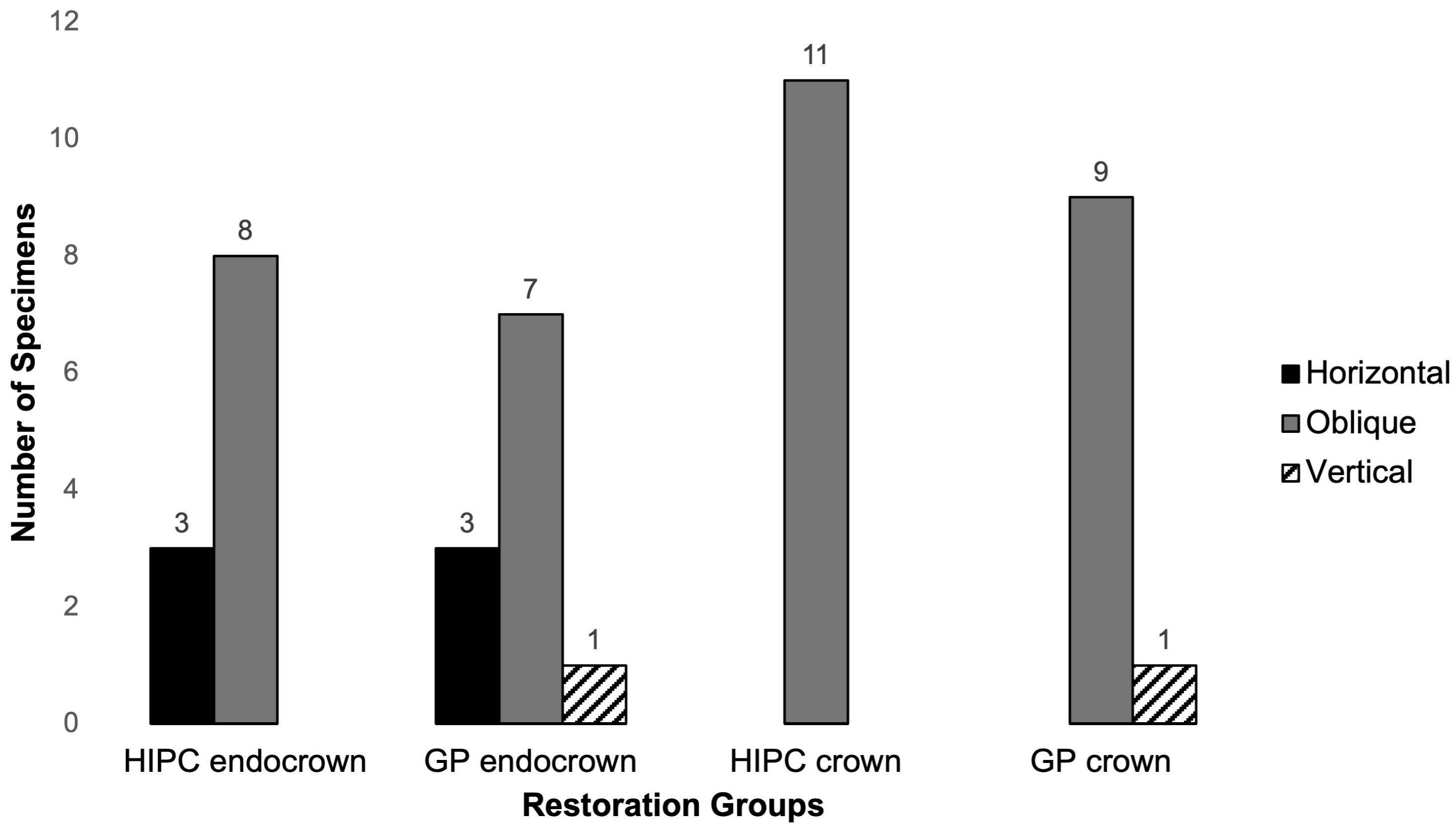
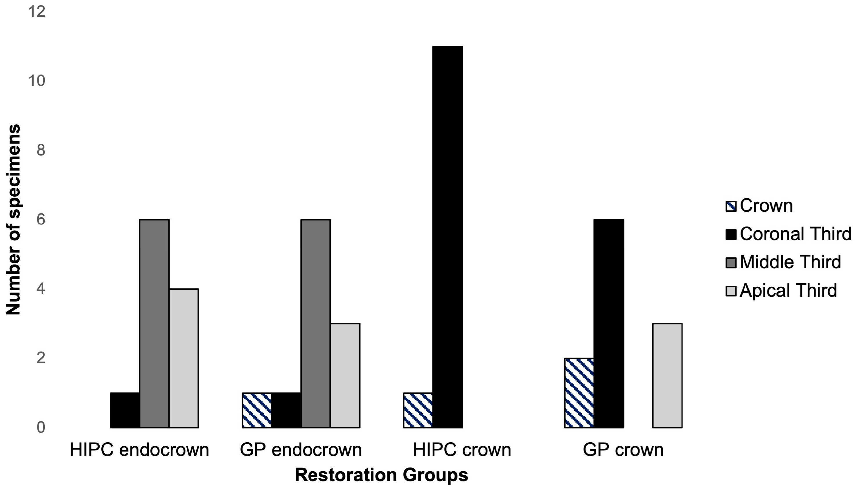
The distribution of fracture types and their localization among the groups are presented in
Figure 1 and
Figure 2. Oblique root fractures were the most frequent failure mode (n = 35), whereas vertical root fractures were the least common (n = 2). No statistically significant differences were observed among groups in terms of fracture type (
p > 0.05).
Regarding fracture localization, no significant differences were detected between materials within the same restoration assembly (endocrowns: p > 0.999; crowns: p = 0.104). However, within the same material, tested restorative assembly significantly influenced fracture localization. breCAM.HIPC crowns exhibited a significantly higher frequency of coronal third fractures compared with breCAM.HIPC endocrowns (p < 0.0001). Similarly, Gradia Plus crowns showed more coronal third fractures, whereas Gradia Plus endocrowns demonstrated a higher incidence of middle third fractures (p = 0.016).
4. Discussion
The present study evaluated the effect of restoration assemblies and material types on the fracture resistance of indirect composite restorations in simulated revitalized immature maxillary central incisors. The results demonstrated that restoration assembly had a significant influence on fracture resistance, whereas material type did not significantly affect the outcomes. Endocrown restorations consistently exhibited higher fracture resistance values than crown restorations, regardless of the material used. These findings suggest that, in structurally compromised teeth following revitalization, the fracture resistance of the restoration is more strongly influenced by restorative assembly-related factors than by the intrinsic properties of the restorative material.
The higher fracture resistance observed in the tested endocrown assembly under the experimental conditions of the present study suggests that this restorative assembly may provide greater resistance to fracture in structurally compromised teeth. Unlike conventional fiber-reinforced crown restorations, endocrowns extend into the pulp chamber, providing both macroretention through the internal geometry of the pulp chamber and micromechanical retention through adhesive cementation [
14]. This combination results in a larger effective bonding surface area and enhanced retention. The increased adhesive interface may improve stress distribution under functional loading, thereby reducing stress concentration at the cervical region [
7,
8,
29,
30,
31]. In contrast, conventional crown restorations supported by fiber-reinforced cores include additional tooth–core and core–crown interfaces, and differences in stiffness among restorative components may influence stress transfer and fracture behavior [
14]. Although the Trinia fiber-reinforced core was incorporated to reinforce the crown groups and to reproduce the conventional restorative approach for severely compromised immature anterior teeth, this additional reinforcement did not appear to compensate for the lower fracture resistance observed in the tested restorative assemblies. One possible explanation is that any reinforcing effect provided by the fiber-reinforced core may have been offset by the more complex restorative assembly and the presence of additional bonded interfaces, which may influence stress transfer and interfacial stress concentration [
32,
33]. In addition, crown preparation and the geometry of the Trinia core may have reduced the amount of remaining coronal tooth structure and altered force transmission under oblique loading. Together, these factors, along with the additional tooth–core and core–crown bonding interfaces, may have created potential weak points and limited the reinforcing effect expected from the fiber-reinforced core. However, because finite element studies have shown that stress distribution is also affected by factors such as loading direction, restoration geometry, and restorative material, the contribution of the fiber-reinforced composite core incorporated in the crown groups cannot be excluded [
33,
34]. Therefore, the higher fracture resistance observed in the present study may partly reflect differences in the structural configuration of the tested restorative assemblies rather than restoration design alone. Accordingly, the findings should be interpreted as reflecting differences between the tested restorative assemblies rather than the isolated effect of restoration design.
Although no statistically significant material-related effect was detected, numerical differences were observed between the tested materials, particularly within the endocrown groups. The relatively small sample size and within-group variability may have limited the ability to detect potential material-related differences. Therefore, the present findings should be interpreted as indicating that no statistically significant differences were detected under the conditions of this study rather than demonstrating equivalence between the tested materials. Although a recent systematic review and meta-analysis reported that different restorative materials may exhibit comparable fracture resistance behavior [
17], the mechanical properties of the investigated materials, including elastic modulus and energy absorption capacity, were not directly evaluated in the present study. Accordingly, explanations based on intrinsic material properties should be regarded as literature-based and hypothesis-generating rather than direct findings of this investigation. Within these limitations, differences between the tested restorative assemblies appeared to have a greater influence on fracture resistance than material-related differences detected under the present experimental conditions.
Although revitalization procedures promote continued root development and may improve dentinal thickness, the fracture resistance of such teeth remains compromised compared with fully developed teeth. Finite element analyses have demonstrated that tissue regeneration following revitalization can contribute to improved stress distribution along the root structure; however, this effect may not be sufficient to fully restore the fracture resistance of immature teeth to that of mature counterparts [
7,
8]. Previous studies have also indicated that fracture resistance values of revitalized teeth are comparable to those treated with apexification, although neither approach fully restores the mechanical strength of mature teeth [
35,
36,
37]. Accordingly, comparisons with previous studies should be interpreted cautiously because differences in treatment protocols, restorative approaches, and experimental models may influence the reported fracture resistance values. In particular, several previous studies evaluated direct restorative techniques, fiber-reinforced composites, apexification procedures, or mature teeth rather than indirect restorative assemblies in simulated revitalized immature anterior teeth [
24,
25,
35,
36,
37]. Biodentine was selected in the present study due to its favorable handling properties and reduced risk of discoloration compared with mineral trioxide aggregate [
38,
39]. Therefore, restorative strategies play a critical role in reinforcing these structurally weakened teeth and compensating for their inherent biomechanical limitations.
In the present study, fiber-reinforced composite cores were used in the crown groups to simulate clinical conditions in extensively damaged teeth. Previous studies have reported that fiber-reinforced systems, such as Trinia, may enhance fracture resistance by providing a dentin-like elastic modulus and improved stress absorption [
26,
27]. However, in contrast to these findings, the crown groups in the present study demonstrated lower fracture resistance values compared with endocrown restorations. This discrepancy may be attributed to the multi-component structure of crown restorations, in which the presence of core and crown interfaces may reduce the effective bonding area and alter stress distribution patterns. Additionally, the reduced thickness of the overlying restorative material in crown configurations may further compromise their fracture resistance under oblique loading conditions.
Previous studies evaluating direct restorative approaches in revitalized teeth have demonstrated that fiber-reinforced composite materials may improve fracture resistance by acting as stress-absorbing layers within the restoration [
24,
25]. Although indirect composite systems are expected to exhibit superior mechanical properties due to higher filler content and controlled polymerization conditions, the fracture resistance values observed in the endocrown groups in the present study were comparable to those reported for fiber-reinforced direct restorations. This finding suggests that the tested restorative assemblies may play a more critical role than the intrinsic material properties, regardless of whether direct or indirect techniques are employed. To the best of our knowledge, there is currently no study directly comparing endocrown and crown restorations fabricated from different indirect composite materials in revitalized immature anterior teeth, which limits direct comparison with previous findings.
In clinical conditions, the type and location of fractures in immature maxillary central incisors are of particular importance because of their aesthetic position [
35]. In the present research, forty-four teeth demonstrated root fractures at the coronal, middle, or apical thirds, regardless of the experimental group. The predominance of root fractures may be explained by the structurally compromised morphology of immature teeth, including thin dentinal walls and incomplete root maturation, which reduce their resistance to functional and traumatic loading [
2,
8]. In addition, finite element analyses have demonstrated that immature maxillary central incisors are subjected to high tensile stress concentrations in the cervical and coronal root regions under oblique loading, predisposing these structurally weakened teeth to crack initiation and propagation [
7]. The 135° oblique loading configuration used in the present study may also have contributed to the predominance of root fractures by generating combined compressive, tensile, and shear stresses in the cervical root region. In addition, embedding the roots in acrylic resin, even with a simulated periodontal ligament layer, may not fully reproduce the viscoelastic support provided by alveolar bone and the periodontal ligament in vivo, which may have influenced stress concentration and fracture propagation.
Crown restorations supported by Trinia fiber-reinforced cores exhibited a significantly higher incidence of coronal root fractures, whereas middle-third fractures were more frequently observed in the endocrown groups. This distinction is clinically relevant because fracture location may influence the available treatment options and the long-term prognosis of the affected tooth. Although coronal-third root fractures may be manageable in selected cases using crown lengthening or surgical or orthodontic extrusion, the clinical management of middle-third root fractures may be more challenging because of their deeper extension within the root. Nevertheless, the restorability of root fractures depends on multiple factors, including fracture extent, remaining tooth structure, ferrule availability, and periodontal support. Therefore, fracture location alone should not be considered the sole determinant of clinical prognosis. Although previous studies have also reported a high incidence of non-restorable root fractures [
25,
35,
38], differences in experimental models, restorative approaches, and fracture classification criteria should be considered when comparing findings across studies. Accordingly, although the tested endocrown restorations demonstrated higher fracture resistance under the experimental conditions of this study, the observed fracture patterns should be interpreted with caution and should not be considered direct evidence of clinical superiority.
The fracture resistance values obtained in this study exceeded the average maximum bite forces reported for individuals with normal occlusion and approached those observed under parafunctional conditions [
7]. While the crown groups demonstrated values within the upper physiological range, endocrown restorations exhibited substantially higher resistance levels. However, these findings should be interpreted cautiously, as in vitro loading conditions do not fully replicate the dynamic and cyclic forces encountered in the oral environment. In particular, the mean fracture resistance values observed in the endocrown groups (approximately 687 N for breCAM.HIPC and 781 N for Gradia Plus) were notably higher than those of the fiber-reinforced crown groups (approximately 453–458 N), suggesting that the tested endocrown assembly may provide greater resistance to fracture under the experimental conditions of the present study.
The findings of this study should be interpreted within the limitations of an in vitro experimental design. Although the experimental model was designed to standardize key clinical steps of revitalization therapy, artificially standardized immature teeth and the revitalization protocol cannot fully reproduce the biological healing process, or the tissue formed following regenerative endodontic procedures. Therefore, the specimens should be considered as immature teeth treated using a standardized revitalization protocol rather than clinically healed revitalized teeth. Furthermore, the absence of positive and negative control groups limited direct comparison of the obtained fracture resistance values with intact mature and untreated immature teeth. As discussed above, the comparison represented two clinically relevant restorative assemblies rather than restoration design alone. Future studies incorporating biologically representative revitalization models, appropriate control groups, cyclic loading protocols, and equivalent internal support structures are warranted to further validate the present findings.
Only static fracture testing was performed, without fatigue loading or chewing simulation and the load was applied at 135° in a single direction, representing only one possible clinical loading scenario for anterior teeth. Therefore, the results primarily reflect the ultimate fracture resistance of the tested restorative assemblies under one standardized loading scenario rather than the long-term behavior of restorations exposed to cyclic and multidirectional intraoral forces [
33]. Under clinical conditions, repeated loading and progressive degradation at the tooth–restoration interface may influence crack initiation and propagation, particularly in structurally compromised immature teeth with thin dentinal walls [
32,
33]. Consequently, although static fracture testing provides valuable information regarding ultimate fracture resistance, it cannot fully reproduce the fatigue-related failure mechanisms that develop over time in the oral environment [
6]. Future studies incorporating chewing simulation, cyclic fatigue loading, multidirectional loading models, and long-term artificial aging are warranted to better evaluate the long-term clinical performance of these restorative approaches.
Although 10,000 thermocycles are often considered to approximate one year of clinical thermal aging, this relationship should be interpreted cautiously because intraoral thermal challenges cannot be directly translated into a fixed number of laboratory cycles [
28,
40]. Therefore, thermocycling should be regarded as an accelerated artificial aging method rather than a direct simulation of clinical service time.
Importantly, the use of a fiber-reinforced composite core in the crown groups, while intended to simulate clinical scenarios of extensively damaged teeth, may have introduced a confounding effect when comparing the tested restorative assemblies. This difference in structural configuration between endocrown and crown groups likely influenced the observed superiority of endocrowns, as the presence of a fiber-reinforced core introduces additional interfaces that can alter stress distribution and fracture behavior. Therefore, the observed differences between the tested restorative assemblies should be interpreted with caution, as they may have been partially influenced by differences in their internal support structures.