Article

Spinal Cord Transcutaneous Stimulation Priming and Lower Limb Neuromuscular Performance in Individuals with Anterior Cruciate Ligament Reconstruction After Their Return to Sport: A Case Series


1
Department of Medicine, University of Udine, 33100 Udine, Italy
2
School of Sport Sciences, University of Udine, 33013 Gemona del Friuli, Italy
3
Department of Neurosciences, Biomedicine and Movement Sciences, University of Verona, 37124 Verona, Italy
4
Department of Theoretical and Applied Sciences, eCampus University, 22060 Como, Italy
5
Fisioterapia Sport Gorizia, 34170 Gorizia, Italy
6
Orthopaedics Clinic, Azienda Sanitaria Universitaria Friuli Centrale (ASUFC), 33100 Udine, Italy
*
Author to whom correspondence should be addressed.
These authors contributed equally to this work.
Appl. Sci. 2026, 16(14), 6913; https://doi.org/10.3390/app16146913
Submission received: 3 June 2026 / Revised: 2 July 2026 / Accepted: 7 July 2026 / Published: 10 July 2026

Abstract

Background: Neuromuscular deficits may persist following anterior cruciate ligament reconstruction (ACLR), even after returning to sport. In this exploratory case series, we investigated the effects of non-invasive spinal cord transcutaneous stimulation (scTS), applied during warm-up for approximately 25 min to prime the nervous system, on neuromuscular performance of the ACLR limb during a simulated half-squat power training session. Methods: Four young, non-professional athletes with time since ACLR between 9.0 and 14.5 months, who were already returned to sport, participated in this study. Force, velocity, power, and electromyography of representative thigh muscles generated by the ACLR and intact limbs during unilateral half-squats on a Smith machine, were assessed over two separate experimental sessions (scTS or sham stimulation priming). Results: Three of the four participants showed power deficit in the ACLR limb during the concentric phase. scTS priming promoted higher power output generated by the ACLR limb in these three participants. Oscillatory-like force pattern during the downward phase was more prominent in the ACLR limb for one subject, and scTS priming attenuated it. Conclusions: These findings support future controlled trials to assess the efficacy of scTS priming as an aid to enhance rehabilitation and physical training after ACLR.

1. Introduction

Anterior cruciate ligament (ACL) injuries are among the most common and debilitating musculoskeletal injuries in athletes [1], with relevant consequences for both the athlete’s and team’s productivity [2]. The incidence of this type of injury is 68.6 per 100,000 person-years [3], with more than half of cases occurring in non-contact scenarios [4]. ACL rupture often requires reconstructive surgery [5] followed by a long recovery period that includes rehabilitation [1], which typically spans 9 to 12 months before a complete return to competition [6]. This period is divided into phases, each generally guided by objective criteria [6], such as symmetry in force and/or power generation between the injured and contralateral sides, as well as the hamstrings-to-quadriceps torque ratio of the injured leg [7,8,9]. Despite long and structured rehabilitation programs, only a subset of ACL-reconstructed athletes returns to their pre-injury level or to competitive-level sport [10]. Additionally, the rate of re-injury remains high, with almost one out of five (~18%) top-level male football players sustaining a second ACL injury after return-to-play [11], suggesting that the indexes currently used to guide return-to-play decision making are not sufficient.
Quadriceps femoris strength deficit is a consequence of ACL injury that can persist despite the lengthy rehabilitation implemented following surgery. For example, up to 40% of patients showed interlimb differences in quadriceps strength at 6 months after ACL reconstruction (ACLR), with moderate differences in isometric and concentric strength, and significant differences in eccentric strength [12]. Important strength deficits in the ACLR limb can also be found more than one year after ACLR [13,14]. Such marked and long-lasting weakness of the quadriceps femoris is not only due to the transient disuse of the ACLR limb. In fact, ACL injury and subsequent reconstructive surgery can lead to deleterious neural adaptations affecting cortical, spinal, and sensory structures, which are generally termed arthrogenic muscle inhibition (AMI) [15]. AMI, in turn, can limit the athlete’s functional performance and the potential of exercise-based rehabilitation to promote positive physiological adaptations [16].
In recent years, non-invasive neuromodulation strategies that are not incorporated into conventional rehabilitation programs, such as non-invasive brain stimulation, have been increasingly investigated after ACLR for their potential to modulate neural excitability; however, the related findings across studies remain inconsistent [15,17]. For example, a two-week transcranial magnetic stimulation protocol started two days after ACLR improved corticospinal pathway excitability and range of motion during knee extension [18]. Similarly, transcranial direct current stimulation applied during a 6-week exercise-based rehabilitation program that started two weeks post-ACLR, reduced maladaptive quadriceps cortical plasticity compared to sham stimulation [16]. On the other hand, a single-session study conducted 4–6 months post-ACLR did not find any effect of transcranial direct current stimulation on corticospinal excitability and isokinetic performance of the quadriceps femoris [19].
Spinal cord transcutaneous stimulation (scTS) is another non-invasive neuromodulation technique originally developed to enhance motor recovery in individuals with spinal cord injuries [20]. In this population, scTS generally increases the excitability of the spinal circuitry controlling the lower limbs, so that peripheral sensory information and residual supraspinal inputs can reemerge as functional sources of control for motor patterns generation.
Exploratory studies in able-bodied, healthy individuals investigated the effects of scTS on motor control and neuromuscular performance, reporting positive effects on countermovement jumps repeated over time [21], as well as on learning and retention of backward locomotion [22] and ballistic plantar flexions [23]. In contrast, negative or no effects were observed on standing postural control [24,25]. More recently, we implemented scTS to prime the nervous system prior to different lower limb motor tasks in young, physically active male individuals. We found that scTS priming improved neuromuscular performance during fatiguing high-intensity efforts [26], whereas no effects were observed on low-level torque steadiness [27] or single-leg stance balance control [28]. Overall, these studies suggest that scTS may increase the excitability of the spinal circuitry, bringing the related neuronal networks closer to their activation threshold [23,29,30,31].
To the best of our knowledge, no studies on the effects of scTS on neuromuscular performance after ACLR are available to date. The main aim of this within-subject-controlled case series was to explore the effects of scTS priming on neuromuscular performance of the ACLR limb during a simulated half-squat power session in young individuals who had already returned to sport. We also compared the neuromuscular performance of the ACLR limb versus the intact limb to support the rationale of this study, which was based on the assumption that critical neuromuscular deficits may persist in the ACLR limb even after lengthy rehabilitation and return to competitive sport practice (return to sport), thereby increasing the risk of re-injury. However, we did not perform neurophysiological assessments to determine the causes and mechanisms of such potential deficits. We hypothesized that the ACLR limb would demonstrate neuromuscular deficits during the concentric and/or eccentric phase of a simulated half-squat power training session compared to the contralateral, intact limb. Furthermore, we hypothesized that scTS priming would improve the neuromuscular performance of the ACLR limb during the simulated power training session as compared to the application of sham spinal stimulation priming. Positive findings from this study could set the stage for well-powered, randomized controlled trials to assess the potential efficacy of scTS priming for enhancing neuromuscular recovery following ACL injury.

2. Materials and Methods

2.1. Research Participants

Four young non-professional athletes, whose characteristics are reported in Table 1, were recruited at the School of Sport Sciences (University of Udine, Udine, Italy) to participate in this study. All subjects underwent surgical ACLR after injury, completed a rehabilitation program, and subsequently returned to regular sport practice prior to participating in the study. At enrolment, time since ACLR ranged between 9.0 and 14.5 months, and participants reported practicing team sports (soccer, rugby), trail running, and/or related conditioning activities for 3.3 ± 1.0 days/week. Individuals had no history of other relevant orthopedic or neurological injuries. The experimental protocol was conducted in accordance with the Declaration of Helsinki and was approved by the Institutional Review Board of the University of Udine (IRB# 197/2023) on 4 October 2023. Before the start of the study, subjects were carefully informed about its purpose and risks, and written informed consent was obtained from all of them.

2.2. Experimental Protocol

The experimental protocol of this study consisted of three laboratory visits, each lasting approximately 1 h 30 min. In the first session, after a brief familiarization with the laboratory equipment and scTS procedures, the subjects underwent an anthropometric assessment. The overload to be used on the Smith machine during the subsequent sessions was then defined. Finally, the subjects practiced the unilateral half-squat power training that would be evaluated during the subsequent two sessions. The second and third experimental sessions were devoted to the assessment of kinetic and electromyographic (EMG) parameters during the unilateral half-squat power training session preceded by the scTS or sham stimulation (sham) priming. The testing order for the scTS or sham priming was randomized (random number generator, MATLAB v. 2025b, the Mathworks, Natick, MA, USA) (Table 1). To limit the potential influence of diurnal variability, each participant completed all sessions under constant environmental conditions (20 °C, 60% humidity), and at a similar time of day, within a tolerance of ±1 h [32]. The interval between consecutive sessions ranged from 6 to 7 days. Participants were instructed to wear the same gymnastic shoes across all sessions. They were also asked to refrain from strenuous physical exercise and alcohol consumption and to maintain their habitual sleeping behavior during the 2 days preceding each experimental session. In addition, participants were instructed to abstain from caffeine and energy drink consumption for at least 3 h prior to each experimental session [33].

2.3. Anthropometric Characteristics

A manual scale (Seca 709, Hamburg, Germany) was used to measure body mass with the participant wearing light underwear and no shoes. A wall-mounted height board was used to measure the stature of participants.

2.4. Unilateral Half-Squat

As previously reported [26], participants performed the explosive unilateral half-squats with overload on a Smith machine (Multipower, Technogym, Cesena, Italy). The guided barbell of this device facilitated balance control during the explosive extensions as well as during the downward phase, which was requested to be controlled and performed at a self-selected velocity. A rigid tactile feedback indicated when 100 degrees of knee flexion was reached during the downward phase of the half-squat. From this position, participants were instructed to perform an explosive lower limb extension. The exercising limb alternated at each repetition. Thus, upon completion of each unilateral lower limb extension, the foot of the uninvolved leg, which was actively flexed, was placed near the supporting foot for the bilateral stance phase lasting approximately 1 to 2 s. To ensure consistent foot placement across half-squat repetitions and experimental sessions, tape markers were applied onto the force platform. The position of the tactile feedback was also defined with respect to the Smith machine and maintained constant between experimental sessions. These strategies were implemented to maximize the reproducibility and consistency of the exercise considered for analysis.

2.5. Selection of the Overload

During the first experimental session, participants began the procedures related to the Smith machine overload selection after approximately 10 min of standardized warm-up, including pedaling on a cycloergometer, joint mobilization, and half-squats (6 bilateral repetitions and 6 unilateral repetitions with 20 kg as overload) [34]. As previously reported by our group [26], participants performed one set of three unilateral half-squat repetitions per leg for each overload that was tested. A linear position transducer (Vitruve, SPEED4LIFTS S.L., Madrid, Spain) was used to assess the mean propulsive upward velocity of the Smith machine barbell. The initial overload of 25 kg was increased by 5 kg for each subsequent set until the mean propulsive velocity of the weaker leg was lower than 0.45 m/s. The corresponding overload was selected and used for the subsequent simulated power training sessions. A 3 min rest period was implemented between sets.

2.6. scTS Cathode Site

As described in detail elsewhere [26,27,28,35], during the first experimental session, the optimal scTS cathode placement site was identified by means of recruitment curve assessment. Briefly, the participant lay relaxed in supine position on a standard examination table while a constant-current stimulator (DS7A, Digitimer, Hertfordshire, UK; maximum output 400 V) was controlled via a trigger box (GeMS TRIGGER BOX, EMS, Bologna, Italy) using a dedicated software (Direct USB for TRIGGER BOX, Version 1.00, EMS, Bologna, Italy). Single-pulse stimulation (1 ms monophasic square-wave) was delivered via self-adhesive electrodes every 4 s. Two 100 × 50 mm electrodes (20021, Axion GmbH, Leonberg, Germany) were placed symmetrically over the iliac crests as anodes, and a circular electrode (diameter: 25 mm; E-CM25, TensCare, Surrey, UK) was applied at the thoracic (T)11-12 or T12-lumbar(L)1 intervertebral space as a cathode. The order of cathode placement at these two sites was randomized, with a 4 min rest interval between stimulations at each site. Stimulation intensity started at 5 mA and was increased in 5 mA increments up to 100 mA, with five pulses delivered at each intensity level. All participants reached and tolerated the maximum intensity of 100 mA without discomfort. The cathode site eliciting preferential recruitment of the vastus lateralis (VL) muscle, as determined by (i) the lower stimulation intensity at motor threshold and (ii) the greater peak-to-peak amplitude of evoked potentials to spinal stimulation [27] was selected for experimental sessions 2 and 3. In order to maximize the reproducibility of cathode electrode placement across experimental sessions, the electrode location was marked on the skin with a permanent ink pen, and pictures of the electrode were also taken.

2.7. Priming Protocol with scTS or Sham Stimulation

The exercise-based intervention previously proposed by our group [26,27,28] was implemented as a priming protocol with the application of scTS or sham stimulation during experimental sessions 2 and 3. The priming protocol consisted of 10 min of quiet standing and a subsequent 15 min of warm-up activities, including stepping in place, joint mobilization, unilateral balance control, and unilateral quarter squats interleaved by periods of quiet standing. Participants were instructed to perform the warm-up at a self-selected intensity that would not lead to fatigue.
The stimulation setup was the same as that described above (see Selection of scTS cathode site) [26,27,28], and the circular electrode was placed on the skin at the T11-12 intervertebral space for S1 and S3, or at T12-L1 for S2 and S4. The electrodes were secured around the participant’s trunk using an elastic bandage. Participants were informed that two different types of stimulation would be applied during the priming protocol in this study; however, they were blinded to the specific purpose of these two different stimulations. As previously described [26,27,28], scTS during priming was characterized by 28 Hz and a monophasic waveform with a pulse width of 1000 µs. Stimulation intensity increased gradually to achieve the highest value that was comfortable and allowed the participant to perform the proposed motor tasks without limitations. In this study, the intensity applied during the scTS priming protocol ranged between 16 mA (S2) and 22 mA (S4). All four subjects tolerated the scTS priming well, and no visible contractions of the lower limb muscles were observed during stimulation.
During sham priming, the stimulation pulse width was 200 µs, and the intensity was gradually increased for approximately one minute to the same value used for scTS priming. This was followed by a 10 s ramp-down period and a subsequent stimulator turn-off. Thus, no stimulation was delivered for the remaining ~24 min of the sham priming protocol [36,37]. The stimulation setup appeared active for the entire duration of the sham priming session, during which the operator periodically asked participants whether the stimulation was comfortable or not, as it was done during the scTS priming protocol. Although credibility and expectancy of the sham stimulation protocol were not formally assessed, participant S3 self-reported the perception of stimulation in the intact limb, along with improved control and readiness of the ACLR limb during sham priming. Furthermore, when participants were individually debriefed after completion of the study to review their results, all of them expressed surprise upon learning that no stimulation had been applied during one of the two power training sessions. The fact that individuals generally habituate to the cutaneous sensation of active scTS within about a minute [36] contributes to the credibility of the sham stimulation protocol implemented in this study.

2.8. Simulated Power Training Session

The unilateral half-squat power training session examined in this study consisted of four sets of 6 repetitions per leg. Three minutes of rest were provided in between sets. At the beginning of each set, the participant positioned the barbell across their shoulders and maintained stable standing for 10 s. After this phase, they were instructed to move the barbell upward ‘as fast as possible’ at each repetition. An operator, who was blinded to the stimulation priming condition, checked the execution of each half-squat repetition and provided strong verbal encouragement to the participant. Audio and visual feedback related to barbell upward velocity were also provided to motivate participants to exert maximal effort. Force plate and surface EMG signals were collected during the power training sessions considered for analysis (experimental sessions 2 and 3). During experimental session 2, Subject 4 requested to skip the fourth set because of the perception of impaired control of the ACLR limb, along with local fatigue. Thus, only for this participant, three sets were considered for analysis during both experimental sessions.

3. Data Acquisition

Kinetic and EMG signals were recorded by a dedicated acquisition system (Smart DX I, BTS Bioengineering, Milan, Italy) and the related software (Smart Motion Capture System Version: 1.10.0469, BTS Bioengineering, Milan, Italy) using a sampling rate of 1000 Hz. Ground reaction forces were collected by a force plate (Kistler, Type 9287CA, Winterthur, Switzerland). The EMG was recorded by a wireless EMG system (BTS FREEEMG1000, BTS Bioengineering Milan, Italy; Input impedance: 100 MΩ; Common Mode Rejection Ratio: >110 dB @50–60 Hz; Sensitivity: 1 µV) with pre-gelled surface electrodes (BlueSensor N-00-S/25, Ambu, Bayan Lepas, Malaysia) positioned with an inter-electrode distance equal to 2 cm.
Prior to electrode placement, the skin was prepared to minimize impedance by shaving the area and rubbing the skin with an abrasive paste. The EMG was collected bilaterally from the vastus lateralis (VL), at two-thirds on the line from the anterior spina iliaca superior to the lateral side of the patella; rectus femoris (RF), midway between the anterior spina iliaca superior and the superior part of the patella; and biceps femoris (long head, BF), midway between the ischial tuberosity and the lateral epicondyle of the tibia [38]. To ensure consistent EMG electrode placement across experimental sessions, electrode locations were marked on the skin with a permanent ink marker, thereby maximizing the reproducibility of the recording setup.

4. Data Analysis

All experimental data were processed and analyzed by the same experienced investigator using LabChart Reader (ADInstruments, Inc., Dunedin, New Zealand). Vertical ground reaction force signals were low-pass filtered at 25 Hz, and EMG signals were band-pass-filtered at 10–499 Hz. All investigated variables were assessed separately for the ACLR or intact lower limb, and were compared to assess: (i) the difference between ACLR and intact lower limb, and (ii) the effect of scTS versus sham priming in the ACLR limb.

4.1. Kinetic Data Analysis

Kinetic data were analyzed as previously reported by our group [26]. The baseline vertical ground reaction force attributable to the participant’s body mass and the additional Smith machine load, representing the constant system mass, was removed from the force signal recorded during the active portion of each power training set to obtain the net vertical ground reaction force [39]. The system mass velocity was then calculated across each entire set by integrating the net vertical ground reaction force–time curve using the trapezoidal method [40]. A dedicated time constant decay function (τ = 1 s) was implemented to avoid small errors due to integration drift [41,42]. Instantaneous power was obtained by multiplying the instantaneous values of velocity and the net vertical ground reaction force [40]. These procedures allowed us to define the eccentric and concentric phase of each unilateral half-squat repetition by identifying: (i) the start of the eccentric phase as the time point at which the velocity becomes negative or crosses from positive to negative; (ii) the onset of the concentric phase, corresponding to the end of the eccentric phase, when the velocity crosses zero while changing from negative to positive; and (iii) the end of the concentric phase as the time point corresponding to the net vertical ground reaction force decreasing to zero [40,43].
Mean force, velocity, and power generated during the concentric phase of each half-squat repetition were considered to assess neuromuscular performance. Furthermore, during the eccentric phase of each repetition, a marker of oscillatory-like pattern of the ground reaction force signal (i.e., force standard deviation frequency, SDf_F) and the mean velocity were assessed as kinetic markers of neuromuscular control. In particular, SDf_F defined the standard deviation of the mean power frequency distribution of the ground reaction force signal, within the selected frequency range (0–500 Hz).

4.2. Electromyography Amplitude Data Analysis

Root-mean-square (RMS) analysis was used to quantify the EMG amplitude of VL, RF, and BF during the concentric and eccentric portions of each unilateral half-squat repetition. For normalization, the RMS values were expressed as a percentage of the EMG amplitude recorded during a submaximal voluntary isometric contraction (sub-MVC) [44,45]. This alternative approach to MVC normalization is suggested when MVC cannot be reliably performed due to the risk of injury, pain, or discomfort. A limit of the sub-MVC normalization is that different synergic muscles may be recruited to different levels during the submaximal reference task, making between-muscle comparisons difficult to interpret. However, normalizing by sub-MVC can still reduce inter-individual variability in factors affecting EMG amplitude, leading to an improved interpretation of the EMG outcome compared to no normalization.
Experimental procedures related to sub-MVC normalization of EMG amplitude were carefully controlled and replicated between experimental sessions. Briefly, the overload selected for the power training sessions was placed onto the Smith machine. With the barbell placed across their shoulders, participants were then asked to flex the lower limbs and achieve a knee angle of 100 deg. Feet position and knee angle were standardized between experimental sessions by the tape markers applied onto the force platform and the tactile feedback, respectively. At this stage, the participant actively flexed the uninvolved lower limb and maintained the unilateral half-squatted position for approximately 4 s. The highest EMG amplitude detected with a 1 s running window was then used for normalization. A total of four sub-MVCs (two with the ACLR and two with the intact limb) were performed at the beginning of Session 2 and 3, after a dedicated 5 min warm-up, with 2 min of rest in between the sub-MVC attempts.

4.3. Muscle Recruitment Data Analysis

For each investigated muscle, the raw EMG recordings were full-wave rectified and subsequently integrated over each concentric and eccentric phase selected for analysis. The resulting integrated EMG signal (iEMG) was then normalized to the peak value reached at the completion of the corresponding phase and expressed as a percentage of that value (Figure S1). Normalized iEMG values extracted at 20%, 40%, 60%, and 80% of each phase were then used to characterize intermuscular coordination through the dispersion index (DI) [46]. The lower DI values indicate smaller differences in EMG recruitment among the considered muscles, which can be interpreted as a marker of increased co-activation. The area under the DI curve was finally considered for analysis (Figure S1).

5. Statistical Analysis

Results are reported as mean and standard deviation of repeated measures within each subject. An autoregressive integrated moving average model with an exogenous variable (ARIMAX) was implemented to assess whether repeated measures collected within each participant were significantly different between the tested conditions. In particular, the effect of (i) lower limb (ACLR vs. Intact) or (ii) stimulation (scTS vs. sham priming) as exogenous variables was tested on the kinetic and EMG parameters assessed in this study. The model assumption of residual independence was checked with the Ljung–Box test and by visual inspection of residuals plotted across repetitions. A p-value lower than 0.05 was considered statistically significant. Cohen’s d effect size (ES) was also calculated for these comparisons. Values of ES lower than 0.20 were defined as negligible, between 0.20 and 0.49 as small, between 0.50 and 0.79 as medium, and equal to or greater than 0.80 as large. Statistical analysis was performed using JASP 0.19 (University of Amsterdam, Amsterdam, the Netherlands).

6. Results

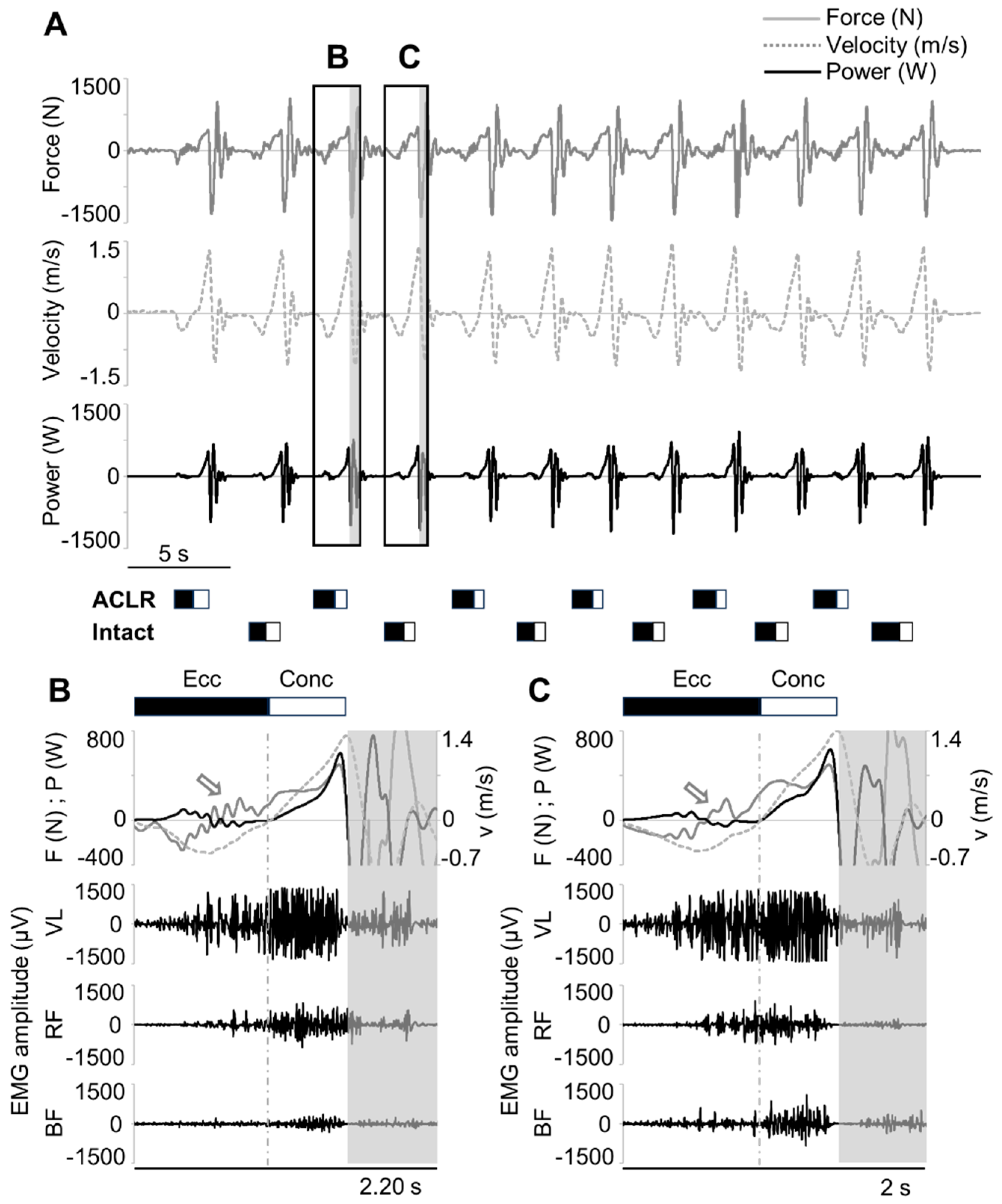
Figure 1A illustrates representative force, velocity, and power profiles recorded throughout one set of unilateral half-squats performed on the Smith machine by participant S4 using 70 kg as overload. An expanded view of a single repetition is presented in Figure 1B,C, where the same kinetic variables are shown together with EMG activity of lower limb muscles for the ACLR and intact limb, respectively.
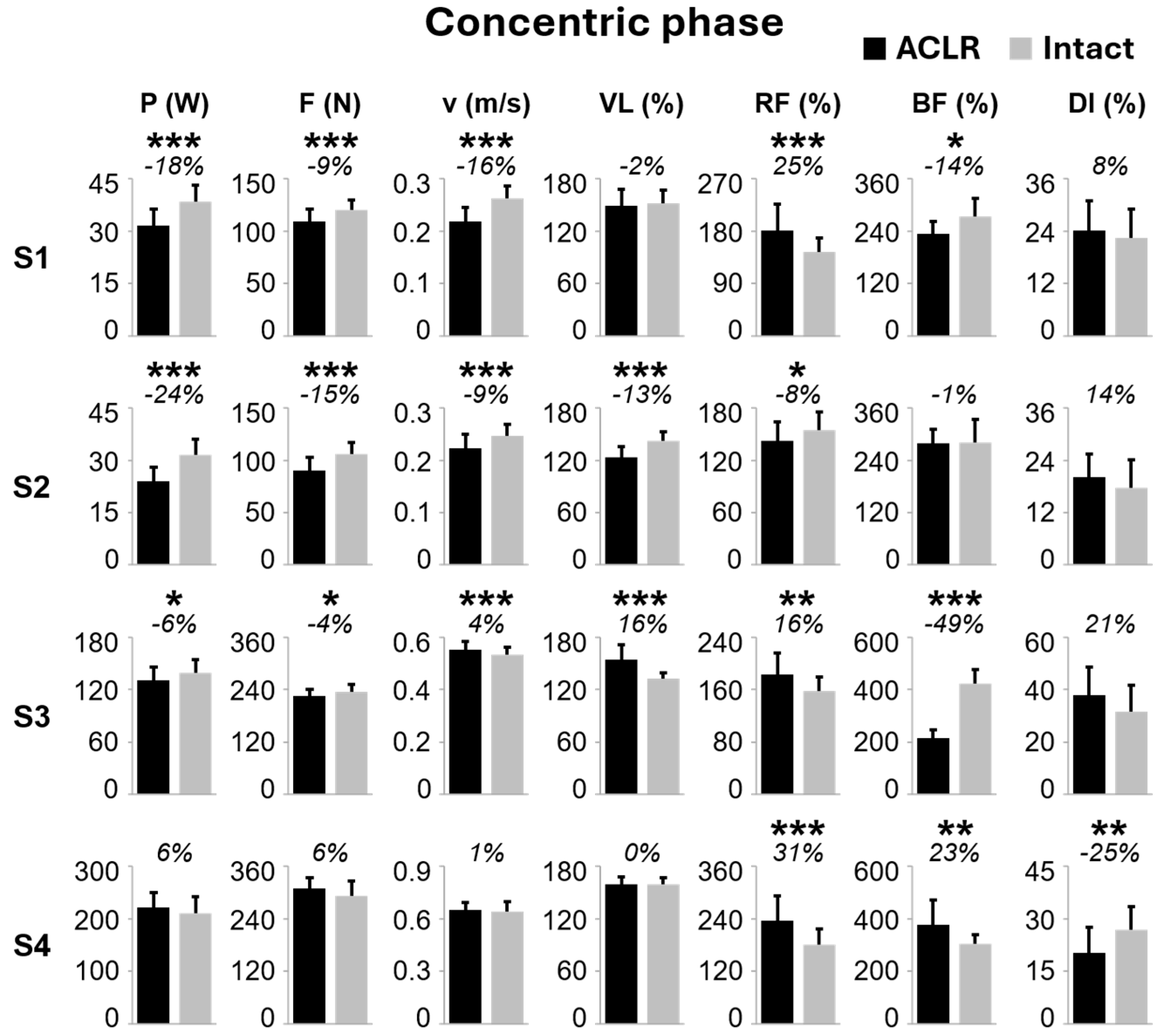
Three of the four participants (S1, S2, S3) showed medium-to-large deficit of power generated during the concentric phase by the ACLR limb compared to the intact limb (Figure 2), which ranged between −24% (p < 0.001, ES = 1.51; S2) and −6% (p = 0.026, ES = 0.51; S3). Such power deficit for the ACLR limb was accompanied by lower force output, with differences ranging between −15% (p < 0.001, ES = 1.12; S2) and −4% (p = 0.015, ES = 0.59; S3). Concentric velocity was also lower for the ACLR limb in these three subjects, with differences ranging between 16% (p < 0.001, ES = 1.45; S1) and 9% (p < 0.001, ES = 0.90; S2) (Figure 2). Inter-limb differences in the EMG activity of representative thigh muscles were observed across participants. It is worth mentioning a common trend for the ACLR side of S1 and S3, which consisted of EMG amplitudes that were higher in RF and lower in BF when compared to the intact limb (Figure 2). Also, the ACLR side of S4, which did not present concentric power deficit, demonstrated higher EMG amplitude of RF (31%, p < 0.001, ES = 0.84) and BF (23%, p < 0.01, ES = 0.94), as well as lower EMG DI (−25%, p < 0.01, ES = 0.58) compared to the intact side.
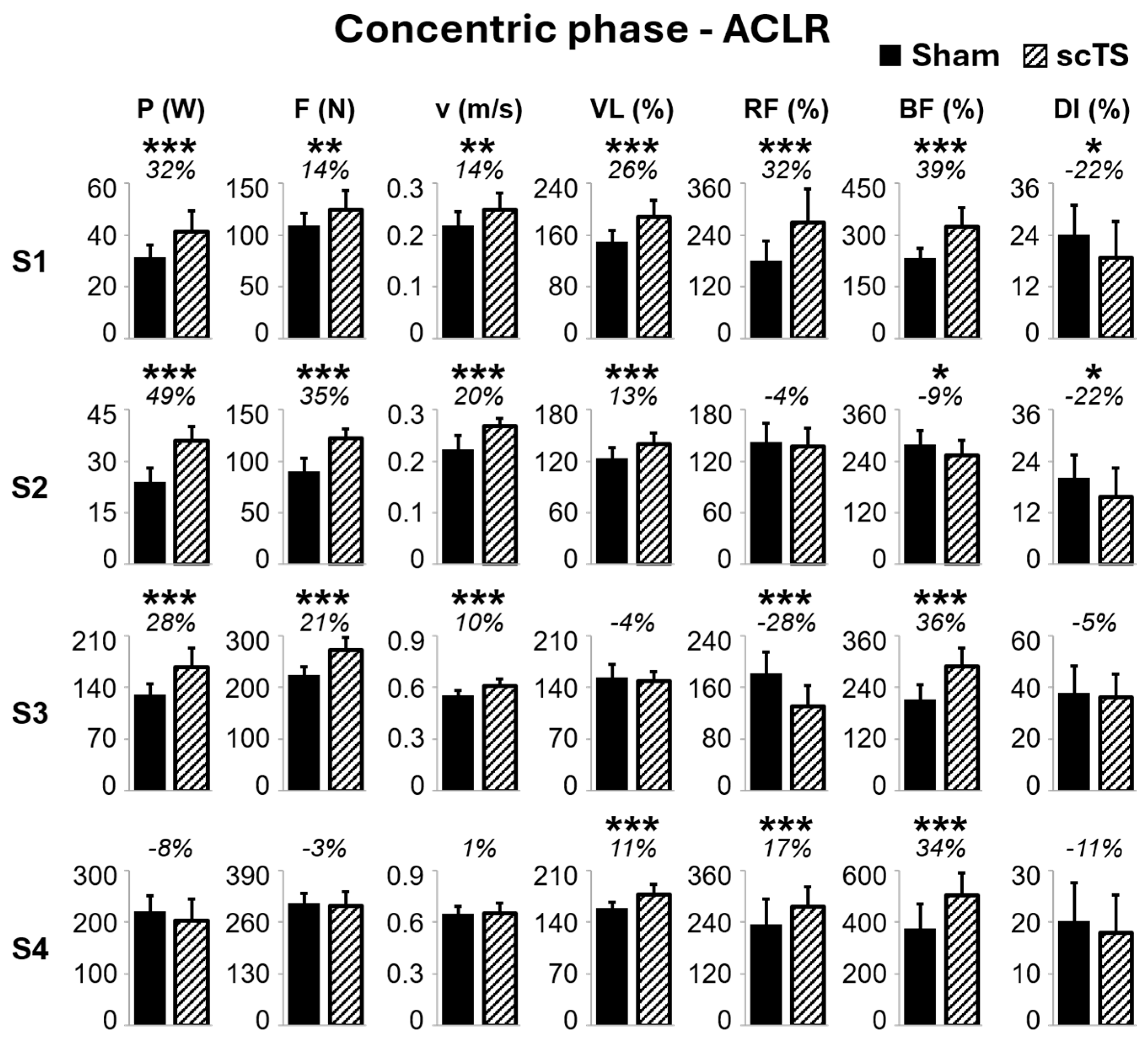
Notably, scTS priming increased the concentric power output of the ACLR limb (Figure 3) in the three participants (S1, S2, S3) who demonstrated a power deficit versus the intact limb during the sham priming (i.e., control) session (Figure 2). Such scTS priming-induced increments in power output ranged between +28% (p < 0.001, ES = 1.43; S3) and +49% (p < 0.001, ES = 2.27; S2), and were accompanied by similar, significant trends of increased force and velocity (Figure 3). Overall, eight of the 12 ACLR limb muscles considered for analysis showed higher EMG amplitude in the scTS priming session compared to the sham priming. However, such a trend was also found in participant S4, who did not show concentric power deficit in the ACLR leg and did not increase power output with scTS priming. On the other hand, lower EMG DI (−22%, p < 0.05) was associated with the increased power output promoted by scTS priming in S1 and S2.
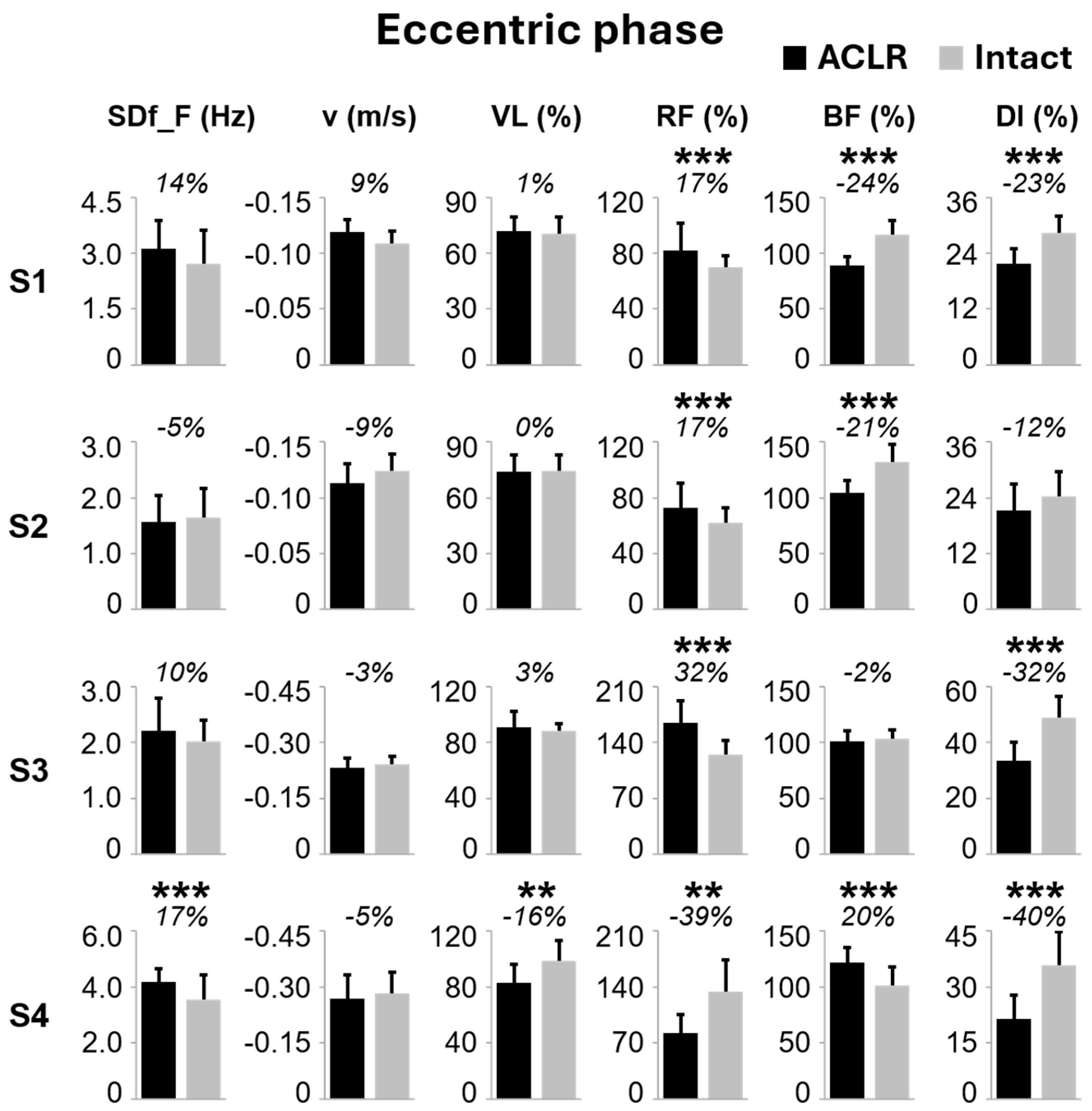
During the eccentric (i.e., downward) phase of the half-squat examined in the control (i.e., sham priming) session (Figure 4), one participant (S4) showed 17% larger SDf_F values in the ACLR limb compared to the intact lower limb (p < 0.001, ES = 0.72). Such a difference in SDf_F can be related to the exemplary difference in force oscillation profiles shown between ACLR and intact limbs (Figure 1B and 1C, respectively). Inter-limb differences in the activation of representative thigh muscles were observed across participants. In the ACLR limb, higher RF EMG amplitude and lower BF EMG amplitude compared to the intact side were found in three subjects (S1, S2, and S3) and two subjects (S1 and S2), respectively. Additionally, the EMG DI was lower in the ACLR limb for three of the four participants (Figure 4), with magnitudes ranging between −40% (p < 0.001, ES = 1.18; S4) and −23% (p < 0.001, ES = 1.45; S1). A similar trend was also observed in S2 (−12%, p = 0.058, ES = 0.44).
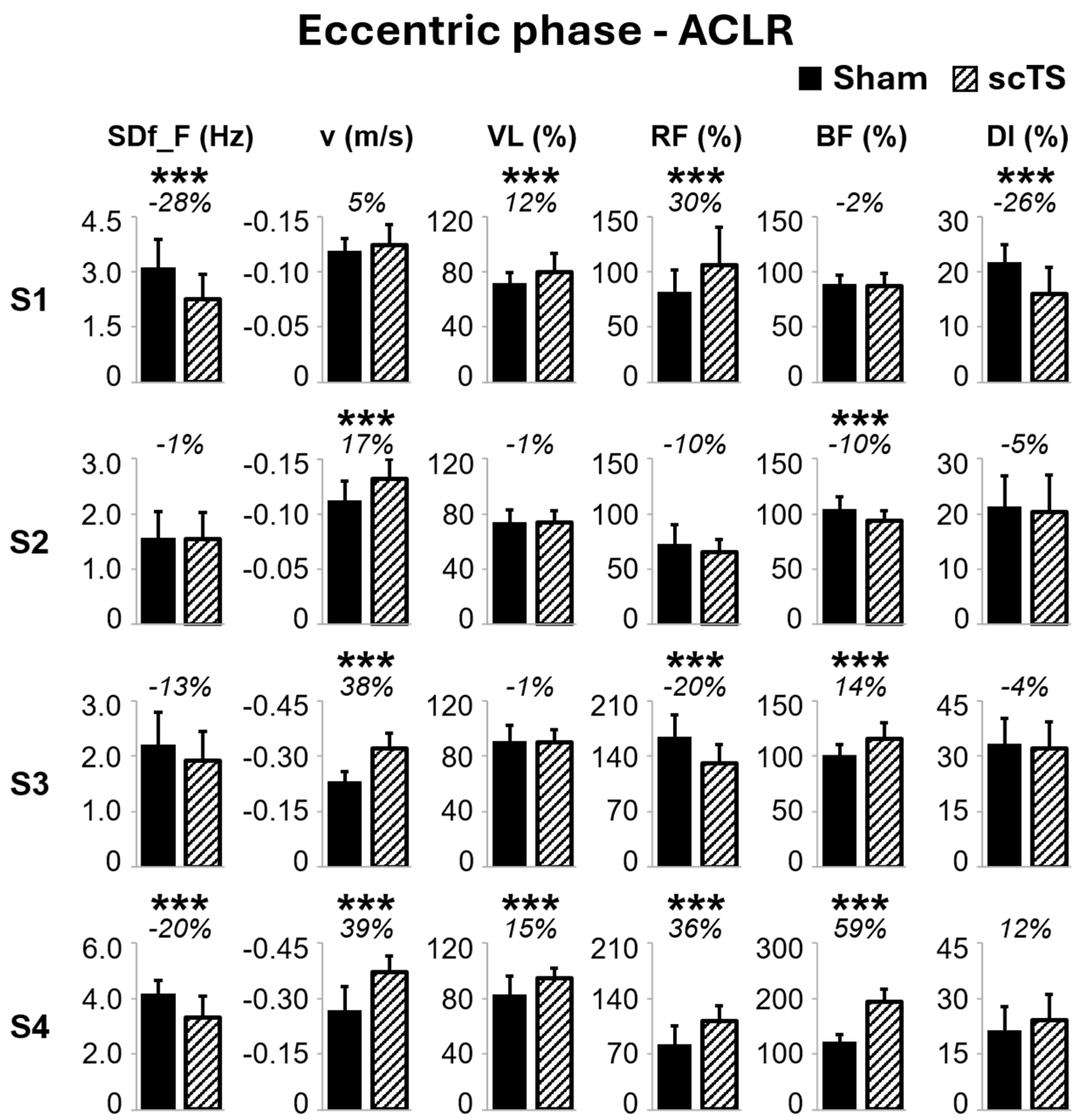
It is worth noting that scTS priming decreased SDf_F in the ACLR limb by 28% in S1 (p < 0.001, ES = 0.97) and by 20% in S4 (p < 0.001, ES = 0.87) when compared to the sham priming (Figure 5), with a similar trend observed in S3 (−13%, p = 0.067, ES = 0.47). Furthermore, scTS priming promoted higher downward velocity in three participants, with magnitudes ranging between +17% (p < 0.001, ES = 0.96; S2) and 39% (p < 0.001, ES = 1.66; S4) when compared to sham priming. Such changes in kinetic outcomes between scTS and sham priming sessions were accompanied by some differences in EMG activity of the thigh muscles considered for analysis, without a consistent trend among participants (Figure 5).

7. Discussion

Three of the four participants included in this case series showed substantial neuromuscular deficits in the ACLR limb compared to the intact limb during the concentric phase of a simulated unilateral half-squat power training session, in spite of the completion of rehabilitation and return to sport. Notably, the same three individuals increased the power output generated by the ACLR limb when scTS priming was implemented prior to the power training session, with differences ranging between 28% and 49% compared to sham priming. We also considered for analysis the downward phase of the half-squat, finding that a marker of vertical ground reaction force oscillation (SDf_F) was larger in the ACLR limb compared to the intact lower limb for the participant who did not show concentric power deficit. Two subjects also showed a decrease in SDf_F for the ACLR limb when scTS priming was implemented.

8. ACLR Results in Long-Term Deficit of Neuromuscular Performance

Following ACLR, deficits in neuromuscular function and structure are commonly observed, such as the reduction in quadriceps muscle cross-sectional area, tissue quality, strength, central activation ratio, and rate of torque development [47,48,49,50,51]. These deficits can persist for several months and even years after the completion of rehabilitation and return to sport [52]. For example, deficits in knee extension peak torque, ranging from 3% to 29%, were found in ACLR team sport athletes compared to a control group more than six months after surgery [52,53]. Residual negative impacts in physical performance have also been reported between 5.6 and 7.6 months following ACLR, ranging from an 11% deficit in single-leg countermovement jump power to a 24% deficit in knee-extension peak torque [54,55]. A similar impairment was also detected two years post-surgical reconstruction of the ACL, with a 13% deficit in peak knee joint power generated during a countermovement jump by the ACLR limb compared to the contralateral side [56].
These findings are consistent with the significant, medium-to-large ACLR limb deficit of power generated by three of the four participants of this study during a simulated unilateral half-squat power training session, which was accompanied by lower velocity and force exertion (Figure 2). For S1 and S2, such an interlimb difference in power output was also larger than 10%, which is generally considered a clinically significant threshold [8,9]. Such impaired lower limb neuromuscular performance, which was detected between 10 and 14.5 months after ACLR in athletes who were already returned to competitive sport practice (Table 1), brings about negative consequences for the athletic performance and risk of re-injury [57,58]. Current evidence suggests that the marked and long-lasting impaired neuromuscular performance following ACL injury may not be due only to the transient disuse of the affected limb. For example, a recent study focused on muscle transcriptomic analysis found that early responses to ACLR were substantially different than those elicited by unilateral lower limb suspension, with reduced extracellular matrix remodeling and induction of denervation-responsive genes observed only after ACLR [59]. Moreover, a number of studies point out that ACLR can result in AMI, negatively affecting spinal, sensory, and cortical functions for over a year [15]. Observed inhibitory mechanisms include altered muscle resting motor threshold, spinal excitability, firing of articular sensory receptors, and cortical activity [15,60,61]. In the present study, we observed inter-limb differences in the EMG amplitude of some thigh muscles (Figure 2). In particular, the ACLR side showed EMG amplitudes that were higher in RF and lower in BF (i.e., biarticular, antagonist muscles) for two subjects (S1 and S3), whereas the VL behavior appeared to be individual-specific. Such findings should be interpreted in the context of the motor task and muscles examined. We assessed lower limb neuromuscular performance by unilateral half-squats because single-leg dynamic tasks appear to be more representative of limb strength due to their higher relative force demands [62] and because this exercise modality is commonly adopted to improve aspects of athletic performance, maximizing the transfer of training adaptations to performance [63]. On the other hand, the multiarticular nature of the half-squat makes it not particularly suitable to investigate isolated neurophysiological mechanisms.

9. scTS Priming Can Enhance ACLR Limb Power Output

In the present study, scTS priming was implemented for approximately 25 min during the warm-up, prior to the simulated power training session, significantly and largely increased (range: 28% to 49%) power output of the ACLR limb in the three participants who showed a deficit of neuromuscular performance with respect to the contralateral (i.e., intact) limb (Figure 3). This outcome, which was accompanied by higher force and velocity outputs (Figure 3), is consistent with previous findings by our group and others, who showed positive effects of scTS priming on neuromuscular performance in physically active individuals during a simulated power training session [26], during repeated countermovement jumps as well as maximal voluntary isometric contractions [21,27]. The increase in force and power observed for the ACLR limb of three participants in the proposed experimental setup may have important implications for athletic performance, return-to-sport readiness, and reduced risk of reinjury. Increased muscle strength and power support the success of returning to sport [64], as these qualities contribute to the performance of a variety of motor tasks and athletic capabilities in many sports [65,66]. In particular, the strength of knee extensors and knee flexors that are also involved in hip extension affects both the half-squat kinetic outcomes of this study and the rehabilitation progression after ACLR [67,68,69], reducing the risk of reinjury at a rate of 3% for every percent point of increment in strength symmetry [58].
scTS is a non-invasive neuromodulation technique that conceivably recruits large, proprioceptive afferent fibers, resulting in the modulation of excitability of the spinal circuitry controlling muscle activation [70,71,72,73,74]. This is of interest because spinal excitability, among other neural features, may be impacted by inhibitory mechanisms following ACLR [15]. We speculate that scTS priming may have increased the excitability of the spinal neuronal elements (i.e., interneurons and motor neurons) involved in the control of the tested motor task, bringing them closer to the activation threshold [23,26,29,31,73]. The difference in EMG activity of the ACLR limb between scTS and sham priming (Figure 3) overall supports this view, as eight of the 12 thigh muscles considered for analysis generated higher EMG amplitude when scTS priming preceded the simulated power training session. In particular, three of the four subjects showed higher BF activation after scTS priming (34 to 39%, p < 0.001; Figure 3). This finding, together with the reduced variability of muscle recruitment among the three tested thigh muscles (i.e., increased co-activation), indicated by the lower EMG DI in two subjects (−22%, p < 0.05), might suggest an increased involvement and co-activation of the hamstring muscles, which is considered a protective strategy as the hamstring may act as an ACL agonist counteracting high anterior tibial shear forces [75]. However, dedicated mechanistic studies should be implemented to clarify this interpretation. Previously, lower EMG DI was also found while comparing unilateral versus bilateral explosive extensions on a sledge ergometer, which helped explain the higher relative power generated during unilateral efforts [46].
The enhancement of ACLR limb power output promoted by scTS priming in this study appears of interest also because of the long time since surgical reconstruction and the fact that the subjects were already returned to competitive sport practice. Previous studies implementing experimental neuromodulation strategies showed potential benefits of transcranial magnetic stimulation or transcranial direct current stimulation in enhancing corticospinal pathway excitability and improving knee extension range of motion when applied in the acute phase [16,18]. Conversely, transcranial direct current stimulation did not demonstrate significant effects on corticospinal excitability or isokinetic quadriceps performance when applied 4 to 6 months post-ACLR [19]. Hence, assessing the efficacy of scTS priming in the earlier phase of rehabilitation post-ACLR may also be of interest for future studies.

10. Half-Squat Downward Phase and Potential Insight into Long-Term Neural Deficits Post ACLR

While long and structured rehabilitative programs are more often implemented after ACLR, the risk of sustaining a subsequent ACL injury is regarded as high, especially for young athletes [76]. This relatively high risk of reinjury persists despite the use of objective criteria considered for returning to sport, which are primarily focused on isometric, concentric, and isokinetic contractions [77], as well as hop tests [78]. In this study, we explored the analysis of the downward phase of the unilateral half-squat because the information available on the effects of ACLR on eccentric motor control is rather scant, and because the nervous system adopts distinct control strategies for concentric or eccentric actions [79]. In particular, during eccentric motor control and without any neurological dysfunction, larger brain areas are activated, and mechanical outputs such as force steadiness are impaired as compared to concentric muscle actions [79].
Here, we assessed a marker of oscillatory-like pattern of the ground reaction force signal (SDf_F) during the downward phase of the half-squat, which was requested to be controlled and performed at a self-selected velocity. We found that SDf_F was larger in the ACLR limb with respect to the intact limb in one of the four subjects (Figure 1B,C and Figure 4). Within this framework, physiological tremor oscillations can be seen as noise oscillations in the neural drive, transmitted to the force output, which interferes with precise voluntary force production [80]. Among the differences found in EMG activity, it is worth mentioning that all four participants showed lower EMG DI in the ACLR limb compared to the intact limb (Figure 4). As mentioned above, lower EMG DI can be interpreted as a marker of reduced variability of muscle recruitment, which is a higher level of co-activation throughout the downward phase. This may reflect a long-term, compensatory neural strategy aimed at enhancing joint stabilization, which is often compromised following ACL reconstruction [81]. Finally, the effect of scTS priming was also assessed during the downward phase of the half-squat, finding that it reduced SDf_F in the ACLR limb for three subjects (Figure 5). SDf_F was included as an exploratory outcome to quantify the variability of the spectral characteristics of force fluctuations across the selected time windows, with higher values reflecting less stable frequency content of force oscillations. However, given the current lack of SDf_F validation against established biomarkers of neuromuscular control, its physiological interpretation should be approached with caution. Additionally, dedicated technology such as high-density EMG could be of particular interest to investigate the physiological mechanisms underlying SDf_F, particularly in terms of descending neural drive variability and cumulative motor unit activity [82].

11. Limitations of the Study and Future Directions

The preliminary observations reported in this case series suggest that scTS priming may warrant further investigation to counteract the long-lasting consequences of ACLR on neuromuscular performance. The study limitations briefly described below should be considered while defining potential experimental protocols in future, larger randomized trials.
Here, we did not directly assess the relationship between kinetic and EMG variables of the study and return-to-sport outcomes. This important aspect may be investigated during dedicated prospective studies that include a long-term follow-up. Also, the mechanisms related to the positive effects of scTS priming on neuromuscular performance are unclear. An important future direction would be to assess neurophysiological markers related to AMI together with neuromuscular performance: this would clarify whether scTS specifically targets the long-term, deleterious neural adaptations brought about by ACL injury and surgical reconstruction. More broadly, this topic is of interest also for non-injured individuals. We have shown a significant and large group effect of scTS priming on neuromuscular performance in young, healthy, and physically active participants [26]; however, there was some intersubject variability that could not be explained by the assessed motor control variables. This approach, for example, could clarify whether the absence of scTS priming effect on power output in S4 (Figure 3) was primarily due to the absence of neuromuscular deficit in the ACLR limb (Figure 2) or to individual-specific responses to scTS.
The findings of this study should also be interpreted with caution due to the heterogeneity of the subjects. For example, different types of ACL reconstruction were implemented, including BPTB and G + ST autografts, which may influence neuromuscular recovery trajectories and asymmetry patterns [83,84]. Sex is another factor that may influence the rehabilitation outcomes and neuromuscular recovery following ACL reconstruction [83,85]. Finally, the fact that this study lacks formal testing of credibility and expectancy of the sham stimulation should also be considered, as well as the lack of blinding of the operator performing data analysis.

12. Conclusions

In this small exploratory case series, we observed neuromuscular deficits in the ACLR limb of individuals who completed rehabilitation and had already returned to competitive sport practice. Such deficits were found during concentric and/or eccentric phases of a simulated half-squat power training session with respect to the intact limb. We also observed that scTS priming was associated with improved neuromuscular performance of the ACLR limb in the three participants who presented with a deficit in power and force generation as compared to the contralateral (intact) limb. These preliminary findings support the feasibility and rationale for a sufficiently powered, randomized, blinded, sham-controlled trial that would include validated neuromuscular outcomes to assess the effects of scTS priming as an aid to enhance post-ACLR rehabilitation and physical training prior to returning to sport.

Supplementary Materials

The following supporting information can be downloaded at https://www.mdpi.com/article/10.3390/app16146913/s1. The description of muscle recruitment data analysis is reported in Figure S1.

Author Contributions

Conceptualization, E.R., F.G. and S.Z.; methodology, N.C., S.Z., L.M., M.D., J.S., F.G., F.M., S.L. and E.R.; validation, E.R. and S.Z.; formal analysis, N.C., S.Z. and E.R.; investigation, N.C., S.Z., L.M., M.D., J.S., F.G., F.M., S.L. and E.R.; resources, S.L.; data curation, N.C., S.Z. and E.R.; writing—original draft preparation, E.R., S.Z. and N.C.; writing—review and editing, N.C., S.Z., L.M., M.D., J.S., F.G., F.M., S.L. and E.R.; visualization, N.C., S.Z. and E.R.; supervision, E.R.; funding acquisition, S.L. All authors have read and agreed to the published version of the manuscript.

Funding

This research was funded under the Regional Programme ESF+ 2021/2027 of the Autonomous Region Friuli Venezia Giulia, PPO 2024, Specific Program No. 20/24—Support for the implementation of PhD programs, CUP G23C25000620008.

Institutional Review Board Statement

The study was conducted in accordance with the Declaration of Helsinki and was approved by the Institutional Review Boards of the University of Udine (IRB# 197/2023).

Informed Consent Statement

Written informed consent was obtained from all subjects involved in the study.

Data Availability Statement

The original contributions presented in this study are included in the article/Supplementary Materials. Further inquiries can be directed to the corresponding author.

Acknowledgments

The authors thank the study participants for their time and commitment.

Conflicts of Interest

Authors have no competing financial interests. The results of the study are presented clearly, honestly, and without fabrication, falsification, or inappropriate data manipulation.

References

  1. Stańczak, M.; Swinnen, B.; Kacprzak, B.; Pacek, A.; Surmacz, J. Neurophysiology of ACL Injury. Orthop. Rev. 2025, 17, 129173. [Google Scholar] [CrossRef] [PubMed]
  2. Forsythe, B.; Lavoie-Gagne, O.Z.; Forlenza, E.M.; Diaz, C.C.; Mascarenhas, R. Return-to-Play Times and Player Performance After ACL Reconstruction in Elite UEFA Professional Soccer Players: A Matched-Cohort Analysis From 1999 to 2019. Orthop. J. Sports Med. 2021, 9, 23259671211008892. [Google Scholar] [CrossRef] [PubMed]
  3. Sanders, T.L.; Maradit Kremers, H.; Bryan, A.J.; Larson, D.R.; Dahm, D.L.; Levy, B.A.; Stuart, M.J.; Krych, A.J. Incidence of Anterior Cruciate Ligament Tears and Reconstruction: A 21-Year Population-Based Study. Am. J. Sports Med. 2016, 44, 1502–1507. [Google Scholar] [CrossRef] [PubMed]
  4. Chia, L.; De Oliveira Silva, D.; Whalan, M.; McKay, M.J.; Sullivan, J.; Fuller, C.W.; Pappas, E. Non-Contact Anterior Cruciate Ligament Injury Epidemiology in Team-Ball Sports: A Systematic Review with Meta-Analysis by Sex, Age, Sport, Participation Level, and Exposure Type. Sports Med. 2022, 52, 2447–2467. [Google Scholar] [CrossRef] [PubMed]
  5. Nutarelli, S.; Sangiorgio, A.; Legrand, T.; Monzoni, F.; Deabate, L.; Delcogliano, M.; Delahunt, E.; Filardo, G. Medial Hamstrings Used as Autograft for ACL Reconstruction Show an Abnormal Neuromuscular Pattern at the Time of Return to Sport. Am. J. Sports Med. 2026, 54, 569–579. [Google Scholar] [CrossRef] [PubMed]
  6. Petterson, S.C.; Buckmire, M.C.; Giordano, A.O. Recovery Stages After Anterior Crucuiate Ligament Reconstruction. Arthrosc. J. Arthrosc. Relat. Surg. 2025, 41, 4393–4395. [Google Scholar] [CrossRef] [PubMed]
  7. Kotsifaki, R.; Korakakis, V.; King, E.; Barbosa, O.; Maree, D.; Pantouveris, M.; Bjerregaard, A.; Luomajoki, J.; Wilhelmsen, J.; Whiteley, R. Aspetar Clinical Practice Guideline on Rehabilitation after Anterior Cruciate Ligament Reconstruction. Br. J. Sports Med. 2023, 57, 500–514. [Google Scholar] [CrossRef] [PubMed]
  8. Harput, G.; Tunay, V.B.; Ithurburn, M.P. Quadriceps and Hamstring Strength Symmetry After Anterior Cruciate Ligament Reconstruction: A Prospective Study. J. Sport Rehabil. 2020, 30, 1–8. [Google Scholar] [CrossRef] [PubMed]
  9. Hiemstra, L.A.; Webber, S.; MacDonald, P.B.; Kriellaars, D.J. Contralateral Limb Strength Deficits after Anterior Cruciate Ligament Reconstruction Using a Hamstring Tendon Graft. Clin. Biomech. 2007, 22, 543–550. [Google Scholar] [CrossRef] [PubMed]
  10. Ardern, C.L.; Webster, K.E.; Taylor, N.F.; Feller, J.A. Return to the Preinjury Level of Competitive Sport after Anterior Cruciate Ligament Reconstruction Surgery: Two-Thirds of Patients Have Not Returned by 12 Months after Surgery. Am. J. Sports Med. 2011, 39, 538–543. [Google Scholar] [CrossRef] [PubMed]
  11. Della Villa, F.; Hägglund, M.; Della Villa, S.; Ekstrand, J.; Waldén, M. High Rate of Second ACL Injury Following ACL Reconstruction in Male Professional Footballers: An Updated Longitudinal Analysis from 118 Players in the UEFA Elite Club Injury Study. Br. J. Sports Med. 2021, 55, 1350–1356. [Google Scholar] [CrossRef] [PubMed]
  12. Vlok, A.; van Dyk, N.; Coetzee, D.; Grindem, H. Exercise Descriptors That Determine Muscle Strength Gains Are Missing From Reported Anterior Cruciate Ligament Reconstruction Rehabilitation Programs: A Scoping Review of 117 Exercises in 41 Studies. J. Orthop. Sports Phys. Ther. 2022, 52, 100–112. [Google Scholar] [CrossRef] [PubMed]
  13. Buckthorpe, M.; La Rosa, G.; Villa, F.D. Restoring knee extensor strength after anterior cruciate ligament reconstruction: A clinical commentary. Int. J. Sports Phys. Ther. 2019, 14, 159–172. [Google Scholar] [CrossRef] [PubMed]
  14. Lisee, C.; Lepley, A.S.; Birchmeier, T.; O’Hagan, K.; Kuenze, C. Quadriceps Strength and Volitional Activation After Anterior Cruciate Ligament Reconstruction: A Systematic Review and Meta-Analysis. Sports Health 2019, 11, 163–179. [Google Scholar] [CrossRef] [PubMed]
  15. Sonnery-Cottet, B.; Saithna, A.; Quelard, B.; Daggett, M.; Borade, A.; Ouanezar, H.; Thaunat, M.; Blakeney, W.G. Arthrogenic Muscle Inhibition after ACL Reconstruction: A Scoping Review of the Efficacy of Interventions. Br. J. Sports Med. 2019, 53, 289–298. [Google Scholar] [CrossRef] [PubMed]
  16. Murphy, M.C.; Sylvester, C.; Whife, C.; D’Alessandro, P.; Rio, E.K.; Vallence, A.-M. Anodal Transcranial Direct Current Stimulation (tDCS) Modulates Quadriceps Motor Cortex Inhibition and Facilitation during Rehabilitation Following Anterior Cruciate Ligament (ACL) Reconstruction: A Triple-Blind, Randomised Controlled Proof of Concept Trial. BMJ Open Sport Exerc. Med. 2024, 10, e002080. [Google Scholar] [CrossRef] [PubMed]
  17. Inukai, Y.; Saito, K.; Sasaki, R.; Tsuiki, S.; Miyaguchi, S.; Kojima, S.; Masaki, M.; Otsuru, N.; Onishi, H. Comparison of Three Non-Invasive Transcranial Electrical Stimulation Methods for Increasing Cortical Excitability. Front. Hum. Neurosci. 2016, 10, 668. [Google Scholar] [CrossRef] [PubMed]
  18. Zhang, S.; Zhao, Y.; Li, W.; Bai, R.; Shi, C.; Han, J.; Zhang, Y.; Mi, J. Effect of Repetitive Transcranial Magnetic Stimulation on Quadriceps Function in Participants in the Early Postoperative Period after Anterior Cruciate Ligament Reconstruction: A Randomized Controlled Trial. BMC Musculoskelet. Disord. 2026, 27, 113. [Google Scholar] [CrossRef] [PubMed]
  19. Zarzycki, R.; Leung, A.; Abraham, R.; Hammoud, S.; Perrone, M.; Kantak, S. Determining the Safety, Feasibility, and Effects of Anodal Transcranial Direct Current Stimulation on Corticospinal Excitability and Quadriceps Performance after Anterior Cruciate Ligament Reconstruction: A Randomized Crossover Design. Ann. Jt. 2025, 10, 3. [Google Scholar] [CrossRef] [PubMed]
  20. Gerasimenko, Y.P.; Lu, D.C.; Modaber, M.; Zdunowski, S.; Gad, P.; Sayenko, D.G.; Morikawa, E.; Haakana, P.; Ferguson, A.R.; Roy, R.R.; et al. Noninvasive Reactivation of Motor Descending Control after Paralysis. J. Neurotrauma 2015, 32, 1968–1980. [Google Scholar] [CrossRef] [PubMed]
  21. Berry, H.R.; Tate, R.J.; Conway, B.A. Transcutaneous Spinal Direct Current Stimulation Induces Lasting Fatigue Resistance and Enhances Explosive Vertical Jump Performance. PLoS ONE 2017, 12, e0173846. [Google Scholar] [CrossRef] [PubMed]
  22. Awosika, O.O.; Sandrini, M.; Volochayev, R.; Thompson, R.M.; Fishman, N.; Wu, T.; Floeter, M.K.; Hallett, M.; Cohen, L.G. Transcutaneous Spinal Direct Current Stimulation Improves Locomotor Learning in Healthy Humans. Brain Stimul. 2019, 12, 628–634. [Google Scholar] [CrossRef] [PubMed]
  23. Yamaguchi, T.; Beck, M.M.; Therkildsen, E.R.; Svane, C.; Forman, C.; Lorentzen, J.; Conway, B.A.; Lundbye-Jensen, J.; Geertsen, S.S.; Nielsen, J.B. Transcutaneous Spinal Direct Current Stimulation Increases Corticospinal Transmission and Enhances Voluntary Motor Output in Humans. Physiol. Rep. 2020, 8, e14531. [Google Scholar] [CrossRef] [PubMed]
  24. Shamantseva, N.; Timofeeva, O.; Gvozdeva, A.; Andreeva, I.; Moshonkina, T. Posture of Healthy Subjects Modulated by Transcutaneous Spinal Cord Stimulation. Life 2023, 13, 1909. [Google Scholar] [CrossRef] [PubMed]
  25. Fava de Lima, F.; Silva, C.R.; Kohn, A.F. Transcutaneous Spinal Direct Current Stimulation (tsDCS) Does Not Affect Postural Sway of Young and Healthy Subjects during Quiet Upright Standing. PLoS ONE 2022, 17, e0267718. [Google Scholar] [CrossRef] [PubMed]
  26. Zaccaron, S.; Mari, L.; D’Alleva, M.; Stafuzza, J.; Lazzer, S.; Rejc, E. Spinal Cord Transcutaneous Stimulation Priming Largely Enhances Lower Limb Performance during a Simulated Power Training Session in Young Active Males. Med. Sci. Sports Exerc. 2026, 58, 331–341. [Google Scholar] [CrossRef] [PubMed]
  27. Zaccaron, S.; Mari, L.; D’Alleva, M.; Stafuzza, J.; Parpinel, M.; Lazzer, S.; Rejc, E. Effects of Neuromuscular Priming with Spinal Cord Transcutaneous Stimulation on Lower Limb Motor Performance in Humans: A Randomized Crossover Sham-Controlled Trial. J. Clin. Med. 2025, 14, 4143. [Google Scholar] [CrossRef] [PubMed]
  28. Zaccaron, S.; D’Alleva, M.; Mari, L.; Stafuzza, J.; Lazzer, S.; Rejc, E. Effects of Spinal Cord Transcutaneous Stimulation Priming on Single-Leg Balance Control. World J. Methodol. 2026, 16, 117099. [Google Scholar] [CrossRef] [PubMed]
  29. Barss, T.S.; Parhizi, B.; Porter, J.; Mushahwar, V.K. Neural Substrates of Transcutaneous Spinal Cord Stimulation: Neuromodulation across Multiple Segments of the Spinal Cord. J. Clin. Med. 2022, 11, 639. [Google Scholar] [CrossRef] [PubMed]
  30. Mayr, W.; Krenn, M.; Dimitrijevic, M.R. Epidural and Transcutaneous Spinal Electrical Stimulation for Restoration of Movement after Incomplete and Complete Spinal Cord Injury. Curr. Opin. Neurol. 2016, 29, 721. [Google Scholar] [CrossRef] [PubMed]
  31. Ibáñez, J.; Angeli, C.A.; Harkema, S.J.; Farina, D.; Rejc, E. Recruitment Order of Motor Neurons Promoted by Epidural Stimulation in Individuals with Spinal Cord Injury. J. Appl. Physiol. 2021, 131, 1100–1110. [Google Scholar] [CrossRef] [PubMed]
  32. Racinais, S.; Blonc, S.; Jonville, S.; Hue, O. Time of Day Influences the Environmental Effects on Muscle Force and Contractility. Med. Sci. Sports Exerc. 2005, 37, 256–261. [Google Scholar] [CrossRef] [PubMed]
  33. de Sousa Fortes, L.; Mazini-Filho, M.; Lima-Júnior, D.; Machado, D.G.S.; Albuquerque, M.R.; Fonseca, F.d.S.; Ferreira, M.E.C. Transcranial Stimulation Improves Volume and Perceived Exertion but Does Not Change Power. Int. J. Sports Med. 2021, 42, 630–637. [Google Scholar] [CrossRef] [PubMed]
  34. Escobar Hincapié, A.; Agudelo Velásquez, C.A.; Ortiz Uribe, M.; García Torres, C.A.; Rojas Jaramillo, A. Unilateral and Bilateral Post-Activation Performance Enhancement on Jump Performance and Agility. Int. J. Environ. Res. Public Health 2021, 18, 10154. [Google Scholar] [CrossRef] [PubMed]
  35. Sayenko, D.G.; Atkinson, D.A.; Dy, C.J.; Gurley, K.M.; Smith, V.L.; Angeli, C.; Harkema, S.J.; Edgerton, V.R.; Gerasimenko, Y.P. Spinal Segment-Specific Transcutaneous Stimulation Differentially Shapes Activation Pattern among Motor Pools in Humans. J. Appl. Physiol. 2015, 118, 1364–1374. [Google Scholar] [CrossRef] [PubMed]
  36. Hawkins, K.A.; DeMark, L.A.; Vistamehr, A.; Snyder, H.J.; Conroy, C.; Wauneka, C.; Tonuzi, G.; Fuller, D.D.; Clark, D.J.; Fox, E.J. Feasibility of Transcutaneous Spinal Direct Current Stimulation Combined with Locomotor Training after Spinal Cord Injury. Spinal Cord. 2022, 60, 971–977. [Google Scholar] [CrossRef] [PubMed]
  37. Skiadopoulos, A.; Famodimu, G.O.; Solomon, S.K.; Agarwal, P.; Harel, N.Y.; Knikou, M. Priming Locomotor Training with Transspinal Stimulation in People with Spinal Cord Injury: Study Protocol of a Randomized Clinical Trial. Trials 2023, 24, 145. [Google Scholar] [CrossRef] [PubMed]
  38. Hermens, H.J.; Freriks, B.; Disselhorst-Klug, C.; Rau, G. Development of Recommendations for SEMG Sensors and Sensor Placement Procedures. J. Electromyogr. Kinesiol. 2000, 10, 361–374. [Google Scholar] [CrossRef] [PubMed]
  39. Cluss, M.; Laws, K.; Martin, N.; Nowicki, T.S.; Mira, A. The Indirect Measurement of Biomechanical Forces in the Moving Human Body. Am. J. Phys. 2006, 74, 102–108. [Google Scholar] [CrossRef]
  40. Kubo, T.; Hirayama, K.; Nakamura, N.; Higuchi, M. Influence of Different Loads on Force-Time Characteristics during Back Squats. J. Sports Sci. Med. 2018, 17, 617–622. [Google Scholar] [CrossRef] [PubMed]
  41. Banks, A.; He, R.; Dillman, L.; McGibbon, C.; Sensinger, J. A Comparison of Force-Plate Based Center of Mass Estimation Algorithms. IEEE Int. Conf. Rehabil. Robot. 2022, 2022, 1–5. [Google Scholar] [CrossRef] [PubMed]
  42. Zatsiorsky, V.M.; King, D.L. An Algorithm for Determining Gravity Line Location from Posturographic Recordings. J. Biomech. 1998, 31, 161–164. [Google Scholar] [CrossRef] [PubMed]
  43. Collins, K.S.; Klawitter, L.A.; Waldera, R.W.; Mahoney, S.J.; Christensen, B.K. Differences in Muscle Activity and Kinetics Between the Goblet Squat and Landmine Squat in Men and Women. J. Strength Cond. Res. 2021, 35, 2661–2668. [Google Scholar] [CrossRef] [PubMed]
  44. Dankaerts, W.; O’Sullivan, P.B.; Burnett, A.F.; Straker, L.M.; Danneels, L.A. Reliability of EMG Measurements for Trunk Muscles during Maximal and Sub-Maximal Voluntary Isometric Contractions in Healthy Controls and CLBP Patients. J. Electromyogr. Kinesiol. 2004, 14, 333–342. [Google Scholar] [CrossRef] [PubMed]
  45. Chapman, A.R.; Vicenzino, B.; Blanch, P.; Knox, J.J.; Hodges, P.W. Intramuscular Fine-Wire Electromyography during Cycling: Repeatability, Normalisation and a Comparison to Surface Electromyography. J. Electromyogr. Kinesiol. 2010, 20, 108–117. [Google Scholar] [CrossRef] [PubMed]
  46. Rejc, E.; Lazzer, S.; Antonutto, G.; Isola, M.; di Prampero, P.E. Bilateral Deficit and EMG Activity during Explosive Lower Limb Contractions against Different Overloads. Eur. J. Appl. Physiol. 2010, 108, 157–165. [Google Scholar] [CrossRef] [PubMed]
  47. Takarada, Y.; Takazawa, H.; Ishii, N. Applications of Vascular Occlusion Diminish Disuse Atrophy of Knee Extensor Muscles. Med. Sci. Sports Exerc. 2000, 32, 2035–2039. [Google Scholar] [CrossRef] [PubMed]
  48. van Melick, N.; van Cingel, R.E.H.; Brooijmans, F.; Neeter, C.; van Tienen, T.; Hullegie, W.; Nijhuis-van der Sanden, M.W.G. Evidence-Based Clinical Practice Update: Practice Guidelines for Anterior Cruciate Ligament Rehabilitation Based on a Systematic Review and Multidisciplinary Consensus. Br. J. Sports Med. 2016, 50, 1506–1515. [Google Scholar] [CrossRef] [PubMed]
  49. Decker, L.M.; Moraiti, C.; Stergiou, N.; Georgoulis, A.D. New Insights into Anterior Cruciate Ligament Deficiency and Reconstruction through the Assessment of Knee Kinematic Variability in Terms of Nonlinear Dynamics. Knee Surg. Sports Traumatol. Arthrosc. 2011, 19, 1693. [Google Scholar] [CrossRef] [PubMed]
  50. Lepley, L.K.; Wojtys, E.M.; Palmieri-Smith, R.M. Combination of Eccentric Exercise and Neuromuscular Electrical Stimulation to Improve Quadriceps Function Post-ACL Reconstruction. Knee 2015, 22, 270–277. [Google Scholar] [CrossRef] [PubMed]
  51. Kuenze, C.; Lisee, C.; Birchmeier, T.; Triplett, A.; Wilcox, L.; Schorfhaar, A.; Shingles, M. Sex Differences in Quadriceps Rate of Torque Development within 1 year of ACL Reconstruction. Phys. Ther. Sport 2019, 38, 36–43. [Google Scholar] [CrossRef] [PubMed]
  52. Maestroni, L.; Read, P.; Turner, A.; Korakakis, V.; Papadopoulos, K. Strength, Rate of Force Development, Power and Reactive Strength in Adult Male Athletic Populations Post Anterior Cruciate Ligament Reconstruction—A Systematic Review and Meta-Analysis. Phys. Ther. Sport 2021, 47, 91–104. [Google Scholar] [CrossRef] [PubMed]
  53. de Almeida, A.M.; Silva, P.R.S.; Pedrinelli, A.; Hernandez, A.J. Aerobic Fitness in Professional Soccer Players after Anterior Cruciate Ligament Reconstruction. PLoS ONE 2018, 13, e0194432. [Google Scholar] [CrossRef] [PubMed]
  54. O’Malley, E.; Richter, C.; King, E.; Strike, S.; Moran, K.; Franklyn-Miller, A.; Moran, R. Countermovement Jump and Isokinetic Dynamometry as Measures of Rehabilitation Status After Anterior Cruciate Ligament Reconstruction. J. Athl. Train. 2018, 53, 687–695. [Google Scholar] [CrossRef] [PubMed]
  55. Lloyd, R.S.; Oliver, J.L.; Kember, L.S.; Myer, G.D.; Read, P.J. Individual Hop Analysis and Reactive Strength Ratios Provide Better Discrimination of ACL Reconstructed Limb Deficits than Triple Hop for Distance Scores in Athletes Returning to Sport. Knee 2020, 27, 1357–1364. [Google Scholar] [CrossRef] [PubMed]
  56. Castanharo, R.; da Luz, B.S.; Duarte, M.; Bitar, A.C.; D’Elia, C.O.; Castropil, W. Males Still Have Limb Asymmetries in Multijoint Movement Tasks More than 2 Years Following Anterior Cruciate Ligament Reconstruction. J. Orthop. Sci. 2011, 16, 531–535. [Google Scholar] [CrossRef] [PubMed]
  57. Haff, G.G.; Stone, M.H. Methods of Developing Power with Special Reference to Football Players. Strength Cond. J. 2015, 37, 2–16. [Google Scholar] [CrossRef]
  58. Grindem, H.; Snyder-Mackler, L.; Moksnes, H.; Engebretsen, L.; Risberg, M.A. Simple Decision Rules Can Reduce Reinjury Risk by 84% after ACL Reconstruction: The Delaware-Oslo ACL Cohort Study. Br. J. Sports Med. 2016, 50, 804–808. [Google Scholar] [CrossRef] [PubMed]
  59. Keeble, A.R.; Gonzalez-Velez, S.; Thomas, N.T.; Owen, A.M.; Stone, A.V.; Johnson, D.L.; Candia, J.; Ferrucci, L.; Narici, M.; Dupont-Versteegden, E.E.; et al. Muscle Atrophy After ACL Reconstruction Involves Molecular Mechanisms Beyond Unloading. J. Appl. Physiol. 2026, 140, 1123–1130. [Google Scholar] [CrossRef] [PubMed]
  60. Rice, D.A.; McNair, P.J. Quadriceps Arthrogenic Muscle Inhibition: Neural Mechanisms and Treatment Perspectives. Semin. Arthritis Rheum. 2010, 40, 250–266. [Google Scholar] [CrossRef] [PubMed]
  61. Baumeister, J.; Reinecke, K.; Weiss, M. Changed Cortical Activity after Anterior Cruciate Ligament Reconstruction in a Joint Position Paradigm: An EEG Study. Scand. J. Med. Sci. Sports 2008, 18, 473–484. [Google Scholar] [CrossRef] [PubMed]
  62. Maestroni, L.; Turner, A.; Papadopoulos, K.; Cohen, D.; Sideris, V.; Graham-Smith, P.; Read, P. Comparison of Strength and Power Characteristics Before ACL Rupture and at the End of Rehabilitation Before Return to Sport in Professional Soccer Players. Sports Health 2023, 15, 814–823. [Google Scholar] [CrossRef] [PubMed]
  63. Speirs, D.E.; Bennett, M.A.; Finn, C.V.; Turner, A.P. Unilateral vs. Bilateral Squat Training for Strength, Sprints, and Agility in Academy Rugby Players. J. Strength Cond. Res. 2016, 30, 386–392. [Google Scholar] [CrossRef] [PubMed]
  64. Buckthorpe, M. Optimising the Late-Stage Rehabilitation and Return-to-Sport Training and Testing Process After ACL Reconstruction. Sports Med. 2019, 49, 1043–1058. [Google Scholar] [CrossRef] [PubMed]
  65. Sarabia, J.M.; Moya-Ramón, M.; Hernández-Davó, J.L.; Fernandez-Fernandez, J.; Sabido, R. The Effects of Training with Loads That Maximise Power Output and Individualised Repetitions vs. Traditional Power Training. PLoS ONE 2017, 12, e0186601. [Google Scholar] [CrossRef] [PubMed]
  66. Suchomel, T.J.; Nimphius, S.; Bellon, C.R.; Stone, M.H. The Importance of Muscular Strength: Training Considerations. Sports Med. 2018, 48, 765–785. [Google Scholar] [CrossRef] [PubMed]
  67. Compagnin, A.; Della Villa, F.; La Rosa, G.; Patterson, S.; Read, P.; Herrington, L.; Di Paolo, S.; Senorski, E.H.; Myer, G.D.; Davison, M.; et al. Force and Power Testing During Anterior Cruciate Ligament Reconstruction Rehabilitation: A World-Wide Survey of Current Practices. Sports Med. 2026, 56, 1283–1300. [Google Scholar] [CrossRef] [PubMed]
  68. Kyritsis, P.; Bahr, R.; Landreau, P.; Miladi, R.; Witvrouw, E. Likelihood of ACL Graft Rupture: Not Meeting Six Clinical Discharge Criteria before Return to Sport Is Associated with a Four Times Greater Risk of Rupture. Br. J. Sports Med. 2016, 50, 946–951. [Google Scholar] [CrossRef] [PubMed]
  69. Palmieri-Smith, R.M.; Lepley, L.K. Quadriceps Strength Asymmetry After Anterior Cruciate Ligament Reconstruction Alters Knee Joint Biomechanics and Functional Performance at Time of Return to Activity. Am. J. Sports Med. 2015, 43, 1662–1669. [Google Scholar] [CrossRef] [PubMed]
  70. Moraud, E.M.; Capogrosso, M.; Formento, E.; Wenger, N.; DiGiovanna, J.; Courtine, G.; Micera, S. Mechanisms Underlying the Neuromodulation of Spinal Circuits for Correcting Gait and Balance Deficits after Spinal Cord Injury. Neuron 2016, 89, 814–828. [Google Scholar] [CrossRef] [PubMed]
  71. Rattay, F.; Minassian, K.; Dimitrijevic, M.R. Epidural Electrical Stimulation of Posterior Structures of the Human Lumbosacral Cord: 2. Quantitative Analysis by Computer Modeling. Spinal Cord. 2000, 38, 473–489. [Google Scholar] [CrossRef] [PubMed]
  72. Benavides, F.D.; Jo, H.J.; Lundell, H.; Edgerton, V.R.; Gerasimenko, Y.; Perez, M.A. Cortical and Subcortical Effects of Transcutaneous Spinal Cord Stimulation in Humans with Tetraplegia. J. Neurosci. 2020, 40, 2633–2643. [Google Scholar] [CrossRef] [PubMed]
  73. Takano, K.; Yamaguchi, T.; Kikuma, K.; Omi, Y.; Katagiri, N.; Sato, T.; Tanabe, S.; Honaga, K.; Kondo, K.; Fujiwara, T. Real-Time Changes in Rate of Torque Development and Corticospinal Excitability during Transcutaneous Spinal Cord Stimulation in Healthy Individuals. J. Electromyogr. Kinesiol. 2026, 86, 103096. [Google Scholar] [CrossRef] [PubMed]
  74. Keesey, R.; Hofstoetter, U.; Hu, Z.; Lombardi, L.; Hawthorn, R.; Bryson, N.; Alashqar, A.; Rowald, A.; Minassian, K.; Seáñez, I. Fundamental Limitations of Kilohertz-Frequency Carriers in Afferent Fibre Recruitment with Transcutaneous Spinal Cord Stimulation. Nat. Biomed. Eng. 2026, 1–18. [Google Scholar] [CrossRef] [PubMed]
  75. Azmi, N.L.; Ding, Z.; Xu, R.; Bull, A.M.J. Activation of Biceps Femoris Long Head Reduces Tibiofemoral Anterior Shear Force and Tibial Internal Rotation Torque in Healthy Subjects. PLoS ONE 2018, 13, e0190672. [Google Scholar] [CrossRef] [PubMed]
  76. Webster, K.E.; Feller, J.A.; Leigh, W.B.; Richmond, A.K. Younger Patients Are at Increased Risk for Graft Rupture and Contralateral Injury after Anterior Cruciate Ligament Reconstruction. Am. J. Sports Med. 2014, 42, 641–647. [Google Scholar] [CrossRef] [PubMed]
  77. Broman, D.; Piussi, R.; Thomeé, R.; Hamrin Senorski, E. A Clinician-Friendly Test Battery with a Passing Rate Similar to a “gold Standard” Return-to-Sport Test Battery 1 Year after ACL Reconstruction: Results from a Rehabilitation Outcome Registry. Phys. Ther. Sport 2023, 59, 144–150. [Google Scholar] [CrossRef] [PubMed]
  78. Abrams, G.D.; Harris, J.D.; Gupta, A.K.; McCormick, F.M.; Bush-Joseph, C.A.; Verma, N.N.; Cole, B.J.; Bach, B.R. Functional Performance Testing After Anterior Cruciate Ligament Reconstruction: A Systematic Review. Orthop. J. Sports Med. 2014, 2, 2325967113518305. [Google Scholar] [CrossRef] [PubMed]
  79. Perrey, S. Brain Activation Associated with Eccentric Movement: A Narrative Review of the Literature. Eur. J. Sport Sci. 2018, 18, 75–82. [Google Scholar] [CrossRef] [PubMed]
  80. Cabral, H.V.; Inglis, J.G.; Cudicio, A.; Cogliati, M.; Orizio, C.; Yavuz, U.S.; Negro, F. Muscle Contractile Properties Directly Influence Shared Synaptic Inputs to Spinal Motor Neurons. J. Physiol. 2024, 602, 2855–2872. [Google Scholar] [CrossRef] [PubMed]
  81. Mengarelli, A.; Gentili, A.; Strazza, A.; Burattini, L.; Fioretti, S.; Di Nardo, F. Co-Activation Patterns of Gastrocnemius and Quadriceps Femoris in Controlling the Knee Joint during Walking. J. Electromyogr. Kinesiol. 2018, 42, 117–122. [Google Scholar] [CrossRef] [PubMed]
  82. Enoka, R.M.; Farina, D. Force Steadiness: From Motor Units to Voluntary Actions. Physiology 2021, 36, 114–130. [Google Scholar] [CrossRef] [PubMed]
  83. Stojanović, M.D.M.; Andrić, N.; Stojanovic, T.J.; Veršić, Š.; Gonzalez, J.C. Limb Strength and Power Asymmetries in Professional Team Sport Athletes at Return-to-Sport Testing Following ACL Reconstruction. Medicina 2026, 62, 654. [Google Scholar] [CrossRef] [PubMed]
  84. Sengoku, T.; Nishikawa, Y.; Watabe, T.; Takata, Y.; Mizuno, Y.; Takemoto, N.; Nishimura, M.; Fujita, K.; Demura, S.; Nakase, J. Relationship Between Quadriceps Neuromuscular Activity and Knee Extensor Strength at Four Months After ACL Reconstruction in High-Level Young Athletes. Orthop. J. Sports Med. 2025, 13, 23259671251398455. [Google Scholar] [CrossRef] [PubMed]
  85. Baldazzi, A.; Rum, L.; Borzuola, R.; Bosio, C.; Pillet, H.; Margheritini, F.; Bergamini, E. Association between Knee Angles at Initial Contact and Post-Landing Knee Ranges of Motion in Athletes with and without Anterior Cruciate Ligament Reconstruction. Sci. Rep. 2026, 16, 8693. [Google Scholar] [CrossRef] [PubMed]
Figure 1. (A) Force, velocity, and power profiles recorded during a representative set of unilateral half-squats performed at the Smith machine with an overload of 70 kg. The eccentric (Ecc, horizontal black bars) and concentric (Conc, horizontal white bars) phases are depicted for each repetition performed by the anterior cruciate ligament reconstructed (ACLR) or intact (Intact) lower limb. Traces of force (F), velocity (v), power (P), and EMG activity of the vastus lateralis (VL), rectus femoris (RF), and biceps femoris (BF) for a representative repetition of the ACLR or intact limb are reported in Panel (B,C), respectively. The gray arrows point out the different oscillatory-like patterns of the force trace between the two lower limbs during the eccentric phase of the unilateral half-squat. The gray shaded area indicates the time window subsequent to the end of the concentric phase during which the barbell rebounds, which is not considered for analysis.
Applsci 16 06913 g001
Figure 2. Mean power (P), force (F), velocity (v), EMG amplitude of the vastus lateralis (VL), rectus femoris (RF) and biceps femoris (BF), and the EMG dispersion index (DI) assessed during the concentric phase of the power training session in the anterior cruciate ligament reconstructed (ACLR) or intact (Intact) lower limb for the four subjects of this study (S1 to S4). EMG amplitude is quantified by root mean square and expressed as a percentage of the highest EMG amplitude detected within a 1 s running window during a 4 s submaximal voluntary isometric contraction (sub-MVC). Results are described as mean and standard deviation for the ACLR lower limb (black) and for the Intact lower limb (gray). For each outcome, the percent difference in the ACLR limb relative to the intact limb is reported in italic above the related histograms. Statistical comparisons between ACLR and intact lower limb were performed using an ARIMAX model: * p < 0.05; ** p < 0.01; *** p < 0.001.
Applsci 16 06913 g002
Figure 3. Mean power (P), force (F), velocity (v), EMG amplitude of the vastus lateralis (VL), rectus femoris (RF) and biceps femoris (BF), and the EMG dispersion index (DI) assessed during the concentric phase of the power training session in the anterior cruciate ligament reconstructed (ACLR) limb following the scTS (solid black) or sham (stripes) priming protocol. S1 to S4 indicate the four subjects of the study. EMG amplitude is quantified by root mean square and expressed as a percentage of the highest EMG amplitude detected within a 1 s running window during a 4 s submaximal voluntary isometric contraction (sub-MVC). Results are described as mean and standard deviation. For each outcome, the percent difference between power training sessions preceded by scTS or sham priming is reported in italic above the related histograms. Statistical comparisons between power training sessions preceded by scTS or sham priming were performed using the ARIMAX model: * p < 0.05; ** p < 0.01; *** p < 0.001.
Applsci 16 06913 g003
Figure 4. Kinetic and EMG outcomes assessed during the controlled, eccentric phase of the unilateral half-squat power training session in the anterior cruciate ligament reconstructed (ACLR, black) or the intact (Intact, gray) lower limb for the four subjects of the study (S1 to S4). SDf_F: force standard deviation frequency; v: mean downward velocity. EMG amplitude of the vastus laterals (VL), rectus femoris (RF), and biceps femoris (BF), as well as the EMG dispersion index (DI), are also reported. EMG amplitude is quantified by root mean square and expressed as a percentage of the highest EMG amplitude detected within a 1 s running window during a 4 s submaximal voluntary isometric contraction (sub-MVC). Results are described as mean and standard deviation. For each outcome, the percent difference in the ACLR limb relative to the intact limb is reported in italic above the related histograms. Statistical comparisons between ACLR and intact lower limb were performed using an ARIMAX model: ** p < 0.01; *** p < 0.001.
Applsci 16 06913 g004
Figure 5. Kinetic and EMG outcomes assessed during the controlled, eccentric phase of the unilateral half-squat power training session in the anterior cruciate ligament reconstructed (ACLR) limb following the scTS (solid black) or sham (stripes) priming protocol. S1 to S4 indicate the four subjects of the study. SDf_F: force standard deviation frequency; v: mean downward velocity. EMG amplitude of the vastus laterals (VL), rectus femoris (RF), and biceps femoris (BF), as well as the EMG dispersion index (DI), are also reported. EMG amplitude is quantified by root mean square and expressed as a percentage of the highest EMG amplitude detected within a 1 s running window during a 4 s submaximal voluntary isometric contraction (sub-MVC). Results are described as mean and standard deviation. For each outcome, the percent difference between power training sessions preceded by scTS or sham priming is reported in italic above the related histograms. Statistical comparisons between power training sessions preceded by scTS or sham priming were performed using an ARIMAX model: *** p < 0.001.
Applsci 16 06913 g005
Table 1. Characteristics of research subjects and study protocol. ID: subject identification; F: female; M: male; ACLR: anterior cruciate ligament reconstruction; G: gracilis; ST semitendinosus; BPTB: bone–patellar tendon–bone; BW: body weight; Sham: sham stimulation; scTS: spinal cord transcutaneous stimulation.
Participants’ CharacteristicsStudy Protocol
IDAge
(Yrs)
SexMass
(kg)
Stature
(cm)
Sport PracticedTime Since ACLR
(Mos)
Graft SourceOverload
(% BW)
Stimulation Order
Session 2Session 3
S120F64161.5Soccer14.5G + ST
(Autograft)
63%ShamscTS
S226F56160.0Trail running10.0G + ST
(Autograft)
71%scTSSham
S322M80175.0Rugby14.0BPTB
(Autograft)
88%ShamscTS
S423M86173.5Resistance training9.0G + ST
(Autograft)
81%scTSSham
Disclaimer/Publisher’s Note: The statements, opinions and data contained in all publications are solely those of the individual author(s) and contributor(s) and not of MDPI and/or the editor(s). MDPI and/or the editor(s) disclaim responsibility for any injury to people or property resulting from any ideas, methods, instructions or products referred to in the content.