1. Introduction
Prostate-specific membrane antigen (PSMA) has emerged as one of the most clinically consequential molecular targets in contemporary prostate cancer care [
1,
2]. Its importance derives not only from its high expression in most prostate cancer lesions, particularly in advanced and treatment-resistant disease, but also from its capacity to support an integrated diagnostic and therapeutic platform [
1,
2]. In practical terms, PSMA-targeted positron emission tomography/computed tomography (PSMA PET/CT) can identify the location and extent of disease with substantially greater sensitivity than many conventional imaging approaches, whereas PSMA-targeted radioligand therapy (RLT) can exploit the same target to deliver tumor-directed radiation in selected patients [
1,
2,
3]. This convergence of imaging and therapy represents one of the clearest contemporary examples of theranostics in solid tumors and has become increasingly relevant to routine prostate cancer management [
1]. From an applied radiopharmaceutical science perspective, however, this platform also depends on ligand design, radionuclide selection, molar activity, radiochemical purity, scanner performance, image reconstruction, and quantitative interpretation, all of which influence whether PSMA uptake can be translated reproducibly into clinical decisions [
4,
5,
6].
The clinical importance of PSMA theranostics lies not merely in the availability of paired imaging and therapeutic agents, but in their capacity to reshape decision-making across the disease course [
1]. A PSMA PET/CT scan may alter whether disease is considered localized, regionally recurrent, oligometastatic, or more widely disseminated [
2,
3]. The same scan may later inform whether RLT is appropriate and how treatment response should be monitored [
1,
2,
3]. In this sense, PSMA theranostics is not simply a technical advance in nuclear medicine. Rather, it provides a clinically meaningful framework that links disease localization, biologic selection, and treatment delivery through a common target [
1].
This development has paralleled the broader transition toward precision-oriented care in prostate cancer [
1]. Traditional disease-state categories, such as localized disease, biochemical recurrence, metastatic hormone-sensitive disease, and metastatic castration-resistant prostate cancer (mCRPC), remain indispensable, but they no longer fully capture the complexity of contemporary management [
1]. Clinicians increasingly consider imaging phenotype, disease burden, prior treatment exposure, tempo of progression, organ reserve, and the feasibility of local or systemic interventions. PSMA theranostics fits naturally within this model because it offers more than an improved scan or a new treatment. It provides a biologically coherent system in which imaging, treatment selection, and post-treatment assessment can be aligned [
1].
The clinical utility of PSMA PET/CT is already well established in several major settings [
1,
2,
3]. In newly diagnosed higher-risk disease, it improves detection of nodal and distant metastases compared with conventional imaging [
2,
3]. In biochemical recurrence, it often localizes disease at prostate-specific antigen levels at which computed tomography and bone scintigraphy are frequently unrevealing [
2,
3]. In oligometastatic or oligorecurrent disease, it can redefine the apparent extent of tumor burden and thereby influence decisions regarding metastasis-directed therapy, broader radiotherapy fields, or earlier systemic treatment [
1]. On the therapeutic side, lutetium-177-labeled PSMA compounds have progressed from encouraging phase 2 experience to randomized phase 2 and phase 3 evidence in mCRPC, with continued expansion into taxane-naive and earlier disease settings [
1].
For a review intended primarily for clinicians, it is, therefore, no longer sufficient to discuss PSMA PET/CT and PSMA-targeted RLT as separate subjects. Their greatest value lies in their interdependence [
1,
2,
3]. Imaging informs treatment eligibility, clarifies where local intervention remains meaningful, and provides a foundation for structured post-treatment monitoring [
1,
2,
3]. At the same time, the clinical delivery of RLT depends on accurate and standardized image interpretation, multidisciplinary coordination, and a clear understanding of what molecular imaging can and cannot determine [
2,
3]. This review therefore focuses on PSMA theranostics as both a clinical and applied science platform. Particular emphasis is placed on radiopharmaceutical design and quality attributes [
4,
5,
6], standardized acquisition and reporting [
2,
3,
7,
8], management-changing applications in biochemical recurrence and oligometastatic disease [
9], practical eligibility assessment for RLT [
1,
2,
3], pivotal therapeutic evidence in mCRPC [
1], and the growing importance of dosimetry, PET-based quantification, and imaging-based response assessment [
1].
The manuscript is organized as follows. First, we summarize the biological, radiochemical, and technical foundations of PSMA theranostics, including ligand design, radionuclide platforms, PET acquisition, and reporting standardization. We then review the evidence supporting PSMA PET/CT in initial staging, biochemical recurrence, salvage planning, and oligometastatic or oligorecurrent disease. Subsequently, we discuss imaging-based eligibility assessment and pivotal clinical trials of PSMA-targeted RLT. Finally, we address dosimetry, post-therapy imaging, PSMA PET-based response assessment, emerging beta- and alpha-emitting platforms, and the practical requirements for broader clinical implementation.
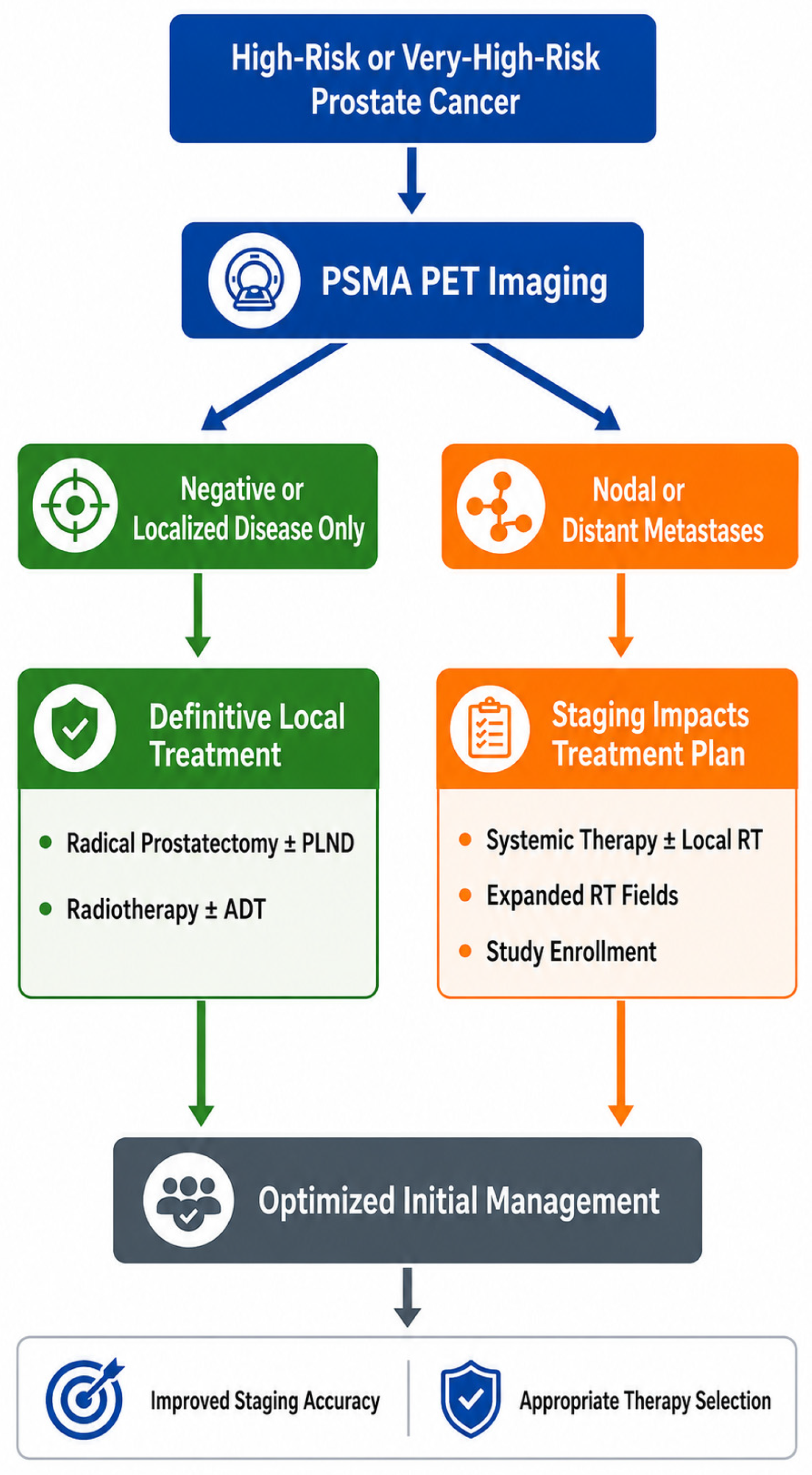
To frame the discussion from a clinician’s perspective, PSMA theranostics may be understood as a workflow that links a specific clinical question to molecular imaging, management adaptation, and, when appropriate, RLT. As a representative example of management-changing use, PSMA PET-guided initial staging in high-risk or very-high-risk prostate cancer is illustrated in
Figure 1.
2. Biological, Radiochemical, and Technical Foundations of PSMA Theranostics
PSMA is a type II transmembrane glycoprotein encoded by FOLH1 and expressed on most prostate cancer cells, with expression generally increasing in higher-grade, metastatic, and castration-resistant disease [
10]. Although the molecular biology of PSMA is well characterized, its practical importance for clinicians lies less in receptor biology itself than in the reproducibility of the clinical system built around it. A PSMA PET/CT study is useful only when it is acquired, interpreted, and reported in a standardized and clinically meaningful manner. Without such standardization, the apparent sensitivity of the technique may foster misplaced confidence, particularly when equivocal uptake, small nodes, or physiologic tracer accumulation are interpreted without sufficient clinical context [
2,
3,
7,
8,
9].
From an applied science perspective, PSMA theranostics also depends on the molecular design and quality attributes of the radiopharmaceutical itself. Most clinically used PSMA-targeted small-molecule ligands are based on a Glu-urea-Lys pharmacophore, which provides PSMA binding, combined with linkers, chelators, or prosthetic groups that influence biodistribution, clearance, tumor uptake, labeling chemistry, and suitability for either diagnostic imaging or therapy [
4,
5,
6]. Thus, PSMA theranostics should not be understood as a single tracer-based technology, but as a family of related radiopharmaceutical platforms in which ligand structure, radionuclide selection, production method, and imaging or therapeutic purpose are closely interconnected.
The choice of radionuclide is particularly important. Gallium-68-labeled PSMA ligands, such as
68Ga-PSMA-11, have been widely used because of their generator-based availability and strong clinical validation. Fluorine-18-labeled ligands, including
18F-DCFPyL and
18F-PSMA-1007, offer a longer physical half-life, cyclotron-based production, and potentially broader distribution from central manufacturing sites [
5,
11]. These features may improve logistics and image quality in some settings, but each tracer also has characteristic biodistribution and interpretive pitfalls. For example,
18F-PSMA-1007 has relatively low urinary excretion and favorable pelvic imaging characteristics, but focal unspecific bone uptake has been reported and should be recognized to avoid overstaging [
11,
12]. Therefore, differences among PSMA tracers should not be treated as merely chemical details; they can influence lesion conspicuity, reader confidence, equivocal findings, and management decisions.
Therapeutic radioligands introduce additional radiochemical and manufacturing considerations.
177Lu-PSMA-617 and
177Lu-PSMA-I&T combine a PSMA-targeted ligand with a beta-emitting radionuclide suitable for systemic radioligand therapy. The clinical performance of these agents depends not only on PSMA expression and patient selection, but also on radionuclide supply, carrier-added or no-carrier-added production routes, molar activity, radiochemical yield, radiochemical purity, stability after synthesis, and the control of radiolysis or thermally mediated degradation products [
6,
13,
14,
15]. These issues are not purely technical. In a clinical theranostic workflow, radiochemical quality influences how reproducibly a prescribed activity can be delivered, how safely the product can be administered, and how consistently trial results can be translated across institutions and regions.
The same principle applies to emerging alpha-emitting PSMA platforms. Actinium-225-labeled PSMA ligands are attractive because alpha particles have high linear energy transfer and short tissue range, but their development is technically more complex than that of conventional beta-emitting agents. The decay chain of
225Ac, daughter redistribution, radiolabeling stability, limited isotope supply, and toxicity concerns such as xerostomia remain important barriers to broad clinical implementation [
16,
17]. These considerations explain why alpha-emitting PSMA therapy remains investigational despite encouraging early clinical activity.
Several key guidance documents have become central to clinical implementation. The joint European Association of Nuclear Medicine and Society of Nuclear Medicine and Molecular Imaging procedure guideline provides detailed recommendations regarding indications, acquisition, interpretation, and reporting of PSMA PET/CT [
2]. The Society of Nuclear Medicine and Molecular Imaging appropriate use criteria translate the evidence base into clinically recognizable scenarios and clarify settings in which PSMA PET/CT is strongly supported [
3]. In parallel, E-PSMA established a structured reporting framework intended to reduce inter-reader variability and improve communication between imaging specialists and treating clinicians [
7]. More recently, a standardized reporting template has been proposed to further improve clarity and consistency in routine practice [
8]. These efforts are not merely methodological refinements. They are what allow PSMA PET/CT to function as a dependable clinical tool rather than as a highly sensitive but variably interpreted test.
From the perspective of the treating clinician, several issues are more relevant than tracer chemistry alone, but tracer chemistry and production quality cannot be ignored. First, which tracer is being used, and are its expected biodistribution and imaging characteristics sufficiently understood? Second, is the scan being interpreted within a framework that distinguishes physiologic uptake from likely malignant disease? Third, does the report describe the findings in a way that supports a management decision? Fourth, were acquisition, reconstruction, reporting, and radiopharmaceutical quality sufficiently standardized to permit comparison with prior scans, trial evidence, or institutional protocols? These questions matter because even excellent molecular imaging becomes less useful when reporting is vague, technical quality is inconsistent, or tracer-specific pitfalls are not considered. The Canadian Urological Association best practice report has been particularly helpful in summarizing physiologic uptake patterns, common interpretive pitfalls, and clinically oriented applications for non-nuclear-medicine specialists [
9].
Another important point is that PSMA theranostics is not synonymous with a single tracer or therapeutic compound. Gallium-68-labeled and fluorine-18-labeled ligands differ somewhat in logistics and biodistribution, and therapeutic radioligands differ in linker design, radionuclide properties, and developmental maturity. Therapeutic radioligands also differ in chelator chemistry, production requirements, and radiochemical quality attributes. PSMA expression is also dynamic and may be modulated shortly after androgen-deprivation treatment, which should be considered when interpreting PET findings obtained close to treatment initiation [
18]. Consequently, the clinical meaning of a PSMA PET/CT result depends not only on whether uptake is present, but also on which radiopharmaceutical was used, how it was produced, when imaging is performed in relation to treatment exposure and disease state, and how the findings were interpreted within the tracer-specific context.
The representative radiopharmaceutical classes and their applied science attributes are summarized in
Table 1 [
4,
5,
6,
7,
8,
9,
10,
11,
12,
13,
14,
15,
16,
17]. This classification is intended to highlight why standardization in PSMA theranostics must include not only image acquisition and reporting, but also radiopharmaceutical design, radionuclide selection, production quality, and tracer-specific interpretive context.
For this reason, the biological, radiochemical, and technical foundations of PSMA theranostics should not be treated as a peripheral preface. They form the basis for all downstream clinical decisions. Standardized acquisition, target-aware interpretation, radiopharmaceutical quality control, and clinically meaningful reporting are what transform PSMA from a molecular target into a practical treatment-enabling platform.
3. Standardization of PSMA PET/CT Acquisition, Interpretation, and Reporting
The clinical value of PSMA PET/CT depends heavily on standardization. As molecular imaging becomes more widely used beyond tertiary referral centers, variation in acquisition, interpretation, and reporting may become a major source of inconsistency. A high-quality scan that is reported ambiguously may be less useful than a moderately complex scan that is reported with structure and therapeutic relevance. Standardization, therefore, represents one of the most important practical advances in PSMA imaging.
The European Association of Nuclear Medicine/Society of Nuclear Medicine and Molecular Imaging guideline provides detailed advice regarding patient selection, radiopharmaceutical administration, uptake time, image acquisition, interpretation, and written reporting [
2]. Although technical details differ somewhat by tracer, the overarching principle is that acquisition should be reproducible enough to support comparability across institutions and time points. This is especially important when PSMA PET/CT findings determine whether a patient proceeds to surgery, radiotherapy, salvage treatment, or RLT. The Society of Nuclear Medicine and Molecular Imaging appropriate use criteria complement this framework by defining when PSMA PET/CT is clinically justified, thereby reducing the risk of indiscriminate or poorly timed use [
3].
Interpretation requires equal discipline. Physiologic uptake in the salivary glands, lacrimal glands, kidneys, bowel, ganglia, and benign musculoskeletal conditions may lead to false-positive interpretation if normal or nonmalignant tracer distribution is not recognized [
7,
8,
9]. Conversely, small-volume disease, low-expression lesions, or treatment-altered tumor biology may produce false-negative or equivocal studies. The practical implication for clinicians is that PSMA PET/CT should not be regarded as infallible simply because it is highly sensitive. Standardized interpretation is necessary precisely because high sensitivity does not eliminate uncertainty.
Structured classification systems have attempted to address this challenge from complementary angles. The Prostate Cancer Molecular Imaging Standardized Evaluation framework provides a disease-mapping approach based on molecular imaging tumor–node–metastasis classification and is intended to harmonize lesion description and staging across PSMA imaging studies [
19]. The PSMA Reporting and Data System was developed as a structured reporting and confidence-scale system to standardize how lesions of varying certainty are categorized in routine practice [
20]. Together with E-PSMA and template-based reporting [
7,
8], these systems reinforce a central point: PSMA PET/CT must not merely identify lesions; it must do so in a format that supports therapeutic action and inter-reader reproducibility.
Before discussing individual disease states in detail, it is useful to summarize the standardized clinical indications for PSMA PET/CT across the prostate cancer care pathway. These indications, together with their typical management implications, are outlined in
Table 2. Against this practical framework, the following sections examine how PSMA PET/CT is applied in specific clinical contexts, beginning with initial staging of localized and higher-risk disease.
4. PSMA PET/CT in Initial Staging of Localized and Higher-Risk Disease
The most established role of PSMA PET/CT in initial staging is in men with unfavorable intermediate-risk, high-risk, or very-high-risk prostate cancer whose treatment plan may be altered by more accurate staging. Conventional computed tomography and bone scintigraphy have limited ability to detect small nodal or distant metastases, and these limitations have historically contributed to understaging and uncertainty regarding the true extent of disease. The proPSMA trial was the landmark prospective randomized study demonstrating the superiority of PSMA PET/CT over conventional imaging for primary staging in high-risk disease, with higher accuracy, fewer equivocal findings, and lower radiation exposure [
33]. This trial was important not only because it showed better diagnostic performance, but also because it reframed PSMA PET/CT as a realistic replacement for conventional staging in appropriate patients rather than as a specialist adjunct.
Subsequent multicenter studies strengthened this position. OSPREY prospectively evaluated fluorine-18-labeled DCFPyL PET/CT and demonstrated high specificity for pelvic nodal metastases, whereas the multicenter phase 3 study of gallium-68-labeled PSMA-11 further clarified the performance of PSMA PET/CT for nodal staging before radical prostatectomy and pelvic lymph node dissection [
34,
35]. Together, these studies support an important clinical inference: when PSMA PET/CT identifies nodal or distant disease in the primary staging setting, the finding is usually meaningful. However, sensitivity remains imperfect for microscopic nodal disease, and a negative scan should not be interpreted as proof that nodal disease is absent in a patient with otherwise high-risk clinicopathologic features.
This limitation is one reason why clinicopathologic risk tools remain relevant even in the PSMA era. External validation of the Candiolo nomogram in high-risk patients treated with carbon-ion radiotherapy and androgen deprivation therapy illustrates that baseline risk heterogeneity remains clinically meaningful even when advanced imaging is available [
36]. In other words, PSMA PET/CT refines disease extent, but it does not eliminate the need for integrated risk assessment based on prostate-specific antigen, biopsy grade group, magnetic resonance imaging findings, and established predictive models.
The role of PSMA PET/CT in intermediate-risk disease remains more nuanced. A recent guideline-of-guidelines review addressing newly diagnosed intermediate-risk prostate cancer highlighted substantial variation in how professional societies position PSMA PET/CT in this setting, particularly when distinguishing favorable from unfavorable intermediate-risk disease [
37]. Current European Association of Urology guidance increasingly incorporates PSMA-based imaging into staging pathways for appropriate patients, especially when management is likely to change [
21]. For clinicians, the most balanced interpretation is that PSMA PET/CT is no longer restricted to the highest-risk patients, but its value is greatest when the result is likely to alter treatment intent, surgical planning, or radiotherapy field design.
5. PSMA PET/CT in Biochemical Recurrence and Salvage Planning
Biochemical recurrence is arguably the setting in which PSMA PET/CT has had the most immediate and intuitive clinical impact. Patients with rising prostate-specific antigen after radical prostatectomy or radiotherapy present a familiar dilemma: there is biochemical evidence of disease, but the location and extent of recurrence are uncertain, and management depends heavily on whether disease is local, regional, oligometastatic, or more diffuse. In this context, PSMA PET/CT is particularly valuable because it can localize recurrence at prostate-specific antigen levels at which conventional imaging is often negative or indeterminate [
38,
39].
Meta-analytic data from Perera and colleagues showed meaningful detection rates for PSMA PET/CT across prostate-specific antigen strata in biochemical recurrence [
38]. The CONDOR trial then provided prospective phase 3 evidence for the performance of fluorine-18-labeled DCFPyL PET/CT in men with biochemical recurrence and noninformative standard imaging [
39]. These findings are clinically important not simply because they show higher sensitivity, but because they increase the likelihood that salvage decisions can be based on anatomic localization rather than biochemical assumption alone.
The clinical importance of this issue is amplified by the heterogeneity of recurrence risk after definitive treatment. In a study of high-risk patients treated with carbon-ion radiotherapy and androgen deprivation therapy, early biochemical recurrence was associated with specific baseline features, reinforcing that recurrence biology is heterogeneous and that surveillance and salvage intensity should not always be uniform [
40]. This broader point is relevant to PSMA-guided salvage care: the consequences of missing or mislocalizing recurrent disease are greatest in patients at highest risk of early progression.
PSMA PET/CT has also shown superiority over non-PSMA positron emission tomography approaches in recurrence assessment. In a prospective comparative imaging study, gallium-68-labeled PSMA-11 PET/CT localized recurrent disease more effectively than fluorine-18-fluciclovine PET/CT in men with early biochemical recurrence after prostatectomy [
41]. This comparison should be interpreted in light of the distinct biological mechanisms of the two tracers:
68Ga-PSMA-11 reflects PSMA expression on prostate cancer lesions, whereas
18F-fluciclovine is a synthetic amino acid analog whose uptake reflects amino acid transport and tumor amino acid metabolism [
42]. Therefore, the superiority of
68Ga-PSMA-11 in this setting should not be regarded merely as a technical difference in PET performance, but as evidence that PSMA-targeted imaging is more closely aligned with the disease biology relevant to localizing early recurrent prostate cancer. For practicing clinicians, the relevance is direct: PSMA PET/CT offers a better chance of converting a biochemical signal into anatomically actionable information.
Importantly, PSMA PET/CT in biochemical recurrence changes management. Calais and colleagues showed that PSMA PET/CT altered intended treatment in a substantial proportion of patients, influencing decisions regarding salvage radiotherapy, nodal treatment, lesion-directed therapy, and systemic treatment [
43]. This management-shaping effect has since been supported by prospective interventional evidence. The randomized phase 3 PSMA-SRT study demonstrated that PSMA PET/CT altered salvage radiotherapy planning in a meaningful proportion of patients, often leading to treatment-plan escalation [
44]. More recently, the PSMAiSRT randomized phase 2 trial suggested that PSMA-guided intensification of salvage radiotherapy after radical prostatectomy improves failure-free survival without significant deterioration in quality of life or unacceptable toxicity [
45].
The case for PSMA-guided salvage planning is further strengthened by longer-term and population-level outcome data. In a nationwide real-world study, use of PSMA PET/CT before salvage radiotherapy was associated with improved overall survival in men with biochemically recurrent prostate cancer [
46]. Long-term follow-up of PSMA PET/CT-guided radiotherapy in recurrent disease has also shown durable biochemical control in a substantial proportion of patients, supporting the clinical relevance of anatomically informed salvage planning beyond early endpoint improvement [
47].
These data explain why the American Urological Association/American Society for Radiation Oncology/Society of Urologic Oncology salvage guideline incorporates advanced molecular imaging into modern recurrence management [
22]. The practical implication is that PSMA PET/CT should be used early enough in the salvage pathway to influence management, not merely late enough to confirm assumptions already embedded in a treatment plan. For clinicians, this is one of the clearest examples of PSMA theranostics functioning as a management-enabling platform rather than as a purely diagnostic refinement.
6. PSMA PET/CT in Oligometastatic and Oligorecurrent Disease
One of the most important conceptual consequences of PSMA PET/CT has been its effect on the definition and management of oligometastatic prostate cancer. Historically, oligometastatic disease was defined primarily by lesion count on conventional imaging. In the PSMA era, however, limited metastatic burden can no longer be regarded as a purely count-based construct. A patient classified as nonmetastatic by conventional imaging may be reclassified as oligometastatic on PSMA PET/CT, whereas another patient thought to have only a few disease sites may prove to harbor more extensive tumor burden. This distinction matters because metastasis-directed therapy, focal radiotherapy, salvage surgery, and selected hybrid approaches are usually considered only when disease appears truly limited.
A recent review by Oka et al. placed PSMA PET/CT within the broader therapeutic evolution of oligometastatic prostate cancer rather than treating it as an isolated diagnostic advance [
23]. The key point is that advanced imaging changes not only lesion visibility, but also therapeutic logic. In oligorecurrent disease, PSMA PET/CT may reveal a lesion distribution compatible with focal treatment and delayed systemic therapy. In another patient, it may identify additional disease that justifies broader radiotherapy fields or earlier systemic intensification. The value of PSMA PET/CT in this setting therefore lies not simply in detecting more lesions, but in providing a more credible basis for deciding whether disease remains biologically and clinically limited enough to justify local strategies.
The randomized ORIOLE trial provided an important conceptual bridge between advanced imaging and metastasis-directed treatment. Although it was not designed primarily as a PSMA PET trial, it showed that untreated lesions identified by PSMA PET were associated with early progression after stereotactic ablative radiotherapy, reinforcing the idea that molecular imaging can reveal clinically relevant disease not seen on conventional imaging [
24]. More recent clinical data also support the feasibility of PSMA PET-guided metastasis-directed radiotherapy, with or without hormonal therapy, in selected patients with oligometastatic prostate cancer [
25].
At the same time, clinicians must avoid overinterpretation. More lesions do not automatically make local therapy futile, and fewer lesions do not guarantee that local treatment alone is sufficient. The biologic distinction between truly oligometastatic disease and early polymetastatic dissemination remains imperfect. Nevertheless, PSMA PET/CT has improved multidisciplinary planning by reducing reliance on the underdetection inherent in conventional imaging. In practice, it has made lesion-directed radiotherapy more anatomically precise, nodal treatment more rational, and follow-up after metastasis-directed intervention more structured.
Collectively, these studies show that the value of PSMA PET/CT lies not only in improved lesion detection, but also in its capacity to alter management across multiple disease states. The landmark studies that have shaped current clinical use are summarized in
Table 3. On this imaging foundation, the next clinical question is how PSMA-based information can be used to determine eligibility for RLT.
7. Imaging-Based Eligibility Assessment for PSMA Radioligand Therapy
Patient selection is the critical bridge between PSMA imaging and PSMA-directed therapy. At first glance, the theranostic paradigm appears simple: if disease is PSMA-positive, a PSMA-targeted radioligand should be effective. In practice, eligibility is considerably more nuanced. The joint European Association of Nuclear Medicine/Society of Nuclear Medicine and Molecular Imaging procedure guideline for lutetium-177-labeled PSMA-targeted radioligand therapy and the Society of Nuclear Medicine and Molecular Imaging consensus statement both emphasize that appropriate candidates should be selected using imaging findings together with disease distribution, prior treatment exposure, marrow reserve, renal function, performance status, and the overall pace of disease [
26,
27].
One of the central challenges is lesion heterogeneity. A patient may satisfy a broad definition of PSMA-positive disease while still harboring lesions with relatively low target expression or biologically aggressive disease that is not uniformly targetable. This issue is clinically important because the efficacy of RLT depends on meaningful target expression across the lesions that matter most. Trial-based eligibility criteria attempted to standardize this problem, but real-world patients often present more complicated patterns, including borderline uptake, diffuse marrow involvement, visceral progression, or mixed treatment history. Thus, imaging eligibility should not be regarded as a simple binary exercise.
From a clinical standpoint, several questions are more useful than a purely formal checklist. Is the dominant disease burden convincingly PSMA-avid? Are there lesions whose biology appears discordant with the overall imaging pattern? Does the patient have sufficient hematologic and renal reserve to tolerate repeated treatment cycles? Is RLT being considered at a clinically appropriate time, or is another therapy needed more urgently? The value of imaging-based eligibility assessment lies precisely in this structured integration of molecular imaging with routine oncologic judgment.
This approach also helps distinguish a theranostic review from a generalized sequencing review. The central issue is not where RLT should sit in a universal treatment hierarchy. Rather, it is whether a patient is an appropriate theranostic candidate. That distinction keeps the focus on the actual clinical identity of PSMA theranostics: a platform in which imaging and treatment selection are inseparably linked.
8. Clinical Evidence for PSMA-Targeted Beta-Emitting Radioligand Therapy
The therapeutic evidence base for beta-emitting PSMA-targeted RLT in mCRPC has developed in a logical sequence, from early signal-generating studies to randomized comparative trials and then to phase 3 validation. In a key early prospective phase 2 study, Hofman and colleagues reported meaningful antitumor activity, pain improvement, and manageable toxicity with lutetium-177-PSMA-617 in heavily pretreated men with mCRPC [
48]. Although this study did not establish comparative efficacy, it provided strong proof of concept that PSMA-targeted therapy could achieve clinically significant benefit in advanced disease.
The next major step was the randomized phase 2 TheraP trial, which compared lutetium-177-PSMA-617 with cabazitaxel in men with mCRPC progressing after docetaxel [
49]. TheraP showed higher prostate-specific antigen response rates and fewer grade 3 or 4 adverse events with RLT, establishing PSMA-targeted treatment as a credible alternative to an active later-line standard. Mature follow-up later confirmed that trial-based molecular imaging selection was prognostically informative and that RLT remained a rational option in appropriately selected patients, although overall survival did not differ significantly between arms in that analysis [
50].
The pivotal phase 3 VISION trial then established lutetium-177-PSMA-617 as a survival-prolonging systemic therapy. In men with PSMA-positive mCRPC previously treated with at least one androgen receptor pathway inhibitor and one or two taxane regimens, the addition of lutetium-177-PSMA-617 to protocol-permitted standard care improved imaging-based radiographic progression-free survival and overall survival [
51]. The subsequent U.S. Food and Drug Administration (FDA) approval summary underscored that regulatory acceptance rested not only on imaging-defined eligibility, but also on a meaningful survival benefit and delayed progression [
52].
The field has since moved earlier in the treatment course. The phase 3 PSMAfore trial showed that in taxane-naive patients with PSMA-positive mCRPC who had progressed after one androgen receptor pathway inhibitor and were considered candidates for androgen receptor pathway inhibitor change, lutetium-177-PSMA-617 improved radiographic progression-free survival with a favorable safety profile relative to switching to another androgen receptor pathway inhibitor [
53]. Final overall survival and safety analyses later clarified the complexity of interpretation in a crossover-permitted trial but did not diminish the central clinical lesson that RLT can provide meaningful disease control in the pre-chemotherapy setting [
54]. Regulatory expansion followed, with the FDA broadening the indication for Pluvicto in 2025 to include patients treated with androgen receptor pathway inhibitor therapy and considered appropriate to delay taxane-based chemotherapy [
55].
For clinicians, the most important conclusion is that the value of PSMA-targeted RLT is inseparable from its imaging-defined framework. The pivotal trials do not simply show that RLT works; they show that molecular imaging-based target selection is central to why it works. That is the essence of theranostics, and it distinguishes interpretation of these studies from a conventional drug-development narrative.
Because the interpretation of PSMA-targeted RLT depends not only on efficacy estimates but also on imaging-based eligibility criteria, comparator choice, and endpoint structure, the principal features of the pivotal trials are summarized in
Table 4. These distinctions are clinically important when translating trial results into routine practice, particularly outside tightly controlled study populations.
9. Clinical Integration Beyond Pivotal Trials
Although a detailed discussion of cross-mechanism treatment sequencing lies outside the main scope of this review, RLT must still be interpreted within the broader systemic treatment landscape. In clinical practice, its use depends not only on trial eligibility criteria, but also on disease tempo, prior therapies, organ reserve, and patient preference. Recent work examining advanced prostate cancer management from a chemotherapy-forward and real-world perspective reinforces that treatment choice is often phenotype-driven rather than determined by a single universal sequence [
56].
Real-world outcome data are, therefore, important. The multi-institutional observational study by Kafka and colleagues showed that
177Lu-PSMA treatment remained effective in routine practice and that an early prostate-specific antigen decline after the first cycles may offer useful prognostic information [
57]. These data do not replace randomized evidence, but they strengthen confidence that RLT can be delivered effectively outside highly selected trial populations.
For clinicians, the practical lesson is that RLT should be integrated thoughtfully rather than dogmatically. It is neither a universally early option nor merely a salvage treatment of last resort. Its strength lies in situations where target expression is convincing, disease biology appears compatible with benefit, and the patient’s prior-treatment context supports safe and meaningful delivery.
10. Dosimetry, Safety, and Response Assessment
One of the defining characteristics of PSMA-targeted RLT, compared with conventional systemic therapy, is the possibility of biologically informed personalization through dosimetry and imaging-based response assessment. Although many pivotal and routine clinical protocols administer lutetium-177-labeled PSMA ligands according to relatively fixed activity schedules, this approach only partly reflects the theranostic principle of patient-specific treatment planning [
26,
58]. A fixed administered activity is practical, reproducible, and suitable for large clinical trials, but it does not directly account for interpatient differences in PSMA-positive tumor burden, lesion uptake, tumor heterogeneity, renal function, salivary-gland exposure, marrow reserve, body habitus, or prior systemic treatment.
This limitation is clinically relevant because the absorbed dose delivered to tumors and organs at risk may vary substantially even when the same nominal activity is administered. Earlier dosimetry work with
177Lu-PSMA-617 showed that tumor absorbed dose is associated with biochemical response, supporting the concept that delivered dose, not merely administered activity, is biologically meaningful [
59]. Conversely, organ-at-risk exposure, particularly to the kidneys, salivary glands, lacrimal glands, and bone marrow, may influence cumulative safety, especially in heavily pretreated patients or in earlier disease states where longer survival increases the relevance of late toxicity [
26,
58,
60]. Therefore, a more flexible evidence-based framework that integrates baseline PSMA PET tumor burden, early post-therapy imaging, organ dosimetry, and clinical reserve may eventually improve treatment adaptation without compromising safety.
The comparative systematic review and meta-analysis by Ells and colleagues highlighted both the promise and the current limitations of dosimetry in PSMA-targeted therapy [
58]. Available studies support the importance of absorbed-dose assessment, but they also demonstrate substantial heterogeneity in methodology, imaging time points, segmentation approaches, and endpoints. Simplified strategies, including single-time point SPECT/CT-based dosimetry, may help reduce workflow burden and support broader implementation, but these approaches still require validation before they can replace more comprehensive dosimetry protocols [
61]. In practical terms, dosimetry is clearly important, but it is not yet fully standardized across institutions, and prospective evidence is still needed to determine whether dosimetry-guided activity adaptation improves survival, toxicity, quality of life, or treatment completion compared with fixed-activity schedules.
Japanese prospective data provide an additional and clinically relevant perspective. Takano and colleagues reported pharmacokinetic and dosimetry data for gallium-68-labeled PSMA-11 and lutetium-177-PSMA-617 in Japanese patients with PSMA-positive mCRPC, demonstrating organ absorbed doses broadly consistent with previously reported international experience and minimal radiation exposure to medical staff and caregivers [
62]. These data are valuable not only because they add population-specific information, but also because they support the practical feasibility and safety of implementing RLT within Japanese clinical settings.
Safety remains a central clinical concern. Hematologic suppression, fatigue, xerostomia, gastrointestinal symptoms, and potential renal effects are the most relevant issues in everyday care. The VISION dosimetry sub-study was especially informative because it showed that cumulative absorbed doses to the kidneys and other organs did not translate into a major clinically significant signal of renal toxicity over the study timeframe [
60]. For clinicians, this finding is reassuring, as renal safety is one of the most common concerns raised when discussing RLT. The available evidence suggests that renal toxicity is not a dominant short-term limiting factor in standard use, although careful monitoring remains appropriate, particularly in medically complex or heavily pretreated patients.
Because PSMA theranostics is defined not only by target selection but also by the possibility of treatment monitoring through dosimetry and molecular imaging, the main dosimetry and response-assessment frameworks are summarized in
Table 5. Among these frameworks, RECIP 1.0 has emerged as one of the most promising approaches for integrating PSMA PET findings into structured post-treatment response assessment.
Response assessment after RLT is also evolving rapidly. Tumor-volume assessment on PSMA PET has shown prognostic value and may complement biochemical markers in evaluating treatment benefit [
29]. Building on this concept, Gafita and colleagues proposed Response Evaluation Criteria in PSMA Imaging (RECIP) 1.0, a PSMA PET-based framework that integrates changes in PSMA-positive tumor volume with the appearance of new lesions [
30]. The prognostic utility of RECIP 1.0 appears to extend across different PET tracers and therapeutic backbones, including fluorine-18-labeled PSMA-1007 PET and lutetium-177-PSMA-I&T [
31]. RECIP 1.0 has also been linked to progression-free survival after lutetium-177-PSMA therapy [
32].
For clinicians, the most relevant implication is that future monitoring of RLT will likely rely on composite assessment rather than on prostate-specific antigen kinetics alone, incorporating clinical course, laboratory data, conventional imaging, and structured PSMA PET-based response evaluation. In this respect, PSMA theranostics is best understood as a longitudinal imaging-treatment system: the same molecular target informs eligibility before treatment, supports delivery during treatment, and increasingly contributes to response assessment after treatment.
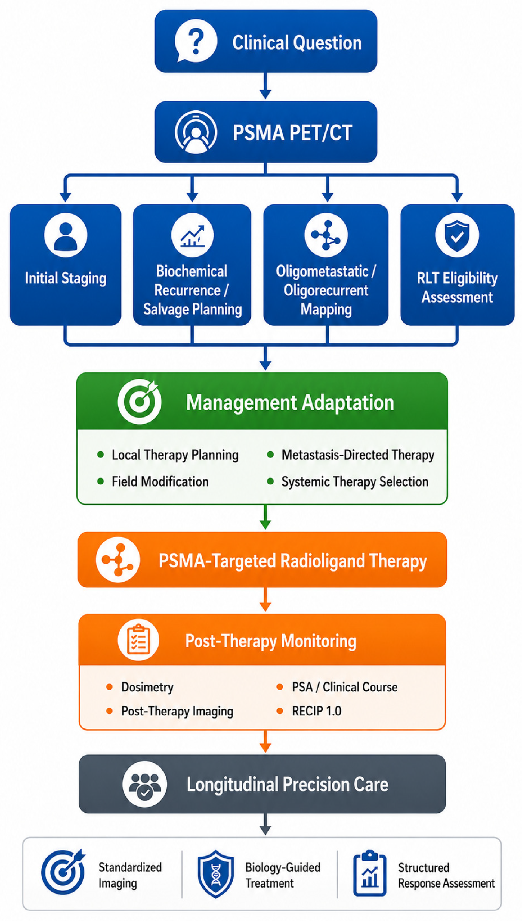
Taken together, these concepts show that PSMA theranostics is best viewed as a longitudinal imaging-to-therapy platform rather than as a single diagnostic or therapeutic step. The integration of staging, recurrence localization, treatment eligibility, and post-treatment monitoring is summarized in
Figure 2.
11. Emerging Trials and Next-Generation Theranostic Platforms
The next major phase of PSMA theranostics is defined by three parallel developments: movement into earlier disease states, expansion to additional beta-emitting ligands, and development of alpha-emitting compounds. Each development has the potential to reshape the clinical role of theranostics, although the maturity of the supporting evidence differs substantially across platforms.
Japanese data have already begun to contribute to this evolving landscape. In a phase 2 open-label, single-arm trial, Izumi and colleagues reported that lutetium-177-PSMA-617 achieved the prespecified efficacy threshold in Japanese patients with progressive PSMA-positive mCRPC treated with or without prior taxane-based chemotherapy, with a safety profile consistent with VISION and PSMAfore [
63]. These results are important because they provide region-specific implementation evidence and support the reproducibility of radioligand outcomes in an Asian population.
At the same time, current European guidance has started to incorporate RLT more explicitly into the advanced disease framework. The 2026 European Society for Medical Oncology clinical practice guideline for advanced and metastatic prostate cancer reflects the growing role of PSMA-based imaging and RLT in contemporary management, even as questions regarding optimal timing and broader implementation remain active areas of study [
28].
Among the most consequential recent developments is PSMAddition. In 2025, Novartis (Basel, Switzerland) reported that the trial met its primary endpoint, demonstrating a statistically significant and clinically meaningful radiographic progression-free survival benefit, together with a positive trend in overall survival, when lutetium-177 vipivotide tetraxetan was added to androgen deprivation therapy plus androgen receptor pathway inhibitor in PSMA-positive metastatic hormone-sensitive prostate cancer [
64]. If confirmed in a full peer-reviewed report, these findings would support the extension of PSMA-targeted RLT from castration-resistant disease into the hormone-sensitive setting and could meaningfully reshape the current treatment continuum.
Additional phase 3 development in taxane-naive mCRPC has also been notable. Lantheus (Bedford, MA, USA) reported positive primary analysis results from the SPLASH trial for lutetium-177-PNT2002, showing significant improvement in radiographic progression-free survival [
65]. Curium (St. Louis, MO, USA) similarly announced that the ECLIPSE trial met its primary endpoint for lutetium-177-PSMA-I&T, also known as lutetium-177 zadavotide guraxetan, in PSMA-positive mCRPC [
66]. Although full peer-reviewed publications are still needed for definitive interpretation, these developments suggest that PSMA-targeted beta-emitting radioligands may be evolving beyond a single-compound paradigm.
Beyond beta-emitting agents, alpha-emitting PSMA ligands are attracting intense interest. The WARMTH Act multicenter retrospective study demonstrated encouraging activity of actinium-225-labeled PSMA therapy in mCRPC, although xerostomia and the retrospective design underscore the need for careful interpretation [
67]. Prospective development is ongoing, including a phase 1 dose-escalation study of actinium-225-PSMA-I&T and the AcTION study of actinium-225-PSMA-617 [
68,
69]. These programs are significant because alpha-emitting compounds may offer higher linear energy transfer and potentially meaningful activity in selected patients, particularly after prior beta-emitting treatment. At present, however, their role remains investigational.
A Japan-specific implementation perspective is also beginning to emerge. In 2026, the Pharmaceuticals and Medical Devices Agency issued an Early Consideration document regarding the clinical development of radiopharmaceuticals for positron emission tomography targeting PSMA, indicating active regulatory attention to the introduction of PSMA PET radiopharmaceuticals into Japanese clinical practice [
70]. This development is important because it highlights that the future of PSMA theranostics will depend not only on trial outcomes, but also on regulatory pathways, companion diagnostic alignment, and practical adoption within national healthcare systems.
12. Future Directions: Quantification, Harmonization, and Clinical Implementation
The future of PSMA theranostics will depend not only on new drugs or broader indications, but also on better measurement and more consistent implementation. One of the most important next steps is improved quantification. Whole-body tumor burden assessment, more reproducible volumetric metrics, and standardized post-therapy imaging may all help refine eligibility assessment, treatment adaptation, and response interpretation. At present, these tools are promising but incompletely harmonized.
Harmonization of response assessment is equally important. RECIP 1.0 represents a meaningful advance, but broader clinical adoption will require greater familiarity, simpler implementation pathways, and continued validation across disease states and treatment settings [
30,
31,
32]. In routine practice, many clinicians remain more comfortable with prostate-specific antigen kinetics and conventional computed tomography-based response terminology. Bridging that gap will require not only more evidence, but also simpler reporting systems and closer collaboration between imaging specialists and treating teams.
Operational implementation is another major issue. As PSMA-based imaging and therapy become more widely available, reproducibility across institutions will become as important as efficacy in expert centers. Standardized reporting templates, multidisciplinary discussion pathways, and practical treatment workflows will all be necessary to ensure that the benefits demonstrated in trials can be translated into routine care [
2,
3,
4,
5,
6]. Artificial intelligence-assisted lesion segmentation, structured reporting support, and improved whole-body quantification may also help reduce inter-reader variability and make theranostic decision-making more reproducible.
Ultimately, the next major advances in PSMA theranostics may arise as much from better clinical operationalization as from additional therapeutic innovation. The field already possesses a powerful biologic target, high-performance imaging, and proven therapeutic agents. The challenge now is to integrate these strengths into systems of care that are scalable, interpretable, and clinically coherent across the full spectrum of prostate cancer practice.
13. Conclusions
PSMA theranostics has evolved from an attractive translational concept into a clinically meaningful platform that now influences prostate cancer management across multiple disease states. Its importance lies not merely in improved lesion detection or in the availability of a new radiopharmaceutical, but in the fact that it links imaging, treatment selection, and treatment monitoring through a common biologic target. This review further emphasizes that this linkage depends on an applied science foundation that includes radiopharmaceutical design, radionuclide selection, production quality, standardized PET acquisition, quantitative interpretation, dosimetry, and structured response assessment. This has important implications for initial staging, biochemical recurrence, oligometastatic decision-making, and systemic therapy in advanced disease.
The practical conclusions of this review can be summarized in four points. First, PSMA PET/CT has become a management-shaping imaging modality in unfavorable intermediate-risk, high-risk, and very-high-risk localized disease, biochemical recurrence, salvage radiotherapy planning, oligometastatic or oligorecurrent disease, and eligibility assessment for PSMA-targeted RLT. Second, landmark imaging studies such as proPSMA, OSPREY, CONDOR, PSMA-SRT, and PSMAiSRT demonstrate that PSMA PET/CT provides not only higher diagnostic confidence but also clinically relevant changes in treatment planning. Third, pivotal RLT trials, including VISION, TheraP, and PSMAfore, establish lutetium-177-labeled PSMA therapy as an evidence-based systemic option in PSMA-positive mCRPC, with ongoing expansion toward earlier disease settings. Fourth, dosimetry, post-therapy imaging, quantitative PET metrics, and RECIP-based response assessment represent the next layer of implementation needed to move PSMA theranostics from protocol-driven use toward more individualized care.
For clinicians, the key insight is that PSMA PET/CT and PSMA-targeted RLT should no longer be viewed as separate innovations. Their greatest value emerges when they are understood as interdependent components of a single clinical framework. Standardized imaging supports management-changing decisions, imaging-based eligibility informs therapeutic delivery, and structured post-treatment assessment increasingly defines how benefit is interpreted over time. At the same time, the clinical meaning of PSMA uptake depends on tracer characteristics, radiochemical quality, disease distribution, prior treatment exposure, and patient-specific organ reserve; therefore, multidisciplinary interpretation remains essential.
As the field advances, the central task will be to move from proof of efficacy to excellence of implementation. Better harmonization of acquisition and reporting, more robust dosimetry and response frameworks, and more deliberate integration into multidisciplinary care will determine how fully PSMA theranostics realizes its promise. Future progress will likely depend not only on new beta- or alpha-emitting agents, but also on reproducible radiopharmaceutical production, validated quantitative imaging workflows, feasible dosimetry-guided treatment adaptation, and harmonized response criteria that can be implemented beyond expert centers. If those steps are achieved, PSMA-based care is likely to remain one of the defining clinical platforms in prostate cancer for years to come.
Author Contributions
Conceptualization, S.I. and T.U.; methodology, T.U.; software, S.I.; validation, N.I., Y.K., T.N., T.S. (Takatoshi Somoto) and R.O.; formal analysis, S.I.; investigation, S.I.; resources, N.I., Y.K., R.I., T.S. (Tatsuharu Sugimoto), Y.S. (Yuka Sugizaki) and S.I.; data curation, N.I., Y.S. (Yuta Suzuki), S.I. and T.E.; writing—original draft preparation, S.I.; writing—review and editing, T.U.; visualization, T.U.; supervision, N.K. and H.S.; project administration, N.K. and H.S. All authors have read and agreed to the published version of the manuscript.
Funding
This research received no external funding.
Institutional Review Board Statement
Not applicable.
Informed Consent Statement
Not applicable.
Data Availability Statement
No new data were created or analyzed in this study. Data sharing is not applicable to this article.
Acknowledgments
ChatGPT (GPT-5.5 Thinking, OpenAI, San Francisco, CA, USA) was used only to assist in the visual drafting and refinement of the schematic figures. All scientific content, figure labels, and final interpretations were reviewed and approved by the authors.
Conflicts of Interest
Hiroyoshi Suzuki reports research funding from Astellas, AstraZeneca, Bayer, Chugai, Eli Lilly, Janssen, MSD, Nihon Kayaku, and Sanofi; advisory fees from AstraZeneca, Bayer, Chugai-Roche, Eli Lilly, Ferring, Janssen, MSD, Novartis, Pfizer, and Sanofi; and lecture fees from Astellas, AstraZeneca, Bayer, Janssen, Novartis, Pfizer, and Sanofi. The other authors declare no conflicts of interest.
Abbreviations
| FDA | U.S. Food and Drug Administration |
| mCRPC | metastatic castration-resistant prostate cancer |
| PET | positron emission tomography |
| PET/CT | positron emission tomography/computed tomography |
| PSA | prostate-specific antigen |
| PSMA | prostate-specific membrane antigen |
| PSMA PET/CT | prostate-specific membrane antigen positron emission tomography/computed tomography |
| RECIP | Response Evaluation Criteria in PSMA Imaging |
| RLT | radioligand therapy |
| SPECT/CT | single-photon emission computed tomography/computed tomography |
References
- Bauckneht, M.; Ciccarese, C.; Laudicella, R.; Mosillo, C.; D’Amico, F.; Anghelone, A.; Strusi, A.; Beccia, V.; Bracarda, S.; Fornarini, G.; et al. Theranostics Revolution in Prostate Cancer: Basics, Clinical Applications, Open Issues and Future Perspectives. Cancer Treat. Rev. 2024, 124, 102698. [Google Scholar] [CrossRef] [PubMed]
- Fendler, W.P.; Eiber, M.; Beheshti, M.; Bomanji, J.; Calais, J.; Ceci, F.; Cho, S.Y.; Fanti, S.; Giesel, F.L.; Goffin, K.; et al. PSMA PET/CT: Joint EANM Procedure Guideline/SNMMI Procedure Standard for Prostate Cancer Imaging 2.0. Eur. J. Nucl. Med. Mol. Imaging 2023, 50, 1466–1486. [Google Scholar] [CrossRef] [PubMed]
- Jadvar, H.; Calais, J.; Fanti, S.; Feng, F.; Greene, K.L.; Gulley, J.L.; Hofman, M.S.; Koontz, B.F.; Lin, D.W.; Morris, M.J.; et al. Appropriate Use Criteria for Prostate-Specific Membrane Antigen PET Imaging. J. Nucl. Med. 2022, 63, 59–68. [Google Scholar] [CrossRef] [PubMed]
- Benešová, M.; Schäfer, M.; Bauder-Wüst, U.; Afshar-Oromieh, A.; Kratochwil, C.; Mier, W.; Haberkorn, U.; Kopka, K.; Eder, M. Preclinical Evaluation of a Tailor-Made DOTA-Conjugated PSMA Inhibitor with Optimized Linker Moiety for Imaging and Endoradiotherapy of Prostate Cancer. J. Nucl. Med. 2015, 56, 914–920. [Google Scholar] [CrossRef] [PubMed]
- Werner, R.A.; Derlin, T.; Lapa, C.; Sheikbahaei, S.; Higuchi, T.; Giesel, F.L.; Behr, S.C.; Drzezga, A.; Kimura, H.; Buck, A.K.; et al. 18F-Labeled, PSMA-Targeted Radiotracers: Leveraging the Advantages of Radiofluorination for Prostate Cancer Molecular Imaging. Theranostics 2020, 10, 1–16. [Google Scholar] [CrossRef] [PubMed]
- Hennrich, U.; Eder, M. [177Lu]Lu-PSMA-617 (PluvictoTM): The First FDA-Approved Radiotherapeutical for Treatment of Prostate Cancer. Pharmaceuticals 2022, 15, 1292. [Google Scholar] [CrossRef] [PubMed]
- Ceci, F.; Oprea-Lager, D.E.; Emmett, L.; Adam, J.A.; Bomanji, J.; Czernin, J.; Eiber, M.; Haberkorn, U.; Hofman, M.S.; Hope, T.A.; et al. E-PSMA: The EANM Standardized Reporting Guidelines v1.0 for PSMA-PET. Eur. J. Nucl. Med. Mol. Imaging 2021, 48, 1626–1638. [Google Scholar] [CrossRef] [PubMed]
- Esfahani, S.A.; Morris, M.J.; Sartor, O.; Frydenberg, M.; Noto, B.; Bouchelouche, K.; Koo, P.J.; Jadvar, H.; Emmett, L.; Hope, T.A.; et al. Standardized Template for Clinical Reporting of PSMA PET/CT Scans. Eur. J. Nucl. Med. Mol. Imaging 2024, 52, 335–341. [Google Scholar] [CrossRef] [PubMed]
- Shaygan, B.; Zukotynski, K.; Bénard, F.; Ménard, C.; Kuk, J.; Sistani, G.; Bauman, G.; Veit-Haibach, P.; Metser, U. Canadian Urological Association Best Practice Report: Prostate-Specific Membrane Antigen Positron Emission Tomography/Computed Tomography and Positron Emission Tomography/Magnetic Resonance in Prostate Cancer. Can. Urol. Assoc. J. 2021, 15, 162–172. [Google Scholar] [CrossRef] [PubMed]
- O’Keefe, D.S.; Bacich, D.J.; Heston, W.D.W. A Perspective on the Evolving Story of PSMA Biology, PSMA-Based Imaging, and Endoradiotherapeutic Strategies. J. Nucl. Med. 2018, 59, 1007–1013. [Google Scholar] [CrossRef] [PubMed]
- Giesel, F.L.; Hadaschik, B.; Cardinale, J.; Radtke, J.; Vinsensia, M.; Lehnert, W.; Kesch, C.; Tolstov, Y.; Singer, S.; Grabe, N.; et al. F-18 Labelled PSMA-1007: Biodistribution, Radiation Dosimetry and Histopathological Validation of Tumor Lesions in Prostate Cancer Patients. Eur. J. Nucl. Med. Mol. Imaging 2017, 44, 678–688. [Google Scholar] [CrossRef] [PubMed]
- Grünig, H.; Maurer, A.; Thali, Y.; Kovacs, Z.; Strobel, K.; Burger, I.A.; Müller, J. Focal Unspecific Bone Uptake on [18F]-PSMA-1007 PET: A Multicenter Retrospective Evaluation of the Distribution, Frequency, and Quantitative Parameters of a Potential Pitfall in Prostate Cancer Imaging. Eur. J. Nucl. Med. Mol. Imaging 2021, 48, 4483–4494. [Google Scholar] [CrossRef] [PubMed]
- Vogel, W.V.; van der Marck, S.C.; Versleijen, M.W.J. Challenges and Future Options for the Production of Lutetium-177. Eur. J. Nucl. Med. Mol. Imaging 2021, 48, 2329–2335. [Google Scholar] [CrossRef] [PubMed]
- Larenkov, A.; Mitrofanov, I.; Rakhimov, M. Improvement of End-of-Synthesis Radiochemical Purity of 177Lu-DOTA-PSMA-Ligands with Alternative Synthesis Approaches: Conversion Upswing and Side-Products Minimization. Pharmaceutics 2024, 16, 1535. [Google Scholar] [CrossRef] [PubMed]
- Hunt, W.; Long, M.; Kamil, U.; Kellapatha, S.; Noonan, W.; Roselt, P.D.; Papa, N.; Emmerson, B.; Hofman, M.S.; Haskali, M.B. Multifactorial Analysis of Radiochemical Purity in High-Activity 177Lu-Labeled Theranostics: Impact of Precursor Source, 177Lu Form, and Production Parameters. EJNMMI Radiopharm. Chem. 2025, 10, 47. [Google Scholar] [CrossRef] [PubMed]
- Bidkar, A.P.; Zerefa, L.; Yadav, S.; VanBrocklin, H.F.; Flavell, R.R. Actinium-225 Targeted Alpha Particle Therapy for Prostate Cancer. Theranostics 2024, 14, 2969–2992. [Google Scholar] [CrossRef] [PubMed]
- Morgenstern, A.; Apostolidis, C.; Kratochwil, C.; Sathekge, M.; Krolicki, L.; Bruchertseifer, F. An Overview of Targeted Alpha Therapy with 225Actinium and 213Bismuth. Curr. Radiopharm. 2018, 11, 200–208. [Google Scholar] [CrossRef] [PubMed]
- Emmett, L.; Yin, C.; Crumbaker, M.; Hruby, G.; Kneebone, A.; Epstein, R.; Nguyen, Q.; Hickey, A.; Ihsheish, N.; O’Neill, G.; et al. Rapid Modulation of PSMA Expression by Androgen Deprivation: Serial 68Ga-PSMA-11 PET in Men with Hormone-Sensitive and Castrate-Resistant Prostate Cancer Commencing Androgen Blockade. J. Nucl. Med. 2019, 60, 950–954. [Google Scholar] [CrossRef] [PubMed]
- Eiber, M.; Herrmann, K.; Calais, J.; Hadaschik, B.; Giesel, F.L.; Hartenbach, M.; Hope, T.A.; Reiter, R.; Maurer, T.; Weber, W.A.; et al. Prostate Cancer Molecular Imaging Standardized Evaluation (PROMISE): Proposed miTNM Classification for the Interpretation of PSMA-Ligand PET/CT. J. Nucl. Med. 2018, 59, 469–478. [Google Scholar] [CrossRef] [PubMed]
- Rowe, S.P.; Pienta, K.J.; Pomper, M.G.; Gorin, M.A. PSMA-RADS Version 1.0: A Step Towards Standardizing the Interpretation and Reporting of PSMA-Targeted PET Imaging Studies. Eur. Urol. 2018, 73, 485–487. [Google Scholar] [CrossRef] [PubMed]
- European Association of Urology. EAU Guidelines on Prostate Cancer; EAU Guidelines Office: Arnhem, The Netherlands, 2025; Available online: https://uroweb.org/guidelines/prostate-cancer (accessed on 5 May 2026).
- Morgan, T.M.; Boorjian, S.A.; Buyyounouski, M.K.; Chapin, B.F.; Chen, D.Y.T.; Cheng, H.H.; Dess, R.T.; Eastham, J.A.; Efstathiou, J.A.; Feng, F.Y.; et al. Salvage Therapy for Prostate Cancer: AUA/ASTRO/SUO Guideline Part I: Introduction and Treatment Decision-Making at the Time of Suspected Biochemical Recurrence after Radical Prostatectomy. J. Urol. 2024, 211, 509–517. [Google Scholar] [CrossRef] [PubMed]
- Oka, R.; Utsumi, T.; Noro, T.; Suzuki, Y.; Iijima, S.; Sugizaki, Y.; Somoto, T.; Kato, S.; Endo, T.; Kamiya, N.; et al. Progress in Oligometastatic Prostate Cancer: Emerging Imaging Innovations and Therapeutic Approaches. Cancers 2024, 16, 507. [Google Scholar] [CrossRef] [PubMed]
- Phillips, R.; Shi, W.Y.; Deek, M.; Radwan, N.; Lim, S.J.; Antonarakis, E.S.; Rowe, S.P.; Ross, A.E.; Gorin, M.A.; Deville, C.; et al. Outcomes of Observation vs. Stereotactic Ablative Radiation for Oligometastatic Prostate Cancer: The ORIOLE Phase 2 Randomized Clinical Trial. JAMA Oncol. 2020, 6, 650–659. [Google Scholar] [CrossRef] [PubMed]
- Chen, W.S.; Tuchayi, A.M.; Sabbagh, A.; Kim, I.; Porter, E.; Ashraf-Ganjouei, A.; Li, Y.R.; Witztum, A.; Rajagopal, A.; Seyedin, S.N.; et al. Utility of Metastasis-Directed Radiotherapy with and without Hormonal Therapy in Management of Oligometastatic Prostate Cancer. JNCI Cancer Spectr. 2025, 9, pkaf096. [Google Scholar] [CrossRef] [PubMed]
- Kratochwil, C.; Fendler, W.P.; Eiber, M.; Hofman, M.S.; Emmett, L.; Calais, J.; Osborne, J.R.; Iravani, A.; Koo, P.; Lindenberg, L.; et al. Joint EANM/SNMMI Procedure Guideline for the Use of 177Lu-Labeled PSMA-Targeted Radioligand Therapy (177Lu-PSMA-RLT). Eur. J. Nucl. Med. Mol. Imaging 2023, 50, 2830–2845. [Google Scholar] [CrossRef] [PubMed]
- Hope, T.A.; Antonarakis, E.S.; Bodei, L.; Calais, J.; Iravani, A.; Jacene, H.; Kesch, C.; Lerner, S.P.; McKay, R.R.; Morris, M.J.; et al. SNMMI Consensus Statement on Patient Selection and Appropriate Use of 177Lu-PSMA-617 Radionuclide Therapy. J. Nucl. Med. 2023, 64, 1417–1423. [Google Scholar] [CrossRef] [PubMed]
- Fizazi, K.; Attard, G.; Azad, A.A.; Baciarello, G.; Beltran, H.; Bjartell, A.; Blanchard, P.; Bossaert, F.; Castro, E.; Compérat, E.; et al. Advanced and Metastatic Prostate Cancer: ESMO Clinical Practice Guideline for Diagnosis, Treatment and Follow-Up. Ann. Oncol. 2026, 37, 590–607. [Google Scholar] [CrossRef] [PubMed]
- Kind, F.; Eder, A.C.; Jilg, C.A.; Hartrampf, P.E.; Meyer, P.T.; Ruf, J.; Michalski, K. Prognostic Value of Tumor Volume Assessment on PSMA PET after 177Lu-PSMA Radioligand Therapy Evaluated by PSMA PET/CT Consensus Statement and RECIP 1.0. J. Nucl. Med. 2023, 64, 605–610. [Google Scholar] [CrossRef] [PubMed]
- Gafita, A.; Rauscher, I.; Weber, M.; Hadaschik, B.; Wang, H.; Armstrong, W.R.; Herrmann, K.; Eiber, M.; Fendler, W.P. Novel Framework for Treatment Response Evaluation Using PSMA PET/CT in Patients with Metastatic Castration-Resistant Prostate Cancer (RECIP 1.0): An International Multicenter Study. J. Nucl. Med. 2022, 63, 1651–1658. [Google Scholar] [CrossRef] [PubMed]
- Hartrampf, P.E.; Hüttmann, T.; Seitz, A.K.; Kübler, H.; Serfling, S.E.; Higuchi, T.; Schlötelburg, W.; Michalski, K.; Gafita, A.; Rowe, S.P.; et al. Prognostic Performance of RECIP 1.0 Based on [18F]PSMA-1007 PET in Prostate Cancer Patients Treated with [177Lu]Lu-PSMA I&T. J. Nucl. Med. 2024, 65, 560–565. [Google Scholar] [CrossRef] [PubMed]
- Gafita, A.; Djaileb, L.; Rauscher, I.; Fendler, W.P.; Hadaschik, B.; Rowe, S.P.; Herrmann, K.; Solnes, L.B.; Calais, J.; Rettig, M.B.; et al. RECIP 1.0 Predicts Progression-Free Survival after [177Lu]Lu-PSMA Radiopharmaceutical Therapy in Patients with Metastatic Castration-Resistant Prostate Cancer. J. Nucl. Med. 2024, 65, 917–922. [Google Scholar] [CrossRef] [PubMed]
- Hofman, M.S.; Lawrentschuk, N.; Francis, R.J.; Tang, C.; Vela, I.; Thomas, P.; Rutherford, N.; Martin, J.M.; Frydenberg, M.; Shakher, R.; et al. Prostate-Specific Membrane Antigen PET-CT in Patients with High-Risk Prostate Cancer before Curative-Intent Surgery or Radiotherapy (proPSMA): A Prospective, Randomised, Multicentre Study. Lancet 2020, 395, 1208–1216. [Google Scholar] [CrossRef] [PubMed]
- Pienta, K.J.; Gorin, M.A.; Rowe, S.P.; Carroll, P.R.; Pouliot, F.; Probst, S.; Saperstein, L.; Preston, M.A.; McCarthy, M.; Eckert, R.; et al. A Phase 2/3 Prospective Multicenter Study of the Diagnostic Accuracy of Prostate Specific Membrane Antigen PET/CT with 18F-DCFPyL in Prostate Cancer Patients (OSPREY). J. Urol. 2021, 206, 52–61. [Google Scholar] [CrossRef] [PubMed]
- Hope, T.A.; Eiber, M.; Armstrong, W.R.; Juarez, R.; Murthy, V.; Lawhn-Heath, C.; Behr, S.C.; Zhang, L.; Dieckmann, W.; Tamada, T.; et al. Diagnostic Accuracy of 68Ga-PSMA-11 PET for Pelvic Nodal Metastasis Detection Prior to Radical Prostatectomy and Pelvic Lymph Node Dissection: A Multicenter Prospective Phase 3 Imaging Trial. JAMA Oncol. 2021, 7, 1635–1642. [Google Scholar] [CrossRef] [PubMed]
- Utsumi, T.; Suzuki, H.; Ishikawa, H.; Hiroshima, Y.; Wakatsuki, M.; Harada, M.; Ichikawa, T.; Akakura, K.; Tsuji, H. External Validation of the Candiolo Nomogram for High-Risk Prostate Cancer Patients Treated with Carbon Ion Radiotherapy plus Androgen Deprivation Therapy: A Retrospective Cohort Study. Jpn. J. Clin. Oncol. 2022, 52, 950–953. [Google Scholar] [CrossRef] [PubMed]
- Carll, J.; Shi, W.; Perera, M.; Lawrentschuk, N.; Chengodu, T.; Woon, D. Guideline of Guidelines: PSMA PET in Staging Newly Diagnosed Intermediate-Risk Prostate Cancer. BJU Int. 2025, 136, 800–804. [Google Scholar] [CrossRef] [PubMed]
- Perera, M.; Papa, N.; Roberts, M.; Williams, M.; Udovicich, C.; Vela, I.; Christidis, D.; Bolton, D.; Hofman, M.S.; Murphy, D.G. Gallium-68 Prostate-Specific Membrane Antigen Positron Emission Tomography in Advanced Prostate Cancer—Updated Diagnostic Utility, Sensitivity, Specificity, and Distribution of Prostate-Specific Membrane Antigen-Avid Lesions: A Systematic Review and Meta-Analysis. Eur. Urol. 2020, 77, 403–417. [Google Scholar] [CrossRef] [PubMed]
- Morris, M.J.; Rowe, S.P.; Gorin, M.A.; Saperstein, L.; Pouliot, F.; Josephson, D.; Wong, J.Y.C.; Pantel, A.R.; Cho, S.Y.; Gage, K.L.; et al. Diagnostic Performance of 18F-DCFPyL-PET/CT in Men with Biochemically Recurrent Prostate Cancer: Results from the CONDOR Phase III, Multicenter Study. Clin. Cancer Res. 2021, 27, 3674–3682. [Google Scholar] [CrossRef] [PubMed]
- Utsumi, T.; Suzuki, H.; Ishikawa, H.; Wakatsuki, M.; Okonogi, N.; Harada, M.; Ichikawa, T.; Akakura, K.; Murakami, Y.; Tsuji, H.; et al. Identification of Early Biochemical Recurrence Predictors in High-Risk Prostate Cancer Patients Treated with Carbon-Ion Radiotherapy and Androgen Deprivation Therapy. Curr. Oncol. 2023, 30, 8815–8825. [Google Scholar] [CrossRef] [PubMed]
- Calais, J.; Ceci, F.; Eiber, M.; Hope, T.A.; Hofman, M.S.; Rischpler, C.; Lawhn-Heath, C.; Czernin, J.; Fendler, W.P. 18F-Fluciclovine PET-CT and 68Ga-PSMA-11 PET-CT in Patients with Early Biochemical Recurrence after Prostatectomy: A Prospective, Single-Centre, Single-Arm, Comparative Imaging Trial. Lancet Oncol. 2019, 20, 1286–1294. [Google Scholar] [CrossRef] [PubMed]
- Parent, E.E.; Schuster, D.M. Update on 18F-Fluciclovine PET for Prostate Cancer Imaging. J. Nucl. Med. 2018, 59, 733–739. [Google Scholar] [CrossRef] [PubMed]
- Calais, J.; Ceci, F.; Eiber, M.; Hope, T.A.; Hofman, M.S.; Rischpler, C.; Lawhn-Heath, C.; Czernin, J.; Fendler, W.P. Impact of 68Ga-PSMA-11 PET/CT on the Management of Prostate Cancer Patients with Biochemical Recurrence. J. Nucl. Med. 2018, 59, 434–441. [Google Scholar] [CrossRef] [PubMed]
- Armstrong, W.R.; Kishan, A.U.; Booker, K.M.; Grogan, T.R.; Elashoff, D.; Lam, E.C.; Clark, K.J.; Steinberg, M.L.; Fendler, W.P.; Hope, T.A.; et al. Impact of Prostate-Specific Membrane Antigen Positron Emission Tomography/Computed Tomography on Prostate Cancer Salvage Radiotherapy Management: Results from a Prospective Multicenter Randomized Phase 3 Trial (PSMA-SRT NCT03582774). Eur. Urol. 2024, 86, 52–60. [Google Scholar] [CrossRef] [PubMed]
- Belliveau, C.; Saad, F.; Duplan, D.; Petit, C.; Delouya, G.; Taussky, D.; Barkati, M.; Lambert, C.; Beauchemin, M.C.; Clavel, S.; et al. Prostate-Specific Membrane Antigen PET-Guided Intensification of Salvage Radiotherapy after Radical Prostatectomy: A Phase 2 Randomized Clinical Trial. JAMA Oncol. 2025, 11, 1431–1438. [Google Scholar] [CrossRef] [PubMed]
- Mogensen, A.W.; Torp-Pedersen, C.; Nørgaard, M.; Zacho, H.D. The Use of PSMA PET/CT Improves Overall Survival in Men with Biochemically Recurrent Prostate Cancer Treated with Salvage Radiotherapy: Real-World Data from an Entire Country. J. Nucl. Med. 2025, 66, 1217–1222. [Google Scholar] [CrossRef] [PubMed]
- Di Giorgio, A.; Siepe, G.; Serani, F.; Di Franco, M.; Malizia, C.; Castellucci, P.; Fanti, S.; Farolfi, A. Long-Term Outcomes of PSMA PET/CT-Guided Radiotherapy in Biochemical Failure Patients Post-Radical Prostatectomy: A 5-Year Follow-Up Analysis. Eur. J. Nucl. Med. Mol. Imaging 2025, 52, 3720–3729. [Google Scholar] [CrossRef] [PubMed]
- Hofman, M.S.; Violet, J.; Hicks, R.J.; Ferdinandus, J.; Thang, S.P.; Akhurst, T.; Iravani, A.; Kong, G.; Ravi Kumar, A.; Murphy, D.G.; et al. [177Lu]Lu-PSMA-617 Radionuclide Treatment in Patients with Metastatic Castration-Resistant Prostate Cancer (LuPSMA Trial): A Single-Centre, Single-Arm, Phase 2 Study. Lancet Oncol. 2018, 19, 825–833. [Google Scholar] [CrossRef] [PubMed]
- Hofman, M.S.; Emmett, L.; Sandhu, S.; Iravani, A.; Joshua, A.M.; Goh, J.C.; Pattison, D.A.; Tan, T.H.; Kirkwood, I.D.; Ng, S.; et al. [177Lu]Lu-PSMA-617 versus Cabazitaxel in Patients with Metastatic Castration-Resistant Prostate Cancer (TheraP): A Randomised, Open-Label, Phase 2 Trial. Lancet 2021, 397, 797–804. [Google Scholar] [CrossRef] [PubMed]
- Hofman, M.S.; Emmett, L.; Sandhu, S.; Iravani, A.; Joshua, A.M.; Pattison, D.A.; Goh, J.C.; Tan, T.H.; Azad, A.; Wood, S.T.; et al. Overall Survival with [177Lu]Lu-PSMA-617 versus Cabazitaxel in Metastatic Castration-Resistant Prostate Cancer (TheraP): Secondary Outcomes of a Randomised, Open-Label, Phase 2 Trial. Lancet Oncol. 2024, 25, 151–161. [Google Scholar] [CrossRef] [PubMed]
- Sartor, O.; de Bono, J.; Chi, K.N.; Fizazi, K.; Herrmann, K.; Rahbar, K.; Tagawa, S.T.; Nordquist, L.T.; Vaishampayan, N.; El-Haddad, G.; et al. Lutetium-177–PSMA-617 for Metastatic Castration-Resistant Prostate Cancer. N. Engl. J. Med. 2021, 385, 1091–1103. [Google Scholar] [CrossRef] [PubMed]
- Fallah, J.; Agrawal, S.; Gittleman, H.; Fiero, M.H.; Subramaniam, S.; John, C.; Chen, W.; Ricks, T.K.; Niu, G.; Fotenos, A.; et al. FDA Approval Summary: Lutetium Lu 177 Vipivotide Tetraxetan for Patients with Metastatic Castration-Resistant Prostate Cancer. Clin. Cancer Res. 2023, 29, 1651–1657. [Google Scholar] [CrossRef] [PubMed]
- Morris, M.J.; Castellano, D.; Herrmann, K.; de Bono, J.S.; Shore, N.D.; Chi, K.N.; Aftab, D.T.; Tagawa, S.T.; de Wit, R.; Choyke, P.L.; et al. 177Lu-PSMA-617 versus a Change of Androgen Receptor Pathway Inhibitor Therapy for Taxane-Naive Patients with Progressive Metastatic Castration-Resistant Prostate Cancer (PSMAfore): A Phase 3, Randomised, Controlled Trial. Lancet 2024, 404, 1227–1239. [Google Scholar] [CrossRef] [PubMed]
- Fizazi, K.; Chi, K.N.; Shore, N.D.; Herrmann, K.; de Bono, J.S.; Castellano, D.; Piulats, J.M.; Fléchon, A.; Wei, X.X.; Mahammedi, H.; et al. Final Overall Survival and Safety Analyses of the Phase III PSMAfore Trial of [177Lu]Lu-PSMA-617 versus Change of Androgen Receptor Pathway Inhibitor in Taxane-Naive Patients with Metastatic Castration-Resistant Prostate Cancer. Ann. Oncol. 2025, 36, 1319–1330. [Google Scholar] [CrossRef] [PubMed]
- U.S. Food and Drug Administration. FDA Expands Pluvicto’s Prostate Cancer Indication. Available online: https://www.fda.gov/drugs/resources-information-approved-drugs/fda-expands-pluvictos-metastatic-castration-resistant-prostate-cancer-indication (accessed on 5 May 2026).
- Noro, T.; Utsumi, T.; Ikeda, R.; Ishitsuka, N.; Somoto, T.; Suzuki, Y.; Iijima, S.; Sugizaki, Y.; Oka, R.; Endo, T.; et al. Chemotherapy-Forward Management of Advanced Prostate Cancer: Taxane Timing, Sequencing and the Real-World Place of Immunotherapy. Cancers 2026, 18, 648. [Google Scholar] [CrossRef] [PubMed]
- Kafka, M.; Horninger, A.; di Santo, G.; Virgolini, I.; Neuwirt, H.; Unterrainer, L.M.; Murphy, D.G.; van der Poel, H.G.; de Santis, M.; Oing, C.; et al. Real-World Outcomes and Predictive Biomarkers for 177Lutetium Prostate-Specific Membrane Antigen Ligand Treatment in Metastatic Castration-Resistant Prostate Cancer: A European Association of Urology Young Academic Urologists Prostate Cancer Working Group Multi-Institutional Observational Study. Eur. Urol. Oncol. 2024, 7, 421–429. [Google Scholar] [CrossRef] [PubMed]
- Ells, Z.; Grogan, T.R.; Czernin, J.; Dahlbom, M.; Calais, J. Dosimetry of [177Lu]Lu-PSMA-Targeted Radiopharmaceutical Therapies in Patients with Prostate Cancer: A Comparative Systematic Review and Meta-Analysis. J. Nucl. Med. 2024, 65, 1264–1271. [Google Scholar] [CrossRef] [PubMed]
- Violet, J.; Jackson, P.; Ferdinandus, J.; Sandhu, S.; Akhurst, T.; Iravani, A.; Kong, G.; Kumar, A.R.; Thang, S.P.; Eu, P.; et al. Dosimetry of 177Lu-PSMA-617 in Metastatic Castration-Resistant Prostate Cancer: Correlations Between Pretherapeutic Imaging and Whole-Body Tumor Dosimetry with Treatment Outcomes. J. Nucl. Med. 2019, 60, 517–523. [Google Scholar] [CrossRef] [PubMed]
- Herrmann, K.; Rahbar, K.; Eiber, M.; Sparks, R.; Baca, N.; Krause, B.J.; Lassmann, M.; Jentzen, W.; Tang, J.; Chicco, D.; et al. Renal and Multiorgan Safety of 177Lu-PSMA-617 in Patients with Metastatic Castration-Resistant Prostate Cancer in the VISION Dosimetry Substudy. J. Nucl. Med. 2024, 65, 71–78. [Google Scholar] [CrossRef] [PubMed]
- Brosch-Lenz, J.; Delker, A.; Völter, F.; Unterrainer, L.M.; Kaiser, L.; Bartenstein, P.; Ziegler, S.; Rahmim, A.; Uribe, C.; Böning, G. Toward Single-Time-Point Image-Based Dosimetry of 177Lu-PSMA-617 Therapy. J. Nucl. Med. 2023, 64, 767–774. [Google Scholar] [CrossRef] [PubMed]
- Takano, S.; Inaki, A.; Hirata, K.; Sparks, R.B.; Sato, M.; Nomura, S.; Hattori, T.; Kambara, H.; Nguyen, Q.; Shiga, T.; et al. Pharmacokinetics and Dosimetry of [177Lu]Lu-PSMA-617 and [68Ga]Ga-PSMA-11 in Japanese Patients with PSMA-Positive mCRPC. Ann. Nucl. Med. 2025, 39, 1201–1212. [Google Scholar] [CrossRef] [PubMed]
- Izumi, K.; Matsumoto, R.; Ito, Y.; Yamasaki, T.; Komaru, A.; Hosono, M.; Kinuya, S.; Mizowaki, T.; Komaru, A.; Nomura, S.; et al. [177Lu]Lu-PSMA-617 in Patients with Progressive PSMA+ mCRPC Treated with or without Prior Taxane-Based Chemotherapy: A Phase 2, Open-Label, Single-Arm Trial in Japan. Cancers 2025, 17, 2351. [Google Scholar] [CrossRef] [PubMed]
- Novartis. Novartis Pluvicto Demonstrates Statistically Significant and Clinically Meaningful rPFS Benefit in Patients with PSMA-Positive Metastatic Hormone-Sensitive Prostate Cancer. Available online: https://www.novartis.com/news/media-releases/novartis-pluvictotm-demonstrates-statistically-significant-and-clinically-meaningful-rpfs-benefit-patients-psma-positive-metastatic-hormone-sensitive-prostate-cancer (accessed on 5 May 2026).
- Lantheus Holdings, Inc. Lantheus Presents Results from the Primary Analysis of Phase 3 Pivotal SPLASH Trial in PSMA-Positive Metastatic Castration-Resistant Prostate Cancer During ESMO Congress 2024. Available online: https://lantheusholdings.gcs-web.com/news-releases/news-release-details/lantheus-presents-results-primary-analysis-phase-3-pivotal (accessed on 5 May 2026).
- Curium. Curium Announces ECLIPSE Trial Has Met Primary Endpoint Demonstrating a Statistically Significant and Clinically Meaningful Benefit for Patients with PSMA-Positive Metastatic Castration-Resistant Prostate Cancer. Available online: https://www.curiumpharma.com/2024/11/13/eclipse-tiral-psma-prostate-cancer/ (accessed on 5 May 2026).
- Sathekge, M.M.; Bruchertseifer, F.; Knoesen, O.; Reyneke, F.; Lawal, I.O.; Lengana, T.; Davis, C.; Mahapane, J.; Corbett, C.; Vorster, M.; et al. Actinium-225-PSMA Radioligand Therapy of Metastatic Castration-Resistant Prostate Cancer (WARMTH Act): A Multicentre, Retrospective Study. Lancet Oncol. 2024, 25, 175–183. [Google Scholar] [CrossRef] [PubMed]
- ClinicalTrials.gov. Phase I Dose Escalation Study to Evaluate Tolerability and Safety of 225Ac-PSMA I&T in Patients with Metastatic Prostate Cancer. Identifier: NCT05902247. Available online: https://clinicaltrials.gov/study/NCT05902247 (accessed on 5 May 2026).
- ClinicalTrials.gov. Study of 225Ac-PSMA-617 in Men with PSMA-Positive Prostate Cancer. Identifier: NCT04597411. Available online: https://clinicaltrials.gov/study/NCT04597411 (accessed on 5 May 2026).
- Pharmaceuticals and Medical Devices Agency. Points to Consider in the Clinical Development of Radiopharmaceuticals for Positron Emission Tomography Targeting Prostate-Specific Membrane Antigen (PSMA-PET) (Early Consideration); Pharmaceuticals and Medical Devices Agency: Tokyo, Japan, 2026. Available online: https://www.pmda.go.jp/files/000279587.pdf (accessed on 5 May 2026).
| Disclaimer/Publisher’s Note: The statements, opinions and data contained in all publications are solely those of the individual author(s) and contributor(s) and not of MDPI and/or the editor(s). MDPI and/or the editor(s) disclaim responsibility for any injury to people or property resulting from any ideas, methods, instructions or products referred to in the content. |




 ,
,