Review

The Evolving Landscape of Targeted Therapies in Systemic Lupus Erythematosus: A Review of Phase 3 Clinical Trials


by
Daliya Tsvetanova Pencheva
1,
Stoimen Dimitrov
2,
Nikolay Stoilov
1,3 and
Mariana Ivanova
1,3,*
1
Clinic of Rheumatology, University Hospital “St. Ivan Rilski”, 1612 Sofia, Bulgaria
2
Clinic of Rheumatology, University Hospital “St. Marina”, Medical University “Prof. Dr. Paraskev Stoyanov”-Varna, 9010 Varna, Bulgaria
3
Department of Internal Diseases, Medical Faculty, Medical University-Sofia, 1431 Sofia, Bulgaria
*
Author to whom correspondence should be addressed.
Appl. Sci. 2026, 16(13), 6458; https://doi.org/10.3390/app16136458
Submission received: 13 May 2026 / Revised: 23 June 2026 / Accepted: 24 June 2026 / Published: 29 June 2026
(This article belongs to the Special Issue Advances in Precision Medicine and AI in Rheumatology and Arthritis)

Abstract

Systemic lupus erythematosus (SLE) is a chronic, heterogeneous autoimmune disease characterized by multisystem involvement and substantial morbidity. Although survival has improved over recent decades, disease burden remains considerable due to cumulative organ damage, comorbidities, and treatment-related toxicity, particularly from long-term glucocorticoid use. Advances in the understanding of SLE immunopathogenesis have led to the development of targeted therapies. Currently approved agents include belimumab and anifrolumab, while obinutuzumab has been approved for lupus nephritis and has also demonstrated significant efficacy in phase III trials in SLE. Several additional agents are in late-stage clinical development, including litifilimab (targeting plasmacytoid dendritic cells), telitacicept and ianalumab (BAFF/APRIL and B-cell modulation), dapirolizumab pegol (CD40L blockade), deucravacitinib and upadacitinib (TYK2/JAK inhibition), and cenerimod (S1P11 modulation). This narrative review summarizes current phase III evidence and emerging therapeutic strategies, highlighting the ongoing transition toward precision medicine and individualized treatment approaches in SLE.

1. Introduction

Systemic lupus erythematosus (SLE) is a chronic, heterogeneous autoimmune disease characterized by dysregulated immune responses and multisystem involvement [1]. Despite improvements in survival over recent decades, patients continue to experience substantial morbidity due to disease flares, cumulative organ damage, and treatment-related toxicity, particularly from prolonged glucocorticoid exposure. The clinical heterogeneity of SLE, together with the complexity of its underlying immunopathogenesis, has historically limited the development of effective targeted therapies [2].
Current management of SLE combines glucocorticoids, antimalarials, conventional immunosuppressive agents, and biologic therapies according to disease severity and organ involvement [3]. Hydroxychloroquine (HCQ) remains the cornerstone of treatment due to its beneficial effects on flare prevention, long-term outcomes, and reduction in damage accrual. Conventional immunosuppressive therapies, including methotrexate, azathioprine, mycophenolate mofetil, cyclophosphamide, and calcineurin inhibitors, continue to play a central role, particularly in organ-threatening disease such as lupus nephritis (LN) [4].
Growing recognition of glucocorticoid-associated toxicity has led to increasing emphasis on steroid minimization strategies, rapid tapering protocols, and earlier introduction of biologic therapies [3]. Current SLE management strategies advocate a treat-to-target approach aiming to achieve remission according to the Definitions of Remission in SLE (DORIS) criteria or, alternatively, lupus low disease activity state (LLDAS), while minimizing glucocorticoid exposure [5,6,7]. Recent recommendations for lupus nephritis from the European Alliance of Associations for Rheumatology (EULAR) have similarly expanded therapeutic strategies, supporting combination approaches and earlier use of targeted therapies to improve renal outcomes and reduce glucocorticoid burden. These developments have been accompanied by the approval of voclosporin as add-on therapy for active LN and, more recently, obinutuzumab following positive phase III clinical trial results [4].
Advances in the understanding of key immunological pathways have led to the development of novel therapeutic agents aimed at specific components of the immune system [8]. Belimumab, targeting B-lymphocyte stimulator (B-lymphocyte stimulator/B-cell activating factor, BLyS/BAFF), was the first biologic approved for SLE and subsequently also for LN, while anifrolumab, targeting interferon alpha and beta receptor subunit 1 (IFNAR1), further expanded the therapeutic armamentarium for moderate-to-severe SLE. Although rituximab failed to meet primary endpoints in randomized clinical trials, it has remained widely used in refractory SLE and severe LN, supported by observational and real-world evidence [8].
Given the expanding pipeline of targeted therapies and the diversity of mechanisms under investigation, a structured overview of ongoing late-phase clinical development is needed. In this context, the present review aims to systematically identify and summarize recruiting phase III clinical trials in SLE, focusing on their mechanisms of action, study populations, and primary endpoints. By organizing therapies according to pathogenic pathways, this review highlights emerging strategies and underscores the transition toward more precise, mechanism-based treatment approaches in SLE.

2. Search Methodology

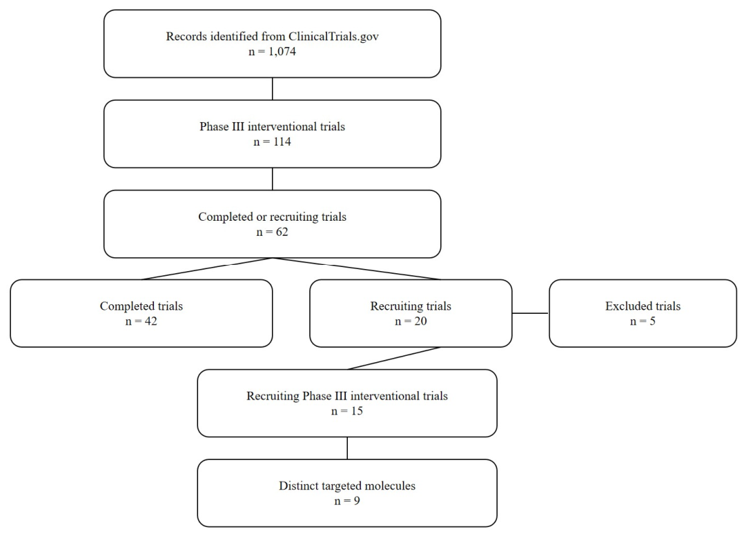
A structured search of the ClinicalTrials.gov database was conducted on 30 November 2025 to identify phase III clinical trials in SLE using the keyword “systemic lupus erythematosus.” The initial search yielded 1074 registered studies. After applying filters for phase III interventional studies, 114 records were identified. Of these, 62 studies met the eligibility criteria based on recruitment status, including 20 recruiting and 42 completed studies. To focus on ongoing research activity, only recruiting studies were included in the final analysis.
Studies were excluded if they evaluated conventional immunosuppressive therapies, investigated non-pharmacological interventions, had an unclear or unspecified mechanism of action, or assessed more than one investigational agent simultaneously. Two investigators (D.P. and S.D.) independently screened the identified records for eligibility, with any discrepancies resolved through consensus. After applying these criteria, 15 eligible phase III interventional studies in SLE were included in the final analysis (n = 15). These studies investigated nine distinct targeted agents (n = 9), which were subsequently grouped according to their principal mechanisms of action (Figure 1).
For each included study, data were extracted on the investigational agent, mechanism of action, ClinicalTrials.gov identifier (NCT number), study population, and primary endpoint. Agents were categorized into major mechanistic groups, including type I interferon pathway inhibition, plasmacytoid dendritic cell targeting, B-cell-targeted therapies, costimulatory pathway blockade, intracellular signaling inhibition (e.g., JAK/TYK2), and S1P receptor modulation. Classification was based on publicly available trial data and supported, when necessary, by published literature to ensure accurate mechanistic grouping.
Following identification of phase III investigational agents, a targeted literature search was performed to summarize the immunopathogenic pathways relevant to each therapeutic target. Searches were conducted in PubMed/MEDLINE using combinations of drug names, molecular targets, and SLE-related keywords. Boolean operators were used to link the investigational molecules with terms such as “pathogenesis,” “pathophysiology,” and “molecular mechanisms.” Priority was given to mechanistic studies, pivotal clinical trials, and recent reviews linking each pathway to SLE pathogenesis. Extracted data were used to support the pathway-based classification of therapies.

3. Pathogenesis as a Basis for Targeted Therapy

The pathogenesis of SLE is driven by a complex interplay between genetic predisposition, epigenetic modifications, and environmental stressors, forming a multifaceted “exposome” [9]. On a fertile background of genetic risk, environmental triggers—such as ultraviolet (UV) light, silica exposure, and viral infections—are thought to initiate aberrant immune activation [10]. For instance, the self-organized criticality theory hypothesizes that repeated antigenic stimulation can push T-cell receptor engagement beyond a stability limit, breaking existing immune anergy and generating autoreactive clones [11].
The transition from subclinical autoimmunity to overt clinical disease relies on a continuous, positive feed-forward loop between the innate and adaptive immune systems [9]. A key initiating step in this cascade is the impaired apoptosis and defective clearance of cellular debris, which leads to an increased exposure of nuclear autoantigens [12]. This accumulation of stimulatory self-nucleic acids activates plasmacytoid dendritic cells (pDCs) and other innate sensors, resulting in sustained type I interferon (IFN-I) pathway activation [10,13]. IFN-I subsequently acts as a potent immune adjuvant that bridges innate and adaptive responses by promoting B-cell differentiation, the expansion of age-associated B cells (ABCs), and the survival of autoantibody-producing plasma cells [9,10]. Concurrently, dysregulated T-cell responses, aberrant co-stimulation, and altered intracellular kinase signaling pathways further amplify and maintain this inflammatory network [14].
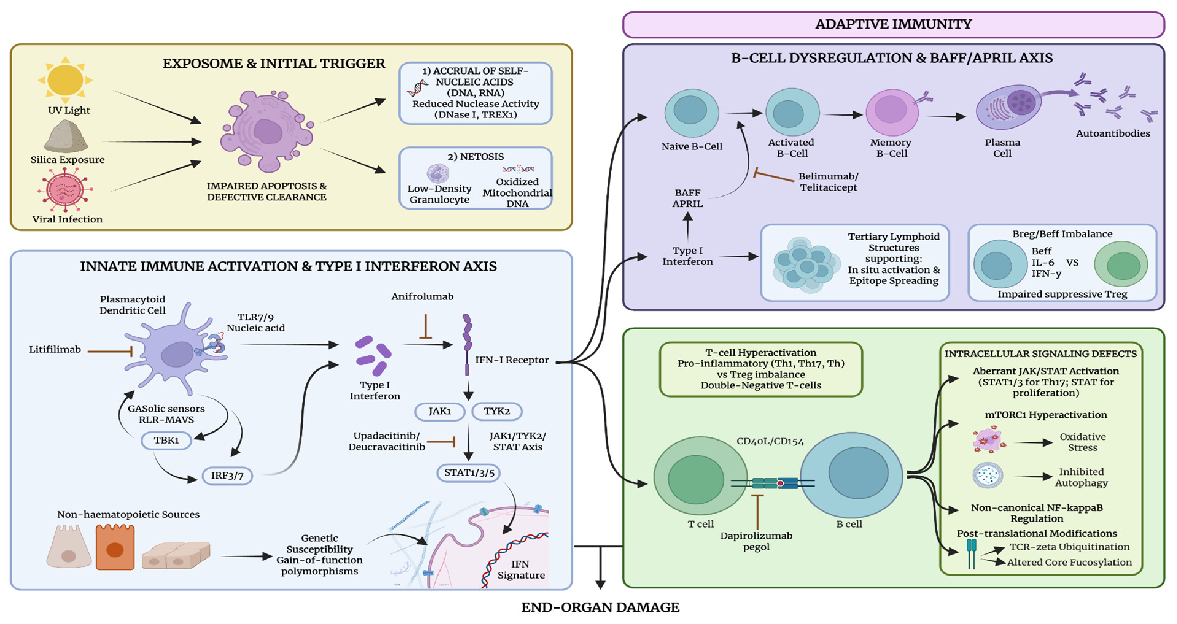
Recognizing these distinct molecular and cellular aberrations has provided the biological rationale for the evolving landscape of targeted therapies [14,15]. To better understand how recent phase 3 clinical trials intervene in this pathogenic cascade, the following subsections will sequentially explore therapies targeting the activation of innate immunity and the IFN-I axis, the dysregulation of the B-cell compartment, and the modulation of T-cell co-stimulation alongside intracellular signal transduction (Figure 2).

3.1. Innate Immune Activation and the Type I Interferon Kinship

Innate immune activation and the subsequent overproduction of IFN-I constitute the primary pathogenic axis in SLE [16]. This chronic inflammatory state is initiated by the abnormal accumulation of endogenous nucleic acids resulting from systemic clearance defects [17]. Specifically, impaired phagocytosis of apoptotic debris and reduced nuclease activity, involving enzymes such as DNase I and TREX1, lead to the persistence of stimulatory self-DNA and RNA [18,19]. Furthermore, low-density granulocytes in SLE exhibit a high propensity for NETosis, extruding decondensed chromatin, antimicrobial peptides like LL-37, and interferogenic oxidized mitochondrial DNA [20,21]. These neutrophil extracellular traps (NETs) directly trigger pDCs and promote aberrant macrophage inflammatory responses [17,21].
Recognition of these ligands occurs through diverse pattern recognition receptors (PRRs). While pDCs produce vast amounts of IFN-alpha via endosomal TLR7/9 pathways, they may become functionally exhausted or inert as the disease progresses [22,23]. Consequently, non-haematopoietic sources, such as keratinocytes and renal tubular cells, alongside other myeloid cells, increasingly contribute to local tissue IFN-I production [24]. Intracellularly, cytosolic sensors like cGAS-STING and RLR-MAVS further detect mitochondrial and retroelement-derived nucleic acids, activating TBK1 and IRF3/7 transcription factors [25,26].
Signaling through the heterodimeric IFN-I receptor (IFNAR) activates the canonical JAK1-TYK2-STAT axis, leading to the induction of a characteristic “IFN signature” [27]. This transcriptional program correlates with autoantibody titers and specific complications like lupus nephritis [28]. Genetic susceptibility is central to this process, as gain-of-function polymorphisms in IRF5, TYK2, and STAT4 enhance either IFN-I production or cellular sensitivity. Additionally, monogenic “type I interferonopathies” serve as clinical models for how constitutive IFN-I activity drives SLE-like autoimmunity [25]. These insights have led to the approval of anifrolumab for receptor blockade and the investigation of JAK inhibitors and blood dendritic cell antigen 2 (BDCA2)-targeted therapies to modulate this innate immune engine [29,30].

3.2. B-Cell Dysregulation and the BAFF/APRIL Axis

B-lineage cells are central drivers of SLE pathogenesis, functioning as autoantibody producers, potent antigen-presenting cells (APCs), and sources of pro-inflammatory cytokines [31]. A fundamental mechanism in the progression of the disease is epitope spreading, where the immune response progressively expands to recognize additional self-epitopes, a process fueled by B-cell-mediated antigen presentation to T cells [32]. This diversification often originates in secondary lymphoid organs, where the failure of central and peripheral tolerance checkpoints allows for the maturation of autoreactive B-cell clones [33].
The survival and terminal differentiation of these cells are governed by the BAFF/APRIL axis [34]. BAFF and a proliferation-inducing ligand (APRIL) signal through three receptors-BAFF-R, transmembrane activator and calcium-modulator and cyclophilin ligand interactor (TACI), and B-cell maturation antigen (BCMA)-with distinct stage-specific expressions [35]. Elevated serum BAFF levels are associated with increased anti-dsDNA titers and disease activity, while APRIL plays a unique role in supporting long-lived plasma cells (LLPCs) and inducing antibody production, particularly IgA [36,37]. In SLE, this axis facilitates the expansion of polyreactive memory B cells, especially those utilizing the IGHV4-34 gene, which is further promoted by type I interferon signaling [38].
The functional balance within the B-cell compartment is severely compromised in SLE, characterized by a “post-activated” phenotype and an imbalance between effector B cells (Beff) and regulatory B cells (Breg) [39]. Excess BAFF drives the differentiation of pro-inflammatory Beff cells (producing IL-6 and IFN-gamma) while impairing the suppressive capacity of IL-10-producing Bregs [40]. This dysregulation also contributes to accelerated atherosclerosis, where B2 cells exhibit pro-atherogenic effects in contrast to the protective role of B1 cell-derived natural IgM [41].
Furthermore, persistent inflammation and antigen exposure lead to the formation of tertiary lymphoid structures (TLS) in target organs such as the skin and kidney [32,42]. These ectopic niches support in situ B-cell activation and epitope diversification, often rendering autoreactive clones resistant to systemic B-cell depletion therapies. These insights provide the rationale for moving beyond pan-B-cell depletion towards strategies targeting specific nodes, such as BAFF inhibition or dual BAFF/APRIL blockade, to simultaneously address the mature B-cell pool and the resilient plasma cell reservoir [33,34]. In addition to BAFF/APRIL signaling, other B-cell regulatory pathways contribute to SLE pathogenesis. CD22 acts as an inhibitory co-receptor that attenuates B-cell receptor signaling and promotes immune tolerance [43]. TLR7 is an endosomal innate immune receptor that recognizes single-stranded RNA and contributes to disease development through activation of autoreactive B cells, promotion of autoantibody production, and induction of type I interferon responses [44]. Together, these pathways further underscore the complexity of B-cell regulation in SLE.

3.3. T-Cell Co-Stimulation and Intracellular Signal Transduction

T-cell hyperactivation and the resulting imbalance between pro-inflammatory (Th1, Th17, Tfh) and regulatory (Treg) subsets are fundamental to the pathogenesis of SLE [45]. This dysfunction is maintained through aberrant co-stimulatory signals and the dysregulation of intracellular signaling cascades, which orchestrate the sustained autoimmune response [46].
A primary driver of this T-cell-dependent B-cell activation is the CD40–CD154 (CD40L) axis [47]. CD154 is persistently overexpressed on SLE T-cells, facilitating the terminal differentiation of B-cells into autoantibody-producing plasma cells and promoting local inflammation in organs like the kidney [48]. While first-generation anti-CD154 antibodies caused thromboembolic complications, second-generation agents like dapirolizumab pegol and the antagonistic anti-CD40 antibody iscalimab are showing safety and efficacy in clinical trials [49,50].
Intracellularly, the Janus kinase/signal transducer and activator of transcription (JAK-STAT) pathway plays a critical role in translating cytokine signals into pathogenic gene expression [51]. Aberrant activation of STAT1 and STAT3 is associated with increased disease activity and Th17 differentiation, while STAT5 phosphorylation patterns correlate with CD4+ T-cell lymphopenia and homeostatic proliferation [52]. Selective tyrosine kinase 2 (TYK2) inhibition has emerged as a promising strategy to block type I interferon and IL-12/23 signaling while uniquely preserving IL-2-mediated Treg cell function [53].
The metabolic state of T-cells is further governed by the mammalian target of rapamycin (mTOR) pathway [54]. mTORC1 hyperactivation is linked to the expansion of pathogenic double-negative (DN) T-cells and the inhibition of autophagy, contributing to oxidative stress and tissue damage [54]. Similarly, the non-canonical NF-kB pathway, regulated by NIK, modulates T-cell differentiation and has been implicated in the loss of self-tolerance [55]. T-cell intrinsic regulators, such as IRF5, further fine-tune this balance, potentially driving the shift from Th2 toward Th1/Th17 responses [56].
Finally, T-cell dysfunction is reinforced by diverse post-translational modifications (PTMs) [57]. SLE T-cells exhibit increased TCR-zeta chain ubiquitination leading to signaling defects, altered protein phosphorylation by PP2A, and changes in core fucosylation that affect T-B cell synaptic interactions [57]. These multifaceted intracellular aberrations provide the rationale for emerging “immune reset” therapies, including CD19-targeted Chimeric antigen receptor (CAR) T-cells and T-cell engagers (TCEs), aimed at eradicating the autoreactive reservoir and restoring long-term tolerance [45,58].

4. Results

The identified phase III clinical trials were grouped according to the predominant immunopathogenic pathway targeted by each therapeutic agent, allowing a pathway-oriented overview of emerging treatment strategies in SLE and LN. The main characteristics of the included molecules, trial populations, and primary efficacy endpoints are summarized in Table 1.

4.1. Drugs Targeting Type I Interferons

Anifrolumab is a human monoclonal antibody targeting IFNAR1. Its efficacy and safety were initially demonstrated in the phase II MUSE trial and subsequently confirmed in the pivotal phase III TULIP-1 and TULIP-2 studies, which led to its regulatory approval for the treatment of moderate-to-severe SLE [59,60]. Long-term extension (LTE) studies have further supported the durability of response and an acceptable safety profile over time [61]. In addition to the original intravenous formulation, a subcutaneous formulation of anifrolumab has also been approved by the US Food and Drug Administration, offering an alternative route of administration. Ongoing phase III trials are further extending the evaluation of anifrolumab [62].
The ongoing IRIS trial (NCT05138133) is a randomized, double-blind, placebo-controlled phase III study evaluating anifrolumab in adults with active proliferative lupus nephritis receiving standard-of-care therapy with mycophenolate mofetil (MMF) and glucocorticoids [63]. Eligible patients have biopsy-confirmed Class III or IV lupus nephritis, with or without concomitant Class V, according to the 2003 ISN/RPS classification, with a renal biopsy obtained within 6 months prior to enrolment or during screening. The primary endpoint is complete renal response (CRR), defined by a urine protein-to-creatinine ratio (UPCR) ≤ 0.5 mg/mg, an estimated glomerular filtration rate (eGFR) ≥ 60 mL/min/1.73 m2 or no decrease from baseline of ≥20%, and no requirement for rescue therapy. Key secondary endpoints include partial renal response, time to renal response, glucocorticoid tapering, and changes in extrarenal disease activity.
In parallel, the BLOSSOM trial (NCT05835310) is a phase III, randomized, double-blind, placebo-controlled study evaluating anifrolumab in children and adolescents aged 5 to <18 years with active systemic lupus erythematosus receiving standard-of-care therapy [64]. The study is designed to assess efficacy over a 52-week treatment period, with the primary endpoint defined as the BILAG-based Composite Lupus Assessment (BICLA) response. Key secondary endpoints include SLEDAI-based measures of disease activity, flare rates, glucocorticoid reduction, and patient-reported outcomes, alongside comprehensive safety and tolerability assessments. Stratification factors include baseline disease activity and background therapy. By extending evaluation to pediatric SLE-a population characterized by more severe disease and earlier organ damage-this trial addresses a critical gap in the evidence base for biologic therapies in younger patients.

4.2. Plasmacytoid Dendritic Cell Targeting

pDCs are a key source of type I interferons in SLE and cutaneous lupus erythematosus (CLE), contributing to immune activation and tissue inflammation. Litifilimab is a humanized IgG1 monoclonal antibody targeting BDCA2, a receptor uniquely expressed on pDCs [65]. Engagement of BDCA2 suppresses the production of type I interferons as well as downstream cytokines and chemokines implicated in lupus pathogenesis.
The efficacy and safety of litifilimab in SLE are being evaluated in two pivotal phase III randomized, double-blind, placebo-controlled trials, TOPAZ-1 and TOPAZ-2, assessing subcutaneous administration in adults with active SLE on background standard-of-care therapy [66]. The primary endpoint is SLE Responder Index-4 (SRI-4) at Week 52, with key secondary endpoints including Joint-50 response, glucocorticoid tapering, organ-specific disease activity measures, and safety.
The AMETHYST trial (NCT05531565) is a phase III, randomized, double-blind, placebo-controlled study evaluating litifilimab in patients with CLE, with or without concomitant systemic lupus erythematosus, receiving standard-of-care therapy [67]. Eligible participants have histologically confirmed CLE either prior to enrolment or during screening. The study comprises two parts with distinct primary endpoints: in Part A, the proportion of patients achieving a Cutaneous Lupus Activity-Investigator’s Global Assessment-Revised (CLA-IGA-R) erythema score of 0 or 1 at Week 16, and in Part B, the proportion achieving a ≥70% reduction in the Cutaneous Lupus Erythematosus Disease Area and Severity Index activity score (CLASI-70) from baseline.

4.3. B-Cell and Plasma Cell Targeting

B cells play a central role in the pathogenesis of SLE through autoantibody production, antigen presentation, and cytokine secretion. Therapeutic strategies aim to disrupt B-cell function either through direct depletion via surface markers such as CD20 or through modulation of survival and differentiation pathways, including BLyS/BAFF, APRIL, and their receptors such as transmembrane activator and calcium modulator and cyclophilin ligand interactor (TACI) [68].
Obinutuzumab is a humanized type II anti-CD20 monoclonal antibody with enhanced antibody-dependent cellular cytotoxicity compared with rituximab, resulting in more potent B-cell depletion [69]. While randomized trials of rituximab did not demonstrate significant benefit in lupus nephritis, obinutuzumab has shown consistent efficacy across clinical studies. In the phase II NOBILITY trial, obinutuzumab added to standard-of-care therapy with MMF and glucocorticoids resulted in higher rates of complete renal response (CRR) at Week 52 compared with placebo [70]. These findings were confirmed in the phase III REGENCY trial, which demonstrated a significant improvement in CRR without new safety signals. Based on these data, obinutuzumab received US Food and Drug Administration approval in 2025 for the treatment of active lupus nephritis [71]. Beyond renal disease, the ALLEGORY trial in systemic lupus erythematosus without active lupus nephritis has also reported positive results, supporting its efficacy in reducing global disease activity [72].
Ongoing studies are further evaluating its role, including the OBILUP trial (NCT04702256), which is designed to assess whether a glucocorticoid-free regimen of obinutuzumab in combination with MMF is non-inferior to a standard regimen including oral corticosteroids and MMF in achieving complete renal response at Week 52 without exceeding a prespecified corticosteroid dose [73]. This study addresses the feasibility of minimizing or avoiding oral glucocorticoids in the induction treatment of lupus nephritis, an approach that may represent a shift in therapeutic strategy if confirmed in randomized controlled settings.
Telitacicept is a recombinant TACI-Fc fusion protein that targets TACI, acting as a dual inhibitor of BLyS/BAFF and APRIL [74]. By simultaneously neutralizing these key survival factors, telitacicept suppresses B-cell maturation, differentiation into plasma cells, and autoantibody production, thereby modulating a central pathogenic pathway in SLE. This dual blockade distinguishes telitacicept from selective BAFF inhibitors by additionally targeting APRIL-driven plasma cell survival. Telitacicept has been approved in China since 2021 for the treatment of SLE [75].
Clinical efficacy has been supported by phase IIb data, in which telitacicept demonstrated significantly higher SLE Responder Index-4 (SRI-4) response rates at Week 48 compared with placebo. The efficacy and safety of telitacicept are being further evaluated in the phase III REMESLE-1 trial, a global, randomized, double-blind, placebo-controlled study in patients with moderate-to-severe SLE, with SRI-4 response at Week 48 as the primary endpoint [76]. At the time of the ClinicalTrials.gov search, a planned phase III study, REMESLE-2 (NCT06456567), was recruiting and therefore met the inclusion criteria. However, the study was subsequently withdrawn prior to enrolment [77].
Ianalumab is a human monoclonal antibody targeting the BAFF receptor, combining inhibition of BAFF-mediated signaling with direct B-cell depletion [78]. By blocking BAFF receptor engagement, ianalumab reduces B-cell survival and autoreactive immune responses, thereby targeting a central pathway in SLE pathogenesis. This mechanism distinguishes it from belimumab, which selectively neutralizes soluble BAFF without inducing B-cell depletion.
The efficacy and safety of ianalumab are being evaluated in two phase III trials, SIRIUS-SLE 1 (NCT05639114) and SIRIUS-SLE 2 (NCT05624749), in patients with moderate-to-severe SLE, with SLE Responder Index-4 (SRI-4) as the primary endpoint [79,80]. In parallel, the SIRIUS-LN trial (NCT05126277) is assessing its role in patients with active lupus nephritis, with stable complete renal response as the primary outcome [81]. Long-term safety and tolerability are being evaluated in the SIRIUS-SLE LTE study (NCT06133972), focusing on treatment-emergent and serious adverse events [82].

4.4. Co-Stimulation Blockade

Dapirolizumab pegol is a polyethylene glycol–conjugated antigen-binding fragment targeting CD40 ligand (CD40L), designed to inhibit the CD40–CD40L interaction, a critical co-stimulatory pathway in SLE pathogenesis [83]. In the phase III PHOENYCS GO trial, a multicenter, randomized, double-blind, placebo-controlled study, patients with moderate-to-severe SLE receiving dapirolizumab pegol in addition to standard-of-care therapy achieved significantly higher BICLA response rates at Week 48 compared with placebo, with a safety profile consistent with previous studies [84].
The ongoing phase III PHOENYCS FLY trial (NCT06617325) is further evaluating its efficacy and safety in patients with moderate-to-severe SLE, with achievement of BICLA response at Week 48 as the primary endpoint [85].

4.5. Intracellular Signaling Pathway Inhibition

Deucravacitinib is an oral, selective, allosteric inhibitor of TYK2, a member of the Janus kinase family that mediates signaling downstream of cytokines central to SLE pathogenesis, including type I interferons, interleukin-12, and interleukin-23 [86]. By binding to the regulatory (pseudokinase) domain of TYK2, deucravacitinib achieves selective inhibition distinct from conventional Janus kinase inhibitors, thereby modulating immune responses implicated in SLE. Its efficacy was demonstrated in the phase II PAISLEY trial, which met its primary and key secondary endpoints in patients with active SLE [87].
The efficacy and safety of deucravacitinib are being evaluated in two phase III, randomized, double-blind, placebo-controlled trials, POETYK SLE-1 (NCT05617677) and POETYK SLE-2 (NCT05620407), in patients with active SLE receiving standard-of-care therapy [88,89]. Both studies assess SLE Responder Index-4 (SRI-4) response at Week 52 as the primary endpoint and incorporate protocol-defined glucocorticoid tapering strategies, with secondary endpoints including changes in organ-specific disease activity and safety outcomes.
Upadacitinib is an oral Janus kinase (JAK) inhibitor that modulates immune responses by targeting the JAK–STAT signaling pathway, which is implicated in the pathogenesis of SLE [90]. The phase II SLEek study demonstrated that upadacitinib 30 mg daily, alone or in combination with elsubrutinib, significantly improved SLE disease activity and reduced flares over 48 weeks, with a favourable safety profile [91]. These findings were further supported by a long-term extension study demonstrating sustained efficacy and acceptable safety through 104 weeks [92]. The phase III SELECT-SLE trial (NCT05843643) is currently evaluating the efficacy and safety of upadacitinib in patients with moderate-to-severe SLE, with achievement of a BICLA response as the primary endpoint [93].

4.6. Immune Trafficking and S1P1 Modulation

Modulation of immune cell trafficking has emerged as a novel therapeutic strategy in SLE. Sphingosine-1-phosphate receptor 1 (S1P1) modulators act by retaining circulating lymphocytes within lymphoid tissues, thereby limiting the migration of potentially pathogenic immune cells to target organs [94]. This mechanism reduces peripheral immune activation without directly depleting immune cells. The clinical relevance of this pathway is supported by the efficacy of the non-selective S1P receptor modulator fingolimod in relapsing forms of multiple sclerosis [95].
Cenerimod is a potent and selective S1P1 receptor modulator designed to reduce lymphocyte egress and modulate immune responses in SLE. Its efficacy and safety are being evaluated in two phase III trials, OPUS-1 (NCT05648500) and OPUS-2 (NCT05672576), in patients with SLE receiving standard-of-care therapy [96,97]. Both studies are randomized, double-blind, placebo-controlled trials, with SLE Responder Index-4 (SRI-4) response at Month 12 as the primary endpoint.

4.7. Patient-Reported Outcomes

Patient-reported outcomes (PROs) were incorporated in 12 of the 15 identified phase III studies, reflecting the growing emphasis on patient-centered assessment in SLE clinical research. The most frequently included PRO was fatigue, evaluated using the Functional Assessment of Chronic Illness Therapy–Fatigue (FACIT-Fatigue) instrument in six trials [98]. Health-related quality of life (HRQoL) measures included the 36-Item Short Form Health Survey (SF-36v2 and SF-36 domains), the Pediatric Quality of Life Inventory (PedsQL), the EuroQol 5-Dimension Questionnaire (EQ-5D), the Cutaneous Lupus Erythematosus Quality of Life questionnaire (CLE-QoL), and the Dermatology Life Quality Index (DLQI) [99,100,101,102,103]. Additional patient-centered measures assessed symptom burden, including pain and itch Numerical Rating Scales (NRS), as well as treatment adherence through the Medication Adherence Self-Report Inventory (MASRI) [104].
Table 1. Phase III targeted therapies in systemic lupus erythematosus (SLE) and lupus nephritis (LN), grouped by pathogenic pathway.
Trial (NCT)Pathogenic PathwayMoleculeMechanism of ActionPopulationPrimary EndpointPatient-Reported Outcomes (PROs)References
1 IRIS (NCT05138133)Type I interferon signalingAnifrolumabAnti-IFNAR1 monoclonal antibodyActive proliferative lupus nephritis (LN)Complete Renal Response (CRR)SF-36v2,
FACIT-Fatigue
[61]
2BLOSSOM (NCT05835310) Pediatric SLEBICLA responsePedsQL[62]
3AMETHYST (NCT05531565)Plasmacytoid dendritic cells (pDCs)LitifilimabAnti-BDCA2 monoclonal antibodyCLE ± SLECLASI-70; CLASI-IGA-R 0/1CLE-QoL, DLQI, pain NRS, itch NRS[65]
4OBILUP (NCT04702256)B cells and plasma cellsObinutuzumabAnti-CD20 monoclonal antibodyLN (≥14 years)Complete Renal Response (CRR)EQ-5D, MASRI[71]
5REMESLE-2 (NCT06456567) TelitaciceptTACI-Fc fusion protein (BAFF/APRIL inhibition)Moderate–severe SLESRI-4FACIT-Fatigue[75]
6SIRIUS-SLE 1 (NCT05639114) IanalumabAnti-BAFF receptor monoclonal antibodySLESRI-4SF-36 Bodily Pain response[77]
7SIRIUS-SLE 2 (NCT05624749) SLESRI-4SF-36 Bodily Pain response[78]
8SIRIUS-LN (NCT05126277) Active LNStable CRRFACIT-Fatigue[79]
9SIRIUS-SLE LTE (NCT06133972) SLESafety (TEAEs/SAEs)NR[80]
10PHOENYCS FLY (NCT06617325)Co-stimulation blockadeDapirolizumab pegolAnti-CD40L monoclonal antibodyModerate–severe SLEBICLA at Week 48FACIT-Fatigue[83]
11POETYK SLE-1 (NCT05617677)Intracellular signaling pathwaysDeucravacitinibSelective TYK2 inhibitorActive SLESRI-4 at Week 52FACIT-Fatigue[86]
12POETYK SLE-2 (NCT05620407) Active SLESRI-4 at Week 52FACIT-Fatigue[87]
13SELECT-SLE (NCT05843643) UpadacitinibSelective JAK1 inhibitorModerate–severe SLEBICLA responseFACIT-Fatigue,
Lupus Pain-NRS,
SF-36 PCS
[91]
14OPUS-1 (NCT05648500) CenerimodS1P1 receptor modulatorSLESRI-4 at Month 12NR[94]
15OPUS-2 (NCT05672576) SLESRI-4 at Month 12NR[95]
Abbreviations: SLE: systemic lupus erythematosus; LN: lupus nephritis; CLE: cutaneous lupus erythematosus; IFNAR1: interferon-α receptor 1; BAFF: B-cell activating factor; APRIL: a proliferation-inducing ligand; S1P1: sphingosine-1-phosphate receptor 1; TYK2: tyrosine kinase 2; JAK: Janus kinase; CRR: complete renal response; SRI-4: Systemic Lupus Erythematosus Responder Index-4; BICLA: BILAG-based Composite Lupus Assessment; BILAG: British Isles Lupus Assessment Group; CLASI: Cutaneous Lupus Erythematosus Disease Area and Severity Index; CLA-IGA-R: Cutaneous Lupus Activity–Investigator’s Global Assessment–Revised; TEAEs: treatment-emergent adverse events; SAEs: serious adverse events; SF-36v2: Short Form-36 Version 2; PCS: Physical Component Summary; FACIT-fatigue: Functional Assessment of Chronic Illness Therapy-Fatigue; PedsQL Generic Core (Physical Functioning Domain) as part of the PRINTO/ACR childhood-onset SLE responder definition; CLE-QoL: Cutaneous Lupus Erythematosus Quality of Life; DLQI: Dermatology Life Quality Index; pain NRS: Numerical Rating Scale (NRS) for Pain in Skin Rash; NRS for Itch in Skin Rash; EQ-5D: EuroQol 5-Dimension Questionnaire, MASRI: Medication Adherence Self-Report Inventory; Lupus Pain-NRS: Lupus Pain Numerical Rating Scale (NRS); NR: Not Reported.

5. Strengths and Limitations

This review offers a multi-dimensional synthesis of the evolving targeted therapy landscape in SLE, bridging the gap between intricate molecular immunopathology—specifically the IFN-I axis, B-cell dysregulation, and intracellular signaling cascades—and the evidence generated by late-phase clinical development. A primary strength of this work is the systematic identification of Phase 3 clinical trials via the ClinicalTrials.gov database, providing a forward-looking framework that captures the transition toward precision medicine as of late 2025. By organizing investigational agents according to their specific pathogenic nodes-ranging from pDC inhibition to selective TYK2/JAK modulation—this review provides clinicians and researchers with a contemporary map of the most transformative era in lupus therapeutics. Furthermore, the detailed integration of B-T cell co-stimulatory dynamics and post-translational modifications provides a robust biological context for understanding the varying success of “immune reset” strategies.
Despite these strengths, several methodological and inherent limitations must be acknowledged. First, a significant limitation of our methodology is the restriction of the search to English-language publications and registries. Given the substantial body of seminal work and large-scale cohort data originating from East Asian and other global populations where SLE prevalence and severity are high, the exclusion of non-English literature may introduce a geographic bias, potentially omitting localized clinical insights or regional management protocols. Secondly, although the search was structured in its execution, this remains a narrative review. The inherent heterogeneity of the SLE literature—characterized by diverse trial endpoints (e.g., SRI-4 vs. BICLA vs. CRR) and varying background standard-of-care regimens—precluded the performance of a formal meta-analysis or a universal quality appraisal of all cited trial data. Furthermore, the search was restricted to ClinicalTrials.gov and may not have captured studies registered exclusively in regional trial registries, potentially resulting in omission of relevant investigational programs.
The clinical evidence is further constrained by the phenotypic diversity of SLE. Many diagnostic and therapeutic conclusions are drawn from populations with specific organ involvement, which may limit their generalizability; for instance, responses in lupus nephritis may follow different biological trajectories compared with cutaneous or neuropsychiatric manifestations. Additionally, as noted in our discussion of B-cell therapies, the “clinical trial paradox”-where agents like rituximab show real-world utility despite failing primary endpoints in randomized controlled trials (RCTs)-highlights the ongoing difficulty in aligning biological rationale with rigid trial designs. Moreover, the reliance on registry data restricts the depth of long-term safety and subgroup analyses, particularly for newer classes of agents such as S1P1 modulators and allosteric TYK2 inhibitors, where the full extent of “off-target” effects or durable remission remains to be established in broader clinical practice. Finally, as many of the identified phase III studies are still ongoing, the long-term efficacy and safety of several investigational agents remain to be fully established upon completion of these trials.

6. Future Perspectives

The treatment paradigm in SLE is rapidly shifting toward precision medicine and mechanism-based interventions aimed at restoring immune homeostasis. Advances in high-dimensional immunophenotyping and biomarker-driven stratification are enabling the alignment of targeted therapies with individual disease endotypes, moving beyond broad, non-selective immunosuppression.
The therapeutic landscape is also expanding toward cellular immunotherapies, particularly CAR T-cell approaches targeting autoreactive B cells [105]. Early-phase studies of CD19-directed CAR T-cell therapy have demonstrated the potential to induce deep immunological remission and prolonged “immune reset” in patients with refractory SLE [106]. Complementing these cellular advances, bispecific TCEs have emerged as a promising “off-the-shelf” alternative for refractory autoimmune disease [107]. These agents redirect T-cell cytotoxicity by simultaneously binding CD3 on T cells and B-cell antigens such as CD19 or BCMA [108]. CD19-directed TCEs, including blinatumomab and A-319, have shown rapid and profound B-cell depletion with subsequent “autoimmune reset” in severe SLE [109], while BCMA-targeted therapies such as teclistamab may eliminate long-lived plasma cells that often escape CD20-directed depletion [107]. Although early clinical data suggest that TCEs can induce remission and normalize serological biomarkers, including anti-dsDNA antibodies and complement levels, the risk of cytokine release syndrome requires careful safety monitoring and inpatient management [108].
Together, these developments may accelerate the transition toward earlier, more individualized use of biologic and targeted therapies in SLE.

7. Conclusions

The therapeutic landscape of systemic lupus erythematosus is undergoing a significant transformation, with multiple targeted agents demonstrating efficacy across distinct immunological pathways. Beyond established therapies targeting the type I interferon and BAFF axes, newer strategies—including plasmacytoid dendritic cell inhibition, co-stimulation blockade, intracellular signaling modulation, and immune trafficking via S1P1 modulation—are expanding the range of potential treatment options.
These developments reflect a shift toward mechanism-based and potentially more individualized treatment approaches, with several agents showing promise in reducing disease activity, limiting organ damage, and enabling glucocorticoid-sparing strategies. Notably, the emergence of therapies targeting B-cell function more precisely, as well as approaches aiming to modulate immune cell trafficking, may address unmet needs in patients with refractory or organ-threatening disease.
Despite these advances, challenges remain, including the need for better patient stratification, harmonization of clinical endpoints, and long-term safety data. Future research should focus on identifying predictive biomarkers, optimizing treatment sequencing, and evaluating combination or personalized therapeutic strategies. Collectively, ongoing phase III trials are expected to further refine the role of targeted therapies and support the transition toward precision medicine in SLE.

Author Contributions

Conceptualization, D.T.P.; methodology, D.T.P. and S.D.; investigation, D.T.P., S.D., N.S. and M.I.; data curation, D.T.P. and S.D.; formal analysis, D.T.P., S.D., N.S. and M.I.; writing—original draft preparation, D.T.P., S.D., N.S. and M.I.; writing—review and editing, D.T.P., S.D., N.S. and M.I.; visualization, S.D.; supervision, D.T.P.; project administration, M.I. All authors have read and agreed to the published version of the manuscript.

Funding

This research received no external funding.

Institutional Review Board Statement

Not applicable.

Informed Consent Statement

Not applicable.

Data Availability Statement

No new data were created. The search strategy was described in the methodology section.

Acknowledgments

The authors acknowledge the support of the University Hospital “St. Ivan Rilski”-Sofia and University Hospital “St. Marina”-Varna. All figures are original and were created by the authors. Figure 1 was designed using Microsoft PowerPoint. Figure 2 is created in BioRender (Dimitrov, S. (2026) https://BioRender.com/oqo797i). Graphical abstract is created in BioRender (Dimitrov, S. (2026) https://BioRender.com/7ptwdvi). Generative AI (Gemini 3 Flash, Google, Mountain View, CA, USA, 2026) was used as a language-editing and formatting assistant during manuscript preparation. It helped refine English phrasing, improve clarity, and align the structure with MDPI formatting requirements. No AI tools were used for data generation, analysis, interpretation, or reference fabrication. All scientific content, interpretation, and conclusions are entirely the authors’ own. The authors take full responsibility for the accuracy and integrity of the submitted work.

Conflicts of Interest

The authors declare no conflicts of interest.

Abbreviations

The following abbreviations are used in this manuscript:
ABCAge-associated B cell
APCAntigen-presenting cell
APRILA proliferation-inducing ligand
BAFFB-cell activating factor
BAFF-RB-cell activating factor receptor
BCMAB-cell maturation antigen
BDCA2Blood dendritic cell antigen 2
BeffEffector B cell
BICLABILAG-based Composite Lupus Assessment
BILAGBritish Isles Lupus Assessment Group
BLySB-lymphocyte stimulator
BregRegulatory B cell
CARChimeric antigen receptor
CD40L/CD154CD40 ligand
cGAS-STINGcyclic GMP–AMP synthase–stimulator of interferon genes
CLA-IGA-RCutaneous Lupus Activity–Investigator’s Global Assessment–Revised
CLASICutaneous Lupus Erythematosus Disease Area and Severity Index
CLECutaneous lupus erythematosus
CLE-QoLCutaneous Lupus Erythematosus Quality of Life questionnaire
CRRComplete renal response
DLQI Dermatology Life Quality Index
DORISDefinitions of Remission in Systemic Lupus Erythematosus
eGFREstimated glomerular filtration rate
EQ-5D EuroQol 5-Dimension Questionnaire
EULAREuropean Alliance of Associations for Rheumatology
FACIT-FatigueFunctional Assessment of Chronic Illness Therapy–Fatigue
HCQHydroxychloroquine
HRQoLHealth-related quality of life
IFN-IType I interferon
IFNAR1Interferon alpha and beta receptor subunit 1
IgAImmunoglobulin A
IgG1Immunoglobulin G1
ILInterleukin
IRFInterferon regulatory factor
ISN/RPSInternational Society of Nephrology/Renal Pathology Society
JAKJanus kinase
LLDASLupus low disease activity state
LLPCLong-lived plasma cell
LNLupus nephritis
MAVSMitochondrial antiviral-signaling protein
MMFMycophenolate mofetil
mTORMammalian target of rapamycin
mTORC1Mammalian target of rapamycin complex 1
NCTNational Clinical Trial number
NETsNeutrophil extracellular traps
NF-κBNuclear factor kappa B
NIKNF-κB-inducing kinase
pDCPlasmacytoid dendritic cell
PedsQLPediatric Quality of Life Inventory
PRRPattern recognition receptor
PTMPost-translational modification
RCTRandomized controlled trial
RLRRIG-I-like receptor
S1P1Sphingosine-1-phosphate receptor 1
SAESerious adverse event
SF-36v236-Item Short Form Health Survey
SLESystemic lupus erythematosus
SRI-4Systemic Lupus Erythematosus Responder Index-4
STATSignal transducer and activator of transcription
TACITransmembrane activator and calcium-modulator and cyclophilin ligand interactor
TBK1TANK-binding kinase 1
TCET-cell engager
TCRT-cell receptor
TEAETreatment-emergent adverse event
TfhT follicular helper cell
ThT helper cell
TLRToll-like receptor
TLSTertiary lymphoid structure
TregRegulatory T cell
TREX1Three prime repair exonuclease 1
TYK2Tyrosine kinase 2
UPCRUrine protein-to-creatinine ratio

References

  1. Kaul, A.; Gordon, C.; Crow, M.K.; Touma, Z.; Urowitz, M.B.; van Vollenhoven, R.; Ruiz-Irastorza, G.; Hughes, G. Systemic Lupus Erythematosus. Nat. Rev. Dis. Primers 2016, 2, 1480–1491. [Google Scholar] [CrossRef]
  2. Felten, R.; Scherlinger, M.; Mertz, P.; Chasset, F.; Arnaud, L. New Biologics and Targeted Therapies in Systemic Lupus: From New Molecular Targets to New Indications. A Systematic Review. Jt. Bone Spine 2023, 90, 105523. [Google Scholar] [CrossRef] [PubMed]
  3. Fanouriakis, A.; Kostopoulou, M.; Andersen, J.; Aringer, M.; Arnaud, L.; Bae, S.-C.; Boletis, J.; Bruce, I.N.; Cervera, R.; Doria, A.; et al. EULAR Recommendations for the Management of Systemic Lupus Erythematosus: 2023 Update. Ann. Rheum. Dis. 2024, 83, 15–29. [Google Scholar] [CrossRef] [PubMed]
  4. Fanouriakis, A.; Kostopoulou, M.; Anders, H.-J.; Andersen, J.; Aringer, M.; Beresford, M.W.; Doria, A.; Frangou, E.; Furie, R.; Gladman, D.D.; et al. EULAR Recommendations for the Management of Systemic Lupus Erythematosus with Kidney Involvement: 2025 Update. Ann. Rheum. Dis. 2026, 85, 75–90. [Google Scholar] [CrossRef] [PubMed]
  5. Parra Sánchez, A.R.; Voskuyl, A.E.; van Vollenhoven, R.F. Treat-to-Target in Systemic Lupus Erythematosus: Advancing towards Its Implementation. Nat. Rev. Rheumatol. 2022, 18, 146–157. [Google Scholar] [CrossRef] [PubMed]
  6. Franklyn, K.; Lau, C.S.; Navarra, S.V.; Louthrenoo, W.; Lateef, A.; Hamijoyo, L.; Wahono, C.S.; Chen, S.L.; Jin, O.; Morton, S.; et al. Definition and Initial Validation of a Lupus Low Disease Activity State (LLDAS). Ann. Rheum. Dis. 2016, 75, 1615–1621. [Google Scholar] [CrossRef] [PubMed]
  7. van Vollenhoven, R.F.; Bertsias, G.; Doria, A.; Isenberg, D.; Morand, E.; Petri, M.A.; Pons-Estel, B.A.; Rahman, A.; Ugarte-Gil, M.F.; Voskuyl, A.; et al. 2021 DORIS Definition of Remission in SLE: Final Recommendations from an International Task Force. Lupus Sci. Med. 2021, 8, e000538. [Google Scholar] [CrossRef] [PubMed]
  8. Scherlinger, M.; Kolios, A.G.A.; Kyttaris, V.C.; Tsokos, G.C. Advances in the Treatment of Systemic Lupus Erythematosus. Nat. Rev. Drug Discov. 2025, 24, 926–944. [Google Scholar] [CrossRef] [PubMed]
  9. Rose, T.; Dörner, T. Drivers of the Immunopathogenesis in Systemic Lupus Erythematosus. Best Pract. Res. Clin. Rheumatol. 2017, 31, 321–333. [Google Scholar] [CrossRef] [PubMed]
  10. Crow, M.K. Pathogenesis of Systemic Lupus Erythematosus: Risks, Mechanisms and Therapeutic Targets. Ann. Rheum. Dis. 2023, 82, 999–1014. [Google Scholar] [CrossRef] [PubMed]
  11. Shiozawa, S. Pathogenesis of Autoimmunity/Systemic Lupus Erythematosus (SLE). Cells 2025, 14, 1080. [Google Scholar] [CrossRef] [PubMed]
  12. Lo, M.S.; Tsokos, G.C. Recent Developments in Systemic Lupus Erythematosus Pathogenesis and Applications for Therapy. Curr. Opin. Rheumatol. 2018, 30, 222–228. [Google Scholar] [CrossRef] [PubMed]
  13. Deng, Y.; Tsao, B.P. Updates in Lupus Genetics. Curr. Rheumatol. Rep. 2017, 19, 68. [Google Scholar] [CrossRef] [PubMed]
  14. Paley, M.A.; Strand, V.; Kim, A.H.J. From Mechanism to Therapies in Systemic Lupus Erythematosus. Curr. Opin. Rheumatol. 2017, 29, 178–186. [Google Scholar] [CrossRef] [PubMed]
  15. Accapezzato, D.; Caccavale, R.; Paroli, M.P.; Gioia, C.; Nguyen, B.L.; Spadea, L.; Paroli, M. Advances in the Pathogenesis and Treatment of Systemic Lupus Erythematosus. Int. J. Mol. Sci. 2023, 24, 6578. [Google Scholar] [CrossRef] [PubMed]
  16. Weidenbusch, M.; Kulkarni, O.P.; Anders, H.-J. The Innate Immune System in Human Systemic Lupus Erythematosus. Clin. Sci. 2017, 131, 625–634. [Google Scholar] [CrossRef] [PubMed]
  17. Ma, C.; Xia, Y.; Yang, Q.; Zhao, Y. The Contribution of Macrophages to Systemic Lupus Erythematosus. Clin. Immunol. 2019, 207, 1–9. [Google Scholar] [CrossRef] [PubMed]
  18. Mustelin, T.; Lood, C.; Giltiay, N.V. Sources of Pathogenic Nucleic Acids in Systemic Lupus Erythematosus. Front. Immunol. 2019, 10, 1028. [Google Scholar] [CrossRef] [PubMed]
  19. Bai, Y.; Tong, Y.; Liu, Y.; Hu, H. Self-dsDNA in the Pathogenesis of Systemic Lupus Erythematosus. Clin. Exp. Immunol. 2018, 191, 1–10. [Google Scholar] [CrossRef] [PubMed]
  20. Barnado, A.; Crofford, L.J.; Oates, J.C. At the Bedside: Neutrophil Extracellular Traps (NETs) as Targets for Biomarkers and Therapies in Autoimmune Diseases. J. Leukoc. Biol. 2016, 99, 265–278. [Google Scholar] [CrossRef] [PubMed]
  21. Watanabe, H.; Matsumoto, Y.; Wada, J. Immunometabolic Regulation of Innate Immunity in Systemic Lupus Erythematosus. Acta Med. Okayama 2025, 79, 147–155. [Google Scholar] [CrossRef] [PubMed]
  22. Matta, B.; Barnes, B.J. Coordination between Innate Immune Cells, Type I IFNs and IRF5 Drives SLE Pathogenesis. Cytokine 2020, 132, 154731. [Google Scholar] [CrossRef] [PubMed]
  23. Panda, S.K.; Kolbeck, R.; Sanjuan, M.A. Plasmacytoid Dendritic Cells in Autoimmunity. Curr. Opin. Immunol. 2017, 44, 20–25. [Google Scholar] [CrossRef] [PubMed]
  24. Psarras, A.; Wittmann, M.; Vital, E.M. Emerging Concepts of Type I Interferons in SLE Pathogenesis and Therapy. Nat. Rev. Rheumatol. 2022, 18, 575–590. [Google Scholar] [CrossRef] [PubMed]
  25. Zhou, Y.; Song, H.-M. Type I Interferon Pathway in Pediatric Systemic Lupus Erythematosus. World J. Pediatr. 2024, 20, 653–668. [Google Scholar] [CrossRef] [PubMed]
  26. Gallucci, S.; Meka, S.; Gamero, A.M. Abnormalities of the Type I Interferon Signaling Pathway in Lupus Autoimmunity. Cytokine 2021, 146, 155633. [Google Scholar] [CrossRef] [PubMed]
  27. Ishihara, R.; Watanabe, R.; Shiomi, M.; Fujita, Y.; Katsushima, M.; Fukumoto, K.; Yamada, S.; Hashimoto, M. The Type I Interferon Axis in Systemic Autoimmune Diseases: From Molecular Pathways to Targeted Therapy. Biomolecules 2025, 15, 1586. [Google Scholar] [CrossRef] [PubMed]
  28. Postal, M.; Vivaldo, J.F.; Fernandez-Ruiz, R.; Paredes, J.L.; Appenzeller, S.; Niewold, T.B. Type I Interferon in the Pathogenesis of Systemic Lupus Erythematosus. Curr. Opin. Immunol. 2020, 67, 87–94. [Google Scholar] [CrossRef] [PubMed]
  29. Lai, B.; Luo, S.-F.; Lai, J.-H. Therapeutically Targeting Proinflammatory Type I Interferons in Systemic Lupus Erythematosus: Efficacy and Insufficiency with a Specific Focus on Lupus Nephritis. Front. Immunol. 2024, 15, 1489205. [Google Scholar] [CrossRef] [PubMed]
  30. Psarras, A.; Emery, P.; Vital, E.M. Type I Interferon-Mediated Autoimmune Diseases: Pathogenesis, Diagnosis and Targeted Therapy. Rheumatology 2017, 56, 1662–1675. [Google Scholar] [CrossRef] [PubMed]
  31. Liu, Z.; Dai, H.; Cui, X.; Liu, Y.; Dong, Z. Importance of B Cells (Review). Int. J. Mol. Med. 2026, 57, 2. [Google Scholar] [CrossRef] [PubMed]
  32. Tariq, F.; Charavanmuttu, Y.; Labiba, K.; Wincup, C. Aberrant B Cell Responses as Drivers of Autoantibody Generation and Epitope Diversification in SLE Pathogenesis. Front. Immunol. 2025, 16, 1731285. [Google Scholar] [CrossRef] [PubMed]
  33. Atisha-Fregoso, Y.; Toz, B.; Diamond, B. Meant to B: B Cells as a Therapeutic Target in Systemic Lupus Erythematosus. J. Clin. Invest. 2021, 131, e149095. [Google Scholar] [CrossRef] [PubMed]
  34. Yang, X.; Liu, S.; Liu, X.-J.; Wen, D.; Zhou, Y.; Zeng, Y.; Li, L.; Deng, Q.; Wang, Y.; Chen, Q.; et al. Biological Targeted Therapy for Lupus Nephritis-the Role of BLYS and APRIL. Ren. Fail. 2025, 47, 2561791. [Google Scholar] [CrossRef] [PubMed]
  35. Wu, J.; Li, Z.; Zheng, S.; Liu, Y.; Zhao, N. BAFF in Autoimmune Kidney Diseases: Pathogenic Pathways, Biomarker Potential, and Therapeutic Horizons. Ren. Fail. 2025, 47, 2577164. [Google Scholar] [CrossRef] [PubMed]
  36. Feng, Y.; Yang, M.; Wu, H.; Lu, Q. The Pathological Role of B Cells in Systemic Lupus Erythematosus: From Basic Research to Clinical. Autoimmunity 2020, 53, 56–64. [Google Scholar] [CrossRef] [PubMed]
  37. Poznyak, A.V.; Gerasimova, E.V.; Orekhov, N.A.; Karimova, A.E.; Vergun, M.A.; Lapshina, K.O.; Sukhorukov, V.N.; Orekhov, A.N. Exploring the Role of APRIL in Autoimmunity: Implications for Therapeutic Targeting in Systemic Lupus Erythematosus, Rheumatoid Arthritis, and Sjögren’s Syndrome. Front. Immunol. 2025, 16, 1523392. [Google Scholar] [CrossRef] [PubMed]
  38. Fujio, K.; Ushijima, T.; Okamura, T.; Ota, M. The Role of Polyreactive Memory B Cells in Systemic Lupus Erythematosus. Int. Immunol. 2025, 37, 189–194. [Google Scholar] [CrossRef] [PubMed]
  39. Dörner, T.; Lipsky, P.E. Beyond Pan-B-Cell-Directed Therapy—New Avenues and Insights into the Pathogenesis of SLE. Nat. Rev. Rheumatol. 2016, 12, 645–657. [Google Scholar] [CrossRef] [PubMed]
  40. Matsushita, T. Regulatory and Effector B Cells: Friends or Foes? J. Dermatol. Sci. 2019, 93, 2–7. [Google Scholar] [CrossRef] [PubMed]
  41. Koulouri, V.; Koutsilieris, M.; Mavragani, C.P. B Cells and Atherosclerosis in Systemic Lupus Erythematosus. Expert Rev. Clin. Immunol. 2019, 15, 417–429. [Google Scholar] [CrossRef] [PubMed]
  42. Fetter, T.; Niebel, D.; Braegelmann, C.; Wenzel, J. Skin-Associated B Cells in the Pathogenesis of Cutaneous Autoimmune Diseases-Implications for Therapeutic Approaches. Cells 2020, 9, 2627. [Google Scholar] [CrossRef] [PubMed]
  43. Dörner, T.; Shock, A.; Goldenberg, D.M.; Lipsky, P.E. The Mechanistic Impact of CD22 Engagement with Epratuzumab on B Cell Function: Implications for the Treatment of Systemic Lupus Erythematosus. Autoimmun. Rev. 2015, 14, 1079–1086. [Google Scholar] [CrossRef] [PubMed]
  44. Vinuesa, C.G.; Shrotri, M.; Rahman, A. TLR7 in Systemic Lupus Erythematosus: Genetics and Emerging Therapies. Nat. Rev. Rheumatol. 2026. [Google Scholar] [CrossRef] [PubMed]
  45. Wu, Y.; Zhang, W.; Liao, Y.; Sun, T.; Liu, Y.; Liu, Y. Immune Cell Aberrations in Systemic Lupus Erythematosus: Navigating the Targeted Therapies toward Precision Management. Cell. Mol. Biol. Lett. 2025, 30, 73. [Google Scholar] [CrossRef] [PubMed]
  46. Balogh, L.; Kovács, V.G.; Nagy, G.; Németh, T. Signaling Pathways in Systemic Lupus Erythematosus and Therapeutic Implications. Front. Immunol. 2025, 16, 1735301. [Google Scholar] [CrossRef] [PubMed]
  47. Ramanujam, M.; Steffgen, J.; Visvanathan, S.; Mohan, C.; Fine, J.S.; Putterman, C. Phoenix from the Flames: Rediscovering the Role of the CD40-CD40L Pathway in Systemic Lupus Erythematosus and Lupus Nephritis. Autoimmun. Rev. 2020, 19, 102668. [Google Scholar] [CrossRef] [PubMed]
  48. Allard, C.C.; Salti, S.; Mourad, W.; Hassan, G.S. Implications of CD154 and Its Receptors in the Pathogenesis and Treatment of Systemic Lupus Erythematosus. Cells 2024, 13, 1621. [Google Scholar] [CrossRef] [PubMed]
  49. Touma, Z.; Aranow, C.; Parodis, I.; Ramsey-Goldman, R.; Schneider, M.; De La Loge, C.; Jimenez, T.; Nejati, M.; Arnaud, L. Dapirolizumab Pegol Demonstrated Improvement in Quality of Life of Patients with Systemic Lupus Erythematosus: Lupusqol Results from a Phase 3 Trial. J. Rheumatol. 2025, 52, 11–12. [Google Scholar] [CrossRef]
  50. Shen, N.; Weinmann-Menke, J.; Malvar, A.; Serra-Roma, A.; Weiss, M.; Danekula, R.; Sips, C.; Rohr, J.; Felten, R.; Gergely, P.; et al. Efficacy, Pharmacokinetics and Safety of Iscalimab (CFZ533) in Patients with Proliferative Lupus Nephritis: A Randomised, Double-Blind, Placebo-Controlled, Phase II Study. RMD Open 2025, 11, e005557. [Google Scholar] [CrossRef] [PubMed]
  51. Goropevšek, A.; Holcar, M.; Avčin, T. The Role of STAT Signaling Pathways in the Pathogenesis of Systemic Lupus Erythematosus. Clin. Rev. Allergy Immunol. 2017, 52, 164–181. [Google Scholar] [CrossRef] [PubMed]
  52. Goropevšek, A.; Holcar, M.; Pahor, A.; Avčin, T. STAT Signaling as a Marker of SLE Disease Severity and Implications for Clinical Therapy. Autoimmun. Rev. 2019, 18, 144–154. [Google Scholar] [CrossRef] [PubMed]
  53. Satoh-Kanda, Y.; Nakayamada, S.; Tanaka, Y. Fine-Tuning SLE Treatment: The Potential of Selective TYK2 Inhibition. RMD Open 2024, 10, e005072. [Google Scholar] [CrossRef] [PubMed]
  54. He, J.; Ma, J.; Ren, B.; Liu, A. Advances in Systemic Lupus Erythematosus Pathogenesis via mTOR Signaling Pathway. Semin. Arthritis Rheum. 2020, 50, 314–320. [Google Scholar] [CrossRef] [PubMed]
  55. Montano, H.; Allen, I.C.; Reilly, C.M. The Path Less Traveled: The Non-Canonical NF-κB Pathway in Systemic Lupus Erythematosus. Front. Immunol. 2025, 16, 1588486. [Google Scholar] [CrossRef] [PubMed]
  56. Brune, Z.; Rice, M.R.; Barnes, B.J. Potential T Cell-Intrinsic Regulatory Roles for IRF5 via Cytokine Modulation in T Helper Subset Differentiation and Function. Front. Immunol. 2020, 11, 1143. [Google Scholar] [CrossRef] [PubMed]
  57. Yang, F.; Lin, J.; Chen, W. Post-Translational Modifications in T Cells in Systemic Erythematosus Lupus. Rheumatology 2021, 60, 2502–2516. [Google Scholar] [CrossRef] [PubMed]
  58. Kurzhagen, J.T.; Laqua, W.; Schett, G.; Grieshaber-Bouyer, R.; Rabb, H.; Noel, S.; Schiffer, M. Tiny Warriors, Big Impact: Targeted Immunotherapy in Kidney Diseases. Kidney Int. 2026, 109, 869–882. [Google Scholar] [CrossRef] [PubMed]
  59. Furie, R.A.; Morand, E.F.; Bruce, I.N.; Manzi, S.; Kalunian, K.C.; Vital, E.M.; Lawrence Ford, T.; Gupta, R.; Hiepe, F.; Santiago, M.; et al. Type I Interferon Inhibitor Anifrolumab in Active Systemic Lupus Erythematosus (TULIP-1): A Randomised, Controlled, Phase 3 Trial. Lancet Rheumatol. 2019, 1, e208–e219. [Google Scholar] [CrossRef] [PubMed]
  60. Morand, E.F.; Furie, R.; Tanaka, Y.; Bruce, I.N.; Askanase, A.D.; Richez, C.; Bae, S.-C.; Brohawn, P.Z.; Pineda, L.; Berglind, A.; et al. Trial of Anifrolumab in Active Systemic Lupus Erythematosus. N. Engl. J. Med. 2020, 382, 211–221. [Google Scholar] [CrossRef] [PubMed]
  61. Strand, V.; Kalunian, K.C.; Lee, K.W.; Seo, C.; Abreu, G.; Tummala, R.; Al-Mossawi, H.; Duncan, E.A.; Lindholm, C. Long-Term Effect of Anifrolumab on Patient-Reported Outcomes in Systemic Lupus Erythematosus (TULIP-LTE): A Randomised, Placebo-Controlled, Phase 3 Long-Term Extension Trial. Lancet Rheumatol. 2025, 7, e485–e494. [Google Scholar] [CrossRef] [PubMed]
  62. SAPHNELO Approved in the US for Subcutaneous Self-Administration as a New Autoinjector for the Treatment of Systemic Lupus Erythematosus. Available online: https://www.astrazeneca-us.com/media/press-releases/2026/SAPHNELO-approved-in-the-US-for-subcutaneous-self-administration-as-a-new-autoinjector-for-the-treatment-of-systemic-lupus-erythematosus.html (accessed on 6 May 2026).
  63. Clinicaltrials.gov. Available online: https://clinicaltrials.gov/study/NCT05138133 (accessed on 5 May 2026).
  64. Clinicaltrials.gov. Available online: https://clinicaltrials.gov/study/NCT05835310 (accessed on 6 May 2026).
  65. Cho, Y.M.; Furie, R. The Development of Litifilimab (BIIB 059) for Cutaneous and Systemic Lupus Erythematosus. Immunotherapy 2024, 16, 15–20. [Google Scholar] [CrossRef] [PubMed]
  66. Vollenhoven, R.F.; Furie, R.A.; Morand, E.F.; Kalunian, K.; Dall’era, M.; Goode-sellers, S.; Joshi, P.; Kong, G.; Wang, T.; Wei, X.; et al. LP-184 Rationale and Design of Two Phase 3, Randomized, Double-Blind, Placebo-Controlled Studies of the Efficacy and Safety of Litifilimab in Adults with Active Systemic Lupus Erythematosus: TOPAZ-1 and TOPAZ-2. Lupus Sci. Med. 2023, 10, A1–A171. [Google Scholar]
  67. Clinicaltrials.gov. Available online: https://clinicaltrials.gov/study/NCT05531565 (accessed on 6 May 2026).
  68. Wu, C.; Wang, Y.; Chen, S.; Lin, Y.; Du, F.; Wang, X.; Chen, S.; Lu, L.; Ye, S.; Ding, H.; et al. Obinutuzumab in Systemic Lupus Erythematosus: A Real-World Experience. Front. Immunol. 2025, 16, 1702550. [Google Scholar] [CrossRef] [PubMed]
  69. Freeman, C.L.; Sehn, L.H. A Tale of Two Antibodies: Obinutuzumab versus Rituximab. Br. J. Haematol. 2018, 182, 29–45. [Google Scholar] [CrossRef] [PubMed]
  70. Furie, R.A.; Aroca, G.; Cascino, M.D.; Garg, J.P.; Rovin, B.H.; Alvarez, A.; Fragoso-Loyo, H.; Zuta-Santillan, E.; Schindler, T.; Brunetta, P.; et al. B-Cell Depletion with Obinutuzumab for the Treatment of Proliferative Lupus Nephritis: A Randomised, Double-Blind, Placebo-Controlled Trial. Ann. Rheum. Dis. 2022, 81, 100–107. [Google Scholar] [CrossRef] [PubMed]
  71. Furie, R.A.; Rovin, B.H.; Garg, J.P.; Santiago, M.B.; Aroca-Martínez, G.; Zuta Santillán, A.E.; Alvarez, D.; Navarro Sandoval, C.; Lila, A.M.; Tumlin, J.A.; et al. Efficacy and Safety of Obinutuzumab in Active Lupus Nephritis. N. Engl. J. Med. 2025, 392, 1471–1483. [Google Scholar] [CrossRef] [PubMed]
  72. Furie, R.A.; Dall’Era, M.; Vital, E.M.; Garg, J.P.; Irazoque Palazuelos, F.; Zuta Santillán, A.E.; Ravelo-Hernández, J.; Santiago, M.B.; Zazueta Montiel, B.; Botha, S.; et al. Efficacy and Safety of Obinutuzumab in Active Systemic Lupus Erythematosus. N. Engl. J. Med. 2026. [Google Scholar] [CrossRef] [PubMed]
  73. Clinicaltrials.gov. Available online: https://clinicaltrials.gov/study/NCT04702256 (accessed on 6 May 2026).
  74. Zeng, L.; Yang, K.; Wu, Y.; Yu, G.; Yan, Y.; Hao, M.; Song, T.; Li, Y.; Chen, J.; Sun, L. Telitacicept: A Novel Horizon in Targeting Autoimmunity and Rheumatic Diseases. J. Autoimmun. 2024, 148, 103291. [Google Scholar] [CrossRef] [PubMed]
  75. Chen, Y.; Lei, X.; Xu, J.; Chen, X.; Pan, H.; Zhang, Q.; Wang, J.; Ren, P.; Lan, L.; Shi, N.; et al. Belimumab versus Telitacicept in Sequential Treatment after Rituximab for Refractory Lupus Nephritis: A Real-World Multicentre Study. Lupus Sci. Med. 2025, 12, e001296. [Google Scholar] [CrossRef] [PubMed]
  76. van Vollenhoven, R.F.; Wang, L.; Merrill, J.T.; Liu, Y.; Bao, C.; Li, F.; Hu, J.; Huang, C.; Zhao, J.; Huang, C.; et al. A Phase 3 Trial of Telitacicept for Systemic Lupus Erythematosus. N. Engl. J. Med. 2025, 393, 1475–1485. [Google Scholar] [CrossRef] [PubMed]
  77. Clinicaltrials.gov. Available online: https://clinicaltrials.gov/study/NCT06456567 (accessed on 6 May 2026).
  78. Wioland, C.; Vedrine, C.; Walter, C.; Marque, F.; Dannequin, T.; Ceci, M.; Buffet, D.; Schmid, C.; Innocenti, E.D.; Robert, G.; et al. Ianalumab’s Dual Mode of Action: Targeting b Cells through Enhanced b Cell Depletion and Blockade of b Cell-Activating Factor Receptor Signaling. J. Rheumatol. 2025, 52, 244. [Google Scholar] [CrossRef]
  79. Clinicaltrials.gov. Available online: https://clinicaltrials.gov/study/NCT05639114 (accessed on 6 May 2026).
  80. Clinicaltrials.gov. Available online: https://clinicaltrials.gov/study/NCT05624749 (accessed on 6 May 2026).
  81. Clinicaltrials.gov. Available online: https://clinicaltrials.gov/study/NCT05126277 (accessed on 6 May 2026).
  82. Clinicaltrials.gov. Available online: https://clinicaltrials.gov/study/NCT06133972 (accessed on 6 May 2026).
  83. Acharya, C.; Magnusson, M.O.; Vajjah, P.; Oliver, R.; Zamacona, M. Population Pharmacokinetics and Exposure-Response for Dapirolizumab Pegol from a Phase 2b Trial in Patients with Systemic Lupus Erythematosus. J. Clin. Pharmacol. 2023, 63, 435–444. [Google Scholar] [CrossRef] [PubMed]
  84. Achievement of Low Disease Activity and Remission in Patients with Systemic Lupus Erythematosus Treated with Dapirolizumab Pegol: 48-Week Results from a Phase 3 Trial. Available online: https://acrabstracts.org/abstract/achievement-of-low-disease-activity-and-remission-in-patients-with-systemic-lupus-erythematosus-treated-with-dapirolizumab-pegol-48-week-results-from-a-phase-3-trial/ (accessed on 6 May 2026).
  85. Clinicaltrials.gov. Available online: https://clinicaltrials.gov/study/NCT06617325 (accessed on 6 May 2026).
  86. Roskoski, R., Jr. Deucravacitinib Is an Allosteric TYK2 Protein Kinase Inhibitor FDA-Approved for the Treatment of Psoriasis. Pharmacol. Res. 2023, 189, 106642. [Google Scholar] [CrossRef] [PubMed]
  87. Morand, E.; Pike, M.; Merrill, J.T.; van Vollenhoven, R.; Werth, V.P.; Hobar, C.; Delev, N.; Shah, V.; Sharkey, B.; Wegman, T.; et al. Deucravacitinib, a Tyrosine Kinase 2 Inhibitor, in Systemic Lupus Erythematosus: A Phase II, Randomized, Double-Blind, Placebo-Controlled Trial. Arthritis Rheumatol. 2023, 75, 242–252. [Google Scholar] [CrossRef] [PubMed]
  88. Clinicaltrials.gov. Available online: https://clinicaltrials.gov/study/NCT05617677 (accessed on 6 May 2026).
  89. Clinicaltrials.gov. Available online: https://clinicaltrials.gov/study/NCT05620407 (accessed on 6 May 2026).
  90. Mohamed, M.-E.F.; Bhatnagar, S.; Parmentier, J.M.; Nakasato, P.; Wung, P. Upadacitinib: Mechanism of Action, Clinical, and Translational Science. Clin. Transl. Sci. 2024, 17, e13688. [Google Scholar] [CrossRef] [PubMed]
  91. Merrill, J.T.; Tanaka, Y.; D’Cruz, D.; Vila-Rivera, K.; Siri, D.; Zeng, X.; Saxena, A.; Aringer, M.; D’Silva, K.M.; Cheng, L.; et al. Efficacy and Safety of Upadacitinib or Elsubrutinib Alone or in Combination for Patients with Systemic Lupus Erythematosus: A Phase 2 Randomized Controlled Trial. Arthritis Rheumatol. 2024, 76, 1518–1529. [Google Scholar] [CrossRef] [PubMed]
  92. Merrill, J.T.; Saxena, A.; Aringer, M.; Tanaka, Y.; Zeng, X.; Cheng, L.; Doan, T.T.; D’Cruz, D.; Masri, K.R.; D’Silva, K.M. Efficacy and Safety of Upadacitinib as Monotherapy or Combined with Elsubrutinib for the Treatment of Systemic Lupus Erythematosus: Results through 104 Weeks in a Long-Term Extension Study. RMD Open 2025, 11, e005742. [Google Scholar] [CrossRef] [PubMed]
  93. Clinicaltrials.gov. Available online: https://clinicaltrials.gov/study/NCT05843643 (accessed on 6 May 2026).
  94. Tian, J.; Huang, T.; Chang, S.; Wang, Y.; Fan, W.; Ji, H.; Wang, J.; Yang, J.; Kang, J.; Zhou, Y. Role of Sphingosine-1-Phosphate Mediated Signalling in Systemic Lupus Erythematosus. Prostaglandins Other Lipid Mediat. 2021, 156, 106584. [Google Scholar] [CrossRef] [PubMed]
  95. Kappos, L.; Radue, E.-W.; O’Connor, P.; Polman, C.; Hohlfeld, R.; Calabresi, P.; Selmaj, K.; Agoropoulou, C.; Leyk, M.; Zhang-Auberson, L.; et al. A Placebo-Controlled Trial of Oral Fingolimod in Relapsing Multiple Sclerosis. N. Engl. J. Med. 2010, 362, 387–401. [Google Scholar] [CrossRef] [PubMed]
  96. Clinicaltrials.gov. Available online: https://clinicaltrials.gov/study/NCT05648500 (accessed on 6 May 2026).
  97. Clinicaltrials.gov. Available online: https://clinicaltrials.gov/study/NCT05672576 (accessed on 6 May 2026).
  98. Kosinski, M.; Gajria, K.; Fernandes, A.W.; Cella, D. Qualitative Validation of the FACIT-Fatigue Scale in Systemic Lupus Erythematosus. Lupus 2013, 22, 422–430. [Google Scholar] [CrossRef] [PubMed]
  99. Ware, J.E., Jr.; Sherbourne, C.D. The MOS 36-Item Short-Form Health Survey (SF-36). I. Conceptual Framework and Item Selection. Med. Care 1992, 30, 473–483. [Google Scholar] [CrossRef] [PubMed]
  100. Hersh, A. Measures of Health-Related Quality of Life in Pediatric Systemic Lupus Erythematosus: Childhood Health Assessment Questionnaire (C-HAQ), Child Health Questionnaire (CHQ), Pediatric Quality of Life Inventory Generic Core Module (PedsQL-GC), Pediatric Quality of Life Inventory Rheumatology Module (PedsQL-RM), and Simple Measure of Impact of Lupus Erythematosus in Youngsters (SMILEY). Arthritis Care Res. 2011, 63, S446–S453. [Google Scholar] [CrossRef] [PubMed]
  101. Rabin, R.; de Charro, F. EQ-SD: A Measure of Health Status from the EuroQol Group. Ann. Med. 2001, 33, 337–343. [Google Scholar] [CrossRef] [PubMed]
  102. Klein, R.; Moghadam-Kia, S.; Taylor, L.; Coley, C.; Okawa, J.; LoMonico, J.; Chren, M.-M.; Werth, V.P. Quality of Life in Cutaneous Lupus Erythematosus. J. Am. Acad. Dermatol. 2011, 64, 849–858. [Google Scholar] [CrossRef] [PubMed]
  103. Finlay, A.Y.; Khan, G.K. Dermatology Life Quality Index (DLQI)--a Simple Practical Measure for Routine Clinical Use. Clin. Exp. Dermatol. 1994, 19, 210–216. [Google Scholar] [CrossRef] [PubMed]
  104. Koneru, S.; Shishov, M.; Ware, A.; Farhey, Y.; Mongey, A.-B.; Graham, T.B.; Passo, M.H.; Houk, J.L.; Higgins, G.C.; Brunner, H.I. Effectively Measuring Adherence to Medications for Systemic Lupus Erythematosus in a Clinical Setting. Arthritis Rheum. 2007, 57, 1000–1006. [Google Scholar] [CrossRef] [PubMed]
  105. Matarneh, A.; Matarneh, B.; Salameh, O.; Akkari, A.; Ghahramani, N.; Trivedi, N. Chimeric Antigen Receptor T Cell Therapy in Systemic Lupus Erythematosus: Mechanisms, Clinical Advances, and Future Directions a Comprehensive Review. Clin. Rev. Allergy Immunol. 2025, 68, 103. [Google Scholar] [CrossRef] [PubMed]
  106. Scherlinger, M.; Nocturne, G.; Radic, M.; Launay, D.; Richez, C.; Bousso, P.; Forcade, E.; Meyer, A.; Jorgensen, C.; Bigenwald, C.; et al. CAR T-Cell Therapy in Autoimmune Diseases: Where Are We and Where Are We Going? Lancet Rheumatol. 2025, 7, e434–e447. [Google Scholar] [CrossRef] [PubMed]
  107. Hagen, M.; Bucci, L.; Böltz, S.; Nöthling, D.-M.; Rothe, T.; Anoshkin, K.; Raimondo, M.G.; Tur, C.; Wirsching, A.; Wacker, J.; et al. BCMA-Targeted T-Cell-Engager Therapy for Autoimmune Disease. N. Engl. J. Med. 2024, 391, 867–869. [Google Scholar] [CrossRef] [PubMed]
  108. Düsing, C.; Györfi, A.-H.; Stütz, A.N.; Lahu, L.-M.; Deicher, F.S.; Li, Y.-N.; Bruch, P.-M.; Matei, A.-E.; Micu, A.; Filla, T.; et al. Bispecific T Cell Engagers for Treatment-Refractory Autoimmune Connective Tissue Diseases. Nat. Med. 2026, 32, 1530–1542. [Google Scholar] [CrossRef] [PubMed]
  109. A-319, a CD3×CD19 T Cell Engager, for the Treatment of Refractory Systemic Lupus Erythematosus: Preliminary Evidence of Autoimmune Reset. Available online: https://acrabstracts.org/abstract/a-319-a-cd3xcd19-t-cell-engager-for-the-treatment-of-refractory-systemic-lupus-erythematosus-preliminary-evidence-of-autoimmune-reset/ (accessed on 6 May 2026).
Figure 1. Flow diagram of study selection. Flow diagram illustrating the identification and selection of phase III clinical trials in SLE from the ClinicalTrials.gov database.
Applsci 16 06458 g001
Figure 2. Key immunopathogenic pathways in SLE and corresponding targeted therapies. Abbreviations: TLR, Toll-like receptor; pDC, plasmacytoid dendritic cell; IFN-I, Type I Interferon; JAK, Janus kinase; TYK2, Tyrosine kinase 2; STAT, Signal transducer and activator of transcription; BAFF, B-cell activating factor; APRIL, A proliferation-inducing ligand; TLS, Tertiary Lymphoid Structures; CD40L, CD40 ligand; mTORC1, Mammalian target of rapamycin complex 1.
Applsci 16 06458 g002
Disclaimer/Publisher’s Note: The statements, opinions and data contained in all publications are solely those of the individual author(s) and contributor(s) and not of MDPI and/or the editor(s). MDPI and/or the editor(s) disclaim responsibility for any injury to people or property resulting from any ideas, methods, instructions or products referred to in the content.