1. Introduction
Mandibular fractures are the most commonly reported injuries within the maxillofacial region [
1,
2]. They are among the most prevalent and challenging conditions encountered in oral and maxillofacial surgery, accounting for approximately 38% of cases and demanding a multidisciplinary approach for optimal management [
3]. Effective management is crucial, as it not only restores facial esthetics but also preserves essential functions such as mastication, speech, and deglutition, directly impacting patients’ quality of life [
4]. The etiological spectrum of mandibular fractures is diverse, encompassing interpersonal violence, motor vehicle accidents, gunshot wounds, sports-related trauma, occupational hazards, and accidental falls [
5,
6]. Classification systems further stratify these injuries by fracture site as unilateral, bilateral, or multiple and by anatomical location at the condyle, coronoid process, and ramus, providing a framework for tailored clinical intervention [
7,
8]. The complexity of the injuries underscores the necessity for precise diagnostic assessment and individualized treatment strategies in maxillofacial fracture surgery.
Among the areas of bone concerned, the angular region of the mandible may have single or multiple fractures [
4]. Proper treatment is essential for functionally and esthetically acceptable outcomes [
9]. Although open reduction and internal fixation (ORIF) with mini-plates is the most prevalent treatment, there is no consensus on the optimal approach [
10,
11]. Plate osteosynthesis has become widely used in mandibular fracture treatment due to its availability, cost-effectiveness, and reliable outcomes [
4], especially for displaced or unstable fractures, and it offers reliable biomechanical performance [
12,
13]. Mini-plate selection based on geometric configuration (trapezoid, deltoid, rhombus, strut, 9-hole trapezoid, and lambda) [
14], and fixation position (unilateral or bilateral) is a crucial consideration in the treatment of maxillofacial fractures [
15]. Notably, no evidence supports the necessity of a specific type or number of mini-plates for mandibular fracture management [
16].
Biomechanical studies indicate that single-plate fixation is highly effective for mandibular fractures [
9], although the choice of single- or double-plate fixation should be tailored to fracture characteristics and patient-specific risk factors [
17]. While bilateral mandibular angle fractures are rare and complex, rigid or non-rigid fixation remain the standard approaches in most cases [
18]. Currently, open reduction with mini-plates and screws is the most frequently recommended treatment strategy, accounting for 48.2% of cases [
4,
15]. Due to their connection to the oral cavity, most mandibular fractures are open and carry a higher risk of inflammatory complications [
10], with infection rates reaching 10% in open fractures and 8% in closed cases, potentially leading to infection, neurosensory deficits, and nutritional issues [
13,
19].
Previous research shows that material selection for fixation plates is another critical consideration, with a variety of metal alloys and polymers being suitable [
15]. Titanium, magnesium, and resorbable magnesium alloy (WE43) screws are used for the osteosynthesis of mandibular fractures [
20,
21,
22]. Wu et al. [
23] introduced a three-titanium-plate fixation method for mandibular extracapsular condylar fractures, achieving precise alignment and stable fixation.
From an engineering perspective, the analysis of fracture location, size, and intersegmental space is essential in improving the biomechanical performance of mandibular fixation. While previous studies focused on the clinical aspects of mandibular fractures and plate fixation [
15], there is a notable gap in engineering research. Investigation of the biomechanical characteristics of plate fixation in mandibular fractures could be crucial in reducing plate removal rates, shortening the duration of surgery, and enhancing fixation stability. Finite Element Analysis (FEA) is widely used to analyze stress, strain, and displacement distributions in fractured mandibles with a fixation system. Numerous studies have shown that FEA results are a valid, accurate, and non-invasive method for the prediction of various parameters of the complex biomechanical performance of human mandibles, such as stress distributions under muscle loads. The use of FEA has underscored the significance of biomechanically optimized fixation in mandibular fractures. Olate et al. [
24] showed that reinforced plate designs enhance stability, while Liu et al. [
25] employed topological optimization to address the biomechanical requirements associated with fixation plates. Further studies have examined screw configuration [
20], fracture risk under impact [
26], and alloy selection [
15] to advance our understanding of effective fixation strategies.
According to the previously mentioned studies, open reduction and internal fixation with mini-plates are widely used for open mandibular fractures. However, clinical studies are showing merely adequate biomechanical performance, leading to treatment failures for mandibular fractures. One challenge associated with current fixation methods is their excessively high cost, particularly in cases where multiple plates are required. Furthermore, the fabrication of customized plates for individual cases is often impractical and inefficient, potentially increasing the duration and complexity of surgery. Variability in fracture pattern—including differences in type, angulation, and anatomical location—further complicates the selection and application of fixation devices, necessitating more advanced techniques. Additionally, the design of commercially available plates imposes limitations on the surgical procedure, such as restricting the surgeon’s options for placement and the number of fixation screws. These factors collectively highlight the need for more adaptable and cost-effective approaches to mandibular fracture management.
Therefore, the current study aims to design a unique fixation system that addresses limitations of affordability, customization, and adaptability. The proposed design process is informed by biomechanical data to ensure acceptable performance across a range of fracture types, angulation characteristics, and anatomical locations. The effectiveness of the design is verified using finite element analysis, demonstrating its practical relevance and impact. Finally, the verified design is subject to a preliminary validation using the fabricated non-functional and functional prototypes, confirming its feasibility and potential for real-world applications.
2. Materials and Methods
The current study is conducted in four main stages. The first stage involved the development of both conceptual and detailed designs for the proposed sagittal split osteotomy fixation of the orthognathic surgery plate and the required tightening screws. Next, a three-dimensional model of a human mandible with a real angular fracture was created. This was followed by a numerical analysis tool to verify the accuracy of the assembled working model. Finally, a prototype of the new plate was manufactured to validate the design, and the fabrication procedures were implemented to produce the proposed plate.
2.1. Design Strategy
Design requirements from engineering and medical perspectives have been determined to guide proposed plate design features. This was based on discussions with the maxillofacial surgical team at the Faculty of Dentistry, Alexandria University, Egypt, to determine the most essential medical requirements, reflecting their knowledge and experience.
These requirements were summarized as follows. The plates should:
- a-
Be manufactured from biocompatible metal materials, such as titanium (highly recommended) or stainless steel;
- b-
Be able to fit with the human mandible for adult anthropometric data;
- c-
Be acted as a general one plate, not custom due to massive cost, nor using more than a single separate plate;
- d-
Be constrained in size compared with commercial single plates, where the plate width, height, and thickness do not exceed 30 mm, 16 mm, and 2mm, respectively;
- e-
Have more than 6 screw holes, as clinical practice suggests that this would provide greater flexibility in plate positioning and allow improved fixation, offering surgeons more options for optimal plate placement;
- f-
Be simple and symmetrical to be easily handled during the surgical operation;
- g-
Have adequate strength that does not exceed the selected material yield strength, in order to withstand the screw-tightening force without risk of material failure;
- h-
Have an appropriate center distance, of 1.5 times the standard diameter of the screw, between the centers of two consecutive holes so as to prevent cracking or fracturing during screw tightening.
A freehand sketch of the proposed plate is presented, informed by studies of the anatomy of the adult human mandible, which show that the distance between mandibular tooth roots and the inferior alveolar canal generally ranges from 1.68 mm to 4.79 mm [
27,
28,
29]. As mentioned in a previous study [
30], this distance is shortest in the posterior region and increases towards the premolars. For first molars and premolars, this distance typically exceeds 4 mm, while for the second molar it ranges from approximately 2.4 mm to 3.6 mm [
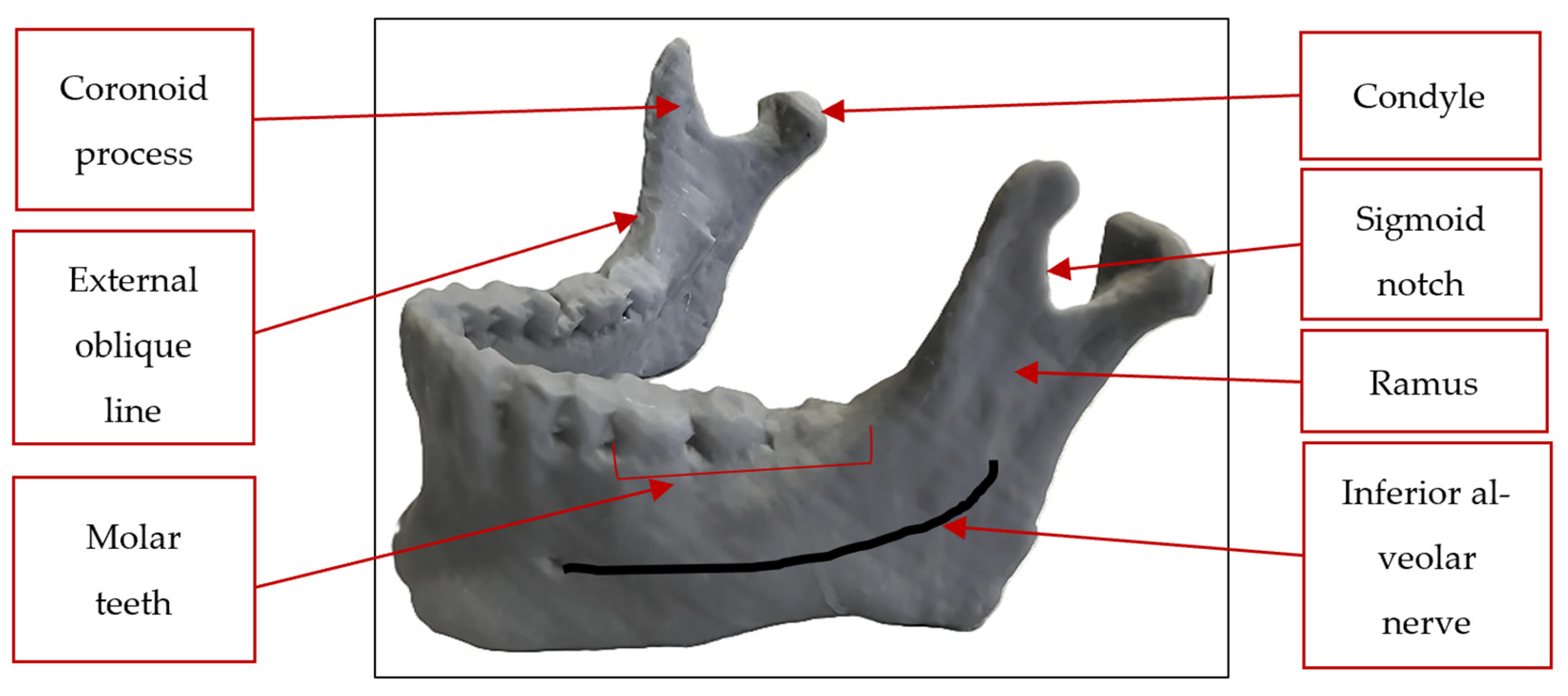
31]. A 3D model of the human mandible is shown in
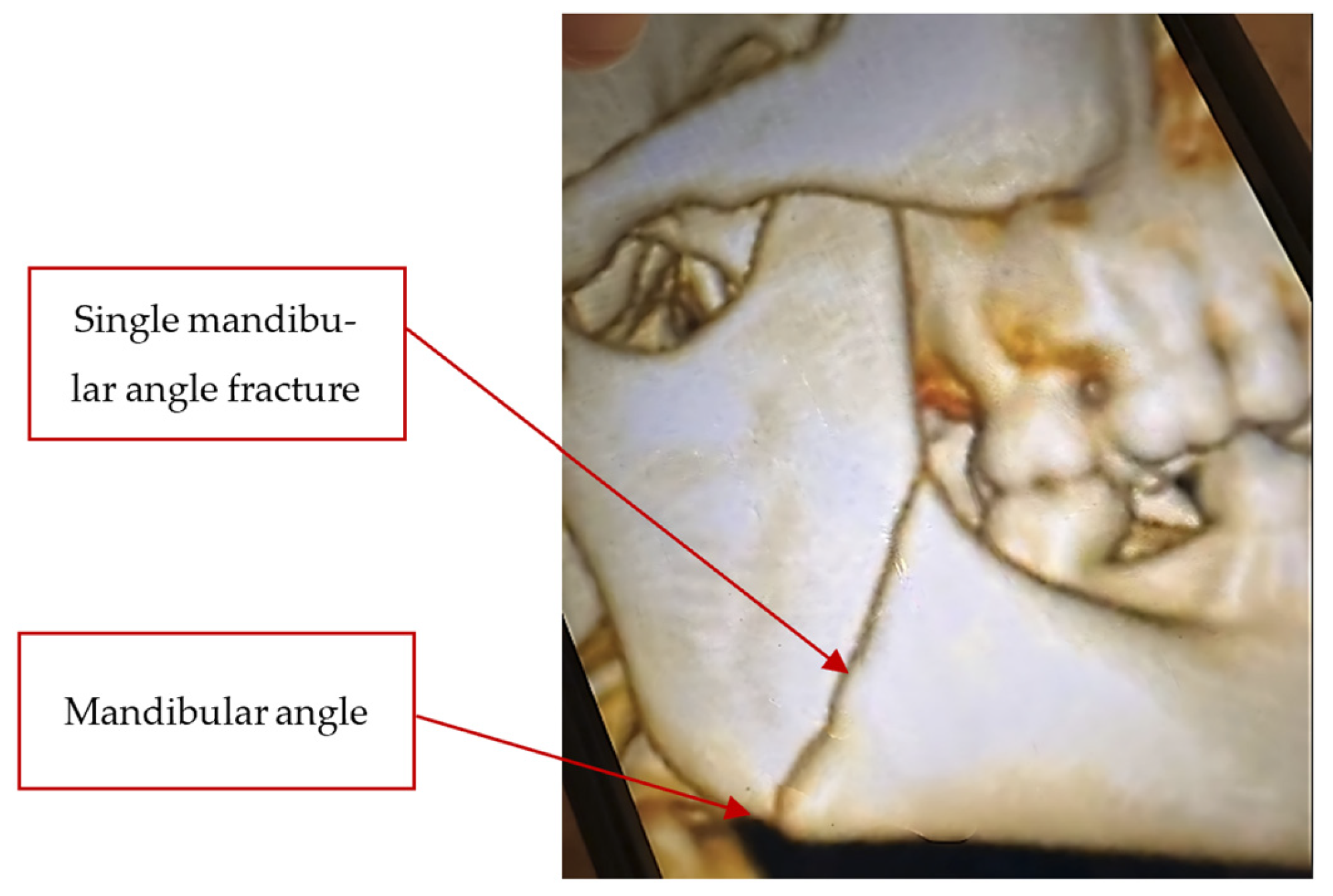
Figure 1, and the left-side angular fracture mandible is illustrated in
Figure 2.
A review of the literature helped to establish the overall dimensions, features, and most appropriate fixation methods for the new-design plate [
32,
33,
34]. According to the usage of screws, it is recommended to use at least 3 tightening screws per bone fragment, with the middle screws positioned at least 5 mm from the defect and anchored in healthy bone [
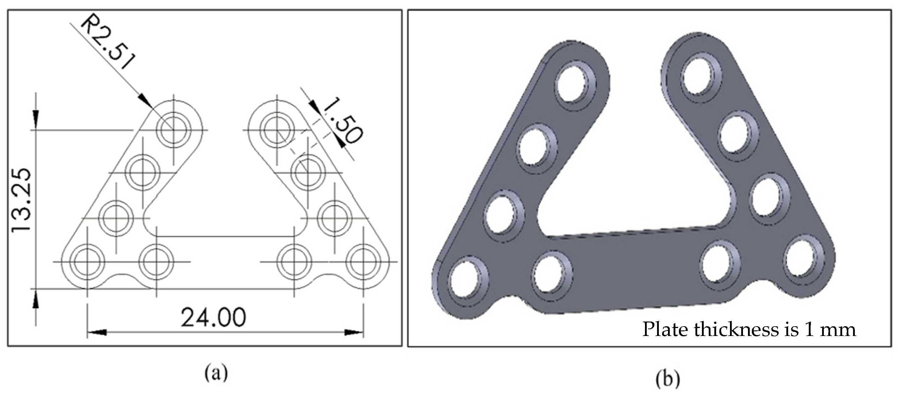
13]. The simple ‘U-shaped’ design with narrow ending segments and ten titanium tightening screws was selected because it satisfies both engineering and medical requirements. Additionally, the screw hole diameter should match the standard M2 screw diameter. This unique shape configuration allows the plate to function as a universal plate. The second step involved creating a detailed design, including the CAD geometric model shown in
Figure 3. This model was created based on the approved concept design using SolidWorks 2024® Premium 2024 × 64 (Dassault Systèmes SolidWorks Corporation, Waltham, MA, USA). The dimensions of the proposed plate were determined based on anthropometric data for the 50th percentile of the human skull (adult men and women), ensuring that it fits the average size.
In the U-shaped pattern of the plate with ten screw holes, the sides of the U function as an assembly of three single mini-plates, thereby streamlining the surgical procedure and significantly reducing the duration of surgery and effort involved. Additionally, this shape offers greater support and stability to the mandible. The number of these holes, according to the advice of oral and maxillofacial surgeons, allows the selection of the most suitable number of tightening screws based on the specific anatomical nature and clinical severity of each case, thereby enhancing the ability to manage significant fracture gaps while minimizing surgical risk. The resulting plate features a U-shaped configuration with four countersunk holes on each side, specifically engineered to accommodate standard self-tapping M2 spherical screw head caps.
2.2. Three-Dimensional Mandibular Angle Fracture Model
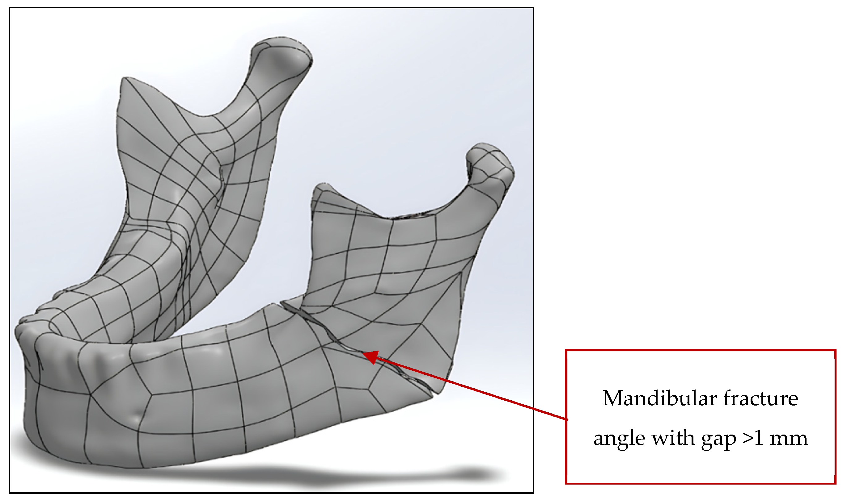
A mandibular CT scan with an angular fracture gap exceeding 1 mm was developed using Mimics Core software 20 (Materialise NV, Leuven, Belgium) for 3D reconstruction and segmentation by the Department of Oral and Maxillofacial Surgery at the Faculty of Dentistry, Alexandria University, Egypt. The detailed 3D mandibular reconstruction is provided in Stereo Lithography (STL) file format and a physical 3D-printed model replicating a true anatomical mandible. The STL file was subsequently imported into SolidWorks for advanced 3D modeling and further analysis, as shown in
Figure 4.
2.3. Construction and Finite Element Analysis of the 3D Model
The FEA was conducted using SolidWorks Simulation (SolidWorks 2024, Dassault Systèmes SolidWorks Corporation, Waltham, MA, USA) on the 3D model of the true angular mandible fracture, the newly designed mini-plate, and the tightening screws. The simulation of the plate fixation position was discussed with the oral and maxillofacial team at Alexandria University, so that full support would be provided and to accelerate healing. Regarding the surgical team’s knowledge and the case studies they handled, it was pointed out that the model chosen represented one of the most severe cases, with an angular fracture gap in the mandible exceeding 1 mm in the superior direction. Therefore, the right and left branches of the U-shaped plate in that model function as a single plate. Each branch is secured with four tightening screws to maintain closure between the two mandibular fracture parts. The tightening screw connecting the mini-plate to the mandible is to be bonded, as in previously published protocols where no relative motion or separation should be allowed at the interface and the nodes at the contacting surfaces were to be fully constrained [
35]. The mandible’s cortical bone is isotropic, homogeneous, and linearly elastic, with a Young’s modulus of 20,000 MPa and a Poisson’s ratio of 0.26 [
36,
37]. Titanium grade 4 was selected as the proposed plate material due to its excellent biocompatibility and non-toxicity upon implantation, as well as its acceptable mechanical properties of a Young’s modulus of 105 GPa, a yield strength of 655 MPa, and a Poisson’s ratio of 0.37, all while being lightweight [
38].
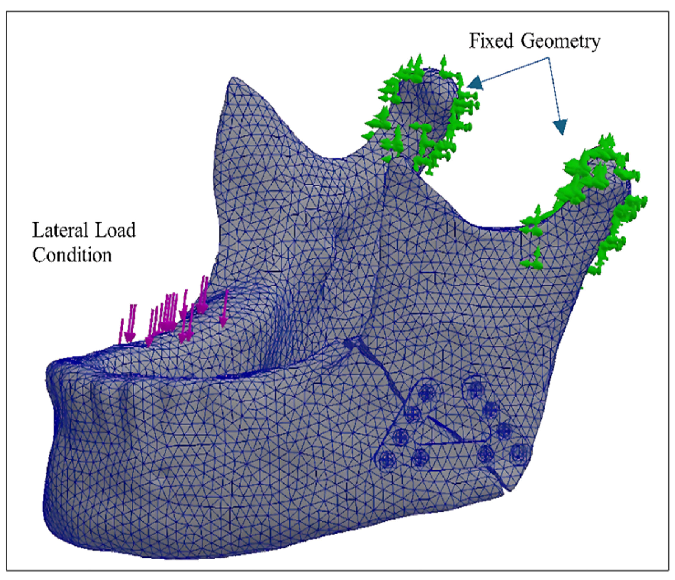
The boundary conditions were designed to closely match real patient conditions in order to ensure meaningful results. These conditions are consistent with those used in previous studies [
36,
37,
39], where a full fixed geometry is applied to the head of the mandible, and a load of 100 N is applied directly to the first molar on the right side of the mandible (the non-fractured side). The load vectors are applied to the first molar area rather than to a specific point. The anatomical location corresponding to the first molar region was identified as approximately 24 mm from the mandibular midline, consistent with the established anteroposterior distance between the intermolar line of the first molars and the mandibular midline. Specifically, this measurement was obtained on the axial plane as the linear distance from the midline passing through the lower central incisors to the intermolar line. The load was applied precisely to this area, as defined by this anatomical measurement. This approach relies on stable skeletal landmarks rather than dental structures, thereby ensuring that the load application point is unambiguously defined and unaffected by the presence or absence of the first molars. This distance is not arbitrary but is firmly supported by anatomical evidence demonstrating minimal variability in the intermolar-to-midline span in adult mandibles. It is consistently reported to be symmetrical between the right and left sides and confined within a narrow range across populations. The value adopted in the present study lies within the documented mean range reported in the literature [
40,
41].
This load represents the resultant forces exerted by the masticatory muscles, simulating the regular chewing forces and the maximum intercuspal position [
42]. This load value was chosen for the model in the current study, from among the values used in previous studies, for the main reasons that it simplified the model and reduced the simulation program runtime. The standard solid linear mesh, which is a high-quality, curvature-based blended mesh, was chosen as recommended in the program user manual, despite the model’s complexity. It consists of a total of 101,758 nodes, an element size of 0.788788 mm, and a total of 66,175 tetrahedral elements, as shown in
Figure 5. This number was determined by the SolidWorks simulation tool through automated mesh-convergence testing, where the solver automatically increased mesh density in critical areas and re-ran the analysis until successive solution results fell within a defined tolerance, thereby ensuring mesh-independent results.
In previous FEA mandible studies, the selection of an appropriate stress theory is considered fundamental for accurate biomechanical evaluation. The distortion-energy theory of stress (‘von Mises stress theory’) is chosen for the evaluation of stresses in the tested model, firstly, consider the ductile nature of the metal in the plate and screws [
43]. This is a widely used method in biomechanical models for the evaluation and prediction of mandibular stress states and bone failures [
44].
2.4. Rapid (Non-Functional) and Functional Prototypes Fabrication
This stage represents a crucial phase in the current study which was implemented to validate the comprehensive engineering design process following the attainment of satisfactory results from simulation analysis. Physical prototypes of the proposed plate were fabricated in order to allow a thorough assessment of its geometrical features, dimensions, and functional performance. Two manufacturing techniques were employed: 3D printing with Fused Deposition Modeling (FDM) and advanced Computer Numerical Control (CNC) machining.
The first prototype was a rapid (non-functional) prototype printed via FDM in the Production Engineering Department at the Faculty of Engineering, Alexandria University, Egypt. A polymer material was used in order to confirm component dimensions, evaluate the assembly process, and approve the final geometry prior to definitive fabrication in titanium. This approach ensured resource efficiency by reducing material wastage and enabling design alterations prior to final production. The new mini-plate and mandible structures were successfully printed using a 3DP-12-4E UP mini 2 3D printer (Tiretime, Beijing, China), with a table dimension of 120 mm × 120 mm × 120 mm. The nozzle temperature was set to 276 °C, and the infill material was set to 80% density with a layer thickness of 0.15 mm in order to achieve high-quality results. The fabrication material used was a thermoplastic Acrylonitrile Butadiene Styrene (ABS) with a filament diameter of 1.75 mm, and the printer was set up to optimize printing time while achieving a suitable surface finish for the final product.
The second prototype was a functional prototype of the proposed design. It was produced so as to satisfy mass-production conditions, necessitating the selection of an advanced fabrication method. CNC machining was chosen, given its reputation for precision and reliability in high-accuracy manufacturing. The material chosen for fabrication is Titanium grade 4, as selected during the design stage. Considering the intricate geometry of the proposed mini-plate, including its small dimensions and multiple 2 mm holes, a 5-axis CNC milling machine was utilized, which was manufactured by the Arab Engineers Co. Ltd., Cairo, Egypt. Among the locally available advanced machine tools, this technology offered superior capability for the reproduction of the plate’s complex features, meeting the highest performance criteria such as dimensional accuracy and surface finish, and thereby ensuring the prototype’s suitability for scalable production.
3. Results and Discussion
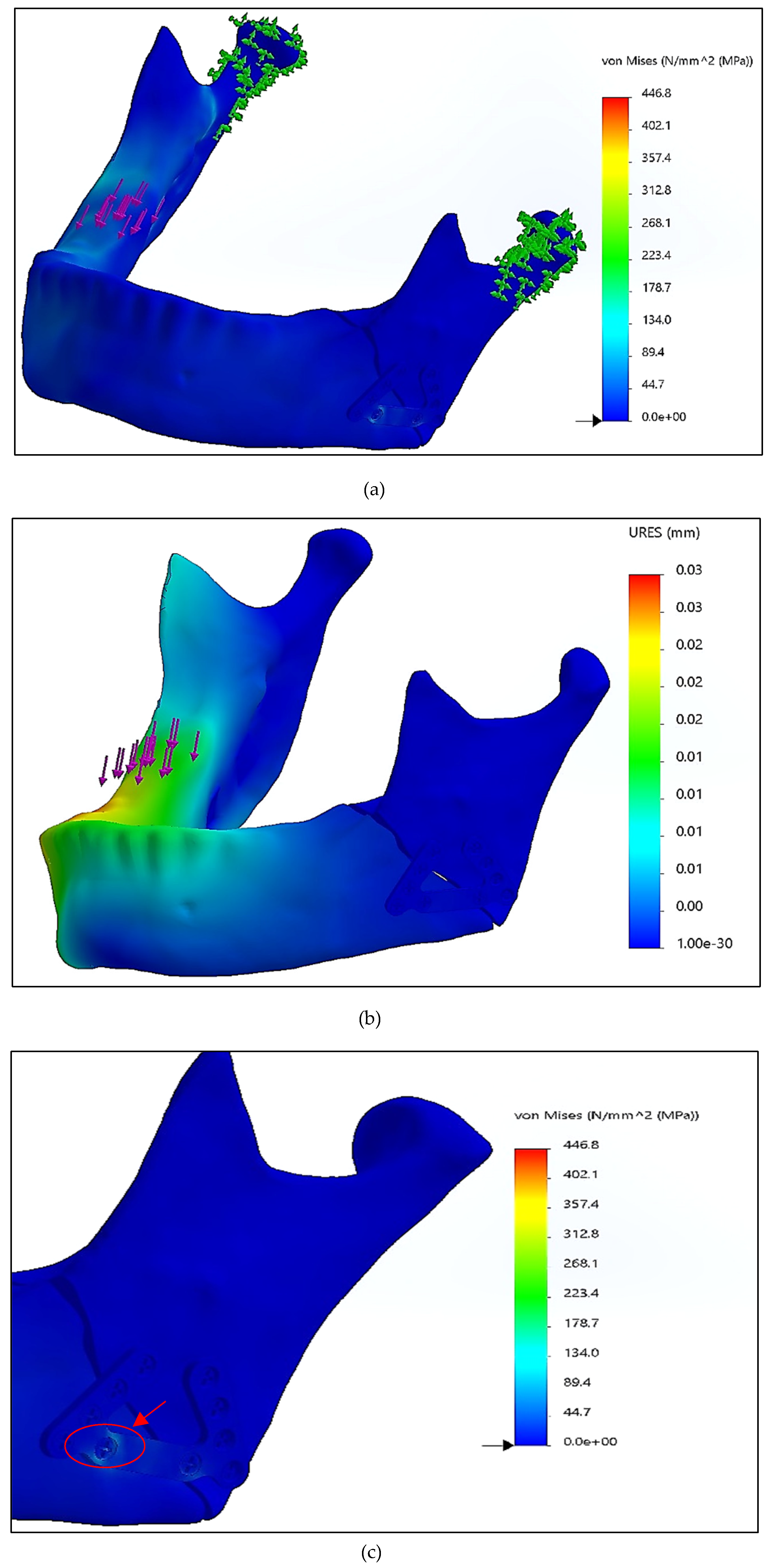
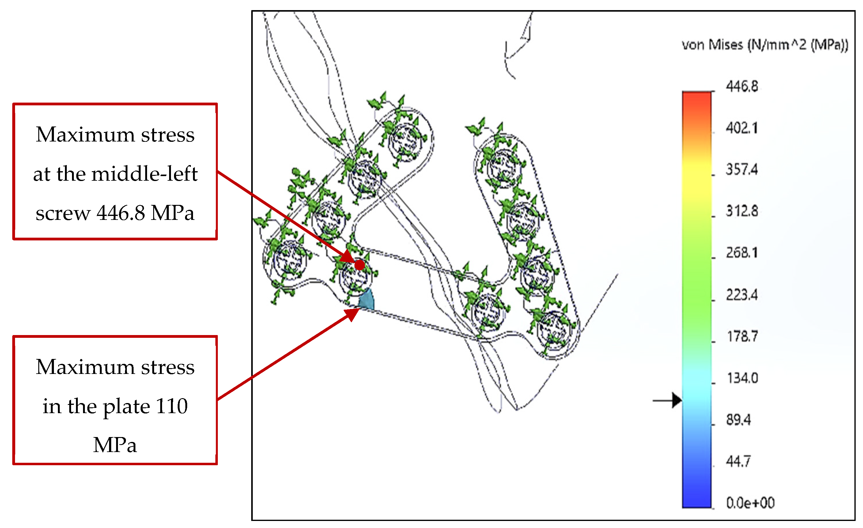
Figure 6 shows a stress distribution pattern that highlights two affected areas. The first area is the localized bone region directly beneath the applied load, as anticipated, given the point of load application. The second affected area is situated in a discrete region of the fixation plate. The maximum stress recorded is 446.8 MPa at the middle-left lower screw, while the maximum stress observed in the designed plate is 110 MPa; both values remain below the yield strength of the selected titanium material. The analysis also identified an ultimate displacement of 0.03 mm on the right side of the mandible, directly beneath the load application area.
The analysis of stress distribution focused on the proposed fixation plate and the associated middle-left lower screw, using SolidWorks’ advanced post-processing capabilities. Specifically, the ‘iso clipping’ feature was employed to isolate and visualize regions of interest. The pattern found is consistent across the models evaluated and is corroborated by the uniform stress scale applied in the analysis, as illustrated in
Figure 7. The FEA results present an innovative, simple, and unique mini-plate design as an alternate treatment for functional stabilization of angular mandibular fractures.
Most previous research on mini-plates for the treatment of angular fractures has focused on comparative studies of existing plates [
36,
39,
45], using specific case studies of mandibular angle fractures. Considering that conditions may change, including the fracture angle and the severity of the fracture gap, this variability may affect the results. However, only a few studies have presented customized fixation plates with numerical analyses for individual patient cases and compared their outcomes with those of conventional mini-plate designs [
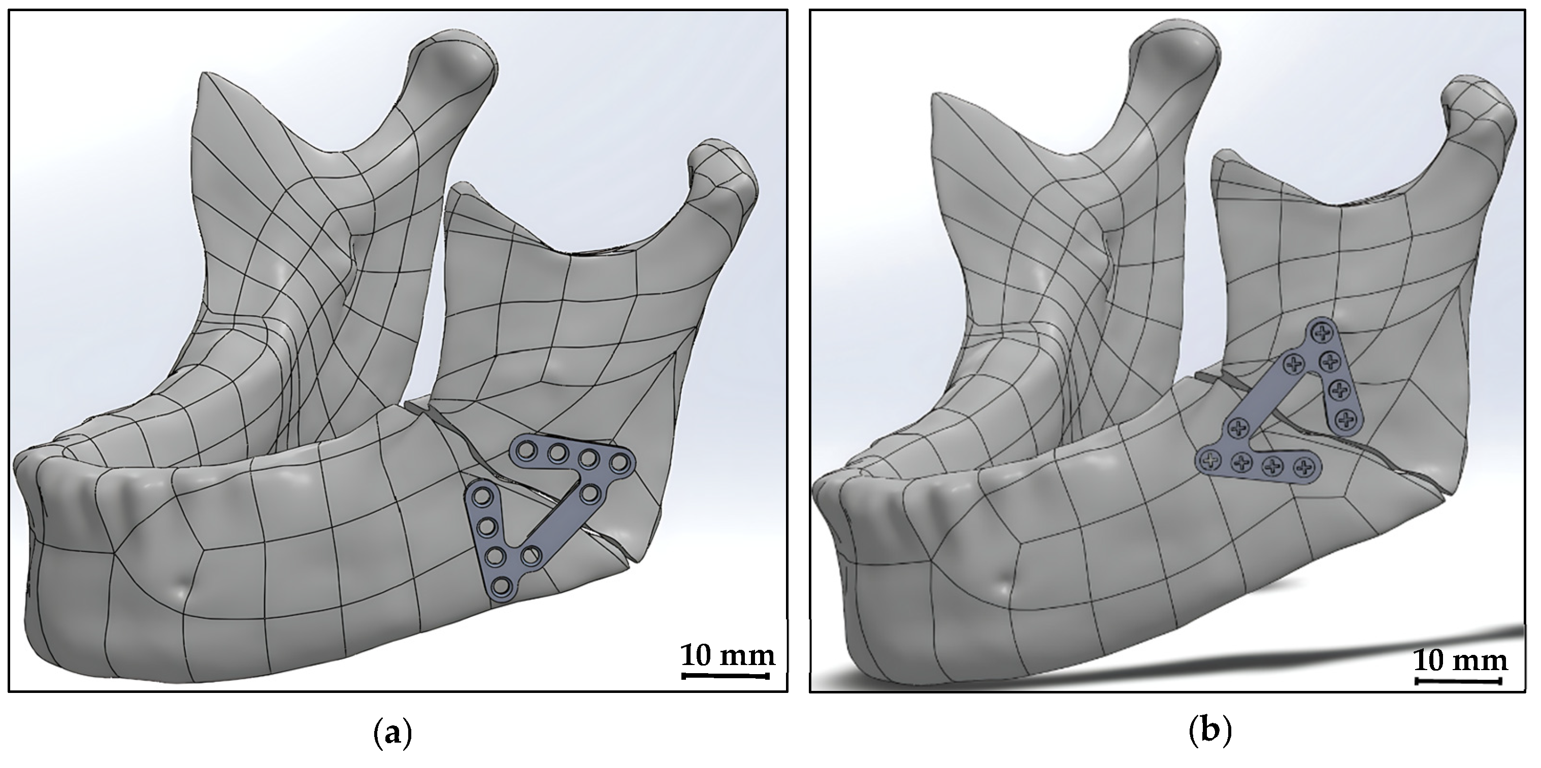
25]. In the interim, the newly designed plate can be oriented in various configurations, including upright, downward, and oblique positions, thereby offering more control over fracture fixation and accommodating a wider range of mandibular fracture patterns.
Figure 8 shows the proposed new-design plate oriented in upright and downward positions.
The simulation analysis results were compared with those of previous studies that used FEA, both for those employing custom fixation plates and for those using established plates to treat mandibular angle fractures. According to von Mises theory, the maximum stress and displacement reported for the new proposed-design plate are 110 MPa and less than 0.01 mm, respectively. In the current study, the maximum stress decreased by 5%, while the displacement decreased by 95%, compared with the results published by Liu et al. [
25] for their customized plate under heavy-load conditions, which is considered the worst-case study. In addition, compared with the findings of another study [
37], the stress and displacement values decreased by 50% and 98.5% respectively. Furthermore, the present study’s results were compared with Abdelwahab et al.’s findings [
39] regarding their three patterns of different existing plates (2.3 plate, double 2.0, and rhombic plate), and our proposed plate achieved lower stress values at 85.3%, 75.5%, and 83.1%, respectively. In 2020, Park et al. [
36] conducted a comprehensive study on the stability of four simulation materials for single mini-plates using FEA. Two of these materials were metals: titanium and magnesium alloy. The study maintained the same boundary conditions as those used in the current research for the working model and the selected stress analysis theory. Compared to Park et al.’s findings, the maximum stress in the plate area decreased by 73.4% and 52.1% for the titanium and magnesium alloy plates, respectively. Finally, the maximum stress observed on the plate proposed in the present study is lower than that detected in previous studies. The reduction in maximum stress ranges from 5% to 85.3% depending on the load conditions applied. Notably, under the same loading conditions, the stress values observed in this study are 60.7% lower than those reported in previous studies. The proposed design for the new mini-plate is therefore considered the most suitable option compared to previous designs due to its acceptable mechanical performance. Fabrication of rapid (non-functional) and functional prototypes in our study, which was not applied in previous studies, further supports the practical applicability of the proposed fixation system.
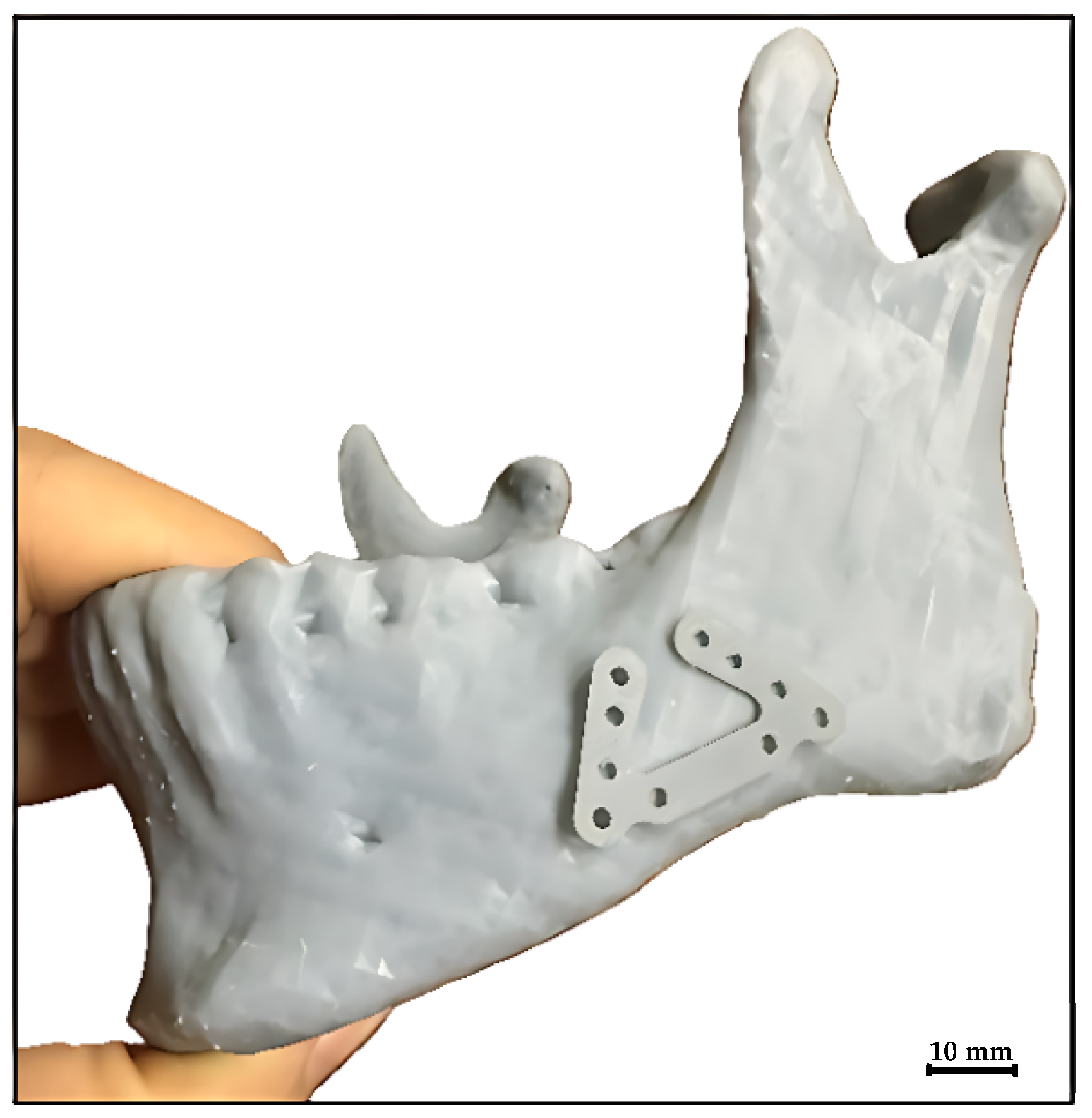
To validate the detailed engineering design process in checking the geometrical features, dimensions, and the function of the proposed plate, physical models were manufactured. The first model, a 3D-printed mandibular model, is shown in
Figure 9. This is a rapid (non-functional) prototype of the new mini-plate and mandible structures which was successfully printed using a 3DP-12-4E UP mini 2 3D printer (Tiretime, Beijing, China).
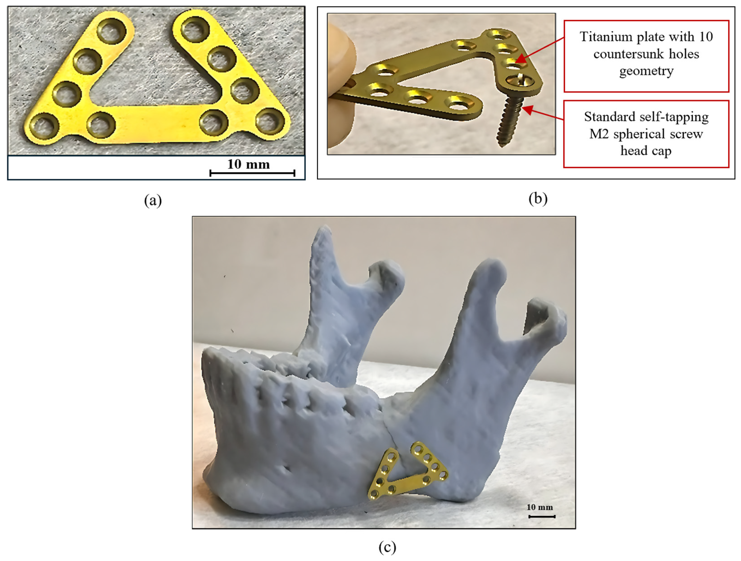
The second model, illustrated in
Figure 10a, represents a fully functional prototype of the proposed mini-plate fabricated from grade 4 titanium. This design ensures that the screw heads are fully seated in the countersunk recesses, as shown in
Figure 10b, providing secure fixation while maintaining a low profile to minimize soft-tissue irritation.
Figure 10c represents the relative position between the functional mini-plate prototype and the FDM-printed mandibular model. The successful fabrication and assembly of this prototype demonstrate both the manufacturability and functional integrity of the proposed mini-plate for clinical application.
4. Limitations and Future Work
The limitations of the current study’s findings include that the results are limited to a computer-aided 3D FEA model, which simplifies a complex biological system. FEA models cannot fully replicate the dynamic and variable forces present in a living patient. The study was also limited by the idealized fracture model, where an angular fracture with a large gap represents one of many severe cases. Real fractures vary widely in shape, size, location, and comminution, which may affect how the plate performs clinically. These results must also be interpreted with caution regarding static loading assumptions, where the analysis simulates bilateral masticatory muscle loading to simplify the model. However, chewing and jaw movements involve complex, dynamic, and asymmetric forces that a static FEA may not fully capture. In addition, for a more accurate representation of the mechanical behavior of the mandible, an anisotropic bone material property could be incorporated into the current model. Furthermore, the study could investigate a range of alternative plate fixation positions, including upward fixation, to assess the plate’s flexibility and biomechanical performance across varying clinical scenarios. Moreover, it is important to visualize the analysis scale specifically for the proposed plate component, as well as the overall stress distribution across the other assembled components (mandible and screws). However, the SolidWorks simulation software (SolidWorks 2024, Dassault Systèmes SolidWorks Corporation, Waltham, MA, USA) used in this study does not support isolating the stress scale for individual components, highlighting the need for advanced finite element analysis software in future work. Finally, although prototypes of the proposed design were produced, experimental validation is not discussed in this paper but is reported in separate research by the maxillofacial surgical team at the Faculty of Dentistry, Alexandria University, Egypt, who received Institutional Review Board approval to use patient data [
46].
In order to further enhance the clinical impact of this research, several recommendations are suggested. A surgical guide kit (Jig) could be used for screw fixation along with a torque meter during the surgical operation. In the interim, the same material should be used for the plate and screw, and the distance between the final hole and the jaw should be larger than three times the hole diameter so as to minimize stress concentration. Additionally, the use of a standard spherical screw head cap could facilitate fixation, and sandblasting the screw stud would roughen the surface, thus allowing for firm bone fixation. Future work will focus on the application of the new mini-plate design using biodegradable materials such as magnesium (Mg) alloys. This would offer the advantages of eliminating the need for secondary removal surgery and promoting gradual load transfer to facilitate natural bone healing.
5. Conclusions
In conclusion, this study introduces a new, unique, U-shaped geometric configuration to improve the quality characteristics of stability, reliability, functionality, dimensional accuracy, and surface finish of plates used to treat angular mandibular fractures. The design is verified through FEA, and a preliminary validation is performed using fabricated prototypes. The proposed design demonstrated acceptable biomechanical performance, including a 60.7% reduction in maximum plate body stress compared to previous studies under the same load conditions. For instance, the results indicate that the maximum stresses observed in the lower-middle screw and the designed plate are 446.8 MPa and 110 MPa, respectively, both of which are below the yield stress of the selected materials. Furthermore, the analysis also identified an ultimate displacement of 0.03 mm on the right side of the mandible, directly beneath the load application area. The increased number and strategic placement of screw holes in the new design enhance intraoperative flexibility and stability, allowing for optimal adaptation to a range of anatomical variations and clinical severities. Additionally, multiple plate orientations are possible, and operative procedures can be streamlined by integrating the functionality of three separately assembled mini-plates into a single construct, thereby reducing surgical time and complexity. Collectively, these advances address all of the requirements of oral and maxillofacial surgery and establish a robust and transferable framework for future developments in fixation system design, as supported by the experimental validation provided in a recently published study by an oral and maxillofacial surgical team collaborating with the authors of the present study. However, despite the significant advances made, it is vital to continue refining analytical models and incorporating clinical data to improve patients’ healthcare outcomes.