


Featured Application
This review provides practical guidance for selecting and validating endospore purification and detection workflows in food and environmental microbiology. An integrated view of sample preparation and detection can improve the comparability of spore resistance and enumeration data across laboratories and support the implementation of rapid methods (e.g., flow cytometry and molecular assays) in routine monitoring and process validation.
Abstract
Bacterial endospores are highly resistant, dormant forms that pose persistent challenges in food safety, environmental microbiology, and industrial hygiene. Accurate evaluation of endospore resistance, physiology, and inactivation depends on both purification and detection methods; however, these processes are typically examined independently, limiting methodological consistency and contributing to variability across studies. In this review, current approaches for endospore purification and detection are critically examined, including washing-based methods, density gradient centrifugation, enzymatic treatments, culture-based enumeration, molecular assays, flow cytometry, and emerging biosensor technologies. In addition, these methods are compared using metrics such as purity, recovery yield, sensitivity, and specificity, and their advantages and limitations are summarized to clarify performance. It is further proposed that endospore purification and detection should be considered as a single, end-to-end analytical workflow and optimized accordingly. Purification strategies influence sample cleanliness and aspects of endospore quality, including viability, structural integrity, and physiological state, which affect detection performance and quantitative accuracy. Based on this integrated perspective, a conceptual framework linking purification efficiency to detection outcomes is presented, along with practical considerations for method selection across relevant application contexts. Finally, gaps in standardization are identified, and future research directions are outlined to improve reproducibility and cross-study comparability in endospore-related studies.
1. Introduction
Bacterial endospores are highly resistant dormant structures produced primarily by Gram-positive genera such as Bacillus [1,2] and Clostridium [3,4]. Their remarkable tolerance to heat [5], desiccation [6], radiation [7], and chemical disinfectants [8] enables long-term persistence under adverse conditions [9], thereby posing persistent challenges in food safety [10], environmental microbiology [11], clinical settings [12], and industrial hygiene [13]. Spore-forming bacteria are frequently implicated in food spoilage [10], foodborne illness, and contamination of processing environments [14], where conventional sanitation procedures often fail to eliminate these dormant forms [15].
Reliable purification and detection of bacterial spores are essential for accurate characterization of their physiological properties [16]. However, laboratory preparations commonly contain substantial proportions of vegetative cells [17], cellular debris [18], and partially germinated spores [19]. Such heterogeneity can significantly distort viability assessments [20], resistance evaluation [21], and molecular analyses [22], leading to inconsistent or misleading results. Importantly, these biases represent a critical and often overlooked source of experimental variability in spore research [23,24].
A variety of purification strategies have been developed, including repeated washing and centrifugation [25], density gradient separation using media such as Percoll or Nycodenz [26], and enzymatic treatments such as lysozyme digestion [27]. While these approaches differ in efficiency, recovery yield, processing time, and cost, they may also exert distinct effects on spore integrity and physiological state [23,24]. Despite their widespread use, purification methods are typically selected based on empirical convention rather than systematic comparison, and no standardized framework currently exists to guide method selection.
In parallel, numerous detection methods have been established, including culture-based enumeration [28], microscopic staining [29], molecular techniques such as PCR [30,31], and emerging analytical platforms such as flow cytometry [32,33] and spectroscopy [34,35]. Although these methods offer varying degrees of sensitivity and specificity, their performance is inherently dependent on the quality of the spore preparation [24,36,37]. Nevertheless, purification and detection are often considered in isolation, thereby obscuring their fundamental interdependence [38,39,40]. This separation further obscures the extent to which purification-induced variability propagates into detection outcomes [41,42], affecting sensitivity, accuracy, and reproducibility. Despite the central role of both purification and detection in spore research [43,44,45], their combined impact on experimental outcomes has not been systematically examined, thereby limiting the ability to interpret data across studies and hindering the development of standardized methodologies.
This review comprehensively evaluates current approaches for bacterial spore purification and detection, with a particular focus on their interdependence. By linking purification strategies to spore quality attributes and subsequent detection performance, this work provides a conceptual framework for improving methodological consistency and supports the rational selection of appropriate techniques in food and environmental microbiology.
2. Spore Purification Methods
Before purification, the preparation of spore-rich cultures is a critical step that can significantly influence downstream purification efficiency and analytical reproducibility. Traditional sporulation approaches based on nutrient depletion and metabolite accumulation in closed culture systems remain widely used for laboratory-scale spore production. Although these methods generally require longer incubation periods (typically 72–96 h), they may promote more synchronous sporulation in heterogeneous cultures and reduce contamination from incompletely sporulated cells [23]. In addition, specialized sporulation media such as Difco Sporulation Medium (DSM) may improve sporulation efficiency and experimental reproducibility under controlled laboratory conditions [46,47]. Therefore, both conventional nutrient depletion approaches and DSM-based methods remain useful depending on the target species, experimental objectives, and required sample throughput.
Preparation of highly purified spore suspensions is a fundamental prerequisite for the accurate evaluation of spore resistance, physiology, and detection sensitivity [48,49,50]. However, complete separation of dormant spores from vegetative cells, cellular debris, and partially germinated forms remain technically challenging [1,51,52]. In practice, spore preparations often exhibit substantial heterogeneity, which can introduce considerable variability into downstream analyses [23,24]. Importantly, spore purification is a critical determinant of experimental outcomes [53,54,55,56]. Different purification strategies can selectively remove, retain, or alter specific subpopulations of spores, thereby influencing apparent resistance profiles (e.g., response to heat, chemical disinfectants, UV irradiation, and desiccation), viability measurements, and detection performance [23,24,57]. Despite its central role, purification is frequently treated as a routine procedure, and its impact on analytical reliability is often underappreciated.
A variety of purification strategies have been developed, differing in complexity, efficiency, and potential effects on spore integrity. However, these methods are rarely evaluated within a unified framework, making it difficult to directly compare their relative performance or select appropriate approaches for specific analytical objectives.
2.1. Washing and Centrifugation
The most widely used approach for spore purification relies on repeated washing and low-speed centrifugation cycles following sporulation [25], often combined with a preliminary heat treatment to inactivate residual vegetative cells. Because optimal heat-treatment conditions vary substantially among species and strains of Bacillus and Clostridium, no single universal temperature can be recommended; however, mild heat treatments around 65–80 °C for 10–30 min are commonly used to reduce vegetative-cell carryover and/or to activate spores prior to downstream analyses. Repeated heating/cooling cycles have been explored in some studies, but their effects appear inconsistent and depend strongly on the physiological state and pre-culture conditions of the spores [58,59,60]. In this method, spores are pelleted by centrifugation and subjected to multiple resuspension and washing steps to remove loosely associated cellular debris and soluble contaminants.
The effectiveness of this approach is primarily based on differences in sedimentation behavior among spores, vegetative cells, and cellular debris [61,62]. Due to their dense and compact structure, mature spores are preferentially recovered in the pellet fraction, whereas lighter debris is progressively removed during washing steps. This principle enables simple physical enrichment without requiring specialized reagents [63,64].
Owing to its simplicity, low cost, and accessibility, this method remains a standard procedure in many laboratories [23,24,63,64]. However, its performance is highly dependent on the efficiency of sporulation and the initial composition of the sample. In cases of incomplete sporulation, substantial contamination with vegetative cells, cell fragments, or partially germinated spores may persist even after repeated washing cycles [23,65,66]. In addition, repeated centrifugation and resuspension steps may impose mechanical stress on spores, potentially affecting recovery yield and, in some cases, altering surface properties relevant to downstream analyses [67,68,69]. Importantly, residual contaminants not completely removed by washing can interfere with detection methods by contributing to background signals in optical or cytometric analyses or introducing nontarget DNA in molecular assays [70].
Therefore, while washing and centrifugation are suitable for routine laboratory applications and preliminary enrichment, this approach alone is often insufficient when high purity is required for precise quantitative measurements or advanced detection platforms. In such cases, additional purification steps, such as density gradient centrifugation or enzymatic treatment, are typically necessary to achieve the desired level of spore quality [23,33].
2.2. Density Gradient Centrifugation
Density gradient centrifugation was developed to improve spore purification efficiency by exploiting differences in buoyant density between spores and other cellular components [71,72]. In this approach, samples are layered onto discontinuous or continuous density gradients generated using media such as Percoll or Nycodenz [24,26,73]. During centrifugation, particles migrate to positions corresponding to their equilibrium densities, enabling the effective separation of spores from vegetative cells, cellular debris, and partially degraded materials.
The high density and compact structure of mature spores generally result in their localization in the lower regions of the gradient, whereas vegetative cells and lighter debris remain in the upper fractions [74,75,76,77]. This density-based partitioning enables more precise and reproducible separation than simple washing procedures. In addition, gradient systems can partially resolve heterogeneity within spore populations, including differences in maturation state or structural integrity, which are not easily distinguishable by conventional centrifugation alone [78]. As a result, density gradient centrifugation is widely employed in studies requiring highly purified and homogeneous spore preparations, such as resistance assays, structural characterization, and advanced analytical applications [79,80]. However, the method involves several practical and technical limitations. Preparation of gradients, optimization of centrifugation parameters, and fraction collection increase procedural complexity and overall processing time [81,82]. Furthermore, recovery yield can be reduced because of losses during fractionation and incomplete recovery from gradient media.
Importantly, separation efficiency is highly sensitive to experimental conditions, including gradient composition, density range, and centrifugation speed and duration. Small variations in these parameters can shift particle distribution within the gradient, leading to inconsistent recovery or incomplete separation when density differences between components are minimal [83,84]. Such variability complicates reproducibility and cross-study comparisons, particularly in the absence of standardized protocols [23,24].
2.3. Enzymatic Treatments
Enzymatic strategies, particularly lysozyme digestion, are widely employed to eliminate residual vegetative cells following initial washing steps [27,85]. Lysozyme specifically hydrolyzes the β-(1,4)-glycosidic linkages between N-acetylmuramic acid and N-acetylglucosamine in peptidoglycan, leading to rapid weakening and lysis of vegetative cell walls [86,87]. In contrast, mature spores are largely resistant to this treatment due to the presence of multiple protective layers, including the cortex, coat, and, in some species, an exosporium, which limits enzyme accessibility [88,89]. The efficiency of lysozyme treatment is strongly influenced by experimental parameters such as enzyme concentration, incubation time, temperature, and buffer composition (e.g., ionic strength, pH), as well as the physiological state of the cells [90,91,92]. For example, sublethally damaged or germinating spores can exhibit increased susceptibility to enzymatic degradation, potentially leading to underestimation of spore recovery [93]. Therefore, careful optimization is required to maximize selective lysis of vegetative cells while preserving spore integrity.
When combined with physical purification steps such as repeated washing or density gradient centrifugation, lysozyme treatment can substantially improve the apparent purity of spore preparations [23,24]. However, excessive enzymatic exposure or inappropriate conditions may partially disrupt spore surface structures, including coat proteins, or alter key physicochemical properties such as hydrophobicity and surface charge. These changes can consequently affect downstream analyses, including antibody binding, flow cytometry, or germination assays [32,33]. Accordingly, enzymatic purification should be applied under controlled conditions and validated for compatibility with the intended analytical techniques.
2.4. Comparative Evaluation of Purification Strategies
Each purification strategy involves inherent trade-offs among purity, recovery yield, processing time, cost, and practical applicability. For instance, simplified isolation procedures incorporating enzymatic digestion (e.g., lysozyme treatment) can effectively remove residual vegetative cells and yield highly purified preparations [27,85]. However, such approaches might reduce overall recovery because of partial loss of spores during repeated washing steps or unintended damage to germination-sensitive subpopulations [90,91,92].
Systematic comparisons of purification workflows have demonstrated that protocols optimized for maximal purity, such as those combining enzymatic digestion with density gradient centrifugation, typically require additional processing steps, specialized reagents, and longer handling times, thereby increasing cost and experimental complexity [79,80,81,83,84].
A major limitation in the current literature is the lack of standardized criteria for assessing spore purity. Reported metrics vary widely, including phase-contrast microscopy, colony-forming unit (CFU) ratios, staining-based methods, and flow cytometric analyses, which makes direct comparison across studies challenging [28,29,30,31,32,33,34,35]. Furthermore, few studies have comprehensively reported purity and recovery yield according to standardized evaluation criteria, further hindering robust methodological evaluation. Therefore, the selection of an appropriate purification strategy should be guided by the intended downstream application. High-precision applications, such as resistance profiling, structural analysis, or antibody-based detection, often require highly purified spores obtained through density gradient centrifugation or combined approaches [23,24]. In contrast, routine enumeration or screening assays may tolerate lower purity levels and benefit from simpler, higher-yield washing-based methods [45,94]. Establishing standardized benchmarks that integrate purity, yield, and practical performance metrics would substantially improve reproducibility and comparability in spore research (Table 1).
Table 1.
Semiquantitative comparison of representative spore purification methods based on commonly reported characteristics in the literature.
3. Bacterial Spore Detection Methods
Reliable detection of bacterial spores is critical for food safety monitoring [13], environmental surveillance [11], and validation of spore purification procedures [23,24]. Spores are highly resistant to heat [5], desiccation [6], and chemical treatment [8], allowing them to persist in food processing environments and evade conventional sanitation strategies. This persistence not only increases the risk of food spoilage [10] and foodborne illness [95] but also complicates accurate microbial risk assessment.
Detection strategies vary widely in sensitivity, specificity, processing time, and ability to distinguish dormant spores from vegetative cells or nonviable particles [96,97,98]. Many conventional culture-based methods require germination prior to detection, which can lead to underestimation because of incomplete or heterogeneous germination [99,100]. Conversely, molecular and staining-based approaches can detect both viable and nonviable cells, thereby potentially overestimating the actual risk [101,102]. Furthermore, the structural similarity between spores and cellular debris presents an additional challenge for accurate discrimination [45,103]. Consequently, there remains a strong demand for rapid, reliable detection methods capable of selectively identifying intact and physiologically relevant spores across diverse sample matrices.
3.1. Culture-Based Enumeration
Culture-based methods remain the conventional standard for spore detection and quantification. Typically, samples undergo heat treatment to inactivate vegetative cells, followed by plating on suitable growth media and enumeration of CFU/mL−1. This approach is widely used because of its simplicity, low cost, and ability to selectively quantify viable spores capable of germination and outgrowth [97,104]. It provides a direct measure of microbiologically relevant viability, which is critical for food safety risk assessment and process validation. Nevertheless, several limitations must be considered. Heat treatment protocols vary widely in temperature, duration, and matrix conditions, which can significantly influence apparent spore counts and hinder comparability across studies. In addition, spores in different physiological states, such as superdormant spores or those subjected to sublethal stress, might exhibit delayed or incomplete germination, resulting in underestimation [58,105,106,107]. Lethally injured or environmentally stressed spores may also fail to form colonies under standard cultivation conditions, further contributing to bias [108,109,110]. Moreover, the efficiency of recovery is strongly affected by the choice of culture medium, germinant, and incubation parameters, all of which can introduce methodological variability. The presence of competing microbiota or inhibitory substances in complex samples can further suppress colony formation. In biofilm-forming species such as Bacillus subtilis, extracellular polymeric substances (EPS) may promote spore aggregation and interfere with accurate enumeration methods. In such cases, repeated washing, mild enzymatic treatment, or mechanical dispersion (e.g., vortexing or sonication) may improve particle separation. In microscopic analyses, the use of an eyepiece reticle or counting grid may further improve counting consistency and reduce operator-dependent variability [111,112]. From a practical perspective, colony development requires extended incubation times, typically ranging from 24 h to several days, making this approach less suitable for rapid or high-throughput applications [113,114,115]. Additionally, in sporicidal efficacy testing, carrier-based methods such as porcelain carrier-based assays have been incorporated into standardized protocols by organizations such as AOAC INTERNATIONAL to evaluate disinfectant efficacy against bacterial spores [116,117,118].
3.2. Microscopic and Staining Techniques
Phase-contrast microscopy provides a rapid and non-destructive means for the qualitative assessment of spore morphology, refractility, and overall preparation purity. Mature endospores typically appear as highly refractile bodies because of their dehydrated core and dense cortex structure, allowing preliminary discrimination from vegetative cells without staining [119,120,121]. In addition, classical endospore staining techniques, such as malachite green staining, Schaeffer–Fulton staining, and related differential staining protocols, facilitate clearer visualization by selectively retaining dye within the spore structure while counterstaining vegetative cells [29,34,35,122]. These microscopy-based methods are widely applied during spore purification workflows to monitor the progression of sporulation, assess the efficiency of purification steps, and confirm the absence of residual vegetative cells [23,24,123]. Furthermore, they can provide insights into morphological heterogeneity, including variations in spore size, shape, and structural integrity, which might reflect differences in sporulation conditions or physiological states. Despite their utility, microscopy-based approaches have several inherent limitations. First, quantitative accuracy is limited, as enumeration is typically based on manual counting or semiquantitative estimation, which is subject to sampling bias and low throughput [124]. Second, differentiation among mature, immature, and structurally compromised spores remains challenging, particularly in heterogeneous preparations, in which intermediate forms may coexist [125]. Third, interpretation is highly operator-dependent and can be influenced by microscope settings, staining efficiency, and observer experience. In addition, residual debris, including cell fragments and extracellular matrix components from incompletely purified samples, can interfere with accurate identification and lead to overestimation of spore content [126].
Recent advances, such as fluorescence microscopy combined with nucleic acid dyes or membrane integrity probes, alongside automated image analysis and machine learning-based classification, have been explored to improve objectivity and throughput. Nevertheless, these approaches are not yet standardized, and they often require specialized equipment and advanced data processing pipelines. Consequently, microscopy is most appropriately employed as a complementary tool for morphological validation and quality control, rather than as a standalone quantitative method [127,128].
3.3. Molecular Detection Methods
PCR-based methodologies, including conventional PCR and quantitative PCR (qPCR), have been extensively employed for the detection of spores due to their high sensitivity and specificity. By targeting species-specific genetic markers, these approaches enable more rapid detection than traditional culture-based methods [30,31].
Nevertheless, a fundamental limitation of molecular detection lies in its inability to discriminate between DNA derived from dormant spores, metabolically active vegetative cells, and nonviable cellular remnants.
DNA released from lysed cells may persist in samples and give rise to false-positive signals, thereby compromising detection accuracy [129]. To address this issue, various pretreatment strategies, such as the use of DNA-intercalating dyes designed to inhibit amplification of extracellular or membrane-compromised DNA, have been developed [130,131,132]. However, complete discrimination between viable and nonviable sources remains challenging to achieve. Moreover, the presence of inhibitory substances in complex food and environmental matrices can adversely affect amplification efficiency, leading to reduced sensitivity and potential underestimation of target organisms. These challenges underscore the critical importance of rigorous sample preparation and appropriate pretreatment procedures to ensure reliable molecular detection [133,134].
3.4. Flow Cytometry and Advanced Analytical Approaches
Flow cytometry has emerged as a powerful high-throughput platform for the rapid enumeration and multiparametric characterization of bacterial spores. By integrating fluorescent staining strategies with intrinsic light-scattering properties, this technique enables discrimination of distinct subpopulations and provides insight into the heterogeneity of spore preparations at the single-particle level [33,135,136,137,138]. Differences in refractive index, size, and internal complexity can be exploited to distinguish dormant spores from vegetative cells or partially germinated forms. However, the reliability of flow cytometry is highly dependent on staining protocols, instrument settings, and gating strategies, which may vary considerably between studies [139,140]. Furthermore, the presence of debris, cell fragments, or structurally compromised spores can lead to misclassification and overestimation of target populations [141,142]. In addition, the requirement for specialized instrumentation, standardized calibration procedures, and experienced operators limits its widespread adoption in routine laboratory or industrial settings.
Spectroscopic approaches, including Raman spectroscopy and other label-free analytical techniques, have attracted increasing attention as complementary tools for spore detection. These methods provide molecular and structural fingerprints based on intrinsic biochemical composition, enabling rapid, nondestructive analysis without requiring exogenous labeling [34,35]. Notably, Raman-based detection can capture signatures associated with dipicolinic acid and other spore-specific components, potentially facilitating highly specific identification [143]. Despite these advantages, challenges remain in terms of sensitivity, reproducibility, and data interpretation, particularly in complex matrices. Consequently, further efforts toward methodological standardization, spectral database development, and integration with advanced data analysis frameworks are required before these techniques can be translated into robust, field-deployable detection systems.
3.5. Emerging Rapid Detection Technologies
In recent years, significant progress in biosensor-based detection technologies has led to notable improvements in analytical sensitivity and substantial reductions in detection times. A diverse array of platforms, including electrochemical, optical, and microfluidic systems, has been actively explored for the detection of bacterial spores in complex food and environmental matrices [144,145,146]. Despite their technological sophistication and growing applicability, these systems generally require highly purified samples to achieve optimal performance. The presence of interfering substances, such as matrix-derived components, particulate debris, and residual vegetative cells, can adversely affect signal integrity, leading to diminished sensitivity and reduced specificity [133,134]. Therefore, it is crucial to carefully evaluate the characteristics and limitations of each detection approach and to select the most suitable method based on the specific analytical objectives and the type of data required (Table 2). In addition, many emerging spore detection platforms rely on specialized instrumentation, such as flow cytometry systems, fluorescence imaging devices, or microfluidic detection platforms. The high initial cost of these instruments, together with maintenance requirements and technical training, may limit their widespread implementation in routine laboratories, industrial quality control settings, or resource-limited environments.
Table 2.
Comparison of methods for the detection and enumeration of bacterial spores.
4. Interdependence Between Purification and Detection
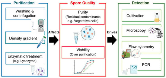
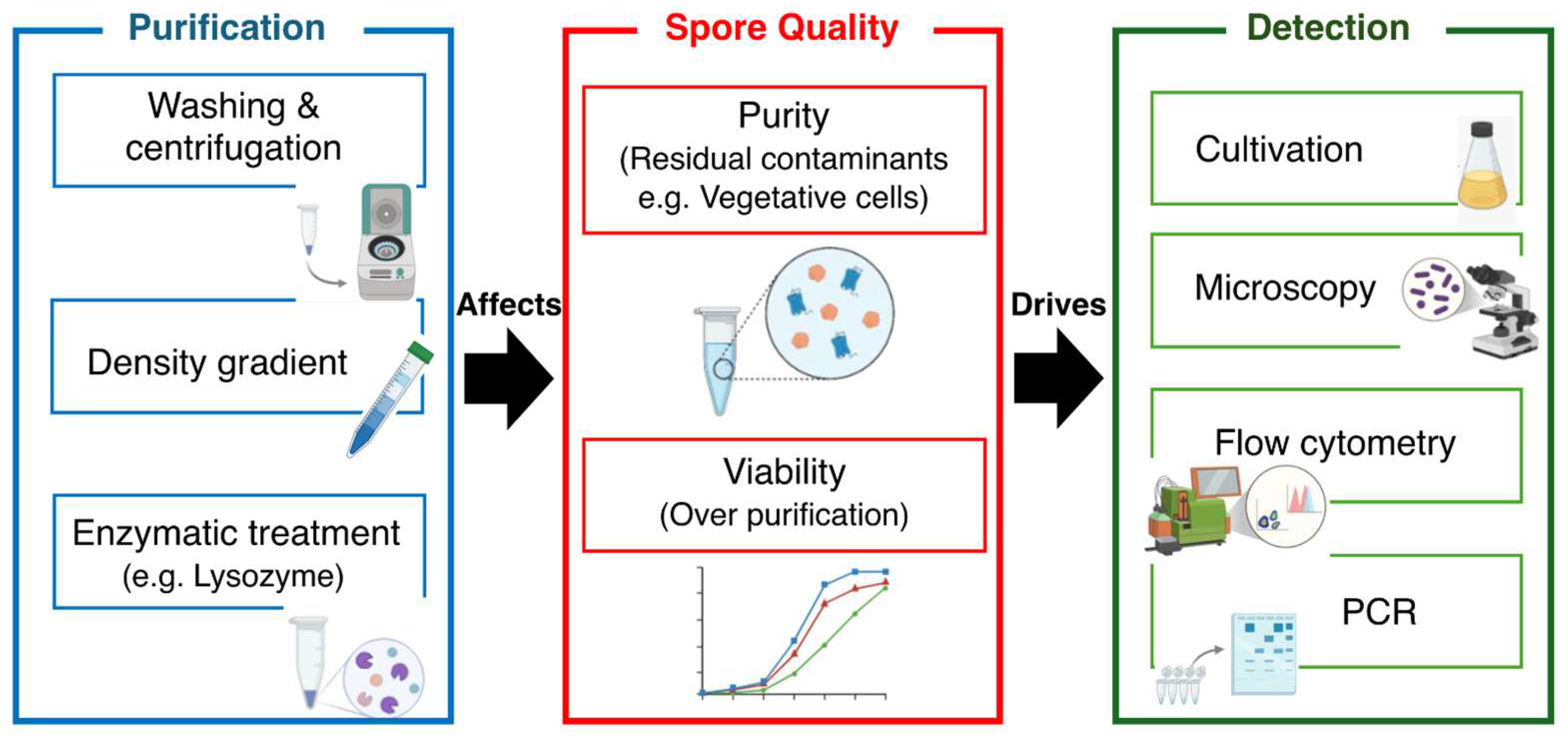
To highlight this interdependence, Figure 1 illustrates how purification choices shape spore quality attributes and subsequently propagate into detection outcomes. Although purification and detection strategies are often described independently, accumulating evidence indicates that these processes are inherently interconnected and should be considered within a unified analytical framework. Variations in purification efficiency can markedly influence downstream detection outcomes, including sensitivity, apparent resistance profiles, and quantitative accuracy. For instance, insufficient removal of vegetative cells may lead to overestimation of viable counts following heat treatment [113,114,115], while residual cellular debris can interfere with optical measurements and flow cytometric discrimination [135,136,137,138]. Furthermore, nucleic acids released from lysed cells may contribute to false-positive signals in molecular assays, thereby inflating estimates of spore abundance [30,31].
Figure 1.
Conceptual framework illustrating the interdependence between spore purification and detection.
Purification strategies influence multiple aspects of spore quality, including purity, viability, structural integrity, and physiological state. These factors collectively determine detection performance across analytical methods. Residual contaminants and excessive purification can introduce bias, potentially leading to inaccurate quantification and misinterpretation of results.
Conversely, overly rigorous purification procedures may compromise spore integrity and viability, resulting in underestimation in culture-based enumeration methods [33]. These bidirectional effects highlight a fundamental trade-off between purity and biological representativeness, emphasizing the need for careful methodological optimization. From a broader perspective, the lack of standardized criteria for evaluating purification efficiency and its impact on detection performance remains a critical limitation across studies.
Future efforts should therefore focus on systematically linking purification conditions with detection outputs across multiple analytical platforms, thereby enabling the development of harmonized protocols and comparable datasets. Establishing such integrative frameworks will be essential for improving reproducibility and ensuring accurate interpretation of spore-related measurements in both research and applied contexts [23,24].
5. Discussion
Despite substantial advances in spore research, methodological standardization remains limited, and this represents a significant barrier to reproducibility and cross-study comparability. One of the most critical challenges lies in the absence of universally accepted criteria for defining and evaluating spore purity. Current studies employ diverse approaches, including microscopic assessment, heat resistance assays, and molecular confirmation—leading to inconsistencies that hinder direct comparison and meta-analytical integration. In addition, intrinsic physiological heterogeneity within spore populations further complicates both purification and detection. Even within a single preparation, spores may differ in maturation state, resistance properties, and germination potential. Such variability can significantly influence purification efficiency and detection sensitivity, particularly when analytical methods assume population uniformity and fail to account for functionally relevant subpopulations.
Matrix-associated effects constitute another persistent challenge. Complex sample matrices, such as food and environmental materials, often contain organic matter, particulate debris, and inhibitory compounds that interfere with both purification efficiency and detection accuracy. Although rapid detection technologies offer considerable promise, their performance frequently depends on highly purified samples and may decline under realistic sample conditions, thereby limiting their practical applicability. For example, in dairy matrices, fats and proteins can co-purify with spores and increase background signals in optical or flow-cytometric analyses, and they may also reduce DNA extraction efficiency and/or inhibit PCR-based assays [30,31,127,128]. In soil and other environmental samples, humic substances and fine particulates can inhibit amplification and complicate discrimination due to overlapping particle characteristics, unless additional cleanup or density-based separation steps are implemented [60,102].
6. Future Directions
Addressing these challenges will require coordinated efforts toward methodological harmonization. Priority should be given to establishing standardized criteria for evaluating spore purity and recovery efficiency, alongside systematic studies that clarify how different purification strategies impact downstream detection performance across analytical platforms. Furthermore, the integration of complementary detection approaches may improve overall robustness and reliability, particularly for rapid and quantitative applications. Advances in analytical technologies, including automation and high-throughput systems, are expected to improve reproducibility; however, their effectiveness will depend on rigorous validation across diverse sample types. Finally, the establishment of consensus benchmarks, such as defined contamination thresholds and viability criteria, will be critical for enabling meaningful cross-study comparisons and driving progress toward greater methodological consistency in spore research.
7. Conclusions
The reliability of bacterial spore research is fundamentally dependent on the accuracy and consistency of purification and detection methodologies. Despite the availability of diverse analytical approaches, methodological variability and the lack of standardization continue to limit reproducibility and hinder comparability across studies. Importantly, purification and detection are not independent processes but are closely interconnected, with each step exerting a direct influence on overall analytical outcomes.
Accordingly, future progress in the field will depend on the adoption of integrated experimental frameworks that consider purification and detection as interdependent components of a unified workflow. The establishment of clear evaluation criteria, coupled with increased methodological transparency, will be essential for improving data reliability. Such efforts will ultimately support more robust and reproducible investigations of bacterial spores, thereby advancing research and applications in food safety, environmental microbiology, and related applied disciplines.
Funding
This research was funded by the Japan Society for the Promotion of Science (JSPS) KAKENHI, grant number 25K24227. The APC was funded by the Toyo Institute of Food Technology.
Institutional Review Board Statement
Not applicable.
Informed Consent Statement
Not applicable.
Data Availability Statement
No new data were created or analyzed in this study. Data sharing is not applicable to this article.
Acknowledgments
The author thanks the members of Toyo Institute of Food Technology for their helpful advice and suggestions throughout the study.
Conflicts of Interest
The author declares no conflicts of interest.
Abbreviations
The following abbreviations are used in this manuscript:
| APC | Article Processing Charge |
| CFU | Colony-Forming Unit |
| DSM | Difco Sporulation Medium |
| DNA | Deoxyribonucleic Acid |
| EPS | Extracellular Polymeric Substances |
| PCR | Polymerase Chain Reaction |
| qPCR | Quantitative Polymerase Chain Reaction |
References
- Wunderlin, T.; Junier, T.; Paul, C.; Jeanneret, N.; Junier, P. Physical isolation of endospores from environmental samples by targeted lysis of vegetative cells. J. Vis. Exp. JoVE 2016, 107, e53411. [Google Scholar]
- Nicholson, W.L.; Munakata, N.; Horneck, G.; Melosh, H.J.; Setlow, P. Resistance of Bacillus endospores to extreme terrestrial and extraterrestrial environments. Microbiol. Mol. Biol. Rev. 2000, 64, 548–572. [Google Scholar] [CrossRef] [Scilit] [PubMed]
- McKenney, P.T.; Driks, A.; Eichenberger, P. The Bacillus subtilis endospore: Assembly and functions of the multilayered coat. Nat. Rev. Microbiol. 2013, 11, 33–44. [Google Scholar] [CrossRef] [Scilit] [PubMed]
- Paredes-Sabja, D.; Shen, A.; Sorg, J.A. Clostridium difficile spore biology: Sporulation, germination, and spore structural proteins. Trends Microbiol. 2014, 22, 406–416. [Google Scholar] [CrossRef] [Scilit] [PubMed]
- Chisholm, J.M.; Putsathit, P.; Riley, T.V.; Lim, S.C. Spore-forming Clostridium (Clostridioides) difficile in wastewater treatment plants in Western Australia. Microbiol. Spectr. 2023, 11, e0358222. [Google Scholar] [CrossRef] [Scilit]
- Setlow, P. Spore Resistance Properties. Microbiol. Spectr. 2014, 2, 201–215. [Google Scholar] [CrossRef] [Scilit]
- Tiloca, G.; Brundu, G.; Ballesteros, D. Bryophyte spores tolerate high desiccation levels and exposure to cryogenic temperatures but contain storage lipids and chlorophyll: Understanding the essential traits needed for the creation of bryophyte spore banks. Plants 2022, 11, 1262. [Google Scholar] [CrossRef] [Scilit]
- Davis, G.M.; Greenhill, A.R.; Munro, T.W.; Horner, J.; Marsden, S.C.; Carter, B.D. Partial protection of Bacillus subtilis spores on simulated Martian media enhances survival against UV radiation. Discov. Space 2025, 129, 3. [Google Scholar] [CrossRef] [Scilit]
- McDonnell, G.; Russell, A.D. Antiseptics and disinfectants: Activity, action, and resistance. Clin. Microbiol. Rev. 1999, 12, 147–179. [Google Scholar] [CrossRef] [Scilit]
- Vreeland, R.H.; Rosenzweig, D.; Powers, D.W. Isolation of a 250 million-year-old halotolerant bacterium from a primary salt crystal. Nature 2000, 407, 897–900. [Google Scholar] [CrossRef] [Scilit]
- Mafe, A.N.; Edo, G.I.; Makia, R.S.; Joshua, O.A.; Akpoghelie, P.O.; Gaaz, T.S.; Jikah, A.N.; Yousif, E.; Isoje, E.F.; Igbuku, U.A.; et al. A review on food spoilage mechanisms, food borne diseases and commercial aspects of food preservation and processing. Food Chem. Adv. 2024, 5, 100852. [Google Scholar] [CrossRef] [Scilit]
- Karanth, S.; Feng, S.; Patra, D.; Pradhan, A.K. Linking microbial contamination to food spoilage and food waste: The role of smart packaging, spoilage risk assessments, and date labeling. Front. Microbiol. 2023, 14, 1198124. [Google Scholar] [CrossRef] [Scilit] [PubMed]
- Lindsay, D.; von Holy, A. Bacterial biofilms within the clinical setting: What healthcare professionals should know. J. Hosp. Infect. 2006, 64, 313–325. [Google Scholar] [CrossRef] [Scilit] [PubMed]
- Bernardi, A.O.; Garcia, M.V.; Copetti, M.V. Food industry spoilage fungi control through facility sanitization. Curr. Opin. Food Sci. 2019, 29, 28–34. [Google Scholar] [CrossRef] [Scilit]
- Gutarowska, B.; Skóra, J.; Stępień, Ł.; Szponar, B.; Otlewska, A.; Pielech-Przybylska, K. Assessment of microbial contamination within working environments of different types of composting plants. J. Air Waste Manag. Assoc. 2015, 65, 466–478. [Google Scholar] [CrossRef] [Scilit]
- Serrano, S.; Grujović, M.; Marković, K.G.; Barreto-Crespo, M.T.; Semedo-Lemsaddek, T. From dormancy to eradication: Strategies for controlling bacterial persisters in food settings. Foods 2025, 14, 1075. [Google Scholar] [CrossRef] [Scilit]
- Talukdar, P.K.; Sarker, M.R. Characterization of putative sporulation and germination genes in Clostridium perfringens food-poisoning strain SM101. Microorganisms 2022, 10, 1481. [Google Scholar] [CrossRef] [Scilit]
- He, H.; Miao, R.; Huang, L.; Jiang, H.; Cheng, Y. Vegetative cells may perform nitrogen fixation function under nitrogen deprivation in Anabaena sp. strain PCC 7120 based on genome-wide differential expression analysis. PLoS ONE 2021, 16, e0248155. [Google Scholar] [CrossRef] [Scilit]
- Nagamine, M.; Ichii, R.; Taira, T.; Teramukai, S.; Konishi, E. Cellular debris on negative liquid-based cytology cervicovaginal smears. Cytopathology 2022, 33, 725–731. [Google Scholar] [CrossRef] [Scilit]
- Corona Ramirez, A.; Bregnard, D.; Junier, T.; Cailleau, G.; Dorador, C.; Bindschedler, S.; Junier, P. Assessment of fungal spores and spore-like diversity in environmental samples by targeted lysis. BMC Microbiol. 2023, 23, 68. [Google Scholar] [CrossRef] [Scilit]
- Wang, Y.; Koopmann, B.; von Tiedemann, A. Methods for assessment of viability and germination of Plasmodiophora brassicae resting spores. Front. Microbiol. 2021, 12, 823051. [Google Scholar] [CrossRef] [Scilit]
- Mafart, P.; Leguérinel, I.; Couvert, O.; Coroller, L. Quantification of spore resistance for assessment and optimization of heating processes: A never-ending story. Food Microbiol. 2010, 27, 568–572. [Google Scholar] [CrossRef] [Scilit]
- Lichten, M. Tetrad, random spore, and molecular analysis of meiotic segregation and recombination. Methods Mol. Biol. 2014, 1205, 13–28. [Google Scholar]
- Li, L.; Jin, J.; Hu, H.; Deveau, I.F.; Foley, S.L.; Chen, H. Optimization of sporulation and purification methods for sporicidal efficacy assessment on Bacillus spores. J. Ind. Microbiol. Biotechnol. 2022, 49, kuac014. [Google Scholar] [CrossRef] [Scilit]
- Kawai, S.; Nakano, M. Optimization of spore purification and quality assessment in Paenibacillus Species. J. Food Prot. 2025, 88, 100595. [Google Scholar] [CrossRef] [Scilit]
- Edwards, A.N.; McBride, S.M. Isolating and purifying Clostridium difficile spores. Methods Mol. Biol. 2016, 1476, 117–128. [Google Scholar]
- Gellert-Mortimer, S.T.; Clarke, G.N.; Baker, H.W.G.; Hyne, R.V.; Johnston, W.I.H. Evaluation of Nycodenz and Percoll density gradients for the selection of motile human spermatozoa. Fertil. Steril. 1988, 49, 335–341. [Google Scholar] [CrossRef] [Scilit]
- Ferraboschi, P.; Ciceri, S.; Grisenti, P. Applications of lysozyme, an innate immune defense factor, as an alternative antibiotic. Antibiotics 2021, 10, 1534. [Google Scholar] [CrossRef] [Scilit] [PubMed]
- Prats, J.; Garcia-Armisen, T.; Larrea, J.; Servais, P. Comparison of culture-based methods to enumerate Escherichia coli in tropical and temperate freshwaters. Lett. Appl. Microbiol. 2008, 46, 243–248. [Google Scholar] [CrossRef] [Scilit] [PubMed]
- Kozuka, S.; Tochikubo, K. Permeability of dormant spores of Bacillus subtilis to malachite green and crystal violet. J. Gen. Microbiol. 1991, 137, 607–613. [Google Scholar] [CrossRef] [Scilit] [PubMed]
- Nakano, M. An improved DNA extraction method for detecting Bacillus subtilis spores in spiked foods and beverages. Int. J. Food Microbiol. 2023, 401, 110280. [Google Scholar] [CrossRef] [Scilit]
- Nakano, M. Spore-DNA localization and extraction efficiencies of Bacillus subtilis for accurate results in quantitative real-time polymerase chain reaction. J. Microorg. Control 2024, 29, 9–15. [Google Scholar] [CrossRef] [Scilit]
- Chen, Z.; Lu, Y.; Cui, J.; Feng, Y.; Dong, H.; Huang, X.; Zhu, C.; Xiong, X.; Chen, H.; Wang, Q.; et al. Monitoring of Bacillus spore-forming dynamics through flow cytometry. Front. Microbiol. 2024, 15, 1450913. [Google Scholar] [CrossRef] [Scilit]
- Kawai, S.; Nakano, M. Analysis of vegetative cells and spore germination in food using flow cytometry. J. Microbiol. Methods 2025, 232–234, 107137. [Google Scholar] [CrossRef] [Scilit]
- Barbiero, D.; Melison, F.; Cocola, L.; Fedel, M.; Andrighetto, C.; De Dea, P.; Poletto, L. Raman spectroscopy applied to early detection of Clostridium infection in milk. Appl. Spectrosc. 2024, 78, 1256–1262. [Google Scholar] [CrossRef] [Scilit] [PubMed]
- Hu, J.; He, L.; Wang, G.; Liu, L.; Wang, Y.; Song, J.; Qu, J.; Peng, X.; Yuan, Y. Rapid and accurate identification of marine bacteria spores at a single-cell resolution by laser tweezers Raman spectroscopy and deep learning. J. Biophotonics 2024, 17, e202300510. [Google Scholar] [CrossRef] [Scilit] [PubMed]
- Wesgate, R.; Rauwel, G.; Criquelion, J.; Maillard, J.Y. Impact of standard test protocols on sporicidal efficacy. J. Hosp. Infect. 2016, 93, 256–262. [Google Scholar] [CrossRef] [Scilit]
- Awasti, N.; Anand, S. A fluorescence spectroscopic method for rapid detection of bacterial endospores: Proof of concept. JDS Commun. 2022, 3, 97–100. [Google Scholar] [CrossRef] [Scilit] [PubMed]
- Haramija, M. Challenges in separations of proteins and small biomolecules and the role of modern mass spectroscopy tools for solving them, as well as bypassing them, in structural analytical studies of complex biomolecular mixtures. Separations 2018, 5, 11. [Google Scholar] [CrossRef] [Scilit]
- Akbar, A.; Malekian, F.; Baghban, N.; Kodam, S.P.; Ullah, M. Methodologies to isolate and purify clinical grade extracellular vesicles for medical applications. Cells 2022, 11, 186. [Google Scholar] [CrossRef] [Scilit]
- Bhalla, N.; Jolly, P.; Formisano, N.; Estrela, P. Introduction to biosensors. Essays Biochem. 2016, 60, 1–8. [Google Scholar] [CrossRef] [Scilit]
- Lemarchand, K.; Berthiaume, F.; Maynard, C.; Harel, J.; Payment, P.; Bayardelle, P.; Masson, L.; Brousseau, R. Optimization of microbial DNA extraction and purification from raw wastewater samples for downstream pathogen detection by microarrays. J. Microbiol. Methods 2005, 63, 115–126. [Google Scholar] [CrossRef] [Scilit]
- Shahidi, F.; Samarasinghe, A. How to assess antioxidant activity? Advances, limitations, and applications of in vitro, in vivo, and ex vivo approaches. Food Prod. Process. Nutr. 2025, 7, 50. [Google Scholar] [CrossRef] [Scilit] [PubMed]
- Li, M.; Liu, S.; Guo, S.; Liang, D.; Li, M.; Zhu, Y.; Zhao, L.; Lee, J.H.; Zhao, G.; Ma, Y.; et al. Selective purification and rapid quantitative detection of spores using a “stepped” magnetic flow device. Anal. Methods 2024, 16, 284–292. [Google Scholar] [CrossRef] [Scilit]
- Castaldi, S.; Donadio, G.; Staiano, I.; Ricca, E.; Isticato, R. The triterpenoid curcumene mediates the relative hydrophilicity of Bacillus subtilis spores. mBio 2025, 16, e0302424. [Google Scholar] [CrossRef] [Scilit]
- Setlow, P. Observations on research with spores of Bacillales and Clostridiales species. J. Appl. Microbiol. 2019, 126, 348–358. [Google Scholar] [CrossRef] [Scilit]
- Serra, C.R.; Earl, A.M.; Barbosa, T.M.; Kolter, R.; Henriques, A.O. Sporulation during growth in a gut isolate of Bacillus subtilis. J. Bacteriol. 2014, 196, 4184–4196. [Google Scholar] [CrossRef] [Scilit] [PubMed]
- Biermann, R.; Beutel, S. Endospore production of Bacillus spp. for industrial use. Eng. Life Sci. 2023, 23, e2300013. [Google Scholar] [CrossRef] [Scilit]
- Yu, B.; Kanaan, J.; Shames, H.; Wicander, J.; Aryal, M.; Li, Y.; Korza, G.; Brul, S.; Kramer, G.; Li, Y.Q.; et al. Identification and characterization of new proteins crucial for bacterial spore resistance and germination. Front. Microbiol. 2023, 14, 1161604. [Google Scholar] [CrossRef] [Scilit]
- Almeida, J.L.; Wang, L.; Morrow, J.B.; Cole, K.D. Requirements for the development of Bacillus anthracis spore reference materials used to test detection systems. J. Res. Natl. Inst. Stand. Technol. 2006, 111, 205–217. [Google Scholar] [CrossRef] [Scilit]
- Bosnar, L.M.; Shindler, A.E.; Wood, J.; Patch, C.; Franks, A.E. Attempts to limit sporulation in the probiotic strain Bacillus subtilis BG01-4TM through mutation accumulation and selection. Access Microbiol. 2023, 5, acmi000419. [Google Scholar] [CrossRef] [Scilit]
- Sakai, K.; Kondo, Y.; Aoki, K.; Goto, Y. Molecular and biophysical perspectives on dormancy breaking: Lessons from yeast spore. Biomolecules 2025, 15, 701. [Google Scholar] [CrossRef] [Scilit]
- Huang, Y.; Swarge, B.N.; Roseboom, W.; Bleeker, J.D.; Brul, S.; Setlow, P.; Kramer, G. Integrative metabolomics and proteomics allow the global intracellular characterization of Bacillus subtilis cells and spores. J. Proteome Res. 2024, 23, 596–608. [Google Scholar] [CrossRef] [Scilit] [PubMed]
- Yang, W.W.; Crow-Willard, E.N.; Ponce, A. Production and characterization of pure Clostridium spore suspensions. J. Appl. Microbiol. 2009, 106, 27–33. [Google Scholar] [CrossRef] [Scilit]
- Stewart, G.C. The exosporium layer of bacterial spores: A connection to the environment and the infected host. Microbiol. Mol. Biol. Rev. 2015, 79, 437–457. [Google Scholar] [CrossRef] [Scilit] [PubMed]
- Pisa, F.; Gionta, M.; Biasi, A.; Pizzuti, M.; Alt, J.R.; Zampaglione, I. Optimization of the preparation of a spiny spore high-concentrated Aspergillus brasiliensis suspension. Lett. Appl. Microbiol. 2025, 78, ovae141. [Google Scholar] [CrossRef] [Scilit]
- Weldy, M.; Evert, C.; Dosa, P.I.; Khoruts, A.; Sadowsky, M.J. Convenient protocol for production and purification of Clostridioides difficile spores for germination studies. STAR Protoc. 2020, 1, 100071. [Google Scholar] [CrossRef] [Scilit]
- Juneja, V.K.; Osoria, M.; Altuntas, E.G.; Taneja, N.K.; Thakur, S.; Kumar, G.D.; Setlow, P. Effects of spore purity on the wet heat resistance of Clostridium perfringens, Bacillus cereus and Bacillus subtilis spores. Food Res. Int. 2024, 177, 113904. [Google Scholar] [CrossRef] [Scilit] [PubMed]
- Wen, J.; Smelt, J.; Vischer, N.O.E.; de Vos, A.L.; Setlow, P.; Brul, S. Heat activation and inactivation of Bacterial spores: Is there an overlap? Appl. Environ. Microbiol. 2022, 88, e0232421. [Google Scholar] [CrossRef] [Scilit]
- Huang, J.; Sheng, K.; Zhang, Y.; Song, M.; Ali, A.; Huang, T.; Huang, M. Inactivation effect of germination combined with cold plasma treatment on Bacillus licheniformis spores. Foods 2023, 12, 4319. [Google Scholar] [CrossRef] [Scilit]
- Tian, P.; Wang, X.; Zhu, G.; Zhang, H.; Zhang, Z. Effects of combining lysozyme, ultrasound, and heat treatments on the inactivation of Bacillus subtilis spores. Arch. Microbiol. 2025, 207, 169. [Google Scholar] [CrossRef] [Scilit]
- Albertsdottir Jonsmoen, U.L.; Allred, J.A.; Malyshev, D.; Segervald, J.; Andersson, M.; Aspholm, M.E. Endospore appendages enhance adhesion of Bacillus cereus sensu lato spores to industrial surfaces, modulated by physicochemical factors. Appl. Environ. Microbiol. 2025, 91, e0094425. [Google Scholar] [CrossRef] [Scilit] [PubMed]
- Torres-Anjel, M.J.; Hedrick, T.I. Spore removal by centrifugation and its effect on ultra-high temperature commercial sterilization of milk. J. Dairy Sci. 1971, 54, 326–330. [Google Scholar] [CrossRef] [Scilit]
- Tavares, M.B.; Souza, R.D.; Luiz, W.B.; Cavalcante, R.C.M.; Casaroli, C.; Martins, E.G.; Ferreira, R.C.C.; Ferreira, L.C.S. Bacillus subtilis endospores at high purity and recovery yields: Optimization of growth conditions and purification method. Curr. Microbiol. 2013, 66, 279–285. [Google Scholar] [CrossRef] [Scilit]
- Koransky, J.R.; Allen, S.D.; Dowell, V.R., Jr. Use of ethanol for selective isolation of sporeforming microorganisms. Appl. Environ. Microbiol. 1978, 35, 762–765. [Google Scholar] [CrossRef] [Scilit]
- Zhao, J.; Krishna, V.; Moudgil, B.; Koopman, B. Evaluation of endospore purification methods applied to Bacillus cereus. Sep. Purif. Technol. 2008, 16, 341–347. [Google Scholar] [CrossRef] [Scilit]
- He, L.M.; Tebo, B.M. Surface charge properties of and Cu(II) adsorption by spores of the marine Bacillus sp. strain SG-1. Appl. Environ. Microbiol. 1998, 64, 1123–1129. [Google Scholar] [CrossRef] [Scilit]
- Delahaye, M.; Lawrence, K.; Ward, S.J.; Hoare, M. An ultra scale-down analysis of the recovery by dead-end centrifugation of human cells for therapy. Biotechnol. Bioeng. 2015, 112, 997–1011. [Google Scholar] [CrossRef] [Scilit] [PubMed]
- Cho, W.I.; Chung, M.S. Bacillus spores: A review of their properties and inactivation processing technologies. Food Sci. Biotechnol. 2020, 29, 1447–1461. [Google Scholar] [CrossRef] [Scilit]
- Nakano, M. 16S rRNA gene primer validation for bacterial diversity analysis of vegetable products. J. Food. Prot. 2018, 81, 848–859. [Google Scholar] [CrossRef] [Scilit]
- Prentice, G.A.; Wolfe, F.H.; Clegg, L.F.L. The use of density gradient centrifugation for the separation of germinated from nongerminated spores. J. Appl. Bacteriol. 1972, 35, 345–349. [Google Scholar] [CrossRef] [Scilit] [PubMed]
- Yamamoto, Y.; Itoh, S.; Yamauchi, Y.; Matsushita, K.; Ikeda, S.; Naruse, H.; Hayashi, M. Density gradient centrifugation for the isolation of cells of multiple lineages. J. Cell. Biochem. 2015, 116, 2709–2714. [Google Scholar] [CrossRef] [Scilit] [PubMed]
- Pertoft, H. Fractionation of cells and subcellular particles with Percoll. J. Biochem. Biophys. Methods 2000, 44, 1–30. [Google Scholar] [CrossRef] [Scilit] [PubMed]
- Warth, A.D. Molecular structure of the bacterial spore. Adv. Microb. Physiol. 1978, 17, 1–45. [Google Scholar] [PubMed]
- Henriques, A.O.; Moran, C.P., Jr. Structure, assembly, and function of the spore surface layers. Annu. Rev. Microbiol. 2007, 61, 555–588. [Google Scholar] [CrossRef] [Scilit]
- Atrih, A.; Foster, S.J. Analysis of the role of bacterial endospore cortex structure in resistance properties and demonstration of its conservation amongst species. J. Appl. Microbiol. 2001, 91, 364–372. [Google Scholar] [CrossRef] [Scilit]
- Beskrovnaya, P.; Sexton, D.L.; Golmohammadzadeh, M.; Hashimi, A.; Tocheva, E.I. Structural, metabolic and evolutionary comparison of bacterial endospore and exospore formation. Front. Microbiol. 2021, 12, 630573. [Google Scholar] [CrossRef] [Scilit]
- Stelder, S.K.; Benito de Moya, C.; Hoefsloot, H.C.J.; de Koning, L.J.; Brul, S.; de Koster, C.G. Stoichiometry, absolute abundance, and localization of proteins in the Bacillus cereus spore coat insoluble fraction determined using a qconcat approach. J. Proteome Res. 2018, 17, 903–917. [Google Scholar] [CrossRef] [Scilit]
- Laue, M.; Han, H.M.; Dittmann, C.; Setlow, P. Intracellular membranes of bacterial endospores are reservoirs for spore core membrane expansion during spore germination. Sci. Rep. 2018, 8, 11388. [Google Scholar] [CrossRef] [Scilit]
- Gauvry, E.; Mathot, A.G.; Couvert, O.; Leguérinel, I.; Jules, M.; Coroller, L. Differentiation of vegetative cells into spores: A kinetic model applied to Bacillus subtilis. Appl. Environ. Microbiol. 2019, 85, e00322-19. [Google Scholar] [CrossRef] [Scilit]
- Seale, R.B.; Flint, S.H.; McQuillan, A.J.; Bremer, P.J. Recovery of spores from thermophilic dairy bacilli and effects of their surface characteristics on attachment to different surfaces. Appl. Environ. Microbiol. 2008, 74, 731–737. [Google Scholar] [CrossRef] [Scilit]
- Da Silva, S.M.; Filliben, J.J.; Morrow, J.B. Parameters affecting spore recovery from wipes used in biological surface sampling. Appl. Environ. Microbiol. 2011, 77, 2374–2380. [Google Scholar] [CrossRef] [Scilit]
- Sun, Y.; Sethu, P. Microfluidic adaptation of density-gradient centrifugation for isolation of particles and cells. Bioengineering 2017, 4, 67. [Google Scholar] [CrossRef] [Scilit]
- Gao, J.; Dong, X.; Wang, Z. Generation, purification and engineering of extracellular vesicles and their biomedical applications. Methods 2020, 177, 114–125. [Google Scholar] [CrossRef] [Scilit]
- Heydenreich, R.; Nacita, J.; Lin, C.W.; O’Dea, F.; Mesnage, S.; Christie, G.; Mathys, A. Revisiting bacterial spore germination in the presence of peptidoglycan fragments. J. Bacteriol. 2025, 207, e0014625. [Google Scholar] [CrossRef] [Scilit]
- Liburdi, K.; Benucci, I.; Esti, M. Lysozyme in wine: An overview of current and future applications. Compr. Rev. Food Sci. Food Saf. 2014, 13, 1062–1073. [Google Scholar] [CrossRef] [Scilit]
- Nawaz, N.; Wen, S.; Wang, F.; Nawaz, S.; Raza, J.; Iftikhar, M.; Usman, M. Lysozyme and its application as antibacterial agent in food industry. Molecules 2022, 27, 6305. [Google Scholar] [CrossRef] [Scilit]
- Blake, C.C.F.; Fenn, R.H.; North, A.C.T.; Phillips, D.C.; Poljak, R.J. Structure of lysozyme. A Fourier map of the electron density at 6 angstrom resolution obtained by x-ray diffraction. Nature 1962, 196, 1173–1176. [Google Scholar] [CrossRef] [Scilit]
- Chipman, D.M.; Pollock, J.J.; Sharon, N. Lysozyme-catalyzed hydrolysis and transglycosylation reactions of bacterial cell wall oligosaccharides. J. Biol. Chem. 1968, 243, 487–496. [Google Scholar] [CrossRef] [Scilit]
- Garajová, K.; Balogová, A.; Dušeková, E.; Sedláková, D.; Sedlák, E.; Varhač, R. Correlation of lysozyme activity and stability in the presence of Hofmeister series anions. Biochim. Biophys. Acta BBA Proteins Proteom. 2017, 1865, 281–288. [Google Scholar] [CrossRef] [Scilit]
- Davidson, P.M.; Matthew, T.; David, J.R.D. Antimicrobials in Food. In Food Science and Technology, 4th ed.; CRC Press: Boca Raton, FL, USA, 2020. [Google Scholar]
- Khorshidian, N.; Khanniri, E.; Koushki, M.R.; Sohrabvandi, S.; Yousefi, M. An overview of antimicrobial activity of lysozyme and its functionality in cheese. Front. Nutr. 2022, 9, 833618. [Google Scholar] [CrossRef] [Scilit]
- Mundra, R.V.; Mehta, K.K.; Wu, X.; Paskaleva, E.E.; Kane, R.S.; Dordick, J.S. Enzyme-driven Bacillus spore coat degradation leading to spore killing. Biotechnol. Bioeng. 2014, 111, 654–663. [Google Scholar] [CrossRef] [Scilit]
- Raguse, M.; Fiebrandt, M.; Stapelmann, K.; Madela, K.; Laue, M.; Lackmann, J.W.; Thwaite, J.E.; Setlow, P.; Awakowicz, P.; Moeller, R. Improvement of biological indicators by uniformly distributing Bacillus subtilis spores in monolayers to evaluate enhanced spore decontamination technologies. Appl. Environ. Microbiol. 2016, 82, 2031–2038. [Google Scholar] [CrossRef] [Scilit]
- Todd, E.C.D. Foodborne diseases: Overview of biological hazards and foodborne diseases. Encycl. Food Saf. 2013, 221–242. [Google Scholar]
- Segervald, J.; Malyshev, D.; Öberg, R.; Zäll, E.; Jia, X.; Wågberg, T.; Andersson, M. Ultra-sensitive detection of bacterial spores via SERS. ACS Sens. 2025, 10, 1237–1248. [Google Scholar] [CrossRef] [Scilit]
- Ni, Y.; Pang, D.; Li, H.; Zhou, G.; Li, Y.; Yao, Y.; Xu, B. Recent progress in typical foodborne spore-forming bacteria detection. Food Chem. 2026, 504, 147858. [Google Scholar] [CrossRef] [Scilit]
- Shafi, Z.; Shahid, M.; Singh, R. Food-borne bacterial pathogens: Emerging approaches in detection and prevention. Arch. Microbiol. 2026, 208, 109. [Google Scholar] [CrossRef] [Scilit]
- Moir, A.; Corfe, B.M.; Behravan, J. Spore germination. Cell. Mol. Life Sci. CMLS 2002, 59, 403–409. [Google Scholar] [CrossRef] [Scilit]
- Lee, G.Y.; Li, W.; Chirwa, U.M.; Shi, J. Effect of substrate characteristics on the growth and sporulation of two biocontrol microorganisms during solid state cultivation. Fermentation 2020, 6, 69. [Google Scholar] [CrossRef] [Scilit]
- Green, L.C.; LeBlanc, P.J.; Didier, E.S. Discrimination between viable and dead Encephalitozoon cuniculi (Microsporidian) spores by dual staining with sytox green and calcofluor white M2R. J. Clin. Microbiol. 2000, 38, 3811–3814. [Google Scholar] [CrossRef] [Scilit]
- Rawsthorne, H.; Dock, C.N.; Jaykus, L.A. PCR-based method using propidium monoazide to distinguish viable from nonviable Bacillus subtilis spores. Appl. Environ. Microbiol. 2009, 75, 2936–2939. [Google Scholar] [CrossRef] [Scilit]
- Ma, Q.; Zhu, Y.; Tu, Q.; Li, M.; Hong, N.; Liu, S.; Sun, L.; Liu, Y.; Zhao, L.; Liang, D. From structural characteristics to metabolic characteristics: Structural morphological differences and proteomic analysis of Clostridium perfringens and its spores. Food Res. Int. 2026, 233, 118960. [Google Scholar] [CrossRef] [Scilit]
- Moore, C.E.; Sona, S.; Poda, S.; Putchhat, H.; Kumar, V.; Sopheary, S.; Stoesser, N.; Bousfield, R.; Day, N.; Parry, C.M. Antimicrobial susceptibility of uropathogens isolated from Cambodian children. Paediatr. Int. Child Health 2016, 36, 113–117. [Google Scholar] [CrossRef]
- Ghosh, S.; Zhang, P.; Li, Y.Q.; Setlow, P. Superdormant spores of Bacillus species have elevated wet-heat resistance and temperature requirements for heat activation. J. Bacteriol. 2009, 191, 5584–5591. [Google Scholar] [CrossRef] [Scilit]
- Løvdal, I.S.; Hovda, M.B.; Granum, P.E.; Rosnes, J.T. Promoting Bacillus cereus spore germination for subsequent inactivation by mild heat treatment. J. Food Prot. 2011, 74, 2079–2089. [Google Scholar] [CrossRef] [Scilit]
- Rodriguez-Palacios, A.; Lejeune, J.T. Moist-heat resistance, spore aging, and superdormancy in Clostridium difficile. Appl. Environ. Microbiol. 2011, 77, 3085–3091. [Google Scholar] [CrossRef] [Scilit]
- Hamilton, M.A.; Bissonnette, G.K. Statistical inferences about injury and persistence of environmentally stressed bacteria. J. Hyg. 1975, 74, 149–155. [Google Scholar] [CrossRef] [Scilit]
- Saito, M.; Takatani, N.; Yoshida, T.; Mariogani, A.; Cho, E.; Matsuoka, H. Effects of injured and dead cells of Escherichia coli on the colony-forming rate of live cells. FEBS Open Bio 2021, 11, 404–412. [Google Scholar] [CrossRef] [Scilit]
- Pazos-Rojas, L.A.; Cuellar-Sánchez, A.; Romero-Cerón, A.L.; Rivera-Urbalejo, A.; Van Dillewijn, P.; Luna-Vital, D.A.; Muñoz-Rojas, J.; Morales-García, Y.E.; Bustillos-Cristales, M.D.R. The Viable but Non-Culturable (VBNC) State, a Poorly Explored Aspect of Beneficial Bacteria. Microorganisms 2023, 12, 39. [Google Scholar] [CrossRef] [Scilit]
- Voběrková, S.; Hermanová, S.; Hrubanová, K.; Krzyžánek, V. Biofilm formation and extracellular polymeric substances (EPS) production by Bacillus subtilis depending on nutritional conditions in the presence of polyester film. Folia Microbiol. 2016, 61, 91–100. [Google Scholar] [CrossRef] [Scilit]
- Chai, Y.; Chu, F.; Kolter, R.; Losick, R. Bistability and biofilm formation in Bacillus subtilis. Mol. Microbiol. 2008, 67, 254–263. [Google Scholar] [CrossRef] [Scilit]
- Thomas, P.; Sekhar, A.C.; Mujawar, M.M. Vulnerability of Bacillus spores and of related genera to physical impaction injury with particular reference to spread-plating. J. Appl. Microbiol. 2014, 117, 1358–1372. [Google Scholar] [CrossRef] [Scilit]
- Warda, A.K.; Tempelaars, M.H.; Abee, T.; Nierop Groot, M.N. Recovery of heat treated Bacillus cereus spores is affected by matrix composition and factors with putative functions in damage repair. Front. Microbiol. 2016, 7, 1096. [Google Scholar] [CrossRef] [Scilit]
- Freire, V.; Del Río, J.; Gómara, P.; Salvador, M.; Condón, S.; Gayán, E. Comparative study on the impact of equally stressful environmental sporulation conditions on thermal inactivation kinetics of B. subtilis spores. Int. J. Food. Microbiol. 2023, 405, 110349. [Google Scholar] [CrossRef] [Scilit]
- White, W.B. AOAC methods of analysis. Food Drug Cosmet. Law J. 1957, 12, 327–341. [Google Scholar]
- Horwitz, W.; Latimer, A.W., Jr. Official Methods of Analysis of AOAC International, 18th ed.; AOAC International: Rockville, MD, USA, 2005. [Google Scholar]
- McCleary, B.V. Measurement of dietary fiber: Which AOAC Official Method of Analysis SM to use. J. AOAC Int. 2023, 106, 917–930. [Google Scholar] [CrossRef] [Scilit]
- Eller, C.; Ordal, Z.J. Interference-contrast and phase-contrast microscopy of sporulation in Clostridium thermosaccharolyticum grown under strict anaerobiosis. J. Bacteriol. 1972, 111, 674–681. [Google Scholar] [CrossRef] [Scilit]
- Kong, L.; Zhang, P.; Setlow, P.; Li, Y.-q. Characterization of Bacterial spore germination using integrated phase contrast microscopy, Raman spectroscopy, and optical tweezers. Anal. Chem. 2010, 82, 3840–3847. [Google Scholar] [CrossRef] [Scilit]
- Wang, X.; Fan, B.; Zhu, T.; Shen, H.; Li, L.; Wang, S.; Yin, X.; Tian, B.; Zhang, J.; Li, B.; et al. Bacillus spore showcasing key antigens of African swine fever potentializes a promising oral vaccine candidate. Appl. Microbiol. Biotechnol. 2025, 109, 162. [Google Scholar] [CrossRef] [Scilit]
- Zhang, C.; Zhang, S.; Diao, H.; Zhao, H.; Zhu, X.; Lu, F.; Lu, Z. Purification and characterization of a temperature- and pH-stable laccase from the spores of Bacillus vallismortis fmb-103 and its application in the degradation of malachite green. J. Agric. Food Chem. 2013, 61, 5468–5473. [Google Scholar] [CrossRef] [Scilit]
- Malyshev, D.; Lee, C.C.; Andersson, M. Evaluating bacterial spore preparation methods for scanning electron microscopy. Microsc. Microanal. 2024, 30, 564–573. [Google Scholar] [CrossRef] [Scilit]
- Borisova, J.; Morshchinin, I.V.; Nazarova, V.I.; Molodkina, N.; Nikitin, N.O. Low-cost microalgae cell concentration estimation in hydrochemistry applications using computer vision. Sensors 2025, 25, 4651. [Google Scholar] [CrossRef] [Scilit]
- Sanchez-Salas, J.L.; Setlow, B.; Zhang, P.; Li, Y.Q.; Setlow, P. Maturation of released spores is necessary for acquisition of full spore heat resistance during Bacillus subtilis sporulation. Appl. Environ. Microbiol. 2011, 77, 6746–6754. [Google Scholar] [CrossRef] [Scilit] [PubMed]
- Stolk, D.; Bloemen, P.; van den Elzen, R.M.; de Bruin, M.; Driessen, C. A potential pitfall in the interpretation of microscope-integrated fluorescence angiography: The center-periphery effect. Med. Sci. 2025, 13, 54. [Google Scholar] [CrossRef] [Scilit] [PubMed]
- McHugh, A.J.; Feehily, C.; Hill, C.; Cotter, P.D. Detection and enumeration of spore-forming bacteria in powdered dairy products. Front. Microbiol. 2017, 8, 109. [Google Scholar] [CrossRef] [Scilit] [PubMed]
- Vidic, J.; Chaix, C.; Manzano, M.; Heyndrickx, M. Food sensing: Detection of Bacillus cereus spores in dairy products. Biosensors 2020, 10, 15. [Google Scholar] [CrossRef] [Scilit]
- Kramer, M.F.; Coen, D.M. Enzymatic amplification of DNA by PCR: Standard procedures and optimization. Curr. Protoc. Toxicol. 2001, 3, A.3C.1–14. [Google Scholar]
- Yu, L.; Xu, Y.; Al-Amin, M.; Jiang, S.; Sample, M.; Prasad, A.; Stephanopoulos, N.; Sulc, P.; Yan, H. Cytodirect: A nucleic acid nanodevice for specific and efficient delivery of functional payloads to the cytoplasm. J. Am. Chem. Soc. 2023, 145, 27336–27347. [Google Scholar] [CrossRef] [Scilit]
- Hongsibsam, T.; Prasertsopon, J.; Siripurkpong, P.; Niyomdecha, N. Comparative evaluation of viability PCR reagents highlights the superior efficacy of PMAxx azo dye for bacterial and viral viability discrimination using real-time and digital PCR. Microbiol. Spectr. 2026, 14, e0400225. [Google Scholar] [CrossRef] [Scilit]
- Nocker, A.; Camper, A.K. Selective removal of DNA from dead cells of mixed bacterial communities by use of ethidium monoazide. Appl. Environ. Microbiol. 2006, 72, 1997–2004. [Google Scholar] [CrossRef] [Scilit]
- Garre, A.; Penalver-Soto, J.L.; Esnoz, A.; Iguaz, A.; Fernandez, P.S.; Egea, J.A. On the use of in-silico simulations to support experimental design: A case study in microbial inactivation of foods. PLoS ONE 2019, 14, e0220683. [Google Scholar] [CrossRef] [Scilit]
- Tarlak, F. The use of predictive microbiology for the prediction of the shelf life of food products. Foods 2023, 12, 4461. [Google Scholar] [CrossRef] [Scilit]
- Priyadarsini, M.; Kushwaha, J.; Pandey, K.P.; Rani, J.; Dhoble, A.S. Application of flow cytometry for rapid, high-throughput, multiparametric analysis of environmental microbiomes. J. Microbiol. Methods 2023, 214, 106841. [Google Scholar] [CrossRef] [Scilit] [PubMed]
- Harada, Y.; Kuwana, R.; Yagi, S.; Okino, Y.; Takamatsu, H. Flow cytometric distinction of mature spores of Bacillus subtilis 168 using Auramine O. Biosci. Biotechnol. Biochem. 2026, 90, 576–584. [Google Scholar] [CrossRef] [Scilit] [PubMed]
- Phillips, A.P.; Martin, K.L. Dual-parameter scatter-flow immunofluorescence analysis of Bacillus spores. Cytometry 1985, 6, 124–129. [Google Scholar] [CrossRef] [Scilit] [PubMed]
- Zheng, X.L.; Xiong, Z.Q.; Wu, J.Q. The use of a simple flow cytometry method for rapid detection of spores in probiotic Bacillus licheniformis-containing tablets. Food Sci. Biotechnol. 2017, 26, 167–171. [Google Scholar] [CrossRef] [Scilit]
- Berg Luecke, L.; Waas, M.; Gundry, R.L. Reliable protocols for flow cytometry analysis of intracellular proteins in pluripotent stem cell derivatives: A fit-for-purpose approach. Curr. Protoc. Stem Cell Biol. 2019, 50, e94. [Google Scholar] [CrossRef] [Scilit]
- Czechowska, K.; Bonilla, D.L.; Cotty, A.; Dankar, A.; Mead, P.E.; Nash, V. Beyond the limits: How is spectral flow cytometry reshaping the clinical landscape and what is coming next? Cells 2025, 14, 997. [Google Scholar] [CrossRef] [Scilit]
- Bruni, C.; Ferrante, L.; Koch, G.; Scoglio, C.; Starace, G. A stochastic model for cell debris in flow cytometry. J. Theor. Biol. 1993, 161, 157–174. [Google Scholar] [CrossRef] [Scilit]
- Darzynkiewicz, Z.; Halicka, H.D.; Zhao, H. Analysis of cellular DNA content by flow and laser scanning cytometry. Adv. Exp. Med. Biol. 2010, 676, 137–147. [Google Scholar]
- Kolomenskii, A.A.; Schuessler, H.A. Raman spectra of dipicolinic acid in crystalline and liquid environments. Spectrochim. Acta Part A Mol. Biomol. Spectrosc. 2005, 61, 647–651. [Google Scholar] [CrossRef] [Scilit]
- Perez-Rodriguez, M.; Serrano-Pertierra, E.; Blanco-Lopez, M.C. Advances in biosensor technologies for the detection of antimicrobial resistance in Staphylococcus aureus. Front. Cell. Infect. Microbiol. 2025, 15, 1741845. [Google Scholar] [CrossRef] [Scilit]
- Wang, S.; Guan, X.; Sun, S. Microfluidic biosensors: Enabling advanced disease detection. Sensors 2025, 25, 1936. [Google Scholar] [CrossRef] [Scilit]
- Bounegru, A.V.; Dinu Iacob, A.; Iticescu, C.; Georgescu, P.L. Electrochemical sensors and biosensors for the detection of pharmaceutical contaminants in natural waters—A comprehensive review. Chemosensors 2025, 13, 65. [Google Scholar] [CrossRef] [Scilit]

Disclaimer/Publisher’s Note: The statements, opinions and data contained in all publications are solely those of the individual author(s) and contributor(s) and not of MDPI and/or the editor(s). MDPI and/or the editor(s) disclaim responsibility for any injury to people or property resulting from any ideas, methods, instructions or products referred to in the content. |
© 2026 by the author. Licensee MDPI, Basel, Switzerland. This article is an open access article distributed under the terms and conditions of the Creative Commons Attribution (CC BY) license.