1. Introduction
The solar radiation spectrum includes such ranges as ultraviolet (UVC, 100–280 nm; UVB 280–315 nm; UVA 315–400 nm), visible light (with a wavelength range of 400 to 700 nm) as well as infrared radiation (IR), including near IR (IR-A; 700–1400 nm), mid IR (IR-B; 1400–3000 nm), and far IR (IR-C; 3000 nm–1 mm) [
1,
2]. The negative impact of UV radiation on the skin is widely known, which is why manufacturers of sunscreen products take into account both protection against UVB radiation, expressed by the SPF (Sun Protection Factor) index, and protection against UVA radiation, determined by the PPD (Persistent Pigment Darkening) index. Ultraviolet radiation can be an unfavourable inflammatory, mutagenic, and carcinogenic agent for the skin, as well as a powerful enhancer of free radical production [
3,
4]. However, IR has recently attracted researchers’ attention due to research findings suggesting its involvement in skin photoaging and increased risk of carcinogenesis. IR has the lowest energy level, but its share of the solar spectrum reaching human skin is approximately 40%. Moreover, infrared radiation can penetrate the deeper layers of the skin, even into the subcutaneous tissue. Therefore, the biological impact on the skin may be significant. IR significantly influences the induction of free radicals in the dermis, reduces the skin’s antioxidant capacity, and also alters the collagen content of the skin’s extracellular matrix (ECM), not only by increasing the expression of collagen-degrading matrix metalloproteinase (MMP) enzymes but also by reducing de novo collagen synthesis [
1,
2,
3,
5].
Effective solutions already exist for protecting skin from UV radiation, thanks to a wide selection of products with varying protection factors. The use of these products in everyday life often contributes to the public’s belief that prolonged exposure to sunlight is possible without negative effects. There is still insufficient knowledge and awareness of the negative effects of IR. It is therefore essential not only to raise consumer awareness, but also to identify and incorporate new bioactive ingredients into suncare products, the effects of which will limit the damage that can occur to the skin as a result of excessive exposure to UV and/or IR. Apart from synthetic ingredients, promising effects in this respect are observed in plant-derived raw materials, due to their bioactive content, including polyphenolic compounds with numerous hydroxyl groups, known for their strong antioxidant and radiation-protective properties [
4,
6,
7]. Furthermore, over the past decades, there has been a growing interest in plant extracts and their increasingly frequent use as active and auxiliary ingredients in cosmetic products. Cosmetic plants also constitute a significant group of plants that are currently gaining in importance as active ingredients in skincare and protective products. Examples of plants that have long been known and used in preventive and skin care include
Crataegus monogyna Jacq. i
Rosmarinus officinalis L. [
8,
9].
Crataegus monogyna Jacq. is a shrub or small tree from the Rosaceae family that grows in Europe, Africa, and Asia. The raw material is hawthorn flower with leaves (
Crataegi inflorescentia) and fruit (
Crataegi fructus). Hawthorn flower with leaf contains flavonoids, procyanidins, phenolic acids, triterpenes, tannins, coumarins, phytosterols, vitamin C and B, pectin, and mineral salts. Hawthorn fruits contain similar active substances, except that they have more flavonoids and procyanidins, and also contain vitamin A and the sugar alcohol sorbitol.
C. monogyna is used in heart support preparations, as well as a sedative, diuretic, anti-inflammatory, antioxidant, antimicrobial, and cardiotonic agent [
8,
10].
Rosmarinus officinalis L. is an aromatic and medicinal plant of the Lamiaceae family native to the Mediterranean region and cultivated worldwide. The raw material is rosemary leaf (
Rosmarini folium) containing flavonoids, phenolic acids, tannins, bitter substances, resins, saponins, phytosterols, as well as essential oil, whose main components are borneol, cineole, limonene, pinene, and camphor. Rosemary is a plant with several biological properties, including anti-inflammatory, antioxidant, antimicrobial, antiproliferative, antitumor, and circulatory-stimulating activities [
9,
11]. Extracts from these raw materials are used not only in the treatment of various diseases but also in skin and appendage care as ingredients in preparations for mature, sensitive, and vascular skin, as well as for the scalp and hair. The bioactive compounds found in hawthorn and rosemary also suggest that these ingredients could be used in sun protection products. However, no data confirm their use as ingredients in formulations that protect the skin from solar radiation, including infrared radiation. Therefore, this study aimed to evaluate the protective effect of extracts from
Crataegus monogyna and
Rosmarinus officinalis against the adverse effects of UV radiation and infrared light on the skin. For this purpose, the hemispheric directional reflectance method was used.
2. Materials and Methods
2.1. Extracts and Extract-Based Hydrogels Preparation
Crataegus monogyna Jacq. flowers with leaves and Rosmarinus officinalis L. leaves collected from the Botanical Garden in Kielce in May 2025 were used in the study. After drying, the raw materials were ground in the grinder (A11 basic, IKA-Werke, Staufen, Germany). Then, hydroethanolic extracts were prepared by ultrasound-assisted extraction. The extracting solvent (50/50 EtOH/water solution (v/v), 60 mL) was added to 2 g of powdered plant material. Extraction was performed for 60 min using an ultrasonic bath (Polsonic 5, Warsaw, Poland). The obtained extracts were filtered using Whatman filter paper (Merck, Darmstadt, Germany).
Two hydrogels containing Crataegus monogyna (HC) or Rosmarinus officinalis (HR) extract were prepared using the following raw materials: Carbopol® Ultrez 10 Polymer (Lubrizol, Cleveland, OH, USA), glycerine (PPH Microfarm, Zabierzów, Poland), triethanolamine (Chempur, Piekary Śląskie, Poland), and aqua. The pH values of the prepared formulations were determined using a pH meter CP-501 with EPS-1 electrode (Elmetron, Zabrze, Poland, SevenCompact S-200, Mettler Toledo, Greisensee, Switzerland).
2.2. Rheological and Textural Assessment of Extract-Based Hydrogels
The rheological properties of hydrogels were analyzed using a Lamy Rheology RM 200 Touch rotational rheometer (Lyon, France) with a CP 2445 measuring system (diameter 24 mm, bevel angle α = 0.45° mm) in a plate-to-plate configuration. The test was conducted at 25 °C (the temperature at which hydrogels are transported and stored) and 32 °C (the temperature of the human skin surface). A constant temperature was maintained using a Lamy Rheology CP-1 Plus laboratory thermostat integrated with the rheometer. The viscosity was determined at shear rates of 30 s−1, 50 s−1, and 100 s−1. Flow curves (shear stress [Pa] = f (shear rate [s−1]) and viscosity curves (viscosity [Pa × s] = f (shear rate [s−1])) of the analyzed formulations were generated using the Rheometic-P Software (Version: 2.1.0.4). The obtained rheograms were analyzed based on selected mathematical models: Casson, Bingham, Ostwald-de Waele, and Herschel–Bulkley. The degree to which the model fits the empirical data was verified using the coefficient of determination, denoted as R2, in accordance with the principle that the higher its value, the better the fit to the model. The area of the obtained hysteresis loops was calculated using the integration method.
A TX-700 texture analyzer (Lamy Rheology Instruments, Lyon, France) configured to operate with Rheotex TX-UK01/2019 software (version: 1.37.0.0) was used for texture testing. An 8 mm diameter hemispherical probe was used in the test. The texture parameters were analysed using the following instrument settings: down speed: 1 mm/s, distance: 5 mm, force to start: 0.05 N, relaxation time: 20 s. The tests were carried out at a temperature of 25 ± 0.1 °C. The theoretical aspects of texture analysis were discussed in an earlier article [
12].
2.3. Measurement Procedure
This study was approved by the Bioethics Committee of the Medical University of Silesia, No. PCN/CBN/0052/KB1/62/22. The study involved a group of 23 healthy women aged between 20 and 26, with Fitzpatrick skin types I to III. All volunteers agreed to participate in the study.
A quantitative comparative analysis of the skin’s reflectance was used to assess potential photoprotective properties of the tested plant extracts. The control group was a hydrogel without the extract. A portion of the hydrogel (approximately 20 mg) was thoroughly smeared on the hairless skin on the inner side of the forearms (skin area of 3 cm2) using a plastic spatula (separate for each hydrogel). To minimize application-related variability, the amount of formulation, application area, anatomical site, application tool, and measurement timing were standardized for all volunteers and formulations. However, because the hydrogel was manually distributed using a plastic spatula, minor differences in film thickness and layer uniformity could not be completely excluded. Reflectance measurements were performed on the same day, before and immediately after hydrogel application; therefore, the study assessed the initial optical effect of the freshly applied formulations rather than the temporal persistence of this effect.
Each woman was examined using a SOC 410 Solar DHR reflectometer (Surface Optics Corporation, San Diego, CA, USA) which measures the directional–hemispherical reflectance of the skin at an incident angle of 20 degrees for seven discrete spectral bands: 335–380 nm, 400–540 nm, 480–600 nm, 590–720 nm, 700–1100 nm, 1000–1700 nm, and 1700–2500 nm. Calibration of the reflectometer was performed using two calibration coupons, certified by ANIST (American National Institute of Standards and Technology).
The SOC 410 Solar measures total, diffuse, and specular reflectance using an integrating sphere that collects light reflected from the sample in different directions. For an incident spectral radiant flux, the energy balance can be expressed as
where R
λ is reflectance, A
λ is absorption, and T
λ is transmission or forward propagation into deeper tissue layers.
Changes in directional–hemispherical reflectance after hydrogel application were used to assess formulation-induced modifications of the optical properties of the skin/formulation system. Reflectance changes were not interpreted as direct proof of photoprotection because reduced reflectance may result from increased absorption within the formulation or skin, increased forward propagation into deeper layers, or changes in scattering.
2.4. The Statistical Analysis
Statistical analysis was performed using Statistica 13 software. Following a preliminary assessment of data distribution using histograms and the Shapiro–Wilk test, deviations from normality were observed in part of the dataset. Therefore, subsequent analyses were carried out using non-parametric tests. To compare the skin, hydrogel, rosemary-enriched hydrogel, and hawthorn-enriched hydrogel, Friedman’s ANOVA test was applied, followed by Dunn’s post hoc test. Statistical significance was set at p < 0.05. The two-tailed Student’s t-test was used for statistical analysis of the rheology and texture test results, using Statistica software version 12.0 (Statsoft, Krakow, Poland). Differences were considered statistically significant at a significance level of p < 0.05. The results that did not reach the significance threshold were marked with the abbreviation NS.
4. Discussion
The search for new and confirmed efficacy of existing plant ingredients in sun protection is particularly important today due to growing consumer concerns about synthetic raw materials and their potentially harmful health effects. A significant problem and challenge for contemporary researchers is assessing the effectiveness of sunscreen ingredients, as the focus has so far been primarily on UV protection, which does not provide complete skin protection. Protection against IR is very difficult, but the use of effective methods for evaluating formulations in vivo could contribute to the development of cosmetics that protect against a broader spectrum of radiation than just UV radiation.
In this study, the effect of carbopol-based hydrogels containing extracts of
Crataegus monogyna and
Rosmarinus officinalis obtained by ultrasonic extraction on the optical properties of the skin was assessed, with particular emphasis on the spectral ranges corresponding to visible and IR. Carbopol is a universal active pharmaceutical ingredient (API) carrier intended for topical administration. It is characterized by good compatibility with many active ingredients and forms stable hydrogels with high viscosity and strong bioadhesion [
16]. Its safety has been confirmed in in vitro and in vivo studies [
17]. Rheological and textural studies of hydrogels with
Crataegus monogyna and
Rosmarinus officinalis extracts based on Carbopol
® Ultrez 10 allowed for the assessment of their application properties and prediction of bioadhesion to the skin. Viscosity significantly affects the residence time of the preparation on the skin and the rate of API penetration into the skin [
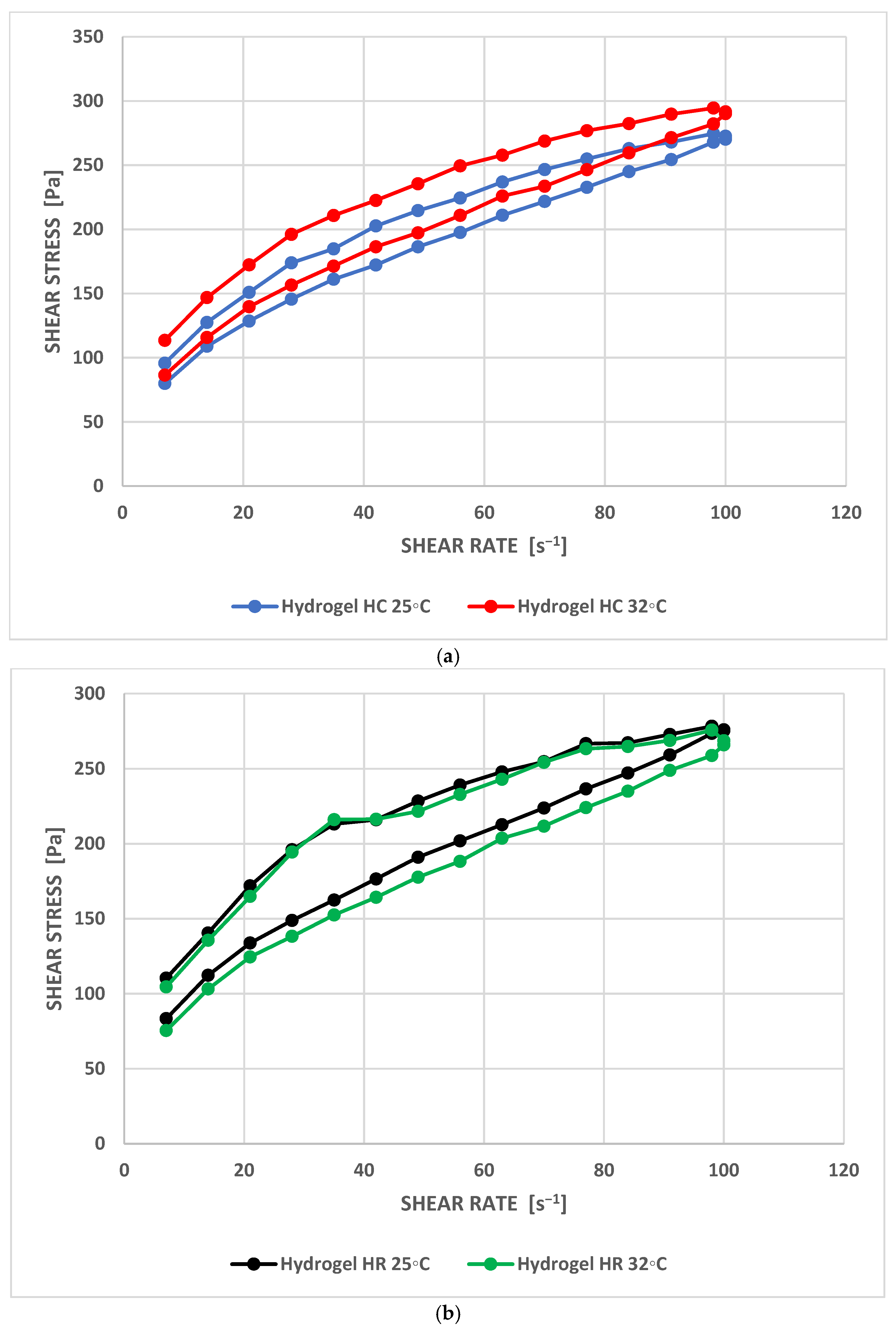
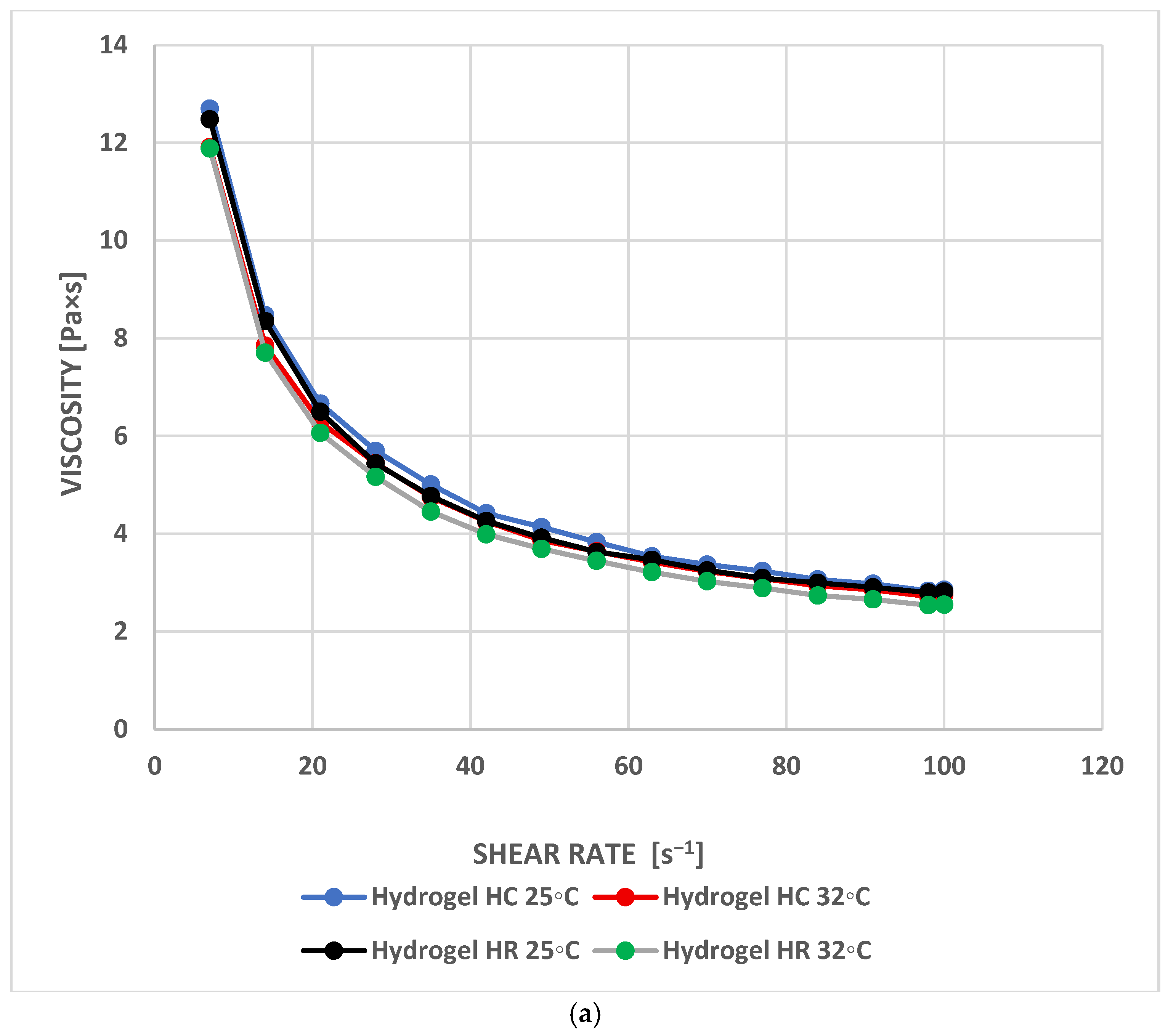
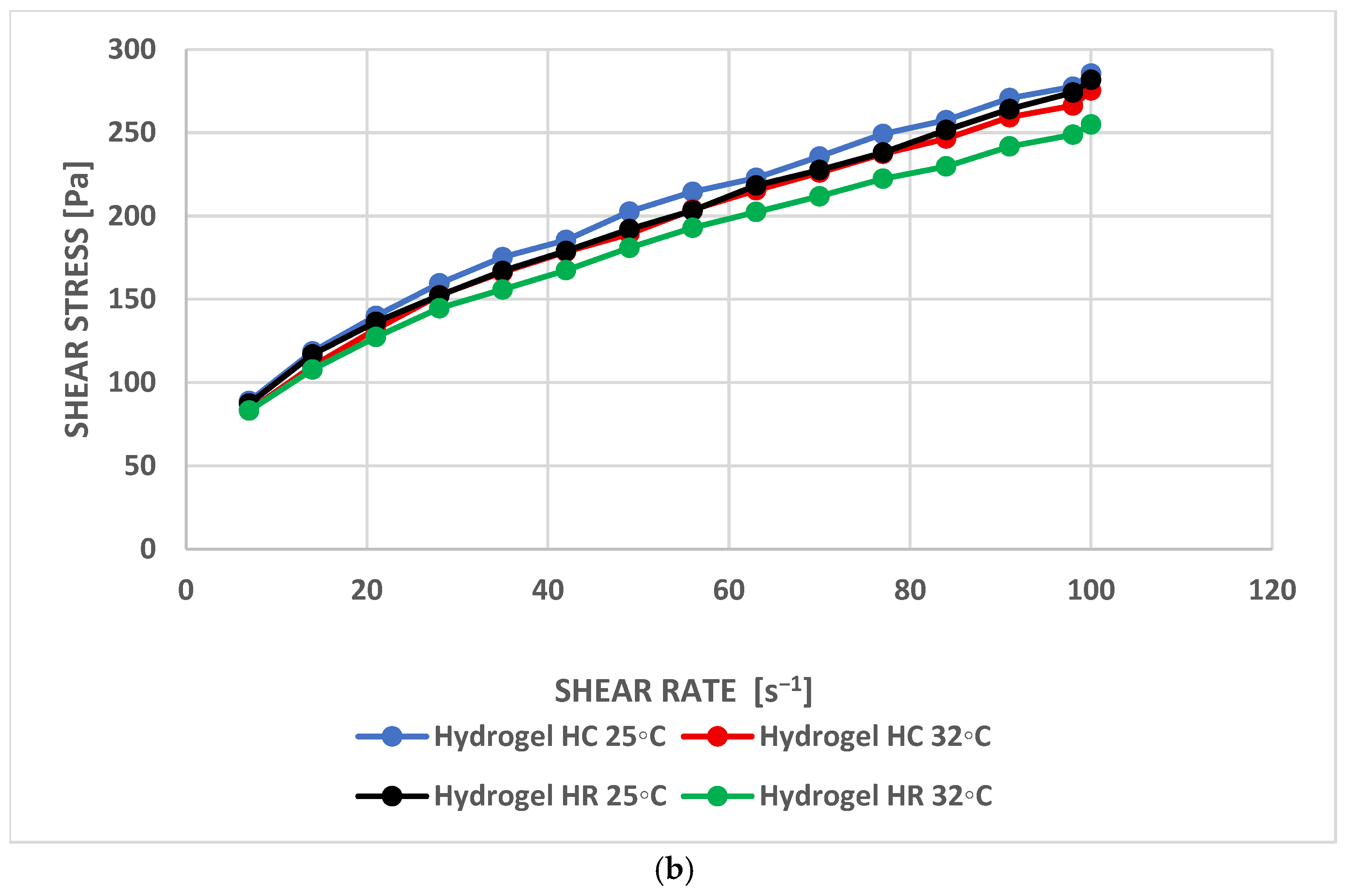
18]. The flow curves were fitted to the Herschel–Bulkley model (
Table 2), and it was found that both hydrogels (regardless of temperature) exhibit pseudoplastic (thinning under shear) properties and have a yield stress (
Table 1,
Figure 2). The shear thinning of the formulation is probably the result of the untangling of the polymer network under the influence of shear stresses. Similar behaviour of Carbopol
®-based hydrogels has been reported in other studies [
12,
19,
20]. The HR exhibited lower viscosity than the HC regardless of the temperature used (
p < 0.05). The lower yield stress of HC (21.6 Pa/25 °C and 21.7 Pa/32 °C vs. HR: 34.4 Pa/25 °C and 28.2 Pa/32 °C) suggests its lighter texture, easier application, and better transdermal diffusion of API. In turn, higher HR yield stress indicates its better adhesive properties to the skin surface.
Textural tests allow the mechanical strength of a formulation to be predicted. Five parameters were selected for analysis: hardness 1, hardness 2, cohesiveness, adhesiveness, elasticity, and relaxation. According to some authors, the optimal preparation should be characterised by low hardness and compressibility, with high cohesiveness and adhesiveness parameters [
21]. Hardness refers to a sample’s mechanical resistance to deformation under the influence of an applied force. The value of this parameter allows us to predict how easily the preparation will be removed from the packaging and applied to the skin, and how long it will remain on the skin after application [
18]. Comparing the values of hardness cycle 1 and hardness cycle 2 (
Table 3), it can be seen that the hydrogel structure was preserved between the two compression cycles. This suggests that the preparation will be easy to dispense from the packaging and spread evenly on the skin. Cohesiveness describes the structural integrity of a sample and estimates its susceptibility to deformation [
22]. Some authors propose that adhesiveness be interpreted as a measure of bioadhesion [
23]. The obtained values of cohesiveness (HR 1.044, HC 1.212,
p = NS) and adhesiveness (HR 0.4 mJ, HC 0.367 mJ,
p = NS) will ensure the restoration of the hydrogel structure after application, prolonged bioadhesion of the preparation to the skin, and, consequently, prolonged retention of the bioactive compound at the site of application [
24]. Elasticity refers to the speed at which a sample returns to its original shape after being compressed. The HC was characterised by greater elasticity (1.143 vs. HR 1.014;
p = NS). The percentage of relaxation is described by the elasticity index [
12]. The higher the value of this parameter, the lower the stiffness of the formulation. The HR showed a higher relaxation value (79.5% vs. HC 75.7,
p < 0.05). The obtained texture parameter values are satisfactory and indicate the expected application effectiveness of the developed hydrogels.
The ability of active ingredients in cosmetic formulations to protect the skin from the entire spectrum of solar radiation can be assessed by determining their effect on the skin’s reflectance using the DHR parameter. In the present study, the DHR method was employed to quantitatively assess changes in the optical properties of the skin following the application of hydrogels containing Crataegus monogyna and Rosmarinus officinalis extracts. The measurements were performed using a SOC 410 Solar DHR reflectometer, which enables precise determination of the skin’s reflectance across a broad spectral range, including UV, visible, and infrared light. This method provides objective data on how cosmetic formulations alter the interaction of solar radiation with the skin, thereby offering valuable insights into their photoprotective potential. The DHR parameter, by capturing both directional and diffuse reflected components, allows for a comprehensive evaluation of skin reflectance that can be linked to the protective effect against harmful radiation. The obtained results demonstrated that the application of both rosemary-enriched and hawthorn-enriched hydrogels led to a reduction in the directional reflectance of the skin across all analyzed spectral ranges. The extract-free hydrogel was used as a vehicle control to distinguish, at least partially, the effect of the hydrogel base from that of the extract-containing formulations. However, the vehicle itself may influence directional–hemispherical reflectance by modifying skin surface hydration, smoothness, film formation, and light scattering. Therefore, the observed reflectance changes should be interpreted as the effect of the complete extract-containing hydrogel formulations rather than the isolated optical effect of the plant extracts alone.
Within the visible light spectrum (400–720 nm) and the UV spectrum (335–380 nm), no statistically significant differences were observed between the reflectance of skin treated with the formulations and that of untreated skin (
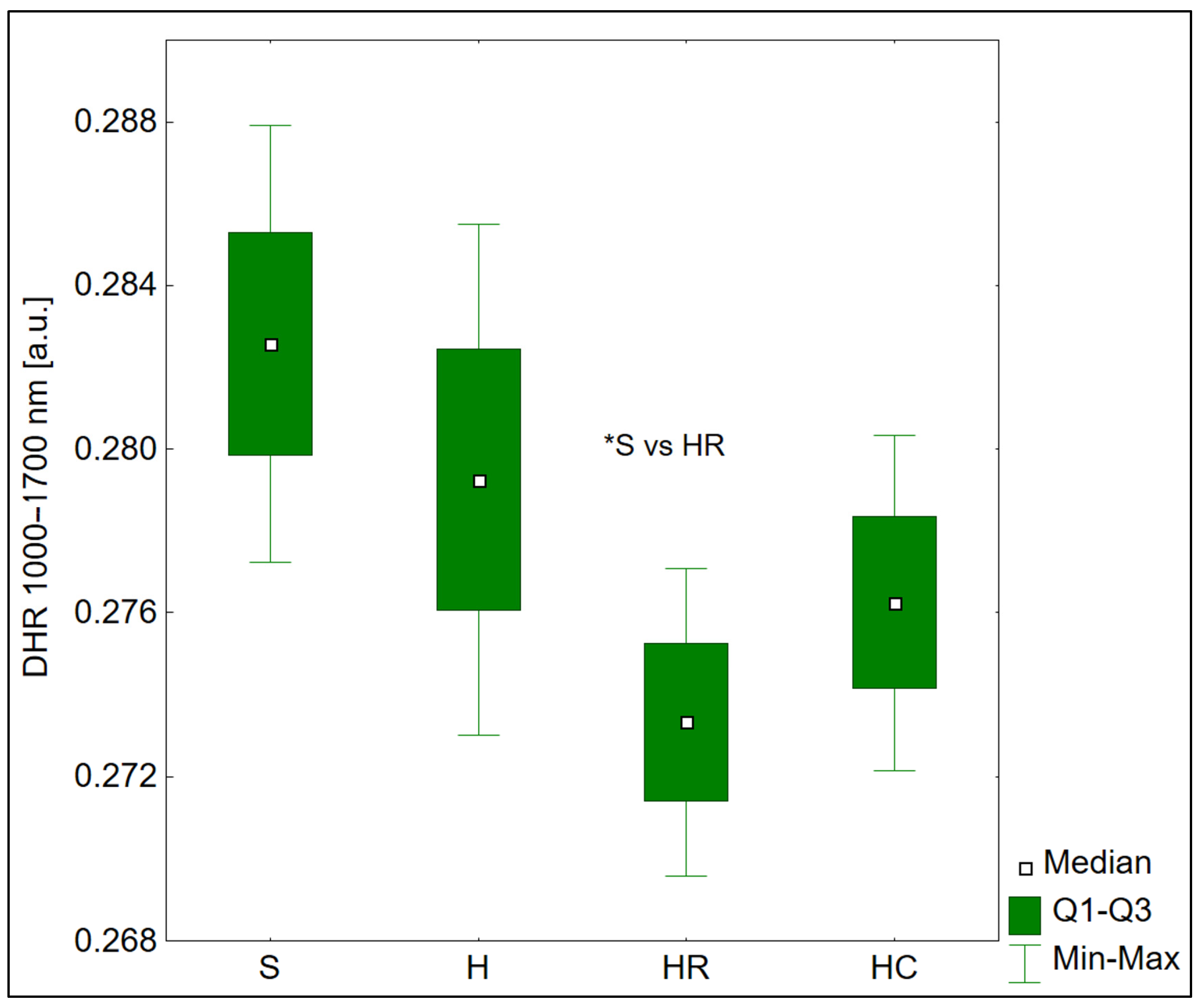
Table 4). This may be related to the limited ability of the tested extracts to modulate the reflection of shorter wavelengths, or to the physical properties of the hydrogel formulations, which may not form a continuous, light-reflecting layer. The most pronounced and statistically significant differences were observed in the infrared (IR) region. In the 1000–1700 nm range, the median reflectance decreased by 3.5% after application of rosemary hydrogel and by 2.3% after application of hawthorn hydrogel, compared to untreated skin (
p = 0.010) (
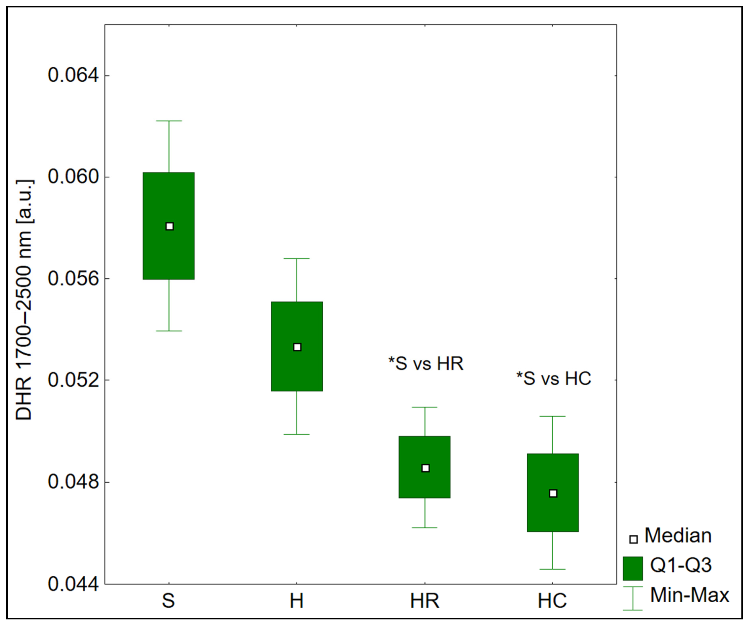
Figure 3). An even more marked effect was observed in the highest IR range (1700–2500 nm), where the median reflectance was reduced by 17.8% for rosemary and 20.3% for hawthorn (
p = 0.001). Post hoc analyses confirmed that these differences were statistically significant compared to the untreated skin (
Figure 4). The observed changes in directional–hemispherical reflectance indicate that the tested hydrogels modified the optical response of the skin/formulation system in the infrared range. However, the interpretation of reduced reflectance requires particular caution. From the standpoint of radiative energy balance, incident radiation may be reflected, absorbed, or transmitted/propagated into deeper tissue layers. Therefore, a decrease in directional–hemispherical reflectance does not by itself demonstrate a protective effect. In contrast, increased reflectance would more directly indicate that a larger fraction of incident radiation is rejected from the skin surface. The reduced IR reflectance observed in the present study indicates that the extract-containing hydrogels modify the optical properties of the skin/formulation system. However, the DHR method alone cannot determine whether the non-reflected radiation was absorbed within the hydrogel layer, absorbed in superficial skin structures, or transmitted deeper into the skin. The DHR method does not provide direct information on the depth of IR penetration within the skin. Photon penetration depth depends on tissue absorption, scattering, anisotropy, refractive index, wavelength, and the layered structure of the skin.
Importantly, no direct quantitative benchmark currently allows a given percentage change in directional–hemispherical reflectance to be converted into a defined level of IR photoprotective efficacy. The 17.8–20.3% reduction in reflectance observed in the 1700–2500 nm range should therefore not be interpreted as a proportional reduction in biological IR damage. From the perspective of radiative energy balance, a decrease in reflectance indicates that a smaller fraction of incident radiation is returned from the skin/formulation system, while the remaining energy may be absorbed within the formulation, absorbed within the skin, transmitted deeper into tissue, or redistributed by scattering. Consequently, reduced reflectance may support a protective mechanism only if the non-reflected radiation is attenuated within the formulation or superficial layers before reaching biologically relevant skin compartments. Since the DHR method does not directly measure absorption, transmission, or depth-resolved energy deposition, the present results should be interpreted as evidence of IR optical modulation rather than direct proof of photoprotective efficacy.
It has been demonstrated in the literature that IR, although characterized by lower energy than UV radiation, penetrates deeper into the skin, reaching the subcutaneous tissue, and induces the expression of MMPs, leading to collagen degradation and photoaging [
2,
25,
26]. The photoprotective activity of plant extracts is mainly attributed to their phenolic compounds, such as flavonoids and phenolic acids [
7]. As far as hawthorn extract is concerned, compounds such as rutin, quercetin, catechin, cyanidin, and chlorogenic acid may play a significant role in this context [
8,
27]. In the case of rosemary extract, noteworthy compounds include rosmarinic, caffeic, and chlorogenic acids, as well as oleanolic acid and luteolin [
9]. Literature data indicate that polyphenolic compounds present in rosemary and hawthorn have protective, antioxidant, and anti-inflammatory properties [
8]. Extracts from
C. monogyna and
R. officinalis may represent valuable raw materials for modern, broad-spectrum dermocosmetic formulations. This is supported by both in vitro and in vivo studies regarding the prevention or treatment of UV-induced photodamage. It has been shown that
Crataegus monogyna extract protects skin cells from radiation, particularly UVA radiation, and causes a dose-dependent reduction in the levels of reactive oxygen species (ROS) induced by UV radiation in cells, as well as inhibiting the production of inflammatory cytokines and MMP-2 and MMP-9 [
28]. The botanical blend, containing
R. officinalis, significantly reduced UV-induced erythema reaction, limited the secretion of MMP-1 and MMP-3 proteins, and lowered levels of ROS and pro-inflammatory interleukins (IL-1, IL-8 and IL-6) [
29]. Our observations are consistent with previous studies highlighting the need for novel compounds with protective activity against IR, as currently available photoprotective products are primarily focused on blocking UV radiation [
2]. It should be emphasized that the tested hydrogels did not produce a meaningful change in skin reflectance in the UV and visible ranges. Therefore, they should not be considered classical UV/visible-light sunscreens. Their potential photoprotective relevance appears to be associated mainly with the infrared spectral range, where more pronounced changes in reflectance were observed. Moreover, the directional–hemispherical reflectance method provides an optical, non-invasive assessment of how a topical formulation modifies the interaction between incident radiation and the skin surface. From a physical point of view, only radiation absorbed by tissue chromophores can induce biological effects, including photochemical reactions, oxidative stress, protein modification, DNA damage, or thermal effects. Radiation reflected away from the skin or formulation surface does not deposit energy in the tissue and therefore has no direct biological activity. For this reason, reflectance-based measurements are useful for evaluating whether a formulation changes the optical balance at the skin surface. However, DHR remains an indirect indicator of photoprotective potential because it does not directly determine the amount of radiation absorbed in the epidermis or dermis, nor does it assess biological endpoints of photodamage. Established methods for evaluating the protective effects of active ingredients include in vitro and in vivo SPF/UVA protection testing, spectrophotometric transmittance and absorbance measurements of formulation films, ex vivo or reconstructed-skin irradiation models, assessment of reactive oxygen species generation, lipid and protein oxidation, DNA damage markers, inflammatory mediators, and matrix metalloproteinase expression. In the present study, the DHR method was selected as a non-invasive in vivo approach allowing rapid quantitative assessment of formulation-induced changes in skin optical properties over a broad spectral range, including UV, visible, and infrared radiation. Therefore, the present results should be interpreted as evidence of formulation-related modification of skin reflectance, particularly in the infrared range, rather than as direct proof of reduced biological photodamage.
5. Limitations of the Study and Future Research Directions
One limitation of the study is that it involves a relatively small number of participants, and the study population is restricted to volunteers with Fitzpatrick skin types I–III. Skin reflectance is strongly dependent on epidermal melanin content. Higher melanin concentration in darker skin types increases light absorption, particularly in the UV and visible ranges, and reduces the intensity of radiation backscattered from the skin surface and deeper layers. Consequently, baseline directional–hemispherical reflectance is expected to be lower in Fitzpatrick skin types IV–VI, and the relative effect of hydrogel application may differ from that observed in lightly pigmented skin. Therefore, the present findings should not be directly generalized to darker skin phototypes, and future studies should include subjects with Fitzpatrick skin types IV–VI to verify whether the observed optical effects are maintained under different absorption–scattering conditions.
Another limitation of the study is the lack of time-resolved measurements after hydrogel application. The reflectance values were recorded only before and immediately after application; therefore, the temporal stability of the observed optical effect was not assessed. Changes associated with hydrogel drying, water evaporation, partial absorption into the skin, redistribution of the formulation layer, or exposure to ambient light may influence reflectance over time. Consequently, the present results should be interpreted as demonstrating an immediate effect on skin directional–hemispherical reflectance rather than sustained photoprotective activity. Future studies should include repeated measurements at defined post-application intervals, such as 15, 30, 60, and 120 min, under controlled environmental conditions.
An additional limitation of the study is the manual application of the hydrogels, which may have introduced variability in film thickness and uniformity and, consequently, may have affected reflectance readings. Future studies should employ a more controlled application method, such as a calibrated applicator, template-guided spreading, or a doctor-blade-type system, to improve the reproducibility of the hydrogel layer on the skin. A further limitation is that the vehicle control did not allow complete separation of extract-specific effects from the physical effects of the hydrogel matrix. Future studies should include additional controls, such as standardized hydrogel films of defined thickness, ex vivo optical assessment of the formulations on inert substrates, and comparison with extract-free and extract-containing vehicles under identical film-forming conditions.
It is important to emphasize that the optical parameters of the skin serve only as an indirect indicator of potential photoprotective activity—they do not directly assess biological effects such as reduced DNA damage, decreased MMP levels, or attenuated inflammation. A valuable addition to future research would be to combine DHR measurements with permeability/absorption measurements and biological markers of skin damage in order to verify the protective significance of the observed optical changes. Future studies should also include a comparison of the efficacy of plant extracts with synthetic filters. It also appears essential to develop formulations that optimise the bioavailability and stability of bioactive compounds in photoprotective products.




 ,
,