Article

Effects of Low-Level Laser Therapy at Different Energy Densities in Patients with Temporomandibular Disorders: A Randomized Clinical Trial


Department of Oral and Maxillofacial Surgery, Faculty of Dentistry, Karadeniz Technical University, Trabzon 61080, Türkiye
*
Author to whom correspondence should be addressed.
Appl. Sci. 2026, 16(11), 5324; https://doi.org/10.3390/app16115324
Submission received: 12 March 2026 / Revised: 4 May 2026 / Accepted: 18 May 2026 / Published: 26 May 2026

Abstract

Temporomandibular disorders (TMDs) are a group of musculoskeletal conditions characterized by pain, restricted mandibular movement, and joint sounds, which may significantly impair quality of life. Among conservative treatment modalities, low-level laser therapy (LLLT) has gained increasing attention due to its noninvasive nature and its documented analgesic and anti-inflammatory effects. Despite growing evidence supporting the clinical effectiveness of LLLT in the management of TMD-related pain and dysfunction, there is still no consensus regarding the optimal energy density parameters to achieve the most favorable therapeutic outcomes. Therefore, the primary objective of this randomized clinical trial was to determine the optimal energy density of low-level laser therapy. This clinical study evaluated the effects of LLLT applied at different energy densities in patients diagnosed with disk displacement with reduction (DDwR) and myofascial pain. A total of 100 patients were divided into two diagnostic groups, each divided into three subgroups: 940 nm, 1.5 W, 90 J; 940 nm, 3 W, 180 J; and a soft diet group. Laser treatment was performed three times per week for three weeks, for a total of nine sessions. Pain intensity, mandibular movements, and joint sounds were assessed at baseline and at one and six months. Comparable and favorable clinical improvements were achieved in both the laser therapy groups and the soft diet group. The 1.5 W-treated group showed the most significant VAS (visual analog scale) parameter reduction at 6 months. Laser treatment outcomes can be summarized as follows: low-level laser therapy was associated with clinical improvement; however, similar positive outcomes were also observed in the soft diet group. These findings indicate that further controlled studies are needed to better clarify the specific role of laser therapy in the management of temporomandibular disorders.

1. Introduction

Temporomandibular joint disorders (TMDs) comprise a complex group of diseases that disrupt the functional and morphological integrity of the temporomandibular joint (TMJ) and related musculoskeletal structures, with multifactorial etiopathogenesis and heterogeneous clinical presentation. Temporomandibular disorders are currently defined according to the Diagnostic Criteria for Temporomandibular Disorders (DC/TMDs) as a group of musculoskeletal conditions involving the temporomandibular joint, masticatory muscles, and associated structures [1]. Clinically, they manifest with symptoms such as pain in the joint and masticatory muscles, decreased mandibular range of motion, and intra-articular sounds [2].
Recent epidemiological data indicate that temporomandibular disorders represent a substantial and growing global health burden. It has been reported that approximately 34% of the global population is affected by a TMD, with prevalence rates varying across continents. Moreover, projections suggest that the global prevalence of TMDs may increase to nearly 44% by 2050, highlighting the increasing clinical and socioeconomic impact of these disorders. This rising prevalence emphasizes the importance of optimizing conservative and non-invasive treatment modalities for TMD management [3].
Management of temporomandibular disorders involves a multimodal approach, including patient education, behavioral modification, pharmacological therapy, physical therapy, occlusal splints, and other conservative interventions. Current evidence emphasizes that conservative and noninvasive management strategies should be considered first-line treatment options, with invasive procedures reserved for refractory cases [4]. LLLT has been reported to induce analgesia through nociceptive modulation, exert anti-inflammatory effects through regulation of inflammatory mediators, and trigger biostimulatory responses by promoting cellular regeneration. This process involves multifaceted biochemical reactions, such as increased ATP synthesis by activating the mitochondrial electron transport chain at the level of complex IV [5,6,7]. Although low-level laser therapy has been associated with anti-inflammatory and regenerative responses, current evidence indicates that these effects are not universal and are highly dependent on treatment parameters such as wavelength, energy density, and irradiation protocol, as well as patient-related factors and the specific subtype of temporomandibular disorder. Consequently, clinical outcomes of LLLT in TMD management remain heterogeneous across reported studies [8].
Various studies in the literature reveal that LLLT increases the mandibular range of motion by optimizing muscle–joint functions [9,10,11]. However, since its direct effect on the biomechanical stability of intra-articular structures is limited, its clinical efficacy on symptoms such as joint sounds and tinnitus is still controversial, and further controlled studies are needed in this field.
Despite the widespread clinical use of low-level laser therapy in the management of temporomandibular disorders, there is still no consensus regarding the optimal energy density required to achieve predictable and sustained clinical outcomes. Previous studies have reported favorable results using a wide range of laser parameters; however, direct comparisons between different energy densities remain limited, particularly in patients with disk displacement with reduction and myofascial pain [9,10,11]. Therefore, the primary aim of this randomized clinical trial was to evaluate and compare the clinical effects of low-level laser therapy applied at different energy densities on pain intensity and mandibular function in patients with temporomandibular disorders. The primary research question was whether variations in laser energy density result in differential therapeutic outcomes when compared with each other and with a conservative soft diet approach.

2. Materials and Methods

This study followed the principles of the Declaration of Helsinki for medical protocols and ethics. The Karadeniz Technical University Institutional Review Board approved the study plan under protocol 2024/1. The trial was registered at ClinicalTrials.gov with the identifier NCT07588841, and the study design and reporting were conducted in accordance with CONSORT guidelines. The patients were informed about the procedure, possible complications, and the materials used, and they signed a detailed written consent form.

2.1. Patient Selection and Grouping

The study population consisted of patients with TMJ disk displacement with reduction and myalgia who were treated at the Karadeniz Technical University Faculty of Dentistry, Department of Oral and Maxillofacial Surgery (Trabzon, Turkey) between 2022 and 2024. Temporomandibular disorders were classified according to the International Classification of Diseases (ICD). In the ICD-10 classification, temporomandibular joint disorders are coded under K07.6 (temporomandibular joint disorders). This classification was used as a reference framework for the clinical diagnosis of patients included in the present study.
Patients with bilateral TMJ pain, TMJ sounds, jaw dysfunction, and pain in the masticatory muscles for at least six months were included in this study. The inclusion criteria were a diagnosis of DDwR and myalgia based on symptoms and clinical and radiographic findings. DDwR was diagnosed based on the presence of joint sounds (clicking) during mandibular opening and closing with reduction confirmed clinically and supported by radiographic findings. Magnetic resonance imaging was used to confirm DDwR in all patients. Myalgia was diagnosed based on pain in the masticatory muscles that was reproducible upon palpation and/or during mandibular function (e.g., chewing or jaw movement). All clinical examinations were performed by an examiner, following standardized DC/TMD procedures [1]. Patients were excluded if they had a history of previous TMJ treatment (e.g., conservative or surgical treatment), congenital or inflammatory joint disease, severe systemic diseases, were edentulous, or were children and adolescents in the active growth period. Patients diagnosed with DDwR or myalgia who refused any treatment (except the soft diet) for any reason were assigned to the soft diet group.
Pain was selected as the primary outcome variable to assess the treatment efficacy. The secondary outcome variables were maximum painless mouth opening (MPMO), maximum unassisted mouth opening (MUMO), maximum assisted mouth opening (MAMO), right and left lateral movements, protrusion, and TMJ sound. Pain was measured using a 10-point visual analog scale (VAS), with 0 indicating no pain and 10 indicating the worst pain. Mouth opening was measured as the distance between the incisal edges of the maxillary and mandibular central incisors using a millimeter scale. Mandibular right and left lateral movements were measured with reference to the dental midline, defined as the line passing through the maxillary and mandibular central incisors. Measurements were recorded in millimeters.
Clinical parameters were recorded for each patient at diagnosis and at the 1- and 6-month follow-up. Outcome assessments were performed by an independent reviewer who was blinded to the group allocation. The independent reviewer was not involved in the application of the intervention and had no knowledge of the laser energy density parameters assigned to the participants. This blinding procedure was implemented to minimize assessment bias.
After obtaining informed consent, patients were divided into two main groups:
  • Group I: Patients diagnosed with disk displacement with reduction.
  • Group II: Patients diagnosed with myofascial pain.
Each main group was divided into three subgroups according to the LLLT parameters applied:
  • Groups Ia and IIa: patients who underwent 940 nm, 1.5 W, 90 J, pulsed LLLT.
  • Groups Ib and IIb: 940 nm, 3 W, 180 J, pulsed LLLT.
  • Groups Ic and IIc: Patients who refused treatment for any reason were assigned to the control group, and only a soft diet was recommended.
There were a total of 50 patients in each main group, with 20 patients in each treatment group and 10 patients in the soft-diet-only group. The sample size was determined according to the number of eligible patients who met the inclusion criteria during the study period rather than by a priori power analysis. This has been acknowledged as a limitation of the study. Potential confounding factors, including medication use, pain characteristics, age, and treatment adherence, were considered during patient selection and follow-up. Patients using analgesic or anti-inflammatory medications during the study period were excluded or instructed to maintain stable medication regimens. Baseline pain characteristics and demographic variables were recorded to ensure comparability between groups. Treatment adherence was monitored throughout the study period. Therefore, no dropout rate was applicable to this study. Patients allocated to the laser treatment groups were assigned using a computer-generated randomization list. The soft diet-only group consisted of patients who declined active laser treatment but agreed to participate in follow-up examinations.

2.2. Laser Treatment Protocol

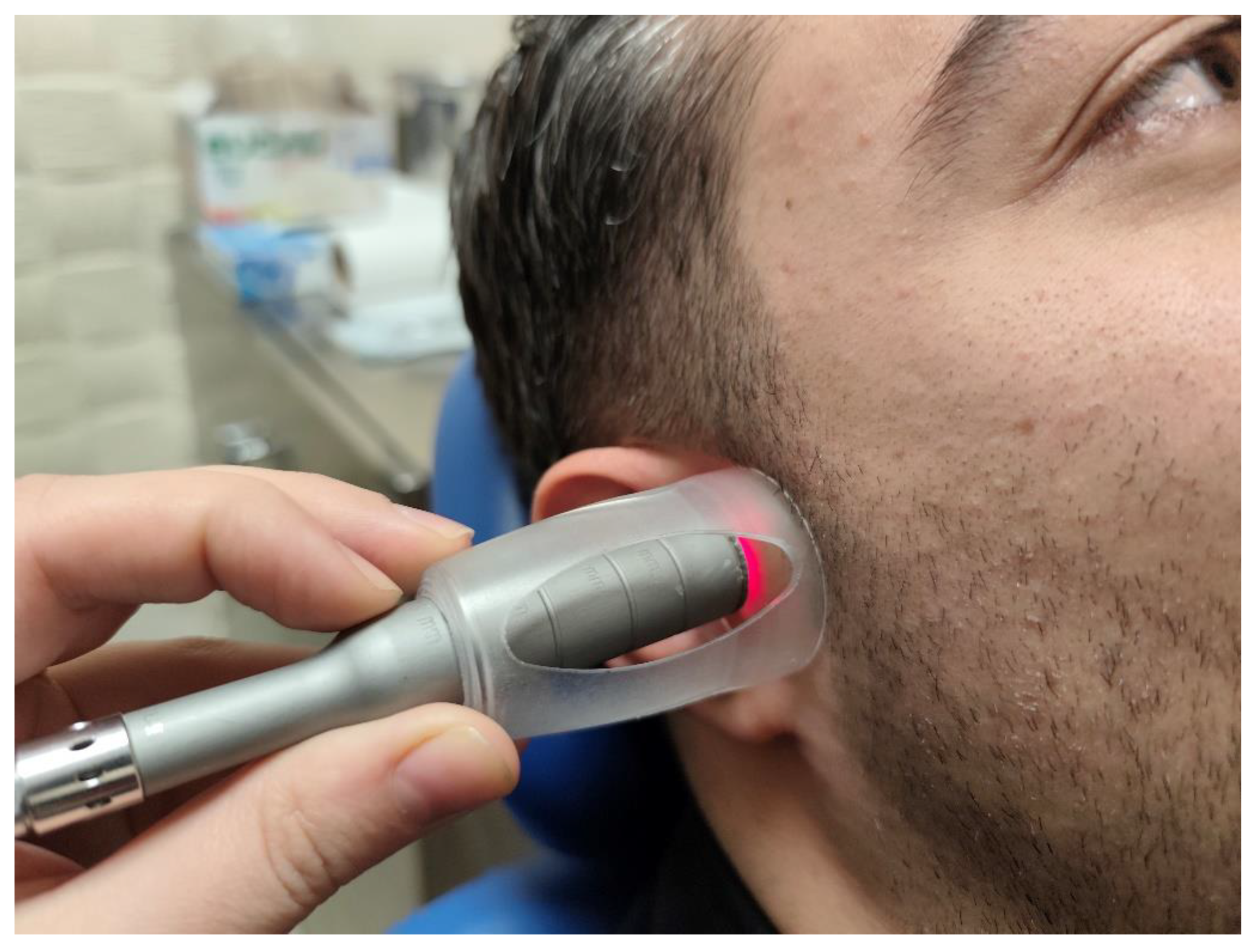
LLLT was performed using a Ga-Al-As diode laser (EzLase 940, Biolase Technology, Lake Forest, CA, USA). The treatment was performed bilaterally on the patients’ TMJ area (an area of approximately 3 cm2, centered 10 mm anterior to the tragus, for 2 min per joint) and the most painful points of the masseter and temporal muscles three times a week for a total of nine sessions (Figure 1). Several studies have demonstrated that low-level laser therapy applied over multiple sessions (generally ranging between 6 and 12 sessions) is effective in reducing pain and improving function in TMD patients [12,13]. Therefore, 9 sessions were selected as appropriate.
Cost-effectiveness analysis was not applicable to this study, as no additional costs were incurred during the procedures.

2.3. Statistical Analysis

All statistical analyses were performed using SPSS for Windows 17.0 (SPSS Inc., Chicago, IL, USA) software. The conformity of continuous variables to normal distribution was evaluated using the Shapiro–Wilk test. The Kruskal–Wallis and Mann–Whitney U tests and Bonferroni corrections were applied for intergroup comparisons. Within-group changes were analyzed using the Wilcoxon test. A chi-square test and Spearman correlation analysis were performed when necessary, and the statistical significance level was accepted as p < 0.05. The Friedman test was used to assess overall differences across repeated measurements within groups. When significant differences were identified, post hoc pairwise comparisons were conducted using the Wilcoxon signed-rank test, with Bonferroni correction applied to control for multiple comparisons.

3. Results

The age, gender, and demographic data of the patients who participated in this study and the comparison between the subgroups are shown in Table 1 and Table 2. The mean age, height, and weight of the participants were not significantly different (p > 0.05) between the subgroups (except for the comparison of the mean age between Group II and Group IIc). The patients did not experience any complications or side effects related to the treatment modalities.
Table 3 and Table 4 show the time-dependent change in the primary variable, pain intensity during jaw function, and secondary variables according to the treatments applied and the comparison between the subgroups. In all subgroups, the decrease in pain intensity at 1 and 6 months was statistically significant (p < 0.05).
The differences in MPMO, protrusion, and right and left lateral movement of the lower jaw were statistically significant in the first and sixth months in all subgroups.
A statistically significant difference was found in the first and sixth months of all subgroups except for after treatment in Group IIb for MUMO.
A statistically significant difference was found in MAMO at the first and sixth months after treatment in all subgroups, except Group IIb and Group IIc.
In the comparison of Groups Ib and Ic between the subgroups in the DDwR group, a significant difference was found in favor of Group Ic in the 1st month left lateral and 6th month MPMO values.
In the comparison between subgroups in the myalgia group, a significant difference was observed in the MAMO variable between groups IIb and IIc in favor of Group IIb in the first month after treatment. Furthermore, a significant difference was observed in the right lateral movement between Group IIa and Group IIc in favor of Group IIa at the first and sixth months after treatment.
Post-treatment MRI scans were obtained, and no changes were observed in the disk displacement in the reduction group.

4. Discussion

It has been suggested that the pathophysiology of TMJ irregularities is not caused by a single process; therefore, it is appropriate to start treatment with conservative methods that offer the least invasive and predictable results. Conservative treatment options mentioned in the literature include a soft diet, physical therapy, nonsteroidal anti-inflammatory drugs, LLLT, and occlusal splints [14,15].
In the studies, different results have shown the effectiveness of low-energy laser application in the treatment of TMJ disorders [11,16,17,18]. This diversity is due to variables such as the type of laser device used, wavelength, application time, frequency, repetition frequency, and laser technique [19]. In most studies, treatment was symptom-oriented and aimed to alleviate patients’ complaints, such as pain, restriction in mouth opening capacity, and joint noise [20,21,22].
It is known that factors such as the point at which the laser probe is positioned, the direction of application, the distance to the anatomical target, and the diameter of the probe are determinants of laser efficacy; however, these factors are not specified in detail in many studies [19]. In this study, these factors are clearly defined: the laser was applied bilaterally to the TMJ region (approximately 3 cm2, centered 10 mm anterior to the tragus and direct skin contact).
Similar to the results of various epidemiological and clinical studies, most patients in our study population were female [23,24]. TMJ disorders are most commonly observed between the ages of 20 and 40 years [25]. In our study, the majority of patients were within this age range.
Patients usually require treatment to reduce their pain during jaw function. One of the most important parameters when evaluating a treatment approach is the reduction or elimination of pain [26,27]. In our study, it was shown that the laser improved the maximum pain values during chewing at the 6-month follow-up in the active treatment groups. The maximum pain value in chewing decreased significantly from 6.65 to 1.60 for group Ia, from 6.70 to 2.50 for group Ib, from 6.40 to 1.20 for group IIa, and from 6.00 to 1.75 for group IIb. These results are consistent with data in the literature and support the efficacy of laser therapy for DDwR and myalgia.
Another important problem is limited mouth opening in TMJ disorders. This limitation negatively affects masticatory function and the quality of life [28]. As lasers reduce pain, edema, and inflammation, they also improve mouth opening [22]. Similar to previous studies, MPMO, MUMO, MAMO, right and left lateral movements, and protrusion values increased in all treatment groups at the 6-month follow-up compared to the baseline values in our study.
The TMJ sounds did not completely disappear after treatment in any of the study groups. This was expected because joint sounds are usually caused by mechanical disruption of the joint and are not expected to be affected by conservative treatment [9]. In addition, the clinical assessment of joint sounds without instrumental verification may limit measurement precision.
Characteristics, such as the wavelength and frequency of the laser beam, varied between studies, leading to variations in the dosage of energy applied to the target area. Taken individually, no specific wavelength or frequency has been directly associated with the favorable effect of laser treatment [29].
In studies performed at different wavelengths, for example, Venancio et al. [30] (780 nm, 30 mW, 6.3 J/cm2), Carrasco et al. [31] (780 nm, 70 mW), Varma et al. [17] (940 nm, 6 W) and Ayyıldız et al. [10] (685 nm, 25 mW, 6.2 J/cm2), significant reductions in pain level were found and maintained in long-term follow-up. In our study, similar results were obtained in pain level reduction using a 940 nm wavelength.
Although different laser parameters have been used in the literature, similar results have been obtained in the treatment groups, which are mostly favorable. Studies by Conti [32] (830 nm, 4 J) and Ahrari [29] (810 nm, 50 mW, 6 J) reported favorable improvement patterns in myogenic and arthrogenic pain. The treatment protocol applied by Sancaklı et al. [33] (820 nm, 500 mW, 4 J) improved both pain and mandibular movement. Pinheiro et al. [11] also reported complete recovery or significant symptom reduction in the vast majority of cases in their study with different wavelengths (632.8 nm, 670 nm, and 830 nm; 12 applications and an average dose of 1.8 J/cm2). The results showed that 154 of 241 patients became completely symptom-free at the end of treatment, 50 patients showed significant improvement, and 37 patients remained symptomatic. Shobha et al. [6] applied a GaAlAs diode laser at an 810 nm wavelength, 0.1 W power, and 6 J/cm2 energy density to the TMJ and masticatory muscles and reported a decrease in pain levels and improvement in clicking sounds. Similarly, Cavalcanti et al. [34] (780 nm, 30 mW, and 35 J/cm2) and Fornaini et al. [35] (904 nm, 15 mW, and 6 J/cm2) showed that laser treatment significantly reduced pain. Gökçen-Röhlig et al. [21] obtained significant improvements in both pain level and functional examination findings in the treatment applied to muscles with 820 nm, 300 mW, and 8 J/cm2 parameters. These results are comparable to those from our study.
Although not all variables were detailed in most studies, it was noted that the dose intensities ranged from 1 to 105 J/cm2. However, these differences in dose intensity do not seem to significantly affect the treatment efficacy. For instance, Carrasco et al. [31] (25 J/cm2, 60 J/cm2, and 105 J/cm2) and Rodrigues da Silva et al. [36] (0.0, 52.5, and 105 J/cm2) used different intensity doses of a Ga-Al-As diode laser (780 nm wavelength, 70 mW power), but the same results were obtained in all three cases. In both studies, a decrease in the pain level and improvement in the mandibular range of motion were observed. In our study, similar results were obtained for the different energy densities. The reason for no significant variations in maximum mouth opening and assisted maximum mouth opening at 1 and 6 months in Group IIb could be due to the high pre-treatment values in these groups.
In our study, clinical findings indicate that, although not all intergroup comparisons reached significance, there were meaningful differences that support dose-dependent effects in specific functional parameters. Therefore, the conclusion regarding an “optimal dose” is based on these significant outcomes rather than a uniform superiority across all variables. Although our study did not include a true placebo group, similar improvements observed in the soft diet group suggest that part of the observed effect of laser therapy may be attributable to a placebo response. This highlights the potential influence of placebo mechanisms in laser treatment for TMD, as supported by previous studies.
The number of applications varied considerably, ranging from three interventions (once a week for three weeks) to 20 (two to three times a week for eight weeks). The session duration varied from 10 s to 10 min per treatment. However, increasing the number of laser applications did not increase the effectiveness of the laser treatment. In fact, Emshoff et al. [16], who used the highest number of applications, reported similar results between laser treatment and placebo. Furthermore, prolonging the duration of laser application did not increase the efficacy when the laser was applied to the TMJ or both the TMJ and masticatory muscles. Because the same application time was used for all treatment groups in our study, no comparison was made on this parameter.
Some studies comparing lasers with other treatment modalities did not show a significant difference. For example, Kato et al. [37] and Kogawa et al. [38] showed that low-level laser therapy may be effective in the treatment of TMD in the short term, especially in terms of increasing mouth opening and reducing pain. In Kato’s study, the laser applied with a wavelength of 830–904 nm, dose of 4 J/cm2, and power of 100 mW provided a significant increase in mouth opening (approximately 5 mm) in both groups compared with Transcutaneous Electrical Nerve Stimulation, but there was no statistically significant difference between the groups [37]. Kogawa also compared laser and microelectric neurostimulation (MENS) treatment with similar parameters (830–904 nm, 4 J/cm2, 100 mW) and reported an increase in mouth opening (from 46.3 mm to 49.4 mm) in the laser group, but a decrease in the MENS group; however, no significant difference was found between the two groups [38]. In our study, both low-level laser therapy and soft diet management were associated with improvements in clinical outcomes in patients with temporomandibular disorders. Given the comparable outcomes between groups, the findings do not allow definitive conclusions regarding the specific effectiveness or superiority of laser therapy. The observed improvements may reflect non-specific therapeutic or contextual effects. These results should therefore be interpreted as exploratory and hypothesis-generating.
Khosla et al. [39] emphasized that compared with TENS, 980 nm, 1 W laser therapy TENS yielded better results. Furthermore, Hotta et al. [40] reported that although applying laser therapy to acupuncture points resulted in electromyographic improvement in masseter muscle activity, no statistically significant increase in mouth opening was observed. These findings demonstrate that the application parameters and protocol consistency are decisive in the effectiveness of laser therapy.
The findings of this study provide clinically relevant information regarding the use of low-level laser therapy at different energy densities in the management of temporomandibular disorders. For clinicians, the results may assist in selecting appropriate noninvasive treatment protocols. For researchers, the study identifies areas requiring further investigation to optimize treatment parameters.
Although our study did not include a true placebo group, similar improvements observed in the soft diet group suggest that part of the observed effect of laser therapy may be attributable to a placebo response. This highlights the potential influence of placebo mechanisms in laser treatment for TMD, as supported by previous studies. It should be noted that TMD symptoms are known to fluctuate and may sometimes remit spontaneously; therefore, over the six-month follow-up, contributions from regression to the mean, spontaneous remission, and behavioral adaptation cannot be excluded as potential factors influencing the observed improvements.
It should be acknowledged, however, that improvement following non-invasive management of temporomandibular disorders is often non-specific. Recent evidence suggests that symptom reduction may occur spontaneously over time, reflecting the fluctuating natural course of TMD, regression to the mean, and contextual or placebo-related effects rather than a direct treatment-specific mechanism. Consequently, the effectiveness of conservative interventions should be interpreted within this broader clinical context.

5. Conclusions

In our study, improvements were observed in both the soft diet and laser therapy groups, with no clear superiority of laser therapy over the control condition. These findings suggest that the observed effects may be related not only to the intervention itself but also to non-specific factors such as the placebo response, patient expectations, and psychological influences associated with receiving treatment.
Given that both groups demonstrated significant clinical improvement, the results should be interpreted with caution. Therefore, it cannot be concluded that low-level laser therapy is definitively superior in the management of TMD based on the present study. Further well-designed studies with larger sample sizes, standardized protocols, and appropriate placebo-controlled designs are required to clarify the specific therapeutic effects of low-level laser therapy in TMD patients.

Author Contributions

Conceptualization, E.M.; methodology, E.M., O.Y.; software, E.M., O.Y.; validation, E.M., O.Y., F.G. and E.U.; formal analysis, E.M., O.Y.; investigation, E.M.; resources, E.M., F.G.; data curation, E.M., F.G.; writing—original draft preparation, E.M., O.Y.; writing—review and editing, E.M., O.Y., F.G. and E.U.; visualization, E.M., F.G., E.U.; supervision, O.Y.; project administration, E.M. All authors have read and agreed to the published version of the manuscript.

Funding

This research received no external funding.

Institutional Review Board Statement

The study was conducted in accordance with the Declaration of Helsinki and approved by Karadeniz Technical University, Faculty of Dentistry, Scientific Research Ethics Committee (protocol 2024/1, number 64929847/7 approved on 6 March 2024). The study was registered in the ClinicalTrials.gov database (Identifier: NCT07588841). The registration was completed after patient enrollment had begun due to an administrative delay during the submission and approval process. Our study has been prepared in accordance with CONSORT guidelines.

Informed Consent Statement

Informed consent was obtained from all individual participants included in the study.

Data Availability Statement

All data generated or analyzed during this study are included in this published article.

Conflicts of Interest

The authors declare no conflicts of interest.

References

  1. Schiffman, E.; Ohrbach, R.; Truelove, E.; Look, J.; Anderson, G.; Goulet, J.P.; List, T.; Svensson, P.; Gonzalez, Y.; Lobbezoo, F.; et al. Diagnostic Criteria for Temporomandibular Disorders (DC/TMD) for Clinical and Research Applications: Recommendations of the International RDC/TMD Consortium Network and Orofacial Pain Special Interest Group. J. Oral Facial Pain Headache 2014, 28, 6–27. [Google Scholar] [CrossRef]
  2. Franklin, C.D. Pathology of the temporomandibular joint. Curr. Diagn. Pathol. 2006, 12, 31–39. [Google Scholar] [CrossRef]
  3. Zieliński, G. Quo Vadis Temporomandibular Disorders? By 2050, the Global Prevalence of TMD May Approach 44%. J. Clin. Med. 2025, 14, 4414. [Google Scholar] [CrossRef] [PubMed]
  4. Gray, R.J.; Davies, S.J.; Quayle, A.A. A clinical approach to temporomandibular disorders. 6 splint therapy. Br. Dent. J. 1994, 177, 135–142. [Google Scholar] [CrossRef]
  5. Gam, A.N.; Thorsen, H.; Lønnberg, F. The effect of low-level laser therapy on musculoskeletal pain: A meta-analysis. Pain 1993, 52, 63–66. [Google Scholar] [CrossRef] [PubMed]
  6. Shobha, R.; Narayanan, V.; Jagadish Pai, B.; Jaishankar, H.; Jijin, M. Low-level laser therapy: A novel therapeutic approach to temporomandibular disorder—A randomized, double-blinded, placebo-controlled trial. Indian J. Dent. Res. 2017, 28, 380. [Google Scholar] [CrossRef]
  7. Basford, J.R. Low intensity laser therapy: Still not an established clinical tool. Lasers Surg. Med. 1995, 16, 331–342. [Google Scholar] [CrossRef]
  8. Hamblin, M.R. Mechanisms and applications of the anti-inflammatory effects of photobiomodulation. AIMS Biophys. 2017, 4, 337–361. [Google Scholar] [CrossRef]
  9. Petrucci, A.; Sgolastra, F.; Gatto, R.; Mattei, A.; Monaco, A. Effectiveness of low-level laser therapy in temporomandibular disorders: A systematic review and meta-analysis. J. Orofac. Pain. 2011, 25, 298–307. [Google Scholar] [PubMed]
  10. Ayyildiz, S.; Emir, F.; Sahin, C. Evaluation of Low-Level Laser Therapy in TMD Patients. Case Rep. Dent. 2015, 2015, 424213. [Google Scholar] [CrossRef]
  11. Pinheiro, A.L.; Cavalcanti, E.T.; Pinheiro, T.I.; Alves, M.J.; Miranda, E.R.; de Quevedo, A.S.; Manzi, C.T.; Vieira, A.L.; Rolim, A.B. Low-Level Laser Therapy Is an Important Tool to Treat Disorders of the Maxillofacial Region. J. Clin. Laser Med. Surg. 1998, 16, 223–226. [Google Scholar] [CrossRef]
  12. Ahmad, S.A.; Hasan, S.; Saeed, S.; Khan, A.; Khan, M. Low-level laser therapy in temporomandibular joint disorders: A systematic review. J. Med. Life 2021, 14, 148–164. [Google Scholar] [CrossRef]
  13. Maia, M.L.D.M.; Bonjardim, L.R.; Quintans, J.D.S.S.; Ribeiro, M.A.G.; Maia, L.G.M.; Conti, P.C.R. Effect of low-level laser therapy on pain levels in patients with temporomandibular disorders: A systematic review. J. Appl. Oral Sci. 2012, 20, 594–602. [Google Scholar] [CrossRef]
  14. Öz, S.; Gökçen-Röhlig, B.; Saruhanoglu, A.; Tuncer, E.B. Management of Myofascial Pain. J. Craniofacial Surg. 2010, 21, 1722–1728. [Google Scholar] [CrossRef] [PubMed]
  15. De Carli, B.M.G.; Magro, A.K.D.; Souza-Silva, B.N.; de Souza Matos, F.; De Carli, J.P.; Paranhos, L.R.; Magro, E.D. The effect of laser and botulinum toxin in the treatment of myofascial pain and mouth opening: A randomized clinical trial. J. Photochem. Photobiol. B 2016, 159, 120–123. [Google Scholar] [CrossRef] [PubMed]
  16. Emshoff, R.; Bösch, R.; Pümpel, E.; Schöning, H.; Strobl, H. Low-level laser therapy for treatment of temporomandibular joint pain: A double-blind and placebo-controlled trial. Oral Surg. Oral Med. Oral Pathol. Oral Radiol. Endodontol. 2008, 105, 452–456. [Google Scholar] [CrossRef] [PubMed]
  17. Varma, S.R.; Al Shayeb, M.; El Kaseh, A.; Kuduruthullah, S.; Ashekhi, A.; Al Khader, E. Effectiveness of Low-level Laser Therapy in the Management of the Temporomandibular Joint Disorders: A Placebo-controlled Trial. World J. Dent. 2018, 9, 316–320. [Google Scholar] [CrossRef]
  18. Heussler, J.K.; Hinchey, G.; Margiotta, E.; Quinn, R.; Butler, P.; Martin, J.; Sturgess, A.D. A double blind randomised trial of low power laser treatment in rheumatoid arthritis. Ann. Rheum. Dis. 1993, 52, 703–706. [Google Scholar] [CrossRef]
  19. Çetiner, S.; Kahraman, S.A.; Yücetas, Ş. Evaluation of Low-Level Laser Therapy in the Treatment of Temporomandibular Disorders. Photomed. Laser Surg. 2006, 24, 637–641. [Google Scholar] [CrossRef]
  20. de Felício, C.M.; Melchior, M.d.O.; da Silva, M.A.M.R. Effects of Orofacial Myofunctional Therapy on Temporomandibular Disorders. CRANIO® 2010, 28, 249–259. [Google Scholar] [CrossRef]
  21. Gökçen Röhlig, B.; Kipirdi, S.; Meriç, U.; Çapan, N.; Keskin, H. Masticatory muscle pain and low-level laser therapy: A double-blind and placebo controlled study. Turk. J. Phys. Med. Rehab. 2011, 57, 31. [Google Scholar]
  22. Kulekcioglu, S.; Sivrioglu, K.; Ozcan, O.; Parlak, M. Effectiveness of low-level laser therapy in temporomandibular disorder. Scand. J. Rheumatol. 2003, 32, 114–118. [Google Scholar] [CrossRef]
  23. Bertolucci, L.E.; Grey, T. Clinical Analysis of Mid-Laser Versus Placebo Treatment of Arthralgic TMJ Degenerative Joints. CRANIO® 1995, 13, 26–29. [Google Scholar] [CrossRef]
  24. McNeill, C. Management of temporomandibular disorders: Concepts and controversies. J. Prosthet. Dent. 1997, 77, 510–522. [Google Scholar] [CrossRef] [PubMed]
  25. Manfredini, D.; Chiappe, G.; Bosco, M. Research Diagnostic Criteria for Temporomandibular Disorders (RDC/TMD) axis I diagnoses in an Italian patient population. J. Oral Rehabil. 2006, 33, 551–558. [Google Scholar] [CrossRef] [PubMed]
  26. Bagis, B.; Ayaz, E.A.; Turgut, S.; Durkan, R.; Özcan, M. Gender Difference in Prevalence of Signs and Symptoms of Temporomandibular Joint Disorders: A Retrospective Study on 243 Consecutive Patients. Int. J. Med. Sci. 2012, 9, 539–544. [Google Scholar] [CrossRef] [PubMed]
  27. Tuncel, U. Repeated sodium hyaluronate injections following multiple arthrocenteses in the treatment of early stage reducing disc displacement of the temporomandibular joint: A preliminary report. J. Cranio-Maxillofac. Surg. 2012, 40, 685–689. [Google Scholar] [CrossRef]
  28. Chisnoiu, A.M.; Picos, A.M.; Popa, S.; Chisnoiu, P.D.; Lascu, L.; Picos, A.; Chisnoiu, R. Factors involved in the etiology of temporomandibular disorders—A literature review. Med. Pharm. Rep. 2015, 88, 473–478. [Google Scholar] [CrossRef]
  29. Ahrari, F.; Madani, A.S.; Ghafouri, Z.S.; Tunér, J. The efficacy of low-level laser therapy for the treatment of myogenous temporomandibular joint disorder. Lasers Med. Sci. 2014, 29, 551–557. [Google Scholar] [CrossRef]
  30. De Abreu Venancio, R.; Camparis, C.M.; De Fátima Zanirato Lizarelli, R. Low intensity laser therapy in the treatment of temporomandibular disorders: A double-blind study. J. Oral Rehabil. 2005, 32, 800–807. [Google Scholar] [CrossRef]
  31. Graciele Carrasco, T.; Oliveira Mazzetto, M.; Galli Mazzetto, R.; Mestriner, W. Low Intensity Laser Therapy in Temporomandibular Disorder: A Phase II Double-Blind Study. CRANIO® 2008, 26, 274–281. [Google Scholar] [CrossRef]
  32. Conti, P.C.R. Low Level Laser Therapy in the Treatment of Temporomandibular Disorders (TMD): A Double-blind Pilot Study. CRANIO® 1997, 15, 144–149. [Google Scholar] [CrossRef]
  33. Sancakli, E.; Gökçen-Röhlıg, B.; Balık, A.; Öngül, D.; Kıpırdı, S.; Keskın, H. Early results of low-level laser application for masticatory muscle pain: A double-blind randomized clinical study. BMC Oral Health 2015, 15, 131. [Google Scholar] [CrossRef]
  34. Cavalcanti, M.F.X.B.; Silva, U.H.; Leal-Junior, E.C.P.; Lopes-Martins, R.A.B.; Marcos, R.L.; Pallotta, R.C.; Diomede, F.; Trubiani, O.; De Isla, N.; Frigo, L. Comparative Study of the Physiotherapeutic and Drug Protocol and Low-Level Laser Irradiation in the Treatment of Pain Associated with Temporomandibular Dysfunction. Photomed. Laser Surg. 2016, 34, 652–656. [Google Scholar] [CrossRef] [PubMed]
  35. Fornaini, C.; Pelosi, A.; Queirolo, V.; Vescovi, P.; Merigo, E. The “at-home LLLT” in temporo-mandibular disorders pain control: A pilot study. Laser Ther. 2015, 24, 47–52. [Google Scholar] [CrossRef]
  36. Antônio Moreira Rodrigues da Silva, M.; Luís Botelho, A.; Vogt Turim, C.; Maria Bettoni Rodrigues da Silva, A. Low Level Laser Therapy as an Adjunctive Technique In the Management of Temporomandibular Disorders. CRANIO® 2012, 30, 264–271. [Google Scholar] [CrossRef]
  37. Kato, M.T.; Kogawa, E.M.; Santos, C.N.; Conti, P.C.R. TENS and low-level laser therapy in the management of temporomandibular disorders. J. Appl. Oral Sci. 2006, 14, 130–135. [Google Scholar] [CrossRef] [PubMed]
  38. Kogawa, E.M.; Kato, M.T.; Santos, C.N.; Conti, P.C.R. Evaluation of the efficacy of low-level laser therapy (LLLT) and the microelectric neurostimulation (MENS) in the treatment of myogenic temporomandibular disorders: A randomized clinical trial. J. Appl. Oral Sci. 2005, 13, 280–285. [Google Scholar] [CrossRef] [PubMed]
  39. Khosla, B.; Singh, A.; Agarwal, N.; Mishra, A. Comparative Evaluation between the Effects of Low-Intensity Laser Therapy and Transcutaneous Electric Nerve Stimulation on Temporomandibular Joint Disorders. Int. J. Sci. Study 2015, 3, 86–92. [Google Scholar]
  40. Hotta, P.T.; Hotta, T.H.; Bataglion, C.; Bataglion, S.A.; de Souza Coronatto, E.A.; Siéssere, S.; Regalo, S.C.H. Emg analysis after laser acupuncture in patients with temporomandibular dysfunction (TMD). Implications for practice. Complement. Ther. Clin. Pract. 2010, 16, 158–160. [Google Scholar] [CrossRef]
Figure 1. Clinical image of photobiomodulation (PBM) application.
Applsci 16 05324 g001
Table 1. Age, gender, and demographic data of patients in the disk displacement with reduction group and comparison between subgroups.
Group IBetween Subgroup
p-Value
Group IaGroup IbGroup IcGroup Ia–Group IbGroup Ia–Group IcGroup Ib–Group Ic
Patients (N)202010
Age28.6 ± 11.42227.1 ± 10.84736.6 ± 17.1990.4290.4220.328
Gender13 F (65%)
7 M (35%)
13 F (65%)
7 M (35%)
5 F (50%)
5 M (50%)
Height (cm)167.35 ± 7.147168.60 ± 9.610171.80 ± 3.9380.8830.1090.131
Weight (kg)63.95 ± 12.64765.40 ± 15.38863.40 ± 9.2040.9890.8120.948
Data are given as the mean (standard deviation).
Table 2. Age, gender, and demographic data of the patients in the myalgia group and comparison between subgroups.
Group IIaBetween Subgroup
p-Value
Group IIaGroup IIbGroup IIcGroup IIa–Group IIbGroup IIa–Group IIcGroup IIb–Group IIc
Patient (N)202010
Age29.55 ± 11.64133.00 ± 11.11639.40 ± 14.3460.3830.039 *0.183
Gender15 F (75%)
5 M (25%)
15 F (75%)
5 M (25%)
6 F (60%)
4 M (40%)
Height (cm)166.6 ± 10.605168.40 ± 6.320170.00 ± 7.0710.3410.2480.746
Weight (kg)62.55 ± 14.09563.95 ± 8.69065.90 ± 11.3770.5290.4480.713
Data are given as the mean (standard deviation). * Statistically significant (p < 0.05).
Table 3. Change in pain intensity and secondary variables over time according to the treatments applied in the DDwR group and comparison between subgroups.
Group I
Group IaGroup IbGroup IcBetween Subgroup p-Value
Group Ia–
Group Ib
Group Ia–
Group Ic
Group Ib–
Group Ic
VAS

Baseline

1 month

6 months

6.65 ± 2.207

1.70 ± 2.080
0.000 *

1.60 ± 2.326
0.000 *

6.70 ± 2.179

2.20 ± 2.419
0.000 *

2.50 ± 2.782
0.000 *

4.20 ± 2.616

1.70 ± 1.889
0.028 *

1.20 ± 2.300
0.035 *

0.779

0.620

0.277

0.019 *

0.948

0.880

0.013 *

0.746

0.231
MPMO

Baseline

1 month

6 months

28.05 ± 4.925

38.60 ± 3.831
0.000 *

39.00 ± 4.316
0.000 *

27.40 ± 8.635

36.45 ± 5.434
0.000 *

37.20 ± 5.606
0.000 *

35.90 ± 4.408

39.60 ± 4.222
0.005 *

41.20 ± 3.155
0.005 *

0.820

0.192

0.201

0.000 *

0.530

0.267

0.005 *

0.109

0.006 *
MUMO

Baseline

1 month

6 months

39.20 ± 5.644

42.75 ± 3.823
0.014 *

42.65 ± 3.407
0.006 *

35.30 ± 8.196

39.85 ± 4.771
0.001 *

40.85 ± 3.815
0.000 *

38.70 ± 4.644

41.40 ± 4.402
0.007 *

42.40 ± 3.471
0.007 *

0.174

0.102

0.149

0.502

0.681

0.983

0.448

0.397

0.231
MAMO

Baseline

1 month

6 months

42.00 ± 4.565

44.65 ± 3.407
0.013 *

44.15 ± 3.066
0.028 *

39.55 ± 7.244

42.95 ± 3.426
0.004 *

43.10 ± 3.042
0.004 *

40.90 ± 3.665

42.30 ± 3.974
0.027 *

42.70 ± 3.683
0.026 *

0.620

0.242

0.461

0.328

0.169

0.422

0.983

0.746

0.948
Right Lateral

Baseline

1 month

6 months

7.65 ± 2.231

9.20 ± 1.576
0.003 *

9.80 ± 1.361
0.001 *

8.10 ± 2.245

9.80 ± 2.142
0.000 *

10.10 ± 1.917
0.000 *

7.60 ± 1.174

9.30 ± 1.337
0.011 *

10.10 ± 1.853
0.009 *

0.414

0.355

0.583

0.880

0.812

0.559

0.422

0.448

0.948
Left Lateral

Baseline

1 month

6 months

7.85 ± 1.954

9.10 ± 2.075
0.001 *

9.25 ± 1.650
0.004 *

6.95 ± 2.328

8.60 ± 1.536
0.001 *

8.90 ± 2.315
0.000 *

7.50 ± 1.780

10.10 ± 1.524
0.005 *

10.00 ± 1.764
0.007 *

0.165

0.398

0.547

0.681

0.198

0.231

0.397

0.024 *

0.198
Protrusion

Baseline

1 month

6 months

6.20 ± 1.824

8.20 ± 1.824
0.000 *

8.75 ± 1.916
0.001 *

6.85 ± 3.014

8.05 ± 2.417
0.006 *

8.85 ± 2.207
0.001 *

6.30 ± 1.337

8.40 ± 1.713
0.005 *

9.00 ± 1.764
0.005 *

0.698

0.841

0.947

0.880

0.746

0.650

0.914

0.713

0.713
Data are given as the mean (standard deviation). MAMO: Maximum Assisted Mouth Opening; MUMO: Maximum Unassisted Mouth Opening; MPMO: Maximum Painless Mouth Opening; VAS: Visual Analog Scale. * Statistically significant (p < 0.05).
Table 4. Change in pain intensity and secondary variables over time according to the treatments applied and comparison between subgroups in the myalgia group.
Group II
Group IIa Group IIb Group IIc Between Subgroup p-Value
Group IIa–Group IIbGroup IIa–Group IIc Group IIb–Group IIc
VAS

Baseline

1 month

6 months

6.40 ± 2.371

1.85 ± 1.843
0.000 *

1.20 ± 1.576
0.000 *

6.00 ± 1.747

2.80 ± 1.852
0.000 *

1.75 ± 2.173
0.000 *

5.90 ± 1.524

2.60 ± 1.776
0.005 *

1.50 ± 1.900
0.005 *

0.529

0.102

0.445

0.530

0.248

0.619

0.812

0.846

0.846
MPMO

Baseline

1 month

6 months

27.55 ± 7.430

36.35 ± 5.383
0.000 *

37.50 ± 5.052
0.000 *

32.90 ± 4.025

38.90 ± 4.678
0.000 *

39.70 ± 4.879
0.000 *

33.90 ± 2.514

38.80 ± 4.131
0.025 *

39.80 ± 4.872
0.022 *

0.014 *

0.108

0.142

0.019 *

0.198

0.214

0.559

0.948

0.948
MUMO

Baseline

1 month

6 months

36.95 ± 6.878

41.10 ± 3.999
0.000 *

41.70 ± 3.420
0.000 *

41.35 ± 5.133

41.85 ± 3.731
0.462

42.20 ± 3.679
0.257

35.80 ± 2.573

39.90 ± 3.843
0.028 *

40.50 ± 4.552
0.032 *

0.052

0.583

0.565

0.448

0.267

0.350

0.010 *

0.267

0.286
MAMO

Baseline

1 month

6 months

41.30 ± 5.592

43.40 ± 3.575
0.001 *

43.70 ± 3.230
0.001 *

42.55 ± 4.740

43.90 ± 1.997
0.113

43.70 ± 2.658
0.188

38.40 ± 2.066

41.30 ± 3.302
0.057

42.00 ± 4.137
0.065

0.779

0.820

0.583

0.055

0.067

0.155

0.010 *

0.031 *

0.267
Right Lateral

Baseline

1 month

6 months

8.20 ± 2.567

10.15 ± 1.755
0.000 *

10.35 ± 1.461
0.000 *

7.45 ± 1.638

9.25 ± 1.585
0.001 *

9.70 ± 1.302
0.000 *

7.30 ± 1.252

8.40 ± 1.350
0.050 *

8.80 ± 1.476
0.033 *

0.355

0.121

0.183

0.039 *

0.015 *

0.017 *

0.880

0.231

0.131
Left Lateral

Baseline

1 month

6 months

8.30 ± 2.697

10.05 ± 2.395
0.001 *

10.20 ± 2.441
0.001 *

8.00 ± 1.974

9.75 ± 1.293
0.000 *

9.90 ± 1.410
0.000 *

6.70 ± 1.494

8.80 ± 1.398
0.004 *

9.40 ± 1.265
0.007 *

0.478

0.698

0.883

0.044 *

0.143

0.475

0.049 *

0.067

0.328
Protrusion

Baseline

1 month

6 months

6.15 ± 1.843

8.75 ± 1.618
0.000 *

9.25 ± 1.517
0.000 *

6.45 ± 2.064

8.20 ± 1.609
0.000 *

8.65 ± 1.461
0.000 *

7.20 ± 2.044

8.70 ± 2.003
0.007 *

9.60 ± 1.955
0.009 *

0.583

0.314

0.253

0.183

0.948

0.530

0.422

0.502

0.183
Data are given as the mean (standard deviation). MAMO: Maximum Assisted Mouth Opening; MUMO: Maximum Unassisted Mouth Opening; MPMO: Maximum Painless Mouth Opening; VAS: Visual Analog Scale. * Statistically significant (p < 0.05).
Disclaimer/Publisher’s Note: The statements, opinions and data contained in all publications are solely those of the individual author(s) and contributor(s) and not of MDPI and/or the editor(s). MDPI and/or the editor(s) disclaim responsibility for any injury to people or property resulting from any ideas, methods, instructions or products referred to in the content.