Review

Menaquinone-7 in Atherosclerosis: Integrated Modulation of Endothelial Dysfunction, Oxidative Stress, and Vascular Inflammation


by
Hayat Hassen
1,2,
Tomasz Tarko
3,* and
Magdalena Franczyk-Żarów
1,*
1
Department of Human Nutrition and Dietetics, Faculty of Food Technology, University of Agriculture in Krakow, Al. Mickiewicz Av. 21, 31-120 Kraków, Poland
2
Department of Human Nutrition, Faculty of Chemical and Food Engineering, Bahir Dar Institute of Technology, Bahir Dar P.O. Box 26, Ethiopia
3
Department of Fermentation Technology and Microbiology, Faculty of Food Technology, University of Agriculture in Krakow, A. Mickiewicz Av. 21, 31-120 Kraków, Poland
*
Authors to whom correspondence should be addressed.
Appl. Sci. 2026, 16(11), 5254; https://doi.org/10.3390/app16115254
Submission received: 19 April 2026 / Revised: 21 May 2026 / Accepted: 21 May 2026 / Published: 24 May 2026

Abstract

Atherosclerosis is a chronic inflammatory arterial disease and the primary underlying cause of cardiovascular morbidity and mortality worldwide. Its development and progression are driven by a mechanistically interconnected triad of endothelial dysfunction, oxidative stress, and vascular inflammation. Current pharmacotherapy, primarily focused on low-density lipoprotein cholesterol (LDL-C) reduction through statin-based and adjunctive therapies, does not fully address the residual inflammatory and calcific components of atherosclerotic risk. Menaquinone-7 (MK-7), a long-chain isoform of vitamin K2 with superior bioavailability and extrahepatic tissue distribution, has emerged as a multi-target modulator of atherogenic processes. Its classical function is to serve as a cofactor for the gamma-carboxylation of vitamin K-dependent proteins (VKDPs), principally matrix Gla protein (MGP), the primary endogenous inhibitor of vascular calcification. Beyond this established pathway, a growing body of experimental evidence indicates that MK-7 may modulate endothelial nitric oxide (NO) production through carboxylation-dependent activation of Growth Arrest-Specific Protein 6 (Gas6) and suppress lipid peroxidation and ferroptosis via Ferroptosis Suppressor Protein 1 (FSP1)-mediated reduction of vitamin K hydroquinone (VKH2). In addition, it may attenuate nuclear factor kappa-B (NF-κB)-driven inflammatory gene transcription in vascular cells. Previous reviews mainly focused on how vitamin K2 influences vascular calcification and cardiovascular outcomes. However, emerging mechanistic evidence linking MK-7 to endothelial dysfunction, oxidative stress, ferroptosis, and vascular inflammation has not been comprehensively integrated. This review summarizes the current knowledge of in vitro, animal, observational, and randomized controlled trial evidence for MK-7 in the context of atherosclerosis. It particularly emphasises mechanistic pathways, the strength of evidence, and translational limitations, highlighting the lack of direct human vascular evidence in several areas.

1. Introduction

Cardiovascular disease (CVD) continues to be the leading cause of premature mortality and disability-adjusted life years worldwide. Data from the Global Burden of Disease Cardiovascular Collaboration indicate that CVD-related deaths increased from approximately 12.4 million in 1990 to 19.8 million in 2022, a pattern influenced by population aging, demographic expansion, and the sustained prevalence of modifiable cardiometabolic risk factors [1]. Among these, elevated systolic blood pressure and adverse dietary patterns, particularly high sodium intake and insufficient whole grain consumption, are the leading contributors to this burden [2]. Atherosclerosis, the chronic inflammatory disease of the arterial intima underlying the majority of CVD cases, manifests clinically as coronary artery disease, ischemic stroke, and peripheral arterial disease [1].
Atherosclerosis is a progressive condition that begins before clinical symptoms appear. Endothelial dysfunction is characterized by impaired NO bioavailability, increased oxidative stress, and higher pro-inflammatory adhesion molecules [3,4,5]. Statins are the main treatment for atherosclerotic CVD by lowering LDL cholesterol. However, many patients on statins experience adverse events even when their lipid levels meet guideline targets. Large clinical trials show that residual inflammatory risk, measured by high-sensitivity C-reactive protein (hsCRP), is at least comparable to residual cholesterol-related risk in predicting cardiovascular outcomes. These findings highlight the importance of strategies that also address inflammation and oxidative stress, offering important additional approaches for managing the disease [6,7].
Vascular calcification, increasingly recognised as an active and regulated component of atherosclerotic plaque progression, further compounds residual risk by increasing arterial stiffness and contributing to structural plaque vulnerability [4]. Current pharmacotherapies do not specifically target vascular calcification, a gap that has driven growing interest in nutritional compounds capable of addressing several atherogenic mechanisms simultaneously.
Vitamin K2, particularly its long-chain isoform menaquinone-7 (MK-7), has become a focus of recent research for its vascular-protective properties, operating through both calcification-dependent and calcification-independent mechanisms. MK-7 is distinguished by superior bioavailability, a prolonged plasma half-life exceeding 72 h, and preferential accumulation in extrahepatic tissues, including the arterial wall. Its primary vascular function is to support gamma-carboxylation of VKDPs, including MGP, the most potent local inhibitor of vascular calcification. Experimental evidence also suggests that MK-7 may exert multi-target effects in early atherogenesis by modulating endothelial function, oxidative stress, and inflammatory signalling within the vascular wall [8,9,10].
To date, no previous review has comprehensively examined the mechanistic actions of MK-7 across the interconnected atherogenic pathways of endothelial dysfunction, oxidative stress, vascular inflammation, and endothelial-to-mesenchymal transition in the context of atherosclerosis. Most existing meta-analyses have focused on how vitamin K supplementation affects markers of vascular calcification and cardiometabolic health [11,12]. Narrative reviews have mainly discussed MGP-related mechanisms of vascular mineralization and calcium homeostasis [13,14]. However, the mechanistic role of MK-7 in early atherogenesis remains insufficiently explored. In particular, the recently characterised carboxylation-independent antioxidant function of vitamin K hydroquinone and its role in ferroptosis suppression have not been addressed in any existing review.
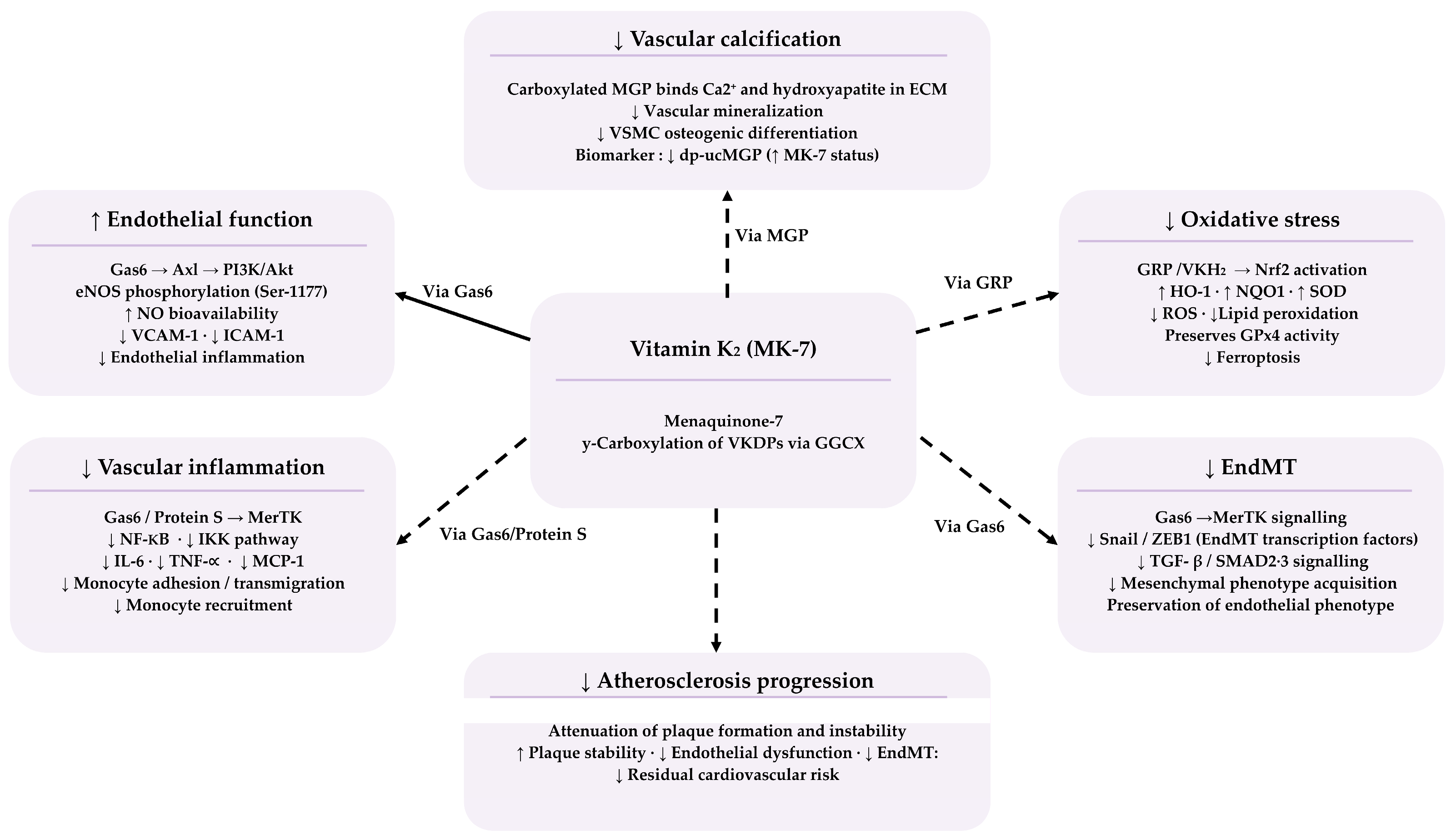
Therefore, it is important to undertake a comprehensive narrative summary of the available evidence to critically evaluate MK-7’s actions across these atherogenic pathways and their translational relevance to early atherosclerosis (Figure 1). This review aims to summarize findings from in vitro, animal, observational, and randomized controlled trials on MK-7 effects on endothelial dysfunction, oxidative stress, ferroptosis, vascular inflammation, and endothelial-to-mesenchymal transition in early atherosclerosis. The findings may guide future research directions and evidence-based nutritional strategies targeting cardiovascular risk reduction.

2. Materials and Methods

2.1. Literature Search Strategy

This narrative review was conducted according to established methodological standards for narrative reviews. A comprehensive search of the relevant literature was conducted using PubMed/MEDLINE, Web of Science, Scopus, and Google Scholar. Boolean operators were used to combine search terms, which included both free-text keywords relevant to the scope of the review and Medical Subject Headings (MeSHs) terms. Keywords included menaquinone-7, MK-7, vitamin K2, atherosclerosis, cardiovascular disease, CVD, endothelial dysfunction, nitric oxide, eNOS, oxidative stress, ferroptosis, NF-κB, vascular inflammation, matrix Gla protein, MGP, dp-ucMGP, vascular calcification, plaque stability, insulin resistance, and endothelial-to-mesenchymal transition.
A representative search string used across databases was (menaquinone-7 OR MK-7 OR vitamin K2 OR vitamin K) AND (atherosclerosis OR endothelial dysfunction OR oxidative stress OR “vascular inflammation OR vascular calcification OR ferroptosis OR insulin resistance”), with minor modifications according to the indexing structure of each database. The search focused on literature published between January 2019 and April 2026, with priority given to recent evidence. The final literature search was conducted in May 2026.
Evidence was narratively synthesized and organized according to an evidence hierarchy, distinguishing between in vitro, animal, observational, and randomized controlled trial findings. Reference lists of relevant reviews and key articles were also screened to identify additional eligible studies.

2.1.1. Inclusion Criteria

Studies published in English investigating the effects of vitamin K2 (MK-7) on endothelial dysfunction, oxidative stress, vascular inflammation, ferroptosis, endothelial-to-mesenchymal transition, or vascular calcification were included. Eligible study designs included in vitro studies, animal models, observational human studies, and randomized controlled trials. Foundational studies published before January 2019 were included where mechanistically relevant.

2.1.2. Exclusion Criteria

Studies investigating vitamin K exclusively in the context of coagulation, bone metabolism, or non-vascular outcomes were excluded. Editorials, letters, case reports, and non-peer-reviewed publications were not considered. Conference abstracts were excluded unless they provided highly relevant emerging mechanistic evidence not yet available in full peer-reviewed publications.

3. Mk-7: Biochemistry, Bioavailability, and Vascularly Relevant Vitamin K-Dependent Proteins

3.1. Structural and Pharmacokinetic Properties

MK-7 belongs to the menaquinone subclass of vitamin K2, characterised by a 2-methyl-1,4-naphthoquinone ring and a seven-unit polyisoprenoid side chain (Figure 2). This configuration results in higher lipophilicity than other forms of vitamin K, which influences its absorption, lipoprotein distribution, and tissue retention.
As a highly lipophilic compound, MK-7 undergoes bile salt-mediated micellarisation in the small intestinal lumen before passive diffusion across the enterocyte brush-border membrane. It is subsequently integrated into nascent chylomicrons and secreted into intestinal lymphatics, thereby bypassing first-pass hepatic uptake [8,16]. Because micellarisation is rate-limiting, the food matrix critically determines absorptive efficiency. Jensen and colleagues assessed vitamin K vitamer bioaccessibility using the validated INFOGEST 2.0 in vitro digestion model. Bioaccessibility was highest in oil-based matrices, followed by supplement formulations, and lowest in plant-source matrices [17]. These findings indicates that lipid-rich food matrices may enhance intestinal solubilisation of menaquinones. However, the model used evaluates in vitro bioaccessibility rather than true in vivo bioavailability. Therefore, validation in controlled human pharmacokinetic studies is required before direct clinical translation.
Compared with MK-4 and phylloquinone, MK-7 demonstrates greater circulating persistence and measurable serum accumulation at nutritional doses. In healthy women receiving an equimolar single dose (420 µg), MK-7 reached peak serum concentrations approximately 6 h after administration and remained detectable for up to 48 h. In contrast, MK-4 was undetectable in serum at all measured time points. Similarly, consecutive 7-day supplementation with 60 µg/day produced significant cumulative increases in serum MK-7 but no measurable change with MK-4 [18,19]. MK-4 at nutritional doses does not substantially contribute to circulating vitamin K status, though the small sample size limits generalizability.
Post-absorptive lipoprotein handling fundamentally differentiates MK-7 from other vitamin K forms. Schurgers and Vermeer demonstrated that phylloquinone is preferentially preserved in triglyceride-rich lipoproteins and cleared hepatically, whereas long-chain menaquinones are progressively transferred to LDL and HDL for redistribution to extrahepatic tissues [16]. Redistribution into LDL and HDL particles is believed to contribute to the prolonged circulating half-life of MK-7, which exceeds 72 h, compared with the substantially shorter half-lives of phylloquinone and MK-4. Du et al. reported a secondary rise in plasma MK-7 concentrations approximately 24 h after ingestion, possibly due to redistribution between lipoproteins or delayed hepatic release. However, this mechanism has not been confirmed by direct lipoprotein fractionation studies [20]. The resulting extended circulatory half-life may support prolonged availability of reduced vitamin K for extrahepatic γ-glutamyl carboxylase (GGCX) activity, particularly within vascular tissues [8,21].
Nanoencapsulation represents a promising strategy for enhancing MK-7 absorption. In a randomized double-blind trial, HyperCelle-encapsulated MK-7 produced significantly higher plasma exposure and peak concentrations compared with unencapsulated natto-derived MK-7 [22]. However, whether improved plasma concentrations translate to enhanced vascular VKDP carboxylation remains untested, and these findings should be considered hypothesis-generating. More broadly, most pharmacokinetic data derive from small studies in healthy volunteers, underrepresenting older adults and those with chronic kidney disease or conditions affecting nutrient absorption [23]. Inter-individual variability in MK-7 absorption has also been documented but remains mechanistically poorly understood [24]. Future studies integrating pharmacokinetic measurements with functional biomarkers such as dp-ucMGP are needed to better define the relationship between MK-7 bioavailability and vascular vitamin K status.

3.2. Key Vascularly Relevant Vitamin K-Dependent Proteins

Among Vitamin K-dependent proteins (VKDPs), MGP is the most clinically and mechanistically significant in the vascular context. MGP is synthesized principally by vascular smooth muscle cells (VSMCs) and, upon gamma-carboxylation, functions as the most potent known local inhibitor of vascular calcification by binding calcium deposits in the extracellular matrix [25,26]. The causal importance of MGP has been established in MGP-knockout mouse models, which develop fatal medial arterial calcification [27,28]. In the circulation, elevated desphospho-uncarboxylated MGP (dp-ucMGP) indicates inadequate activation and is independently associated with arterial stiffness, coronary calcification, and cardiovascular event risk [29,30]. MK-7 supplementation has been shown to lower dp-ucMGP in randomised trials, with effects more pronounced at 180 to 360 μg/day in populations with the greatest baseline vitamin K insufficiency [31].
Growth Arrest-Specific Protein 6 (Gas6) is a VKDP which, upon carboxylation, activates the TAM family of receptor tyrosine kinases (Axl, MerTK, Tyro3), initiating PI3K/Akt survival signalling and eNOS phosphorylation at serine-1177 in vascular endothelial cells [32,33]. Undercarboxylated Gas6 exhibits markedly reduced receptor-binding affinity, attenuating these pro-survival and NO-promoting signals [34].
Gla-Rich Protein (GRP) constitutes an additional calcification inhibitor in the vascular smooth muscle and endothelial compartments, inhibiting VSMC osteochondrogenic differentiation and extracellular vesicle-mediated mineralisation in a carboxylation-dependent manner [35,36,37], while Protein S contributes anti-thrombotic and endothelial anti-inflammatory activities through MerTK signalling [37,38]. Direct evidence for their role specifically mediated by MK-7 supplementation in human atherosclerosis remains limited.

3.3. Dietary Sources, Adequate Intake, and Supplementation

Long-chain menaquinones, including MK-7, are found primarily in fermented foods. Natto, a Japanese traditional food derived from Bacillus subtilis fermentation of soybeans, constitutes the richest known dietary source of MK-7, containing approximately 1,000 μg per 100 g [18]. In contrast, Western dietary sources, including fermented cheeses and certain meats, provide lower concentrations. Average MK-7 intakes in European and North American populations typically fall below 25 μg/day [18,39]. This limited dietary supply is considered insufficient to achieve complete carboxylation of extrahepatic VKDPs, particularly in older adults and individuals with CKD or other conditions impairing vitamin K metabolism [23].
The European Food Safety Authority (EFSA) has established an Adequate Intake (AI) for phylloquinone of 70 μg/day for adults, though no specific AI for MK-7 has been defined [40]. No upper tolerable intake level for vitamin K has been established. Supplementation with MK-7 at doses of 90 to 360 μg/day is validated as effective, bioavailable, and safe, producing consistent reductions in dp-ucMGP and undercarboxylated osteocalcin within weeks to months of initiation [19,20]. Comprehensive toxicological evaluation of MK-7 produced by Bacillus subtilis fermentation, including a 90-day repeated oral dosing study in rats at doses up to 4500 mg/kg/day, found no adverse effects, with the no-observed-adverse-effect level set at the maximum tested dose. Genotoxic activity was similarly not detected across bacterial reversion, chromosomal aberration, and micronucleus assays [41]. Considerable inter-individual variability in MK-7 absorption, influenced by gastrointestinal physiology and co-ingested lipid content, has been documented, with fat-containing matrices such as dairy products potentially augmenting absorption or retention [19,42].

4. Early Atherosclerosis: Pathophysiological Mechanisms

4.1. Endothelial Dysfunction and Oxidative Stress

Endothelial nitric oxide synthase (eNOS) produces constitutive NO, which helps the vascular endothelium maintain an anti-atherogenic character. Exposure to oxidized LDL (ox-LDL), hyperglycaemia, and angiotensin II depletes the obligate eNOS cofactor tetrahydrobiopterin (BH4). This causes eNOS to uncouple and redirect its enzymatic output from NO to superoxide anion (O2). The resulting superoxide reacts with residual NO to form peroxynitrite, a potent oxidant that further depletes BH4 and amplifies vascular reactive oxygen species in a self-perpetuating cycle [5,43]. Loss of NO bioactivity is accompanied by upregulation of VCAM-1, ICAM-1, and E-selectin, facilitating monocyte adhesion and subendothelial inflammation, the obligate first step in plaque formation [5,44].
Oxidative stress in the atherogenic vessel wall reflects a pathological imbalance between ROS production and antioxidant capacity. The NADPH oxidase (NOX) family, uncoupled eNOS, and the mitochondrial electron transport chain represent the principal enzymatic ROS sources in the vascular wall. Among NOX isoforms, NOX1 and NOX2 generate superoxide in a stimulus-dependent fashion, while NOX4 constitutively generates hydrogen peroxide. At moderate levels, NOX4 may exert atheroprotective effects, demonstrating isoform-specific functional heterogeneity important to therapeutic targeting [45,46]. Secondary lipid peroxidation products, including 4-hydroxynonenal (4-HNE) and malondialdehyde (MDA), activate macrophage scavenger receptors and inflammatory signalling, bridging oxidative injury to inflammatory amplification [47].
The counterbalancing antioxidant defence system is central to the regulation of atherogenic oxidative stress. Under physiological conditions, vascular cells are protected by antioxidant enzymes including superoxide dismutases, catalase, glutathione peroxidases, thioredoxins, and peroxiredoxins, which collectively detoxify ROS and lipid hydroperoxides [48,49]. The transcriptional regulation of these antioxidant defences is governed primarily by nuclear factor erythroid 2-related factor 2 (Nrf2). Under basal conditions, Nrf2 is retained in the cytoplasm by Kelch-like ECH-associated protein 1 (Keap1) and targeted for ubiquitin-mediated degradation. In response to oxidative or electrophilic stress, oxidative modification of critical Keap1 cysteine residues impairs Keap1-mediated Nrf2 ubiquitination, enabling Nrf2 to accumulate and translocate to the nucleus. Nuclear Nrf2 subsequently activates antioxidant response element (ARE)-dependent genes, including HO-1, NQO1, glutamate-cysteine ligase (GCL), and ferritin heavy chain (FTH1) [48,50].
In early atherosclerosis, Nrf2 signalling is initially engaged as a compensatory response to ROS accumulation [51,52]. However, sustained atherogenic stimulation and the NF-κB/Nrf2 cross-inhibitory relationship progressively impair Nrf2 activity through dysregulation of the Keap1–Nrf2 axis. This weakens the antioxidant transcriptional response and permitting pathological ROS accumulation [51,53]. In parallel, NF-κB activation suppresses Nrf2-dependent antioxidant gene expression through competition for shared transcriptional co-activators such as CBP/p300 and recruitment of repressor complexes including HDAC3 [54]. This NF-κB/Nrf2 imbalance promotes persistent ROS accumulation, sustained inflammatory signalling, and vascular oxidative injury, potentially contributing to the residual inflammatory and oxidative burden observed in patients with established atherosclerotic disease despite lipid-lowering therapy [6,7]. Importantly, the Nrf2 targets GPx4 and FTH1 additionally confer protection against iron-catalysed lipid peroxidation, providing a mechanistic link between impaired Nrf2 signalling and ferroptotic cell death pathways discussed in Section 4.2.

4.2. Vascular Inflammation and Emerging Mechanisms

The transcription factor NF-κB occupies a central regulatory node in vascular inflammation. Atherogenic stimuli, including ox-LDL, angiotensin II, and ROS activate the IκB kinase (IKK) complex, leading to IκBα degradation and nuclear translocation of the p65/p50 NF-κB heterodimer, which drives transcription of VCAM-1, ICAM-1, MCP-1, IL-6, IL-8, TNF-α, COX-2, and iNOS [44]. ROS-mediated NF-κB activation establishes a self-reinforcing triad with endothelial dysfunction and oxidative stress, which persists independently of LDL-C levels. This pathological cycle drives the residual inflammatory risk documented in statin-treated patients [6,7,44].
Beyond these classical mechanisms, endothelial-to-mesenchymal transition (EndMT) represents an emerging amplifier of atherogenic vascular remodelling, in which endothelial cells progressively acquire mesenchymal characteristics. The transition is driven by TGF-β/SMAD2/3 signalling, NF-κB/Snail activation, and oxidative stress [55,56]. In vivo cell lineage tracing in ApoE-deficient mice has demonstrated that approximately 30% of aortic endothelial cells undergo EndMT during diet-induced atherogenesis [56].
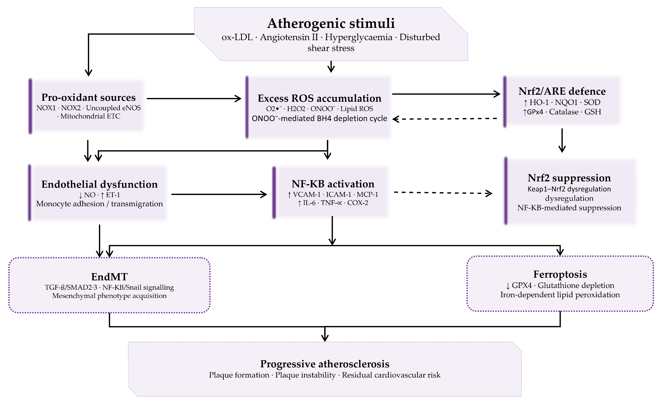
Ferroptosis is an iron-dependent form of cell death characterised by iron-catalysed accumulation of lethal lipid ROS and peroxidation of membrane phospholipid-polyunsaturated fatty acids. In the atherosclerotic vascular wall, ferroptotic death of macrophage foam cells and VSMCs contributes to plaque instability and has been associated with endothelial dysfunction [57,58]. The biochemical execution of ferroptosis depends on the impairment of the glutathione peroxidase 4 (GPX4)/glutathione axis), and in vitro inhibition of ferroptosis has been shown to partially restore eNOS expression and reduce adhesion molecule expression in endothelial cells [57]. The causal contribution of ferroptosis to atherosclerotic plaque formation in vivo remains incompletely explored, limiting definitive therapeutic conclusions. Supporting this, GPX4 overexpression in ApoE-deficient mice did not significantly change plaque size or composition [58]. The convergence of pro-oxidant activity, endothelial dysfunction, NF-κB-driven inflammation, impaired Nrf2-mediated antioxidant defence, EndMT, and ferroptosis in progressive atherosclerosis is illustrated schematically in Figure 3.

5. Mk-7 Modulation of the Atherogenic Triad: Mechanistic Evidence

5.1. MK-7 and Endothelial Function: The Gas6-Axl-eNOS Axis

Experimental evidence indicates that MK-7 supports endothelial function through a carboxylation-dependent mechanism involving Gas6 and the TAM receptor signalling axis. Upon adequate gamma-carboxylation by GGCX, Gas6 engages the Axl receptor on vascular endothelial cells, initiating PI3K/Akt signalling that phosphorylates eNOS at serine-1177, substantially augmenting enzymatic NO output [9,32,59]. Undercarboxylated Gas6 exhibits lower Axl receptor-binding affinity and reduces downstream Akt-eNOS phosphorylation [34]. Subclinical vitamin K insufficiency, reflected by higher circulating dp-ucMGP, is prevalent in Western populations and independently associated with endothelial dysfunction and increased cardiovascular risk [31,60]. By promoting GGCX-dependent Gas6 carboxylation, MK-7 may therefore preserve Gas6-Axl-eNOS axis continuity under conditions of suboptimal vitamin K status.
In ApoE/LDLR double-knockout mice, low-dose MK-7 improved endothelium-dependent vasodilation, increased aortic NO production, and reduced brachiocephalic artery media thickness, without affecting atherosclerotic plaque size [61]. Direct evidence that exogenous MK-7 augments Gas6-mediated eNOS activation in human vascular endothelium has not been established through dedicated translational studies.

5.2. MK-7 and Oxidative Stress: The FSP1-VKH2 Ferroptosis-Suppressive Pathway

A recently identified antioxidant function of vitamin K, independent of γ-carboxylation, has broaden its vascular relevance. Mishima et al. (2022) demonstrated that the reduced hydroquinone form of vitamin K (VKH2) functions as a substrate in the Ferroptosis Suppressor Protein 1 (FSP1/AIFM2), a flavoprotein oxidoreductase that regenerates VKH2 from its quinone form [62]. In this context, VKH2 functions as a potent lipophilic radical-trapping antioxidant. It directly intercepts phospholipid peroxyl radicals within cellular membranes. Importantly, the FSP1-dependent recycling pathway is distinct from the classical VKORC1-mediated vitamin K cycle and is not inhibited by warfarin. The study characterised this pathway for vitamin K hydroquinone broadly rather than MK-7 specifically [63]. Accordingly, the relevance of MK-7 to the FSP1–VKH2 axis remains indirect and hypothesis-driven, based on its higher bioavailability and tissue accumulation.
This FSP1-VKH2 system has been implicated in the suppression of ferroptotic cell death in the atherogenic vascular wall, where ferroptotic death of macrophage foam cells and VSMCs amplifies lipid core expansion and impairs fibrous cap integrity [57]. By replenishing the membrane-accessible VKH2 pool, MK-7 may reduce ferroptotic cell death in the arterial wall, given its superior tissue accumulation relative to shorter-chain menaquinones.
Supporting in vitro evidence was reported by Cirilli et al. (2020), MK-7(10 μM) reduced ROS production, apoptosis, and senescence in cigarette smoke extract-treated HUVECs and was more effective than MK-4 and phylloquinone [64]. The concentration used is above the physiological range, and the artificial injury model limits translational relevance. Therefore, confirmation in human vascular tissue or clinical supplementation studies remains a critical unmet research priority.

5.3. MK-7 and Vascular Inflammation: NF-κB Suppression and EndMT

In vascular and hepatic cell models, MK-7 attenuates pro-inflammatory signalling through suppression of the IKK/IκBα axis and NF-κB-dependent gene transcription. Reductions in pro-inflammatory cytokine expression, including IL-6, MCP-1, and VCAM-1, have been observed in experimental models of vascular injury [44,63]. In a study specifically examining MK-7 in human coronary artery smooth muscle cells, dose-dependent reductions in NF-κB and IL-6 mRNA expression were demonstrated following oxLDL exposure. This provides MK-7-specific evidence for vascular NF-κB suppression, although the effect was observed in smooth muscle rather than endothelial cells [65]. The precise molecular target through which MK-7 modulates IKK activity, and whether these effects requires Gla-protein carboxylation or the menaquinone structure, remains unresolved. In addition, randomized clinical trials have not systematically evaluated inflammatory cytokine or adhesion molecule responses as primary vascular endpoints, limiting direct translational evidence in humans [66,67]. Of note, much of the available evidence for NF-κB suppression derives from general cellular models investigating menaquinones broadly. Direct validation of MK-7-specific IKK/NF-κB inhibition in human vascular endothelial cells remains limited [65].
Given the established roles of NF-κB, oxidative stress, and TGF-β-associated signalling in endothelial-to-mesenchymal transition (EndMT), MK-7 may theoretically influence EndMT-related processes via indirect modulation of pathways. Gas6-mediated MerTK activation reduces TGF-β-induced SMAD2/3 phosphorylation in endothelial cells, while NF-κB suppression reduces IL-6 and TNF-α concentrations that amplify EndMT through JAK/STAT3 signalling [55,56]. Additionally, the FSP1-VKH2 antioxidant axis may prevent oxidative stress-driven transcriptional reprogramming that promotes mesenchymal transition [62]. Preliminary evidence from a peer-reviewed journal supplement suggested that vitamin K2 may attenuate EndMT-associated changes in human coronary artery endothelial cells. These changes include reductions in mesenchymal markers and inflammatory signalling [68]. However, the findings require confirmation in full peer-reviewed studies conducted under physiologically relevant conditions. Accordingly, direct evidence indicates MK-7-specific modulation of EndMT in human vascular endothelial models remains limited.

5.4. MK-7, Insulin Resistance, and Vascular Inflammatory Risk

Beyond dyslipidaemia, insulin resistance has long been recognised as an independent driver of vascular inflammation and plaque development. Impaired insulin signalling reduces the activity of the IRS-1/PI3K pathway, which decreases nitric oxide synthase activation and endothelial dysfunction, while preservation of MAPK pathway signalling promotes a pro-inflammatory state within the arterial wall [69]. Observational clinical data from patients with carotid artery stenosis indicate that higher insulin resistance, as measured by the triglyceride-glucose index. It is linked to both vulnerable plaque characteristics and an increased risk of cerebrovascular events. Mediation analysis further suggests that plaque vulnerability partially explains the relationship between insulin resistance and stroke risk. The findings suggest a possible connection between dysglycaemia and plaque instability [70].
Emerging randomized controlled trials suggest that MK-7 (90 µg/day) supplementation may improve glycaemic parameters in individuals with type 2 diabetes. MK-7 supplementation significantly decreased fasting blood glucose, fasting insulin, and HbA1c in 60 participants during a six-month randomized controlled trial [71]. In a 12-week double-blind randomized controlled trial, supplementation with 360 µg/day MK-7 did not significantly improve insulin resistance-related atherogenic indices after adjustment for baseline values. Although some reductions were observed before adjustment [72]. According to a recent meta-analysis of randomized controlled trials, MK-7 supplementation improved HbA1c, insulin levels, and HOMA-IR, but no significant effect was observed for fasting blood glucose [12]. Critically, no randomised trial has evaluated whether MK-7-induced improvements in glycaemic markers result in quantifiable reductions in vascular inflammation or plaque stability. As such, the causal relationship between MK-7 supplementation and reductions in atherosclerotic risk associated with insulin resistance remains unexplored.

6. Matrix Gla Protein and Vascular Calcification

The primary vascular function of MK-7 is the activation of MGP through gamma-carboxylation and the consequent inhibition of vascular calcification. Vascular calcification is now recognised as a regulated, bone-like process controlled by osteogenic signals, including bone morphogenetic proteins (BMPs), Wnt/Runx2 transcriptional pathways, and calcification inhibitors, such as MGP and osteopontin [28,73]. In atherosclerosis, calcification shows mechanistic and temporal heterogeneity. Microcalcifications (<0.5 mm), enriched in regions of high macrophage infiltration, enhance local mechanical stress and plaque vulnerability, whereas macrocalcifications are associated with more stable, fibrous plaques. This pattern indicates that vascular calcification is linked to the inflammatory and cellular processes of plaque development, rather than constituting a purely passive end-stage phenomenon [74,75].
The mechanistic role of MGP in preventing vascular mineral deposition is unequivocally established by MGP-knockout mouse models, which develop fatal widespread arterial calcification [25]. In humans, dp-ucMGP, a biomarker of inactive MGP, is independently associated with coronary artery, ascending thoracic aortic, and descending thoracic aortic calcification in Multi-Ethnic Study of Atherosclerosis participants [29,30]. A prospective community-based cohort has further indicated that each standard deviation increase in dp-ucMGP is associated with approximately 23% higher CVD incidence and 40% higher all-cause mortality over approximately 10 years [76].
It is important to recognise, however, that preventing calcification alone does not fully prevent or reverse early atherosclerosis. Endothelial injury, oxidised lipid accumulation, and chronic inflammation and early progression occur before, and independently of, vascular mineral deposition. MGP’s classical role is therefore to limit ossification in advanced lesions rather than endothelial and inflammatory mechanisms of plaque initiation [4,28,77].

7. In Vitro, Animal, and Clinical Studies

7.1. In Vitro Studies

In vitro evidence for MK-7 in endothelial and vascular cell models is limited but mechanistically informative. Cirilli et al. (2020) investigated the effect of MK-7’s in Human Umbilical Vein Endothelial Cells exposed to cigarette smoke extract, demonstrating significant reductions in ROS generation, apoptosis, and senescence markers at a concentration of 10 μM [64]. MK-7 demonstrated higher cytoprotection than MK-4 and phylloquinone in this model, supporting the hypothesis of differential potency among vitamin K forms in protecting against endothelial oxidative injury. Nevertheless, the supraphysiological MK-7 concentration, artificial injury model, and absence of measurements of NO production, VCAM-1 expression, or VKDP carboxylation limit the translational value of the finding. The concentration (10 µM) applied exceeds physiologically achievable plasma MK-7 levels following standard supplementation, typically in the low nanomolar range after 180–360 µg/day [31,63]. This limits direct extrapolation of these findings to human vascular physiology.
The identification of the FSP1-VKH2 axis by Mishima et al. (2022) provides the most mechanistically significant in vitro evidence for MK-7’s vascular relevance [62]. These findings establish a carboxylation-independent antioxidant mechanism for vitamin K hydroquinone that encompasses ferroptosis suppression. This is directly relevant to the atherogenic vascular wall, where iron-dependent lipid peroxidation-driven cell death contributes to endothelial dysfunction and plaque progression [78,79]. Additional evidence from vascular and macrophage cell models indicates that menaquinones inhibit IKK/NF-κB activation and reduce pro-inflammatory cytokine and adhesion molecule expression [11,37].

7.2. Animal Models

Animal studies provide the most direct evidence for MK-7 effects on endothelial function and vascular calcification. Bar et al. (2019) showed in ApoE/LDLR double-knockout mice that low-dose MK-7 improved endothelium-dependent vasodilation and increased aortic NO production without affecting plaque regression, consistent with the Gas6-Axl-eNOS mechanism [61].
Florea et al. (2021) studied ApoE-knockout mice on an atherogenic diet, assessing plaque calcification [80]. Mice receiving MK-7 showed active microcalcification tracer uptake similar to chow-fed controls and significantly lower than warfarin-treated or continued high-fat diet controls, indicating that MK-7 halted active calcification progression. These findings are consistent with MGP-mediated calcification inhibition, though the dose was very high relative to human supplementation ranges and inflammatory endpoints were not directly measured [80]. El-Sherbiny et al. (2022) reported that vitamin K2 administration in aged rats reduced hepatic inflammation, with significant reductions in COX-2, iNOS, and TNF-α immunostaining in treated versus untreated aged animals [63].
Non-physiological doses, genetically modified disease models, and the absence of direct vascular inflammatory measures in most studies limit translation to human atherosclerosis [81].

7.3. Randomized Controlled Trials

Randomised controlled trial evidence for MK-7 in human vascular endpoints is growing, but most studies report neutral primary outcomes. Reported benefits are generally limited to pre-specified subgroups and individuals with greater baseline vitamin K insufficiency or cardiovascular burden (Table 1).
Knapen et al. (2015) conducted a 3-year double-blind RCT in healthy postmenopausal women, administering 180 μg/day MK-7 versus placebo [82]. The authors revealed that MK-7 supplementation significantly reduced the age-related increase in carotid-femoral pulse wave velocity (cfPWV), with a stronger effect in women with higher baseline arterial stiffness. Circulating dp-ucMGP was significantly reduced throughout the intervention [82]. A 12-month double-blind RCT in 165 women with low vitamin K status (180 μg/day MK-7) found no significant difference in the primary endpoint of cfPWV between groups. However, the post-hoc analysis indicated that postmenopausal women with higher baseline arterial stiffness showed a significant reduction in stiffness and brachial blood pressure, along with decreased dp-ucMGP across all participants [83].
A 24-week multicentre randomized controlled trial in haemodialysis patients with arterial stiffness found no significant difference in carotid-femoral pulse wave velocity between MK-7 (375 µg/day) and standard care. Nevertheless, a subgroup analysis in diabetic haemodialysis patients showed that MK-7 significantly reduced cfPWV and decreased the rate of arterial stiffness progression relative to controls [84]. An 18-month RCT study in haemodialysis patients assessing the effect of MK-7 (360 μg three times weekly) on coronary artery calcification (CAC) score revealed a significant reduction in dp-ucMGP. However, no significant difference in CAC score progression was observed between groups [67]. These findings are consistent with those of Bartstra et al. (2020), who found no effect of 6-month MK-7 supplementation (360 μg/day) on arterial calcification progression or bone mineral density in patients with type 2 diabetes and pre-existing CVD [66].
MK-7 administered over 270 days (90 μg/day with cholecalciferol) in 42 non-dialyzed CKD patients significantly reduced the progression of common carotid artery intima-media thickness compared with vitamin D alone, accompanied by marked reductions in dp-ucMGP. No significant effect on CACS progression was observed [85]. In a small open-label randomized controlled trial in coronary artery disease patients, MK-7 supplementation was associated with favourable changes in CAC score in approximately half of treated patients, compared with CAC progression in 80% of controls [86]. Across all RCTs, MK-7 supplementation reduces dp-ucMGP, although corresponding improvements in clinical vascular endpoints have not been uniformly confirmed.

7.4. Observational and Epidemiological Studies

Large prospective cohort studies provide consistent, though observational, support for the role of vitamin K2 in atherosclerotic CVD prevention. Bellinge et al. (2021) reported a 14% lower risk of ASCVD hospitalization in the highest versus lowest dietary vitamin K2 quintile across 53,372 participants followed for 21 years, with inverse associations observed for coronary heart disease, ischemic stroke, and peripheral arterial disease [87]. Similarly, inverse associations observed between dietary menaquinone intake and coronary heart disease risk were documented in both Rotterdam Study and PROSPECT-EPIC cohort [88,89]. In addition to dietary assessments, biomarker-based evidence further supports this relationship, as vitamin K insufficiency, defined by increased dp-ucMGP, has been independently associated with higher CVD incidence and all-cause mortality [78]. Supporting this evidence, another study found that serum MK-7 and MK-4 levels are significantly lower in patients with acute coronary syndrome than healthy controls, with a reduction in unstable angina [90].
Table 1. Summary of key studies evaluating MK-7 in vascular biology.
ReferenceStudy TypePopulation/ModelMK-7 Dose/InterventionPrimary Endpoint(s)Key Findings
[63]In vitroHUVECs; cigarette smoke extract model10 μM MK-7ROS, apoptosis, senescence markersMK-7 reduced ROS, decreased apoptosis, lowered p21 and β-galactosidase vs. controls; outperformed MK-4 and K1
[62]In vitro/animalMultiple cell lines; murine modelsVKH2 (reduced vitamin K); FSP1 pathway characterisationLipid peroxidation; ferroptosis markersVKH2 functions as lipophilic radical-trapping antioxidant via FSP1; suppresses ferroptotic cell death independently of VKORC1
[61]Animal (in vivo)ApoE/LDLR/ mice (pre- and established plaque stages)0.05–10 mg/kg/day MK-7Endothelium-dependent vasodilation; NO production; media thicknessLow-dose MK-7 improved acetylcholine- and flow-induced vasodilation; increased aortic NO by EPR; reduced brachiocephalic media thickness; no effect on plaque size
[80]Animal (in vivo)ApoE/ mice on atherogenic diet (12 weeks)≈400 μg/day MK-7 (100 μg/g feed)18F-NaF PET (active microcalcification)MK-7-treated mice showed 18F-NaF uptake comparable to chow-fed controls, significantly lower than warfarin or HFD controls; warfarin markedly increased tracer uptake
[64]Animal (in vivo)Aged male Wistar ratsVitamin K2 (dose not fully specified)Hepatic COX-2, iNOS, TNF-α expressionHepatic sections showed marked downregulation of COX-2, iNOS, and TNF-α in treated vs. untreated aged animals
[88]Observational (prospective cohort)Rotterdam Study (4,807 participants)Dietary menaquinone intake (FFQ)CHD mortality; incident CHD; all-cause mortality; severe aortic calcification.Higher menaquinone (not K1) intake is inversely associated with CHD mortality, all-cause mortality and aortic calcification
[89]Observational (prospective cohort)PROSPECT-EPIC (16,057 women)Dietary vitamin K2 intake (FFQ)Coronary heart disease incidence9% lower CHD risk per 10 μg/day higher K2 intake
[87]Observational (prospective cohort)Danish Diet, Cancer & Health Study (53,372 participants; 21-year follow-up)Dietary vitamin K2 intake (FFQ)ASCVD hospitalizations (IHD, stroke, PAD)14% lower ASCVD hospitalization risk in the highest vs. lowest K2 quintile; similar inverse association for K1
[76]Observational (prospective cohort)Community-based cohort (~10-year follow-up)Plasma dp-ucMGP (biomarker of vitamin K insufficiency)CVD incidence; all-cause mortalityEach SD increase in dp-ucMGP: +23% CVD incidence, +40% all-cause mortality
[90]Observational (case-control)CAD patients vs. healthy controls; CAD subtypes (STEMI, NSTEMI, unstable angina, stable angina)Serum MK-4 and MK-7 measurement (UPLC-MS/MS)Serum MK-7 levels across CAD subtypesLower serum MK-7 and MK-4 in ACS vs. controls; unstable angina had lowest levels; MK-7 showed greatest intergroup difference
[91]RCTHealthy postmenopausal women (244; 3-year trial)180 μg/day MK-7 vs. placeboCarotid-femoral pulse wave velocity (cfPWV)Significant attenuation of age-related cfPWV increase in MK-7 group; larger effect in women with high baseline stiffness; dp-ucMGP reduced
[83]RCTLow vitamin K-status women (165; 12 months)180 μg/day MK-7 vs. placebocfPWV; brachial blood pressure; dp-ucMGPOverall cfPWV non-significant; postmenopausal women with high baseline stiffness showed significant reduction in stiffness and BP; dp-ucMGP significantly reduced across all participants
[84]RCT (multicentre)Haemodialysis patients with arterial stiffness (96; 24 weeks)375 μg/day MK-7 vs. standard carecfPWVNo significant difference in cfPWV overall; diabetic subgroup showed significant cfPWV reduction and lower progression rate
[67] RCTMaintenance haemodialysis patients (18 months)MK-7 vs. standard careCoronary artery calcification (CAC) scoredp-ucMGP significantly lower in MK-7 group; no significant difference in CAC score progression
[66]RCTT2DM patients with CVD history (68; 6 months)360 μg/day MK-7 vs. placeboTotal arterial calcification mass; BMDNo significant effect on calcification progression or BMD decline vs. placebo; authors note need for pre-selected vitamin K-insufficient participants
[86]RCT (small; open-label)CAD patients (30; 6 months)Oral MK-7 supplementation vs. no treatmentCAC score (Agatston, non-contrast CT)46.6% of MK-7 group had decreased CAC score; 53.3% stable; 80% of controls showed increased CAC; 20% stable
Abbreviations: ASCVD, atherosclerotic cardiovascular disease; BMD, bone mineral density; CAC, coronary artery calcification; CACS, coronary artery calcification score; CCA-IMT, common carotid artery intima-media thickness; cfPWV, carotid-femoral pulse wave velocity; CKD, chronic kidney disease; dp-ucMGP, desphospho-uncarboxylated matrix Gla protein; EPR, electron paramagnetic resonance; FFQ, food-frequency questionnaire; HUVEC, human umbilical vein endothelial cell; HFD, high-fat diet; MK-7, menaquinone-7; NO, nitric oxide; PAD, peripheral arterial disease; RCT, randomized controlled trial; ROS, reactive oxygen species; SD, standard deviation; STEMI, ST-elevation myocardial infarction; T2DM, type 2 diabetes mellitus; VKH2, vitamin K hydroquinone.

8. Translational Implications and Research Priorities

Supplementation with MK-7 at doses of 90 to 360 μg/day showed higher bioavailability over that attainable through typical Western diets, increased serum MK-7 concentrations and reduced VKDP undercarboxylation biomarkers within weeks [19,42]. Dose-response evidence from healthy adults demonstrates that 180 and 360 μg/day produced dose-dependent reductions in dp-ucMGP of approximately 31% and 46%, respectively [31]. MK-7-fortified functional foods offer a practical delivery strategy; MK-7-fortified yoghurt has been shown to improve vitamin K status, and fat-containing food matrices may enhance absorption or retention [42,91]. Current evidence suggests that the populations most likely to benefit are those with documented vitamin K insufficiency, postmenopausal women with elevated baseline arterial stiffness, and patients with CKD or diabetes. Current evidence shows that specific populations may benefit, including those with vitamin K deficiency, postmenopausal women with elevated baseline arterial stiffness, and patients with CKD or diabetes [66,83,84,85]. However, it is important to emphasize that these observations are mainly derived from subgroup or post-hoc analyses and should therefore be considered hypothesis-generating rather than confirmatory. A threshold effect in vitamin K-replete populations may limit the ability to demonstrate benefits in trials that do not pre-screen for vitamin K insufficiency.

9. Conclusions

MK-7 is a mechanistically plausible regulator of several pathophysiological mechanisms associated with early atherosclerosis. Its well-characterised role in activating MGP to inhibit vascular calcification is supported by animal models, biomarker studies, and randomized controlled trials showing reductions in dp-ucMGP. Beyond calcification, experimental in vitro and animal evidence reported MK-7 may enhance endothelial NO bioavailability via the Gas6-Axl-eNOS axis, suppress ferroptotic lipid peroxidation through the FSP1-VKH2 pathway, attenuate NF-κB-driven vascular inflammation, and modulate EndMT. These findings collectively support the biological plausibility of MK-7 as a multi-target vascular agent, although clinical validation in adequately powered trials with hard vascular endpoints remains limited.
Despite growing mechanistic support, the existing evidence base contains several critical gaps that currently limit clinical translation of MK-7’s multi-target vascular effects. First, no dedicated mechanistic human RCTs have evaluated endothelial function, circulating inflammatory biomarkers, or oxidative stress markers as primary endpoints; existing trials have relied on arterial stiffness or calcification scores, leaving the pathophysiological triad central to this review unaddressed in the human vascular environment. Second, the FSP1-VKH2 ferroptosis-suppressive pathway and EndMT suppression by MK-7 remain unvalidated in human vascular tissue or clinical supplementation studies. Third, optimal dose, treatment duration, and patient selection criteria remain prospectively undefined, and hard cardiovascular outcomes have not been studied as primary endpoints. Fourth, whether MK-7 exhibits additive or synergistic vascular benefits when combined with established anti-inflammatory or antioxidant interventions requires investigation. Available trial data suggest that co-supplementation strategies require systematic evaluation before adoption [66,72].

Author Contributions

Conceptualization, H.H. and M.F.-Ż.; methodology, H.H.; validation, H.H., T.T.; data curation, H.H.; writing—original draft preparation, H.H. and M.F.-Ż.; writing—review and editing, H.H. and M.F.-Ż.; supervision, M.F.-Ż. and T.T. All authors have read and agreed to the published version of the manuscript.

Funding

This research received no external funding.

Data Availability Statement

No new data were created or analysed in this study. Data sharing is not applicable to this article.

Conflicts of Interest

The authors declare no conflicts of interest.

References

  1. Mensah, G.A.; Fuster, V.; Murray, C.J.L.; Roth, G.A.; Mensah, G.A.; Abate, Y.H.; Abbasian, M.; Abd-Allah, F.; Abdollahi, A.; Abdollahi, M.; et al. Global Burden of Cardiovascular Diseases and Risks, 1990–2022. J. Am. Coll. Cardiol. 2023, 82, 2350–2473. [Google Scholar] [CrossRef] [PubMed]
  2. Tsampasian, V.; Bloomfield, G.S. The Evolving Global Burden of Cardiovascular Diseases: What Lies Ahead. Eur. J. Prev. Cardiol. 2025, 32, 1016–1017. [Google Scholar] [CrossRef]
  3. Berenji Ardestani, S.; Eftedal, I.; Pedersen, M.; Jeppesen, P.B.; Nørregaard, R.; Matchkov, V.V. Endothelial Dysfunction in Small Arteries and Early Signs of Atherosclerosis in ApoE Knockout Rats. Sci. Rep. 2020, 10, 15296. [Google Scholar] [CrossRef]
  4. Libby, P.; Buring, J.E.; Badimon, L.; Hansson, G.K.; Deanfield, J.; Bittencourt, M.S.; Tokgözoğlu, L.; Lewis, E.F. Atherosclerosis. Nat. Rev. Dis. Prim. 2019, 5, 56. [Google Scholar] [CrossRef]
  5. Xu, S.; Ilyas, I.; Little, P.J.; Li, H.; Kamato, D.; Zheng, X.; Luo, S.; Li, Z.; Liu, P.; Han, J.; et al. Endothelial Dysfunction in Atherosclerotic Cardiovascular Diseases and Beyond: From Mechanism to Pharmacotherapies. Pharmacol. Rev. 2021, 73, 924–967. [Google Scholar] [CrossRef]
  6. Ridker, P.M.; Bhatt, D.L.; Pradhan, A.D.; Glynn, R.J.; MacFadyen, J.G.; Nissen, S.E. Inflammation and Cholesterol as Predictors of Cardiovascular Events among Patients Receiving Statin Therapy: A Collaborative Analysis of Three Randomised Trials. Lancet 2023, 401, 1293–1301. [Google Scholar] [CrossRef]
  7. Ridker, P.M.; Lei, L.; Louie, M.J.; Haddad, T.; Nicholls, S.J.; Lincoff, A.M.; Libby, P.; Nissen, S.E. Inflammation and Cholesterol as Predictors of Cardiovascular Events Among 13 970 Contemporary High-Risk Patients with Statin Intolerance. Circulation 2024, 149, 28–35. [Google Scholar] [CrossRef] [PubMed]
  8. Halder, M.; Petsophonsakul, P.; Akbulut, A.C.; Pavlic, A.; Bohan, F.; Anderson, E.; Maresz, K.; Kramann, R.; Schurgers, L. Vitamin K: Double Bonds beyond Coagulation Insights into Differences between Vitamin K1 and K2 in Health and Disease. Int. J. Mol. Sci. 2019, 20, 896. [Google Scholar] [CrossRef]
  9. Willems, B.A.G.; Vermeer, C.; Reutelingsperger, C.P.M.; Schurgers, L.J. The Realm of Vitamin K Dependent Proteins: Shifting from Coagulation toward Calcification. Mol. Nutr. Food Res. 2014, 58, 1620–1635. [Google Scholar] [CrossRef]
  10. Jadhav, N.; Ajgaonkar, S.; Saha, P.; Gurav, P.; Pandey, A.; Basudkar, V.; Gada, Y.; Panda, S.; Jadhav, S.; Mehta, D.; et al. Molecular Pathways and Roles for Vitamin K2-7 as a Health-Beneficial Nutraceutical: Challenges and Opportunities. Front. Pharmacol. 2022, 13, 896920. [Google Scholar] [CrossRef] [PubMed]
  11. Li, T.; Wang, Y.; Tu, W.P. Vitamin K Supplementation and Vascular Calcification: A Systematic Review and Meta-Analysis of Randomized Controlled Trials. Front. Nutr. 2023, 10, 1115069. [Google Scholar] [CrossRef] [PubMed]
  12. Nikpayam, O.; Jafari, A.; Faghfouri, A.; Pasand, M.; Noura, P.; Najafi, M.; Sohrab, G. Effect of Menaquinone-7 (MK-7) Supplementation on Anthropometric Measurements, Glycemic Indices, and Lipid Profiles: A Systematic Review and Meta-Analysis of Randomized Controlled Trials. Prostaglandins Other Lipid Mediat. 2025, 177, 106970. [Google Scholar] [CrossRef]
  13. Kapadia, S.; Hariri, E.; Kassis, N.; Iskandar, J.-P.; Schurgers, L.J.; Saad, A.; Abdelfattah, O.; Bansal, A.; Isogai, T.; Harb, S.C. Open Access Vitamin K 2-a Neglected Player in Cardiovascular Health: A Narrative Review. Heart 2021, 8, 1715. [Google Scholar] [CrossRef]
  14. Schurgers, L.J.; Dissel, P.E.P.; Spronk, H.M.H.; Soute, B.A.M.; Dhore, C.R.; Cleutjens, J.P.M.; Vermeer, C. Role of Vitamin K and Vitamin K-Dependent Proteins in Vascular Calcification. Z. Kardiol. 2001, 90, 57–63. [Google Scholar] [CrossRef]
  15. Menaquinone 7|C46H64O2|CID 5287554—PubChem. Available online: https://pubchem.ncbi.nlm.nih.gov/compound/5287554 (accessed on 15 May 2026).
  16. Schurgers, L.J.; Teunissen, K.J.F.; Hamulyák, K.; Knapen, M.H.J.; Vik, H.; Vermeer, C. Vitamin K–Containing Dietary Supplements: Comparison of Synthetic Vitamin K1 and Natto-Derived Menaquinone-7. Blood 2007, 109, 3279–3283. [Google Scholar] [CrossRef]
  17. Jensen, M.B.; Biltoft-Jensen, A.P.; Jakobsen, J. In Vitro Bioaccessibility of Vitamin K (Phylloquinone and Menaquinones) in Food and Supplements Assessed by INFOGEST 2.0—Vit K. Curr. Res. Food Sci. 2022, 5, 306–312. [Google Scholar] [CrossRef]
  18. Schurgers, L.J.; Vermeer, C. Determination of Phylloquinone and Menaquinones in Food. Effect of Food Matrix on Circulating Vitamin K Concentrations. Haemostasis 2000, 30, 298–307. [Google Scholar] [CrossRef] [PubMed]
  19. Sato, T.; Schurgers, L.J.; Uenishi, K. Comparison of Menaquinone-4 and Menaquinone-7 Bioavailability in Healthy Women. Nutr. J. 2012, 11, 93. [Google Scholar] [CrossRef]
  20. Du, F.; Yan, M.; Duan, L.; Xie, G.; Yao, X.; Hu, W.; Liu, Y.; Meng, M.; Chen, J.; Shao, D. The Study of Bioavailability and Endogenous Circadian Rhythm of Menaquinone-7, a Form of Vitamin K2, in Healthy Subjects. Br. J. Nutr. 2023, 130, 1885–1897. [Google Scholar] [CrossRef] [PubMed]
  21. Berkner, K.L.; Runge, K.W. Vitamin K-Dependent Protein Activation: Normal Gamma-Glutamyl Carboxylation and Disruption in Disease. Int. J. Mol. Sci. 2022, 23, 5759. [Google Scholar] [CrossRef]
  22. Cong, R.; Kim, J.W.; Lee, N.Y.; Chun, Y.S.; Kim, J.K.; Kim, B.K.; Hong, S.B.; Shim, S.M. Oral Bioavailability Enhancement of MK-7 from Natto Extract via HyperCelle Nanoencapsulation in a Clinical Study. Appl. Biol. Chem. 2025, 68, 65. [Google Scholar] [CrossRef]
  23. Roumeliotis, S.; Dounousi, E.; Salmas, M.; Eleftheriadis, T.; Liakopoulos, V. Vascular Calcification in Chronic Kidney Disease: The Role of Vitamin K- Dependent Matrix Gla Protein. Front. Med. 2020, 7, 533649. [Google Scholar] [CrossRef]
  24. Knapen, M.H.J.; Vermeer, C.; Theuwissen, E. Pharmacokinetics of Menaquinone-7 (Vitamin K2) in Healthy Volunteers. J. Clin. Trials 2014, 4, 1–6. [Google Scholar] [CrossRef]
  25. Jono, S.; Ikari, Y.; Vermeer, C.; Dissel, P.; Hasegawa, K.; Shioi, A.; Taniwaki, H.; Kizu, A.; Nishizawa, Y.; Saito, S. Matrix Gla Protein Is Associated with Coronary Artery Calcification as Assessed by Electron-Beam Computed Tomography. Thromb. Haemost. 2004, 91, 790–794. [Google Scholar] [CrossRef]
  26. Kumric, M.; Borovac, J.A.; Kurir, T.T.; Martinovic, D.; Separovic, I.F.; Baric, L.; Bozic, J. Role of Matrix Gla Protein in the Complex Network of Coronary Artery Disease: A Comprehensive Review. Life 2021, 11, 737. [Google Scholar] [CrossRef] [PubMed]
  27. Luo, G.; Ducy, P.; McKee, M.D.; Pinero, G.J.; Loyer, E.; Behringer, R.R.; Karsenty, G. Spontaneous Calcification of Arteries and Cartilage in Mice Lacking Matrix GLA Protein. Nature 1997, 386, 78–81. [Google Scholar] [CrossRef]
  28. Kida, Y.; Yamaguchi, I.; Heineke, J.; Tan, X.; Mequanint, K. The Vascular Protective Effect of Matrix Gla Protein during Kidney Injury. Front. Mol. Med. 2022, 2, 970744. [Google Scholar] [CrossRef]
  29. Berlot, A.A.; Fu, X.; Shea, M.K.; Tracy, R.; Budoff, M.; Kim, R.S.; Naveed, M.; Booth, S.L.; Kizer, J.R.; Bortnick, A.E. Matrix Gla Protein and the Long-Term Incidence and Progression of Coronary Artery and Aortic Calcification in the Multi-Ethnic Study of Atherosclerosis. Atherosclerosis 2024, 392, 117505. [Google Scholar] [CrossRef] [PubMed]
  30. Berlot, A.A.; Fu, X.; Shea, M.K.; Tracy, R.; Budoff, M.; Kim, R.S.; Naveed, M.; Booth, S.L.; Kizer, J.R.; Bortnick, A.E. Inactive Matrix Gla Protein and Cardiovascular Outcomes: The Multi-Ethnic Study of Atherosclerosis. J. Am. Heart Assoc. 2025, 14, 36459. [Google Scholar] [CrossRef]
  31. Dalmeijer, G.W.; van der Schouw, Y.T.; Magdeleyns, E.; Ahmed, N.; Vermeer, C.; Beulens, J.W.J. The Effect of Menaquinone-7 Supplementation on Circulating Species of Matrix Gla Protein. Atherosclerosis 2012, 225, 397–402. [Google Scholar] [CrossRef]
  32. Bellido-Martín, L.; de Frutos, P.G. Vitamin K-Dependent Actions of Gas6. Vitam. Horm. 2008, 78, 185–209. [Google Scholar] [CrossRef] [PubMed]
  33. Furukawa, M.; Wang, X.; Ohkawara, H.; Fukatsu, M.; Alkebsi, L.; Takahashi, H.; Harada-Shirado, K.; Shichishima-Nakamura, A.; Kimura, S.; Ogawa, K.; et al. A Critical Role of the Gas6-Mer Axis in Endothelial Dysfunction Contributing to TA-TMA Associated with GVHD. Blood Adv. 2019, 3, 2128. [Google Scholar] [CrossRef] [PubMed]
  34. Tjwa, M.; Bellido-Martin, L.; Lin, Y.; Lutgens, E.; Plaisance, S.; Bono, F.; Delesque-Touchard, N.; Hervé, C.; Moura, R.; Billiau, A.D.; et al. Gas6 Promotes Inflammation by Enhancing Interactions between Endothelial Cells, Platelets, and Leukocytes. Blood 2008, 111, 4096–4105. [Google Scholar] [CrossRef]
  35. Viegas, C.; Carreira, J.; Maia, T.M.; Macedo, A.L.; Matos, A.P.; Neves, J.; Simes, D. Gla Rich Protein (GRP) Mediates Vascular Smooth Muscle Cell (VSMC) Osteogenic Differentiation, Extracellular Vesicle (EV) Calcification Propensity, and Immunomodulatory Properties. Int. J. Mol. Sci. 2024, 25, 12406. [Google Scholar] [CrossRef]
  36. Galunska, B.; Yotov, Y.; Nikolova, M.; Angelov, A. Extrahepatic Vitamin K-Dependent Gla-Proteins–Potential Cardiometabolic Biomarkers. Int. J. Mol. Sci. 2024, 25, 3517. [Google Scholar] [CrossRef]
  37. Xie, Y.; Li, S.; Wu, D.; Wang, Y.; Chen, J.; Duan, L.; Li, S.; Li, Y. Vitamin K: Infection, Inflammation, and Auto-Immunity. J. Inflamm. Res. 2024, 17, 1147–1160. [Google Scholar] [CrossRef]
  38. Fraineau, S.; Monvoisin, A.; Clarhaut, J.; Talbot, J.; Simonneau, C.; Kanthou, C.; Kanse, S.M.; Philippe, M.; Benzakour, O. The Vitamin K–Dependent Anticoagulant Factor, Protein S, Inhibits Multiple VEGF-A–Induced Angiogenesis Events in a Mer- and SHP2-Dependent Manner. Blood 2012, 120, 5073–5083. [Google Scholar] [CrossRef]
  39. Vermeer, C.; Raes, J.; van ’t Hoofd, C.; Knapen, M.H.J.; Xanthoulea, S. Menaquinone Content of Cheese. Nutrients 2018, 10, 446. [Google Scholar] [CrossRef]
  40. Turck, D.; Bresson, J.L.; Burlingame, B.; Dean, T.; Fairweather-Tait, S.; Heinonen, M.; Hirsch-Ernst, K.I.; Mangelsdorf, I.; McArdle, H.J.; Naska, A.; et al. Dietary Reference Values for Vitamin K. EFSA J. 2017, 15, e04780. [Google Scholar] [CrossRef]
  41. Hwang, S.B.; Choi, M.J.; Lee, H.J.; Han, J.J. Safety Evaluation of Vitamin K2 (Menaquinone-7) via Toxicological Tests. Sci. Rep. 2024, 14, 5440. [Google Scholar] [CrossRef] [PubMed]
  42. Knapen, M.H.J.; Braam, L.A.J.L.M.; Teunissen, K.J.; Van’t Hoofd, C.M.; Zwijsen, R.M.L.; Van Den Heuvel, E.G.H.M.; Vermeer, C. Steady-State Vitamin K2 (Menaquinone-7) Plasma Concentrations after Intake of Dairy Products and Soft Gel Capsules. Eur. J. Clin. Nutr. 2016, 70, 831–836. [Google Scholar] [CrossRef]
  43. Janaszak-Jasiecka, A.; Płoska, A.; Wierońska, J.M.; Dobrucki, L.W.; Kalinowski, L. Endothelial Dysfunction Due to ENOS Uncoupling: Molecular Mechanisms as Potential Therapeutic Targets. Cell. Mol. Biol. Lett. 2023, 28, 21. [Google Scholar] [CrossRef]
  44. Kong, P.; Cui, Z.Y.; Huang, X.F.; Zhang, D.D.; Guo, R.J.; Han, M. Inflammation and Atherosclerosis: Signaling Pathways and Therapeutic Intervention. Signal Transduct. Target. Ther. 2022, 7, 131. [Google Scholar] [CrossRef]
  45. Forrester, S.J.; Kikuchi, D.S.; Hernandes, M.S.; Xu, Q.; Griendling, K.K. Reactive Oxygen Species in Metabolic and Inflammatory Signaling. Circ. Res. 2018, 122, 877. [Google Scholar] [CrossRef]
  46. Konior, A.; Schramm, A.; Czesnikiewicz-Guzik, M.; Guzik, T.J. NADPH Oxidases in Vascular Pathology. Antioxid. Redox Signal. 2014, 20, 2794. [Google Scholar] [CrossRef] [PubMed]
  47. Batty, M.; Bennett, M.R.; Yu, E. The Role of Oxidative Stress in Atherosclerosis. Cells 2022, 11, 3843. [Google Scholar] [CrossRef] [PubMed]
  48. Bellezza, I.; Giambanco, I.; Minelli, A.; Donato, R. Nrf2-Keap1 Signaling in Oxidative and Reductive Stress. Biochim. Biophys. Acta Mol. Cell Res. 2018, 1865, 721–733. [Google Scholar] [CrossRef]
  49. Kattoor, A.J.; Pothineni, N.V.K.; Palagiri, D.; Mehta, J.L. Oxidative Stress in Atherosclerosis. Curr. Atheroscler. Rep. 2017, 19, 42. [Google Scholar] [CrossRef]
  50. Siti, H.N.; Kamisah, Y.; Kamsiah, J. The Role of Oxidative Stress, Antioxidants and Vascular Inflammation in Cardiovascular Disease (a Review). Vasc. Pharmacol. 2015, 71, 40–56. [Google Scholar] [CrossRef] [PubMed]
  51. Alonso-piñeiro, J.A.; Gonzalez-rovira, A.; Sánchez-gomar, I.; Moreno, J.A.; Durán-ruiz, M.C. Nrf2 and Heme Oxygenase-1 Involvement in Atherosclerosis Related Oxidative Stress. Antioxidants 2021, 10, 1463. [Google Scholar] [CrossRef]
  52. Huang, Z.; Wu, M.; Zeng, L.; Wang, D. The Beneficial Role of Nrf2 in the Endothelial Dysfunction of Atherosclerosis. Cardiol. Res. Pract. 2022, 2022, 4287711. [Google Scholar] [CrossRef]
  53. Dhyani, N.; Tian, C.; Gao, L.; Rudebush, T.L.; Zucker, I.H. Nrf2-Keap1 in Cardiovascular Disease: Which Is the Cart and Which the Horse? Physiology 2024, 39, 288–301. [Google Scholar] [CrossRef]
  54. Liu, G.H.; Qu, J.; Shen, X. NF-ΚB/P65 Antagonizes Nrf2-ARE Pathway by Depriving CBP from Nrf2 and Facilitating Recruitment of HDAC3 to MafK. Biochim. Et Biophys. Acta (BBA) Mol. Cell Res. 2008, 1783, 713–727. [Google Scholar] [CrossRef]
  55. Kovacic, J.C.; Dimmeler, S.; Harvey, R.P.; Finkel, T.; Aikawa, E.; Krenning, G.; Baker, A.H. Endothelial to Mesenchymal Transition in Cardiovascular Disease: JACC State-of-the-Art Review. J. Am. Coll. Cardiol. 2019, 73, 190–209. [Google Scholar] [CrossRef]
  56. Evrard, S.M.; Lecce, L.; Michelis, K.C.; Nomura-Kitabayashi, A.; Pandey, G.; Purushothaman, K.R.; D’Escamard, V.; Li, J.R.; Hadri, L.; Fujitani, K.; et al. Endothelial to Mesenchymal Transition Is Common in Atherosclerotic Lesions and Is Associated with Plaque Instability. Nat. Commun. 2016, 7, 11853. [Google Scholar] [CrossRef] [PubMed]
  57. Bai, T.; Li, M.; Liu, Y.; Qiao, Z.; Wang, Z. Inhibition of Ferroptosis Alleviates Atherosclerosis through Attenuating Lipid Peroxidation and Endothelial Dysfunction in Mouse Aortic Endothelial Cell. Free Radic. Biol. Med. 2020, 160, 92–102. [Google Scholar] [CrossRef] [PubMed]
  58. Coornaert, I.; Breynaert, A.; Hermans, N.; De Meyer, G.R.Y.; Martinet, W. GPX4 Overexpression Does Not Alter Atherosclerotic Plaque Development in ApoE Knock-out Mice. Vasc. Biol. 2024, 6, e230020. [Google Scholar] [CrossRef] [PubMed]
  59. Lemke, G. Biology of the TAM Receptors. Cold Spring Harb. Perspect. Biol. 2013, 5, a009076. [Google Scholar] [CrossRef]
  60. Riphagen, I.J.; Keyzer, C.A.; Drummen, N.E.A.; de Borst, M.H.; Beulens, J.W.J.; Gansevoort, R.T.; Geleijnse, J.M.; Muskiet, F.A.J.; Navis, G.; Visser, S.T.; et al. Prevalence and Effects of Functional Vitamin K Insufficiency: The PREVEND Study. Nutrients 2017, 9, 1334. [Google Scholar] [CrossRef]
  61. Bar, A.; Kus, K.; Manterys, A.; Proniewski, B.; Sternak, M.; Przyborowski, K.; Moorlag, M.; Sitek, B.; Marczyk, B.; Jasztal, A.; et al. Vitamin K2-MK-7 Improves Nitric Oxide-Dependent Endothelial Function in ApoE/LDLR−/− Mice. Vasc. Pharmacol. 2019, 122–123, 106581. [Google Scholar] [CrossRef]
  62. Mishima, E.; Ito, J.; Wu, Z.; Nakamura, T.; Wahida, A.; Doll, S.; Tonnus, W.; Nepachalovich, P.; Eggenhofer, E.; Aldrovandi, M.; et al. A Non-Canonical Vitamin K Cycle Is a Potent Ferroptosis Suppressor. Nature 2022, 608, 778–783. [Google Scholar] [CrossRef]
  63. El-Sherbiny, M.; Atef, H.; Helal, G.M.; Al-Serwi, R.H.; Elkattawy, H.A.; Shaker, G.A.; Said, E.; Abulfaraj, M.; Albalawi, M.A.; Elsherbiny, N.M. Vitamin K2 (MK-7) Intercepts Keap-1/Nrf-2/HO-1 Pathway and Hinders Inflammatory/Apoptotic Signaling and Liver Aging in Naturally Aging Rat. Antioxidants 2022, 11, 2150. [Google Scholar] [CrossRef]
  64. Cirilli, I.; Orlando, P.; Marcheggiani, F.; Dludla, P.V.; Silvestri, S.; Damiani, E.; Tiano, L. The Protective Role of Bioactive Quinones in Stress-Induced Senescence Phenotype of Endothelial Cells Exposed to Cigarette Smoke Extract. Antioxidants 2020, 9, 1008. [Google Scholar] [CrossRef]
  65. Aksoy, A.; Al Zaidi, M.; Repges, E.; Becher, M.U.; Müller, C.; Oldenburg, J.; Zimmer, S.; Nickenig, G.; Tiyerili, V. Vitamin K Epoxide Reductase Complex Subunit 1-Like 1 (VKORC1L1) Inhibition Induces a Proliferative and Pro-Inflammatory Vascular Smooth Muscle Cell Phenotype. Front. Cardiovasc. Med. 2021, 8, 708946. [Google Scholar] [CrossRef] [PubMed]
  66. Bartstra, J.W.; Draaisma, F.; Zwakenberg, S.R.; Lessmann, N.; Wolterink, J.M.; van der Schouw, Y.T.; de Jong, P.A.; Beulens, J.W.J. Six Months Vitamin K Treatment Does Not Affect Systemic Arterial Calcification or Bone Mineral Density in Diabetes Mellitus 2. Eur. J. Nutr. 2020, 60, 1691–1699. [Google Scholar] [CrossRef]
  67. Haroon, S.; Davenport, A.; Ling, L.H.; Tai, B.C.; Teo, L.L.S.; Schurgers, L.; Chen, Z.; Shroff, R.; Fischer, D.C.; Khatri, P.; et al. Randomized Controlled Clinical Trial of the Effect of Treatment with Vitamin K2 on Vascular Calcification in Hemodialysis Patients (Trevasc-HDK). Kidney Int. Rep. 2023, 8, 1741–1751. [Google Scholar] [CrossRef]
  68. Al Zaidi, M.; Repges, E.; Tiyerili, V.; Oldenburg, J.; Jansen, F.; Nickenig, G.; Zimmer, S.; Aksoy, A. Vitamin K2 and the Vitamin-K-Cycle-Enzyme VKORC1L1 Improve Endothelial Regeneration by Inhibiting Endothelial-Mesenchymal Transition, Inflammatory Cell Activation and Ferroptosis. Eur. Heart J. 2024, 45, ehae666.3820. [Google Scholar] [CrossRef]
  69. Di Pino, A.; Defronzo, R.A. Insulin Resistance and Atherosclerosis: Implications for Insulin-Sensitizing Agents. Endocr. Rev. 2019, 40, 1447. [Google Scholar] [CrossRef]
  70. Cao, J.; Zhou, D.; Yao, Z.; Zeng, Y.; Zheng, J.; Tang, Y.; Huang, J.; Liu, Z.; Huo, G. Insulin Resistance, Vulnerable Plaque and Stroke Risk in Patients with Carotid Artery Stenosis. Sci. Rep. 2024, 14, 30453. [Google Scholar] [CrossRef]
  71. Zhang, Y.; Liu, L.; Wei, C.; Wang, X.; Li, R.; Xu, X.; Zhang, Y.; Geng, G.; Dang, K.; Ming, Z.; et al. Vitamin K2 Supplementation Improves Impaired Glycemic Homeostasis and Insulin Sensitivity for Type 2 Diabetes through Gut Microbiome and Fecal Metabolites. BMC Med. 2023, 21, 174. [Google Scholar] [CrossRef] [PubMed]
  72. Rahimi Sakak, F.; Moslehi, N.; Abdi, H.; Mirmiran, P. Effects of Vitamin K2 Supplementation on Atherogenic Status of Individuals with Type 2 Diabetes: A Randomized Controlled Trial. BMC Complement. Med. Ther. 2021, 21, 134. [Google Scholar] [CrossRef]
  73. Tyson, J.; Bundy, K.; Roach, C.; Douglas, H.; Ventura, V.; Segars, M.F.; Schwartz, O.; Simpson, C.L. Mechanisms of the Osteogenic Switch of Smooth Muscle Cells in Vascular Calcification: WNT Signaling, BMPs, Mechanotransduction, and EndMT. Bioengineering 2020, 7, 88. [Google Scholar] [CrossRef]
  74. Jiang, H.; Li, L.; Zhang, L.; Zang, G.; Sun, Z.; Wang, Z. Role of Endothelial Cells in Vascular Calcification. Front. Cardiovasc. Med. 2022, 9, 895005. [Google Scholar] [CrossRef]
  75. Shi, X.; Gao, J.; Lv, Q.; Cai, H.; Wang, F.; Ye, R.; Liu, X. Calcification in Atherosclerotic Plaque Vulnerability: Friend or Foe? Front. Physiol. 2020, 11, 501317. [Google Scholar] [CrossRef]
  76. Willeit, K.; Santer, P.; Tschiderer, L.; Pechlaner, R.; Vermeer, C.; Willeit, J.; Kiechl, S. Association of Desphospho-Uncarboxylated Matrix Gla Protein with Incident Cardiovascular Disease and All-Cause Mortality: Results from the Prospective Bruneck Study. Atherosclerosis 2022, 353, 20–27. [Google Scholar] [CrossRef] [PubMed]
  77. Sutton, N.R.; Malhotra, R.; Hilaire, C.S.; Aikawa, E.; Blumenthal, R.S.; Gackenbach, G.; Goyal, P.; Johnson, A.; Nigwekar, S.U.; Shanahan, C.M.; et al. Molecular Mechanisms of Vascular Health: Insights from Vascular Aging and Calcification. Arterioscler. Thromb. Vasc. Biol. 2023, 43, 15–29. [Google Scholar] [CrossRef]
  78. Zhang, H.; Zhou, S.; Sun, M.; Hua, M.; Liu, Z.; Mu, G.; Wang, Z.; Xiang, Q.; Cui, Y. Ferroptosis of Endothelial Cells in Vascular Diseases. Nutrients 2022, 14, 4506. [Google Scholar] [CrossRef] [PubMed]
  79. Jinson, S.; Zhang, Z.; Lancaster, G.I.; Murphy, A.J.; Morgan, P.K. Iron, Lipid Peroxidation, and Ferroptosis Play Pathogenic Roles in Atherosclerosis. Cardiovasc. Res. 2025, 121, 44–61. [Google Scholar] [CrossRef]
  80. Florea, A.; Sigl, J.P.; Morgenroth, A.; Vogg, A.; Sahnoun, S.; Winz, O.H.; Bucerius, J.; Schurgers, L.J.; Mottaghy, F.M. Sodium [18F]Fluoride PET Can Efficiently Monitor In Vivo Atherosclerotic Plaque Calcification Progression and Treatment. Cells 2021, 10, 275. [Google Scholar] [CrossRef] [PubMed]
  81. Zeng, L.; Chi, J.; Zhu, M.; Hao, H.; Long, S.; Liu, Z.; Zhang, C. Rodent Models for Atherosclerosis. Int. J. Mol. Sci. 2025, 27, 378. [Google Scholar] [CrossRef]
  82. Knapen, M.H.J.; Braam, L.A.J.L.M.; Drummen, N.E.; Bekers, O.; Hoeks, A.P.G.; Vermeer, C. Menaquinone-7 Supplementation Improves Arterial Stiffness in Healthy Postmenopausal Women. A Double-Blind Randomised Clinical Trial. Thromb. Haemost. 2015, 113, 1135–1144. [Google Scholar] [CrossRef] [PubMed]
  83. de Vries, F.; Bittner, R.; Maresz, K.; Machuron, F.; Gåserød, O.; Jeanne, J.F.; Schurgers, L.J. Effects of One-Year Menaquinone-7 Supplementation on Vascular Stiffness and Blood Pressure in Post-Menopausal Women. Nutrients 2025, 17, 815. [Google Scholar] [CrossRef]
  84. Naiyarakseree, N.; Phannajit, J.; Naiyarakseree, W.; Mahatanan, N.; Asavapujanamanee, P.; Lekhyananda, S.; Vanichakarn, S.; Avihingsanon, Y.; Praditpornsilpa, K.; Eiam-Ong, S.; et al. Effect of Menaquinone-7 Supplementation on Arterial Stiffness in Chronic Hemodialysis Patients: A Multicenter Randomized Controlled Trial. Nutrients 2023, 15, 2422. [Google Scholar] [CrossRef]
  85. Kurnatowska, I.; Grzelak, P.; Masajtis-Zagajewska, A.; Kaczmarska, M.; Stefañczyk, L.; Vermeer, C.; Maresz, K.; Nowicki, M. Effect of Vitamin K2 on Progression of Atherosclerosis and Vascular Calcification in Nondialyzed Patients with Chronic Kidney Disease Stages 3–5. Pol. Arch. Intern. Med. 2015, 125, 631–640. [Google Scholar] [CrossRef] [PubMed]
  86. El Ameen, I.M.; Ibrahim, A.S.; Madkour, S.S.; Hatata, A.I. Effect of Vitamin K2 on Progression of Coronary Artery Calcification by Multislice CT Examination. QJM Int. J. Med. 2024, 117, hcae175.927. [Google Scholar] [CrossRef]
  87. Bellinge, J.W.; Dalgaard, F.; Murray, K.; Connolly, E.; Blekkenhorst, L.C.; Bondonno, C.P.; Lewis, J.R.; Sim, M.; Croft, K.D.; Gislason, G.; et al. Vitamin K Intake and Atherosclerotic Cardiovascular Disease in the Danish Diet Cancer and Health Study. J. Am. Heart Assoc. 2021, 10, e020551. [Google Scholar] [CrossRef]
  88. Geleijnse, J.M.; Vermeer, C.; Grobbee, D.E.; Schurgers, L.J.; Knapen, M.H.J.; Van Der Meer, I.M.; Hofman, A.; Witteman, J.C.M. Dietary Intake of Menaquinone Is Associated with a Reduced Risk of Coronary Heart Disease: The Rotterdam Study. J. Nutr. 2004, 134, 3100–3105. [Google Scholar] [CrossRef]
  89. Gast, G.C.M.; de Roos, N.M.; Sluijs, I.; Bots, M.L.; Beulens, J.W.J.; Geleijnse, J.M.; Witteman, J.C.; Grobbee, D.E.; Peeters, P.H.M.; van der Schouw, Y.T. A High Menaquinone Intake Reduces the Incidence of Coronary Heart Disease. Nutr. Metab. Cardiovasc. Dis. 2009, 19, 504–510. [Google Scholar] [CrossRef]
  90. Ahmed, S.A.; Yar, A.A.; Ghaith, A.M.; Alahmadi, R.N.; Almaleki, F.A.; Alahmadi, H.S.; Almaramhy, W.H.; Alsaedi, A.M.; Alraddadi, M.K.; Ismail, H.M. Prevalence of Vitamin K2 Deficiency and Its Association with Coronary Artery Disease: A Case–Control Study. Diseases 2025, 13, 12. [Google Scholar] [CrossRef]
  91. Knapen, M.H.J.; Braam, L.A.J.L.M.; Teunissen, K.J.; Zwijsen, R.M.L.; Theuwissen, E.; Vermeer, C. Yogurt Drink Fortified with Menaquinone-7 Improves Vitamin K Status in a Healthy Population. J. Nutr. Sci. 2015, 4, e35. [Google Scholar] [CrossRef]
Figure 1. Multi-target vascular actions of vitamin K2 (MK-7) in the pathophysiology of early atherosclerosis. Solid arrows indicate activation or promotion of signalling pathways. Broken (dashed) arrows indicate inhibitory or suppressive pathways.
Applsci 16 05254 g001
Figure 2. Menaquinone-7 (MK-7) chemical structure [15]. Chemical name: 2-Methyle-3-[(2E,6E)-3,7,11,15,19,23,27-heptamethyl 2,6,10,14,18,22,26-docosaheptaenyl]-1,4-naphthoquinone. Molecular formula: C46H64O2. Molecular Weight: 648.96 g/mol.
Applsci 16 05254 g002
Figure 3. Molecular mechanisms driving atherogenesis and progressive atherosclerosis. Solid arrows indicate activation or promotion of downstream signalling pathways. Broken (dashed) arrows indicate inhibitory or suppressive interactions. Broken dashed border boxes indicate an emerging mechanism (experimental/animal model evidence).
Applsci 16 05254 g003
Disclaimer/Publisher’s Note: The statements, opinions and data contained in all publications are solely those of the individual author(s) and contributor(s) and not of MDPI and/or the editor(s). MDPI and/or the editor(s) disclaim responsibility for any injury to people or property resulting from any ideas, methods, instructions or products referred to in the content.