


Abstract
Traumatised immature permanent teeth with pulp necrosis pose a clinical challenge due to incomplete root development and limited treatment options. This retrospective observational study evaluated the clinical and radiographic outcomes of regenerative endodontic treatment (RET) incorporating an additional calcium hydroxide-based step for blood clot stabilisation compared with apexification in traumatised immature teeth. A total of 49 teeth treated between 2015 and 2025 were analysed. Clinical and radiographic parameters were recorded at baseline and reassessed at 6- and 12-month follow-up, including root length, dentinal wall thickness, apical diameter, periapical status, and apical closure. RET was associated with significantly greater increases in dentinal wall thickness (p = 0.002), root length (p = 0.010), and reductions in apical diameter (p < 0.001) over time compared with apexification. Apical closure was observed significantly more frequently in the RET group (p < 0.001). Periapical healing improved in both groups, while overall clinical outcomes appeared broadly similar. Within the limitations of this retrospective observational study, RET incorporating an additional calcium hydroxide-based step for blood clot stabilisation appears to be a clinically applicable approach for promoting root development in traumatised immature teeth. Further prospective and controlled studies are required to confirm these findings and to better define the role of this adjunctive step.
1. Introduction
Immature permanent teeth affected by pulp necrosis following dental trauma pose a considerable clinical challenge, largely due to incomplete root development and reduced structural integrity [1,2]. Conventional apexification has long been employed as a treatment approach; however, it does not support continued root development and has been associated with structurally weakened teeth [3,4]. Regenerative endodontic treatment (RET) has been introduced as a biologically oriented alternative aimed at enabling continued root development [5,6]. The outcomes of regenerative endodontic treatment are strongly influenced by the properties of the materials used, particularly intracanal medicaments, scaffold formation, and bioactive cements, which play a critical role in infection control, stem cell viability, and tissue regeneration [7,8]. In this context, the formation and stability of the intracanal blood clot are of particular importance, as it serves as a natural scaffold supporting stem cell migration, differentiation, and subsequent tissue development [9,10]. Given this role, strategies aimed at improving blood clot stability may be clinically relevant, particularly in traumatised immature teeth where its formation and stability can be difficult to control.
Previous studies have reported improved radiographic outcomes following RET compared with apexification, including increased root length, dentinal wall thickness, and apical closure, while clinical healing rates remain comparable between the two approaches [11,12,13,14,15,16]. However, the reported outcomes vary considerably across studies, partly due to differences in treatment protocols, scaffold strategies, and clinical conditions, as well as ongoing debate regarding the predictability and long-term effectiveness of regenerative approaches [17,18,19,20,21].
A wide range of regenerative protocols has been described, including the use of different intracanal medicaments, scaffold materials, and procedural variations, with no clear consensus on the optimal approach [22,23]. As part of these protocols, the intracanal blood clot plays a key role as a biological scaffold; however, its stability may be difficult to control, particularly in traumatised immature teeth with impaired vascularisation. Clinical outcomes are therefore influenced by variations in procedural steps and local conditions. Minor adjunctive procedural steps may be introduced in routine clinical practice to improve procedural control and safety without fundamentally altering the underlying treatment concept. Furthermore, many studies include heterogeneous case populations, whereas traumatised immature teeth represent a distinct and clinically more challenging subgroup due to compromised vascular supply and a higher risk of complications [1,24,25].
Therefore, the aim of the present retrospective observational study was to evaluate the clinical and radiographic outcomes of regenerative endodontic treatment performed using a protocol incorporating an additional calcium hydroxide-based step for blood clot stabilisation, compared with apexification, in traumatised immature teeth.
2. Materials and Methods
2.1. Study Design and Ethical Considerations
This retrospective observational study analysed clinical and radiographic records of patients treated between 2015 and 2025 at the Department of Paediatric and Preventive Dentistry, University Hospital Centre Zagreb, Croatia. Treatment allocation reflected routine clinical practice over time, with a shift from apexification in earlier years towards increased use of regenerative approaches in later years. The study was conducted in accordance with the Declaration of Helsinki and was approved by the Ethics Committee of the School of Dental Medicine, University of Zagreb (Class: 003-01/25-05/14; Reg. No.: 251-60-4/119-10; approved on 3 December 2025). Given the retrospective design and the use of anonymised data without prospective intervention, trial registration was not applicable. Only existing clinical and radiographic records were used. Written informed consent for the use of medical and dental records for scientific and educational purposes had been obtained from parents or legal guardians of all minor patients at the time of clinical treatment.
2.2. Study Population
The study population consisted of patients aged <18 years with traumatised immature permanent single-rooted teeth with pulp necrosis, consecutively identified from existing clinical and radiographic records.
Included teeth had been treated either with regenerative endodontic treatment incorporating an additional calcium hydroxide-based step for blood clot stabilisation as part of routine clinical care or with conventional apexification.
2.3. Inclusion and Exclusion Criteria
Inclusion criteria were traumatised immature permanent single-rooted teeth with pulp necrosis, a clinically restorable crown, available baseline and follow-up clinical and radiographic records, and a minimum follow-up of 12 months. Patients had to be aged ≤18 years at the time of treatment. The presence of periapical radiolucency or root resorption as a complication of dental trauma was not considered an exclusion criterion, as these findings commonly accompany pulp necrosis in traumatised immature teeth and have also been included in previous regenerative endodontic studies [26,27].
Exclusion criteria included teeth with non-restorable crowns, significant developmental anomalies (e.g., dentinal dysplasia, dentinogenesis imperfecta, taurodontism), systemic conditions known to affect healing, active orthodontic treatment during follow-up, multi-rooted permanent teeth, and incomplete clinical or radiographic documentation.
2.4. Regenerative Endodontic Treatment
Regenerative endodontic treatment was performed in accordance with previously published protocols for regenerative endodontic procedures in traumatised immature teeth [22], with a minor adaptation consisting of an additional blood clot stabilisation step using calcium hydroxide. This additional step was consistently incorporated into the local clinical protocol to support blood clot stability and facilitate handling during the procedure.
In this retrospective study, treatment was performed as part of routine clinical care using a three-step approach aimed at infection control, induction of intracanal bleeding, and the establishment of a coronal seal. At the first visit, minimal mechanical instrumentation and irrigation with sodium hypochlorite solution (NaOCl, 1.5–2.5%; Gram-Mol d.o.o., Zagreb, Croatia), sterile saline solution (sodium chloride 9 mg/mL, 0.9%; Croatian Institute of Transfusion Medicine, Zagreb, Croatia), and 17% EDTA (MD-Cleanser, Meta Biomed, Cheongju, Republic of Korea) were performed, followed by placement of a double antibiotic paste containing ciprofloxacin (Ciprinol® 250 mg, KRKA, Novo mesto, Slovenia) and metronidazole (Medazol® 250mg, Belupo, Koprivnica, Croatia) and temporary sealing for 7–14 days. At the second visit, after removal of the intracanal medicament, bleeding was induced from the periapical tissues, and the canal was filled with blood to the level of the coronal third; the clot was stabilised with calcium hydroxide paste (Calcicur; VOCO GmbH, Cuxhaven, Germany), followed by temporary sealing. At the third visit, a calcium silicate-based material, white mineral trioxide aggregate (MTA Angelus®, Angelus, Londrina, PR, Brazil) was placed over the stabilised clot, and a definitive coronal restoration was completed.
2.5. Conventional Apexification
Teeth in the control group were treated with conventional apexification using calcium hydroxide paste (Calcicur; VOCO GmbH, Cuxhaven, Germany) as part of routine clinical care. Apexification was performed as a multi-visit procedure with periodic intracanal calcium hydroxide dressing changes, typically at intervals of 2–3 months, with the aim of inducing apical barrier formation. Following this treatment phase, the root canal was obturated using standard endodontic techniques and restored coronally.
2.6. Radiographic and Clinical Measurements
For each included tooth, clinical and radiographic parameters were recorded at baseline and at follow-up visits conducted at 6 and 12 months after treatment, using existing clinical records and standardised periapical radiographs.
2.7. Baseline Data Collection
Baseline data included patient age, tooth type (maxillary central or lateral incisors), type of dental trauma (crown fracture injuries, luxation injuries, or avulsion), and stage of root development according to the Cvek classification (stages 1–5) [28]. Baseline clinical status was recorded as the presence or absence of symptoms, defined as pain, swelling, fistula, tooth mobility, or sensitivity to percussion or palpation. The presence of at least one positive finding was classified as symptomatic.
2.8. Radiographic Measurements
Radiographic measurements were performed using dedicated imaging software (Scanora 5.1; Soredex, Tuusula, Finland) by a single calibrated examiner. The examiner was blinded to the image sequence, and measurements were repeated after 48 h for reliability assessment. Radiographic evaluation was performed on baseline and follow-up periapical radiographs obtained at 6 and 12 months after treatment. Only radiographs of sufficient diagnostic quality and comparable projection were included in the analysis [29].
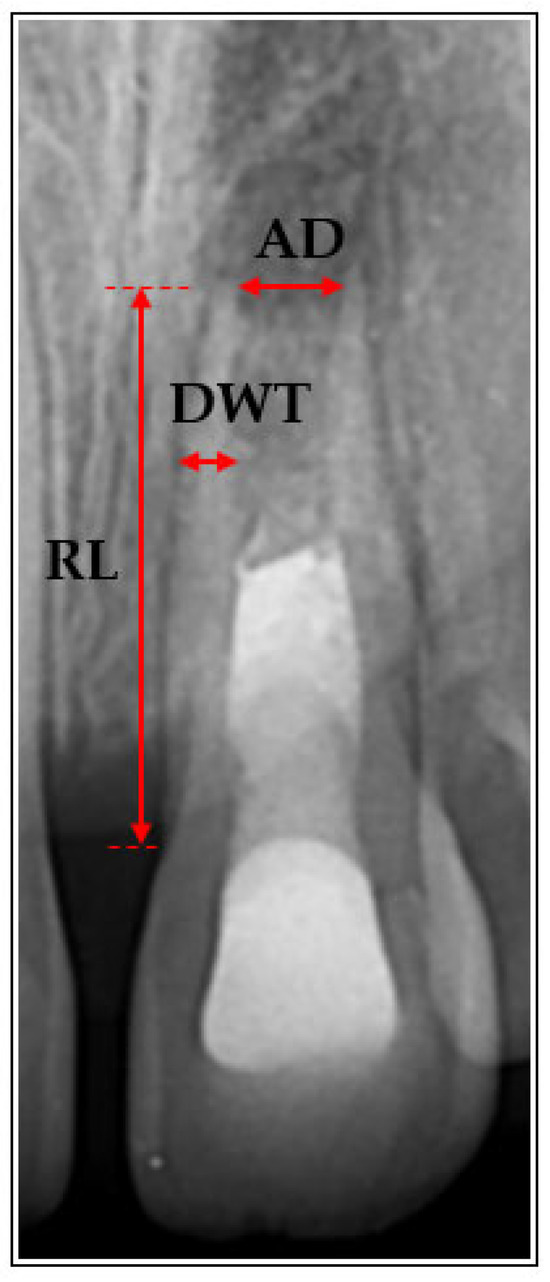
Measurements were conducted using a standardised approach consistent with previously published methodologies for the assessment of immature teeth [30], including linear measurements of root length from the cemento-enamel junction to the apex, dentinal wall thickness at a predefined level of approximately two-thirds of the root length, and apical diameter measured at the root apex [31].
The following absolute radiographic parameters were assessed:
- Root length (RL, mm), defined as the distance from the cemento-enamel junction to the root apex.
- Dentinal wall thickness (DWT, mm), measured at a standardised point at approximately two-thirds of the root length on the root wall best visible on the radiograph, with the same wall consistently used for all measurements within each tooth.
- Apical diameter (AD, mm), defined as the width of the apical foramen measured at the root apex.
- Periapical status, assessed using the Periapical Index (PAI; scores 1–5) [32].
- Root resorption, recorded as absent or present at baseline and classified at follow-up as absent, arrested, or progressive.
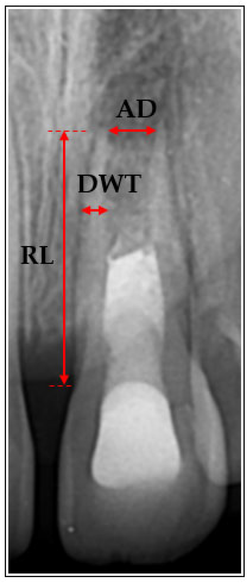
Radiographic measurements of root length, dentinal wall thickness, and apical diameter were performed at predefined locations, as illustrated in Figure 1.
Figure 1.
Schematic illustration of radiographic measurements performed on periapical radiographs. Root length (RL), dentinal wall thickness (DWT), and apical diameter (AD) were measured at predefined, standardised locations.
At the final follow-up, apical closure was assessed qualitatively on periapical radiographs and classified as follows:
- 0—no apical closure: persistence of an open apical foramen, without radiographic evidence of narrowing;
- 1—partial apical closure: reduction in apical diameter with radiographic evidence of narrowing, without complete closure;
- 2—complete apical closure: radiographic absence of an apical opening, including complete or irregular apical barrier formation.
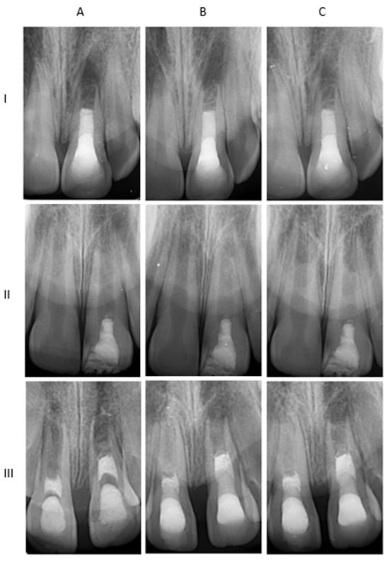
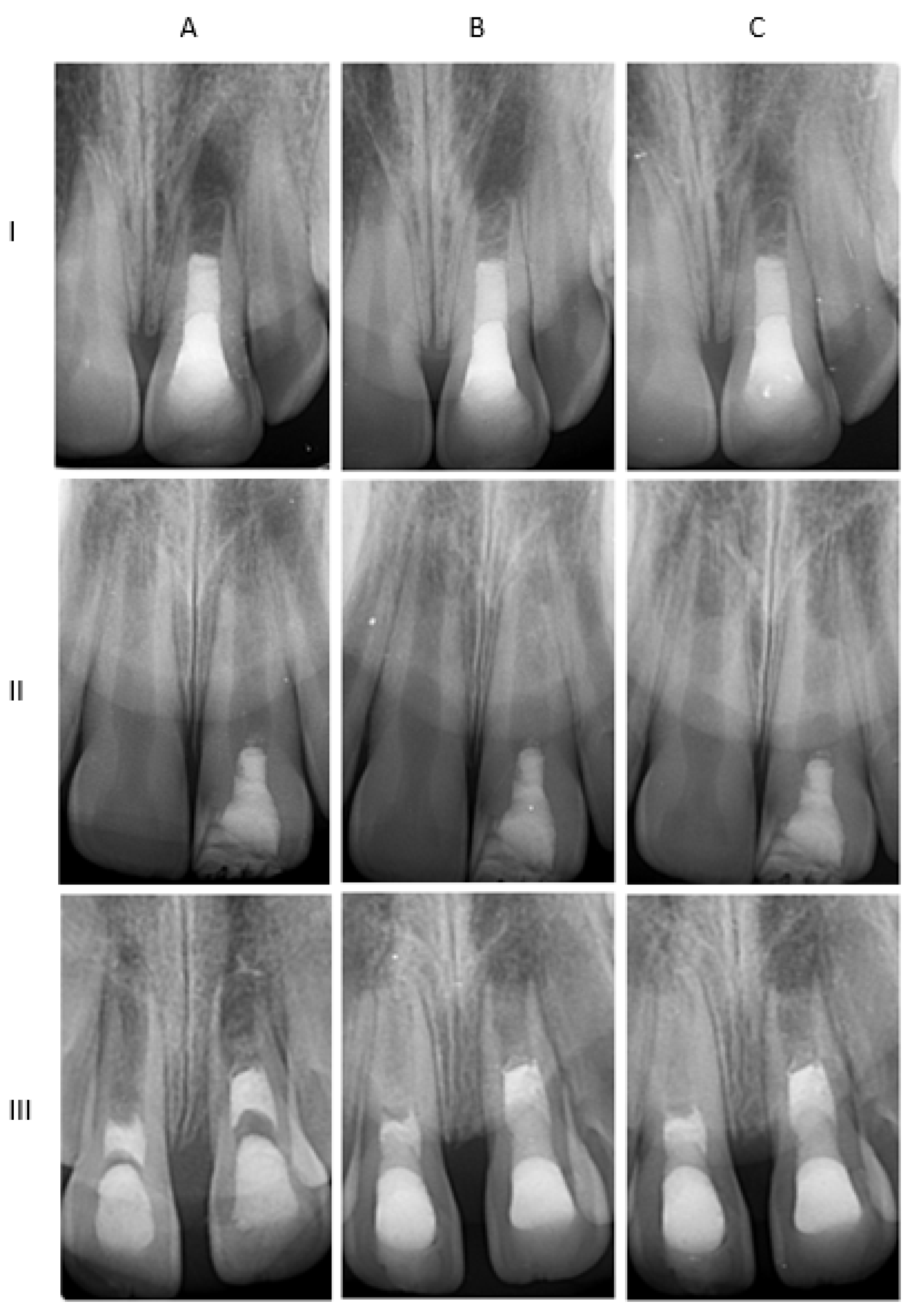
A representative example of baseline and follow-up periapical radiographs used for longitudinal assessment is shown in Figure 2.
Figure 2.
Representative periapical radiographs of three cases (I–III) obtained at baseline (A), 6 months (B), and 12 months (C), showing radiographic changes over time.
2.9. Changes over Time
Radiographic changes were calculated as the difference between follow-up (6 and 12 months) and baseline measurements:
- ΔRL (mm) = RL at follow-up—RL at baseline
- ΔDWT (mm) = DWT at follow-up—DWT at baseline
- ΔAD (mm) = AD at follow-up—AD at baseline
Negative ΔAD values indicated a reduction in apical diameter, whereas negative ΔRL values reflected a decrease in root length. Δ values were analysed only when both baseline and follow-up measurements were available and considered reliable.
2.10. Outcome Measures
2.10.1. Primary Outcome—Biological Outcome
The primary outcome was biological healing, assessed at the final follow-up (12 months) based on clinical findings, periapical status, and resorptive changes [13,24,30]. Outcomes were classified as
- Success (complete healing): absence of clinical symptoms, PAI ≤ 2, and no evidence of progressive root resorption.
- Survival (incomplete healing): absence of clinical symptoms with stable or improved periapical status compared with baseline (no increase in PAI score), with arrested resorption or no radiographic deterioration.
- Failure: persistence or development of clinical symptoms, worsening periapical status (increase in PAI score), and/or progressive root resorption.
2.10.2. Secondary Outcome—Regenerative Outcome
The secondary outcome was regenerative root development, evaluated based on changes in radiographic parameters (ΔRL, ΔDWT, ΔAD) between baseline and follow-up examinations at 6 and 12 months, as well as qualitative assessment of apical closure [13,30]. Outcomes were classified as follows:
- Favourable root development: Clear radiographic evidence of continued root maturation, defined as an increase in root length of ≥0.5 mm (ΔRL ≥ 0.5 mm) and/or evidence of partial or complete apical closure, with or without an increase in dentinal wall thickness.
- Limited root development: Minor radiographic changes not meeting the criteria for favourable development (i.e., ΔRL < 0.5 mm), without evidence of deterioration.
- Unfavourable root development: Radiographic evidence of deterioration, including a decrease in root length (ΔRL < 0), an increase in apical diameter (ΔAD > 0), a decrease in dentinal wall thickness (ΔDWT < 0), or progressive root resorption.
The threshold of 0.5 mm for root length increase was selected to represent a clinically meaningful change exceeding expected measurement variability, in line with previously reported radiographic assessment approaches in regenerative endodontic studies [11,30].
2.11. Statistical Analysis
Statistical analysis was performed using the SPSS software package, version 25.0 (IBM, Armonk, NY, USA). Descriptive statistics were used to summarise the data. Continuous variables were presented as mean and standard deviation (SD), while categorical variables were expressed as frequencies and percentages.
Between-group comparisons of continuous variables (root length, dentinal wall thickness, and apical diameter, including their changes over time) were performed using the independent samples t-test, as the data distributions were considered acceptable for parametric analysis. Comparisons between groups were primarily based on changes over time (Δ values), allowing each tooth to serve as its own reference point when assessing treatment-related effects. For baseline root length, results were consistent across parametric and non-parametric approaches. Categorical variables (tooth type, trauma type, symptoms, resorption, apical closure, biological outcome, and regenerative outcome) were compared using the chi-square test, with post hoc pairwise comparisons (z-test) where appropriate. The level of statistical significance was set at α = 0.05.
Intra-examiner reliability was assessed using the intraclass correlation coefficient (ICC) based on a two-way mixed-effects model. Root length, dentinal wall thickness, and apical diameter were measured for the whole sample (n = 49) and re-measured after a 48h interval by the same examiner. Intra-examiner reliability showed very high agreement, with ICC values of 0.961 for root length, 0.948 for dentinal wall thickness, and 0.979 for apical diameter. Effect size (Cohen’s d) was calculated for between-group comparisons.
3. Results
A total of 49 teeth from patients aged <18 years were included in the study. Of these, 29 teeth were treated with regenerative endodontic treatment (RET) and 20 with apexification.
3.1. Baseline Characteristics
The mean age of patients was similar between groups (RET: 8.9 ± 2.1 years; apexification: 9.4 ± 2.2 years). Baseline clinical and radiographic characteristics did not differ significantly. A difference was observed in baseline root length (RL_baseline), which was slightly lower in the RET group (13.1 ± 1.8 mm vs. 14.0 ± 1.3 mm; p = 0.041), whereas dentinal wall thickness and apical diameter were comparable between groups. No significant differences were observed between groups at baseline for Cvek stage or PAI scores. Detailed baseline characteristics are presented in Table 1.
Table 1.
Baseline characteristics of included teeth according to treatment group.
3.2. Radiographic Outcomes
At 6 months, no significant differences in root length or apical diameter were observed between groups. However, dentinal wall thickness was significantly greater in the RET group compared with the apexification group (p = 0.013). At 12 months, dentinal wall thickness remained significantly greater in the RET group (p = 0.005), while no statistically significant differences were observed for root length or apical diameter (Table 2). Among the evaluated radiographic parameters, dentinal wall thickness demonstrated the most consistent and statistically significant differences between groups at both follow-up time points.
Table 2.
Radiographic parameters at 6 and 12 months after treatment.
3.3. Changes in Radiographic Parameters over Time
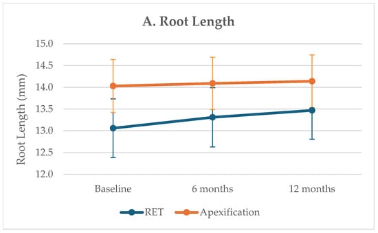
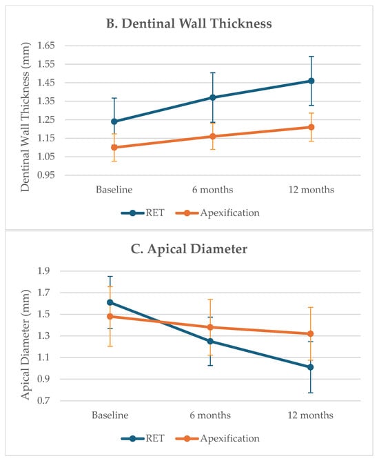
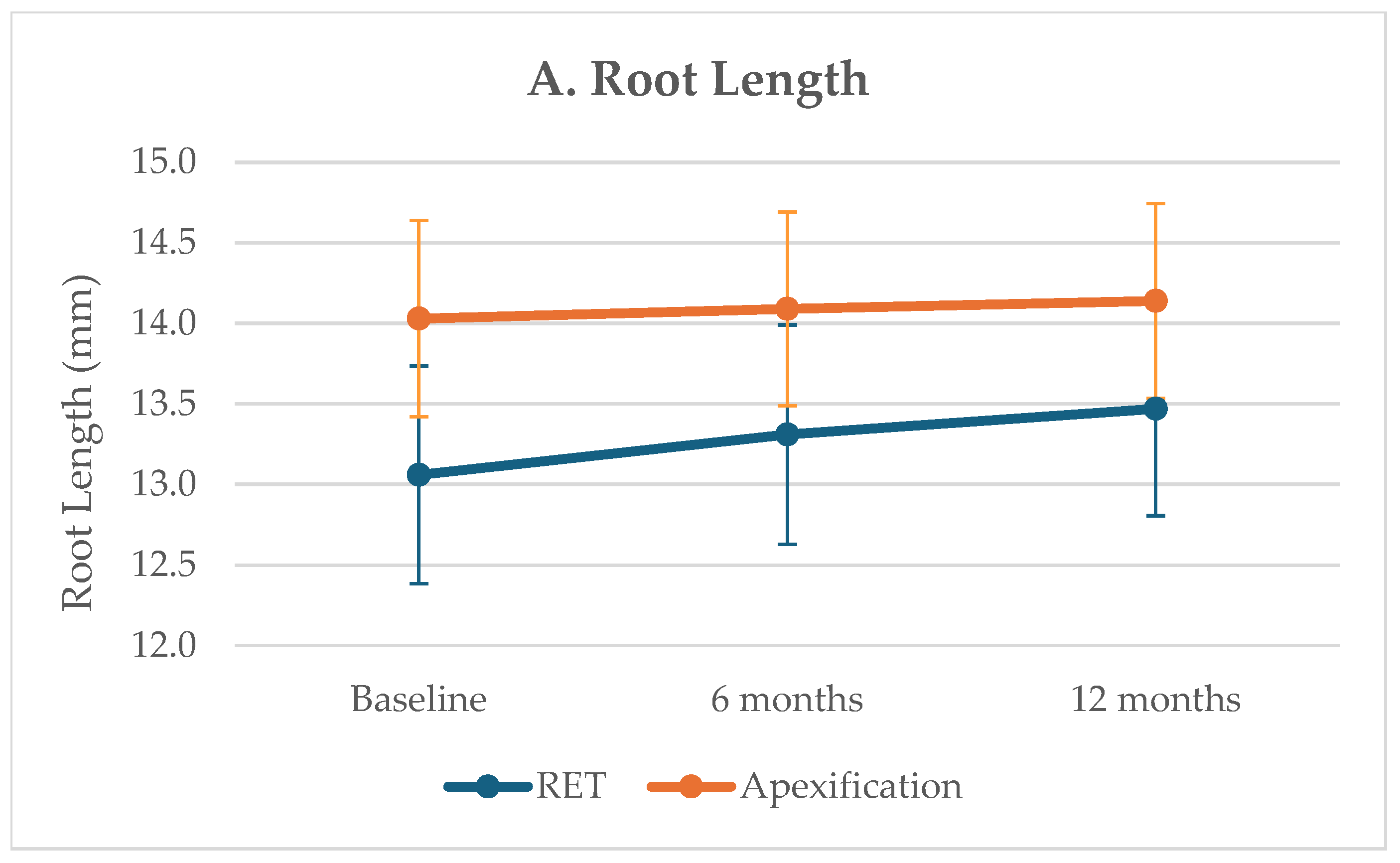
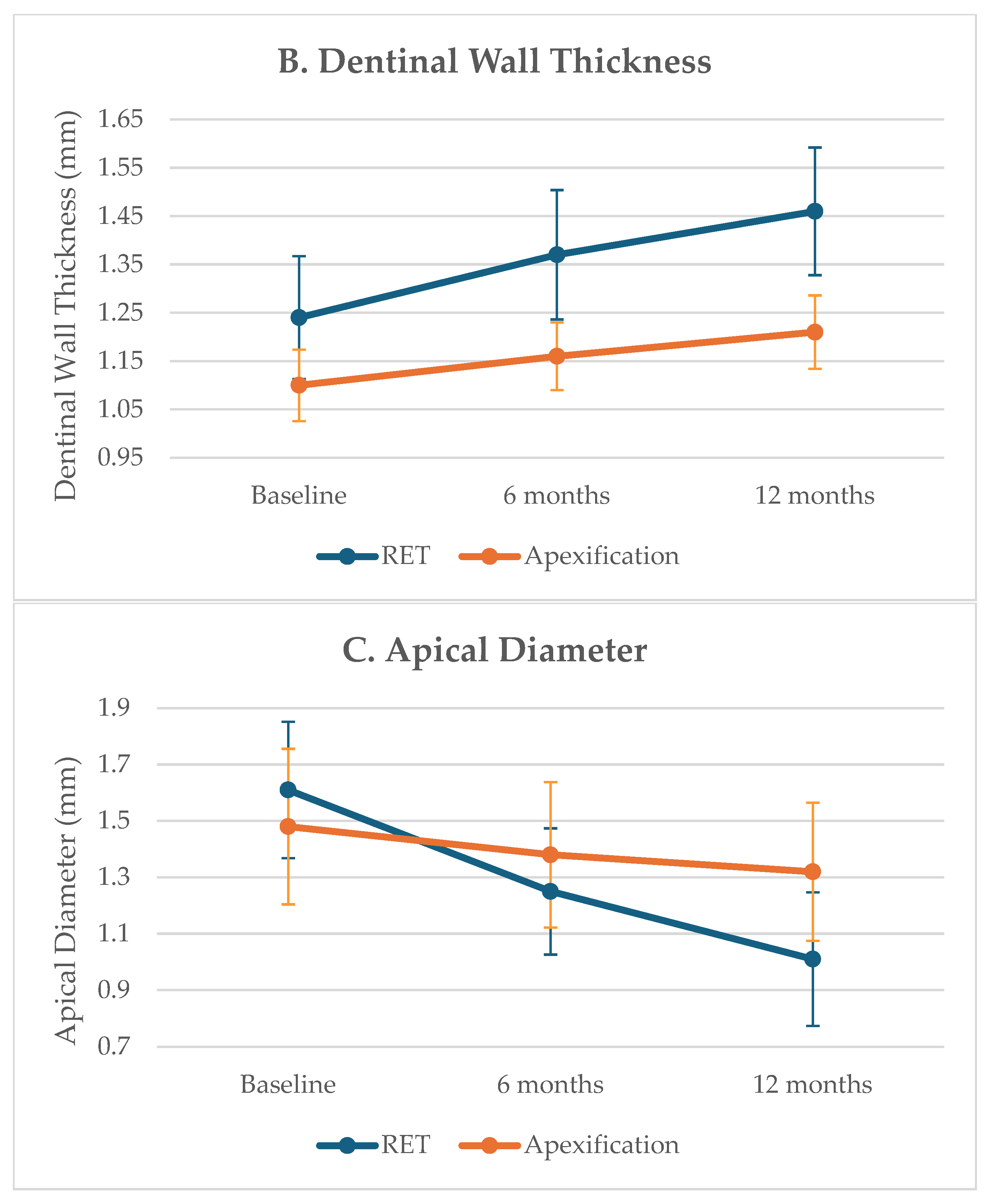
Analysis of changes over time showed greater increases in the RET group compared with apexification (Figure 3). The RET group demonstrated a significantly greater increase in root length (ΔRL: 0.42 ± 0.50 mm vs. 0.11 ± 0.09 mm; p = 0.010) and dentinal wall thickness (ΔDWT: 0.21 ± 0.14 mm vs. 0.11 ± 0.04 mm; p = 0.002), as well as a greater reduction in apical diameter (ΔAD: −0.60 ± 0.39 mm vs. −0.16 ± 0.13 mm; p < 0.001) (Table 3). The difference in ΔDWT corresponded to a large effect size (Cohen’s d = 0.97), with a statistical power of approximately 89%.
Figure 3.
Changes in (A) root length (RL), (B) dentinal wall thickness (DWT), and (C) apical diameter (AD) over time in the RET and apexification groups at baseline, 6 months, and 12 months. Error bars represent 95% confidence intervals.
Table 3.
Radiographic changes over time (Δ values), representing the difference between final follow-up and baseline measurements.
3.4. Categorical Clinical and Radiographic Variables
Baseline characteristics were similar between groups, with no differences in tooth type or symptoms. A higher proportion of avulsion injuries was observed in the RET group (34.5% vs. 5.0%), and baseline resorption was present only in this group (41.4%).
At follow-up, clinical outcomes appeared broadly similar between groups, although numerical differences in outcome distribution were observed. Arrested root resorption and crown discolouration were observed only in the RET group. Detailed distributions are presented in Table 4.
Table 4.
Comparison of categorical variables between groups. Data are presented as frequencies (n) and percentages (%).
3.5. Clinical and Radiographic Outcomes
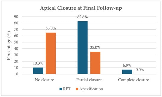
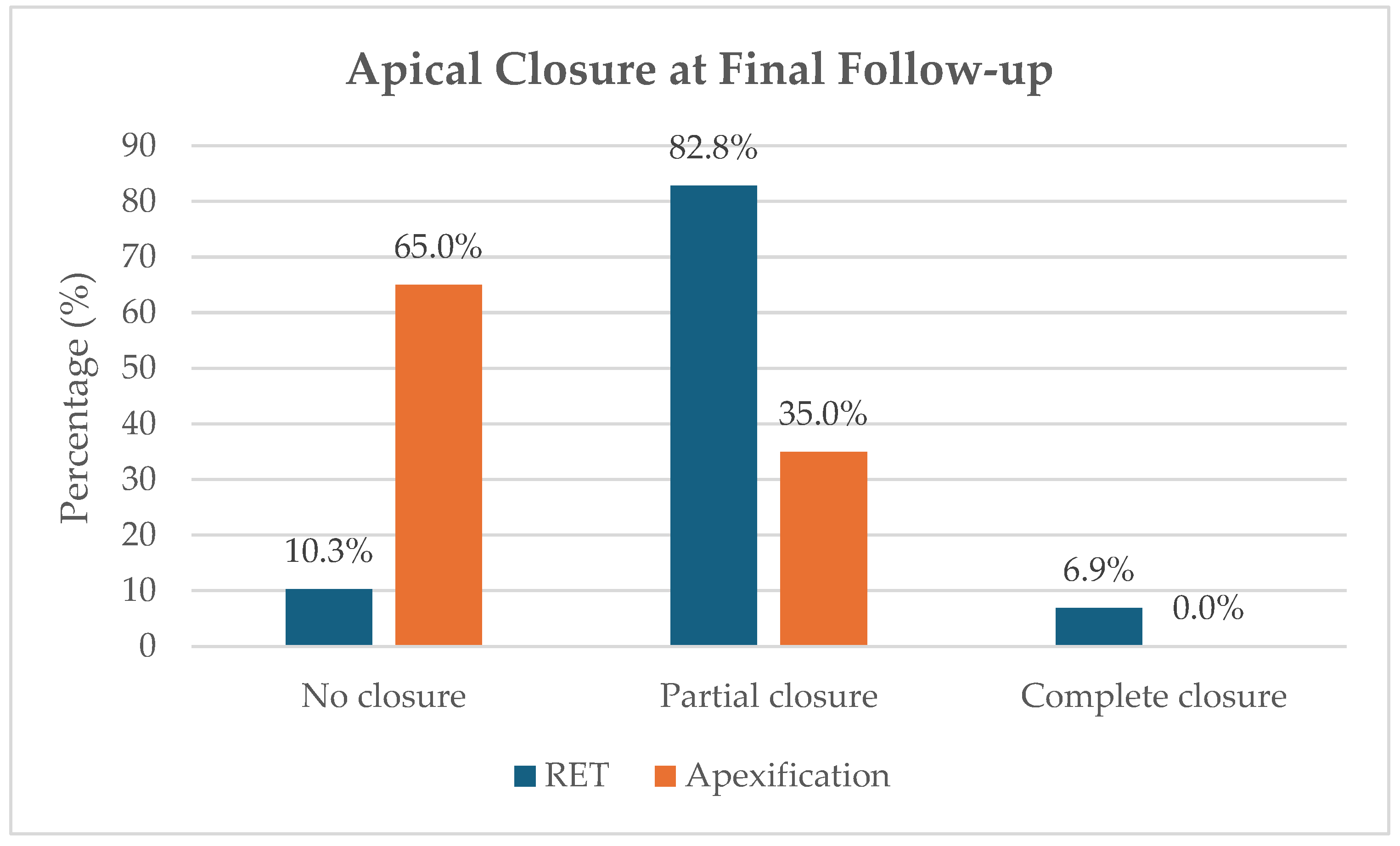
At the final follow-up, the RET group demonstrated a significantly higher degree of apical closure compared with the apexification group (p < 0.001; Figure 4). PAI scores improved in both groups, with more favourable values observed in the RET group (p = 0.031). No statistically significant difference was observed in biological outcomes between groups (p = 0.084). In contrast, regenerative outcomes were significantly more favourable in the RET group (p = 0.001), consistent with the distributions presented in Table 4.
Figure 4.
Distribution of apical closure at the final follow-up in the RET and apexification groups.
4. Discussion
The present study evaluated the clinical and radiographic outcomes of regenerative endodontic treatment (RET) incorporating an additional calcium hydroxide-based step compared with apexification in traumatised immature teeth. The findings indicate that RET resulted in more favourable radiographic outcomes, including greater root development and apical closure, while clinical outcomes appeared broadly similar between the two treatment approaches. However, differences in the distribution of “success” and “survival” outcomes were observed. These results are consistent with a recent umbrella review by Tewari et al., as well as with earlier clinical studies [11,12,13,14,15,16].
More specifically, radiographic parameters reflecting root maturation showed more pronounced changes in the RET group. Increased dentinal wall thickness and a reduction in apical diameter, recognised indicators of successful regenerative outcomes, were significantly greater in the RET group. Although dentinal wall thickness was not used as a defining criterion in the outcome classification, it emerged as the most consistently improved radiographic parameter. This is clinically relevant, as increased dentinal wall thickness contributes to improved structural integrity and may reduce the risk of fracture in immature teeth. Overall, these findings suggest that structural reinforcement, as reflected by increased dentinal wall thickness, may represent a key advantage of regenerative approaches.
In addition, apical closure was more frequently observed in the RET group, further supporting the regenerative potential of this approach. The predominance of partial apical closure observed in this study may reflect the biological variability of regenerative healing, particularly in traumatised teeth. Similar findings have been reported in a retrospective study on traumatised immature teeth by Cheng et al., in which partial apical closure was also frequently observed [33]. The concept of revascularisation, originally introduced by Banchs and Trope [5], provides the biological basis for these regenerative processes.
However, these radiographic advantages were not paralleled by differences in clinical outcomes, as clinical healing appeared broadly similar between groups. This highlights the distinction between clinical healing and true regenerative success, as resolution of symptoms and periapical healing may be achieved with both treatment approaches, whereas continued root development appears to be more closely associated with regenerative procedures. These findings are consistent with previous studies reporting comparable periapical healing between RET and apexification, as reflected by similar PAI scores and alternative radiographic assessment methods [30,34], as well as with the findings of a recent umbrella review [13]. However, apexification has been associated with long-term complications, including an increased risk of tooth fracture and premature tooth loss, particularly in structurally compromised immature teeth [35]. Notably, a tendency towards more favourable outcomes following RET has also been reported [12,36], although these findings should be interpreted cautiously given the variability in study designs and clinical protocols.
Differences between the findings of the present study and previously published data may be partly explained by variations in study populations and clinical conditions. While several studies have evaluated regenerative endodontic treatment in mixed cohorts of immature teeth with different aetiologies, including both caries- and trauma-related cases [37], the present study focused exclusively on traumatised immature teeth, which represent a more complex clinical scenario due to compromised vascular supply and a higher risk of complications, such as root resorption. This may have influenced the observed pattern of healing, particularly the predominance of partial rather than complete apical closure. These differences in baseline characteristics, particularly those related to trauma severity and the presence of resorption, should be considered when interpreting the results.
Differences in trauma type and severity may further contribute to variability in regenerative outcomes. Traumatised teeth often exhibit greater disruption of the apical tissues and stem cell niche, which may limit continued root development despite successful infection control [25]. These factors may also affect baseline comparability between treatment groups and should therefore be considered when interpreting the findings.
Notably, the RET group included a higher proportion of teeth with more severe trauma, including avulsion injuries and pre-existing resorption, both of which are known to negatively affect healing potential. This imbalance reflects real-world clinical decision-making, in which regenerative approaches are more frequently selected for complex cases. Therefore, the observed radiographic differences between groups should be interpreted in the context of potential selection bias and residual baseline confounding. In addition, given the retrospective design and limited sample size, the ability to statistically control for these baseline differences was limited.
Nevertheless, the overall pattern observed in the present study, characterised by favourable radiographic changes following RET combined with broadly similar clinical healing, remains consistent with previously reported trends [15,37,38,39], suggesting the potential applicability of regenerative approaches even in clinically challenging trauma-related conditions.
Given the variability of reported outcomes, it is important to note that no universally accepted classification system for regenerative outcomes currently exists, and most studies report individual radiographic parameters such as root length, dentinal wall thickness, and apical closure [13,37]. In the present study, these commonly reported parameters were structured into a clinically interpretable framework to facilitate outcome assessment. This approach was intended to provide a structured description of radiographic changes over time.
Root resorption was observed only in the RET group at baseline, reflecting the traumatic origin of the cases included in this study. This is consistent with previous evidence indicating that traumatic dental injuries are a major predisposing factor for the development of external root resorption [24]. The presence of baseline resorption exclusively in the RET group likely reflects case selection rather than treatment-related differences, as regenerative procedures are more commonly indicated in teeth with advanced trauma-related complications [15]. At follow-up, resorption was arrested in all affected cases, suggesting a possible contribution of regenerative treatment to the stabilisation of resorptive processes. This may be explained by effective infection control and the resolution of inflammation, which are essential for halting resorptive activity [22].
Crown discolouration was observed only in the RET group and is likely related to the use of calcium silicate-based materials, such as mineral trioxide aggregate, which has been consistently associated with tooth discolouration in regenerative procedures [17].
The outcomes of regenerative endodontic treatment are strongly influenced by the properties of the materials used, particularly intracanal medicaments and bioactive cements [7,19,40]. A specific feature of the present study was the use of an additional calcium hydroxide-based step for blood clot stabilisation as part of the applied RET protocol. The study was not designed to isolate the effect of this step, but rather to evaluate outcomes associated with the protocol as a whole. From a biological perspective, calcium hydroxide has been associated with antimicrobial effects and with conditions that may support a favourable regenerative microenvironment. Its high alkalinity has been shown to disrupt bacterial cell membranes and proteins, while previous studies have also suggested potential compatibility with stem cell survival and tissue healing [26,41]. In this context, the additional calcium hydroxide-based step may have contributed to procedural stability and infection control during treatment. However, the present findings should not be interpreted as evidence of a specific independent effect of this adjunctive step.
Previous studies have explored a variety of scaffold-based and adapted approaches to enhance regenerative potential, including the use of platelet-rich fibrin [31,42], platelet-rich plasma, and induced blood clot formation, all of which have been associated with favourable clinical outcomes [43,44]. In addition, scaffold type may influence regenerative outcomes, with biologically active scaffolds such as platelet-rich fibrin showing a tendency towards improved radiographic results, although no single approach has been shown to be consistently superior. Furthermore, biocompatible irrigation and intracanal protocols that preserve stem cell viability and support the formation of a functional scaffold are considered important for successful regenerative procedures [8,45].
Within this context, the additional calcium hydroxide-based step used for blood clot stabilisation in the present study may be considered a minor adjunctive component of the applied RET. Given the variability of existing regenerative protocols [21,38,39], this approach represents a simple and clinically applicable adjunctive approach that does not require additional equipment or laboratory procedures. The observed outcomes suggest that this approach may be compatible with favourable clinical performance. However, these findings should be interpreted with caution due to the retrospective nature of the study and should be considered descriptive of clinical practice rather than evidence of superiority [20,21]. Further prospective and controlled studies are needed to better evaluate the potential benefits of this adjunctive step.
This study has several limitations, including its retrospective design, relatively small sample size, and variability in trauma type. It should be noted that the reported statistical power primarily relates to continuous radiographic outcomes, whereas the study may have been underpowered to detect differences in categorical clinical outcomes. The inclusion of clinically heterogeneous cases may have contributed to variability in baseline conditions and treatment responses, thereby reflecting the complexity of real-world clinical practice. In addition, the 12-month follow-up period, while clinically relevant for assessing early healing and radiographic changes, may not have been sufficient to capture continued root maturation or long-term outcomes, including late complications such as fracture or progressive crown discolouration.
Radiographic assessment was based on two-dimensional periapical radiographs, which are subject to inherent limitations, including projection errors and geometric distortion [29]. Despite the use of a standardised measurement protocol, minor variations in image acquisition may have affected measurement accuracy.
Within these limitations, regenerative endodontic treatment incorporating an additional calcium hydroxide-based step appears to be a clinically applicable approach for promoting root development in traumatised immature teeth while maintaining broadly similar clinical outcomes to apexification. However, these findings should be interpreted with caution, and further prospective and controlled studies are required to confirm these findings and to better define the role of such adjunctive steps.
5. Conclusions
Within the limitations of this retrospective study, regenerative endodontic treatment was associated with improved radiographic outcomes, including greater root development and apical closure, compared with apexification in traumatised immature teeth, while clinical outcomes appeared broadly similar. These findings suggest the potential of regenerative approaches to promote continued root maturation in clinically challenging trauma-related conditions.
Author Contributions
Conceptualization, P.B.S., H.J. and D.G.; methodology, P.B.S. and H.J.; validation, P.B.S., J.S. and J.B.; resources, T.Š. and K.G.; formal analysis, P.B.S., D.G., D.T., T.Š. and K.G.; investigation, P.B.S., J.S., J.B. and D.T.; data curation, J.S., J.B. and D.T.; writing—original draft preparation, P.B.S.; writing—review and editing, P.B.S., D.T., D.G., T.Š., K.G. and H.J.; supervision, H.J.; Project administration, P.B.S. and D.G. All authors have read and agreed to the published version of the manuscript.
Funding
This research received no external funding.
Institutional Review Board Statement
The study was conducted in accordance with the Declaration of Helsinki and was approved by the Ethics Committee of the School of Dental Medicine, University of Zagreb (Class: 003-01/25-05/14; Reg. No.: 251-60-4/119-10; approved on 3 December 2025).
Informed Consent Statement
Informed consent was obtained from all subjects involved in the study.
Data Availability Statement
All the data used in this study are available on request from the corresponding author.
Conflicts of Interest
The authors declare no conflicts of interest.
Abbreviations
The following abbreviations are used in this manuscript:
| AD | Apical diameter |
| DWT | Dentinal wall thickness |
| ICC | Intraclass correlation coefficient |
| MTA | Mineral trioxide aggregate |
| PAI | Periapical Index |
| RET | Regenerative endodontic treatment |
| RL | Root length |
| SD | Standard deviation |
| ΔAD | Change in apical diameter |
| ΔDWT | Change in dentinal wall thickness |
| ΔRL | Change in root length |
References
- Wigler, R.; Kaufman, A.Y.; Lin, S.; Steinbock, N.; Hazan-Molina, H.; Torneck, C.D. Revascularization: A treatment for permanent teeth with necrotic pulp and incomplete root development. J. Endod. 2013, 39, 319–326. [Google Scholar] [CrossRef] [Scilit] [PubMed]
- Andreasen, J.O.; Farik, B.; Munksgaard, E.C. Long-term calcium hydroxide as a root canal dressing may increase risk of root fracture. Dent. Traumatol. 2002, 18, 134–137. [Google Scholar] [CrossRef] [Scilit]
- Rafter, M. Apexification: A review. Dent. Traumatol. 2005, 21, 1–8. [Google Scholar] [CrossRef] [Scilit] [PubMed]
- Guerrero, F.; Mendoza, A.; Ribas, D.; Aspiazu, K. Apexification: A systematic review. J. Conserv. Dent. 2018, 21, 462–465. [Google Scholar] [CrossRef] [Scilit]
- Banchs, F.; Trope, M. Revascularization of immature permanent teeth with apical periodontitis: New treatment protocol? J. Endod. 2004, 30, 196–200. [Google Scholar] [CrossRef] [Scilit]
- Diogenes, A.; Ruparel, N.B.; Shiloah, Y.; Hargreaves, K.M. Regenerative endodontics: A way forward. J. Am. Dent. Assoc. 2016, 147, 372–380. [Google Scholar] [CrossRef] [Scilit]
- Sequeira, D.B.; Diogo, P.; Gomes, B.P.F.A.; Peça, J.; Santos, J.M.M. Scaffolds for dentin–pulp complex regeneration. Medicina 2023, 60, 7. [Google Scholar] [CrossRef] [Scilit]
- Kharchi, A.S.; Tagiyeva-Milne, N.; Kanagasingam, S. Regenerative endodontic procedures, disinfectants and outcomes: A systematic review. Prim. Dent. J. 2020, 9, 65–84. [Google Scholar] [CrossRef] [Scilit]
- Lovelace, T.W.; Henry, M.A.; Hargreaves, K.M.; Diogenes, A. Evaluation of the delivery of mesenchymal stem cells into the root canal space of necrotic immature teeth after clinical regenerative endodontic procedure. J. Endod. 2011, 37, 133–138. [Google Scholar] [CrossRef] [Scilit]
- Hargreaves, K.M.; Diogenes, A.; Teixeira, F.B. Treatment options: Biological basis of regenerative endodontic procedures. J. Endod. 2013, 39, S30–S43. [Google Scholar] [CrossRef] [Scilit] [PubMed]
- Jeeruphan, T.; Jantarat, J.; Yanpiset, K.; Suwannapan, L.; Khewsawai, P.; Hargreaves, K.M. Comparison of radiographic and survival outcomes of immature teeth treated with regenerative endodontic or apexification methods. J. Endod. 2012, 38, 1330–1336. [Google Scholar] [CrossRef] [Scilit]
- Lin, J.; Zeng, Q.; Wei, X.; Zhao, W.; Cui, M.; Gu, J.; Lu, J.; Yang, M.; Ling, J. Regenerative endodontics versus apexification in immature permanent teeth with apical periodontitis: A randomized controlled study. J. Endod. 2017, 43, 1821–1827. [Google Scholar] [CrossRef] [Scilit]
- Tewari, N.; Devi, P.; Sampath, S.; Mathur, V.P.; Tsilingaridis, G.; Wikström, A.; Rahul, M.; Bansal, K. Comparative effectiveness of regenerative endodontic treatment versus apexification: An umbrella review. Dent. Traumatol. 2025, 41, 263–282. [Google Scholar] [CrossRef] [Scilit]
- American Association of Endodontists. Regenerative endodontics: A position statement. J. Endod. 2016, 42, 1340–1343. [Google Scholar]
- Saoud, T.M.A.; Zaazou, A.; Nabil, A.; Moussa, S.; Lin, L.M.; Gibbs, J.L. Clinical and radiographic outcomes of traumatized immature permanent necrotic teeth after revascularization. J. Endod. 2014, 40, 1946–1952. [Google Scholar] [CrossRef] [Scilit]
- Panda, P.; Mishra, L.; Govind, S.; Panda, S.; Lapinska, B. Clinical outcome and comparison of regenerative and apexification intervention in immature necrotic teeth: A systematic review and meta-analysis. J. Clin. Med. 2022, 11, 3909. [Google Scholar] [CrossRef] [Scilit] [PubMed]
- Staffoli, S.; Plotino, G.; Nunez Torrijos, B.G.; Grande, N.M.; Bossù, M.; Gambarini, G.; Polimeni, A. Regenerative endodontic procedures using contemporary materials. Materials 2019, 12, 908. [Google Scholar] [CrossRef] [Scilit] [PubMed]
- Nazzal, H.; Duggal, M.S. Regenerative endodontics: A true paradigm shift or a bandwagon about to be derailed? Eur. Arch. Paediatr. Dent. 2017, 18, 3–15. [Google Scholar] [CrossRef] [Scilit]
- Pulyodan, M.K.; Paramel Mohan, S.; Valsan, D.; Divakar, N.; Moyin, S.; Thayyil, S. Regenerative endodontics: A paradigm shift in clinical endodontics. J. Pharm. Bioallied Sci. 2020, 12, S20–S26. [Google Scholar] [CrossRef] [Scilit]
- Keinan, D.; Nuni, E.; Bronstein Rainus, M.; Ben Simhon, T.; Dakar, A.; Slutzky-Goldberg, I. Retreatment of failed regenerative endodontic therapy: Outcome and treatment considerations. Cureus 2024, 16, e75147. [Google Scholar] [CrossRef] [Scilit]
- Asgary, S.; Shamszadeh, S.; Nosrat, A.; Aminoshariae, A.; Sabeti, M. Management strategies for immature teeth with pulp necrosis: An umbrella review of systematic reviews. Iran. Endod. J. 2024, 19, 242–253. [Google Scholar] [CrossRef] [Scilit] [PubMed]
- Wei, X.; Yang, M.; Yue, L.; Huang, D.; Zhou, X.; Wang, X.; Zhang, Q.; Qiu, L.; Huang, Z.; Wang, H.; et al. Expert consensus on regenerative endodontic procedures. Int. J. Oral Sci. 2022, 14, 55. [Google Scholar] [CrossRef] [Scilit]
- Kahler, B.; Chugal, N.; Lin, L.M. Alkaline materials and regenerative endodontics: A review. Materials 2017, 10, 1389. [Google Scholar] [CrossRef] [Scilit]
- Andreasen, J.O.; Andreasen, F.M. Root resorption following traumatic dental injuries. Proc. Finn. Dent. Soc. 1992, 88, 95–114. [Google Scholar]
- Geisler, T. Clinical considerations for regenerative endodontic procedures. Dent. Clin. N. Am. 2012, 56, 603–626. [Google Scholar] [CrossRef] [Scilit]
- Kim, S.G.; Malek, M.; Sigurdsson, A.; Lin, L.M.; Kahler, B. Regenerative endodontics: A comprehensive review. Int. Endod. J. 2018, 51, 1367–1388. [Google Scholar] [CrossRef] [Scilit]
- Nagy, M.M.; Tawfik, H.E.; Hashem, A.A.R.; Abu-Seida, A.M. Regenerative potential of immature permanent teeth with necrotic pulps after different regenerative protocols. J. Endod. 2014, 40, 192–198. [Google Scholar] [CrossRef] [Scilit] [PubMed]
- Cvek, M. Prognosis of luxated non-vital maxillary incisors treated with calcium hydroxide and filled with gutta-percha. Endod. Dent. Traumatol. 1992, 8, 45–55. [Google Scholar] [PubMed]
- Yen, M.; Yeung, A.W.K. The performance of paralleling technique and bisecting angle technique for taking periapical radiographs: A systematic review. Dent. J. 2023, 11, 155. [Google Scholar] [CrossRef] [Scilit]
- Chen, S.J.; Chen, L.P. Radiographic outcome of necrotic immature teeth treated with two endodontic techniques: A retrospective analysis. Biomed. J. 2016, 39, 366–371. [Google Scholar] [CrossRef] [Scilit]
- Turjanski, D.; Lisjak, D.; Bučević Sojčić, P.; Valpotić, J.; Borojević Renić, T.; Goršeta, K.; Glavina, D. Advanced platelet-rich fibrin sealed with glass ionomer cement in regenerative endodontics. Materials 2025, 18, 4421. [Google Scholar] [CrossRef] [Scilit]
- Orstavik, D.; Kerekes, K.; Eriksen, H.M. The periapical index: A scoring system for radiographic assessment of apical periodontitis. Endod. Dent. Traumatol. 1986, 2, 20–34. [Google Scholar] [CrossRef] [Scilit]
- Cheng, J.; Yang, F.; Li, J.; Hua, F.; He, M.; Song, G. Treatment outcomes of regenerative endodontic procedures in traumatized immature permanent necrotic teeth: A retrospective study. J. Endod. 2022, 48, 1129–1136. [Google Scholar] [CrossRef] [Scilit]
- Pereira, A.C.; Oliveira, M.L.; Cerqueira-Neto, A.C.C.L.; Vargas-Neto, J.; Nagata, J.Y.; Gomes, B.P.F.A.; Ferraz, C.C.R.; de Almeida, J.F.A.; de-Jesus-Soares, A. Outcomes of traumatised immature teeth treated with apexification or regenerative endodontic procedure. Aust. Endod. J. 2021, 47, 178–187. [Google Scholar] [CrossRef] [Scilit]
- Wikström, A.; Brundin, M.; Mohmud, A.; Anderson, M.; Tsilingaridis, G. Outcomes of apexification in immature traumatised necrotic teeth and risk factors for premature tooth loss: A 20-year longitudinal study. Dent. Traumatol. 2024, 40, 658–671. [Google Scholar] [CrossRef] [Scilit] [PubMed]
- Kyaw, M.S.; Zhang, Y.; Li, X.; Wang, Y.; Chen, Y.; Liu, Z. Endodontic regeneration therapy: Current strategies and tissue engineering solutions. Cells 2025, 14, 422. [Google Scholar] [CrossRef] [Scilit] [PubMed]
- Lopes, L.B.; Neves, J.A.; Botelho, J.; Machado, V.; Mendes, J.J. Regenerative endodontic procedures: An umbrella review. Int. J. Environ. Res. Public Health 2021, 18, 754. [Google Scholar] [CrossRef] [Scilit]
- Theekakul, C.; Banomyong, D.; Osiri, S.; Sutam, N.; Ongchavalit, L.; Jantarat, J. Treatment outcomes and prognostic factors of regenerative endodontic procedures in immature permanent teeth. J. Endod. 2024, 50, 1569–1578. [Google Scholar] [CrossRef] [Scilit]
- Žižka, R.; Belák, Š.; Šedý, J.; Fačevicová, K.; Voborná, I.; Marinčák, D. Clinical and radiographic outcomes of immature teeth treated with different regenerative protocols: A retrospective cohort study. J. Clin. Med. 2021, 10, 1600. [Google Scholar] [CrossRef] [Scilit] [PubMed]
- Dissanayaka, W.L.; Zhang, C. Scaffold-based and scaffold-free strategies in dental pulp regeneration. J. Endod. 2020, 46, S81–S89. [Google Scholar] [CrossRef] [Scilit]
- Mohammadi, Z.; Shalavi, S.; Yazdizadeh, M. Antimicrobial activity of calcium hydroxide in endodontics: A review. Chonnam Med. J. 2012, 48, 133–140. [Google Scholar] [CrossRef] [Scilit]
- Wakhloo, T.; Shukla, S.; Chug, A.; Dhar, M. Platelet-rich fibrin-mediated regeneration of necrotic immature permanent teeth: A clinical study. Int. J. Clin. Pediatr. Dent. 2022, 15, 402–406. [Google Scholar] [CrossRef] [Scilit]
- Yoshpe, M.; Kaufman, A.Y.; Lin, S.; Ashkenazi, M. Regenerative endodontics using platelet-rich fibrin in immature teeth. Eur. Arch. Paediatr. Dent. 2021, 22, 527–534. [Google Scholar] [CrossRef] [Scilit] [PubMed]
- Murray, P.E. Platelet-rich plasma and platelet-rich fibrin in regeneration of immature permanent teeth: A meta-analysis. Front. Bioeng. Biotechnol. 2018, 6, 139. [Google Scholar] [CrossRef] [Scilit] [PubMed]
- Ruparel, N.B.; Teixeira, F.B.; Ferraz, C.C.R.; Diogenes, A. Direct effect of intracanal medicaments on survival of stem cells of the apical papilla. J. Endod. 2012, 38, 1372–1375. [Google Scholar] [CrossRef] [Scilit] [PubMed]

Disclaimer/Publisher’s Note: The statements, opinions and data contained in all publications are solely those of the individual author(s) and contributor(s) and not of MDPI and/or the editor(s). MDPI and/or the editor(s) disclaim responsibility for any injury to people or property resulting from any ideas, methods, instructions or products referred to in the content. |
© 2026 by the authors. Licensee MDPI, Basel, Switzerland. This article is an open access article distributed under the terms and conditions of the Creative Commons Attribution (CC BY) license.