Review

Therapeutic Potential of Selected Isoquinoline Alkaloids: Berbamine, Tetrandrine, Fangchinoline, and Sinomenine, in Neuropathic Pain Management


by
Anna Gumieniczek
* and
Aleksandra Kozińska
Department of Medicinal Chemistry, Faculty of Pharmacy, Medical University of Lublin, Jaczewskiego 4, 20-090 Lublin, Poland
*
Author to whom correspondence should be addressed.
Appl. Sci. 2026, 16(10), 4985; https://doi.org/10.3390/app16104985
Submission received: 9 April 2026 / Revised: 11 May 2026 / Accepted: 14 May 2026 / Published: 16 May 2026
(This article belongs to the Special Issue Bioactive Natural Compounds: From Discovery to Applications)

Abstract

Pharmacotherapy of neuropathic pain (NP) remains challenging due to its heterogeneous etiology, lack of objective diagnostic tools, and the limited efficacy of currently available treatments, including antidepressants, anticonvulsants, and local anesthetics. Therefore, the search for novel therapies with improved analgesic efficacy and reduced adverse effects is of growing importance. In this context, natural alkaloids have emerged as promising candidates, demonstrating analgesic potential in both diabetes-induced neuropathy and various experimental models of NP. This review outlines NP pathophysiology, emphasizing maladaptive changes within the somatosensory nervous system, including peripheral and central sensitization, as well as glial cell activation. Furthermore, it discusses the mechanisms through which alkaloids may modulate NP-related pathways, with particular focus on their interactions with ion channels, signaling pathways, inflammatory responses, and oxidative stress. A literature search was conducted using the Scopus, Google Scholar and PubMed databases for papers published between 2015 and 2026, using the keywords “alkaloids” and “neuropathic pain”, and focused on recent findings regarding the antinociceptive effects of berbamine, tetrandrine, fangchinoline, and sinomenine, and their derivatives. The analysis indicates that, despite promising preclinical evidence, further rigorous preclinical and clinical studies are necessary to fully assess their therapeutic potential in the treatment of NP.

1. Introduction

1.1. Pathophysiology of Neuropathic Pain (NP)

According to the taxonomy established by the International Association for the Study of Pain (IASP), neuropathic pain (NP) is defined as pain caused by a lesion or disease affecting the somatosensory nervous system [1]. In contrast to nociceptive pain, NP is characterized by maladaptive neuronal excitability and impaired modulation of nociceptive signaling. Clinically, it manifests as hyperalgesia (an exaggerated response to a painful stimulus) and allodynia (pain elicited by a normally non-noxious stimulus) [2,3]. The etiology of NP is highly heterogeneous and includes inflammatory conditions, malignancies, mechanical injury, immune dysfunction, chemotherapy-induced peripheral neuropathy, and metabolic disorders, particularly diabetic peripheral neuropathy [4,5]. At the molecular level, damage to sensory neurons induces significant alterations in expression and functional properties of voltage-gated (VG) ion channels, especially sodium Nav1.3, Nav1.7, Nav1.8, and Nav1.9, as well as potassium (Kv) and calcium (Cav) channels [5,6]. These neuroplastic changes occur primarily within the dorsal root ganglia (DRG), leading to a lowered depolarization threshold and generation of ectopic discharges in nociceptors [5]. This process is further exacerbated by enhanced purinergic signaling. Under conditions of cellular stress, extracellular ATP activates ionotropic P2X receptors, including P2X3, P2X4, and P2X7. Among them, the P2X3 receptor plays a particularly important role in the development and maintenance of NP. Initially identified in regions of the central nervous system such as spinal cord, nucleus tractus solitarius, cerebral cortex, and thalamus, P2X3 receptors are predominantly expressed in small- and medium-diameter sensory neurons. Subsequent studies have demonstrated their presence in the peripheral nervous system, including DRG, sympathetic ganglia, and trigeminal nerves [7,8].
Concomitantly, a robust neuroinflammatory response is initiated within the spinal dorsal horn (SDH) [6,9]. Activated microglia and astrocytes release a wide range of proinflammatory mediators, including tumor necrosis factor-α (TNF-α), interleukin-1β (IL-1β), and interleukin-6 (IL-6). The production of these cytokines is regulated by intracellular signaling pathways, notably the p38 mitogen-activated protein kinase (MAPK) cascade and the nuclear factor kappa B (NF-κB) transcription factor pathway [5,10]. In parallel, excessive generation of reactive oxygen species (ROS), combined with impaired antioxidant defense mechanisms, such as reduced activity of superoxide dismutase (SOD) and depletion of glutathione (GSH), leads to oxidative damage to nerve fibers. This process is particularly prominent in diabetic and chemotherapy-induced peripheral neuropathy [5].
Ultimately, these pathological processes contribute to a disruption of the balance between excitatory and inhibitory neurotransmission. A marked reduction in γ-aminobutyric acid (GABA)-ergic and glycinergic inhibitory signaling is observed, accompanied by enhanced activation of N-methyl-D-aspartate (NMDA) receptors [5,6,11,12]. This imbalance promotes the activation of the extracellular signal-regulated kinase/cAMP response element-binding protein (ERK/CREB) pathway, facilitating long-term potentiation and the persistence of central sensitization [5,6].
In general, two principal types of nociceptive fibers are involved in pain transmission: myelinated Aδ fibers, which mediate the rapid transmission of sharp, well-localized pain, and unmyelinated C fibers, which conduct slower, diffuse sensations associated with dull and aching pain. Upon entering the spinal cord, nociceptive signals synapse with second-order neurons in the dorsal horn, where neurotransmitters such as substance P, glutamate, and calcitonin gene-related peptide (CGRP) facilitate signal amplification and contribute to the development of central sensitization. This process underlies abnormal pain perception, including hyperalgesia and allodynia. Second-order neurons project to the thalamus, where pain and temperature information is further processed and subsequently relayed to cortical regions responsible for sensory discrimination and affective–emotional responses. In NP, dysfunction within these pathways, often resulting from persistent peripheral nerve injury and sustained central sensitization, can lead to maladaptive reorganization of nociceptive circuits. This “rewiring” contributes to the persistence of pain and reduced responsiveness to conventional analgesic therapies [13,14].
Berbamine, fangchinoline, tetrandrine, and sinomenine have been widely studied for their diverse medical applications, including anticancer, anti-inflammatory and analgesic activities, with several studies exploring their potential mechanisms of action. The literature also includes some papers reporting on the use of their plant-derived preparations for pain treatment. However, they primarily focus on traditional medicine formulations rather than isolated alkaloids. Notably, no comprehensive reviews have summarized findings from recent experimental studies using well-established experimental models of NP that have investigated their potential targets and specific mechanisms of action. The present review addresses this gap by examining the effects of berbamine, fangchinoline, tetrandrine, and sinomenine as well as their derivatives on the pathophysiological mechanisms underlying NP, based on reliable experimental models.

1.2. Data Acquisition Methodology

A literature search was conducted using the Scopus, Google Scholar, and PubMed databases. The primary search covered publications from 2015 to 2026 and was conducted using the keywords “alkaloids” and “neuropathic pain”. Articles were included if they met at least one of the following criteria: (i) original experimental studies evaluating the effects of the selected alkaloids in well-established models of NP; (ii) studies investigating molecular mechanisms relevant to NP, including neuroinflammation, oxidative stress, ion channel modulation, glial activation, chemokine signaling, or opioid receptor-related pathways; (iii) review articles providing mechanistic or pharmacological context for the selected compounds; or (iv) studies describing closely related inflammatory or neuroprotective mechanisms when direct NP data were unavailable. Articles were excluded if they were not written in English, did not address any of the selected alkaloids, focused exclusively on crude herbal preparations without identifying the active alkaloid component, lacked mechanistic or pharmacological relevance to NP, or were not available as peer-reviewed scientific publications. Although the primary focus was placed on studies published between 2015 and 2026, several earlier publications were also included when they provided fundamental mechanistic evidence that could not be replaced by more recent studies. These earlier studies were retained to ensure mechanistic completeness and to provide an appropriate scientific context for interpreting more recent preclinical findings.

1.3. Experimental Models of Neuropathic Pain (NP)

The etiology of NP determines its classification into central and peripheral forms, which in turn the guides selection of appropriate experimental models in preclinical studies [15]. Central NP arises from dysfunction or injury within the central nervous system (CNS), caused by conditions such as ischemic stroke, spinal cord injury, or neurodegenerative processes associated with multiple sclerosis [16]. In contrast, peripheral neuropathy results from primary lesions of peripheral nerve fibers, induced by mechanical trauma, metabolic disorders such as diabetes, or toxic insults including chemotherapy-induced neurotoxicity [5,17].
Among the experimental models employed in alkaloid research, surgical nerve injury paradigms are most commonly used, including chronic constriction injury (CCI), partial sciatic nerve ligation (PSNL), and spinal nerve ligation (SNL). These models reliably reproduce the key NP symptoms, such as mechanical allodynia and thermal hyperalgesia [15]. Their pathophysiology involves the rapid release of proinflammatory mediators, including cytokines (TNF-α, IL-1β, IL-6), nerve growth factor (NGF), ATP, prostaglandin E2 (PGE2), inflammatory enzymes such as cyclooxygenase-2 (COX-2), and matrix metalloproteinases (MMP-2, MMP-9). Oxidative and nitrosative stress, through the generation of ROS and nitric oxide (NO), further exacerbate these processes, leading to peripheral and central sensitization via activation of NMDA receptors and protein kinase C (PKC) [6,12,17,18]. A critical feature of NP pathogenesis is Wallerian degeneration, during which macrophages recruited to the injury site phagocytose myelin debris, amplifying neuroinflammatory responses and sustaining pain hypersensitivity [8,9,17].
One of the most widely used chronic NP models is the CCI, which involves partial ligation of the sciatic nerve, resulting in intraneural edema and axonal injury. This model produces persistent NP symptoms, including mechanical allodynia and thermal hyperalgesia. The CCI induces extensive anatomical and neurochemical changes in the DRG, spinal cord, and potentially higher CNS structures. These alterations include sympathetic fiber sprouting in the DRG, modifications in gene expression within DRG neurons, activation of spinal glial cells, and changes in cerebral blood flow, among others [19].
The PSNL is another commonly employed model of chronic NP in rodents. It is characterized by mechanical and thermal hypersensitivity, ongoing pain, and alterations in limb temperature, closely mimicking human NP symptoms such as mechanical allodynia, cold and heat hyperalgesia, and spontaneous pain behaviors (e.g., paw licking). The procedure is relatively fast and straightforward to perform. Unilateral ligation enables comparison between ipsilateral and contralateral limbs and facilitates assessment of central sensitization. In this model, a non-absorbable nylon suture is used to ligate the dorsal third of the sciatic nerve, inducing persistent neuropathic hypersensitivity [20].
The SNI model is a robust and prolonged model of NP in which the common peroneal and tibial nerve branches of the sciatic nerve are ligated and axotomized, while the sural nerve branch is left intact. This results in hypersensitivity in the non-injured skin territories (innervated by the sural nerve, and to a lesser extent by the saphenous nerve) which lie adjacent to the denervated areas. This model enables the elucidation of key mechanisms underlying NP, including the roles of brain plasticity and glial activation. The major advantage of this model is the stability of the pain phenotype over time, allowing for longitudinal assessment of genetic, epigenetic, proteomic and behavioral changes [21].
Another commonly used model of NP is the chemotherapy-induced peripheral neuropathy, exemplified by paclitaxel (PX)-induced NP (PX-NP) and oxaliplatin (OX)-induced NP (OX-NP). This model typically manifests as sensory disturbances, including mechanical and cold allodynia, numbness, and pain, although motor and autonomic impairments may also occur [22]. The underlying mechanism is believed to involve overexpression of proinflammatory mediators in the spinal cord and sciatic nerve, disrupting ion channel function and intracellular signaling. Recent studies demonstrate that the PX readily crosses the blood–brain barrier and predominantly accumulates in the DRG. This accumulation, together with mitochondrial morphological alterations and local inflammation, contributes to the development of neuropathy. Emerging evidence also indicates that PX affects ion channel function, neurotransmitter and neuromodulator signaling, pro- and anti-inflammatory pathways, transcription factors activity, and mitochondrial function, collectively driving NP [22].
Neuropathy and pain can also be provoked in streptozotocin (STZ)-induced diabetes mellitus, a widely used metabolic model of NP. Elevated glucose levels promote the generation of ROS, leading to oxidative damage in peripheral nerves, including the DRG neurons. This oxidative stress disrupts normal cellular functions and contributes to neuronal apoptosis. Concurrently, neuroinflammation, characterized by microglial activation and the release of proinflammatory cytokines in both the spinal cord and peripheral nerves, sensitizes nociceptors, further increasing pain perception and promoting neurodegeneration [23].
While these models are not without limitations, they are essential for identifying new analgesic compounds and improving our understanding of clinically challenging pain syndromes [10]. The use of these models enables comprehensive evaluation of the antinociceptive effects of natural compounds, including their influence on neuronal plasticity and modulation of inflammatory mediators in NP pathophysiology [5,17].

2. Berbamine, Tetrandrine and Fangchinoline (Bis-Benzylisoquinolines)

2.1. Berbamine Potential in the Treatment of Neuropathic Pain (NP)

Berbamine isolated from Berberis roots is structurally classified as a bis-benzylisoquinoline alkaloid according to the MeSH hierarchy. Its chemical structure comprises two benzylisoquinoline units linked by two oxygen bridges, with the methoxy (-OCH3) groups at the C-6, C-6’ and C-7 positions, and the hydroxyl (-OH) group at the C-12 position (Figure 1).
Berbamine is best known for its potential applications in oncology, hematology, and cardiovascular medicine, particularly in the treatment of leukopenia and the inhibition of cancer cell growth. It has also been extensively studied for its anti-inflammatory properties [22,24,25], whereas only a limited number of studies have investigated its potential for the treatment of NP. The neuroprotective effects of berbamine were evaluated in the STZ-induced NP model in rats. Treatment at doses of 5 and 15 mg/kg significantly increased the paw withdrawal threshold and reduced thermal hyperalgesia compared with diabetic control animals. These effects were attributed to inhibition of the nuclear transcription factor NF-κB (NF-κB), which consequently decreased the production of proinflammatory mediators, including COX-2 and TNF-α. Furthermore, berbamine improved antioxidant defenses by increasing GSH levels and reducing oxidative stress markers, such as inducible nitric oxide synthase (iNOS) and lipid peroxidation, thereby preventing the ROS-mediated neuronal damage. Importantly, attenuation of oxidative stress and inflammation was associated with reduced neurodegeneration and neuroinflammation in both the sciatic nerve and spinal cord [25]. Similar mechanisms were proposed for protective effects of berbamine in the PX-NP model in rats, where NF-κB downregulation led to reduced expression of COX-2 and TNF-α, ultimately alleviating pain severity [22]. These data suggest that berbamine exerts its neuroprotective effects through modulation of NF-κB, TNF-α, and COX-2 expression via an MAPK-dependent signaling pathway. Advanced in silico analyses, including molecular docking and molecular dynamics (MD) simulations, identified direct interactions between berbamine and the MAPK binding domain with a binding affinity of 6.9 kcal/mol. Additionally, under simulated physiological conditions, the alkaloid established a robust network of hydrogen bonds with key amino acid residues within the MAPK active site, notably Cys119, Gly120, Asp161, and Met179 [26].
In addition to the previously described mechanisms, emerging evidence suggests that berbamine may modulate NP through newly discovered CNS pathways. It is known that the knockdown of spinal transmembrane protein 34 (TMEM34) markedly alleviates NP, whereas overexpression of TMEM34 promotes pain and induces allodynia in rats [27]. The literature also indicates a correlation between TMEM34 and serum/glucocorticoid-inducible kinase-1 (SGK1), a well-known regulator of pain and inflammation. Moreover, the Forkhead box O3 (FOXO3) transcription factor, which is inhibited by SGK1, appears to be involved in NP pathophysiology [28]. In the CCI model of NP, the spinal SGK1/FOXO3 axis was shown to contribute to NP pathology, with TMEM34 acting as an upstream mediator, while berbamine attenuated NP by modulating this pathway. Both intrathecal (1, 3, and 5 mM) and intragastric (5, 11, and 20 mg/kg) administration of berbamine significantly improved thermal analgesia, with efficacy comparable to that of gabapentin. Mechanistically, overexpression of TMEM34 partially reversed the analgesic effects of berbamine (20 mg/kg) and counteracted its regulation of TMEM34, phosphorylated SGK1 (p-SGK1), and FOXO3. These findings suggest that berbamine can alleviate NP by downregulating TMEM34 and p-SGK1 while upregulating FOXO3 expression [27].

2.2. Tetrandrine and Fangchinoline Potential in the Treatment of Neuropathic Pain (NP)

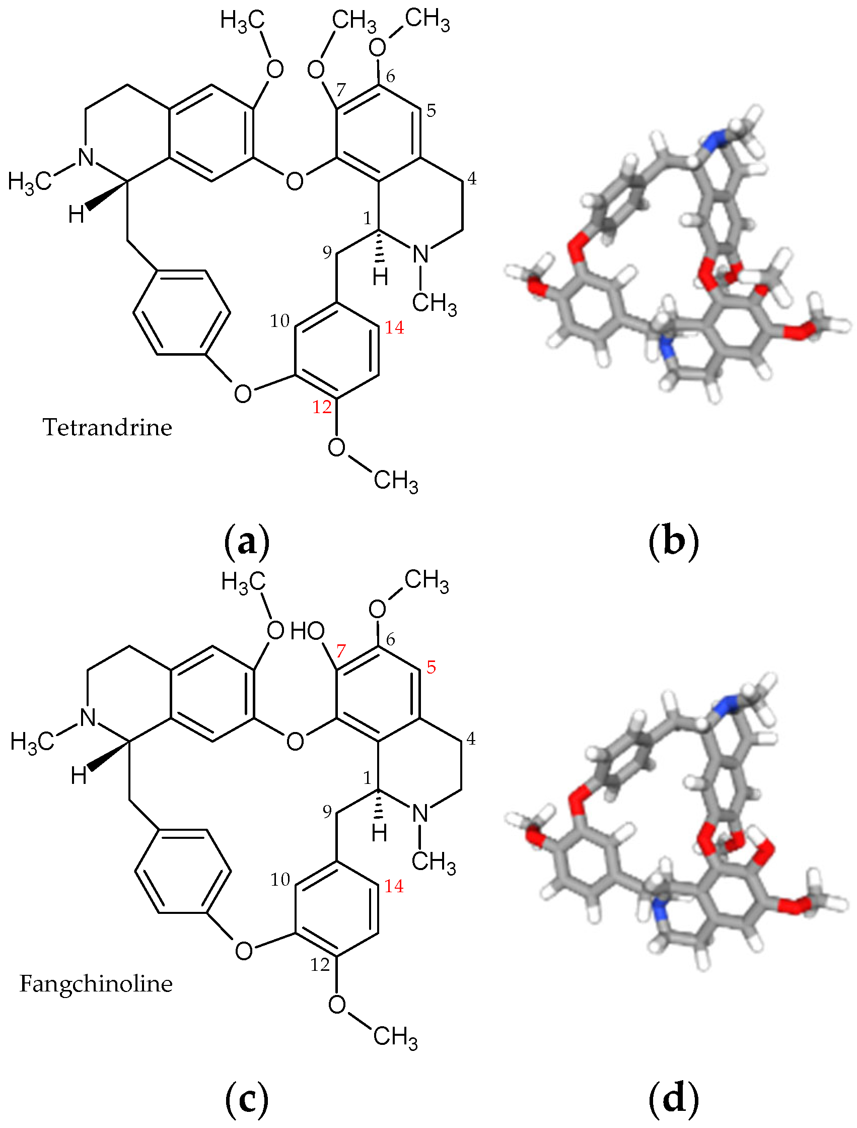
Further insights into the therapeutic potential of isoquinoline alkaloids come from the studies on tetrandrine and fangchinoline actions. Both are major bis-benzylisoquinoline alkaloids, similar to berbamine, isolated from the roots of Stephania tetrandra S. Moore [29]. Their molecular structures are identical except for a single substitution at the C-7 position: tetrandrine contains a methoxy group (-OCH3), whereas fangchinoline possesses a hydroxyl group (-OH) at this position (Figure 2).
Tetrandrine has long been used in traditional medicine to treat malaria, edema, wet beriberi, dysuria, eczema, and inflamed sores. It also demonstrated anticancer properties in vitro and in vivo, as well as antioxidant, anti-inflammatory, immunosuppressive, and analgesic effects [29,30]. Mechanistically, tetrandrine was shown to inhibit phosphorylation of inhibitor of nuclear factor kappa-B kinase (IKKβ), suppress the COX-2/PGE2 pathway, and reduce the production and activation of proinflammatory cytokines, including interleukins and TNF-α [31,32].
Recent studies explored the antinociceptive effects of tetrandrine in experimental neuropathy. In a mouse model of OX-NP, mechanical allodynia was assessed using von Frey filaments, and motor function was evaluated via rotarod testing. RNA sequencing and qPCR analyses of spinal cord tissue identified key inflammation-related genes, including Arg2, Cxcl12, H2-Q6, Kdr, and Nfkbia. Notably, Arg2, which is implicated in microglial activation and pain behavior following nerve injury, showed no significant changes in either RNA sequencing or qPCR analyses, while Nfkbia, a negative regulator of NF-κB and neuroinflammation, was significantly upregulated by tetrandrine in qPCR [33]. These results suggest that tetrandrine may alleviate OX-NP by upregulating Nfkbia and modulating Arg2-associated pathways. Importantly, administration of tetrandrine at 45 mg/kg did not impair motor function or induce sedation, highlighting its safety profile. These studies underscore the multifaceted mechanisms by which tetrandrine may attenuate NP, from gene modulation to immune response regulation, and support its potential as a promising therapeutic candidate for the OX-NP and related neuropathies [32,33,34].
Administration of tetrandrine at 45 mg/kg significantly improved mechanical allodynia, with efficacy comparable to that of pregabalin in the SNI model in rats. Western blot and immunofluorescence analyses demonstrated that tetrandrine inhibited spinal protein expression of CKLF1, p-NF-κB/NF-κB, p-IKK/IKK, and pro-inflammatory cytokines IL-1β and TNF-α, while enhancing the level of the anti-inflammatory cytokine IL-10. The most evidence-based interpretation of these results is that CKLF1 primarily acts as a mediator of spinal microglial activation downstream of the IKK/NF-κB signaling rather than as a directly validated neuronal effector. CKLF1 is known to play a critical role in both peripheral and central inflammatory processes [35,36]. More broadly, CKLF1 is a CCR4 ligand with chemotactic properties implicated in neuron-to-microglia/macrophage signaling in various CNS injury settings. Thus, immune cell recruitment and crosstalk remain biologically plausible mechanisms. However, direct actions on nociceptive neurons in the SNI model have not yet been clearly demonstrated [35,37,38].
Fangchinoline, the second major bis-benzylisoquinoline alkaloid from Stephania roots, exhibited antitumor, antifungal, and antiviral activities. Additionally, it demonstrated protective effects in STZ-induced diabetes models, where it ameliorated nephropathy and retinopathy. It normalized retinal morphology, reduced inflammatory mediators expression, decreased oxidative stress, suppressed NF-κB activity, and lowered the apoptosis index in diabetic rats compared with untreated controls [39,40]. It was also reported to reduce the production of proinflammatory mediators in a rat model of rheumatoid arthritis. In vitro, this effect appears to be mediated through inhibition of NF-κB, while in vivo, fangchinoline significantly decreases levels of TNF-α, IL-6, MMP-3, and PGE2 [41]. These findings suggest that fangchinoline may exert its effects by modulating cytokine signaling, chemokine pathways, and MAPK-related mechanisms, highlighting its potential relevance to NP pathophysiology. However, the current evidence base for fangchinoline in NP remains limited as no studies have evaluated its effects in canonical NP models such as CCI, PSNL, SNI models, with behavioral analgesic outcomes. Therefore, fangchinoline should currently be considered a compound with anti-inflammatory and antioxidant properties that may be relevant to NP pathophysiology, but for which direct preclinical validation in NP models is still lacking [41,42,43,44]. Overall, these findings highlight fangchinoline as a promising candidate for further investigation in experimental NP models.

3. Sinomenine and Its Therapeutic Potential in Neuropathic Pain (NP)

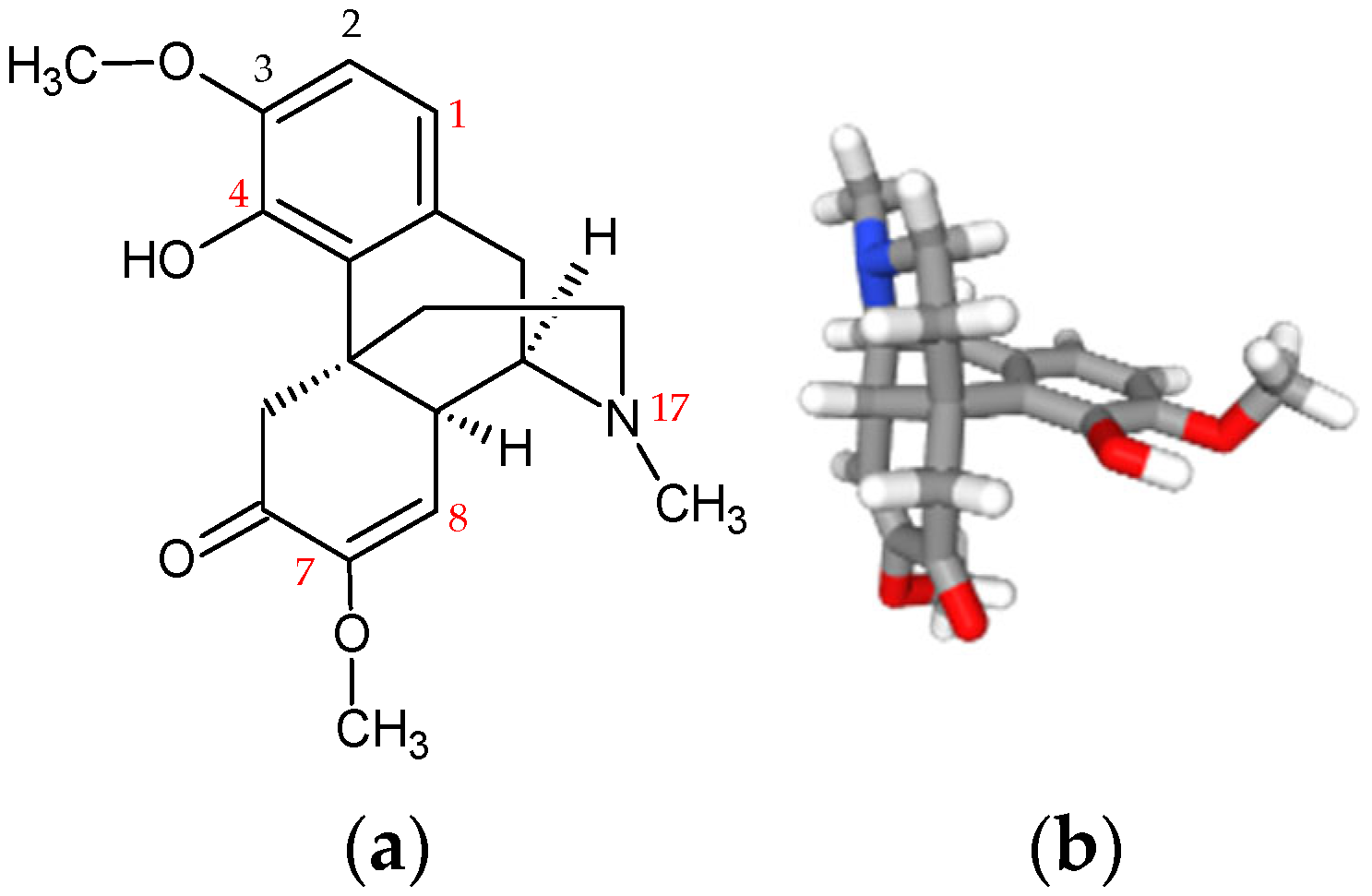
Sinomenine is the primary pharmacologically active alkaloid isolated from Sinomenium acutum, a plant traditionally used in East Asian ethnomedicine in the form of root and stem decoctions. It has been shown to exert significant anti-inflammatory and immunomodulatory effects, with mechanisms involving inhibition of inflammatory mediators and the modulation of immune cell activity [17]. Structurally, sinomenine is classified as a morphinan alkaloid according to the MeSH hierarchy (Figure 3). It shares significant structural similarities with classical opioid receptor agonists, and in total synthesis and structure–activity relationship (SAR) analyses, morphinan alkaloids, including sinomenine, are considered part of the broader isoquinoline series. This classification reflects their distinct physicochemical properties and affinity for specific molecular targets within nociceptive pathways [17,45].

3.1. Inhibition of the Neuroinflammatory Cascade and Immunological System

Sinomenine demonstrated substantial antinociceptive effects in the CCI model in rats. A single intraperitoneal (i.p.) dose of 20 mg/kg significantly elevated pain thresholds within 2–2.5 h post-injection, whereas a 40 mg/kg dose accelerated the onset of effect to approximately 1.5 h [17]. Chronic oral administration over 14 days also produced significant reductions in both mechanical allodynia and thermal hyperalgesia [46].
A key factor in the pathogenesis of NP is activation of microglial cells in the SDH, which triggers the unfolded protein response (UPR) to counteract endoplasmic reticulum (ER) stress and restore cellular homeostasis. Within this pathway, inositol-requiring enzyme 1α (IRE1α) is activated via its kinase and RNase domains. The kinase domain phosphorylates JNK or IKK, thereby enhancing inflammatory signaling and apoptosis, whereas the RNase domain mediates regulated IRE1α-dependent decay (RIDD) of selected mRNAs and miRNAs or generates the transcriptionally active spliced X-box binding protein 1 (XBP1s), which regulates UPR target genes [46,47]. Animal studies demonstrated that sinomenine can potently inhibit microglial activation. In the STZ-induced NP model in rats, it was administered intraperitoneally at a dose of 1 mL/kg. Assessment of COX-2, IRE1α, spliced X-box binding protein 1 (XBP1s), TNF-α, IL-1β, and IL-6 in spinal cord tissue and microglial supernatants revealed that it inactivated the IRE1α–XBP1s pathway and downregulated COX-2, thereby alleviating pain symptoms [10]. Additionally, sinomenine reduced levels of the NLRP1 inflammasome complex. Microglial deactivation, coupled with inflammasome inhibition, resulted in a dose-dependent decrease in mature proinflammatory cytokines IL-1β and IL-18, thereby limiting neuroinflammation and mitigating chronic NP symptoms [7,9,10,48].
Sinomenine also exhibits a unique capacity to modulate immune responses by promoting a phenotypic shift in both innate and adaptive immunity. It induces microglia and macrophages to transition from the proinflammatory M1 phenotype to the anti-inflammatory, neuroprotective M2 phenotype, characterized by increased Arg1 and IL-10 expression. In the adaptive immune system, sinomenine suppresses differentiation of proinflammatory Th17 cells while enhancing regulatory T cell (Treg) generation, largely through activation of the aryl hydrocarbon receptor (AhR). Additionally, it helps restore Th1/Th2 balance by inhibiting Th1 cell activity and interferon-γ (IFN-γ) secretion [17,48].

3.2. Modulation of Ion-Channels, Receptors and Neurotransmitters

The STZ-induced NP model is widely regarded as the gold standard for studying diabetic polyneuropathy. Hyperglycemia in this model triggers oxidative stress, damages both C and Aδ fibers, and alters the expression of ion channels [15,17]. Sinomenine has been shown to inhibit the protein expression of Nav1.7 and Nav1.8, as well as Kv1.1 and Kv2.1, thereby decreasing neuronal hyperexcitability within the DRG [48]. Notably, it does not significantly affect calcium channels, such as the Cav1.3 subtype, demonstrating pharmacological selectivity [9]. In addition to these effects, sinomenine’s peripheral antinociceptive properties are mediated through modulation of ionotropic purinergic receptors. The alkaloid inhibits the activity of P2X3, P2X4, and P2X7 receptors, which plays a critical role in the transduction and amplification of pain signals in response to the extracellular ATP release under pathological conditions [6,7,8,48].
The glutamatergic system, the principal excitatory network in the CNS, plays a pivotal role in the maintenance of chronic pain through activation of NMDA receptors, which are key mediators of both peripheral and central sensitization [10]. Sinomenine has been shown to modulate this system by restoring the balance between excitatory and inhibitory neurotransmission in the CCI model. More specifically, it has been shown to suppress pathological neuronal activity by reducing sensory neuron hyperexcitability, inhibiting glial overactivation, and normalizing metabolic processes, collectively lowering extracellular glutamate levels and preventing aberrant nociceptive signaling [11,17,48]. Within the CNS, sinomenine also exhibits notable selectivity for the GluN2B (NR2B) subunit of NMDA receptors [5]. Inhibition of this pathway disrupts downstream signaling cascades, including ERK/CREB and mTOR pathways, ultimately suppressing maladaptive synaptic plasticity associated with pain chronification [5,7,49].
Following nerve injury, both peripheral and central sensitization are key mechanisms in the pathogenesis of NP, involving hyperexcitability of primary sensory neurons and alterations in central synaptic transmission. Specifically, excitatory synaptic activity is enhanced, while inhibitory neurotransmission is diminished in central neurons. Changes in neurotransmitters, particularly glutamate and GABA, within the SDH play a critical role in chronic pain development. Under normal conditions, painful stimuli evoke action potentials in primary afferent neurons (Aδ and C fibers), which activate SDH pain-transmission neurons. Inhibitory interneurons in the SDH, containing GABA or glycine, suppress excitatory interneurons that drive pain transmission. After nerve injury, GABA levels decline, inhibitory interneuron activity is reduced, and GABA receptor expression is downregulated [5]. This loss of inhibitory “tone” leads to a neurochemical imbalance, reinforcing excitatory interneuron activity and contributing to neuronal hyperexcitability. Such hyperexcitability underlies mechanical hypersensitivity, including hyperalgesia and allodynia, whereas restoration of inhibitory signaling can suppress the pathological discharges characteristic of NP [13,50]. The relevance of inhibitory neurotransmission was further demonstrated using sinomenine and its primary active metabolite, N-demethylsinomenine, in the CCI model. In this model, reduced inhibitory tone of GABA- and glycine-containing interneurons led to enhanced excitatory interneuron activity and hyperexcitability of pain-transmission neurons. Acute administration of N-demethylsinomenine (10–40 mg/kg, i.p.) dose-dependently attenuated mechanical allodynia. Pretreatment with bicuculline, a selective GABAA receptor antagonist, almost completely abolished the anti-allodynic effect of N-demethylsinomenine at 40 mg/kg. These findings indicate that sinomenine and its metabolite exert their analgesic effects, at least in part, through modulation of GABAA receptor-mediated inhibitory signaling, with receptor blockade effectively eliminating their anti-allodynic activity [51].

3.3. Structural Regeneration, Neuroprotection, and Advanced Metabolic Modulation

The therapeutic efficacy of sinomenine in NP extends beyond acute synaptic modulation to encompass nerve tissue repair and the stabilization of metabolic and immunological homeostasis. It plays a key role in restoring the structural integrity of peripheral nerves. Morphometric and histological analyses demonstrate that sinomenine promotes remyelination and counteracts axonal atrophy, as evidenced by increased myelin sheath thickness and axonal diameter. These effects are closely associated with upregulation of myelin basic protein (MBP) and neurofilament heavy chain (NF-H), key markers of nerve fiber regeneration [11]. Additionally, sinomenine exerts potent neuroprotective effects by modulating apoptotic pathways, enhancing neuronal survival through upregulation of the anti-apoptotic protein Bcl-2, while simultaneously downregulating pro-apoptotic factors, including Bax and cleaved caspase-3 [9].
Metabolomic analyses reveal that sinomenine profoundly remodels the chemical composition of cerebrospinal fluid (CSF) and plasma. It modulates 14 key metabolic pathways, including the biosynthesis of phenylalanine, tyrosine, and tryptophan, as well as cysteine and methionine metabolism. In plasma, sinomenine significantly increases levels of key neurotransmitters and their precursors, including 5-hydroxytryptophan, dopamine, norepinephrine, epinephrine, serotonin, glutamate, and glutamine, while decreasing levels of 3-methoxy-4-hydroxymandelate and N-methylaspartic acid. In the CSF, it restores normetanephrine balance and regulates L-DOPA and N-methyl-L-glutamic acid levels. Furthermore, by normalizing the purinergic environment in the spinal cord, sinomenine mitigates the deleterious effects of excessive extracellular ATP, thereby contributing to its antinociceptive actions [7,11].

3.4. Pharmacological Profile of Sinomenine

Although sinomenine is a morphinan alkaloid with structural similarity to morphine, the available pharmacological evidence does not support its classification as a broad opioid receptor agonist. Direct evidence exists primarily for μ-opioid receptor activity: in μ-opioid receptor-transfected CHO cells, sinomenine displaced [(3H)]naloxone in a concentration-dependent manner and increased receptor phosphorylation, and its antinociceptive effects in acute pain models were attenuated by μ-opioid antagonists. In contrast, comparable data on affinity and efficacy at δ- or κ-opioid receptors are lacking, and in vivo studies suggest that these receptors are not major contributors to its analgesic effects [52,53]. Importantly, this limited μ-receptor activity does not appear to account for most of sinomenine’s efficacy in chronic NP models, where mechanisms such as GABAA receptor modulation, ER stress signaling, and suppression of spinal neuroinflammation are more prominently implicated [10,49,54,55]. Some evidence indicates that μ-opioid receptor activation may contribute to the antinociceptive effects of sinomenine and to inhibition of spinal ERK1/2 signaling. However, the overall evidence supports a model in which sinomenine acts as a weak or context-dependent μ-opioid receptor modulator in acute pain, while its anti-neuropathic effects are mediated predominantly through non-opioid mechanisms [52].
Unlike classical opioids, long-term administration of sinomenine does not induce analgesic tolerance [7,9,17,49,50]. Animal studies indicated that even at high therapeutic doses, sinomenine did not cause sedation (except at extremely high doses, e.g., 80 mg/kg), motor coordination deficits, or physical dependence [5,17,50]. Notably, N-demethylsinomenine exhibited an even more favorable safety profile, remaining non-sedative at doses up to 80 mg/kg. Moreover, sinomenine was shown to inhibit morphine-induced addiction and attenuate withdrawal symptoms, highlighting its potential as a safer alternative for chronic pain management [51].
A recent study further expanded the therapeutic potential of sinomenine by demonstrating that its co-administration with ligustrazine (LGZ) produced a robust synergistic effect in a CCI model. The combination therapy attenuated nociceptive hypersensitivity more effectively than either compound alone, suggesting that lower doses may be sufficient to achieve optimal analgesic efficacy. Because no measurable toxicity was observed in any experimental group, direct comparison of adverse effects between monotherapy and combination therapy was precluded. However, this synergy-based treatment strategy that reduces total drug exposure may still offer a safer and more effective alternative to conventional analgesic approaches. Furthermore, the combined administration of LGZ and sinomenine was associated with improved overall animal condition, as reflected by greater body weight gain in the high-dose combination group [11].

4. Structural Modifications in Isoquinoline Alkaloids

All compounds discussed in the present review, berbamine, tetrandrine, fangchinoline, and sinomenine, contain multiple sites amenable to structural modification (Figure 1, Figure 2 and Figure 3). As such, they represent valuable scaffolds for the development of novel derivatives with enhanced biological activity. However, most of these derivatives were synthesized and evaluated primarily for their cytotoxic effects against various cancer cell lines, and some for their anti-inflammatory action. Only a limited number of studies have investigated their analgesic properties or their specific effects in NP models.
Structural modifications of berbamine were primarily focused on the C-12 hydroxyl group, with many resulting derivatives demonstrating enhanced antitumor activity. Subsequently, a series of analogs substituted at both the C-12-OH and C-5 positions were developed. Molecular docking studies revealed the relationship between the structures of these compounds and potent inhibition of Ca2+/calmodulin-dependent protein kinase II γ (CaMKIIγ), a target implicated in cancer progression [56,57,58]. Notably, CaMKII is also involved in synaptic plasticity and central sensitization, key processes in chronic pain [59], highlighting the need for further investigation of these berbamine derivatives in NP models.
Regarding tetrandrine modifications, the primary focus was on its cytotoxic activity against various cancer cell lines, as well as its relatively high toxicity associated with the formation of reactive intermediates, oxidative stress, and apoptosis. In the proposed metabolic pathway, the methoxy group at the C-12 position is first demethylated to yield the corresponding phenolic metabolite, which can subsequently undergo enzymatic oxidation to form a para-quinone methide [60,61,62]. Based on this liability, substitution of the C-12 methoxy group with metabolically stable trifluoromethoxy or chloro- substituents was proposed. These derivatives were shown to inhibit proliferation of leukemia cells, primarily through disruption of mitochondrial membrane potential, thereby activating early apoptosis pathways. In parallel, studies of these tetrandrine derivatives with stable substitutions at the C-12 position suggested that mechanisms beyond formation of reactive metabolites may contribute to its toxicity [63]. Other structural optimizations of tetrandrine was focused on substitution at the C-14 position of the benzene ring. Derivatives bearing L-amino acid-urea or sulfonamide moieties significantly improved antiproliferative potency against cancer cells [64]. In general, compounds bearing electron-withdrawing substituents at the C-14 amino position exhibited enhanced antiproliferative activity. Their mechanism of action was proposed to involve increased expression of the proapoptotic protein Bax, together with downregulation of anti-apoptotic proteins Bcl-xL and Bcl-2 [63,64].
The derivatization strategy for fangchinoline involved protection of the C-7 phenolic position with different substituents, followed by introduction of halogen or nitrile groups at the C-5 and/or C-14 positions to generate first- and second-generation analogues. These derivatives exhibited significantly higher inhibitory activity against various cancer cells than the parent compound [65]. In addition, fangchinoline, along with its derivatives, were investigated as anti-inflammatory agents in a lipopolysaccharide/nigericin (LPS/NIG)-induced model of IL-1β release and NLRP3 inflammasome activation in human leukemia cells. Both alkaloids displayed moderate anti-inflammatory activity, with inhibition rates of 50.5% for fangchinoline and 40.7% for tetrandrine at 5 µM. A series of bis-benzylisoquinoline analogues with diverse substitution patterns at the C-7 position were then synthesized and evaluated in the same system. Initial modifications, including methyl, ethyl, isobutyl, and ethoxyethyl ethers, led to slight decreases in activity, whereas the propyl ether derivative showed a markedly improved inhibitory rate of 71.8%. Based on this result, novel aminopropyl ether derivatives were developed; among them, 7-phthalimidepropylfangchinoline exhibited strong inhibitory activity against IL-1β activation, with an inhibition rate of 76.1% at 5 µM and an IC50 value of 3.7 µM. Interestingly, this compound did not inhibit NF-κB or MAPK signaling pathways and was not a direct caspase-1 inhibitor. Therefore, it was proposed that its mechanism of action may involve direct interaction with NLRP3, thereby impairing ASC inflammasome assembly and ultimately suppressing IL-1β release [66].
Importantly, no studies have yet investigated the effects of fangchinoline and tetrandrine derivatives in validated NP models, representing a significant gap in the current literature.
As far as sinomenine-derived compounds are concerned, they displayed markedly improved anticancer potency in breast, lung, liver, and prostate cancers. These derivatives were mainly obtained through structural modifications at the C-1, C-4, C-7, and C-8 positions of the parent molecule. In addition, selective reduction of the α,β-unsaturated ketone moiety, previously proposed as a key pharmacophore in sinomenine’s structure, could also be considered as a strategy to generate more potent analogues [45].
A large number of sinomenine derivatives were also synthesized and evaluated to enhance its anti-inflammatory, immunosuppressive, and analgesic activities. The C-1 position, C-4 hydroxyl group, and the nitrogen atom represent key sites for structural modification in this context. A series of C-1 substituted derivatives was reported with potent anti-inflammatory effects. Among them, 1-formyl-sinomenine exhibited greater inhibitory activity on IL-2 release in rat splenocytes compared with the parent compound [67]. Other minor alterations, such as esterification of the C-4 hydroxyl group or N-17-demethylation, were also shown to significantly enhance anti-rheumatoid arthritis activity [45]. In addition to single-site modifications, the synthesis of sinomenine homodimers and hybrid molecules emerged as an effective strategy for developing improved antirheumatic agents. For example, sinomenine–pyrazine hybrids significantly reduced the expression of multiple cytokines and suppressed key inflammatory signaling pathways [67,68]. Furthermore, a series of sinomenine 1-bromo-4-hydroxy ester derivatives incorporating cinnamic acid moieties was designed to improve analgesic and anti-inflammatory activity, as well as bioavailability and lipophilicity balance. Their analgesic effects were evaluated using the hot plate and acetic acid-induced writhing tests, while anti-neuroinflammatory activity was assessed in a xylene-induced mouse ear edema model associated with elevated TNF-α, IL-1β, and PGE2 levels. Several of these derivatives exhibited superior analgesic and anti-inflammatory effects compared with sinomenine [69]. However, there is currently a lack of studies evaluating these derivatives in well-established NP models. To date, only N-demethylsinomenine, a natural metabolite of sinomenine, has been investigated in the CCI, where acute intraperitoneal administration significantly has attenuated mechanical allodynia, showing greater efficacy than the parent compound [51].
Comparative analysis of berbamine, tetrandrine, fangchinoline, and sinomenine suggests that anti-inflammatory and analgesic efficacy is governed by a combination of bis-benzylisoquinoline scaffold rigidity, electronic distribution, and functional group accessibility. Considering further modifications of these alkaloids, a strategic roadmap can be proposed for their potential application in NP therapy, with an emphasis on SAR studies. For berbamine, targeted optimization of substitutions at the C-12-OH and C-5 positions, as well as increasing rigidity, may be pursued. In the case of tetrandrine and fangchinoline, further structural optimization should integrate modifications at the C-12 and C-14 positions, prioritize metabolically stable substitutions, and systematically evaluate their effects on both analgesic efficacy and toxicity mechanisms. In addition, novel substitutions at the C-5, C-7 and C-14 positions may also be explored to optimize pharmacological activity and mechanistic selectivity. Finally, a roadmap for structural modification of sinomenine should prioritize systematic optimization at the C-1, C-4, C-7, C-8, and nitrogen positions, as well as controlled modification of the α,β-unsaturated ketone moiety, together with the design of novel hybrid compounds, with the aim of further enhancing anti-inflammatory and analgesic activities. Importantly, both existing and newly designed derivatives should be extensively evaluated in appropriate NP models to validate their therapeutic potential.

5. Conclusions, Limitations and Future Directions

The reviewed literature indicates that the development of novel treatments with improved analgesic efficacy and fewer adverse effects compared with currently available therapies remains a major challenge in the management of NP. In this context, natural alkaloids such as berbamine, fangchinoline, tetrandrine, and sinomenine have emerged as promising candidates, as demonstrated by their multidirectional activities. These compounds modulate multiple key NP-related pathways, including inflammatory processes, oxidative stress, ion channel function, and intracellular signaling cascades. In particular, berbamine is associated with modulation of the TMEM34/p-SGK1/FOXO3 signaling axis, while sinomenine is linked to regulation of the IRE1α–XBP1s ER stress pathway. Collectively, these alkaloids also influence voltage-gated ion channels, as well as ionotropic purinergic receptors, including P2X3, P2X4, and P2X7. Beyond symptomatic pain relief, sinomenine additionally promotes nerve repair processes, including remyelination and upregulation of myelin-associated proteins such as MBP. Importantly, unlike classical opioids, long-term administration of sinomenine and its metabolite N-demethylsinomenine does not appear to induce analgesic tolerance, physical dependence, or significant sedation at therapeutic doses.
A comparative analysis indicates that, among the alkaloids discussed, sinomenine and tetrandrine currently possess the most robust direct experimental evidence supporting their efficacy in NP models, although their mechanistic profiles are markedly distinct. Fangchinoline shows the weakest evidence base for NP, as the available data are predominantly derived from rheumatoid arthritis and osteoarthritis models, as well as cytoprotective studies not directly related to neuropathy. Berbamine, in contrast, remains a mechanistically intriguing candidate; however its evidence base is limited to fewer NP-specific studies and partial extrapolation from broader anti-inflammatory research, particularly involving NF-κB signaling. Accordingly, future research should address compound-specific gaps. For berbamine, efforts should focus on more precise identification and validation of its molecular targets within NF-κB-related pathways. For tetrandrine, further studies are needed to confirm the role of the CKLF1/CCR4 axis and to define the relevant cellular targets in NP. For fangchinoline, direct evaluation in established NP models such as CCI, PSNL, SNI, and diabetic neuropathy remains essential. Finally, research on sinomenine should aim to further delineate the contribution of its opioid-related activity relative to its predominant anti-inflammatory and neuroimmune mechanisms, alongside systematic assessment of its translational potential in clinical NP settings. Overall, sinomenine currently represents the most translationally advanced candidate among the compounds reviewed, followed by tetrandrine. Berbamine remains mechanistically promising but insufficiently characterized in NP-specific contexts, while fangchinoline should presently be regarded as exploratory with respect to NP treatment.

Author Contributions

Conceptualization, A.G. and A.K.; methodology, A.G. and A.K.; writing—original draft preparation, A.G. and A.K.; writing—review and editing, A.G. and A.K.; supervision, A.G. All authors have read and agreed to the published version of the manuscript.

Funding

This research received no external funding.

Data Availability Statement

No new data were created or analyzed in this study. Data sharing is not applicable to this article.

Conflicts of Interest

The authors declare no conflicts of interest.

Abbreviations

The following abbreviations are used in this manuscript:
NPNeuropathic pain
IASPInternational Association for the Study of Pain
VGVoltage-gated
DRGDorsal root ganglia
TNF-αTumor necrosis factor-α
SDHSpinal dorsal horn
IL-1βInterleukin 1β
MAPKMitogen-activated protein kinase
NF-κBNuclear factor kappa B
ERK/CREBExtracellular signal-regulated kinase/cAMP response element-binding protein
ROSReactive oxygen species
SODSuperoxide dismutase
GSHGlutathione
GABAγ-Aminobutyric acid
NMDAN-Methyl-D-aspartate
CGRPCalcitonin gene-related peptide
CNSCentral nervous system
CCIChronic constriction injury
PSNLPartial sciatic nerve ligation
SNLSpinal nerve ligation
NGFNerve growth factor
PGE2Prostaglandin E2
COX-2Cyclooxygenase-2
MMPMatrix metalloproteinase
NONitric oxide
PKCProtein kinase C
PXPaclitaxel
OXOxaliplatin
STZStreptozotocin
MESHMedical Subject Headings
iNOSInducible nitric oxide synthase
MDMolecular dynamics
PP2CαPhosphatase 2Cα (PP2Cα)
AMPKAMP-activated protein kinase
TMEM34Spinal transmembrane protein 34
SGK1Serum/glucocorticoid-inducible kinase-1
FOXO3Forkhead Box O3
IKKβInhibitor of nuclear factor kappa-B kinase
CKLF1Chemokine-like factor 1
CCR4Chemokine receptor CC4
SARStructure–activity relationship
UPRUnfolded protein response
EREndoplasmic reticulum
IRE1αInositol-requiring enzyme 1α
JNKc-Jun N-terminal kinase
XBP1X-box binding protein 1
RIDDRegulated IRE1α-dependent decay
MBPMyelin basic protein
NF-HNeurofilament heavy chain
CSFCerebrospinal fluid
LGZLigustrazine
CaMKIIγCa2+/calmodulin-dependent protein kinase II γ
LPSLipopolysaccharide
NIGNigericin

References

  1. International Association for the Study of Pain (IASP). What is Neuropathic Pain? 2022. Available online: https://www.iasp-pain.org/wp-content/uploads/2022/10/What-is-Neuropathic-Pain.pdf (accessed on 13 March 2026).
  2. Woolf, C.J. Pain: Moving from symptom control toward mechanism-specific pharmacologic management. Ann. Intern. Med. 2004, 140, 441–451. [Google Scholar] [CrossRef]
  3. Hall, G.C.; Carroll, D.; Parry, D.; McQuay, H.J. Epidemiology and treatment of neuropathic pain: The UK primary care perspective. Pain 2006, 122, 156–162. [Google Scholar] [CrossRef]
  4. Gilron, I.; Baron, R.; Jensen, T. Neuropathic pain: Principles of diagnosis and treatment. Mayo Clin. Proc. 2015, 90, 532–545. [Google Scholar] [CrossRef]
  5. Zhu, C.; Liu, N.; Tian, M.; Ma, L.; Yang, J.; Lan, X.; Ma, H.; Niu, J.; Yu, J. Effects of alkaloids on peripheral neuropathic pain: A review. Chin. Med. 2020, 15, 106. [Google Scholar] [CrossRef]
  6. Dong, C.; Zhang, W.; Luo, H. Association between P2X3 receptors and neuropathic pain: As a potential therapeutic target for therapy. Biomed. Pharmacother. 2022, 150, 113029. [Google Scholar] [CrossRef] [PubMed]
  7. Ai, X.; Dong, X.; Guo, Y.; Yang, P.; Hou, Y.; Bai, J.; Zhang, S.; Wang, X. Targeting P2 receptors in purinergic signaling: A new strategy of active ingredients in traditional Chinese herbals for diseases treatment. Purinergic Signal. 2021, 17, 229–240. [Google Scholar] [CrossRef]
  8. Guo, W.; Zhang, J.; Feng, Y. Treatment of neuropathic pain by traditional Chinese medicine: An updated review on their effect and putative mechanisms of action. Phytother. Res. 2024, 38, 2962–2992. [Google Scholar] [CrossRef]
  9. Chen, W.; Chen, J.; Lu, Y.; Chen, Y.; Liu, X.; Yang, F. Fiber-electrospun hydrogel therapy for DNP: A synergistic electrospun-hydrogel composite for alleviating diabetic neuropathic pain via MMP9 regulation and sodium channel inhibition. Bioeng. Transl. Med. 2025, e70050. [Google Scholar] [CrossRef]
  10. Chen, J.; Guo, P.; Liu, X.; Liao, H.; Chen, K.; Wang, Y.; Qin, J.; Yang, F. Sinomenine alleviates diabetic peripheral neuropathic pain through inhibition of the inositol-requiring enzyme 1 alpha-X-box binding protein 1 pathway by downregulating prostaglandin-endoperoxide synthase 2. J. Diabetes Investig. 2023, 14, 364–375. [Google Scholar] [CrossRef] [PubMed]
  11. Yuan, Z.; Zhao, X.; Zhang, Y.; Jiao, Y.; Liu, Y.; Gao, C.; Zhang, J.; Ma, Y.; Wang, Z.; Li, T. Using integrated network pharmacology and metabolomics to reveal the mechanisms of the combined intervention of ligustrazine and sinomenine in CCI-induced neuropathic pain rats. Int. J. Mol. Sci. 2025, 26, 2604. [Google Scholar] [CrossRef] [PubMed]
  12. Okumo, T.; Takayama, Y.; Maruyama, K.; Kato, M.; Sunagawa, M. Senso-immunologic prospects for complex regional pain syndrome treatment. Front. Immunol. 2022, 12, 786511. [Google Scholar] [CrossRef]
  13. Karcz, M.; Abd-Elsayed, A.; Chakravarthy, K.; Aman, M.M.; Strand, N.; Malinowski, M.N.; Latif, U.; Dickerson, D.; Suvar, T.; Lubenow, T.; et al. Pathophysiology of pain and mechanisms of neuromodulation: A narrative review (A Neuron Project). J. Pain Res. 2024, 17, 3757–3790. [Google Scholar] [CrossRef]
  14. Sic, A.; Manzar, A.; Knezevic, N.N. The role of phytochemicals in managing neuropathic pain: How much progress have we made? Nutrients 2024, 16, 4342. [Google Scholar] [CrossRef] [PubMed]
  15. Quintans, J.S.S.; Antoniolli, Â.R.; Almeida, J.R.G.S.; Santana-Filho, V.J.; Quintans-Júnior, L.J. Natural products evaluated in neuropathic pain models—A systematic review. Basic Clin. Pharmacol. Toxicol. 2014, 114, 442–450. [Google Scholar] [CrossRef]
  16. Basbaum, A. Chemogenetic management of neuropathic pain. Brain 2017, 140, 2522–2525. [Google Scholar] [CrossRef]
  17. Lai, W.-D.; Wang, S.; You, W.-T.; Chen, S.-J.; Wen, J.-J.; Yuan, C.-R.; Zheng, M.-J.; Jin, Y.; Yu, J.; Wen, C.-P. Sinomenine regulates immune cell subsets: Potential neuro-immune intervene for precise treatment of chronic pain. Front. Cell Dev. Biol. 2022, 10, 1041006. [Google Scholar] [CrossRef]
  18. Li, X.-Y.; Fang, Z.-M.; Guo, S.-J.; Jia, L.-N.; Sun, T.; Ma, J.-L.; Zheng, Y.-L. NMDA receptors in neuropathic pain: From mechanisms to therapeutic strategies. Neuroscientist 2026, 32, 126–142. [Google Scholar] [CrossRef] [PubMed]
  19. Rusanescu, G.; Mao, J. Peripheral nerve injury induces brain neurogenesis and remodelling. J. Cell. Mol. Med. 2017, 21, 299–314. [Google Scholar] [CrossRef]
  20. Korah, H.E.; Cheng, K.; Washington, S.M.; Flowers, M.E.; Stratton, H.J.; Patward, A. Partial sciatic nerve ligation: A mouse model of chronic neuropathic pain to study the antinociceptive effect of novel therapies. J. Vis. Exp. 2022, 188, e64555. [Google Scholar]
  21. Rought, R.; Rodrigues, R.; Sharif-Naeini, R.; Stone, L.S.; Coderre, T.J.; Ribeiro-da-Silva, A.; Millecamps, M. New and less invasive surgical approach to the mouse spared nerve injury model of peripheral. J. Neurosci. Meth. 2026, 431, 110751. [Google Scholar] [CrossRef]
  22. Faheem, M.; Khan, A.-U.; Waqas Saleem, M.; Shah, F.A.; Ali, F.; Waheed Khan, A.; Li, S. Neuroprotective effect of natural compounds in paclitaxel-induced chronic inflammatory pain. Molecules 2022, 27, 4926. [Google Scholar] [CrossRef]
  23. Ivanova, N.; Hristov, M.; Gateva, P. Rodent models of diabetic neuropathy, role of calcium homeostasis in pain and KB-R7943 as a potential therapeutic. Int. J. Mol. Sci. 2025, 26, 2094. [Google Scholar] [CrossRef] [PubMed]
  24. Farooqi, A.A.; Wen, R.; Attar, R.; Taverna, S.; Butt, G.; Xu, B. Regulation of cell-signaling pathways by berbamine in different cancers. Int. J. Mol. Sci. 2022, 23, 2758. [Google Scholar] [CrossRef] [PubMed]
  25. Jahan, F.; Alvi, S.S.; Islam, M.H. Berberis aristata and its secondary metabolites: Insight into nutraceutical and therapeutical applications. Pharmacol. Res. Mod. Chin. Med. 2022, 5, 100184. [Google Scholar] [CrossRef]
  26. Faheem, M.; Khan, A.-U.; Shah, F.A.; Li, S. Investigation of natural compounds for therapeutic potential in streptozotocin-induced diabetic neuroinflammation and neuropathic pain. Front. Pharmacol. 2022, 13, 1019033. [Google Scholar] [CrossRef]
  27. Yao, C.; Zhang, Z.; Zhang, S.; Jiang, H.; Wang, R.; Liu, J.; Xie, H.; Dai, W. Berbamine alleviates neuropathic pain via suppressing spinal TMTM34/SGK1/FOXO3 axis. Phytomedicine 2025, 140, 156619. [Google Scholar] [CrossRef]
  28. Liu, B.; Li, N.; Duan, G. Emerging role of serum glucocorticoid-regulated kinase 1 in pathological pain. Front. Mol. Neurosci. 2021, 14, 683527. [Google Scholar] [CrossRef]
  29. Chan, E.W.C.; Wong, S.K.; Chan, H.T. An overview on the chemistry, pharmacology and anticancer properties of tetrandrine and fangchinoline (alkaloids) from Stephania tetrandra roots. J. Integr. Med. 2021, 19, 311–316. [Google Scholar] [CrossRef]
  30. Jiang, Y.; Liu, M.; Liu, H.; Liu, S. A critical review: Traditional uses, phytochemistry, pharmacology and toxicology of Stephania tetrandra S. Moore (Fen Fang Ji). Phytochem. Rev. 2020, 19, 449–489. [Google Scholar] [CrossRef]
  31. Luan, F.; He, X.; Zeng, N. Tetrandrine: A review of its anticancer potentials, clinical settings, pharmacokinetics and drug delivery systems. J. Pharm. Pharmacol. 2020, 72, 1459–1665. [Google Scholar] [CrossRef]
  32. Bhagya, N.K.; Chandrashekar, R. Tetrandrine, a molecule of wide bioactivity. Phytochemistry 2016, 125, 5–13. [Google Scholar] [CrossRef]
  33. Zhang, Z.-L.; Wu, Z.-Y.; Liu, F.-Y.; Chen, H.-Y.; Zhai, S.-D. Tetrandrine alleviates oxaliplatin-induced mechanical allodynia via modulation of inflammation-related genes. Front. Mol. Neurosci. 2024, 17, 1333842. [Google Scholar] [CrossRef] [PubMed]
  34. Yin, Y.; Linh Ph, T.; Shin, J.; Shin, N.; Kang, D.-W.; Yeul Lee, S.; Lee, W.; Kim, C.-S.; Ryong Kim, S.; Hong, J.; et al. Arginase 2 deficiency promotes neuroinflammation and pain behaviours following nerve injury in mice. J. Clin. Med. 2020, 9, 305. [Google Scholar] [CrossRef] [PubMed]
  35. Li, Y.; Yu, H.; Feng, J. Role of chemokine-like factor 1 as an inflammatory marker in diseases. Front. Immunol. 2023, 14, 1085154. [Google Scholar] [CrossRef]
  36. Zhang, Z.-L.; Wang, Z.-T.; Shi, J.; Pu, X.-P.; Zhai, S.-D. Tetrandrine attenuates SNI-induced mechanical allodynia by inhibiting spinal CKLF1. Neuropharmacology 2023, 238, 109673. [Google Scholar] [CrossRef]
  37. Wang, Z.-Z.; Li, G.; Chen, X.-Y.; Zhao, M.; Yuan, Y.-H.; Wang, X.-L.; Chen, N.-H. Chemokine-like factor 1, a novel cytokine, induces nerve cell migration through the non-extracellular Ca2+-dependent tyrosine kinases pathway. Brain Res. 2010, 1308, 24–34. [Google Scholar] [CrossRef] [PubMed]
  38. Chen, C.; Chu, S.-F.; Ai, Q.-D.; Zhang, Z.; Guan, F.-F.; Wang, S.-S.; Dong, Y.-X.; Zhu, J.; Jian, W.-X.; Chen, N.-H. CKLF1 aggravates focal cerebral ischemia injury at early stage partly by modulating microglia/macrophage toward M1 polarization through CCR4. Cell Mol. Neurobiol. 2019, 39, 651–669. [Google Scholar] [CrossRef]
  39. Yang, L.; Wang, X.; Ma, Z.; Sui, Y.; Liu, X. Fangchinoline inhibits growth and biofilm of Candida albicans by inducing ROS overproduction. J. Cell. Mol. Med. 2024, 28, e18354. [Google Scholar] [CrossRef]
  40. Wu, Q.; Liu, H.; Ming, Z. Fangchinoline ameliorates diabetic retinopathy by inhibiting receptor for advanced glycation end-products (RAGE)-nuclear factor kappa-light-chain-enhancer of activated B cells (NF-kB) pathway in streptozotocin (STZ)-induced diabetic rats. Med. Sci. Monit. 2019, 25, 1113–1121. [Google Scholar] [CrossRef]
  41. He, W.; Li, X.; Ding, Q.; Zhang, T.; Zheng, J.; Lu, X.; Li, J.; Jin, C.; Xu, A. Fangchinoline alleviates the progression of osteoarthritis through the nuclear factor kappa B signaling pathway. Toxicol. Appl. Pharmacol. 2025, 496, 117241. [Google Scholar] [CrossRef]
  42. Shan, L.; Tong, L.; Hang, L.; Fan, H. Fangchinoline supplementation attenuates inflammatory markers in experimental rheumatoid arthritis-induced rats. Biomed. Pharmacother. 2019, 111, 142–150. [Google Scholar] [CrossRef] [PubMed]
  43. Villa, T.; Kim, M.; Oh, S. Fangchinoline has an anti-arthritic effect in two animal models and in IL-1β-stimulated human FLS cells. Biomol. Ther. 2020, 28, 414–422. [Google Scholar] [CrossRef]
  44. Jiang, Y.; Liu, J.; Zhou, Z.; Liu, K.; Liu, C. Fangchinoline protects against renal injury in diabetic nephropathy by modulating the MAPK signaling pathway. Exp. Clin. Endocrinol. Diabetes 2020, 128, 499–505. [Google Scholar] [CrossRef] [PubMed]
  45. Hou, W.; Huang, J.; Huang, H.; Liu, S.; Dai, W.; Tang, J.; Chen, X.; Lu, X.; Zheng, Q.; Zhou, Z.; et al. Bioactivities and mechanisms of action of sinomenine and its derivatives: A comprehensive review. Molecules 2024, 29, 540. [Google Scholar] [CrossRef]
  46. He, N.; Qu, Y.-J.; Li, D.-Y.; Yue, S.-W. RIP3 inhibition ameliorates chronic constriction injury-induced neuropathic pain by suppressing JNK signaling. Aging 2021, 13, 24417–24431. [Google Scholar] [CrossRef]
  47. Lee, H.; Eynullazada, K.; Ou, Q.; Shin, J.; Roy, S.; Engin, F. Defining the role of β-cell IRE1α/XBP1 pathway and its gene regulatory network components in non-obese diabetic mice. Nat. Commun. 2025, 6, 10574. [Google Scholar] [CrossRef]
  48. Jiang, S.; Li, S.; Pang, S.; Liu, M.; Sun, H.; Zhang, N.; Liu, J. A systematic review: Sinomenine. Heliyon 2024, 10, e29976. [Google Scholar] [CrossRef]
  49. Zhen, W.; Zhen, H.; Wang, Y.; Chen, L.; Niu, X.; Zhang, B.; Yang, Z.; Peng, D. Mechanism of ERK/CREB pathway in pain and analgesia. Front. Mol. Neurosci. 2023, 16, 1156674. [Google Scholar] [CrossRef]
  50. Li, Z.; Li, X.; Liu, J.; Sun, R.; Ye, Y.; Xiang, H.; Luo, F.; Li, S.; Luo, A. Molecular mechanisms of chronic pain and therapeutic interventions. MedComm 2025, 6, e70325. [Google Scholar] [CrossRef]
  51. Zhou, Z.; Qiu, N.; Ou, Y.; Wei, Q.; Tang, W.; Zheng, M.; Xing, Y.; Li, J.-J.; Ling, Y.; Li, J.; et al. N-Demethylsinomenine, an active metabolite of sinomenine, attenuates chronic neuropathic and inflammatory pain in mice. Sci. Rep. 2021, 11, 9300. [Google Scholar] [CrossRef] [PubMed]
  52. Komatsu, T.; Katsuyama, S.; Takano, F.; Okamura, T.; Sakurada, C.; Tsuzuki, M.; Ogawa, K.; Kubota, A.; Morinaga, O.; Tabata, K.; et al. Possible involvement of the μ opioid receptor in the antinociception induced by sinomenine on formalin-induced nociceptive behavior in mice. Neurosci. Lett. 2019, 699, 103–108. [Google Scholar] [CrossRef] [PubMed]
  53. Wang, M.H.; Chang, C.-K.; Cheng, J.-H.; Wu, H.-T.; Li, Y.-X.; Cheng, J.-T. Activation of opioid μ-receptor by sinomenine in cell and mice. Neurosci. Lett. 2008, 443, 209–212. [Google Scholar] [CrossRef]
  54. Zhu, Q.; Sun, Y.; Zhu, J.; Fang, T.; Zhang, W.; Li, J.-X. Antinociceptive effects of sinomenine in a rat model of neuropathic pain. Sci. Rep. 2014, 4, 7270. [Google Scholar] [CrossRef] [PubMed]
  55. Ling, L.; Luo, M.; Yin, H.; Tian, Y.; Wang, T.; Zhang, B.; Yin, L.; Zhang, Y.; Bian, J. Sinomenine ameliorated microglial activation and neuropathic pain after chronic constriction injury via TGF-β1/ALK5/Smad3 signalling pathway. J. Cell Mol. 2024, 22, e70214. [Google Scholar] [CrossRef]
  56. Lujan, B.; Zhang, M.; Cao, Y.; Kacker, A.; Mai, L.; Wu, S.; Alexander, T.; Huang, W.; Kou, K.G.M. Semisynthesis of bersavine and berbamine derivatives that target the CaMKIIγ:cMyc axis for lymphoma therapy. Org. Biomol. Chem. 2025, 23, 4403–4408. [Google Scholar] [CrossRef]
  57. Wu, S.-G.; Zhang, G.-L. Synthesis and antitumor activity in vitro of novel berbamine derivatives. J. Asian Nat. Prod. Res. 2021, 23, 681–691. [Google Scholar] [CrossRef]
  58. Xu, S.; Wu, S.; Zhang, M.; Xie, J.; Lin, M.; Jin, L.; Zhang, J.; Wang, Y.; Fan, M.; Fang, Z.; et al. Pharmacological profiling of a berbamine derivative for lymphoma treatment. Blood Adv. 2024, 8, 309–323. [Google Scholar] [CrossRef]
  59. Zheng, Y.; Gu, S.; Li, X.; Tan, J.; Liu, S.; Jiang, Y.; Zhang, C.; Gao, L.; Yang, H.-T. Berbamine postconditioning protects the heart from ischemia/reperfusion injury through modulation of autophagy. Cell Death Dis. 2017, 8, e2577. [Google Scholar] [CrossRef] [PubMed]
  60. Schütz, R.; Müller, M.; Geisslinger, F.; Vollmar, A.; Bartel, K.; Bracher, F. Synthesis, biological evaluation and toxicity of novel tetrandrine analogues. Eur. J. Med. Chem. 2020, 207, 112810. [Google Scholar] [CrossRef]
  61. Chow, L.W.C.; Cheng, K.-S.; Leong, F.; Cheung, C.-W.; Shiao, L.-R.; Leung, Y.-M.; Wong, K.-L. Enhancing tetrandrine cytotoxicity in human lung carcinoma A549 cells by suppressing mitochondrial ATP production. Naunyn Schmiedebergs Arch. Pharmacol. 2019, 392, 427–436. [Google Scholar] [CrossRef]
  62. Wang, C.-H.; Yang, J.-M.; Guo, Y.-B.; Shen, J.; Pei, X.-H. Anticancer activity of tetrandrine by inducing apoptosis in human breast cancer cell line MDA-MB-231 in vivo. Evid. Based Complement. Alternat. Med. 2020, 2020, 6823520. [Google Scholar] [CrossRef]
  63. Song, J.; Lan, J.; Chen, C.; Hu, S.; Song, J.; Liu, W.; Zeng, X.; Lou, H.; Ben-David, Y.; Pan, W. Design, synthesis and bioactivity investigation of tetrandrine derivatives as potential anti-cancer agents. Med. Chem. Commun. 2018, 9, 1131–1141. [Google Scholar] [CrossRef]
  64. Hu, S.-C.; Yang, J.; Chen, C.; Song, J.-R.; Pa, W.-D. Design, synthesis of novel tetrandrine-14-l-amino acid and tetrandrine-14-l-amino acid-urea derivatives as potential anti-cancer agents. Molecules 2020, 25, 1738. [Google Scholar] [CrossRef]
  65. Liu, Y.; Xia, B.; Lan, J.; Hu, S.; Huang, L.; Chen, C.; Zeng, X.; Lou, H.; Lin, C.; Pan, W. Design, synthesis and anticancer evaluation of fangchinoline derivatives. Molecules 2017, 22, 1923. [Google Scholar] [CrossRef]
  66. Liu, T.; Zeng, Q.; Zhao, X.; Wei, W.; Li, Y.; Deng, H.; Song, D. Synthesis and biological evaluation of fangchinoline derivatives as anti-inflammatory agents through inactivation of inflammasome. Molecules 2019, 24, 1154. [Google Scholar] [CrossRef]
  67. Zhang, C.; Zhang, S.; Liao, J.; Gong, Z.; Chai, X.; Lyu, H. Towards better sinomenine-type drugs to treat rheumatoid arthritis: Molecular mechanisms and structural modification. Molecules 2022, 27, 8645. [Google Scholar] [CrossRef]
  68. Gao, F.; Dai, Z.; Zhang, T.; Gu, Y.; Cai, D.; Lu, M.; Zhang, Z.; Zeng, Q.; Shang, B.; Xu, B.; et al. Synthesis and biological evaluation of novel sinomenine derivatives as anti-inflammatory and analgesic agent. RSC Adv. 2022, 12, 30001–30007. [Google Scholar] [CrossRef]
  69. Wu, M.; Zhang, Z.; Li, Z.; Zhao, Z. Semi-synthesis and biological evaluation of novel sinomenine derivatives analgesics. Molecules 2025, 30, 3802. [Google Scholar] [CrossRef]
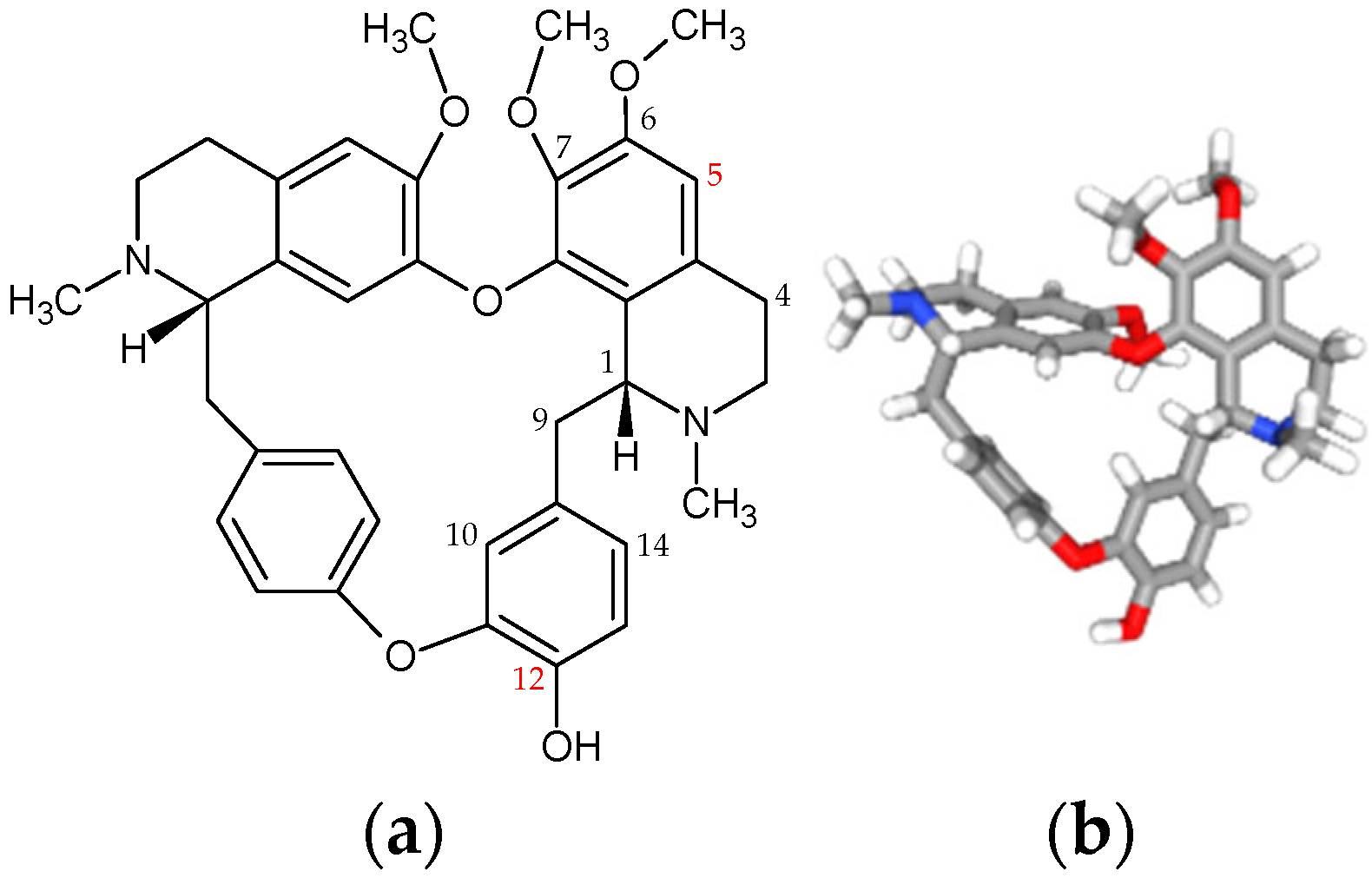
Figure 1. Chemical structures of berbamine, with key positions indicated. (a) Atoms used for structural numbering are shown in black; positions suitable for modification are highlighted in red. (b) The 3D image adapted from PubChem (NCBI), CID 275182.
Applsci 16 04985 g001
Figure 2. Chemical structures of tetrandrine and fangchinoline, with key positions indicated. (a,c) Atoms used for structural numbering are shown in black; positions suitable for modification are highlighted in red. (b,d) The 3D images adapted from PubChem (NCBI), CID 73078 (tetrandrine) and 73481 (fangchinoline).
Applsci 16 04985 g002
Figure 3. Chemical structure of sinomenine with key positions indicated. (a) Atoms used for structural numbering are shown in black; positions suitable for modification are highlighted in red. (b) The 3D image adapted from PubChem (NCBI), CID 5459308.
Applsci 16 04985 g003
Disclaimer/Publisher’s Note: The statements, opinions and data contained in all publications are solely those of the individual author(s) and contributor(s) and not of MDPI and/or the editor(s). MDPI and/or the editor(s) disclaim responsibility for any injury to people or property resulting from any ideas, methods, instructions or products referred to in the content.