Review

Integrated Roles of Hypoxia Signaling, Lipid-Handling, and Extracellular Matrix Remodeling Genes in Myocardial Infarction and Heart Failure: A Gene-Centric Translational Review

by
Rafał Celiński
1,
Janusz Kocki
2,
Anna Grzywa-Celińska
3,
Katarzyna Dos Santos Szewczyk
4,* and
Anna Berecka-Rycerz
5,*
1
Department of Cardiology, Independent Public Provincial Specialist Hospital in Chełm, 22-100 Chełm, Poland
2
Department of Clinical Genetics, Medical University of Lublin, 20-080 Lublin, Poland
3
Chair and Department of Pneumonology, Oncology and Allergology, Medical University of Lublin, 20-093 Lublin, Poland
4
Department of Pharmaceutical Botany, Medical University of Lublin, 20-093 Lublin, Poland
5
Department of Medicinal Chemistry, Medical University of Lublin, 20-090 Lublin, Poland
*
Authors to whom correspondence should be addressed.
Appl. Sci. 2026, 16(10), 4806; https://doi.org/10.3390/app16104806
Submission received: 30 March 2026 / Revised: 2 May 2026 / Accepted: 4 May 2026 / Published: 12 May 2026
(This article belongs to the Special Issue Therapeutic Applications and Biology of Extracellular Vesicles)

Abstract

Heart failure (HF) and myocardial infarction (MI) are interconnected syndromes with overlapping pathogenic pathways, including ischemia, neurohormonal activation, and maladaptive remodeling. Hypoxia-response genes, lipid-handling genes, and extracellular matrix (ECM) genes each influence these processes. Understanding their integrated roles can uncover biomarkers and targets. A systematic literature search was conducted (PubMed, Web of Science, and Scopus; 2000–2026; English-only, following PRISMA guidelines) to identify studies on key genes in hypoxia signaling, lipid metabolism, and ECM remodeling in MI/HF. Acute hypoxia (via HIFs) orchestrates metabolic adaptation and inflammation, but chronic HIF activation drives fibrosis and dysfunction. In parallel, genes controlling triglyceride and cholesterol handling (e.g., LPL, APOC3) influence energy supply and vascular risk. Variants in these genes modulate plasma lipids and MI/HF risk. For example, genetic loss-of-function in APOC3 lowers triglycerides and reduces coronary risk. ECM-related genes (e.g., COL4A1, LRP1) govern fibrosis and vascular integrity. Mutations in COL4A1 cause cardiomyocyte hypertrophy and severe fibrosis, while LRP1 regulates matrix remodeling and is upregulated in ischemic myocardium. Throughout, gene functions span acute repair versus chronic maladaptation. Findings derive from mixed sources: rodent models and cell studies demonstrate mechanistic links, while human genetics and cohorts link gene variants to HF/MI outcomes. Many promising biomarkers (e.g., circulating ITGA1) are preliminary, lacking large prospective validation. Not all cited therapeutic ideas have been tested in the treatment of human cardiac disease. The literature mix of species, models, and patient cohorts introduces heterogeneity.

1. Introduction

Cardiovascular diseases remain the leading global health burden. In 2021, at least 38% of premature deaths from noncommunicable diseases were caused by cardiovascular conditions [1,2]. In particular, heart failure (HF) and myocardial infarction (MI) are major contributors to morbidity and mortality worldwide. The pathogenesis of HF and MI involves metabolic shifts, inflammatory signaling, extracellular matrix remodeling, and fibrosis. Preventing adverse cardiac remodeling is challenging because multiple pathways converge [1,2,3,4,5,6,7,8].
We hypothesize that oxygen-sensing mechanisms (hypoxia signaling), lipid metabolism genes, and ECM regulators interact synergistically to influence disease trajectories after MI. Therefore, in this review, we focus on representative genes grouped into three axes (hypoxia regulators, metabolic mediators, ECM modulators) to illustrate how these axes interact in driving remodeling after MI. We describe gene functions, disease associations, and potential for biomarkers or therapy. Where possible, we highlight crosstalk between pathways (e.g., hypoxia influencing lipid handling or fibrosis) to construct a unified mechanistic framework. We explicitly state our objectives and guiding questions: (1) Which genes at the intersection of hypoxia signaling and metabolism are most critical in MI/HF? (2) How do these pathways converge on ECM remodeling? (3) Can an integrated model link hypoxia → metabolism → ECM to predict outcomes? Beyond gene aggregation, our work proposes a phase-based model linking molecular pathways to HF progression, providing novel insights for translational research and bridging molecular mechanisms with disease-stage-specific clinical interpretation.

2. Methodology (Literature Search and Selection)

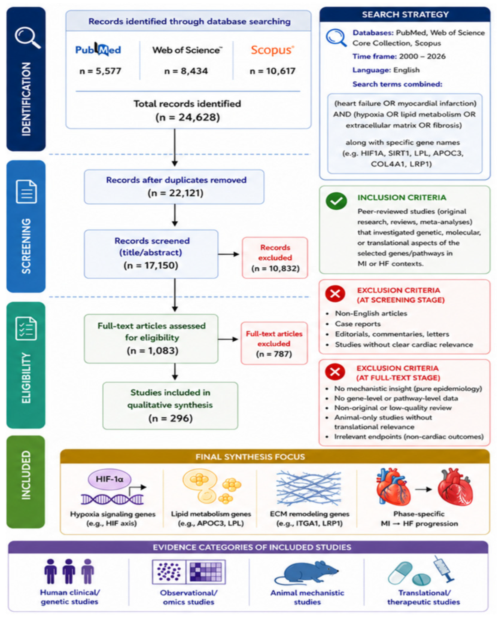
A comprehensive literature search was performed across PubMed, Web of Science, and Scopus (January 2010 to February 2026). Search terms combined “(heart failure OR myocardial infarction) AND (hypoxia OR lipid metabolism OR extracellular matrix OR fibrosis)” along with specific gene names (e.g., HIF1A, SIRT1, LPL, APOC3, COL4A1, LRP1). We followed PRISMA guidelines (Figure 1) for transparency. Inclusion criteria: peer-reviewed studies (original research, reviews, meta-analyses) that investigated genetic, molecular, or translational aspects of the selected genes/pathways in MI or HF contexts. Exclusion criteria: non-English articles, case reports, editorials, or studies without clear cardiac relevance. Two authors independently screened titles/abstracts and reviewed full texts, extracting data on gene function, expression changes, polymorphisms, and therapeutic implications. Discrepancies were resolved by consensus. This targeted strategy aimed to verify recent findings (past 5–10 years), focusing on human and relevant animal studies, and mapping common themes.

3. Hypoxia Regulators and Metabolic Stress Genes

Table 1 summarizes key genes across hypoxia, lipid metabolism and ECM remodeling discussed in this section and later sections.

3.1. HIF Axis (HIF1A, EPAS1/HIF2α, HIF3A) and Oxygen-Dependent Regulators (PHD/EGLN1–3, HIF1AN/FIH, VHL)

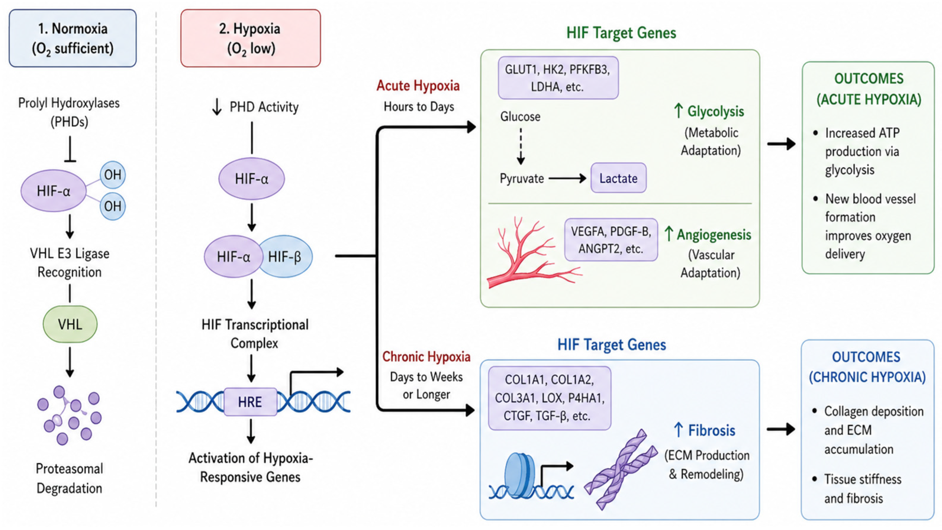
Cellular adaptation to hypoxia is largely governed by hypoxia-inducible factors (HIFs). HIF-1α (encoded by HIF1A) and HIF-2α (EPAS1) are oxygen-sensitive transcription factors that become stabilized when oxygen levels drop, triggering a broad transcriptional program to restore oxygen homeostasis [1,35]. In normoxia, these HIF-α subunits are hydroxylated and targeted for proteasomal degradation via prolyl hydroxylases (PHDs) and the von Hippel—Lindau (VHL) ubiquitin ligase complex [16,35,36,37,38]. Under hypoxic conditions, HIF-α accumulates, translocates to the nucleus, dimerizes with HIF-1β, and binds hypoxia-response elements in DNA to induce target genes [35,36,37,38]. HIF target genes promote adaptive processes such as anaerobic glycolysis (e.g., upregulating GLUT1, HK1, LDHA), angiogenesis (e.g., VEGF-A), erythropoiesis (EPO), iron metabolism (HO-1), and cell survival/apoptosis regulation (BNIP3, IGF1). In the heart, these responses can be double-edged: they support short-term survival in ischemia but may contribute to long-term pathology if overactivated [3,8,13,34,35,36,37,38].
A schematic overview of the HIF signaling cascade, including its regulatory components and downstream adaptive versus maladaptive responses, is presented in Figure 2.

3.1.1. Role in Myocardial Infarction

Transient HIF-1α activation during acute ischemia is generally cardioprotective. Stabilization of HIF-1α in the ischemic heart shifts metabolism toward glycolysis (preserving ATP when oxygen is scarce), stimulates angiogenesis, and can reduce cell death [39,40,41,42,43]. Experimental studies show that moderate HIF-1α augmentation limits infarct size and preserves cardiac function after MI [44,45]. For example, myocardium-targeted HIF-1α gene delivery in mice attenuated cardiomyocyte apoptosis and dysfunction following MI, in part by upregulating antioxidant defenses (heme oxygenase-1) and suppressing a pro-apoptotic mediator (BNIP3) [46]. Similarly, mice with inhibited HIF degradation (e.g., PHD knockout) have shown smaller infarcts and improved post-MI remodeling [42,44,45,47,48,49,50,51,52]. These findings suggest that boosting the hypoxia response during an acute MI can mitigate tissue injury. In line with this, pharmacological HIF activators are being explored: for instance, prolyl hydroxylase inhibitors (HIF stabilizers) like roxadustat given at reperfusion have demonstrated reduced infarct size and improved ejection fraction in rodents [53]. There is even early clinical interest—a Phase I trial of an HIF-activating agent (the peptide LRP1 agonist SP16, which indirectly enhances HIF signaling via anti-inflammatory pathways) showed safety and hinted at potential benefits in MI patients [33].

3.1.2. Role in Heart Failure

By contrast, chronic or excessive HIF-α signaling in the heart can be maladaptive. Prolonged HIF-1α upregulation has been linked to adverse remodeling, in part through sustained shifts in metabolism and promotion of cell death pathways [35,42,50]. Transgenic mice with chronically high HIF-1α (achieved by deleting PHD2, a HIF suppressor) developed dilated cardiomyopathy, indicating that unrestrained HIF signaling can drive pathological cardiac hypertrophy and failure [54]. One mechanism involves HIF-1α–p53 crosstalk—a recent study reported that excessive HIF-1α expression after MI triggered p53-dependent apoptosis, leading to thinning of the infarcted wall and cardiac rupture [16,55]. This illustrates the delicate balance—while HIF helps hearts cope with acute ischemia, its sustained activation may exacerbate cardiomyocyte dropout and arrhythmias in chronic hypoxia [47]. Notably, HIF-1α induces the pro-apoptotic gene BNIP3, which, if persistently elevated, contributes to mitochondrial dysfunction and cell death seen in chronic ischemic cardiomyopathy [47,56]. Thus, the timing and degree of HIF activation are critical: intermittent hypoxia or brief HIF stimulation can precondition and protect the heart, whereas continuous hypoxia drives arrhythmogenesis and HF progression [51,57].
HIF-2α (EPAS1) plays a somewhat complementary role. It shares many targets with HIF-1α but also uniquely regulates genes like EPO (erythropoietin) and enzymes of catecholamine metabolism [35,57,58]. Knockout models indicate HIF-2α is essential for proper cardiac development and stress adaptation—for example, mice lacking Epas1 have impaired catecholamine homeostasis and are prone to HF under stress [35]. However, HIF-2’s role in adult heart disease is less studied than HIF-1. Some evidence suggests endothelial HIF-2α supports microvascular integrity: mice with endothelial Hif2a deletion showed exacerbated cardiac injury, inflammation, and mortality after pressure overload [59]. This aligns with the idea that HIF-2α in vessels helps maintain perfusion during hypertrophic stress. Overall, both HIF-1 and HIF-2 pathways are implicated in ischemic preconditioning, a phenomenon where prior hypoxic episodes make the heart more resilient to MI [57]. Therapeutically, targeting HIF pathways is complex—some HF patients (e.g., with anemia or chronic kidney disease) might benefit from HIF stimulation (as HIF stabilizers combat anemia and may improve oxygen delivery), whereas others may be harmed by overactivation [16]. Ongoing research is clarifying whether carefully timed HIF activation (e.g., during acute MI or shortly after) can be exploited without inducing long-term harm [60,61].
HIF-3α (encoded by the HIF3A gene) is expressed in various forms and can act as a negative regulator of HIF signaling. Although HIF3A is low in the adult heart under basal conditions [35]. In vitro, studies suggest that HIF-3α can limit classic HIF target gene expression by competing for HIF-1α DNA binding or cofactors [62,63,64]. HIF3A gene expression can be induced by metabolic signals (e.g., insulin) and may provide feedback inhibition to prevent excessive HIF-1 activity. Its exact role in cardiac ischemia remains under investigation, but HIF3A may function analogously to PHDs/FIH in fine-tuning the hypoxic response [35,62,63,64].
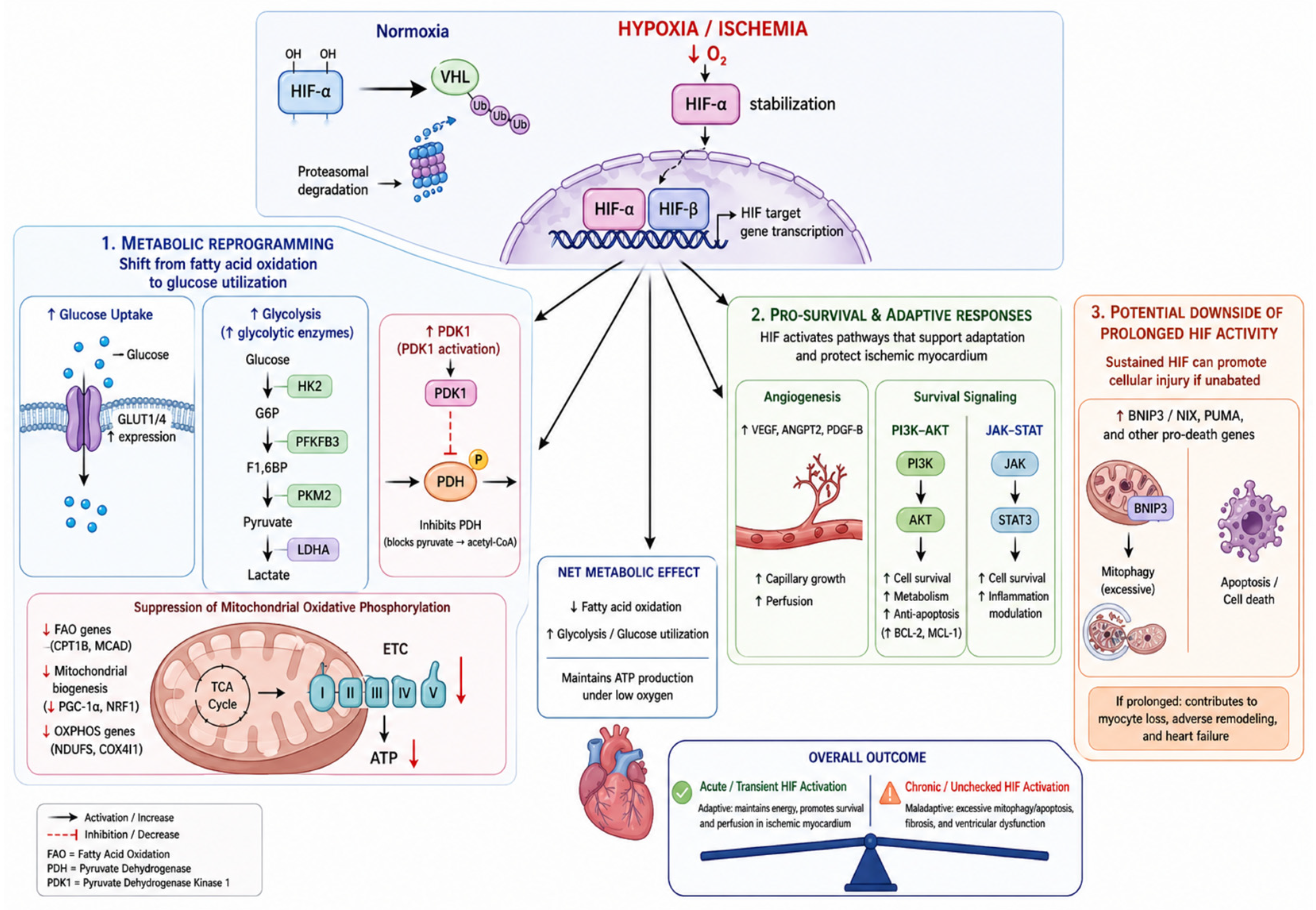
HIF signaling broadly shifts myocardial substrate utilization from fatty acids toward glucose to adapt to low oxygen [65]. The HIF-driven metabolic reprogramming of cardiomyocytes, including the shift from mitochondrial oxidative phosphorylation to glycolysis, is illustrated in Figure 3. HIF also promotes angiogenesis and cell survival pathways (PI3K-AKT, JAK-STAT) that can protect the ischemic myocardium [57,66]. However, prolonged HIF activity may upregulate mediators like BNIP3 that trigger mitophagy or apoptosis if unabated [67].

3.1.3. Therapeutic Implications (HIF Stabilization vs. Safety)

To contextualize the translational relevance of hypoxia-related pathways, Table 2 summarizes current therapeutic strategies. Agents are grouped by direct gene targeting (e.g., APOC3, LRP1) versus pathway-linked modulation (e.g., PHD–HIF inhibitors; NAD+ boosting supporting sirtuin activity). The Table emphasizes trial phase/status, studied indication, and key evidence, while noting when cardiovascular indications are exploratory or non-primary.

3.2. SIRT1—A Metabolic Longevity Factor in the Heart

Sirtuin-1 (SIRT1) is a NAD+-dependent deacetylase that links cellular energy status to gene expression. SIRT1 deacetylates numerous transcription factors and enzymes (e.g., PGC-1α, FOXO, p53, NF-κB), generally enhancing antioxidant defenses and mitochondrial biogenesis while dampening apoptosis [15,70,71,72,73,74]. It is often termed a “longevity factor” because of its role in stress resistance and caloric restriction benefits. In the heart, SIRT1 is low in the fetal stage, upregulated in exercise and mild stress, but tends to decline with aging and advanced HF [74,75]. Adequate SIRT1 activity appears protective against both ischemic injury and maladaptive hypertrophy.

3.2.1. Ischemia/Reperfusion Injury

Pioneering studies by Alcendor et al. [76] showed that SIRT1 activation protects cardiomyocytes from oxidative stress and apoptosis. In a 2010 mouse study, cardiac-specific Sirt1 knockout led to significantly larger infarcts after ischemia/reperfusion (I/R)—infarct size as a percentage of area-at-risk increased from ~15% in controls to ~44% in Sirt1-null hearts [77]. These Sirt1-null mice also had worse post-ischemic contractile dysfunction. Conversely, transgenic mice overexpressing SIRT1 in the heart had much smaller infarcts (~15% vs. 36% in wild-type) and fewer apoptotic cells (TUNEL-positive nuclei ~4% vs. 10%) following I/R. SIRT1-overexpressors also exhibited better functional recovery during reperfusion [73]. Mechanistically, SIRT1 upregulated several anti-apoptotic and antioxidant molecules (e.g., Mn-SOD, thioredoxin-1, Bcl-x_L) and suppressed pro-death effectors (Bax, cleaved caspase-3) in the reperfused heart. Notably, SIRT1’s interaction with the transcription factor FOXO1 was critical: SIRT1 deacetylated and activated FOXO1, which in turn increased Mn-Sod expression and reduced ROS accumulation [73]. The net effect was lower oxidative DNA damage and greater cardiomyocyte survival after MI. Thus, SIRT1 activation blunts acute ischemic injury, a finding mirrored by pharmacological activators like resveratrol. In rodent MI models, resveratrol (which activates SIRT1) improved cardiac function and reduced fibrosis, partially via activating AMP-activated kinase (AMPK) and downstream metabolic benefits [78].

3.2.2. Heart Failure and Remodeling

In chronic HF, SIRT1 expression is often downregulated, and this deficit may contribute to worsening oxidative stress and cell death. A clinical study of 163 HF patients found that SIRT1 mRNA in peripheral blood dropped significantly in HF, with the lowest levels in decompensated HF patients [79]. These low SIRT1 levels strongly correlated with high oxidative stress markers (increased oxidant index, low antioxidant capacity) in the same patients. The authors concluded that significantly decreased SIRT1 expression is linked to increased oxidative stress in failing hearts, particularly in decompensated heart failure [79]. This supports the idea that SIRT1 helps keep oxidative damage in check during compensated HF, and its further loss may tip the balance toward decompensation. From a mechanistic viewpoint, SIRT1’s targets in the heart include pro-hypertrophic factors (like NF-κB and myocardin) and metabolic regulators (PGC-1α). SIRT1 can deacetylate and inhibit NF-κB, thereby reducing inflammation and fibrosis [80]. It also activates PGC-1α to maintain mitochondrial function. Consistently, mice with moderate overexpression of SIRT1 are protected against age-related cardiac hypertrophy and interstitial fibrosis. However, intriguingly, extremely high SIRT1 overexpression (a level ~10-fold above normal) was reported to impair cardiac function, possibly by excessive deacetylation of cytoskeletal proteins and derepression of pathological genes (a reminder that more is not always better) [81,82].
Clinically, evidence for SIRT1’s impact comes from genetic association and human tissue studies. A polymorphism in SIRT1 gene (e.g., rs7069102) has been associated with coronary artery disease risk in some populations [83], although findings are not yet conclusive. In failing human hearts, SIRT1 gene expression is often found reduced, especially in aged patients, which correlates with worsened mitochondrial quality control [84]. This „downregulation of SIRT1 as an aging change” in HF suggests that therapeutic upregulation of SIRT1 could be beneficial in those with advanced HF and metabolic syndrome [84]. Indeed, low SIRT1 activity is thought to contribute to impaired autophagy and increased oxidative damage in the failing myocardium. In contrast, in patients with hypertrophic cardiomyopathy, some data show elevated SIRT1, possibly as a compensatory response or related to altered metabolism. In essence, SIRT1 exerts a dual-edged role—a low-to-moderate level is cardioprotective, whereas an excessively high level might be detrimental (though such high levels are unlikely in physiological settings).
Therapeutically, boosting SIRT1 is an attractive strategy for metabolic modulation in HF. Small-molecule SIRT1 activators (e.g., SRT2104) have been tested in clinical trials. SRT2104 was well-tolerated but did not significantly improve cardiac outcomes in a short-term HF trial [75,85]. Nonetheless, SGLT2 inhibitors (a new class of HF drugs) may activate SIRT1 indirectly by creating a “fasting-like” metabolic state. This has been proposed as part of the mechanism by which SGLT2 inhibitors improve HF outcomes (often termed “nutritional ketosis” or increased NAD+ levels activating SIRT1) [75]. The research highlights show that AAV-mediated gene therapies aimed at improving cardiac function post-MI in pigs (such as knocking down Hippo signaling or overexpressing SCF) are a rapidly advancing area of preclinical research [86]. Complementing mechanistic models, NAD+ repletion has shown preclinical reversal of HFpEF features, strengthening the plausibility of NAD+-SIRT axes as therapeutic leverage points [18]. Ongoing research is exploring if directly activating SIRT1 (or preventing its decline) in HF patients can synergize with existing therapies to enhance cardiac energy efficiency and prevent adverse remodeling.
In summary, SIRT1 serves as a crucial metabolic and stress response regulator in the heart, promoting cardiomyocyte longevity and efficient energy use. Its activation correlates with cardioprotection against ischemia, hypertrophy, and ageing, whereas loss of SIRT1 exacerbates these conditions [70]. However, maintaining SIRT1 within an optimal range is key—highlighting the sophisticated control needed if we aim to harness SIRT1 for HF or post-MI therapies [75]. Measurements of SIRT1 or its activity (e.g., deacetylation status of target proteins) are not yet used clinically, but they hold potential as biomarkers of cardiac metabolic health or response to therapy in the future.

3.3. MYC Gene—Stress-Responsive Transcriptional Driver Across Cell Types

The proto-oncogene MYC encodes the transcription factor MYC, which is a master regulator of cell proliferation, growth, and metabolism. In healthy adult hearts, MYC gene expression is low; cardiomyocytes are terminally differentiated and do not typically express cell cycle promoters such as MYC. However, MYC is often reactivated in response to cardiac stress and disease. Studies have shown MYC gene upregulation in various forms of heart disease—including hypertrophic cardiomyopathy, pressure-overload hypertrophy, and dilated cardiomyopathy—often before overt pathology develops [87]. MYC’s involvement in the heart is multifaceted: it can promote cardiomyocyte hypertrophy, drive fibroblast proliferation, alter metabolism, and induce apoptosis under certain conditions [87].
In models of pressure overload (e.g., aortic stenosis), the MYC gene is one of the early response genes that is activated during the hypertrophic phase. MYC protein contributes to the switch from fatty acid oxidation to glycolysis in hypertrophic hearts, akin to a Warburg-like metabolic shift seen in rapidly growing cells [87]. By upregulating glycolytic enzymes and glutamine metabolism, MYC ensures an ample supply of biosynthetic building blocks for growth. However, this metabolic reprogramming can be maladaptive in the heart, leading to energy inefficiency. Additionally, MYC can directly control the expression of cell cycle regulators and mitochondrial genes, influencing cardiomyocyte size and number. For instance, MYC activation in adult cardiomyocytes can induce a degree of cell cycle re-entry, which usually does not complete and may trigger apoptotic pathways or DNA damage responses [77].
Evidence for MYC’s pathological role of MYC in HF comes from both gene manipulation studies and observational human data. Transgenic mice with cardiac-specific MYC overexpression develop enlarged hearts, and if MYC is chronically active, contractile failure eventually occurs. Conversely, inhibition of MYC has been proposed as a therapeutic strategy—in experimental models, blocking MYC activity (via dominant-negative constructs or small molecules) attenuated cardiac hypertrophy and improved function in pressure overload [87]. The MYC gene has also been linked to HF comorbidities, such as diabetes and cancer. Diabetic hearts exhibit elevated MYC signaling, which may exacerbate metabolic stress. Conversely, cancer therapies targeting MYC may have off-target cardiac effects that are still being investigated.
Notably, MYC can influence cardiac angiogenesis and oxidative stress. MYC activation upregulates VEGF and angiogenic factors, attempting to match new capillary growth to hypertrophying muscles. However, if this response is insufficient, the result is relative hypoxia and HIF activation, creating a feedback loop between MYC and HIF in advanced hypertrophy. MYC also drives the expression of mitochondrial biogenesis factors but in a dysregulated fashion, sometimes leading to excess ROS production and oxidative damage [87]. Indeed, the MYC gene has been implicated in cardiomyocyte apoptosis through mitochondrial pathways when highly expressed.
Clinically, direct measurement of myocardial MYC is not performed, but its fingerprint can be observed—patients with HF often show increased expression of MYC target genes (such as cyclins and glycolytic enzymes) in the heart tissue. There is interest in MYC as part of the gene expression profile that predicts HF prognosis. Moreover, a recent bioinformatics study identified MYC (along with another transcription factor C/EBPβ) as a key node in the gene network distinguishing failing hearts, suggesting it as a potential biomarker or therapeutic target [79].
Encouragingly, novel therapeutics that inhibit MYC, such as small molecules that disrupt MYC-MAX dimerization, are being developed in the oncology field (e.g., small molecules that disrupt MYC-MAX dimerization). If safe and specific, these drugs could theoretically be repurposed for the treatment of cardiovascular diseases. A 2019 review posited MYC inhibition as an „attractive paradigm” for treating hypertrophic cardiomyopathy and HF, given its central role in disease initiation and maintenance [88]. Any such approach would have to be finely controlled because MYC is essential for normal cell turnover in many tissues.
Importantly, newer studies expand MYC’s relevance beyond cardiomyocytes. For example, one study reported HF-specific fibroblast programs contributing to dysfunction via a MYC–CXCL1–CXCR2 axis, implying that MYC-centered transcriptional hubs may link fibroblast phenotype to inflammatory signaling and functional deterioration [19]. In parallel, another study reported that MYC overexpression improved post-MI recovery associated with cardiomyocyte hyperplasia in a mouse model, suggesting that context, timing, and cellular targets critically determine whether MYC activation is beneficial or harmful [20].
In summary, MYC is a central orchestrator of pathological cardiac remodeling. By promoting hypertrophy, proliferation signals, and metabolic shifts, MYC genes contribute to the development and progression of HF [79]. It represents a link between classic growth signaling and the metabolic and apoptotic dysregulation observed in failing hearts. Ongoing research is exploring MYC-modulating therapies and whether short-term inhibition during stress might blunt adverse remodeling without harming the necessary cellular functions. Given MYC’s involvement of MYC in cancer, the cardiovascular safety of MYC inhibitors requires careful evaluation. Nonetheless, the MYC gene stands out as a convergence point of pathways in HF, underscoring the overlap between oncogenic and cardiogenic mechanisms in the diseased heart.

3.4. CDC6—MI-Associated, Senescence-Linked Biomarker Candidate

Unlike many tissues, the adult mammalian heart has very limited regenerative capacity—adult cardiomyocytes are largely post-mitotic. However, after injury, there is evidence of transient cell cycle re-entry or activation of senescence programs in certain cardiac cells, which may influence healing. CDC6 (Cell Division Cycle 6) is a DNA replication licensing factor that plays a key role in permitting cells to enter S-phase. In the context of heart disease, CDC6 has recently emerged as a novel player in the response to MI [21].
A 2025 study by Wen et al. used bioinformatics and animal models to show that CDC6 is upregulated in the early stages of MI and may act as an “acute senescence trigger” that paradoxically aids cardiac repair [21]. In their analysis, CDC6 was identified as one of two key genes (along with PLK1) overlapping between MI-induced genes and senescence-associated genes. Post-MI, Cdc6 expression was elevated in mouse hearts within the first few days. Importantly, circulating CDC6 levels (from patient blood samples) also rose early after MI, suggesting it could serve as an early biomarker of myocardial injury and cellular senescence. The authors reported that CDC6 had high diagnostic value for acute MI, distinguishing MI patients from controls, and its expression varied over time post-infarction in line with the progression of injury and repair. Mechanistically, early after MI, a subset of cardiomyocytes and cardiac progenitor cells may attempt to re-enter the cell cycle (as an abortive attempt at regeneration) and in doing so, activating a senescence program that aids in wound healing. CDC6, traditionally a DNA replication licensing factor, appears to play a role in this process—its overexpression in an in vitro ischemia model (oxygen-glucose deprivation in cardiomyocytes) boosted the expression of genes mediating cardiac repair [21]. Conversely, clearing senescent cells (with a senolytic drug, ABT263) blunted the expression of those repair genes, suggesting that the early, transient presence of senescent-like cells is beneficial for tissue repair after MI [21]. This concept aligns with emerging views that not all senescence is bad—a transient senescent response can be part of normal wound healing, whereas chronic senescence (and persistence of senescent cells) is detrimental.
From a therapeutic standpoint, CDC6 itself is not a direct drug target (given its essential cell cycle functions and oncogenic potential if unsuppressed), but its behavior highlights new pathways. Modulating the balance between regeneration and senescence in the heart is a frontier of HF research. Strategies like transiently encouraging cardiomyocyte cell cycle activity (e.g., by cyclin overexpression or cell cycle gene therapy) have had some success in animal models of MI [89,90]. The CDC6 findings underscore that timing is crucial: boosting proliferation or senescence at specific phases could improve repair, while the same at a wrong time could worsen remodeling.
Therefore, the CDC6 gene has emerged as a link between cell cycle dynamics and cardiac repair after MI. Its early upregulation following infarction appears to promote a reparative senescent response that helps limit damage and initiate healing [21]. This finding contrasts with the traditional view that cell cycle activation in the adult heart is purely pathological. Clinically, CDC6 shows promise as a novel early MI biomarker and potentially as a gauge of the heart’s regenerative response to MI. Further studies are needed to translate these findings. However, the dual role of cell cycle regulators, such as CDC6, in acute versus chronic settings is an important consideration for future cardiac therapies.
In summary, hypoxia regulators like HIF1A/EPAS1 and metabolic guardians like SIRT1 form the frontline of the heart’s defense against ischemic injury, orchestrating adaptive metabolic shifts and stress resistance. Meanwhile, genes like CDC6 remind us that the heart’s response to injury also involves cell cycle and senescence decisions in non-myocyte cells (and perhaps rare myocytes). Understanding these processes offers hope for innovative treatments—for instance, preconditioning the heart via HIF pathways, enhancing SIRT1 activity to rejuvenate aging hearts, or transiently overcoming cell cycle blocks to regenerate myocardium.

3.4.1. Strength of Evidence (Hypoxia Pathways)

Evidence comes from animal studies and clinical observations. For example, knocking out HIF-1α in mouse hearts worsens infarct size, while pharmacologically stabilizing HIF-1α (using PHD inhibitors) can reduce injury [91,92,93,94]. In humans, higher myocardial HIF-1α levels correlate with severity of ischemic damage. Gene expression profiling of failing hearts reveals upregulation of HIF target genes, linking hypoxia response to disease severity. That said, some findings conflict: a few studies show that HIF-1α overactivation under normoxia (pseudohypoxia) can be protective or deleterious depending on context. The overall consensus is that HIF is central to the acute response, but chronic HIF-driven fibrogenesis and inflammation are supported by multiple studies [91,92,93,94,95].

3.4.2. Limitations (Hypoxia Pathways)

Most data are preclinical or observational: human trials of HIF modulators in MI/HF are scarce, so translational relevance is uncertain. Moreover, hypoxia effects vary by cell type and timing; what helps myocytes acutely may harm endothelial or fibroblast cells long-term. There is also complexity in different HIF isoforms (HIF-1 vs. HIF-2α) and their tissue-specific roles. Notably, some hypoxia genes (e.g., EGLN polymorphisms) were studied in small cohorts with inconsistent results. We must also distinguish heart-specific hypoxia effects from systemic hypoxia (e.g., lung disease)—most gene studies focus on cardiac tissue or blood markers, limiting generalizability.

4. Lipid Metabolism Genes in HF and MI

The failing or ischemic heart’s metabolism undergoes a profound shift. In a healthy heart, fatty acids are the primary fuel (providing ~60–70% of ATP), with glucose and other substrates making up the rest. In HF, the heart often becomes “energy-starved”—there is reduced fatty acid oxidation, a partial reliance on glucose, and overall ATP production is decreased [23,35]. Perturbations in systemic lipid handling can also influence cardiac outcomes: circulating triglyceride-rich lipoproteins and their composition affect atherosclerosis (and thus MI risk), and they may indirectly affect cardiac energy supply during stress.

4.1. Lipoprotein Lipase (LPL)—Gatekeeper of Cardiac Fat Utilization

Lipoprotein lipase (LPL) is an enzyme anchored to capillary endothelium (especially in muscle, heart, and adipose tissue) that hydrolyzes triglycerides (TG) in circulating chylomicrons and very-low-density lipoproteins (VLDL) [96,97,98,99]. By releasing free fatty acids from TG, LPL allows uptake of these fatty acids into myocardium to be used for β-oxidation and energy production [100]. The heart expresses a unique isoform of LPL and can rapidly increase LPL activity under conditions like exercise or fasting to draw in more fatty acids. Given the heart’s high energy demand, LPL is critical—approximately 50–70% of the heart’s ATP in normal conditions comes from oxidation of fatty acids delivered by LPL activity [23].
In the context of HF, a well-recognized metabolic change is a decline in fatty acid oxidation capacity and a relative increase in glucose utilization (a reversion toward the “fetal” metabolic profile) [35]. Some studies have observed that cardiac LPL expression/activity decreases in advanced HF, which would limit fatty acid uptake and might contribute to energy deficit. On the other hand, heart failure patients often exhibit hypertriglyceridemia and insulin resistance, factors that can downregulate LPL. The net impact of LPL in HF appears context dependent. For instance, in early pressure-overload hypertrophy, cardiac LPL might actually increase to supply more fuel; but in late HF, LPL falls, and the failing heart cannot adequately use fats, partly due to mitochondrial dysfunction [101,102].
Animal models provide direct insights. Cardiac-specific Lpl knockout (hLPL0) mice develop severe cardiomyopathy and failure, underscoring that the heart absolutely requires triglyceride-derived fatty acids for normal function [103]. These mice have abundant circulating TG (because muscle and heart cannot clear it), yet their hearts are energy starved. A remarkable study “rescued” such mice by increasing glucose delivery/uptake: crossing the hLPL0 mice with mice overexpressing the GLUT1 glucose transporter (thus enhancing cardiac glucose utilization) partly compensated for the loss of fatty acids and prevented the heart failure [104,105]. In other words, failing to transport or use triglyceride leads to an energy deficiency that causes HF, which can be ameliorated by providing an alternative fuel (glucose) [103,106]. This experiment elegantly demonstrates the principle of metabolic flexibility—the heart can burn different fuels, but it needs one or the other. In hLPL0 without a backup, the energy shortfall is lethal [103].
Beyond energy supply, LPL can also influence lipotoxicity. If LPL is too active in certain settings (e.g., diabetes), excessive fatty acid uptake by cardiomyocytes can lead to lipid accumulation in the heart (myocardial steatosis) and cause lipotoxic damage (via ceramides, etc.). Thus, both LPL deficiency and LPL excess can be harmful: an optimal balance is needed. In advanced HF patients, paradoxically, higher circulating lipids (and perhaps higher LPL in peripheral tissues) correlates with better outcomes—the so-called “lipid paradox”—possibly because those patients have better nutritional status or more metabolic reserve. However, this epidemiological observation does not negate the fundamental importance of LPL for cardiac energetics [23,107,108,109].
From a genetic and clinical perspective, LPL variants have been implicated in coronary artery disease (CAD) and MI risk. Certain mutations in LPL (or its regulators) cause familial hypertriglyceridemia, which can accelerate atherosclerosis. For example, the common LPL variant S447X, which increases LPL activity, is associated with lower TG levels and has been linked with modest protection against CAD. Conversely, rare loss-of-function LPL mutations (like those causing familial chylomicronemia) lead to extremely high TG but interestingly do not strongly predispose to CAD (possibly because chylomicrons are too large to penetrate the arterial wall readily) [110,111,112,113,114]. More relevant are mutations in LPL regulators—discussed below (APOC3, APOA5, etc.)—which do affect CAD risk.
In summary, in the failing heart, reduced LPL activity contributes to the metabolic shift and energy deficit, while in the context of systemic metabolism, adequate LPL function is cardioprotective by preventing hypertriglyceridemia and atherosclerosis. Therapies targeting LPL are not direct (as LPL is widely expressed and crucial for general metabolism), but interventions like fibrates (PPARα agonists) can raise muscle LPL and have been used to lower TGs. Gene therapy for LPL deficiency (alipogene tiparvovec) was approved for familial pancreatitis, hinting at future possibilities of enhancing LPL in specific tissues [115]. For HF patients, strategies to boost myocardial energy substrate uptake—whether fatty acids via LPL or glucose via insulin sensitization—remain an area of intense research.

4.2. APOC3 and APOA5—Regulators of Triglycerides and MI Risk

Apolipoprotein C-III (APOC3) and apolipoprotein A-V (APOA5) are two hepatically produced proteins that modulate plasma triglyceride levels by influencing lipoprotein lipase and the clearance of TG-rich lipoproteins. They have almost opposing actions: APOC3 is an inhibitor of lipoprotein lipase and hepatic uptake of TG-rich particles, whereas APOA5 is a facilitator of lipolysis [110,111,112,113,114]. Variants in these genes have significant impact on plasma TG and have been linked to the risk of MI and HF (mainly via atherosclerosis and possibly through direct metabolic effects on the heart).
APOC3 is a small apolipoprotein (79 amino acids) that attaches to VLDL and chylomicrons and inhibits LPL as well as hepatic remnant uptake. Elevated APOC3 levels cause increased plasma TG. Notably, human genetics provided compelling evidence that APOC3 influences coronary disease risk. In 2014, two large studies examined rare loss-of-function (LOF) mutations in APOC3. Individuals heterozygous for APOC3 LOF had roughly 50% lower triglycerides than non-carriers and a dramatically lower incidence of ischemic heart disease [110]. In a combined analysis of ~110,000 people, carriers of any APOC3 LOF mutation showed a 40% reduction in coronary heart disease (CHD) risk compared to non-carriers (OR ~0.60) [110]. This association was highly significant and independent of other factors [116]. Another study in a Danish population found similar results: lifelong low APOC3 conferred ~41% lower risk of MI [117]. These findings cemented APOC3 as a causal contributor to MI risk, likely through its effect on triglyceride-rich lipoproteins.
These human data have spurred development of APOC3 inhibitors as drugs. An antisense oligonucleotide targeting APOC3 (volanesorsen) was approved for familial chylomicronemia syndrome; it lowers TG by ~70% [25]. Newer RNA interference therapies (like olpasiran for Lp(a) and another for APOC3) are in trials for broader hypertriglyceridemia [118]. While it is not yet proven that APOC3 inhibition reduces MI in the general population, genetics strongly predict it should. Interestingly, high APOC3 is not only atherogenic but also might promote inflammation—some studies found APOC3 directly activates monocytes and endothelial cells. In HF, severe hypertriglyceridemia (as seen in diabetes or obesity) could exacerbate cardiac steatosis and insulin resistance, though direct APOC3 effects on the myocardium are not well-characterized. However, there is evidence that lowering APOC3 (and TG) improves vascular function and might alleviate the lipid-induced impairment of cardiac muscle seen in metabolic syndrome [25,26,68].
APOA5 is another potent modulator of TG levels, identified in 2001. It is relatively low abundance in plasma, but even trace amounts have large effects on TG metabolism—APOA5 accelerates LPL-mediated TG hydrolysis and hepatic uptake of remnants [114,119]. Common variants in APOA5 are among the strongest common genetic determinants of plasma TG levels. The most studied SNP is -1131T>C in the APOA5 promoter (rs662799). This variant (C allele) reduces APOA5 expression, leading to higher TGs. Meta-analyses show that APOA5 -1131C is associated with increased risk of CAD/MI. For instance, a 2015 meta-analysis of 21,378 cases and 28,428 controls found the -1131C allele conferred ~27% higher odds of CHD (OR ~1.27 per allele). The association was significant in Asians and Caucasians alike. In that analysis, men appeared to have a stronger effect than women [120]. Another earlier meta-analysis similarly found APOA5 -1131T>C significantly associated with CAD, especially in East Asians [119]. The biological interpretation is that less APOA5 (C allele) = higher TG and remnant cholesterol = more atherogenic lipoproteins fueling CAD. Indeed, carriers of the risk allele have higher TG and often lower HDL.
It is important to note the APOA5/A4/C3/A1 gene cluster on chromosome 11q, which contains APOA1, APOC3, APOA4, APOA5 together—haplotypes often affect multiple of these. For example, a common haplotype with APOA5 -1131C tends to co-segregate with APOC3 promoter variants in some ethnic groups. This can complicate attribution, but functional assays clearly show APOA5 variants altering TG levels, so the link to CAD is plausible. In terms of magnitude, the impact of APOA5 variants on MI risk (OR ~1.1–1.3 for common SNPs) is modest compared to APOC3 LOF (OR ~0.6), highlighting that rare large-effect mutations (APOC3 LOF) can have bigger impacts than common SNPs with small effect on trait (APOA5) [110,121,122,123].
From a heart failure perspective, systemic hypertriglyceridemia (which APOC3 and APOA5 influence) can contribute to coronary microvascular dysfunction and comorbidities that worsen HF, especially HFpEF. Patients with metabolic syndrome often have HFpEF, and APOA5 polymorphisms are more frequent in metabolic syndrome [124]. Additionally, extremely high TG (as in diabetic hypertriglyceridemia) can promote pancreatic dysfunction and obesity, indirectly affecting HF outcomes. No direct role of APOA5 in myocardial metabolism is known (cardiomyocytes do not express it), but one could speculate that differences in lipid delivery to the heart via APOA5/APOC3 could alter substrate availability in HF [125,126].

Therapeutic Implications

APOC3 is now a drug target—ongoing cardiovascular outcome trials (e.g., with an APOC3 antisense, or with angiopoietin-like protein 3 inhibitors that also lower TG) will reveal if lowering TG beyond LDL control yields MI reduction. Given the genetic evidence, it is expected to benefit, particularly in patients with high TG or diabetes. For APOA5, there is no direct therapy, but it reinforces that TG levels matter. In clinical practice, fibrates and high-dose omega-3 fatty acids (fish oil) can reduce TG and are sometimes used in hypertriglyceridemic patients at risk (though outcome trials with older fibrates were mixed). The recent REDUCE-IT trial (with icosapent ethyl, a form of EPA fish oil) showed a sizable reduction in MI among patients with high TG on statins, indirectly supporting the APOC3/APOA5 hypothesis that TG-rich lipoproteins are causal in residual risk [110,121,127,128,129,130].
In summary, triglyceride metabolism genes are significant modifiers of MI risk: lowering TG via APOC3 or raising it via APOA5 has clear consequences evidenced by human studies. In heart failure, managing dyslipidemia (especially TG-rich lipoproteins and remnant cholesterol) may help prevent ischemic events and could improve outcomes by reducing ectopic fat deposition in tissues. Future therapies targeting these pathways (e.g., APOC3 antisense, APOA5 mimetics, ANGPTL inhibitors) hold promise in the cardiometabolic arena [131,132].

4.3. Cholesterol and Lipoprotein Genes: APOA1, APOE, LIPC

Apart from triglycerides, cholesterol transport and high-density lipoproteins (HDL) are also relevant to heart failure (HF) and myocardial infarction (MI). While LDL cholesterol („bad cholesterol”) is a well-established causal factor for atherosclerosis and MI, none of the classic LDL-related genes (for example, LDLR and APOB) were included in our list [133]. Instead, we focused on APOA1 (which encodes APOA-I, the major protein of HDL), APOE (APOE, important for remnant clearance and Alzheimer’s risk allele E4’s role in heart disease) [125,134,135], and LIPC (hepatic lipase, an enzyme that remodels both HDL and LDL) [126].
Apolipoprotein A-I (APOA1) is principal protein component of HDL particles. APOA-I facilitates reverse cholesterol transport, the process by which excess cholesterol in peripheral tissues (such as lipid-laden macrophages in atherosclerotic plaques) is picked up by HDL and delivered back to the liver for excretion. It also imparts antioxidant and anti-inflammatory properties to HDL. Higher plasma APOA-I/HDL levels generally correlate with a lower risk of coronary artery disease. Indeed, large epidemiological studies have long shown that HDL-C is inversely associated with MI risk. However, recent trials have raised HDL (for example CETP inhibitors) failed to show benefit, leading to the recognition that HDL function may be more important than just HDL quantity [135,136,137]. In heart failure, HDL and APOA-I have additional significance. Patients with HF often have depressed HDL levels and dysfunctional HDL particles (with altered protein cargo). Low serum APOA-I in HF is associated with worse outcomes and higher mortality [135]. This might be partly because low HDL/APOA-I reflects poor overall health (e.g., inflammation, malnutrition)—as mentioned, a phenomenon termed the „cholesterol paradox” has been observed where HF patients with higher cholesterol and HDL sometimes survive longer [135,138,139]. The paradox likely arises because in severe HF, liver production of APOA-I and cholesterol drops and inflammatory catabolism rises, so low levels indicate a sicker patient [135,138,139].
Functionally, APOA-I and HDL have direct beneficial effects on the heart and blood vessels. HDL particles can directly improve endothelial function (via NO production), inhibit LDL oxidation, and modulate glucose uptake. APOA-I has been shown to binds to receptors on heart cells (such as SR-BI and S1P receptors) to activate pro-survival signaling pathways [73,135,140]. In some animal models, infusions of APOA-I or HDL reduced infarct size and improved cardiac function post-MI by enhancing cholesterol efflux from macrophages and possibly by direct cardioprotective signaling. One study demonstrated that gene transfer of human APOA1 in mice reduced diabetic cardiomyopathy and cardiac inflammation [135]. Additionally, APOA-I mimetic peptides have been tested in preclinical HF models, showing reductions in myocardial fibrosis and inflammation. For example, HDL/APOA-I interactions can inhibit TNF-α and other cytokine signaling pathways in the heart.
There is growing interest in therapies that enhance functional HDL or APOA-I in HF. Approaches such as infusion of reconstituted HDL (rHDL) [141] or synthetic APOA-I Milano [142] are being studied post-MI to stabilize plaques and support myocardial repair, with trials like CSL112 evaluating effects on recurrent events. While no large HF trials have targeted HDL raising, exercise may modestly increase APOA-I and improve outcomes [143]. Importantly, simply increasing HDL levels is ineffective without improving function, as shown by CETP inhibitors. Therefore, current strategies focus on enhancing APOA-I function, including antioxidant activity and cholesterol efflux capacity [144]. This is particularly relevant since oxidative modifications in HF often render HDL dysfunctional [135,145,146].
Beyond ischemic risk, apolipoproteins may provide prognostic value in heart failure. A 2025 cohort study found that APOA-I levels were associated with outcomes, especially in HFmrEF and HFpEF [22]. Similarly, the APOB/APOA-I ratio has shown prognostic relevance for HF mortality, supporting the role of lipid-related biomarkers in risk stratification [27]. Although HDL-targeted therapies offer a translational perspective, their clinical benefit remains uncertain, highlighting that these markers are informative but not yet clearly actionable [147].
Apolipoprotein E (APOE) is an apolipoprotein present on chylomicron and VLDL remnants and HDL. It is crucial for mediating the uptake of remnants by the liver via LDL and related receptors. The human APOE gene has three common isoforms (ε2, ε3, and ε4). APOE4 is infamous for increasing Alzheimer’s risk, but it also influences the lipid profile. ε4 carriers tend to have higher LDL and higher risk of atherosclerosis than wild-type ε3. APOE2 binds the LDL receptor poorly and can predispose (if homozygous with additional factors) to type III hyperlipoproteinemia, which is characterized by elevated remnants and premature atherosclerosis. In terms of coronary disease: epidemiologic studies indicate APOE ε4 allele is a significant risk factor for CAD/MI, while ε2 may be slightly protective (unless the person develops type III dyslipidemia) [148,149]. The mechanism is primarily via lifelong differences in cholesterol levels—ε4 raises LDL modestly and perhaps impairs HDL function, thereby accelerating plaque formation [150,151]. A meta-analysis of APOE polymorphisms found that ε4 carriers had a higher MI incidence, and even among MI patients, ε4 might portend worse outcomes (some studies have linked ε4 to poor recovery after cardiac surgery or MI, possibly due to its pro-inflammatory profile).
Although APOE has been less studied in heart failure, it may indirectly influence outcomes. For example, higher APOE levels (predominantly from the liver, as the heart produces little APOE) may help remove cholesterol from the heart and vasculature. Some animal studies suggest that APOE has anti-inflammatory properties in the heart; APOE-knockout mice (a model of atherosclerosis) have been noted to develop more cardiac fibrosis with angiotensin II infusion than wild-type mice, implying that APOE modulates cardiac remodeling. Moreover, APOE is expressed by macrophages, in the infarcted heart, macrophage-derived APOE could aid in clearing apoptotic debris and calming inflammation, which is somewhat analogous to LRP1’s effects. Supporting this, one study found that the lack of APOE led to worse post-MI healing in mice, with larger infarcts and more inflammation (though that could be partly due to hyperlipidemia) [151,152].
From a therapeutic perspective, we cannot change an individual’s APOE genotype but understanding it can inform us about risk. Interestingly, in the era of PCSK9 inhibitors and precision medicine, some have considered whether ε4 carriers derive benefits from certain lipid-lowering therapies. Regardless, aggressive LDL-lowering (statins, ezetimibe, PCSK9 inhibitors) is crucial to prevent MI in individuals with unfavorable lipid genetics, including APOE variants.
Hepatic lipase (LIPC) is an enzyme produced by the liver and is also bound to liver endothelial cells. It hydrolyzes phospholipids and triglycerides in intermediate-density lipoproteins (IDL) [153,154] and HDL, playing a role in converting IDL to LDL and remodeling HDL2 to HDL3 [127]. Variants in the LIPC gene can influence HDL and LDL levels [155]. For example, loss-of-function in LIPC often results in elevated HDL-C (because less HDL is catabolized) [153,154], but HDL can be dysfunctional or larger HDL2 particles [127,156]. Paradoxically, some LIPC polymorphisms that raise HDL levels are linked to a higher CAD risk, suggesting that increased HDL levels are not protective or that other mechanisms are at play. Conversely, other LIPC variants are associated with low HDL and high TG levels, contributing to the metabolic syndrome phenotype [155,156,157].
Although the LIPC gene primarily acts in the liver, it indirectly influences cardiac lipid supply. High hepatic lipase activity promotes smaller, denser LDL and lower HDL—an atherogenic profile linked to coronary disease. Conversely, reduced activity has more favorable lipid effects [158,159,160]. Accordingly, hepatic lipase may be considered proatherogenic when overactive. LIPC polymorphisms have been associated with coronary stenosis and MI history [155]. For example, the -514C>T promoter variant lowers hepatic lipase expression, increases HDL levels, and is linked to reduced CAD risk in some populations [154,155]. While not directly affecting cardiac function, the resulting dyslipidemia—small dense LDL, high TG, and low HDL—is common in ischemic cardiomyopathy, often associated with metabolic syndrome [157].
In terms of biomarkers or therapy, hepatic lipase is not directly measured clinically but contributes to HDL-C levels [127,154]. A 2019 epidemiologic and Mendelian randomization study linked hepatic lipase activity, LDL triglycerides, and coronary artery disease, highlighting causal inference beyond LDL-C alone [29]. Therapeutically, inhibiting hepatic lipase could raise HDL, but increasing HDL without improving function may be ineffective. Ongoing genetic and phenotypic studies, including gain-of-function LIPC variants linked to hypocholesterolemia, show that favorable lipid profiles can arise through diverse mechanisms that do not necessarily translate into clinical benefit [158].

4.3.1. Strength of Evidence (Lipid Pathways)

Genetic and epidemiologic data strongly link lipid genes to cardiovascular outcomes. Genome-wide association and Mendelian randomization studies confirm LPL, APOC3 and APOA5 influence MI risk. Clinical trials of TG-lowering therapies (fibrates, omega-3) have had mixed results, but new drugs targeting APOC3 (antisense inhibitors) show promise in reducing TG and possibly CVD events. In HF specifically, metabolic studies find downregulation of fatty acid oxidation genes and upregulation of glycolysis, indicating a substrate shift. Elevated circulating TG or remnant cholesterol correlates with coronary events, although direct causation in HF progression remains under investigation [161,162,163,164,165,166,167,168]. Some HDL-boosting trials (e.g., CETP inhibitors) failed to improve outcomes, reflecting our incomplete understanding.

4.3.2. Limitations (Lipid Pathways)

Data on lipid genes in HF are often indirect: most research links lipids to CAD, and HF is a later consequence. Few studies dissect gene function in the failing heart; animal models of HF (pressure overload, MI) show altered cardiac lipid uptake (e.g., changes in LPL expression) but clinical relevance is uncertain. Confounders (diet, comorbidities, medications like statins) complicate human studies. Also, candidate gene studies can suffer from small samples and population heterogeneity. In some cases, epidemiologic findings (e.g., low HDL associated with higher HF mortality) do not establish a direct mechanistic role for specific genes. Consequently, while the association between dyslipidemia and HF is clear, the causal roles of each lipid gene need more validation.

5. Extracellular Matrix and Vascular Remodeling Genes

Adverse remodeling of the myocardium after injury—characterized by fibrosis (ECM deposition), scar formation, and changes in ventricular architecture—is a key process in both HF progression and post-MI recovery. Similarly, the coronary vasculature’s response (arteriolar remodeling, collateral growth, endothelial function) influences long-term outcomes. Several genes that regulate the extracellular matrix (ECM) and vessel wall have been implicated in these processes [169]. We focus on COL4A1 (Collagen Type IV Alpha-1), ITGA1 (Integrin α1), and LRP1 (LDL Receptor-Related Protein 1), highlighting how each contributes to structural and functional remodeling in the heart and vessels.

5.1. COL4A1—Collagen IV and Microvascular Integrity

Type IV collagen is a major component of basement membranes, forming a scaffold in capillaries, muscle cells, and other tissues [170]. COL4A1 encodes the α1 chain of type IV collagen, which polymerizes (with α2) into networks that support endothelial cells and myocytes at their basal surface. In the heart, collagen IV is found in the basement membrane of cardiomyocytes and in the walls of small vessels. While fibrillar collagens (types I and III) are more abundant and central to scar formation, type IV collagen is critical for microvascular stability and cell–matrix adhesion [171].
Genetic studies unexpectedly identified COL4A1 in vascular disease contexts. Certain COL4A1 mutations cause small-vessel disease in the brain (leading to strokes or hemorrhages), and genome-wide association studies (GWAS) have linked the COL4A1/COL4A2 locus to coronary artery disease [171,172,173]. Notably, a GWAS variant (rs4773144 in COL4A2, tightly linked with COL4A1) was associated with CHD risk [24]. Functional follow-up demonstrated that this variant affects collagen IV expression and plaque stability. In vascular smooth muscle cells and endothelial cells cultured from individuals with different genotypes, the risk allele (G) led to lower expression of COL4A1/COL4A2. Less collagen IV in the fibrous cap of atherosclerotic plaques was observed in carriers of the G allele, resulting in thinner, more rupture-prone plaques. Indeed, coronary plagues from COL4A1/2 risk genotype patients had less collagen content and their fibrous caps were prone to disruption. Furthermore, vessels with less collagen IV showed higher smooth muscle cell apoptosis rates. These mechanistic links explain why the COL4A1/COL4A2 variant carriers had higher rates of MI—essentially, weaker plaque caps that rupture more easily, precipitating MI. Thus, type IV collagen contributes to plaque stability, and genetic reduction in COL4A1 can elevate MI risk by promoting plaque rupture [24,174].
Beyond atherosclerosis, COL4A1 mutations in humans cause a syndrome with systemic basement membrane defects, sometimes including hematuria, cerebral small vessel disease, and coronary artery tortuosity. In mice, Col4a1 mutations led to fragile blood vessels and cardiac fibrosis secondary to vascular leakage. These underscore COL4A1’s role in maintaining vascular integrity—microhemorrhages or chronic ischemia from leaky microvessels could stimulate fibrosis and HF over time [24,175].
In the context of post-MI remodeling, collagen IV is upregulated during angiogenesis and scar maturation, but its precise role is less studied than collagen I/III. We do know that effective repair requires a balance of ECM proteins: too little collagen (as in Ehlers-Danlos or scurvy) leads to cardiac rupture after MI, whereas too much leads to a stiff scar and diastolic dysfunction. COL4A1 likely helps endothelial cells form new capillaries in the infarct border zone by providing basement membrane support. Impaired COL4A1 could mean poor revascularization of the infarct, thereby larger infarcts and more hypoxia, fueling a vicious cycle in HF [176,177].
In summary, COL4A1 is not the prototypical “fibrosis gene” (like collagen I or TGF-β), but it influences cardiovascular outcomes by supporting microvascular and plaque structural integrity. Variants diminishing its expression associate with higher risk of MI, and possibly HF (though direct HF associations are not well established, one could postulate that chronic microvascular disease from COL4A1 defects could contribute to HFpEF by inducing myocardial fibrosis) [24]. Therapeutically, one does not target collagens directly, but understanding a patient’s COL4A1 genotype might inform risk (e.g., those with the high-risk genotype might benefit especially from collagen-stabilizing treatments or more aggressive atherosclerosis prevention) [178,179,180].

5.2. ITGA1—Integrin α1 and Cardiac Fibrosis

Integrins are transmembrane receptors that connect the extracellular matrix to the cell’s cytoskeleton and activate intracellular signaling [181,182]. ITGA1 encodes integrin α1, which pairs with β1 to form the α1β1 integrin—a receptor for collagens (especially type I and IV) and laminin [183]. Integrin α1β1 is expressed on various cells including cardiac fibroblasts, vascular smooth muscle, and to some extent cardiomyocytes. It plays a role in sensing the collagen matrix and modulating cell behavior in response to it [182,183,184].
In the normal heart, integrin α1β1 helps attach cells to the collagen-rich matrix and modulates signals for growth and survival [30,183]. In fibroblasts, α1β1 integrin engagement can actually limit collagen production through a negative feedback loop involving downregulation of TGF-β signaling (as shown in kidney fibrosis models: Itga1-knockout mice had more fibrosis after injury due to unchecked collagen synthesis). In the injured heart (MI), expression of integrin α1 has been shown to increase in the infarct area. One study found that in the week following MI in rats, the α1 integrin subunit was upregulated in the peri-infarct zone, coincident with collagen deposition, while another collagen-binding integrin (α5β1) was even more widely upregulated. This suggests α1β1 integrin is part of the early scar-forming response, potentially helping fibroblasts migrate and organize the new collagen matrix [185,186,187].
Recent clinical research highlights integrin α1 as a biomarker and mediator in HF with preserved EF (HFpEF), especially in diabetic patients. A 2024 study [30] performed proteomics in diabetics with and without HFpEF and identified ITGA1 as a top differentiator. Patients with type 2 diabetes who had HFpEF showed significantly higher plasma integrin α1 levels than those without HFpEF. Moreover, elevated plasma ITGA1 correlated with worse left ventricular remodeling and diastolic dysfunction measures. Prospectively, diabetic patients with higher ITGA1 had faster progression of cardiac dysfunction and a higher risk of hospitalization for HF (adjusted HR ~2.33 for HF hospitalization comparing highest vs. lowest ITGA1 tertile). These findings imply that integrin α1 might be involved in the development of myocardial fibrosis and stiffness that characterizes HFpEF, particularly in the setting of diabetes. Mechanistically, diabetes is associated with interstitial fibrosis (due to advanced glycation end-products, inflammation, etc.), and integrin α1β1 could modulate fibrogenic signaling. Proteomic analysis of myocardial tissue in that study suggested that high ITGA1 is linked to activation of TGF-β pathways and other profibrotic signaling in the myocardium [30].
It is somewhat counterintuitive, as earlier basic studies hinted integrin α1 can be antifibrotic (by limiting collagen overproduction). The discrepancy might be due to context—in acute injury, α1 integrin signals through ERK and Akt to promote cell survival and limit fibrosis. But in chronic metabolic disease, persistent high integrin signaling might drive a pathological steady state of fibroblast activation. Another possibility is that soluble ITGA1 (or ectodomain shed) is what was measured in plasma, which might reflect ongoing matrix remodeling in the heart [188].
Regardless, these clinical data position integrin α1 as a candidate biomarker for fibrosis-related cardiac dysfunction. If future studies validate this, measuring ITGA1 could help identify diabetic patients at risk for HFpEF early, enabling preventative therapy. On the interventional side, targeting integrins is tricky (they are ubiquitous and important in many processes). However, integrin αv (another integrin on myofibroblasts) has been targeted by antibodies to reduce fibrosis in preclinical models [189]. Perhaps specific blockade of α1β1 could be beneficial in diseases of excessive collagen deposition—indeed, a recent preclinical paper suggested that genetic deletion of α1β1 in mice attenuated interstitial fibrosis in pressure overload (along with α2 and α11 deletion) [190]. This aligns with reducing fibroblast adhesion to collagen might hamper their activation. On the flip side, completely abolishing integrin α1 might impair appropriate wound healing after MI (risking scar weakness). Thus, any therapy would require fine tuning.
In summary, ITGA1 is a key mediator of cell-ECM interactions in the heart. Its expression rises after MI in the scar, and chronically in diabetic hearts. Clinically, high integrin α1 levels mark a profibrotic state and predict HFpEF progression [30]. This makes it both a biomarker of myocardial fibrosis and a potential therapeutic target (if we can safely modulate its signaling). Integrins reside at the nexus of mechanical and chemical signaling, so studying α1β1 further could reveal novel insights into how mechanical stiffness in the ECM translates into cellular responses in HF [191,192].

5.3. LRP1—Modulator of Inflammation and Remodeling Post-MI

LDL receptor-related protein 1 (LRP1) is a remarkably versatile cell-surface receptor. It is involved in endocytosis of numerous ligands (lipoproteins, proteases, growth factors complexes) and triggers intracellular signaling [160,193]. LRP1 is expressed in many cell types relevant to MI and HF—macrophages, fibroblasts, vascular smooth muscle cells, endothelial cells, and even cardiomyocytes (to a lesser extent) [193]. Its broad actions include regulating lipid uptake, protease activity, and inflammatory pathways [194,195,196,197].
Several lines of evidence point to LRP1 as a protective factor in cardiovascular injury response:
In the infarcted heart: LRP1 expression increases in areas of injury. Macrophages upregulate LRP1 upon engulfing apoptotic cells, which help trigger anti-inflammatory signaling and tissue repair. Mice with myeloid-specific Lrp1 deletion have been shown to have impaired clearance of dead cells and a hyper-inflammatory response after tissue injury (increasing secondary damage). LRP1 on cardiac fibroblasts and SMCs modulates TGF-β and PDGF signaling—loss of LRP1 there leads to excessive TGF-β activity and uncontrolled matrix accumulation or aneurysm formation. In one study, mice lacking Lrp1 in smooth muscle developed exaggerated neointimal formation and heart stress responses [152,160,193,198,199].
Atherosclerosis and vascular injury: Macrophage Lrp1 knockout in atherosclerotic mice resulted in larger necrotic cores and less stable plaques (due to defective efferocytosis of dead cells and heightened protease secretion) [176,193,199,200,201,202]. Conversely, overexpression of LRP1 in macrophages promotes plaque stability. LRP1 in SMCs protects against aneurysm by attenuating PDGF signaling (without LRP1, PDGF causes hypermigration and degradation of elastic lamina) [203]. These vascular roles indirectly benefit the heart by preventing plaque rupture (MI trigger) and preserving vessel integrity (preventing ischemic injury to myocardium).
Human genetics/biomarkers: Variants in LRP1 have been associated with vascular disease phenotypes, including abdominal aortic aneurysm in genome-wide association studies [204]. Experimental and genetic studies further suggest that impaired LRP1 signaling contributes to vascular remodeling and atherosclerosis, while its activation exerts protective, anti-inflammatory effects [160,197]. These properties have motivated the development of LRP1-targeted therapies in myocardial infarction models [205].
One of the most exciting developments is LRP1 agonism as a therapy for acute MI. The discovery that certain ligands (like activated α2-macroglobulin or the serine protease plasmin) binding to LRP1 can activate pro-survival kinases (Akt/ERK) in cells prompted the design of an LRP1-activating peptide called SP16 [33]. SP16 is derived from stress protein and specifically activates LRP1 signaling without causing endocytosis. In mice, administration of SP16 at the time of myocardial reperfusion led to smaller infarcts and better cardiac function recovery [33,193]. Essentially, LRP1 activation helped to resolve inflammation faster and protect cardiomyocytes (likely via downstream PI3K/Akt pathway and induction of anti-inflammatory cytokines) [33]. SP16-treated hearts had more M2 “reparative” macrophages and less neutrophil infiltration [206]. Importantly, these preclinical successes have moved to human testing: a Phase I trial of SP16 in STEMI patients was completed, showing safety and some indications of reduced inflammation and troponin release [33,202]. Although still at an early stage, this strategy represents a novel approach—targeting endogenous repair pathways through activation of a receptor involved in the resolution of post-injury inflammation.
LRP1 also intersects with ECM remodeling. It binds and internalizes matrix metalloproteinases (MMPs) and their inhibitors, and it can regulate plasmin generation (through uPA/uPAR clearance) [196]. One study showed LRP1 on fibroblasts keeps a lid on plasminogen activation; without LRP1, fibroblasts had excessive ECM proteolysis due to unrestrained urokinase activity [207,208]. Thus, LRP1 prevents excessive matrix degradation, which is important after MI—a well-timed matrix turnover is needed to clear debris but also to allow stable scar formation. Indeed, LRP1 knockout fibroblasts caused aberrant ECM remodeling in a wound model.
In summary, LRP1 acts as a central regulator of the injury response, facilitating the clearance of deleterious molecules (e.g., proteases and apoptotic debris) while promoting pro-survival and anti-inflammatory signaling. In acute MI, more LRP1 activity correlates with smaller infarcts and better healing, whereas loss of Lrp1 leads to worse inflammation, bigger scars, and possibly higher risk of rupture or dysfunction [198,202]. Harnessing LRP1’s “calm-down” signaling is an innovative therapeutic angle. Additionally, LRP1 influences chronic remodeling in pressure overload or HF by modulating TGF-β. Some studies suggest that when vascular SMCs lose Lrp1, they undergo a phenotypic switch that could contribute to myocardial fibrosis via secreting pro-fibrotic factors [198].
In a broader context, LRP1 integrates metabolic, inflammatory, and remodeling pathways. It also binds apolipoprotein E (APOE) and facilitates clearance of remnant lipoproteins, linking it to lipid metabolism. Notably, some protective effects of APOE and alpha-1 antitrypsin in experimental models appear to be mediated through LRP1 activation. Given its pleiotropic roles, systemic activation of LRP1 may require careful safety monitoring due to potential off-target effects in the brain and other tissues. However, targeted, transient delivery to the myocardium during MI (e.g., via short-lived peptides) represents a promising therapeutic strategy.

5.3.1. Strength of Evidence (ECM Pathways)

Animal models strongly support ECM gene roles. The Col4a1+/svc mouse exhibits adult-onset cardiomyopathy with hypertrophy and fibrosis, a clear mechanistic demonstration [6]. Experimental MI or pressure overload in rodents consistently show upregulation of collagens (I/III/IV), fibronectin, and integrins; genetic or pharmacologic inhibition (e.g., of MMPs) can attenuate remodeling. In humans, myocardial biopsies from HF patients reveal similar ECM gene upregulation. The studies cited above demonstrate conserved LRP1 signaling: murine Lrp1 deficiency exacerbates fibrosis, whereas LRP1 agonists attenuate infarct injury [209,210,211]. Additionally, epidemiologic data link markers of fibrosis (circulating collagen peptides, TIMP levels) to HF prognosis.

5.3.2. Limitations (ECM Pathways)

Translating ECM findings is challenging. Many matrix genes have ubiquitous roles, so systemic manipulation risks side effects. Human genetic evidence is limited: COL4A1 mutations are rare, and most HF patients do not carry known ECM gene variants. Much of the data comes from animal or cellular models of acute MI or hypertension; chronic human studies are scarce. The interplay between vascular ECM and myocardial ECM is complex (e.g., arterial stiffness affects afterload). Moreover, anti-fibrotic therapies (e.g., TGF-β inhibitors) remain experimental. Thus, while ECM gene dysregulation is clearly associated with remodeling, proving causation in patients is harder. Heterogeneity in HF phenotypes (e.g., HFpEF vs. HFrEF) also means ECM pathways may play different roles.
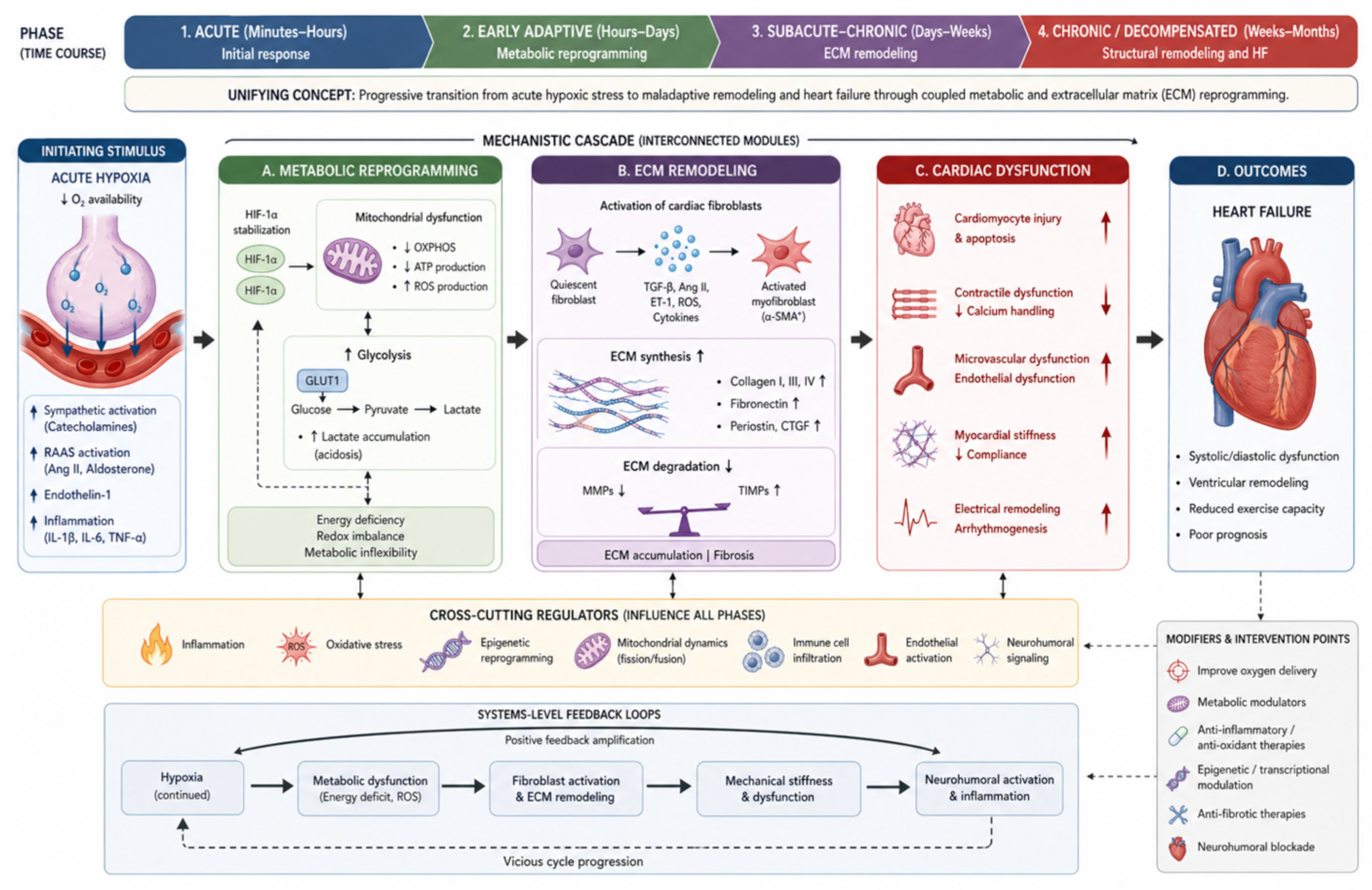
The genes reviewed, spanning hypoxic response, metabolic regulation, lipid han-dling, and extracellular matrix organization, illustrate the multifactorial nature of heart failure and myocardial infarction pathology. Heart disease is not the result of a single molecular pathway but rather a complex interplay of interwoven processes. Under-standing these processes gene-by-gene provides insight into disease mechanisms and uncovers biomarkers and therapeutic targets. An integrated, phase-based schematic linking hypoxia signaling, metabolic reprogramming, inflammatory activation, and extracellular matrix remodeling in the progression from myocardial infarction to heart failure is presented in Figure 4.

6. Interplay Between Pathways

The pathophysiology of myocardial infarction (MI) and heart failure (HF) cannot be fully explained by isolated pathway activity. Instead, these processes emerge from a dynamically interacting network in which hypoxia signaling, metabolic regulation, lipid handling, inflammation, and extracellular matrix (ECM) remodeling are tightly coupled through feedback loops and stage-dependent regulatory dominance.

6.1. Hypoxia—Metabolism Feedback Loop

Acute ischemia stabilizes HIF-1α, promoting a shift toward glycolysis while suppressing mitochondrial oxidative phosphorylation [9]. This metabolic reprogramming is reinforced by activation of AMP-activated protein kinase (AMPK), which supports energy conservation under conditions of ATP depletion. However, sustained HIF activation leads to maladaptive consequences, including mitochondrial dysfunction, reactive oxygen species accumulation, and activation of pro-apoptotic pathways such as BNIP3.
A key mediator of this transition is lactate accumulation, which not only reflects enhanced glycolysis but also acts as a signaling molecule. Lactate promotes the release and activation of transforming growth factor-β (TGF-β) from ECM, thereby stimulating fibroblast activation and initiating fibrotic remodeling. This establishes a positive feedback loop linking hypoxia, metabolic reprogramming, and ECM deposition [212].

6.2. AMPK—mTOR Regulatory Axis

The balance between AMPK and mechanistic target of rapamycin (mTOR) represents a central metabolic switch governing cellular adaptation. Under conditions of energy deficit, AMPK inhibits mTORC1 through phosphorylation of TSC2 and raptor, promoting autophagy, mitochondrial maintenance, and metabolic efficiency. Conversely, nutrient availability and growth signals restore mTOR activity, driving anabolic processes and cellular growth [212,213,214,215].
In chronic HF, this balance is frequently disrupted, leading to sustained mTOR activation despite ongoing energetic stress. This “metabolic mismatch” or “lock-in” state is characterized by suppression of AMPK activity, reduced SIRT1 signaling, depletion of NAD+ pools, and progressive epigenetic drift, ultimately contributing to hypertrophy, fibrosis, and functional decline [212,216].

6.3. Lipid—Energy Coupling

Lipid-handling genes (e.g., LPL, APOC3, APOA5) regulate substrate availability and systemic metabolic stress, thereby influencing cardiac energy homeostasis. Impaired lipid utilization results in energetic deficits, whereas excessive lipid uptake promotes lipotoxicity, mitochondrial dysfunction, and oxidative stress. Importantly, lipid metabolism is closely linked to hypoxia signaling. Alterations in substrate utilization affect oxygen demand and redox balance, positioning lipid metabolism as a critical modulator of cardiac adaptation during ischemia and HF progression [9,215].

6.4. Lipid Metabolism, Inflammation, and ECM Coupling

Beyond energy metabolism, lipid handling intersects with inflammatory signaling, forming an additional regulatory layer linking hypoxia to ECM remodeling. Hypoxia and metabolic reprogramming exert profound effects on lipid metabolism and immune cell function. HIF-1α, together with PPARγ, promotes the expression of the scavenger receptor CD36, thereby enhancing lipid uptake in macrophages and cardiomyocytes. In parallel, oxidized low-density lipoprotein (oxLDL) activates NF-κB signaling, leading to the production of pro-inflammatory cytokines. Enhanced glycolysis in M1 macrophages, driven by HIF-1α, further amplifies the inflammatory response. These activated macrophages secrete mediators such as TGF-β and VEGF, which stimulate fibroblast activation and proliferation [9,217].
Collectively, lipid and inflammatory signaling pathways are tightly coupled with cellular metabolism and ECM remodeling. This integration establishes a reinforcing positive feedback loop—hypoxia → lactate accumulation → TGF-β activation → fibroblast stimulation → ECM deposition—that accelerates disease progression.

6.5. ECM—Mechanotransduction Feedback

ECM remodeling is not merely a structural consequence but an active regulator of cellular signaling. Increased matrix stiffness enhances integrin-mediated mechanotransduction (e.g., via ITGA1), activating downstream pathways such as PI3K–AKT and mTOR. Simultaneously, alterations in collagen composition (e.g., COL4A1) influence oxygen diffusion and microvascular integrity, further reinforcing hypoxic stress. Emerging evidence suggests that mechanotransduction pathways, including Hippo/YAP signaling, may link ECM stiffness to metabolic reprogramming in cardiomyocytes, although these mechanisms remain incompletely understood [212,217,218].

6.6. Phase—Dependent Dominance and Systems Integration

These interconnected pathways exhibit phase-dependent dominance during disease progression. In the acute phase, hypoxia signaling and AMPK-mediated metabolic adaptation are predominant, supporting cell survival. In the subacute phase, inflammatory and metabolic interactions determine the balance between repair and maladaptation. In the chronic phase, ECM remodeling and mechanotransduction become dominant drivers of dysfunction, reinforcing fibrosis, metabolic rigidity, and persistent hypoxia. This transition from metabolic to structural dominance reflects a systems-level shift that has been increasingly recognized in the literature, including models linking lactate accumulation and TGF-β signaling to fibrotic remodeling [212,217].

6.7. Systems Implication

Taken together, these findings indicate that MI and HF progression arise from the breakdown of coordinated regulation across interconnected biological systems. The presence of multiple reinforcing feedback loops explains why interventions targeting single pathways often yield limited or context-dependent effects. A systems-level perspective is therefore essential for identifying stage-specific therapeutic strategies and improving translational outcomes.
Many of the pathways described are well supported by experimental models (e.g., the role of lactate in TGF-β activation or the oscillatory dynamics of the AMPK/mTOR axis); however, robust evidence from human studies remains limited. In particular, there is a lack of validated clinical biomarkers reflecting epigenetic regulation or ECM turnover dynamics. Accordingly, it is important to clearly distinguish which observations are based on mechanistic inference, and which have established clinical relevance. For example, proteomic and metabolomic analyses suggest potential markers of AMPK/mTOR activity (e.g., phosphorylated AMPK, NAD+ levels), but their diagnostic and therapeutic utility has yet to be validated in clinical settings. Similarly, biomarkers of fibrosis (such as circulating collagen-derived peptides) are available, but their direct linkage to the underlying molecular mechanisms driving heart failure remains insufficiently established [9,212].
To integrate the complex interplay between hypoxia signaling, metabolic regulation, and extracellular matrix remodeling, Table 3 summarizes key mechanistic interactions across these axes. The table highlights how individual pathways converge at different stages of myocardial infarction and heart failure progression, emphasizing both adaptive and maladaptive responses.

6.8. Biomarkers and Translational Perspectives

Integrating findings across hypoxia signaling, metabolic regulation, and extracellular matrix remodeling highlights a set of candidate biomarkers with potential diagnostic and prognostic relevance in myocardial infarction and heart failure. These markers span multiple biological levels, from established clinical indicators (e.g., cardiac troponins, NT-proBNP) to emerging molecular signatures reflecting early repair processes, inflammation, and fibrotic remodeling. Importantly, several candidates—such as CDC6, soluble LRP1, and ITGA1—capture distinct phases of disease progression, from acute injury to chronic remodeling, and may enable more precise patient stratification. While Table 3 (above) summarizes the mechanistic interactions between hypoxia, metabolism, and ECM remodeling, clinical translation requires biomarkers that capture these processes across disease stages. Table 4 presents candidate biomarkers reflecting key pathways of the hypoxia-metabolism-ECM axis, organized according to their underlying mechanisms, temporal dynamics, measurement methods, and level of clinical validation. This structured overview highlights the gap between mechanistic insights and clinically actionable tools, emphasizing the need for further validation of emerging biomarkers.
Overall, the biomarkers presented in Table 4 reflect different stages of disease progression, from acute hypoxic stress (e.g., HIF-1α, lactate) to chronic metabolic and fibrotic remodeling (e.g., SIRT1, TGF-β, ITGA1). Notably, most candidates remain in the preclinical or early clinical validation phase, highlighting a significant gap between mechanistic understanding and routine clinical application. Future studies should focus on multi-marker panels integrating hypoxia, metabolic, and ECM signals to improve risk stratification and therapeutic targeting in MI and HF.
These developments align with emerging non-invasive and embedded monitoring technologies capable of assessing tissue integrity and physiological dynamics in real time. Recent advances in bioelectronic platforms and intelligent embedded sensor architectures demonstrate how continuous monitoring of structural integrity, signal stability, and physiological parameters can be translated into biomedical applications, including cardiovascular disease management and chronic heart failure monitoring [226,227].

7. Conclusions and Future Directions

Over the past decade, significant advances have clarified the roles of hypoxia regulators, lipid-handling proteins, and extracellular matrix (ECM)/vascular genes in cardiovascular disease. Hypoxia-inducible signaling, mediated by HIF1A and EPAS1, has emerged as a key determinant of cellular fate under ischemic stress, conferring protection in the acute phase while contributing to maladaptive remodeling when chronically activated. Metabolic regulators such as SIRT1 further emphasize the importance of cellular energy sensing in the heart, supporting therapeutic strategies that recapitulate the effects of caloric restriction or exercise. The identification of CDC6 in acute myocardial infarction introduces an additional dimension, linking cell-cycle arrest and senescence to reparative processes—an apparent paradox that underscores the importance of temporal and contextual regulation in cardiac regeneration.
In parallel, lipids once considered merely associative risk factors, particularly triglycerides, are now recognized as causal contributors to cardiovascular disease, largely through insights from human genetics. APOC3 and APOA5 exemplify how modulation of triglyceride metabolism can directly influence myocardial infarction risk. As APOC3-targeted therapies enter clinical practice, their potential impact on myocardial infarction and heart failure is anticipated, particularly through the prevention of atherosclerotic disease. Emerging questions remain as to whether such metabolic interventions may also benefit patients with chronic heart failure, in whom altered lipid metabolism and cachexia are common—for example, by reducing ectopic lipid accumulation or improving skeletal muscle energetics.
Finally, the ECM and vascular genes discussed, including COL4A1, ITGA1, and LRP1, underscore that the pathogenesis and treatment of heart failure extend beyond cardiomyocytes. Effective intervention requires preservation of microvascular integrity, regulation of fibrosis, and appropriate resolution of inflammation. Collagen IV plays a crucial role in maintaining vascular stability, influencing plaque integrity and susceptibility to rupture. Integrin-mediated signaling in fibroblasts modulates the balance between fibrotic stiffening and adaptive remodeling. LRP1, functioning as a central regulatory receptor, illustrates the therapeutic potential of enhancing endogenous repair mechanisms. In this context, LRP1 agonists such as SP16 may represent a novel class of cardioprotective agents that promote resolution of injury rather than simply inhibiting upstream pathogenic processes.
From a pharmacological perspective, many of the genes discussed are emerging as therapeutic targets or biomarkers within precision medicine frameworks:
  • HIF stabilizers. Already approved for the treatment of renal anemia, these agents are being explored for ischemic tissue repair. A key challenge lies in optimizing dosing and timing to harness acute protective effects while avoiding adverse consequences of sustained HIF activation. A potential future strategy may involve short-term administration of HIF prolyl hydroxylase inhibitors following myocardial infarction to enhance angiogenesis and myocardial salvage [16,57,219,228,229,230,231];
  • SIRT1 activators. Although compounds such as resveratrol and synthetic SRT agents have yielded mixed results, targeting the NAD+–SIRT1 axis remains an attractive strategy. Combination approaches, for example, with SGLT2 inhibitors, may offer synergistic benefits by promoting a favorable metabolic shift toward increased ketone and glucose utilization and reduced oxidative stress [75];
  • APOC3 inhibitors and triglyceride-lowering therapies. These agents are well positioned to reduce residual cardiovascular risk in patients with hypertriglyceridemia or diabetes. Ongoing and future studies will determine whether they also reduce heart failure hospitalizations in these populations. Given strong genetic evidence linking APOC3 to atherosclerosis, such therapies may substantially lower myocardial infarction incidence, with secondary benefits for heart failure prevalence [109,114,117,129,232];
  • Antifibrotic strategies targeting integrins or TGF-β signaling. Agents such as nintedanib, a broad tyrosine kinase inhibitor, are already used in pulmonary fibrosis and are under investigation in HFpEF. More selective approaches, including integrin αv antagonists, are in early-stage development. Insights from ITGA1 suggest that modulation of integrin α1β1 signaling may attenuate pathological fibrosis, although the pleiotropic roles of integrins necessitate careful evaluation. In parallel, circulating ITGA1 may have utility as a biomarker to identify high-risk metabolic patients who could benefit from intensified therapy [30];
  • LRP1-targeted therapies. Should agents such as SP16 demonstrate efficacy in larger myocardial infarction trials, they may represent a novel adjunct to reperfusion strategies. In addition, alpha-1 antitrypsin, an endogenous LRP1 ligand, is being repurposed for acute myocardial infarction, leveraging its anti-inflammatory properties mediated via LRP1. Following completion of early-phase studies, subsequent trials will evaluate effects on infarct size and ventricular function. Positive results would support the broader concept of therapeutically enhancing endogenous resolution pathways and could stimulate development of LRP1 agonists for other inflammatory cardiovascular conditions, including myocarditis and non-ischemic heart failure [32,202].
A recurring theme across these pathways is the importance of timing and balance. For instance, HIF1A and CDC6 confer short-term adaptive benefits but may become detrimental with sustained activation. SIRT1 is protective within a physiological range yet potentially harmful when overactivated. Integrin α1 signaling may attenuate acute injury responses but contribute to chronic fibrosis; and while LRP1 exerts beneficial effects in acute injury, the consequences of prolonged activation remain insufficiently defined. These observations underscore the need for precision medicine, in which interventions are delivered at the appropriate disease stage and tailored to specific patient subsets. Advances in patient profiling—including genomics, proteomics (e.g., ITGA1 assays), and imaging modalities assessing hypoxia or fibrosis—are increasingly enabling such targeted approaches.
In summary, hypoxia regulators, lipid metabolism genes, and ECM/vascular pathways constitute an interconnected network governing cardiac adaptation to stress and injury. An ischemic event such as myocardial infarction activates HIF- and SIRT1-mediated responses, induces lipid mobilization and inflammatory signaling involving APOC3/APOA5 and LRP1, and initiates structural remodeling driven by collagens and integrins. Each of these components represents both a mechanistic contributor and a potential therapeutic target. In the coming years, therapies targeting HIF signaling, metabolic pathways, and fibrotic or inflammatory processes are likely to be evaluated in myocardial infarction and heart failure, building on this mechanistic foundation. Integrating these multidimensional strategies—enhancing acute stress adaptation, optimizing systemic metabolism, and modulating chronic remodeling—offers the potential not only to improve survival but also to preserve cardiac function and quality of life in patients following myocardial infarction or with heart failure.

Author Contributions

Conceptualization, R.C.; writing—original draft preparation, R.C., A.G.-C. and K.D.S.S.; writing—review and editing, R.C., J.K. and K.D.S.S.; visualization, A.B.-R.; supervision, J.K. and K.D.S.S. All authors have read and agreed to the published version of the manuscript.

Funding

This research received no external funding.

Institutional Review Board Statement

Not applicable.

Informed Consent Statement

Not applicable.

Data Availability Statement

The data presented in this study are available on request.

Conflicts of Interest

The authors declare no conflicts of interest.

References

  1. Kryeziu, B.; Poniku, A.; Henein, M.Y.; Ibrahimi, P.; Batalli, A.; Pllana, E.; Morina, D.; Berlajolli, A.; Krasniqi, J.; Elezi, S.; et al. Epidemiological 11-year dynamics study of acute myocardial infarction: A cohort study in a country with a transitional healthcare system. Glob. Heart 2026, 21, 14. [Google Scholar] [CrossRef]
  2. Available online: https://www.who.int/news-room/fact-sheets/detail/cardiovascular-diseases-(cvds) (accessed on 23 April 2026).
  3. Belaidi, E.; Morand, J.; Gras, E.; Pépin, J.L.; Godin-Ribuot, D. Targeting the ROS-HIF-1-endothelin axis as a therapeutic approach for the treatment of obstructive sleep apnea-related cardiovascular complications. Pharmacol. Ther. 2016, 168, 1–11. [Google Scholar] [CrossRef]
  4. Li, Q.O.Y.; Soro-Arnaiz, I.; Aragones, J. Age-dependent obesity and mitochondrial dysfunction. Adipocyte 2017, 6, 161–166. [Google Scholar] [CrossRef]
  5. Lee, Y.S.; Kim, J.W.; Osborne, O.; Oh, D.Y.; Sasik, R.; Schenk, S.; Chen, A.; Chung, H.; Murphy, A.; Watkins, S.M.; et al. Increased adipocyte O2 consumption triggers HIF-1α, causing inflammation and insulin resistance in obesity. Cell 2014, 157, 1339–1352. [Google Scholar] [CrossRef] [PubMed]
  6. Sun, K.; Halberg, N.; Khan, M.; Magalang, U.J.; Scherer, P.E. Selective inhibition of hypoxia-inducible factor 1α ameliorates adipose tissue dysfunction. Mol. Cell. Biol. 2013, 33, 904–917. [Google Scholar] [CrossRef] [PubMed]
  7. Luo, Z.; Tian, M.; Yang, G.; Tan, Q.; Chen, Y.; Li, G.; Zhang, Q.; Li, Y.; Wan, P.; Wu, J. Hypoxia signaling in human health and diseases: Implications and prospects for therapeutics. Signal Transduct. Target. Ther. 2022, 7, 218. [Google Scholar] [CrossRef] [PubMed]
  8. Boland, E.; Hoyle, A.; Robertson-Gray, O.; Fuller, W.; Swift, J.; Cantini, M.; Walker, M.; Huethorst, E.; MacDonald, E.; Loughrey, C.; et al. Biomechanical and compositional basement membrane defects due to a Col4a1 mutation affect cardiac morphology and function. Matrix Biol. 2025, 141, 82–100. [Google Scholar] [CrossRef]
  9. Li, H.; Xiao, F.; Zhou, C.; Zhu, T.; Wang, S. Metabolic adaptations and therapies in cardiac hypoxia: Mechanisms and clinical implications/potential strategies. JACC Basic Transl. Sci. 2025, 10, 862–878. [Google Scholar] [CrossRef]
  10. Liang, Y.; Ruan, W.; Jiang, Y.; Smalling, R.; Yan, X.; Eltzschig, H.K. Interplay of hypoxia-inducible factors and oxygen therapy in cardiovascular medicine. Nat. Rev. Cardiol. 2023, 20, 723–737. [Google Scholar] [CrossRef]
  11. Koeppen, M.; Lee, J.W.; Seo, S.W.; Brodsky, K.S.; Kreth, S.; Yang, I.V.; Buttrick, P.M.; Eckle, T.; Eltzschig, H.K. Hypoxia-inducible factor 2-alpha-dependent induction of amphiregulin dampens myocardial ischemia-reperfusion injury. Nat. Commun. 2018, 9, 816. [Google Scholar] [CrossRef]
  12. Mylonis, I.; Simos, G.; Paraskeva, E. Hypoxia-inducible factors and the regulation of lipid metabolism. Cells 2019, 8, 214. [Google Scholar] [CrossRef]
  13. Tian, L.; Wang, M.; Liu, M.; Pang, Y.; Zhao, J.; Zheng, B.; Wang, Y.; Zhao, W. Cardiovascular and renal safety outcomes of hypoxia-inducible factor prolyl-hydroxylase inhibitor roxadustat for anemia patients with chronic kidney disease: A systematic review and meta-analysis. Ren. Fail. 2024, 46, 2313864. [Google Scholar] [CrossRef]
  14. Deeks, E.D. Belzutifan: First approval. Drugs 2021, 81, 1921–1927. [Google Scholar] [CrossRef]
  15. Kane, A.E.; Sinclair, D.A. Sirtuins and NAD+ in the development and treatment of metabolic and cardiovascular diseases. Circ. Res. 2018, 123, 868–885. [Google Scholar] [CrossRef]
  16. Sato, T.; Takeda, N. The roles of HIF-1α signaling in cardiovascular diseases. J. Cardiol. 2023, 81, 202–208. [Google Scholar] [CrossRef]
  17. Packer, M. Critical reanalysis of the mechanisms underlying the cardiorenal benefits of SGLT2 inhibitors and reaffirmation of the nutrient deprivation signaling/autophagy hypothesis. Circulation 2022, 146, 1383–1405. [Google Scholar] [CrossRef]
  18. Tong, D.; Schiattarella, G.G.; Jiang, N.; Altamirano, F.; Szweda, P.A.; Elnwasany, A.; Lee, D.I.; Yoo, H.; Kass, D.A.; Szweda, L.I.; et al. NAD+ repletion reverses heart failure with preserved ejection fraction. Circ. Res. 2021, 128, 1629–1641. [Google Scholar] [CrossRef] [PubMed]
  19. Komuro, J.; Hashimoto, H.; Katsuki, T.; Kusumoto, D.; Katoh, M.; Ko, T.; Ito, M.; Katagiri, M.; Kubota, M.; Yamada, S.; et al. Heart failure-specific cardiac fibroblasts contribute to cardiac dysfunction via the MYC-CXCL1-CXCR2 axis. Nat. Cardiovasc. Res. 2025, 4, 1135–1151. [Google Scholar] [CrossRef] [PubMed]
  20. Villa del Campo, C.; Raiola, M.; Sierra, R.; Ignacio, M.S.; Peña-Peña, J.; Esteban, I.; Tottes, M. Myc overexpression improves recovery from myocardial infarction associated with cardiomyocyte hyperplasia in the mouse heart. Commun. Biol. 2025, 8, 1069. [Google Scholar] [CrossRef] [PubMed]
  21. Wen, Y.; Zhang, X.; Wang, J.; Ma, C.; Wu, L.; Wang, L. CDC6 as early biomarker for myocardial infarction with acute cellular senescence and repair mechanisms. Sci. Rep. 2025, 15, 14130. [Google Scholar] [CrossRef]
  22. He, X.; Li, F.; Zhang, J.; Wen, Z.; Li, Y.; Gao, W.; Chen, H.; Yu, Y.; Zheng, Y.; Xie, X. The level of apolipoprotein A-I Is associated with a prognosis in patients with chronic heart failure especially in HFmrEF and HFpEF: A retrospective cohort study. Int. J. Gen. Med. 2025, 18, 4735–4751. [Google Scholar] [CrossRef]
  23. Borén, J.; Taskinen, M.R.; Björnson, E.; Packard, C.J. Metabolism of triglyceride-rich lipoproteins in health and dyslipidaemia. Nat. Rev. Cardiol. 2022, 19, 577–592. [Google Scholar] [CrossRef] [PubMed]
  24. Yang, W.; Ng, F.L.; Chan, K.; Pu, X.; Poston, R.N.; Ren, M.; An, W.; Zhang, R.; Wu, J.; Yan, S.; et al. Coronary-heart-disease-associated genetic variant at the COL4A1/COL4A2 locus affects COL4A1/COL4A2 expression, vascular cell survival, atherosclerotic plaque stability and risk of myocardial infarction. PLoS Genet. 2016, 12, e1006127. [Google Scholar] [CrossRef] [PubMed]
  25. Witztum, J.L.; Gaudet, D.; Freedman, S.D.; Alexander, V.J.; Digenio, A.; Williams, K.R.; Yang, Q.; Hughes, S.G.; Geary, R.S.; Arca, M.; et al. Volanesorsen and triglyceride levels in familial chylomicronemia syndrome. N. Engl. J. Med. 2019, 381, 531–542. [Google Scholar] [CrossRef] [PubMed]
  26. Hooper, A.J.; Bell, D.A.; Burnett, J.R. Olezarsen, a liver-directed APOC3 ASO therapy for hypertriglyceridemia. Expert Opin. Pharmacother. 2024, 25, 1861–1866. [Google Scholar] [CrossRef]
  27. Li, S.; Xie, X.; Zeng, X.; Wang, S.; Lan, J. Serum apolipoprotein B to apolipoprotein A-I ratio predicts mortality in patients with heart failure. ESC Heart Fail. 2024, 11, 99–111. [Google Scholar] [CrossRef]
  28. Zheng, Q.; Wang, Y.; Yang, H.; Sun, L.; Fu, X.; Wei, R.; Liu, Y.N.; Liu, W.J. Efficacy and safety of daprodustat for anemia therapy in chronic kidney disease patients: A systematic review and meta-analysis. Front. Pharmacol. 2021, 11, 573645. [Google Scholar] [CrossRef]
  29. Silbernagel, G.; Scharnagl, H.; Kleber, M.E.; Delgado, G.; Stojakovic, T.; Laaksonen, R.; Erdmann, J.; Rankinen, T.; Bouchard, C.; Landmesser, U.; et al. LDL triglycerides, hepatic lipase activity, and coronary artery disease: An epidemiologic and Mendelian randomization study. Atherosclerosis 2019, 282, 37–44. [Google Scholar] [CrossRef]
  30. Su, M.; Hou, Y.; Cai, S.; Li, W.; Wei, Y.; Wang, R.; Wu, M.; Liu, M.; Chang, J.; Yang, K.; et al. Elevated ITGA1 levels in type 2 diabetes: Implications for cardiac function impairment. Diabetologia 2024, 67, 850–863. [Google Scholar] [CrossRef]
  31. Aman, J.; Margadant, C. Integrin-dependent cell–matrix adhesion in endothelial health and disease. Circ. Res. 2023, 132, 355–378. [Google Scholar] [CrossRef]
  32. Toldo, S.; Austin, D.; Mauro, A.G.; Mezzaroma, E.; Van Tassell, B.W.; Marchetti, C.; Carbone, S.; Mogelsvang, S.; Gelber, C.; Abbate, A. Low-density lipoprotein receptor-related protein-1 is a therapeutic target in acute myocardial infarction. JACC Basic Transl. Sci. 2017, 2, 561–574, Erratum in JACC Basic Transl. Sci. 2018, 3, 162. https://doi.org/10.1016/j.jacbts.2018.01.012. [Google Scholar] [CrossRef] [PubMed]
  33. Potere, N.; Del Buono, M.G.; Niccoli, G.; Crea, F.; Toldo, S.; Abbate, A. Developing LRP1 agonists into a therapeutic strategy in acute myocardial infarction. Int. J. Mol. Sci. 2019, 20, 544. [Google Scholar] [CrossRef] [PubMed]
  34. Van Tassell, B.W.; Wohlford, G.F.; Del Buono, M.G.; Damonte, J.I.; Markley, R.; Turlington, J.; Kadariya, D.; Talasaz, A.; Ho, J.; Marawan, A.; et al. Safety, tolerability, and effects of a single subcutaneous administration of SP16—A SERPIN-like, small peptide agonist of the low-density lipoprotein-like receptor 1- on the acute inflammatory response in patients with ST-segment elevation myocardial infarction. J. Cardiovasc. Pharmacol. 2022, 80, 672–678. [Google Scholar] [CrossRef] [PubMed]
  35. Knutson, A.K.; Williams, A.L.; Boisvert, W.A.; Shohet, R.V. HIF in the heart: Development, metabolism, ischemia, and atherosclerosis. J. Clin. Investig. 2021, 131, e137557. [Google Scholar] [CrossRef]
  36. Wang, G.L.; Semenza, G.L. Purification and characterization of hypoxia-inducible factor 1. J. Biol. Chem. 1995, 270, 1230–1237. [Google Scholar] [CrossRef]
  37. Ivan, M.; Kondo, K.; Yang, H.; Kim, W.; Valiando, J.; Ohh, M.; Salic, A.; Asara, J.M.; Lane, W.S.; Kaelin, W.G., Jr. HIFalpha targeted for VHL-mediated destruction by proline hydroxylation: Implications for O2 sensing. Science 2001, 292, 464–468. [Google Scholar] [CrossRef]
  38. Jaakkola, P.; Mole, D.R.; Tian, Y.M.; Wilson, M.I.; Gielbert, J.; Gaskell, S.J.; von Kriegsheim, A.; Hebestreit, H.F.; Mukherji, M.; Schofield, C.J.; et al. Targeting of HIF-alpha to the von Hippel-Lindau ubiquitylation complex by O2-regulated prolyl hydroxylation. Science 2001, 292, 468–472. [Google Scholar] [CrossRef]
  39. Wu, J.; Chen, P.; Li, Y.; Ardell, C.; Der, T.; Shohet, R.; Chen, M.; Wright, G.L. HIF-1α in heart: Protective mechanisms. Am. J. Physiol. Heart Circ. Physiol. 2013, 305, H821–H828. [Google Scholar] [CrossRef]
  40. Ockaili, R.; Natarajan, R.; Salloum, F.; Fisher, B.J.; Jones, D.; Fowler, A.A., 3rd; Kukreja, R.C. HIF-1 activation attenuates postischemic myocardial injury: Role for heme oxygenase-1 in modulating microvascular chemokine generation. Am. J. Physiol. Heart Circ. Physiol. 2005, 289, H542–H548. [Google Scholar] [CrossRef]
  41. Philipp, S.; Jürgensen, J.S.; Fielitz, J.; Bernhardt, W.M.; Weidemann, A.; Schiche, A.; Pilz, B.; Dietz, R.; Regitz-Zagrosek, V.; Eckardt, K.U.; et al. Stabilization of hypoxia inducible factor rather than modulation of collagen metabolism improves cardiac function after acute myocardial infarction in rats. Eur. J. Heart Fail. 2006, 8, 347–354. [Google Scholar] [CrossRef]
  42. Mirtschink, P.; Krishnan, J.; Grimm, F.; Sarre, A.; Hörl, M.; Kayikci, M.; Fankhauser, N.; Christinat, Y.; Cortijo, C.; Feehan, O.; et al. HIF-driven SF3B1 induces KHK-C to enforce fructolysis and heart disease. Nature 2015, 522, 444–449. [Google Scholar] [CrossRef]
  43. Morand, J.; Arnaud, C.; Pépin, J.L.; Godin-Ribuot, D. Chronic intermittent hypoxia promotes myocardial ischemia-related ventricular arrhythmias and sudden cardiac death. Sci. Rep. 2018, 8, 2997. [Google Scholar] [CrossRef]
  44. Xie, L.; Pi, X.; Wang, Z.; He, J.; Willis, M.S.; Patterson, C. Depletion of PHD3 protects heart from ischemia/reperfusion injury by inhibiting cardiomyocyte apoptosis. J. Mol. Cell. Cardiol. 2015, 80, 156–165. [Google Scholar] [CrossRef]
  45. Date, T.; Mochizuki, S.; Belanger, A.J.; Yamakawa, M.; Luo, Z.; Vincent, K.A.; Cheng, S.H.; Gregory, R.J.; Jiang, C. Expression of constitutively stable hybrid hypoxia-inducible factor-1α protects cultured rat cardiomyocytes against simulated ischemia-reperfusion injury. Am. J. Physiol. Physiol. 2005, 288, C314–C320. [Google Scholar] [CrossRef]
  46. Datta Chaudhuri, R.; Banik, A.; Mandal, B.; Sarkar, S. Cardiac-specific overexpression of HIF-1α during acute myocardial infarction ameliorates cardiomyocyte apoptosis via differential regulation of hypoxia-inducible pro-apoptotic and anti-oxidative genes. Biochem. Biophys. Res. Commun. 2021, 537, 100–108. [Google Scholar] [CrossRef] [PubMed]
  47. Bouhamida, E.; Morciano, G.; Perrone, M.; Kahsay, A.E.; Della Sala, M.; Wieckowski, M.R.; Fiorica, F.; Pinton, P.; Giorgi, C.; Patergnani, S. The interplay of hypoxia signaling on mitochondrial dysfunction and inflammation in cardiovascular diseases and cancer: From molecular mechanisms to therapeutic approaches. Biology 2022, 11, 300. [Google Scholar] [CrossRef] [PubMed]
  48. Tian, X.; Zhou, N.; Yuan, J.; Lu, L.; Zhang, Q.; Wei, M.; Zou, Y.; Yuan, L. Heat shock transcription factor 1 regulates exercise-induced myocardial angiogenesis after pressure overload via HIF-1α/VEGF pathway. J. Cell. Mol. Med. 2020, 24, 2178–2188. [Google Scholar] [CrossRef] [PubMed]
  49. Zhang, Y.; Xu, Y.; Zhou, K.; Kao, G.; Yan, M.; Xiao, J. Hypoxia-inducible transcription factor-1α inhibition by topotecan protects against lipopolysaccharide-induced inflammation and apoptosis of cardiomyocytes. BioMed. Eng. OnLine 2021, 20, 88. [Google Scholar] [CrossRef]
  50. Hölscher, M.; Schäfer, K.; Krull, S.; Farhat, K.; Hesse, A.; Silter, M.; Lin, Y.; Pichler, B.J.; Thistlethwaite, P.; El-Armouche, A.; et al. Unfavourable consequences of chronic cardiac HIF-1α stabilization. Cardiovasc. Res. 2012, 94, 77–86. [Google Scholar] [CrossRef]
  51. Stanley, W.C.; Recchia, F.A.; Lopaschuk, G.D. Myocardial substrate metabolism in the normal and failing heart. Physiol. Rev. 2005, 85, 1093–1129. [Google Scholar] [CrossRef]
  52. Kolwicz, S.C., Jr.; Tian, R. Glucose metabolism and cardiac hypertrophy. Cardiovasc. Res. 2011, 90, 194–201. [Google Scholar] [CrossRef]
  53. Li, X.; Jiang, S.; Gu, X.; Liu, X.; Shang, S.; Zhang, J.; Pang, K.; Li, W. Assessment of the safety of Roxadustat for cardiovascular events in chronic kidney disease-related anemia using meta-analysis and bioinformatics. Front. Pharmacol. 2024, 15, 1380326. [Google Scholar] [CrossRef]
  54. Minamishima, Y.A.; Moslehi, J.; Bardeesy, N.; Cullen, D.; Bronson, R.T.; Kaelin, W.G., Jr. Somatic inactivation of the PHD2 prolyl hydroxylase causes polycythemia and congestive heart failure. Blood 2008, 111, 3236–3244. [Google Scholar] [CrossRef]
  55. Ikeda, M.; Ide, T.; Tadokoro, T.; Miyamoto, H.D.; Ikeda, S.; Okabe, K.; Ishikita, A.; Sato, M.; Abe, K.; Furusawa, S.; et al. Excessive hypoxia-inducible factor-1α expression induces cardiac rupture via p53-dependent apoptosis after myocardial infarction. J. Am. Heart Assoc. 2021, 10, e020895. [Google Scholar] [CrossRef] [PubMed]
  56. Zhang, Y.; Liu, D.; Hu, H.; Zhang, P.; Xie, R.; Cui, W. HIF-1α/BNIP3 signaling pathway-induced-autophagy plays protective role during myocardial ischemia-reperfusion injury. Biomed. Pharmacother. 2019, 120, 109464. [Google Scholar] [CrossRef] [PubMed]
  57. Scortegagna, M.; Ding, K.; Oktay, Y.; Gaur, A.; Thurmond, F.; Yan, L.J.; Marck, B.T.; Matsumoto, A.M.; Shelton, J.M.; Richardson, J.A.; et al. Multiple organ pathology, metabolic abnormalities and impaired homeostasis of reactive oxygen species in Epas1−/− mice. Nat. Genet. 2003, 35, 331–340. [Google Scholar] [CrossRef]
  58. Majmundar, A.J.; Wong, W.J.; Simon, M.C. Hypoxia-inducible factors and the response to hypoxic stress. Mol. Cell 2010, 40, 294–309. [Google Scholar] [CrossRef] [PubMed]
  59. Ullah, K.; Ai, L.; Li, Y.; Liu, L.; Zhang, Q.; Pan, K.; Humayun, Z.; Piao, L.; Sitikov, A.; Zhao, Q.; et al. ARNT-dependent HIF-2α signaling protects cardiac microvascular barrier integrity and heart function post-myocardial infarction. Commun. Biol. 2025, 8, 440. [Google Scholar] [CrossRef]
  60. Jatho, A.; Zieseniss, A.; Brechtel-Curth, K.; Yamamoto, A.; Coleman, M.L.; Vergel Leon, A.M.; Biggs, D.; Davies, B.; Pugh, C.W.; Ratcliffe, P.J.; et al. Precisely tuned inhibition of HIF prolyl hydroxylases is key for cardioprotection after ischemia. Circ. Res. 2021, 128, 1208–1210. [Google Scholar] [CrossRef]
  61. Zhao, Y.; Xiong, W.; Li, C.; Zhao, R.; Lu, H.; Song, S.; Zhou, Y.; Hu, Y.; Shi, B.; Ge, J. Hypoxia-induced signaling in the cardiovascular system: Pathogenesis and therapeutic targets. Signal Transduct. Target. Ther. 2023, 8, 431. [Google Scholar] [CrossRef]
  62. Zhang, N.; Fu, Z.; Linke, S.; Chicher, J.; Gorman, J.J.; Visk, D.; Haddad, G.G.; Poellinger, L.; Peet, D.J.; Powell, F.; et al. The asparaginyl hydroxylase factor inhibiting HIF-1alpha is an essential regulator of metabolism. Cell Metab. 2010, 11, 364–378. [Google Scholar] [CrossRef]
  63. Sim, J.; Cowburn, A.S.; Palazon, A.; Madhu, B.; Tyrakis, P.A.; Macías, D.; Bargiela, D.M.; Pietsch, S.; Gralla, M.; Evans, C.E.; et al. The factor inhibiting HIF asparaginyl hydroxylase regulates oxidative metabolism and accelerates metabolic adaptation to hypoxia. Cell Metab. 2018, 27, 898–913. [Google Scholar] [CrossRef] [PubMed]
  64. Drevytska, T.; Gonchar, E.; Okhai, I.; Lynnyk, O.; Mankovska, I.; Klionsky, D.; Dosenko, V. The protective effect of Hif3a RNA interference and HIF-prolyl hydroxylase inhibition on cardiomyocytes under anoxia-reoxygenation. Life Sci. 2018, 202, 131–139. [Google Scholar] [CrossRef] [PubMed]
  65. Papandreou, I.; Cairns, R.A.; Fontana, L.; Lim, A.L.; Denko, N.C. HIF-1 mediates adaptation to hypoxia by actively downregulating mitochondrial oxygen consumption. Cell Metab. 2006, 3, 187–197. [Google Scholar] [CrossRef] [PubMed]
  66. Fuhrmann, D.; Brüne, B. Mitochondrial composition and function under the control of hypoxia. Redox Biol. 2017, 12, 208–215. [Google Scholar] [CrossRef]
  67. Dorn, G.W.; Kirshenbaum, L.A. Cardiac reanimation: Targeting cardiomyocyte death by BNIP3 and NIX/BNIP3L. Oncogene 2008, 27, S158–S167. [Google Scholar] [CrossRef]
  68. Ballantyne, C.M.; Vasas, S.; Azizad, M.; Clifton, P.; Rosenson, R.S.; Chang, T.; Melquist, S.; Zhou, R.; Mushin, M.; Leeper, N.J.; et al. Plozasiran, an RNA interference agent targeting APOC3, for mixed hyperlipidemia. N. Engl. J. Med. 2024, 391, 899–912. [Google Scholar] [CrossRef]
  69. Singh, A.K.; Cizman, B.; Carroll, K.; McMurray, J.J.V.; Perkovic, V.; Jha, V.; Johansen, K.L.; Lopes, R.D.; Macdougall, I.C.; Obrador, G.T.; et al. Efficacy and safety of daprodustat for treatment of anemia of chronic kidney disease in incident dialysis patients: A randomized clinical trial. JAMA Intern. Med. 2022, 182, 592–602. [Google Scholar] [CrossRef]
  70. Matsushima, S.; Sadoshima, J. The role of sirtuins in cardiac disease. Am. J. Physiol. Heart Circ. Physiol. 2015, 309, H1375–H1389. [Google Scholar] [CrossRef]
  71. Tanno, M.; Sakamoto, J.; Miura, T.; Shimamoto, K.; Horio, Y. Nucleocytoplasmic shuttling of the NAD+-dependent histone deacetylase SIRT1. J. Biol. Chem. 2007, 282, 6823–6832. [Google Scholar] [CrossRef]
  72. Wang, C.; Chen, L.; Hou, X.; Li, Z.; Kabra, N.; Ma, Y.; Nemoto, S.; Finkel, T.; Gu, W.; Cress, W.D.; et al. Interactions between E2F1 and SirT1 regulate apoptotic response to DNA damage. Nat. Cell Biol. 2006, 8, 1025–1031. [Google Scholar] [CrossRef]
  73. Hsu, J.J.; Ziaeian, B.; Fonarow, G.C. Heart failure with mid-range (borderline) ejection fraction: Clinical implications and future directions. JACC Heart Fail. 2017, 5, 763–771. [Google Scholar] [CrossRef] [PubMed]
  74. Akkafa, F.; Halil Altiparmak, I.; Erkus, M.E.; Aksoy, N.; Kaya, C.; Ozer, A.; Sezen, H.; Oztuzcu, S.; Koyuncu, I.; Umurhan, B. Reduced SIRT1 expression correlates with enhanced oxidative stress in compensated and decompensated heart failure. Redox Biol. 2015, 6, 169–173. [Google Scholar] [CrossRef][Green Version]
  75. Packer, M. Cardioprotective effects of Sirtuin-1 and its downstream effectors: Potential role in mediating the heart failure benefits of SGLT2 (Sodium-Glucose Cotransporter 2) inhibitors. Circ. Heart Fail. 2020, 13, e007197. [Google Scholar] [CrossRef] [PubMed]
  76. Alcendor, R.R.; Gao, S.; Zhai, P.; Zablocki, D.; Holle, E.; Yu, X.; Tian, B.; Wagner, T.; Vatner, S.F.; Sadoshima, J. Sirt1 regulates aging and resistance to oxidative stress in the heart. Circ. Res. 2007, 100, 1512–1521. [Google Scholar] [CrossRef] [PubMed]
  77. Ahuja, P.; Zhao, P.; Angelis, E.; Ruan, H.; Korge, P.; Olson, A.; Wang, Y.; Jin, E.S.; Jeffrey, F.M.; Portman, M.; et al. Myc controls transcriptional regulation of cardiac metabolism and mitochondrial biogenesis in response to pathological stress in mice. J. Clin. Investig. 2010, 120, 1494–1505. [Google Scholar] [CrossRef]
  78. Gu, X.S.; Wang, Z.B.; Ye, Z.; Lei, J.P.; Li, L.; Su, D.F.; Zheng, X. Resveratrol, an activator of SIRT1, upregulates AMPK and improves cardiac function in heart failure. Genet. Mol. Res. 2014, 13, 323–335. [Google Scholar] [CrossRef]
  79. Wang, H.; Wang, X.; Xu, L.; Cao, H. Identification of transcription factors MYC and C/EBPβ mediated regulatory networks in heart failure based on gene expression omnibus datasets. BMC Cardiovasc. Disord. 2020, 20, 250. [Google Scholar] [CrossRef]
  80. Zhou, R.; Barnes, K.; Gibson, S.; Fillmore, N. Dual-edged role of SIRT1 in energy metabolism and cardiovascular disease. Am. J. Physiol. Heart Circ. Physiol. 2024, 327, H1162–H1173. [Google Scholar] [CrossRef]
  81. Sundaresan, N.R.; Pillai, V.B.; Gupta, M.P. Emerging roles of SIRT1 deacetylase in regulating cardiomyocyte survival and hypertrophy. J. Mol. Cell. Cardiol. 2011, 51, 614–618. [Google Scholar] [CrossRef]
  82. Hsu, C.P.; Zhai, P.; Yamamoto, T.; Maejima, Y.; Matsushima, S.; Hariharan, N.; Shao, D.; Takagi, H.; Oka, S.; Sadoshima, J. Silent information regulator 1 protects the heart from ischemia/reperfusion. Circulation 2010, 122, 2170–2182. [Google Scholar] [CrossRef]
  83. Altaher, A.M.; Foad, A.M.; Youssef, W.; Ahmed, A.E. Association of SIRT1 (rs7069102) gene polymorphism with premature myocardial infarction in young Egyptian patients. Egypt. J. Med. Hum. Genet. 2024, 25, 121. [Google Scholar] [CrossRef]
  84. Lu, T.M.; Tsai, J.Y.; Chen, Y.C.; Huang, C.Y.; Hsu, H.L.; Weng, C.F.; Shih, C.C.; Hsu, C.P. Downregulation of Sirt1 as an aging change in advanced heart failure. J. Biomed. Sci. 2014, 21, 57. [Google Scholar] [CrossRef] [PubMed]
  85. Chang, N.; Li, J.; Lin, S.; Zhang, J.; Zeng, W.; Ma, G.; Wang, Y. Emerging roles of SIRT1 activator, SRT2104, in disease treatment. Sci. Rep. 2024, 14, 5521. [Google Scholar] [CrossRef] [PubMed]
  86. Ishikawa, K.; Fish, K.; Aguero, J.; Yaniz-Galende, E.; Jeong, D.; Kho, C.; Tilemann, L.; Fish, L.; Liang, L.; Eltoukhy, A.A.; et al. Stem cell factor gene transfer improves cardiac function after myocardial infarction in swine. Circ. Heart Fail. 2015, 8, 167–174. [Google Scholar] [CrossRef]
  87. Wolfram, J.A.; Lesnefsky, E.J.; Hoit, B.D.; Smith, M.A.; Lee, H.G. Therapeutic potential of c-Myc inhibition in the treatment of hypertrophic cardiomyopathy. Ther. Adv. Chronic Dis. 2011, 2, 133–144. [Google Scholar] [CrossRef]
  88. Bushweller, J.H. Targeting transcription factors in cancer—From undruggable to reality. Nat. Rev. Cancer 2019, 19, 611–624. [Google Scholar] [CrossRef]
  89. Zhai, P.; Sadoshima, J. Cardiomyocyte senescence and the potential therapeutic role of senolytics in the heart. J. Cardiovasc. Aging 2024, 4, 18. [Google Scholar] [CrossRef]
  90. Urbanek, K.; Torella, D.; Sheikh, F.; De Angelis, A.; Nurzynska, D.; Silvestri, F.; Beltrami, C.A.; Bussani, R.; Beltrami, A.P.; Quaini, F.; et al. Myocardial regeneration by activation of multipotent cardiac stem cells in ischemic heart failure. Proc. Natl. Acad. Sci. USA 2005, 102, 8692–8697. [Google Scholar] [CrossRef]
  91. Lee, P.; Chandel, N.S.; Simon, M.C. Cellular adaptation to hypoxia through hypoxia inducible factors and beyond. Nat. Rev. Mol. Cell Biol. 2020, 21, 268–283. [Google Scholar] [CrossRef]
  92. Bruick, R.K. Oxygen sensing in the hypoxic response pathway: Regulation of the hypoxia-inducible transcription factor. Genes Dev. 2003, 17, 2614–2623. [Google Scholar] [CrossRef]
  93. Tekin, D.; Dursun, A.D.; Xi, L. Hypoxia inducible factor 1 (HIF-1) and cardioprotection. Acta Pharmacol. Sin. 2010, 31, 1085–1094. [Google Scholar] [CrossRef]
  94. Giaccia, A.; Siim, B.G.; Johnson, R.S. HIF-1 as a target for drug development. Nat. Rev. Drug Discov. 2003, 2, 803–811. [Google Scholar] [CrossRef] [PubMed]
  95. Singh, K.P.; Kannojiya, N.; Singh, A.; Nishad, U.; Kumar Mishra, D.R.P. Pathophysiological mechanisms of congestive heart failure: A comprehensive review. IJISRT Int. J. Innov. Sci. Res. Technol. 2025, 10, 1410–1416. [Google Scholar] [CrossRef]
  96. Shang, R.; Rodrigues, B. Lipoprotein lipase and its delivery of fatty acids to the heart. Biomolecules 2021, 11, 1016. [Google Scholar] [CrossRef] [PubMed]
  97. Augustus, A.S.; Buchanan, J.; Park, T.S.; Hirata, K.; Noh, H.L.; Sun, J.; Homma, S.; D’armiento, J.; Abel, E.D.; Goldberg, I.J. Loss of lipoprotein lipase-derived fatty acids leads to increased cardiac glucose metabolism and heart dysfunction. J. Biol. Chem. 2006, 281, 8716–8723. [Google Scholar] [CrossRef] [PubMed]
  98. Kashiwazaki, K.; Hirano, T.; Yoshino, G.; Kurokawa, M.; Tajima, H.; Adachi, M. Decreased release of lipoprotein lipase is associated with vascular endothelial damage in NIDDM patients with microalbuminuria. Diabetes Care 1998, 21, 2016–2020. [Google Scholar] [CrossRef]
  99. Yagyu, H.; Chen, G.; Yokoyama, M.; Hirata, K.; Augustus, A.; Kako, Y.; Seo, T.; Hu, Y.; Lutz, E.P.; Merkel, M.; et al. Lipoprotein lipase (LpL) on the surface of cardiomyocytes increases lipid uptake and produces a cardiomyopathy. J. Clin. Investig. 2003, 111, 419–426. [Google Scholar] [CrossRef]
  100. Kristensen, K.K.; Leth-Espensen, K.Z.; Kumari, A.; Grønnemose, A.L.; Lund-Winther, A.M.; Young, S.G.; Ploug, M. GPIHBP1 and ANGPTL4 utilize protein disorder to orchestrate order in plasma triglyceride metabolism and regulate compartmentalization of LPL activity. Front. Cell Dev. Biol. 2021, 9, 702508. [Google Scholar] [CrossRef]
  101. Sun, Q.; Karwi, Q.G.; Wong, N.; Lopaschuk, G.D. Advances in myocardial energy metabolism: Metabolic remodelling in heart failure and beyond. Cardiovasc. Res. 2024, 120, 1996–2016. [Google Scholar] [CrossRef]
  102. Khan, R.S.; Lin, Y.; Hu, Y.; Son, N.H.; Bharadwaj, K.G.; Palacios, C.; Chokshi, A.; Ji, R.; Yu, S.; Homma, S.; et al. Rescue of heart lipoprotein lipase-knockout mice confirms a role for triglyceride in optimal heart metabolism and function. Am. J. Physiol. Endocrinol. Metab. 2013, 305, E1339–E1347. [Google Scholar] [CrossRef] [PubMed]
  103. Zhang, R.; Zhang, K. An updated ANGPTL3-4-8 model as a mechanism of triglyceride partitioning between fat and oxidative tissues. Prog. Lipid Res. 2022, 85, 101140. [Google Scholar] [CrossRef] [PubMed]
  104. Abdurrachim, D.; Luiken, J.J.; Nicolay, K.; Glatz, J.F.; Prompers, J.J.; Nabben, M. Good and bad consequences of altered fatty acid metabolism in heart failure: Evidence from mouse models. Cardiovasc. Res. 2015, 106, 194–205. [Google Scholar] [CrossRef] [PubMed]
  105. Nakamura, M.; Liu, T.; Husain, S.; Zhai, P.; Warren, J.S.; Hsu, C.P.; Matsuda, T.; Phiel, C.J.; Cox, J.E.; Tian, B.; et al. Glycogen synthase kinase-3α promotes fatty acid uptake and lipotoxic cardiomyopathy. Cell Metab. 2019, 29, 1119–1134.e12. [Google Scholar] [CrossRef]
  106. Péterfy, M. Lipase maturation factor 1: A lipase chaperone involved in lipid metabolism. Biochim. Biophys. Acta 2012, 1821, 790–794. [Google Scholar] [CrossRef]
  107. Doolittle, M.H.; Ehrhardt, N.; Péterfy, M. Lipase maturation factor 1: Structure and role in lipase folding and assembly. Curr. Opin. Lipidol. 2010, 21, 198–203. [Google Scholar] [CrossRef]
  108. Mao, H.Z.; Ehrhardt, N.; Bedoya, C.; Gomez, J.A.; DeZwaan-McCabe, D.; Mungrue, I.N.; Kaufman, R.J.; Rutkowski, D.T.; Péterfy, M. Lipase maturation factor 1 (lmf1) is induced by endoplasmic reticulum stress through activating transcription factor 6α (Atf6α) signaling. J. Biol. Chem. 2014, 289, 24417–24427. [Google Scholar] [CrossRef]
  109. The TG and HDL Working Group of the Exome Sequencing Project, National Heart, Lung, and Blood Institute. Loss-of-function mutations in APOC3, triglycerides, and coronary disease. N. Engl. J. Med. 2014, 371, 22–31. [Google Scholar] [CrossRef]
  110. Sharma, V.; Witkowski, A.; Witkowska, H.E.; Dykstra, A.; Simonsen, J.B.; Nelbach, L.; Beckstead, J.A.; Pullinger, C.R.; Kane, J.P.; Malloy, M.J.; et al. Aberrant hetero-disulfide bond formation by the hypertriglyceridemia-associated p.Gly185Cys APOA5 variant (rs2075291). Arterioscler. Thromb. Vasc. Biol. 2014, 34, 2254–2260. [Google Scholar] [CrossRef]
  111. Miyauchi, S.; Utsunomiya, Y.; Okamoto, M.; Akesaka, K.; Ono, K.; Hara, Y.; Miyazaki, M.; Nakaguchi, H.; Miyake, T.; Furukawa, S.; et al. Reversibly reduced lipoprotein lipase in APOA5 c.553G>T-related hypertriglyceridemia successfully treated with pemafibrate. Endocrinol. Diabetes Metab. Case Rep. 2026, 2026, EDM250084. [Google Scholar] [CrossRef]
  112. He, H.; Lei, L.; Chen, E.; Dong, J.; Zhang, K.; Yang, J. The c.553G>T genetic variant of the APOA5 gene and altered triglyceride levels in the Asian population: A meta-analysis of case-control studies. Genet. Test. Mol. Biomark. 2016, 20, 758–765. [Google Scholar] [CrossRef]
  113. Tang, Y.; Sun, P.; Guo, D.; Ferro, A.; Ji, Y.; Chen, Q.; Fan, L. A genetic variant c.553G > T in the apolipoprotein A5 gene is associated with an increased risk of coronary artery disease and altered triglyceride levels in a Chinese population. Atherosclerosis 2006, 185, 433–437. [Google Scholar] [CrossRef]
  114. Gaudet, D.; Méthot, J.; Kastelein, J. Gene therapy for lipoprotein lipase deficiency. Curr. Opin. Lipidol. 2012, 23, 310–320. [Google Scholar] [CrossRef]
  115. Kathiresan, S. Rare loss-of-function mutations in the APOC3 gene, plasma triglycerides, and risk for coronary heart disease. Atherosclerosis 2014, 235, E30–E31. [Google Scholar] [CrossRef]
  116. Wulff, A.B.; Nordestgaard, B.G.; Tybjærg-Hansen, A. APOC3 loss-of-function mutations, remnant cholesterol, low-density lipoprotein cholesterol, and cardiovascular risk: Mediation- and meta-analyses of 137 895 individuals. Arterioscler. Thromb. Vasc. Biol. 2018, 38, 660–668. [Google Scholar] [CrossRef] [PubMed]
  117. Gaudet, D.; Alexander, V.J.; Baker, B.F.; Brisson, D.; Tremblay, K.; Singleton, W.; Geary, R.S.; Hughes, S.G.; Viney, N.J.; Graham, M.J.; et al. Antisense inhibition of apolipoprotein C-III in patients with hypertriglyceridemia. N. Engl. J. Med. 2015, 373, 438–447. [Google Scholar] [CrossRef] [PubMed]
  118. Zhou, J.; Xu, L.; Huang, R.S.; Huang, Y.; Le, Y.; Jiang, D.; Yang, X.; Xu, W.; Huang, X.; Dong, C.; et al. Apolipoprotein A5 gene variants and the risk of coronary heart disease: A case-control study and meta-analysis. Mol. Med. Rep. 2013, 8, 1175–1182. [Google Scholar] [CrossRef] [PubMed][Green Version]
  119. Ye, H.; Zhou, A.; Hong, Q.; Tang, L.; Xu, X.; Xin, Y.; Jiang, D.; Dai, D.; Li, Y.; Wang, D.W.; et al. Positive association between APOA5 rs662799 polymorphism and coronary heart disease: A case-control study and meta-analysis. PLoS ONE 2015, 10, e0135683. [Google Scholar] [CrossRef]
  120. Pennacchio, L.A.; Olivier, M.; Hubacek, J.A.; Cohen, J.C.; Cox, D.R.; Fruchart, J.C.; Krauss, R.M.; Rubin, E.M. An apolipoprotein influencing triglycerides in humans and mice revealed by comparative sequencing. Science 2001, 294, 169–173. [Google Scholar] [CrossRef]
  121. Talmud, P.J.; Hawe, E.; Martin, S.; Olivier, M.; Miller, G.J.; Rubin, E.M.; Pennacchio, L.A.; Humphries, S.E. Relative contribution of variation within the APOC3/A4/A5 gene cluster in determining plasma triglycerides. Hum. Mol. Genet. 2002, 11, 3039–3046. [Google Scholar] [CrossRef]
  122. Mendoza-Barberá, E.; Julve, J.; Nilsson, S.K.; Lookene, A.; Martín-Campos, J.M.; Roig, R.; Lechuga-Sancho, A.M.; Sloan, J.H.; Fuentes-Prior, P.; Blanco-Vaca, F. Structural and functional analysis of APOA5 mutations identified in patients with severe hypertriglyceridemia. J. Lipid Res. 2013, 54, 649–661. [Google Scholar] [CrossRef] [PubMed]
  123. Mozafari, S.; Ashoori, M.; Emami Meybodi, S.M.; Solhi, R.; Mirjalili, S.R.; Firoozabadi, A.D.; Soltani, S. Association between APOA5 polymorphisms and susceptibility to metabolic syndrome: A systematic review and meta-analysis. BMC Genom. 2024, 25, 590. [Google Scholar] [CrossRef]
  124. Fukase, T.; Dohi, T. A narrative review of impacts of apolipoproteins on atherosclerotic coronary plaques. npj Cardiovasc. Health 2026, 3, 4. [Google Scholar] [CrossRef] [PubMed]
  125. Ma, Z.; Zhong, J.; Tu, W.; Li, S.; Chen, J. The functions of apolipoproteins and lipoproteins in health and disease. Mol. Biomed. 2024, 5, 53. [Google Scholar] [CrossRef]
  126. Rader, D.J.; Hovingh, G.K. HDL and cardiovascular disease. Lancet 2014, 384, 618–625. [Google Scholar] [CrossRef]
  127. Toth, P.P.; Granowitz, C.; Hull, M.; Liassou, D.; Anderson, A.; Philip, S. High triglycerides are associated with increased cardiovascular events, medical costs, and resource use: A real-world administrative claims analysis of statin-treated patients with high residual cardiovascular risk. J. Am. Heart Assoc. 2018, 7, e008740. [Google Scholar] [CrossRef]
  128. Bhatt, D.L.; Steg, P.G.; Brinton, E.A.; Jacobson, T.A.; Miller, M.; Tardif, J.C.; Ketchum, S.B.; Doyle, R.T.; Murphy, S.A.; Soni, P.N.; et al. Rationale and design of REDUCE-IT: Reduction of cardiovascular events with icosapent ethyl-intervention trial. Clin. Cardiol. 2017, 40, 138–148. [Google Scholar] [CrossRef] [PubMed]
  129. Bhatt, D.L.; Steg, P.G.; Miller, M.; Brinton, E.A.; Jacobson, T.A.; Ketchum, S.B.; Doyle, R.T., Jr.; Juliano, R.A.; Jiao, L.; Granowitz, C.; et al. Cardiovascular risk reduction with icosapent ethyl for hypertriglyceridemia. N. Engl. J. Med. 2019, 380, 11–22. [Google Scholar] [CrossRef]
  130. Graham, M.J.; Lee, R.G.; Brandt, T.A.; Tai, L.J.; Fu, W.; Peralta, R.; Yu, R.; Hurh, E.; Paz, E.; McEvoy, B.W.; et al. Cardiovascular and metabolic effects of ANGPTL3 antisense oligonucleotides. N. Engl. J. Med. 2017, 377, 22–232. [Google Scholar] [CrossRef]
  131. Musunuru, K.; Pirruccello, J.P.; Do, R.; Peloso, G.M.; Guiducci, C.; Sougnez, C.; Garimella, K.V.; Fisher, S.; Abreu, J.; Barry, A.J.; et al. Exome sequencing, ANGPTL3 mutations, and familial combined hypolipidemia. N. Engl. J. Med. 2010, 363, 2220–2227. [Google Scholar] [CrossRef]
  132. Imes, C.C.; Austin, M.A. Low-density lipoprotein cholesterol, apolipoprotein B, and risk of coronary heart disease: From familial hyperlipidemia to genomics. Biol. Res. Nurs. 2013, 15, 292–308. [Google Scholar] [CrossRef] [PubMed]
  133. Abudukeremu, A.; Chen, Q.; Pan, Z.; Liu, X.; Huang, T.; Jiang, Y.; Li, H.; Sun, R.; Pan, H.; Wen, K.; et al. Different association of HDL-C, apoA-I, and HDL-C/apoA-I with multiple outcomes in HFrEF patients. Int. J. Cardiol. 2025, 422, 132929. [Google Scholar] [CrossRef] [PubMed]
  134. Behbodikhah, J.; Ahmed, S.; Elyasi, A.; Kasselman, L.J.; De Leon, J.; Glass, A.D.; Reiss, A.B. Apolipoprotein B and cardiovascular disease: Biomarker and potential therapeutic target. Metabolites 2021, 11, 690. [Google Scholar] [CrossRef] [PubMed]
  135. Diab, A.; Valenzuela Ripoll, C.; Guo, Z.; Javaheri, A. HDL composition, heart failure, and its comorbidities. Front. Cardiovasc. Med. 2022, 9, 846990. [Google Scholar] [CrossRef]
  136. Tavazzi, L.; Maggioni, A.P.; Marchioli, R.; Barlera, S.; Franzosi, M.G.; Latini, R.; Lucci, D.; Nicolosi, G.L.; Porcu, M.; Tognoni, G.; et al. Effect of rosuvastatin in patients with chronic heart failure (the GISSI-HF trial): A randomised, double-blind, placebo-controlled trial. Lancet 2008, 372, 1231–1239. [Google Scholar] [CrossRef]
  137. Kjekshus, J.; Apetrei, E.; Barrios, V.; Böhm, M.; Cleland, J.G.; Cornel, J.H.; Dunselman, P.; Fonseca, C.; Goudev, A.; Grande, P.; et al. Rosuvastatin in older patients with systolic heart failure. N. Engl. J. Med. 2007, 357, 2248–2261. [Google Scholar] [CrossRef]
  138. Horwich, T.B.; Kalantar-Zadeh, K.; MacLellan, R.W.; Fonarow, G.C. Albumin levels predict survival in patients with systolic heart failure. Am. Heart J. 2008, 155, 883–889. [Google Scholar] [CrossRef]
  139. May, H.T.; Muhlestein, J.B.; Carlquist, J.F.; Horne, B.D.; Bair, T.L.; Campbell, B.A.; Kfoury, A.G.; Lyon, J.L.; Kim, H.; Renlund, D.G. Relation of serum total cholesterol, C-reactive protein levels, and statin therapy to survival in heart failure. Am. J. Cardiol. 2006, 98, 653–658. [Google Scholar] [CrossRef]
  140. Borlaug, B.A.; Redfield, M.M. Diastolic and systolic heart failure are distinct phenotypes within the heart failure spectrum. Circulation 2011, 123, 2006–2014. [Google Scholar] [CrossRef]
  141. Gibson, C.M.; Chi, G.; Duffy, D.; Bahit, M.C.; White, H.; Korjian, S.; Alexander, J.H.; Lincoff, A.M.; Anschuetz, G.; Girgis, I.G.; et al. ApoA-I infusions and burden of ischemic events after acute myocardial infarction: Insights from the AEGIS-II trial. J. Am. Coll. Cardiol. 2024, 84, 2185–2192. [Google Scholar] [CrossRef]
  142. Nissen, S.E.; Tsunoda, T.; Tuzcu, E.M.; Schoenhagen, P.; Cooper, C.J.; Yasin, M.; Eaton, G.M.; Lauer, M.A.; Sheldon, W.S.; Grines, C.L.; et al. Effect of recombinant ApoA-I Milano on coronary atherosclerosis in patients with acute coronary syndromes: A randomized controlled trial. JAMA 2003, 290, 2292–2300. [Google Scholar] [CrossRef] [PubMed]
  143. REVEAL Collaborative Group; Bowman, L.; Chen, F.; Sammons, E.; Hopewell, J.C.; Wallendszus, K.; Stevens, W.; Valdes-Marquez, E.; Wiviott, S.; Cannon, C.P.; et al. Randomized evaluation of the effects of anacetrapib through lipid-modification (REVEAL)—A large-scale, randomized, placebo-controlled trial of the clinical effects of anacetrapib among people with established vascular disease: Trial design, recruitment, and baseline characteristics. Am. Heart J. 2017, 187, 182–190. [Google Scholar] [CrossRef]
  144. Khera, A.V.; Cuchel, M.; de la Llera-Moya, M.; Rodrigues, A.; Burke, M.F.; Jafri, K.; French, B.C.; Phillips, J.A.; Mucksavage, M.L.; Wilensky, R.L.; et al. Cholesterol efflux capacity, high-density lipoprotein function, and atherosclerosis. N. Engl. J. Med. 2011, 364, 127–135. [Google Scholar] [CrossRef] [PubMed]
  145. Bergt, C.; Fu, X.; Huq, N.P.; Kao, J.; Heinecke, J.W. Lysine residues direct the chlorination of tyrosines in YXXK motifs of apolipoprotein A-I when hypochlorous acid oxidizes high density lipoprotein. J. Biol. Chem. 2004, 279, 7856–7866. [Google Scholar] [CrossRef]
  146. Besler, C.; Heinrich, K.; Rohrer, L.; Doerries, C.; Riwanto, M.; Shih, D.M.; Chroni, A.; Yonekawa, K.; Stein, S.; Schaefer, N.; et al. Mechanisms underlying adverse effects of HDL on eNOS-activating pathways in patients with coronary artery disease. J. Clin. Investig. 2011, 121, 2693–2708. [Google Scholar] [CrossRef] [PubMed]
  147. Mishra, M.; De Geest, B. High-density lipoprotein-targeted therapies for heart failure. Biomedicines 2020, 8, 620. [Google Scholar] [CrossRef]
  148. Paudel, P.; Sah, A.; Paudel, P. ApoE polymorphism analysis in health and disease of South Asian populations: A systematic review and meta-analysis. Clin. Genet. 2025, 108, 619–640. [Google Scholar] [CrossRef]
  149. Song, Y.; Stampfer, M.J.; Liu, S. Meta-analysis: Apolipoprotein E genotypes and risk for coronary heart disease. Ann. Intern. Med. 2004, 141, 137–147. [Google Scholar] [CrossRef]
  150. Chen, Q.; Reis, S.E.; Kammerer, C.M.; McNamara, D.M.; Holubkov, R.; Sharaf, B.L.; Sopko, G.; Pauly, D.F.; Bairey Merz, C.N.; Kamboh, M.I.; et al. APOE polymorphism and angiographic coronary artery disease severity in the Women’s Ischemia Syndrome Evaluation (WISE) study. Atherosclerosis 2003, 169, 159–167. [Google Scholar] [CrossRef]
  151. Mahley, R.W.; Angelin, B. Type III hyperlipoproteinemia: Recent insights into the genetic defect of familial dysbetalipoproteinemia. Adv. Intern. Med. 1984, 29, 385–411. [Google Scholar]
  152. Samouillan, V.; Garcia, E.; Benitez-Amaro, A.; La Chica Lhoëst, M.T.; Dandurand, J.; Actis Dato, V.; Guerra, J.M.; Escolà-Gil, J.C.; Chiabrando, G.; Enrich, C.; et al. Inhibitory effects of LRP1-based immunotherapy on cardiac extracellular matrix biophysical alterations induced by hypercholesterolemia. J. Med. Chem. 2023, 66, 6251–6262. [Google Scholar] [CrossRef] [PubMed]
  153. Jansen, H.; Verhoeven, A.J.; Sijbrands, E.J. Hepatic lipase: A pro- or anti-atherogenic protein? J. Lipid Res. 2002, 43, 1352–1362. [Google Scholar] [CrossRef]
  154. Zambon, A.; Deeb, S.S.; Pauletto, P.; Crepaldi, G.; Brunzell, J.D. Hepatic lipase: A marker for cardiovascular disease risk and response to therapy. Curr. Opin. Lipidol. 2003, 14, 179–189. [Google Scholar] [CrossRef]
  155. Isaacs, A.; Sayed-Tabatabaei, F.A.; Njajou, O.T.; Witteman, J.C.; van Duijn, C.M. The -514 C->T hepatic lipase promoter region polymorphism and plasma lipids: A meta-analysis. J. Clin. Endocrinol. Metab. 2004, 89, 3858–3863. [Google Scholar] [CrossRef]
  156. Voight, B.F.; Peloso, G.M.; Orho-Melander, M.; Frikke-Schmidt, R.; Barbalic, M.; Jensen, M.K.; Hindy, G.; Hólm, H.; Ding, E.L.; Johnson, T.; et al. Plasma HDL cholesterol and risk of myocardial infarction: A mendelian randomisation study. Lancet 2012, 380, 572–580. [Google Scholar] [CrossRef]
  157. Grundy, S.M. Metabolic syndrome: Connecting and reconciling cardiovascular and diabetes worlds. J. Am. Coll. Cardiol. 2006, 47, 1093–1100. [Google Scholar] [CrossRef]
  158. Lim, G.B. LIPC variant in combined hypocholesterolaemia. Nat. Rev. Cardiol. 2022, 19, 642. [Google Scholar] [CrossRef] [PubMed]
  159. Alex, L.; Tuleta, I.; Hernandez, S.C.; Hanna, A.; Venugopal, H.; Astorkia, M.; Humeres, C.; Kubota, A.; Su, K.; Zheng, D.; et al. Cardiac pericytes acquire a fibrogenic phenotype and contribute to vascular maturation after myocardial infarction. Circulation 2023, 148, 882–898. [Google Scholar] [CrossRef]
  160. Herz, J.; Strickland, D.K. LRP: A multifunctional scavenger and signaling receptor. J. Clin. Investig. 2001, 108, 779–784. [Google Scholar] [CrossRef] [PubMed]
  161. Jiang, S.; Ren, Z.; Yang, Y.; Liu, Q.; Zhou, S.; Xiao, Y. The GPIHBP1-LPL complex and its role in plasma triglyceride metabolism: Insights into chylomicronemia. Biomed. Pharmacother. 2023, 169, 115874. [Google Scholar] [CrossRef]
  162. Wadstrom, B.N.; Pedersen, K.M.; Wulff, A.B.; Nordestgaard, B.G. Elevated remnant cholesterol, plasma triglycerides, and cardiovascular and non-cardiovascular mortality. Eur. Heart J. 2023, 44, 1432–1445. [Google Scholar] [CrossRef] [PubMed]
  163. Klempfner, R.; Erez, A.; Sagit, B.Z.; Goldenberg, I.; Fisman, E.; Kopel, E.; Shlomo, N.; Israel, A.; Tenenbaum, A. Elevated triglyceride level is independently associated with increased all-cause mortality in patients with established coronary heart disease: Twenty-two-year follow-up of the bezafibrate infarction prevention study and registry. Circ. Cardiovasc. Qual. Outcomes 2016, 9, 100–108. [Google Scholar] [CrossRef]
  164. Lawler, P.R.; Kotrri, G.; Koh, M.; Goodman, S.G.; Farkouh, M.E.; Lee, D.S.; Austin, P.C.; Udell, J.A.; Ko, D.T. Real-world risk of cardiovascular outcomes associated with hypertriglyceridaemia among individuals with atherosclerotic cardiovascular disease and potential eligibility for emerging therapies. Eur. Heart J. 2020, 41, 86–94. [Google Scholar] [CrossRef] [PubMed]
  165. Kozdag, G.; Ertas, G.; Emre, E.; Akay, Y.; Celikyurt, U.; Sahin, T.; Gorur, G.; Karauzum, K.; Yilmaz, I.; Ural, D.; et al. Low serum triglyceride levels as predictors of cardiac death in heart failure patients. Tex. Heart Inst. J. 2013, 40, 521–528. [Google Scholar]
  166. Lindman, A.S.; Veierod, M.B.; Tverdal, A.; Pedersen, J.I.; Selmer, R. Nonfasting triglycerides and risk of cardiovascular death in men and women from the Norwegian counties study. Eur. J. Epidemiol. 2010, 25, 789–798. [Google Scholar] [CrossRef] [PubMed]
  167. Kintscher, U.; Foryst-Ludwig, A.; Haemmerle, G.; Zechner, R. The role of adipose triglyceride lipase and cytosolic lipolysis in cardiac function and heart failure. Cell Rep. Med. 2020, 1, 100001. [Google Scholar] [CrossRef]
  168. Ren, Q.W.; Teng, T.H.K.; Ouwerkerk, W.; Yi-Kei Tse, Y.K.; Tsang, C.T.W.; Wu, M.Z.; Tse, H.F.; Voors, A.A.; Tromp, J.; Lam, C.S.P.; et al. Triglyceride levels and its association with all-cause mortality and cardiovascular outcomes among patients with heart failure. Nat. Commun. 2025, 16, 1408. [Google Scholar] [CrossRef]
  169. Daseke, M.J., 2nd; Tenkorang, M.A.A.; Chalise, U.; Konfrst, S.R.; Lindsey, M.L. Cardiac fibroblast activation during myocardial infarction wound healing: Fibroblast polarization after MI. Matrix Biol. 2020, 91–92, 109–116. [Google Scholar] [CrossRef]
  170. Yurchenco, P.D. Basement membranes: Cell scaffoldings and signaling platforms. Cold Spring Harb. Perspect. Biol. 2011, 3, a004911. [Google Scholar] [CrossRef]
  171. Adi, D.; Xie, X.; Ma, Y.T.; Fu, Z.Y.; Yang, Y.N.; Li, X.M.; Xiang, Y.; Liu, F.; Chen, B.D. Association of COL4A1 genetic polymorphisms with coronary artery disease in Uygur population in Xinjiang, China. Lipids Health Dis. 2013, 12, 153. [Google Scholar] [CrossRef]
  172. Tarasov, K.V.; Sanna, S.; Scuteri, A.; Strait, J.B.; Orrù, M.; Parsa, A.; Lin, P.I.; Maschio, A.; Lai, S.; Piras, M.G.; et al. COL4A1 is associated with arterial stiffness by genome-wide association scan. Circ. Cardiovasc. Genet. 2009, 2, 151–158. [Google Scholar] [CrossRef]
  173. Dichgans, M.; Malik, R.; König, I.R.; Rosand, J.; Clarke, R.; Gretarsdottir, S.; Thorleifsson, G.; Mitchell, B.D.; Assimes, T.L.; Levi, C.; et al. Shared genetic susceptibility to ischemic stroke and coronary artery disease: A genome-wide analysis of common variants. Stroke 2014, 45, 24–36. [Google Scholar] [CrossRef]
  174. Sorokin, L. The impact of the extracellular matrix on inflammation. Nat. Rev. Immunol. 2010, 10, 712–723. [Google Scholar] [CrossRef] [PubMed]
  175. Gould, D.B.; Phalan, F.C.; Breedveld, G.J.; van Mil, S.E.; Smith, R.S.; Schimenti, J.C.; Aguglia, U.; van der Knaap, M.S.; Heutink, P.; John, S.W. Mutations in Col4a1 cause perinatal cerebral hemorrhage and porencephaly. Science 2005, 308, 1167–1171. [Google Scholar] [CrossRef]
  176. Frangogiannis, N.G. The inflammatory response in myocardial injury, repair, and remodelling. Nat. Rev. Cardiol. 2014, 11, 255–265. [Google Scholar] [CrossRef]
  177. Kuo, D.S.; Labelle-Dumais, C.; Gould, D.B. COL4A1 and COL4A2 mutations and disease: Insights into pathogenic mechanisms and potential therapeutic targets. Hum. Mol. Genet. 2012, 21, R97–R110. [Google Scholar] [CrossRef] [PubMed]
  178. López, B.; González, A.; Ravassa, S.; Beaumont, J.; Moreno, M.U.; San José, G.; Querejeta, R.; Díez, J. Circulating biomarkers of myocardial fibrosis: The need for a reappraisal. J. Am. Coll. Cardiol. 2015, 65, 2449–2456. [Google Scholar] [CrossRef] [PubMed]
  179. Jeanne, M.; Gould, D.B. Genotype-phenotype correlations in pathology caused by collagen type IV alpha 1 and 2 mutations. Matrix Biol. 2017, 57–58, 29–44. [Google Scholar] [CrossRef]
  180. Hudson, M.P.; Armstrong, P.W.; Ruzyllo, W.; Brum, J.; Cusmano, L.; Krzeski, P.; Lyon, R.; Quinones, M.; Theroux, P.; Sydlowski, D.; et al. Effects of selective matrix metalloproteinase inhibitor (PG-116800) to prevent ventricular remodeling after myocardial infarction: Results of the PREMIER (Prevention of Myocardial Infarction Early Remodeling) trial. J. Am. Coll. Cardiol. 2006, 48, 15–20. [Google Scholar] [CrossRef]
  181. Hynes, R.O. Integrins: Bidirectional, allosteric signaling machines. Cell 2002, 110, 673–687. [Google Scholar] [CrossRef]
  182. Barczyk, M.; Carracedo, S.; Gullberg, D. Integrins. Cell Tissue Res. 2010, 339, 269–280. [Google Scholar] [CrossRef]
  183. Gardner, H. Integrin α1β1. Adv. Exp. Med. Biol. 2014, 819, 21–39. [Google Scholar] [CrossRef] [PubMed]
  184. Chen, C.; Li, R.; Ross, R.S.; Manso, A.M. Integrins and integrin-related proteins in cardiac fibrosis. J. Mol. Cell. Cardiol. 2016, 93, 162–174. [Google Scholar] [CrossRef] [PubMed]
  185. Nawata, J.; Ohno, I.; Isoyama, S.; Suzuki, J.; Miura, S.; Ikeda, J.; Shirato, K. Differential expression of alpha 1, alpha 3 and alpha 5 integrin subunits in acute and chronic stages of myocardial infarction in rats. Cardiovasc. Res. 1999, 43, 371–381. [Google Scholar] [CrossRef]
  186. Meagher, P.B.; Lee, X.A.; Lee, J.; Visram, A.; Friedberg, M.K.; Connelly, K.A. Cardiac fibrosis: Key role of integrins in cardiac homeostasis and remodeling. Cells 2021, 10, 770. [Google Scholar] [CrossRef]
  187. Niestrawska, J.A.; Augustin, C.M.; Plank, G. Computational modeling of cardiac growth and remodeling in pressure overloaded hearts-Linking microstructure to organ phenotype. Acta Biomater. 2020, 106, 34–53. [Google Scholar] [CrossRef] [PubMed]
  188. Zhang, S.; Zhang, Q.; Lu, Y.; Chen, J.; Liu, J.; Li, Z.; Xie, Z. Roles of integrin in cardiovascular diseases: From basic research to clinical implications. Int. J. Mol. Sci. 2024, 25, 4096. [Google Scholar] [CrossRef]
  189. Moita, M.R.; Silva, M.M.; Diniz, C.; Serra, M.; Hoet, R.M.; Barbas, A.; Simão, D. Transcriptome and proteome profiling of activated cardiac fibroblasts supports target prioritization in cardiac fibrosis. Front. Cardiovasc. Med. 2022, 9, 1015473. [Google Scholar] [CrossRef]
  190. Grenier, C.; Lin, I.H.; Peters, D.; Pozzi, A.; Lennon, R.; Naylor, R.W. Integrin alpha1 beta1 promotes interstitial fibrosis in a mouse model of polycystic kidney disease. bioRxiv 2024. [Google Scholar] [CrossRef]
  191. Zile, M.R.; Baicu, C.F.; Ikonomidis, J.S.; Stroud, R.E.; Nietert, P.J.; Bradshaw, A.D.; Slater, R.; Palmer, B.M.; Van Buren, P.; Meyer, M.; et al. Myocardial stiffness in patients with heart failure and a preserved ejection fraction: Contributions of collagen and titin. Circulation 2015, 131, 1247–1259. [Google Scholar] [CrossRef]
  192. Henderson, N.C.; Arnold, T.D.; Katamura, Y.; Giacomini, M.M.; Rodriguez, J.D.; McCarty, J.H.; Pellicoro, A.; Raschperger, E.; Betsholtz, C.; Ruminski, P.G.; et al. Targeting of αv integrin identifies a core molecular pathway that regulates fibrosis in several organs. Nat. Med. 2013, 19, 1617–1624. [Google Scholar] [CrossRef]
  193. Lillis, A.P.; Van Duyn, L.B.; Murphy-Ullrich, J.E.; Strickland, D.K. LDL receptor-related protein 1: Unique tissue-specific functions revealed by selective gene knockout studies. Physiol. Rev. 2008, 88, 887–918. [Google Scholar] [CrossRef]
  194. Potere, N.; Del Buono, M.G.; Mauro, A.G.; Abbate, A.; Toldo, S. Low density lipoprote, receptor-related protein-1 in cardiac inflammation and infarct healing. Front. Cardiovasc. Med. 2019, 6, 51. [Google Scholar] [CrossRef] [PubMed]
  195. Gonias, S.L.; Campana, W.M. LDL receptor-related protein-1: A regulator of inflammation in atherosclerosis, cancer, and injury to the nervous system. Am. J. Pathol. 2014, 184, 18–27. [Google Scholar] [CrossRef] [PubMed]
  196. Strickland, D.K.; Ranganathan, S. Diverse role of LDL receptor-related protein in the clearance of proteases and in signaling. J. Thromb. Haemost. 2003, 1, 1663–1670. [Google Scholar] [CrossRef] [PubMed]
  197. Mao, H.; Xie, L.; Pi, X. Low-density lipoprotein receptor-related protein-1 signaling in angiogenesis. Front. Cardiovasc. Med. 2017, 4, 34. [Google Scholar] [CrossRef]
  198. Boucher, P.; Gotthardt, M.; Li, W.P.; Anderson, R.G.; Herz, J. LRP: Role in vascular wall integrity and protection from atherosclerosis. Science 2003, 300, 329–332. [Google Scholar] [CrossRef]
  199. Liu, L.; Henry, J.; Liu, Y.; Jouve, C.; Hulot, J.S.; Georges, A.; Bouatia-Naji, N. LRP1 repression by SNAIL results in ECM remodeling in genetic risk for vascular diseases. Circ. Res. 2024, 135, 1084–1097. [Google Scholar] [CrossRef]
  200. Prabhu, S.D.; Frangogiannis, N.G. The biological basis for cardiac repair after myocardial infarction: From inflammation to fibrosis. Circ. Res. 2016, 119, 91–112. [Google Scholar] [CrossRef]
  201. Yancey, P.G.; Blakemore, J.; Ding, L.; Fan, D.; Overton, C.D.; Zhang, Y.; Linton, M.F.; Fazio, S. Macrophage LRP-1 controls plaque cellularity by regulating efferocytosis and Akt activation. Arterioscler. Thromb. Vasc. Biol. 2010, 30, 787–795. [Google Scholar] [CrossRef]
  202. Huang, W.; Gui, D. Low density lipoprotein receptor-related protein 1 (LRP1) is a multifunctional receptor in heart. Int. J. Cardiol. 2020, 320, 140. [Google Scholar] [CrossRef]
  203. Hardy, S.A.; Liesinger, L.; Patrick, R.; Poettler, M.; Rech, L.; Gindlhuber, J.; Mabotuwana, N.S.; Ashour, D.; Stangl, V.; Bigland, M.; et al. Extracellular matrix protein-1 as a mediator of inflammation-induced fibrosis after myocardial infarction. JACC Basic Transl. Sci. 2023, 8, 1539–1554. [Google Scholar] [CrossRef]
  204. Bown, M.J.; Jones, G.T.; Harrison, S.C.; Wright, B.J.; Bumpstead, S.; Baas, A.F.; Gretarsdottir, S.; Badger, S.A.; Bradley, D.; Burnand, K.; et al. Abdominal aortic aneurysm is associated with a variant in low-density lipoprotein receptor-related protein 1. Am. J. Hum. Genet. 2011, 89, 619–627. [Google Scholar] [CrossRef]
  205. Madrigal-Matute, J.; de Bruijn, J.; van Kuijk, K.; Riascos-Bernal, D.F.; Diaz, A.; Tasset, I.; Martín-Segura, A.; Gijbels, M.J.J.; Sander, B.; Kaushik, S.; et al. Protective role of chaperone-mediated autophagy against atherosclerosis. Proc. Natl. Acad. Sci. USA 2022, 119, e2121133119. [Google Scholar] [CrossRef] [PubMed]
  206. Zaruba, M.M.; Staggl, S.; Ghadge, S.K.; Maurer, T.; Gavranovic-Novakovic, J.; Jeyakumar, V.; Schönherr, P.; Wimmer, A.; Pölzl, G.; Bauer, A.; et al. Roxadustat attenuates adverse remodeling following myocardial infarction in mice. Cells 2024, 13, 1074. [Google Scholar] [CrossRef]
  207. Gaultier, A.; Hollister, M.; Reynolds, I.; Hsieh, E.H.; Gonias, S.L. LRP1 regulates remodeling of the extracellular matrix by fibroblasts. Matrix Biol. 2010, 29, 22–30. [Google Scholar] [CrossRef] [PubMed]
  208. Schnieder, J.; Mamazhakypov, A.; Birnhuber, A.; Wilhelm, J.; Kwapiszewska, G.; Ruppert, C.; Markart, P.; Wujak, L.; Rubio, K.; Barreto, G.; et al. Loss of LRP1 promotes acquisition of contractile-myofibroblast phenotype and release of active TGF-β1 from ECM stores. Matrix Biol. 2020, 88, 69–88. [Google Scholar] [CrossRef] [PubMed]
  209. Cal, R.; Castellano, J.; Revuelta-Lopez, E.; Aledo, R.; Barriga, M.; Farre, J.; Vilahur, G.; Nasarre, L.; Hove-Madsen, L.; Badimon, L.; et al. Low-density lipoprotein receptor-related protein 1 mediates hypoxia-induced very low density lipoprotein-cholesteryl ester uptake and accumulation in cardiomyocytes. Cardiovasc. Res. 2012, 94, 469–479. [Google Scholar] [CrossRef]
  210. Cal, R.; Juan-Babot, O.; Brossa, V.; Roura, S.; Galvez-Monton, C.; Portoles, M.; Rivera, M.; Cinca, J.; Badimon, L.; Llorente-Cortés, V. Low density lipoprotein receptor-related protein 1 expression correlates with cholesteryl ester accumulation in the myocardium of ischemic cardiomyopathy patients. J. Transl. Med. 2012, 10, 160. [Google Scholar] [CrossRef]
  211. Ju, S.; Park, S.; Lim, L.; Choi, D.H.; Song, H. Low density lipoprotein receptor-related protein 1 regulates cardiac hypertrophy induced by pressure overload. Int. J. Cardiol. 2020, 299, 235–242. [Google Scholar] [CrossRef]
  212. Mănescu, D.C.; Plăstoi, C.D.; Petre, R.L.; Mărgărit, I.R.; Mănescu, A.M.; Pîrvan, A. Metabolic overdrive in elite sport: A systems model of AMPK–mTOR oscillation, NAD+ economy, and epigenetic drift. Int. J. Mol. Sci. 2026, 27, 1817. [Google Scholar] [CrossRef]
  213. Hardie, D.G. AMPK: A key regulator of energy balance in the single cell and the whole organism. Int. J. Obes. 2008, 32, S7–S12. [Google Scholar] [CrossRef] [PubMed]
  214. Saxton, R.A.; Sabatini, D.M. mTOR signaling in growth, metabolism, and disease. Cell 2017, 168, 960–976, Erratum in Cell 2017, 169, 361–371. https://doi.org/10.1016/j.cell.2017.03.035. [Google Scholar] [CrossRef]
  215. Sciarretta, S.; Forte, M.; Frati, G.; Sadoshima, J. New insights into the role of mTOR signaling in the cardiovascular system. Circ. Res. 2018, 122, 489–505. [Google Scholar] [CrossRef] [PubMed]
  216. Cantó, C.; Auwerx, J. Targeting sirtuin 1 to improve metabolism: All you need is NAD(+)? Pharmacol. Rev. 2012, 64, 166–187. [Google Scholar] [CrossRef] [PubMed]
  217. Lee, S.A.; Cho, G.J.; Kim, D.; Kim, D.H. Biophysical interplay between extracellular matrix remodeling and hypoxia signaling in regulating cancer metastasis. Front. Cell Dev. Biol. 2024, 12, 1335636. [Google Scholar] [CrossRef]
  218. Santinha, D.; Vilaça, A.; Estronca, L.; Schüler, S.C.; Bartoli, C.; De Sandre-Giovannoli, A.; Figueiredo, A.; Quaas, M.; Pompe, T.; Ori, A.; et al. Remodeling of the cardiac extracellular matrix proteome during chronological and pathological aging. Mol. Cell. Proteom. 2024, 23, 100706. [Google Scholar] [CrossRef]
  219. Semenza, G.L. Hypoxia-inducible factor 1 and cardiovascular disease. Annu. Rev. Physiol. 2014, 76, 39–56. [Google Scholar] [CrossRef]
  220. Thygesen, K.; Alpert, J.S.; Jaffe, A.S.; Chaitman, B.R.; Bax, J.J.; Morrow, D.A.; White, H.D. Executive Group on behalf of the Joint European Society of Cardiology (ESC)/American College of Cardiology (ACC)/American Heart Association (AHA)/World Heart Federation (WHF) Task Force for the Universal Definition of Myocardial Infarction. Fourth Universal Definition of Myocardial Infarction. Circulation 2018, 13, e618–e651, Erratum in Circulation 2018, 138, e652. https://doi.org/10.1161/CIR.0000000000000632. [Google Scholar] [CrossRef]
  221. McDonagh, T.A.; Metra, M.; Adamo, M.; Gardner, R.S.; Baumbach, A.; Böhm, M.; Burri, H.; Butler, J.; Čelutkienė, J.; Chioncel, O.; et al. 2021 ESC Guidelines for the diagnosis and treatment of acute and chronic heart failure. Eur. Heart J. 2021, 42, 3599–3726, Erratum in Eur. Heart J. 2021, 42, 4901. https://doi.org/10.1093/eurheartj/ehab670. [Google Scholar] [CrossRef]
  222. de Gonzalo-Calvo, D.; Elosua, R.; Vea, A.; Subirana, I.; Sayols-Baixeras, S.; Marrugat, J.; Llorente-Cortés, V. Soluble low-density lipoprotein receptor-related protein 1 as a biomarker of coronary risk: Predictive capacity and association with clinical events. Atherosclerosis 2019, 287, 93–99. [Google Scholar] [CrossRef]
  223. Heallen, T.; Zhang, M.; Wang, J.; Bonilla-Claudio, M.; Klysik, E.; Johnson, R.L.; Martin, J.F. Hippo pathway inhibits Wnt signaling to restrain cardiomyocyte proliferation and heart size. Science 2011, 332, 458–461. [Google Scholar] [CrossRef]
  224. Yu, F.X.; Zhao, B.; Guan, K.L. Hippo pathway in organ size control, tissue homeostasis, and cancer. Cell 2015, 163, 811–828. [Google Scholar] [CrossRef]
  225. Xiao, Y.; Hill, M.C.; Zhang, M.; Martin, T.J.; Morikawa, Y.; Wang, S.; Moise, A.R.; Wythe, J.D.; Martin, J.F. Hippo Signaling Plays an Essential Role in Cell State Transitions during Cardiac Fibroblast Development. Dev. Cell 2018, 45, 153–169.e6. [Google Scholar] [CrossRef]
  226. Pullano, S.A.; Critello, D.C.; Fiorillo, A.S. Triboelectric-induced Pseudo-ICG for cardiovascular risk assessment on flexible electronics. Nano Energy 2020, 67, 104278. [Google Scholar] [CrossRef]
  227. Laganà, F. Design and simulation-based validation of an embedded acquisition architecture for in situ PCB integrity monitoring in biomedical devices. Electronics 2026, 15, 833. [Google Scholar] [CrossRef]
  228. Chen, N.; Hao, C.; Peng, X.; Lin, H.; Yin, A.; Hao, L.; Tao, Y.; Liang, X.; Liu, Z.; Xing, C.; et al. Roxadustat for anemia in patients with kidney disease not receiving dialysis. N. Engl. J. Med. 2019, 381, 1001–1010. [Google Scholar] [CrossRef] [PubMed]
  229. Allison, S.J. Transplantation: Successful adoptive transfer of tolerance in miniature pigs. Nat. Rev. Nephrol. 2013, 9, 247. [Google Scholar] [CrossRef]
  230. Eckle, T.; Köhler, D.; Lehmann, R.; El Kasmi, K.; Eltzschig, H.K. Hypoxia-inducible factor-1 is central to cardioprotection: A new paradigm for ischemic preconditioning. Circulation 2008, 118, 166–175. [Google Scholar] [CrossRef]
  231. Semenza, G.L. Targeting HIF-1 for cancer therapy. Nat. Rev. Cancer 2003, 3, 721–732. [Google Scholar] [CrossRef]
  232. Keech, A.; Simes, R.J.; Barter, P.; Best, J.; Scott, R.; Taskinen, M.R.; Forder, P.; Pillai, A.; Davis, T.; Glasziou, P.; et al. Effects of long-term fenofibrate therapy on cardiovascular events in 9795 people with type 2 diabetes mellitus (the FIELD study): Randomised controlled trial. Lancet 2005, 366, 1849–1861, Erratum in Lancet 2006, 368, 1420. Erratum in Lancet 2006, 368, 1415. https://doi.org/10.1016/S0140-6736(06)69594-9. [Google Scholar] [CrossRef]
Figure 1. PRISMA flow diagram. Created in BioRender. Dos Santos Szewczyk, K. (2026) https://BioRender.com/8gwxtoa (accessed on 29 March 2026).
Applsci 16 04806 g001
Figure 2. Overview of HIF signaling under hypoxia, highlighting stabilization of HIF-α, activation of hypoxia-responsive genes, and the balance between adaptive responses and pathological remodeling in the heart. PHD—prolyl hydroxylase domain enzyme; VHL—von Hippel-Lindau protein; HRE—hypoxia response element; ECM—extracellular matrix; CTGF—connective tissue growth factor; TGF-β—transforming growth factor β. Hypoxia stabilizes HIF-α→Gene activation→Adaptive responses (acute) or Pathological remodeling (chronic). Created in BioRender. Dos Santos Szewczyk, K. (2026) https://BioRender.com/qgsiu4h (accessed on 29 March 2026).
Applsci 16 04806 g002
Figure 3. HIF-driven metabolic reprogramming in cardiomyocytes: shift from oxidative phosphorylation to glycolysis under hypoxia. HIF—hypoxia-inducible factor; PDK1—pyruvate dehydrogenase kinase 1; PDH—pyruvate dehydrogenase; GLUT1—glucose transporter 1; LDHA—lactate dehydrogenase A; VEGF—vascular endothelial growth factor; PI3K—phosphoinositide 3-kinase; AKT—protein kinase B; JAK—Janus kinase; STAT—signal transducer and activator of transcription; ROS—reactive oxygen species; ATP—adenosine triphosphate. Created in BioRender. Dos Santos Szewczyk, K. (2026) https://BioRender.com/qgsiu4h (accessed on 29 March 2026).
Applsci 16 04806 g003
Figure 4. Integrated phase-based model linking hypoxia signaling, metabolic reprogramming, inflammation, and extracellular matrix remodeling in the progression from myocardial infarction to heart failure. RAAS—renin–angiotensin–aldosterone system; IL—interleukin; TNF-α—tumor necrosis factor alpha; HIF-1α—hypoxia-inducible factor-1α; OXPHOS—oxidative phosphorylation; ATP—adenosine triphosphate; ROS—reactive oxygen species; GLUT1—glucose transporter type 1; TGF-β—transforming growth factor-beta; Ang II—angiotensin II, ET-1—endothelin 1; α-SMA—alpha-smooth muscle actin; ECM—extracellular matrix; CTGF—connective tissue growth factor; MMPs—matrix metalloproteinases; TIMPs—tissue inhibitors of metalloproteinases. Created in BioRender. Dos Santos Szewczyk, K. (2026) https://BioRender.com/qgsiu4h (accessed on 29 March 2026).
Applsci 16 04806 g004
Table 1. Functional classification and clinical relevance of key genes involved in myocardial infarction and heart failure.
GeneCategoryMain RoleAcute MIChronic HFClinical/Research RelevanceReference(s)
Hypoxia and Regulation
HIF1AHypoxiaMaster hypoxia regulator; metabolic adaptationPhase-dependent targeting; caution in chronic use[9,10,11,12]
EPAS1 (HIF2α)HypoxiaCell-specific hypoxia signaling; repair ↑ (protective)Selective targeting under investigation[9,11]
HIF3AHypoxiaModulates HIF signaling; (often inhibitory)↕/unclear↕/unclearNeeds further study (biomarker potential) [11,12]
EGLN1-3Hypoxia regulationOxygen sensors (PHD enzymes) controlling HIF ↓ (functional)Relevant to HIF-PHI drugs (e.g., CKD anemia)[11,13]
HIF1AN (FIH)Hypoxia regulationInhibits HIF transcription↓ (functional)Limited direct clinical targeting[9,11]
VHLHypoxia regulationDegrades HIF proteinsIndirect relevance (oncology drugs exist)[11,14]
Stress and Cell Regulation
SIRT1Stress metabolismRegulates mitochondria, autophagy ↑ (protective)↕ with agingLinked to NAD+ therapies, SGLT2i [15,16,17,18]
MYCTranscriptionCell growth, inflammation, fibroblast activity ↕ (context-dependent)Cell-specific targeting needed[14,19,20]
CDC6Cell cycleEarly repair/senescence marker↑ (early)UnclearPotential biomarker [21,22]
Lipid Metabolism
LPLLipid handlingTriglyceride breakdownTarget indirectly (e.g., APOC3)[23,24]
APOC2Lipid handlingActivates LPL Rare disease relevance[23,24]
APOA5Lipid handlingSupports TG metabolismPrecision lipid profiling[23]
APOC3Lipid handlingInhibits LPLActive drug target (ASO/siRNA)[22,23,24,25,26]
APOA1Lipid handlingHDL structure; anti-inflammatory Prognostic biomarker [22,27]
APOELipid handlingLipid clearance; genetic riskRisk stratification [22]
LMF1Lipid handlingLPL maturationRare genetic disorders[24]
GPIHBP1Lipid handlingLPL transport (endothelium)Supports LPL-targeted therapies[24]
LIPCLipid handlingLipoprotein remodelingPhenotype-specific target[28,29]
ECM/Vascular and Repair
COL4A1ECM/vascularBasement membrane integrityVascular risk link [24]
ITGA1ECM/vascularCell–matrix interaction; fibrosisHFpEF/metabolic risk biomarker[9,12,30,31]
LRP1ECM/repairRepair signaling; efferocytosis Drug target + biomarker[12,15,32,33,34]
MI—myocardial infarction; HF—heart failure; HIF1A—hypoxia-inducible factor 1-alpha; EPAS1—endothelial PAS domain-containing protein 1; HIF3A—hypoxia-inducible factor 3-alpha; HIF—hypoxia-inducible factor; EGLN1—Egl-9 family hypoxia inducible factor 1; PHD2—prolyl hydroxylase domain protein 2; HIF-PHI—hypoxia-inducible factor prolyl hydroxylase inhibitor; EGLN2—Egl-9 family hypoxia inducible factor 2; PHD1—prolyl hydroxylase domain protein 1; EGLN3—Egl-9 family hypoxia inducible factor 3; PHD3—prolyl hydroxylase domain protein 3; HIF1AN—hypoxia-inducible factor 1-alpha inhibitor; VHL—von Hippel-Lindau disease; E3—ubiquitin ligase; SIRT1—Sirtuin 1; NAD+—nicotinamide adenine dinucleotide; SGLT2i—sodium-glucose cotransporter-2 inhibitor; MYC—proto-oncogene; CDC6—cell division cycle 6; LPL—lipoprotein lipase; HTG—hypertriglyceridemia; TG—triglycerides; FA—supply of fatty acids; APOC—apolipoprotein C; APOA—apolipoprotein A; ASO—antisense oligonucleotide; siRNA—small interfering RNA; HDL—high-density lipoprotein; APOE—apolipoprotein E; LMF1—lipase maturation factor 1; GPIHBP1—glycosylphosphatidylinositol-anchored high-density lipoprotein-binding protein 1; LIPC—hepatic lipase encoded by the LIPC gene; COL4A1—collagen type IV alpha 1 chain; ECM—extracellular matrix; ITGA1—integrin alpha-1; HFpEF—heart failure with preserved ejection fraction; T2D—type 2 diabetes; LRP1—low-density lipoprotein receptor-related protein 1; FIH—factor inhibiting HIF; SLPL—lymphoplasmacytic lymphoma; ER—endoplasmic reticulum; ↑ increase; ↓ decrease; ↕ variable/context-dependent.
Table 2. Translational landscape of therapeutic strategies targeting hypoxia-related and metabolic pathways in myocardial infarction and heart failure: mechanisms, clinical status, and safety considerations.
AgentModalityTargetCore MechanismStatus (February 2026)IndicationKey EffectSafety/NotesReference(s)
VolanesorsenASOAPOC3↓APOC-3→↓trigliceridesPhase 3 completedFamilial chylomicronemia Strong TG reductionThrombocytopenia monitoring required[24,25]
PlozasiransiRNAAPOC3Hepatic APOC3 silencingPhase 2b Mixed hyperlipidemiaSignificant TG lowering Long-term safety ongoing[22,68]
OlezarsenASOAPOC3Next-gen APOC3 antisensePhase 2/3 HypertriglyceridemiaTG loweringRefer to trial data[26]
SP16Peptide agonistLRP1Prosurvival, anti-inflammatory signalingEarly clinicalSTEMI Exploratory benefit signalsSmall sample size[12,34]
RoxadustatSmall moleculeEGLN/PHD–HIF axisHIF stabilization (PHI)ApprovedCKD anemiaNo major CV risk increaseHypertension signal[13]
DaprodustatSmall moleculeEGLN/PHD–HIF axisHIF-PHIPhase 3/approved (regions)CKD anemiaEffective for anemiaCV safety context-dependent[69]
BelzutifanSmall moleculeEPAS1/HIF-2αDirect HIF-2α inhibitionApproved (oncology)VHL tumors/RCCProof of HIF2 targetingNot cardiac therapy[14]
ASO—antisense oligonucleotide; siRNA—small interfering RNA; APOC3—apolipoprotein C-III; TG—triglycerides; LRP1—low-density lipoprotein receptor–related protein 1; STEMI—ST-elevation myocardial infarction; HIF—hypoxia-inducible factor; PHD—prolyl hydroxylase domain enzyme; HIF-PHI—hypoxia-inducible factor prolyl hydroxylase inhibitor; CKD—chronic kidney disease; CV—cardiovascular; RCC—renal cell carcinoma; VHL—von Hippel–Lindau (disease/gene).
Table 3. Key pathway interactions and levels of evidence.
Pathway InteractionCore MechanismFeedback/ConsequenceLevel of Evidence (Mechanistic vs. Clinical)Reference(s)
Hypoxia (HIF)—MetabolismHIF-1α/2α ↑ glycolysis (GLUT1, PDK1, LDHA) and ↓ OXPHOS → lactate accumulation; also modulates lipid metabolismLactate → TGF-β activation → fibroblast activation → ECM deposition (positive loop)Strong preclinical evidence; no validated clinical biomarkers[9,217]
AMPK–mTOR axisAMPK (energy deficit) inhibits mTORC1; nutrients/reactivation restore mTOR signalingChronic mTOR activation → “lock-in” (↓AMPK, ↓SIRT1, NAD+ depletion, epigenetic drift)Supported by mechanistic and systems models; clinical validation limited[212,213,214,215]
Lipid metabolism—InflammationHIF-1α/PPARγ ↑ CD36 → lipid uptake; oxLDL activates NF-κB → cytokines (TNF, IL-1)M1 macrophage activation → TGF-β, VEGF → fibroblast stimulation and inflammationMechanistic evidence (MI/HF models); limited clinical biomarkers[9,215]
ECM remodeling—MetabolismHypoxia → fibroblast activation via lactate–TGF-β loop; HIF induces ECM-modifying enzymesMatrix stiffening → integrin/mechanotransduction signaling (e.g., Hippo/YAP)Clinical fibrosis markers exist; mechanistic links still under investigation[217,218]
Multi-pathway integrationAMPK–mTOR interacts with NAD+/SIRT1 and epigenetic regulationLoss of metabolic oscillation → persistent remodeling and dysfunctionSystems-level concept; translational relevance emerging[212,216]
HIF—hypoxia-inducible factor; GLUT1—glucose transporter 1; PDK1—pyruvate dehydrogenase kinase 1; LDHA—lactate dehydrogenase A; OXPHOS—oxidative phosphorylation; ECM—extracellular matrix; TGF-β—transforming growth factor beta; AMPK—AMP-activated protein kinase; mTOR—mechanistic target of rapamycin; mTORC1—mechanistic target of rapamycin complex 1; TSC2—tuberous sclerosis complex 2; PARP—poly(ADP-ribose) polymerase; SIRT1—sirtuin 1; NAD+—nicotinamide adenine dinucleotide; PPARγ—peroxisome proliferator-activated receptor gamma; CD36—cluster of differentiation 36 (fatty acid translocase); oxLDL—oxidized low-density lipoprotein; NF-κB—nuclear factor kappa-light-chain-enhancer of activated B cells; TNF—tumor necrosis factor; IL-1—interleukin-1; VEGF—vascular endothelial growth factor; PLOD—procollagen-lysine,2-oxoglutarate 5-dioxygenase; Hippo/YAP—Hippo signaling pathway/Yes-associated protein.
Table 4. Candidate biomarkers reflecting hypoxia—metabolism—ECM axis in MI and HF. MI—myocardial infarction; HF—heart failure; HFpEF—heart failure with preserved ejection fraction; T2D—type 2 diabetes; CDC6—cell division cycle 6 (DNA replication regulator protein); sLRP1—soluble low-density lipoprotein receptor–related protein 1; ITGA1—integrin subunit alpha 1; APOA-I—apolipoprotein A-I; APOB—apolipoprotein B; TG—triglycerides; TRL—triglyceride-rich lipoproteins; ACS—acute coronary syndrome; HIF—Hypoxia-inducible factor.
BiomarkerPathway AxisBiological SampleClinical RelevanceEvidence LevelReference
HIF-1αHypoxia signalingMyocardial tissue (primarily)Indicator of acute ischemia and hypoxic stressPreclinical/limited clinical[9,219]
LactateHypoxia–metabolism couplingBlood (plasma/serum)Marker of tissue hypoxia and metabolic stress; prognostic in acute settingsStrong clinical[9,220]
p-AMPKEnergy sensing (AMPK axis)Tissue/experimental assaysReflects cellular energy status; potential therapeutic target markerPreclinical/emerging[212,213]
mTOR activity markers (e.g., p-S6K)Anabolic signaling (mTOR)Tissue/experimental assaysIndicator of metabolic dysregulation and hypertrophic signalingPreclinical/limited clinical[214,215]
NAD+/NAD+-related metabolitesRedox metabolism (SIRT1 axis)Blood/tissueReflect metabolic resilience and mitochondrial functionEmerging clinical[216]
SIRT1Epigenetic–metabolic regulationBlood cells/tissueAssociated with metabolic adaptation and cardioprotectionEmerging clinical[216]
CD36Lipid uptakeBlood/tissueReflects altered lipid handling and lipotoxicityPreclinical/associative clinical[221,222]
oxLDLLipid–inflammation axisBlood (plasma)Marker of oxidative stress and vascular inflammationModerate clinical[221]
Inflammatory cytokines (e.g., TNF, IL-1β)InflammationBlood (serum)Indicators of systemic inflammation and HF progressionEstablished clinical (non-specific)[221]
TGF-βFibrotic signalingBlood/tissueCentral mediator of fibrosis and remodelingModerate clinical[218]
Collagen-derived peptides (e.g., PICP, PIIINP)ECM remodelingBlood (serum)Biomarkers of myocardial fibrosis and ECM turnoverModerate–strong clinical[217,218]
TIMP/MMP balanceECM turnoverBlood (serum)Reflects ECM degradation vs. deposition balanceModerate clinical[218]
ITGA1Mechanotransduction (ECM signaling)Tissue/experimentalAssociated with ECM stiffness and cell–matrix signalingPreclinical[218]
YAP/TAZ activityMechanotransduction (Hippo pathway)Tissue/experimentalKey regulator of mechanotransduction linking ECM stiffness to metabolic and fibrotic remodelingPreclinical[223,224,225]
Disclaimer/Publisher’s Note: The statements, opinions and data contained in all publications are solely those of the individual author(s) and contributor(s) and not of MDPI and/or the editor(s). MDPI and/or the editor(s) disclaim responsibility for any injury to people or property resulting from any ideas, methods, instructions or products referred to in the content.