Technical Note

Innovative Stackable Multijet-Printed Templates for Precise Veneer Preparation: A Dental Technique

1
Department of Restorative Dentistry, Faculty of Dentistry, The National University of Malaysia, Kuala Lumpur 50300, Malaysia
2
Ants Dental Laboratory, Kuala Lumpur 53300, Malaysia
*
Author to whom correspondence should be addressed.
Appl. Sci. 2025, 15(9), 4975; https://doi.org/10.3390/app15094975
Submission received: 22 March 2025 / Revised: 27 April 2025 / Accepted: 28 April 2025 / Published: 30 April 2025
(This article belongs to the Section Additive Manufacturing Technologies)

Abstract

:
Achieving precise veneer preparation is essential for optimal esthetic outcome and bonding strength. Advancements in digital technology enable the design and fabrication of stereolithographic templates to guide veneer preparation, significantly improving precision compared to conventional free-hand and silicone guide techniques. Therefore, this study outlines a digital workflow for designing and fabricating a series of Multijet-printed templates following the four essential steps of veneer preparation. The proposed workflow integrates virtual tooth preparation, virtual template design, and fabrication using an additive manufacturing approach and veneer preparation guided by the stackable Multijet-printed template. The result showed that the tooth reduction depth closely matched the required tooth reduction volume in three-dimensional accuracy analysis when using this innovative approach. This marks a transformative development in CAD/CAM dentistry, offering a more predictable and precise approach for veneer preparation, ultimately leading to improved clinical outcomes.

1. Introduction

Dental aesthetics are a primary concern for many patients. Porcelain laminate veneers (PLVs) offer a minimally invasive approach to rehabilitate anterior teeth by enhancing their color and contour, thereby improving both aesthetics and functionality [1,2,3]. PLVs have remarkable clinical success when bonded to enamel [4,5], making precise control of tooth reduction depths essential.
Conventionally, various veneer preparation methods have been described. The most commonly used techniques have been free-hand or those utilizing silicone guides, both of which require a high degree of clinical experience and skill [6,7]. However, the free-hand technique can lead to uncontrolled reduction and has been associated with under-preparation, resulting in over-contoured laminated porcelain veneers [8]. Additionally, silicone guides are primarily used to verify the tooth reduction depth after preparation, providing limited guidance during the actual tooth preparation process to achieve an optimal tooth reduction depth [9].
With advancements in computer-assisted design/computer-assisted manufacturing (CAD/CAM) technology, tooth reduction templates can now be designed and 3D printed to guide tooth preparation to a desired depth [10,11,12,13,14], offering improved accuracy compared to conventional free-hand and silicone guide techniques. However, most previously reported tooth reduction templates, incorporating various designs such as multiple depth-guiding holes, cross-shaped, or window access, primarily assist in creating initial depth grooves on tooth surfaces. The subsequent reduction between these grooves still requires a free-hand technique after template removal. As a result, operator-dependent variations remain a limitation of current computer-assisted veneer preparation methods. This has been highlighted by Gao et al. [15], who compared tooth reduction accuracy using four different guided veneer preparation techniques.
To address the current limitations in veneer preparation, this article introduces an innovative, stackable tooth reduction template, printed using a Multijet system that guides clinicians through the following four critical stages: incisal reduction, incisal and cervical halves of labial reduction, and cervical margin reduction. Unlike previous designs, these templates feature calibrated bur-guiding channels and handpiece access windows tailored to specific burs, allowing operator-independent control over reduction depths for preserving enamel integrity. By aligning precisely with virtual preparation plans generated through CAD software, this guided veneer preparation technique supports the ongoing transition toward fully digital, patient-specific restorative protocols in modern dentistry.

2. Materials and Methods

The stackable tooth reduction templates were designed in a sequential manner, guiding the preparation process through incisal reduction, labial reduction at both incisal and cervical halves, and cervical margin preparation. These templates followed a butt joint design with chamfer margins, ensuring reduction depths of 1.5 mm for the incisal edge, 0.3 mm for the margin, and 0.5 mm for the labial surface [16,17]. The template fabricating process involved virtual tooth preparation, template design, and 3D printing.

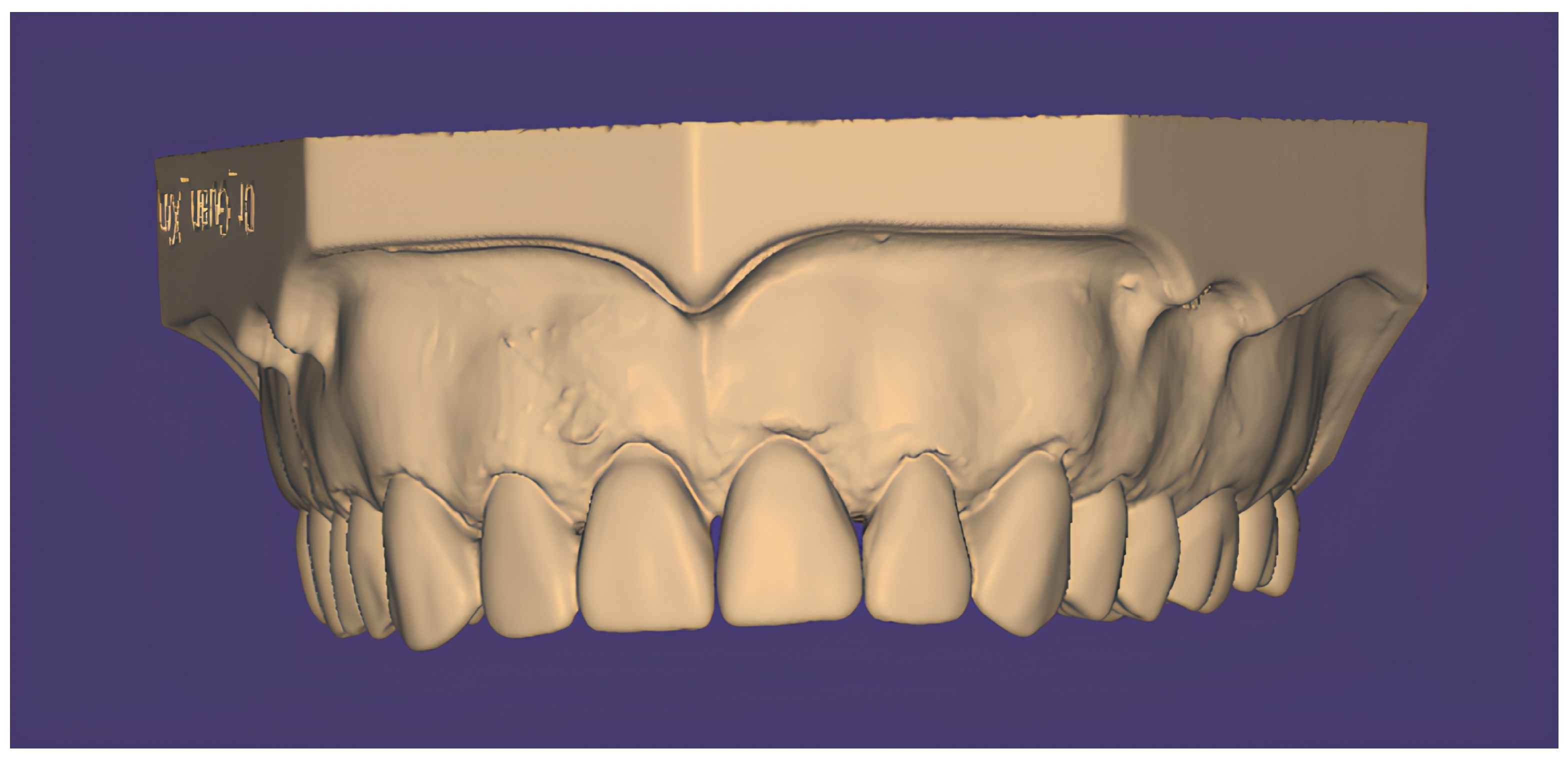
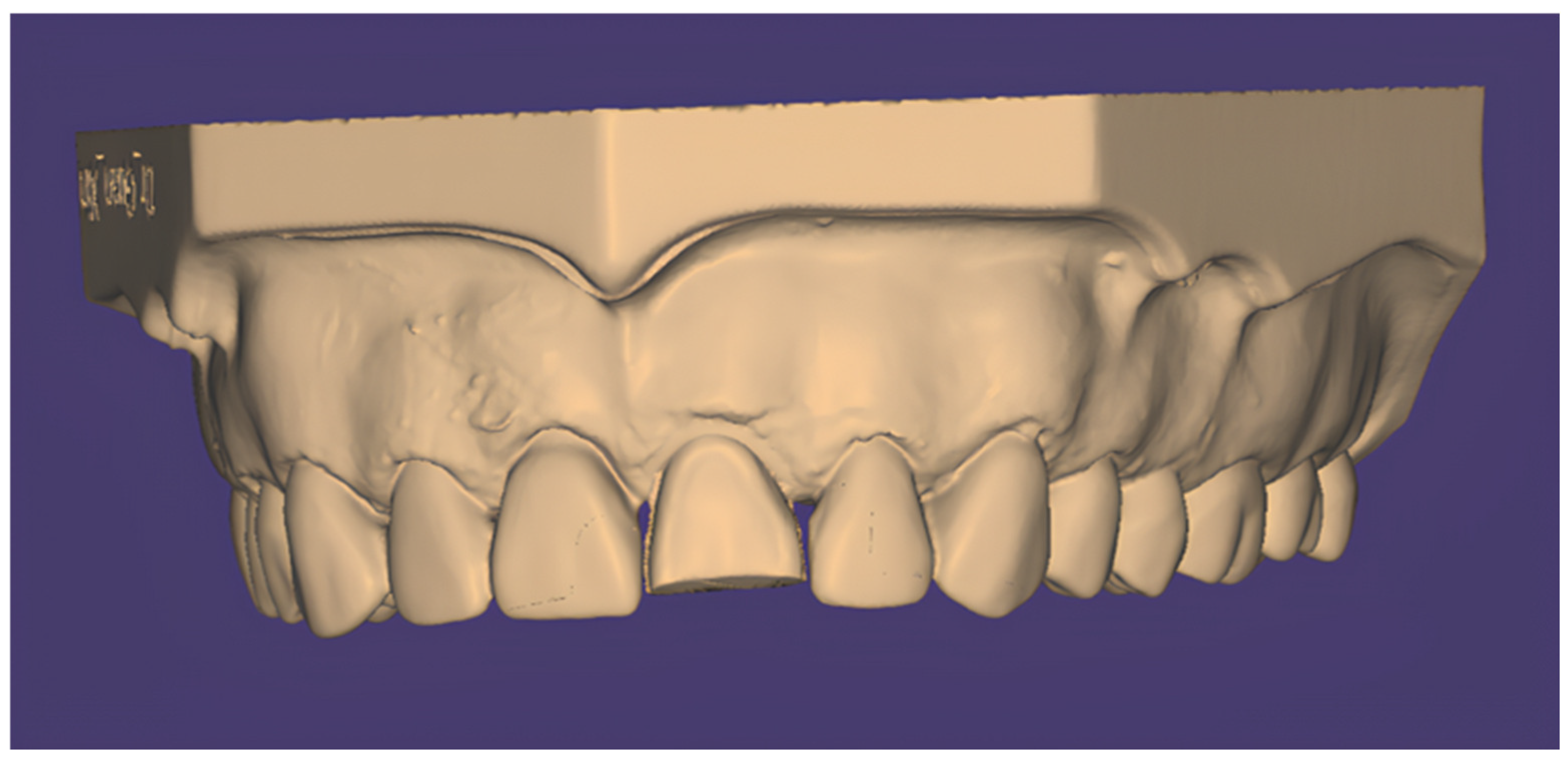
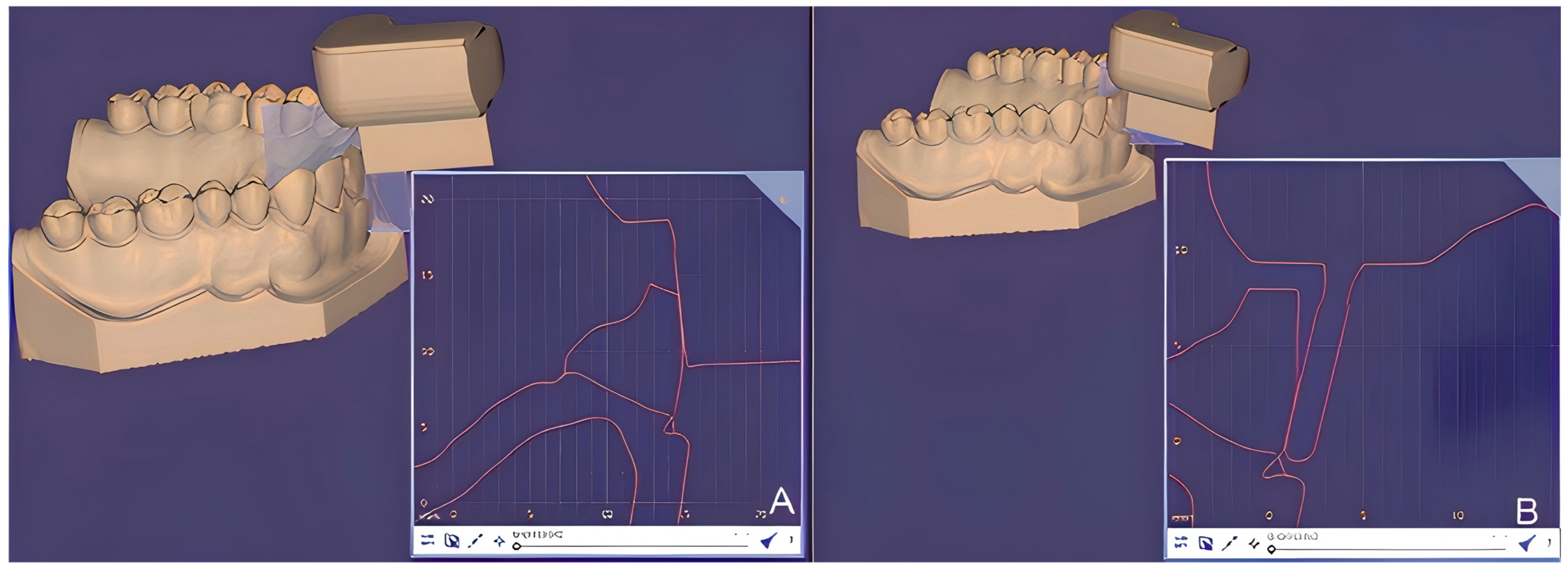
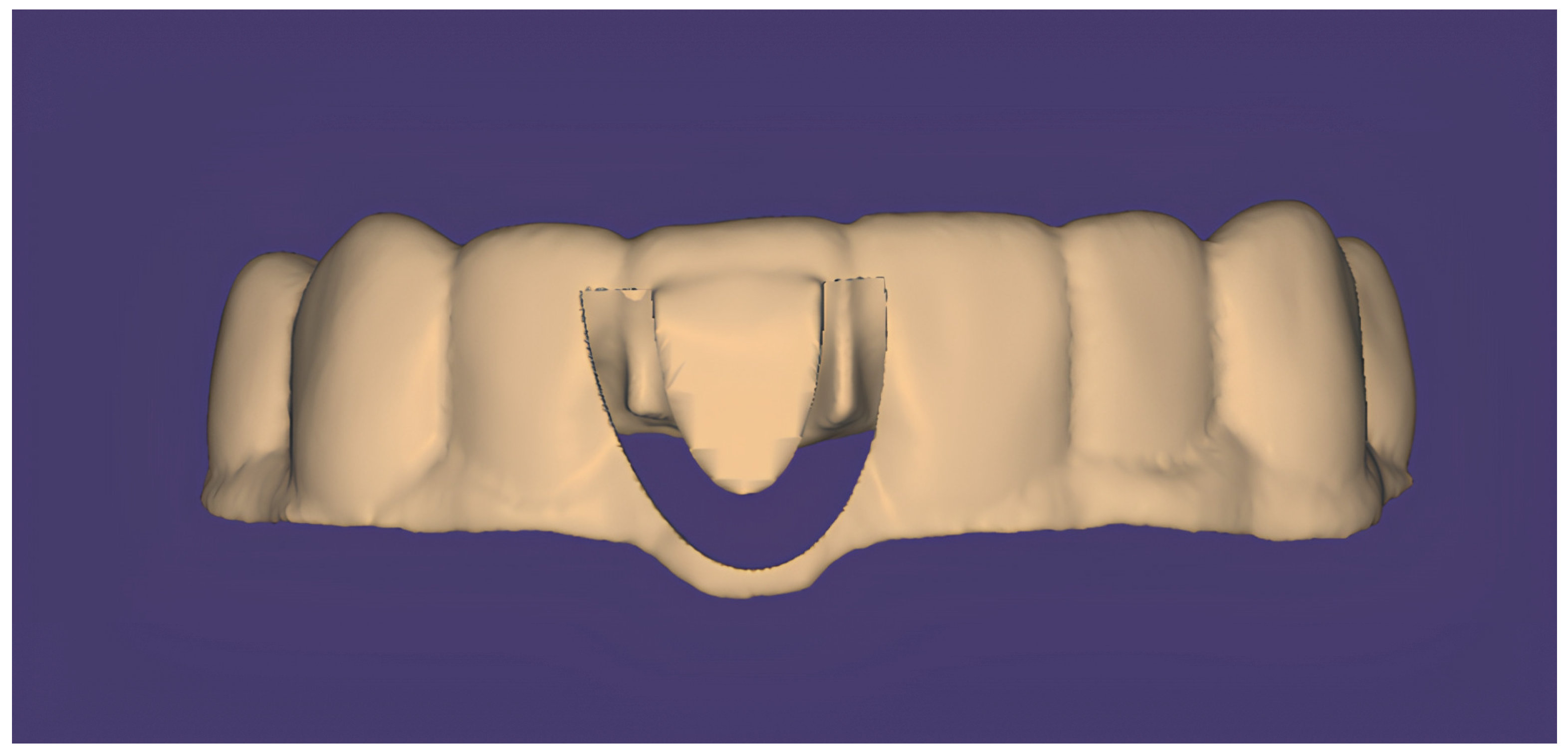
2.1. Tooth Reduction Template Design for Incisal Reduction

A maxillary complete-dentate cast simulating a patient’s dental arch was scanned using a laboratory scanner (3Shape Trios E4; 3Shape, Copenhagen, Denmark) and saved in standard triangle language (STL) format (Figure 1). The maxillary right central incisor was designated the target tooth for veneer preparation.
The first tooth reduction template was designed specifically for incisal reduction using dental design software (DentalCAD 3.1 Rijeka; exocad GmbH, Darmstadt, Germany). The incisal edge of the maxillary right central incisor was virtually reduced by 1.5 mm using the cutting tool (Materialise Magics 27; Materialise, Leuven, Belgium; Figure 2). This was followed by designing the tooth reduction template, extending from the maxillary right first premolar to the left first premolar (Figure 3).

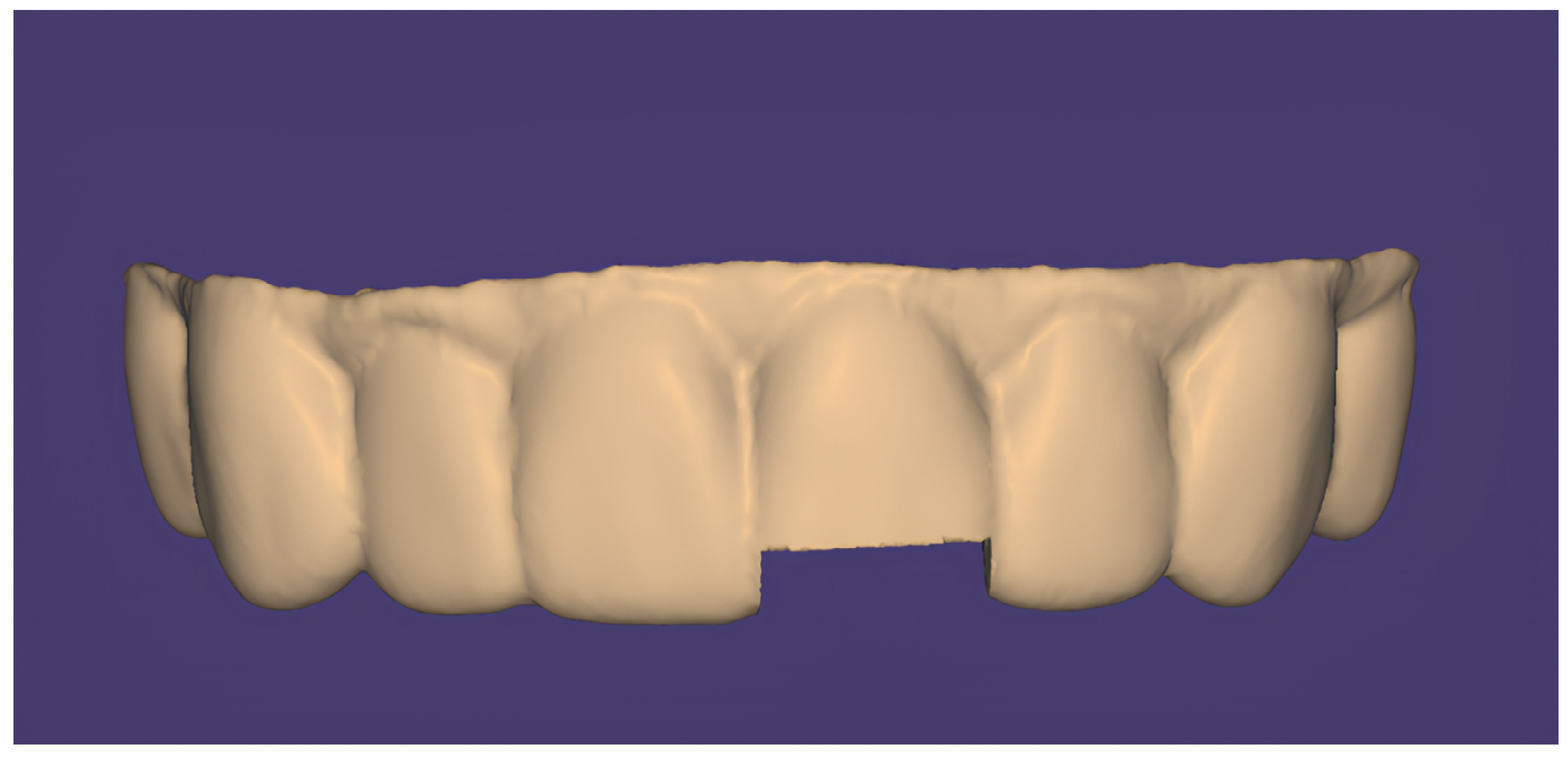
2.2. Tooth Reduction Template Design for Labial Reduction

The second and third templates, used for labial reduction at the incisal half and cervical half, were designed using dental design software (DentalCAD 3.1 Rijeka, exocad GmbH). A high-speed handpiece with chamfer bur (G/TR-32; Daobang, Foshan, China) was scanned using a laboratory scanner (3Shape Trios E4; 3Shape, Copenhagen, Denmark) and saved in STL format (Figure 4).
A virtual labial reduction was performed at both incisal and cervical halves by applying an inward offset of 0.5 mm following the planned labial reduction depth using the “Freeform Shape Marking and Remeshing” tool (Materialise Magics; Materialise, Leuven, Belgium). A tooth reduction template was then designed with a window to accommodate the handpiece head and an integrated bur channel measuring 8 mm in length and 1 mm in width to precisely control handpiece and bur movement during labial preparation (Figure 5A,B). The virtual positioning of the chamfer bur was verified at both the incisal half (Figure 6A) and cervical half (Figure 6B), ensuring alignment with the inclination of the labial surfaces.
The tooth reduction template design was then completed by extending from the maxillary right first premolar to the left first premolar for both incisal (Figure 7) and cervical halves (Figure 8).

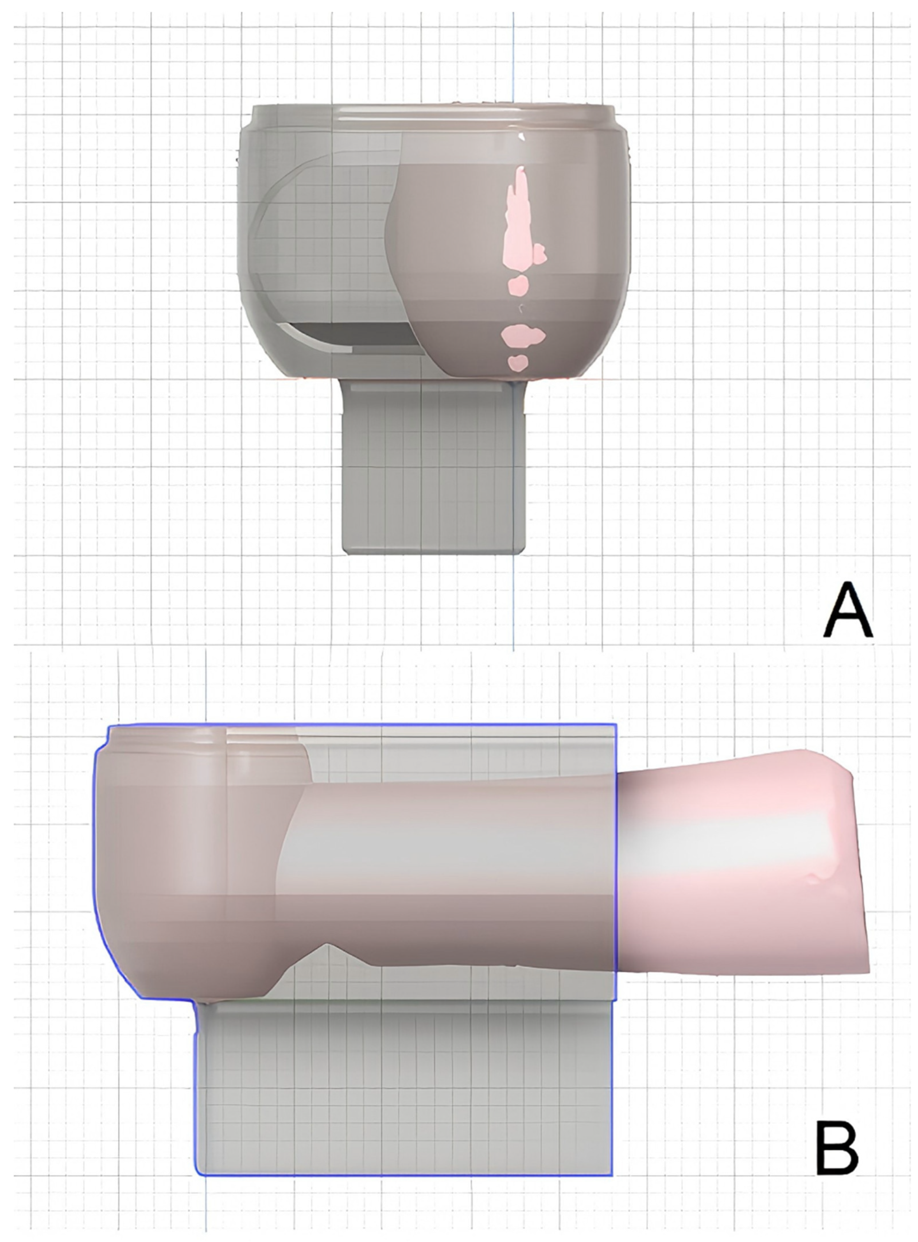
2.3. Tooth Reduction Template Design for Cervical Margin Reduction

The tooth reduction template for cervical margin reduction was the fourth template designed. Virtual reduction at the cervical margin was performed by applying an inward offset of 0.3 mm using the “Freeform Shape Marking and Remeshing” tool (Materialise Magics; Materialise, Leuven, Belgium). A diamond bur with a stopper (G/EX-58, Daobang, FG0807D, China) was modified to a length of 0.3 mm (Figure 9), scanned using a laboratory scanner (3Shape Trios E4; 3Shape, Copenhagen, Denmark), and saved in the STL format. A tooth reduction template incorporating a 3 mm wide bur channel was then designed to accommodate the modified bur (Figure 10).

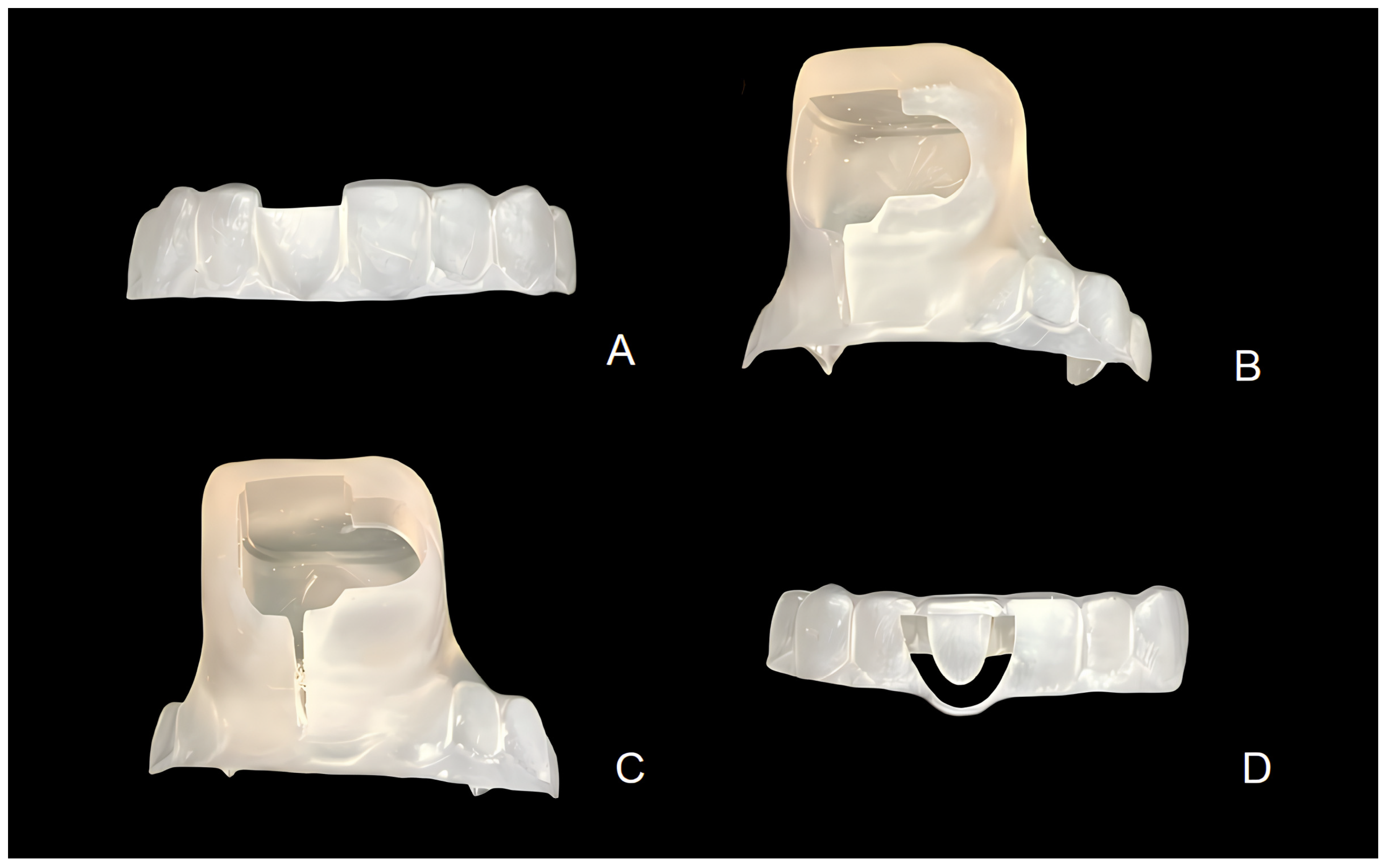
2.4. Tooth Reduction Template Fabrication

All four tooth reduction templates were printed with resin material (VisiJet M3 Crystal 3D Material, 3D Systems, Rock Hill, SC, USA) using a 3D printer (ProJet MJP 3600; 3D Systems, USA) (Figure 11A–D).

2.5. Veneer Preparation Guided via Stackable Templates
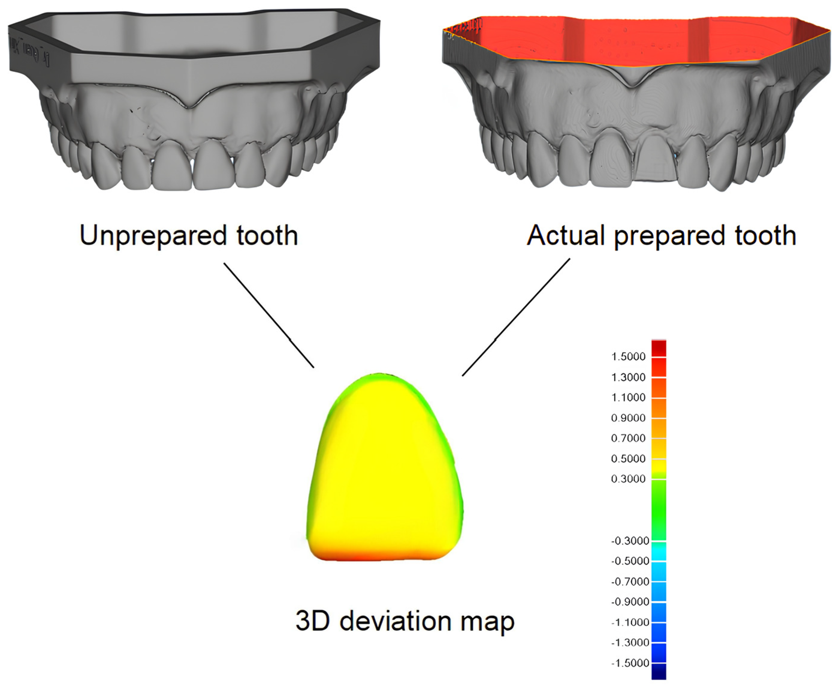
Veneer preparation on the maxillary left central incisor was guided via the templates following four essential steps: incisal reduction, incisal half labial reduction, cervical half labial reduction, and cervical reduction. Before proceeding with the veneer preparation, each template was seated on a dental cast following a 90° path of insertion relative to the occlusal plane of the dental cast. Accurate template positioning was further verified through visual inspection, facilitated by the translucency of the template.
The incisal reduction was performed using the incisal reduction template as a guide. A diamond chamfer bur (G/TR-32; Daobang, Foshan, China) was used to achieve the targeted 1.5 mm incisal reduction (Figure 12A). The labial surface was then sequentially prepared under the guidance of two labial reduction templates, using the diamond chamfer bur (G/TR-32, Daobang, FG0807D, China; Figure 12B,C) to ensure a uniform reduction of approximately 0.5 mm. Finally, the margin reduction template was employed along with a modified diamond bur (G/EX-58; Daobang, Foshan, China; Figure 12D) to precisely prepare a chamfer margin of 0.3 mm.

3. Results

To assess the accuracy of veneer preparation using the four templates, the cast with the prepared maxillary left center incisor was scanned using a lab scanner (D700L; 3Shape A/S, Copenhagen, Denmark) in STL format. The STL files of the prepared maxillary left center incisor were superimposed with the virtual referenced unprepared cast using 3D metrology software (Geomagic Control X, 2017.0.1; 3D Systems, Rock Hill, SC, USA). The 3D deviation analysis showed that the tooth reduction depth followed the virtual plan, whereby the labial reduction achieved a 0.5 mm depth, the cervical margin reduction achieved a 0.3 mm depth, and the incisal reduction achieved a 1.5 mm depth (Figure 13).

4. Discussion

This article demonstrates a digital workflow for designing and fabricating stackable Multijet-printed templates to provide comprehensive guidance for veneer preparation in four essential steps: incisal, labial, and cervical margin preparation. The core concept of this design is to enhance the accuracy of the tooth preparation depth by incorporating a virtual tooth preparation plan with precisely engineered bur-guiding channels within the template.
Several previous studies have reported different designs of tooth reduction templates used for veneer preparation [11], including a cross-shaped design [13], multiple cylindrical guide tubes [10], and an open-window design at the labial surfaces [14]. However, all of these templates only provide tooth reduction depth grooves, and the subsequent tooth preparation still primarily relies on the free-hand technique after removal of the template, which minimizes the accuracy of veneer preparation [15,18]. Therefore, to improve the accuracy of guided veneer preparation, we designed four distinct templates to guide the clinician through the four key steps of veneer preparation. Each template is tailored to address specific areas of the tooth preparation process, with corresponding depth requirements.
Color map analysis demonstrates promising outcomes for veneer preparation utilizing stackable tooth reduction templates (RMS = 1.38 ± 0.03 mm). The color map provides a visual representation of the reduction achieved across four different tooth surfaces, offering a clear assessment of preparation uniformity. Our tooth reduction template optimizes the margin preparation (±0.3 mm), addressing inaccuracies that are commonly found in veneer preparation using other tooth reduction templates [15,18]. The results of this study also demonstrated well-controlled preparation on the labial surface (±0.5 mm), with seamless transitions at the incisal–cervical labial junction. The map highlights well-distributed reductions, suggesting that the templates effectively guide the clinician toward achieving consistent and controlled preparation depths. This aligns with a study by Gao et al. [19], which demonstrated that guided veneer preparation using tooth reduction templates resulted in less tooth reduction compared to free-hand and silicone guides. Accurate tooth reduction, as virtually planned, is important to ensure that the required tooth reduction volume is maintained, ultimately enhancing aesthetics and bonding strength within the enamel.
Several factors influence the accuracy of stackable tooth reduction templates in veneer preparation, particularly during the design of the template, the 3D printing process, and the veneer preparation phase. When designing a tooth reduction template, it is important to incorporate the estimated tooth reduction volume based on the thickness of the veneer material. This ensures uniform restorative spaces, leading to improved aesthetics. In this study, to achieve this aim, we calculated the actual tooth reduction depth at each designed site and planned the reduction virtually on digital dental casts prior to designing the tooth reduction templates. Basic virtual tooth preparation can typically be performed using the “inward offset” tool in CAD software [10,19]. Alternatively, more precise virtual tooth preparation using virtual burs, as reported by previous studies, enables accurate chair-side tooth preparation simulations. This approach allows for the design and fabrication of definitive veneers for immediate cementation in a single visit [14,20].
Furthermore, to improve control over bur movements during veneer preparation, bur-guiding channels were integrated into the tooth reduction templates for labial and cervical margin preparation. For labial preparation, an L-shaped bur-guiding channel was designed alongside handpiece access windows to enhance the control of the handpiece and bur during veneer preparation. To ensure proper alignment of the handpiece access window and bur-guiding channels with the burs’ dimensions, a commonly used high-speed handpiece and the specific burs for veneer preparation must be scanned in STL format for template design. However, for the cervical margin preparation, a cost-effective modification of a diamond bur with a stopper was proposed to fit into the curved bur-guiding channel within the template, optimizing margin preparation accuracy. A similar approach is observed in a commercially available First-fit system for veneer preparation; however, unlike our approach, a First-fit system requires a specially designed handpiece to accommodate the rotary instrument access windows, leading to higher equipment costs. Verifying the virtual positioning of the burs, as described in this study, is recommended. This step ensures that the actual bur position aligns correctly with the intended tooth surfaces, thereby improving the precision of veneer preparation.
In addition, two distinct templates for labial reduction were designed to maintain the two planes of the labial surface during veneer preparation—one for the incisal half and another for the cervical half. Nevertheless, the inclusion of handpiece access windows in both tooth reduction templates increases their bulkiness, which may pose challenges for patients with limited mouth opening.
Several factors associated with 3D printing also affect the accuracy of templates in tooth reduction, including the 3D printing technology employed, parameter settings during the printing process, and post-processing procedures [21,22,23]. Compared to Stereolithography (SLA) and Digital Light Processing (DLP) technologies, the Multijet Printing (MJP) technology used in this study demonstrates advantages in high-speed printing, excellent surface smoothness, and exceptional detail resolution. In this study, a class VI biocompatible photopolymer resin was used with UV curing and a 45° build orientation to ensure high dimensional stability and printing precision [24,25], which are critical for accurate tooth reduction guidance. The translucency of the resin material is beneficial in further facilitating visual inspections of bur positioning and movement during the tooth preparation process, thus enhancing clinical control and accuracy [26,27].
The stackable tooth reduction template introduced in this study offers several clinically significant advantages in veneer preparation. First, by standardizing reduction depths across critical regions (incisal edge: 1.5 mm, labial surface: 0.5 mm, cervical margins: 0.3 mm) through guided bur movement, this approach significantly reduces operator dependence, a persistent limitation of conventional veneer preparation techniques [6,7]. This allows clinicians with varying levels of clinical experience to consistently achieve enamel-preserving preparations, especially at the cervical margins, which are critical for long-term bonding success [4,5], while potentially minimizing postoperative sensitivity and the risk of restoration failure. Second, the sequential guidance in veneer preparation improves clinical efficiency by eliminating the need for repeated depth checks required when using conventional silicone guides. In addition, this innovative technique is compatible with standard burs and integrates easily with CAD/CAM workflows, making it well suited for modern digital prosthodontics and enabling same-day veneer fabrication when combined with intraoral scanning [17,21].
While the innovative guided veneer preparation approach presented in this study shows promising results, certain limitations should be acknowledged. This study primarily focuses on the digital workflow for developing stackable tooth reduction templates without providing a comparative analysis against other veneer preparation techniques. Therefore, further in vitro or clinical studies are required to evaluate the accuracy of veneer preparation using stackable tooth reduction templates compared to conventional free-hand preparation, silicone-guided preparation, and other types of tooth reduction templates.

5. Conclusions

The use of stackable Multijet-printed templates can be a potential approach to further improve the precision and predictability of veneer preparation.

Author Contributions

Conceptualization, X.G. and I.M.T.; methodology, X.G., C.L., T.F.S., Y.H.B. and I.M.T.; writing—original draft preparation, X.G.; writing—reviewing and editing, I.M.T.; validation, I.M.T. and Y.H.B. All authors have read and agreed to the published version of the manuscript.

Funding

This research received no external funding.

Institutional Review Board Statement

Not applicable.

Informed Consent Statement

Not applicable.

Data Availability Statement

Not applicable.

Conflicts of Interest

The authors declare no conflicts of interest.

References

  1. Dumfahrt, H.; Schäffer, H. Porcelain laminate veneers. A retrospective evaluation after 1 to 10 years of service: Part II—Clinical results. Int. J. Prosthodont. 2000, 13, 9–18. [Google Scholar] [PubMed]
  2. Friedman, M.J. Masters of esthetic dentistry: Porcelain veneer restorations: A clinician’s opinion about a disturbing trend. J. Esthet. Restor. Dent. 2001, 13, 318. [Google Scholar] [CrossRef] [PubMed]
  3. Strassler, H.E. Minimally invasive porcelain veneers: Indications for a conservative esthetic dentistry treatment modality. Gen. Dent. 2007, 55, 686–694. [Google Scholar] [PubMed]
  4. Peumans, M.; De Munck, J.; Fieuws, S.; Lambrechts, P.; Vanherle, G.; Van Meerbeek, B. A prospective ten-year clinical trial of porcelain veneers. J. Adhes. Dent. 2004, 6, 65–76. [Google Scholar]
  5. Piemjai, M.; Arksornnukit, M. Compressive fracture resistance of porcelain laminates bonded to enamel or dentin with four adhesive systems. J. Prosthodont. 2007, 16, 457–464. [Google Scholar] [CrossRef] [PubMed]
  6. Cherukara, G.P.; Davis, G.R.; Seymour, K.G.; Zou, L.; Samarawickrama, D.Y. Dentin exposure in tooth preparations for porcelain veneers: A pilot study. J. Prosthet. Dent. 2005, 94, 414–420. [Google Scholar] [CrossRef]
  7. Gürel, G. Predictable, precise, and repeatable tooth preparation for porcelain laminate veneers. Pract. Proced. Aesthet. Dent. 2003, 15, 17–24. [Google Scholar]
  8. Sisler, Z.S. Preparation Guides: 10 Steps to Maximize Success for Veneer Preparation. J. Cosmet. Dent. 2020, 35, 26–33. [Google Scholar]
  9. Guzman Perez, G.; Jurado, C.A.; Azpiazu Flores, F.; Afrashtehfar, K.I.; Tsujimoto, A. Minimally Invasive Laminate Veneer Therapy for Maxillary Central Incisors. Medicina 2023, 59, 603. [Google Scholar] [CrossRef]
  10. Liu, C.; Guo, J.; Gao, J.; Yu, H. Computer-Assisted Tooth Preparation Template and Pre-Designed Restoration: A Digital Workflow. Int. J. Comput. Dent. 2020, 23, 351–362. [Google Scholar]
  11. Guan, X.; Beh, Y.H.; Tew, I.M. Computer-Assisted Porcelain Laminate Veneer Preparation: A Scoping Review of Stereolithographic Template Design and Fabrication Workflows. Dent. J. 2024, 12, 302. [Google Scholar] [CrossRef] [PubMed]
  12. Zhong, Y.L.; Bai, H.F.; Zhao, Y.J.; Wang, Y.; Ye, H.Q.; Sun, Y.C. 3D Evaluation of Accuracy of Tooth Preparation for Laminate Veneers Assisted by Rigid Constraint Guides Printed by Selective Laser Melting. Chin. J. Dent. Res. 2020, 23, 183–189. [Google Scholar]
  13. Robles, M.; Jurado, C.A.; Azpiazu-Flores, F.X.; Villalobos-Tinoco, J.; Afrashtehfar, K.I.; Fischer, N.G. An innovative 3D printed tooth reduction guide for precise dental ceramic veneers. J. Funct. Biomater. 2023, 14, 216. [Google Scholar] [CrossRef] [PubMed]
  14. Figueira, J.; Guaqueta, N.; Ramirez, D.I.; Kois, J. Veneer tooth preparation utilizing a novel digital designed workflow: A case report. J. Esthet. Restor. Dent. 2023, 35, 803–809. [Google Scholar] [CrossRef] [PubMed]
  15. Gao, J.; He, J.; Fan, L.; Lu, J.; Xie, C.; Yu, H. Accuracy of reduction depths of tooth preparation for porcelain laminate veneers assisted by different tooth preparation guides: An in vitro study. J. Prosthodont. 2022, 31, 593–600. [Google Scholar] [CrossRef]
  16. Chai, S.Y.; Bennani, V.; Aarts, J.M.; Lyons, K. Incisal preparation design for ceramic veneers: A critical review. J. Am. Dent. Assoc. 2018, 149, 25–37. [Google Scholar] [CrossRef]
  17. Alothman, Y.; Bamasoud, M.S. The success of dental veneers according to preparation design and material type. Open Access Maced. J. Med. Sci. 2018, 6, 2402. [Google Scholar] [CrossRef]
  18. Luo, T.; Zhang, J.; Fan, L.; Huang, Y.; Yu, J.; Yu, H. A digital workflow with the virtual enamel evaluation and stereolithographic template for accurate tooth preparation to conservatively manage a case of complex exogenous dental erosion. J. Esthet. Restor. Dent. 2022, 34, 733–740. [Google Scholar] [CrossRef]
  19. Gao, J.; Li, J.; Liu, C.; Fan, L.; Yu, J.; Yu, H. A stereolithographic template for computer-assisted teeth preparation in dental esthetic ceramic veneer treatment. J. Esthet. Restor. Dent. 2020, 32, 763–769. [Google Scholar] [CrossRef]
  20. Silva, B.P.D.; Stanley, K.; Gardee, J. Laminate veneers: Preplanning and treatment using digital guided tooth preparation. J. Esthet. Restor. Dent. 2020, 32, 150–160. [Google Scholar] [CrossRef]
  21. Etemad-Shahidi, Y.; Qallandar, O.B.; Evenden, J.; Alifui-Segbaya, F.; Ahmed, K.E. Accuracy of three-dimensionally printed full-arch dental models: A systematic review. J. Clin. Med. 2020, 9, 3357. [Google Scholar] [CrossRef] [PubMed]
  22. Jeong, M.; Radomski, K.; Lopez, D.; Liu, J.T.; Lee, J.D.; Lee, S.J. Materials and applications of 3D printing technology in dentistry: An overview. Dent. J. 2023, 12, 1. [Google Scholar] [CrossRef] [PubMed]
  23. Arnold, C.; Monsees, D.; Hey, J.; Schweyen, R. Surface quality of 3D-printed models as a function of various printing parameters. Materials 2019, 12, 1970. [Google Scholar] [CrossRef]
  24. Morón-Conejo, B.; López-Vilagran, J.; Cáceres, D.; Berrendero, S.; Pradíes, G. Accuracy of five different 3D printing workflows for dental models comparing industrial and dental desktop printers. Clin. Oral Investig. 2023, 27, 2521–2532. [Google Scholar] [CrossRef] [PubMed]
  25. Gao, H.; Yang, Z.; Lin, W.S.; Tan, J.; Chen, L. The effect of build orientation on the dimensional accuracy of 3D-printed mandibular complete dentures manufactured with a multijet 3D printer. J. Prosthodont. 2021, 30, 684–689. [Google Scholar] [CrossRef]
  26. Bammani, S.S.; Birajdar, P.R.; Metan, S.S. Application of CAD and SLA Method in Dental Prosthesis. AMAE Int. J. Man. Mat. Sci. 2013, 3, 5. [Google Scholar]
  27. Dawood, A.; Marti Marti, B.; Sauret-Jackson, V. 3D printing in dentistry. Br. Dent. J. 2015, 219, 521–529. [Google Scholar] [CrossRef]
Figure 1. A laboratory scan of a maxillary complete-dentate cast generated for further tooth reduction template designs.
Applsci 15 04975 g001
Figure 2. Virtual incisal reduction of 1.5 mm at the maxillary right central incisor.
Applsci 15 04975 g002
Figure 3. Design of tooth reduction template for incisal reduction.
Applsci 15 04975 g003
Figure 4. Virtual high-speed handpiece and chamfer bur.
Applsci 15 04975 g004
Figure 5. An access window to accommodate the handpiece head with an L-shaped bur-guiding channel. (A) Top view; (B) side view.
Applsci 15 04975 g005
Figure 6. Verification of virtual chamfer bur position at the (A) incisal half and (B) cervical half of the labial surface after virtual labial reduction.
Applsci 15 04975 g006
Figure 7. Tooth reduction template design for labial preparation at the incisal half.
Applsci 15 04975 g007
Figure 8. Tooth reduction template design for labial preparation at the cervical half.
Applsci 15 04975 g008
Figure 9. A modified bur with a stopper and a length of 0.3 mm.
Applsci 15 04975 g009
Figure 10. Tooth reduction template design for cervical margin preparation.
Applsci 15 04975 g010
Figure 11. Three-dimensional printed stereolithographic templates for (A) incisal reduction, (B) incisal half labial reduction, (C) cervical half labial reduction, and (D) cervical margin reduction.
Applsci 15 04975 g011
Figure 12. Tooth preparation guided by the templates: (A) incisal reduction, (B) incisal half labial reduction, (C) cervical half labial reduction, and (D) margin reduction.
Applsci 15 04975 g012
Figure 13. Amount of tooth volume loss observed in the 3D deviation map.
Applsci 15 04975 g013
Disclaimer/Publisher’s Note: The statements, opinions and data contained in all publications are solely those of the individual author(s) and contributor(s) and not of MDPI and/or the editor(s). MDPI and/or the editor(s) disclaim responsibility for any injury to people or property resulting from any ideas, methods, instructions or products referred to in the content.

