Article

Computational Prediction of Ginsenosides Targeting ADGRG3/GPR97 in Cancer and Immune Pathways: A Multi-Faceted In Silico Approach

Division of General Education, Seokyeong University, Seoul 02173, Republic of Korea
Appl. Sci. 2025, 15(8), 4332; https://doi.org/10.3390/app15084332
Submission received: 9 January 2025 / Revised: 2 April 2025 / Accepted: 9 April 2025 / Published: 14 April 2025
(This article belongs to the Special Issue Advanced Phytochemistry and Its Applications)

Abstract

:
Ginsenosides are bioactive secondary metabolites in ginseng, which have gained popularity for their usage in traditional Oriental medicine. Many studies have reported that ginsenosides exert their effects through multiple pathways, such as GPCR-related pathways. However, focusing on their specific interactions with ADGRG3 (GPR97) can provide possible insights to inform targeted intervention strategies in oncology and immunotherapy through the tumor–immune microenvironment interactions. Thus, this study employed an integrative in silico computational strategy to investigate ginsenosides as possible targets of ADGRG3. First, gene expression was analyzed using multiple databases such as TCGA, cBioPortal, and TIMER, revealing the differential expression of ADGRG3 across cancers, with notable overexpression in leukemia. Then, the virtual screening of 128 ginsenosides identified five top candidates (Rg3, Rk3, F5, Rg7, and F1) that showed strong binding energy (−10.7 −10.6, −10.5, −10.4, and −10.3 kcal/mol, respectively) with ADGRG3, as determined through in silico molecular docking (MD). Computational approaches such as molecular dynamics simulations (MDSs), free binding energy calculations (MM-PBSA), and ADMET profiling confirmed the stability of these complexes’ favorable ADMET predictions, respectively, which warrants further experimental validation through in vitro and in vivo pharmacokinetic studies. Finally, the computational protein–protein interaction and pathway enrichment analyses of ADGRG3 demonstrated immune-related pathways, such as neutrophil degranulation and GPCR signaling, emphasizing its role in cancer progression and immune modulation. These computational findings predict ADGRG3 as a viable target for cancer and immune pathways and ginsenosides as natural ligands. Further in vitro and in vivo preclinical and clinical studies are warranted to validate the interactions of ADGRG3 with ginsenosides.

1. Introduction

The human genome provides a wealth of potential therapeutic targets, making druggable targets essential in drug discovery. Advances in genomics have expanded the concept of the “druggable genome” to include molecular entities that can be manipulated to enhance health, revolutionizing the drug discovery process from “forward pharmacology” to “reverse pharmacology” [1,2,3,4,5,6]. Novel druggable targets identified through genome sequencing have significantly accelerated drug development [5,7]. Tools such as AlphaFold, an AI-based protein structure prediction method, further enhance this process by enabling large-scale drug screening, improving target–ligand interaction studies, and facilitating the identification of molecules from the druggable genome/proteome [3,8]. These system-based technologies, including cellular gene expression profiling and in silico modeling, have led to more efficient pipelines to predict novel therapeutics.
G-protein-coupled receptors (GPCRs), the largest family of membrane receptors, have been extensively studied in cancer- and immune-related diseases due to their crucial roles in cellular signaling and disease pathways [3,7,9,10,11,12,13,14]. Among them, the adhesion G-protein-coupled receptor G3 (ADGRG3 or GPR97) has gained interest due to its potential involvement in cancer progression and immune modulation [15,16,17]. The classical adhesion structure of GRP97 has seven transmembrane helices, an N-terminal fragment, and a C-terminal fragment that results from autoproteolytic cleavage at a GPCR-proteolytic site contained in a juxtamembranous GPCR autoproteolysis-inducing domain [16]. ADGRG3/GPR97 has been identified in leukocytes, including eosinophils, neutrophils, and mast cells [18], and it plays a role in macrophage-related diseases and obesity-induced metabolic disorder [19]. Additionally, it has been documented that ADGRG3/GPR97 is expressed in spinal cord endothelial cells in naive mice and is elevated in spinal cord endothelial cells and spinal cord-infiltrating CD4+ T cells of MOG-immunized mice during the onset of disease [20]. ADGRG3/GPR97 is essential for controlling the destiny of B cells, particularly for controlling the activities of nuclear factor (NF)-κB and constitutive cAMP response element-binding protein [21]. These results suggest that ADGRG3/GPR97 may play a role in the emergence of immune diseases and cancers. However, unlike well-studied GPCRs such as ADGRG1, ADGRG3 has not been thoroughly researched, despite evidence of its differential expression in cancers such as leukemia and its association with immune cell pathways [17,22]. Notably, a recent study demonstrated that another adhesion GPCR, ADGRE2, plays a critical role in Acute Myeloid Leukemia (AML) by promoting leukemic stem cell survival and tumor progression via the MEK/AP1/DUSP1 axis. This role was experimentally validated through both in vitro and in vivo studies, where ADGRE2 knockdown in AML cell lines and patient-derived cells significantly impaired cell proliferation, induced apoptosis, and reduced leukemic stem cell (LSC) frequency. Moreover, in xenograft mouse models, ADGRE2 silencing delayed leukemia progression and prolonged survival, providing strong evidence for the functional relevance of adhesion GPCRs in AML pathogenesis [23]. Given the structural and functional similarities among adhesion GPCRs, this finding underscores the potential role of ADGRG3 in leukemia, suggesting that it may similarly contribute to disease progression and serve as a promising target for further investigation.
Natural products have historically been a cornerstone of drug discovery, especially in oncology, due to their structural diversity and biological activities [24,25]. Triterpenoids, a prominent class of plant-derived compounds, demonstrate significant potential as anticancer agents [26]. These natural substances have presented cytotoxicity against tumor cells and efficacy in preclinical studies, influencing intracellular signaling pathways involved in cancer [27,28,29,30,31,32]. Research has shown that triterpenoids influence multiple intracellular signaling pathways, exhibiting chemopreventive and antitumor properties in various cancer models [33]. For instance, oleanolic acid derivatives modulated proliferation, apoptosis, and inflammation in breast cancer, while mistletoe triterpenoids enhanced melanoma treatment outcomes in animal models [34]. However, challenges such as poor solubility and bioavailability limit their development [35,36]. Future studies should prioritize systematic target identification and structure–activity relationship analyses to optimize their therapeutic potential.
Among triterpenoids, ginsenosides from Panax species are promising due to their multitarget abilities, steroid-like structures, and favorable properties [37,38,39,40]. Despite these advantages, research on ginsenosides faces challenges, including structural diversity (over 400 isoforms) and varying efficacy between major and minor ginsenosides. Moreover, ginsenosides have demonstrated anti-inflammatory and anticancer properties, and their structural similarities to synthetic steroids such as dexamethasone reinforce their potential to modulate GPCRs [41,42]. Adhesion GPCRs (aGPCRs) play a key role in tumor microenvironment modulation, immune cell trafficking, and tumor progression. They have long extracellular domains that facilitate interactions with the extracellular matrix, influencing cell adhesion, migration, and invasion—processes that are critical in cancer. Moreover, many GPCRs signal through classical Gα proteins (Gs, Gi, Gq, and G12/13). ADGRG3 shows unique autoproteolysis-dependent signaling (via the GPS motif); can influence multiple pathways, such as cAMP regulation, RhoA activation, and NF-κB signaling; and specifically allows for targeted analysis of its non-canonical pathways. However, ginsenosides’ specific interactions with ADGRG3 remain unexplored, presenting an opportunity to investigate these natural compounds as modulators with potentially safer profiles than synthetic drugs [42,43,44,45].
In recent years, computational biology has transformed drug discovery by integrating in silico molecular docking (MD), molecular dynamics simulations (MDSs), and ADMET predictions with gene expression and pathway analyses [44,46,47,48,49,50]. In silico technologies such as MD and MDS are well-known examples of virtual screening techniques based on a structure, and these have been proven important in elucidating the relationships between biomolecules [51]. These methods provide reliable predictable insights into target–ligand interactions while reducing experimental time and cost. Additionally, publicly available datasets, such as those from TCGA and cBioPortal, further enhance research by offering comprehensive gene expression, mutation, and survival data to validate therapeutic targets in cancer and inflammation.
This study employed a multi-step in silico approach to assess ginsenosides as natural ligands of ADGRG3. First, gene expression analyses using TCGA, cBioPortal, UALCAN, and TIMER validated ADGRG3’s relevance in cancer and immune pathways. Next, 128 ginsenosides and four control ligands (beclomethasone dipropionate, dexamethasone, HCY, and prednisolone) were screened for their ability to bind to ADGRG3 through in silico MD. Beclomethasone dipropionate is an approved drug that acts as an agonist against ADGRG3 (https://www.genecards.org/cgi-bin/carddisp.pl?gene=ADGRG3#drugs_compounds, accessed on 25 April 2023 and 20 February 2025). Computational ADMET analysis was conducted to evaluate pharmacokinetics, while MD simulations assessed binding stability. Online protein–protein interaction (PPI) and pathway enrichment analyses were performed to contextualize ADGRG3 within cancer- and immune-related pathways. Additionally, the gene expression analysis of ADGRG3 and its interacting genes in AML was performed using the publicly available GEO dataset, which includes data from 506 diverse samples. This analysis highlighted ADGRG3’s expression patterns and its role in inflammatory signaling within the leukemia tumor microenvironment.

2. Material and Methods

2.1. Gene Expression Analysis of ADGRG3 in Various Cancers

The expression and clinical relevance of ADGRG3 (GPR97) in various cancers were assessed using multiple databases such as Drugbanks, ProteinAtlas, and Druggable Targets (accessed on 2 February 2023). The Cancer Genome Atlas (TCGA) [46,52] provided data on the differential expression of ADGRG3 between tumor and normal tissues, highlighting its upregulation or downregulation in cancer (accessed on 15 March 2023). cBioPortal [53] (accessed on 17 March 2023) was used to examine genetic alterations in ADGRG3, including mutations and amplifications, and their associations with mRNA expression in various cancers. UALCAN [54] (accessed on 20 March 2023) was used for a more detailed analysis of ADGRG3 expression according to clinical attributes such as cancer stage, sample type, gender, and age, along with the evaluation of the impact of ADGRG3 expression on patients’ survival. TIMER (Tumor Immune Estimation Resource) [55] (accessed on 20 March 2023) offered insights into ADGRG3’s role in the tumor microenvironment (TME) through the analysis of its associations with immune cell infiltration, highlighting the immunological context of ADGRG3 across cancer types.

2.2. Virtual Screening

For the virtual screening of ginsenosides against ADGRG3, ADGRG3 was selected as the target, and its relevance in cancer was confirmed through gene expression analysis (Section 2.1). Ginsenosides, chosen for their structural similarity to steroid drugs and their known pharmacological benefits in cancer and immune modulation, were screened against ADGRG3. The 7D77 [56] crystal structure of ADGRG3 (Protein Data Bank, (PDB), 2.90 Å resolution) was used for docking to ensure reliable interaction modeling. The ADGRG3 structure was prepared for docking by removing water molecules and heteroatoms, adding hydrogens, and assigning appropriate charges. As the 3D structures of most ginsenosides are not readily available in public databases, 3D conformations of 128 ginsenosides [57] were constructed using ChemDraw professional 20.0. Four control ligands (beclomethasone dipropionate, dexamethasone, HCY, and prednisolone) were screened for their ability to bind to ADGRG3 through MD, Beclomethasone dipropionate (PubChem ID: 21700), an approved small-molecule drug reported as an agonist against ADGRG3 in gene cards (https://www.genecards.org/cgi-bin/carddisp.pl?gene=ADGRG3#drugs_compounds, accessed on 25 April 2023 and 20 February 2025), and the glucocorticoid prednisolone (PubChem ID: 5755) were used as the controls. Dexamethasone (PubChem ID: 5743), an anticancer and anti-inflammatory drug that is structurally similar to ginsenosides, and hydrocortisone (HCY), the co-crystallized ligand in the 7D77 PDB structure, served as the references for ADGRG3 binding [58,59]. The grid box coordinates and size parameters used for MD are presented in Supplementary Table S1.
DogSiteScorer [60] (accessed on 25 April 2023) was used to predict potential binding sites within ADGRG3, ensuring accurate binding pocket identification. The active site prediction and further details for ADGRG3 using DoGSiteScorer are listed in Supplementary Table S2. Virtual screening of the prepared ADGRG3 structure and ginsenoside ligands was performed with AutoDock Vina 1.2.5 [61] on the Linux platform, using script-based automation for all 128 ginsenosides.

2.3. ADMET Analysis

To assess the pharmacokinetic and pharmacodynamic properties of the top-ranked ginsenoside ligands and control drugs from virtual screening, ADMET (Absorption, Distribution, Metabolism, Excretion, and Toxicity) analysis was conducted using ADMETlab 3.0 [62] (accessed on 5 May 2023). This analysis predicted the drug-likeness and safety profiles for each compound, ensuring suitability for further MDSs. Using the SMILES format for each top-ranked ligand and the control drugs (dexamethasone and hydrocortisone (HYC)), key ADMET parameters such as absorption, bioavailability, blood–brain barrier permeability, hepatic metabolism, and toxicity were evaluated to prioritize compounds with favorable profiles. This ADMET screening provided an initial assessment of pharmacological viability, aiding in the selection of the best-performing ginsenosides and control drugs for detailed MD simulations to explore binding stability and interactions with ADGRG3.

2.4. Molecular Dynamics Simulation

MDSs were performed using GROMACS 2023.1 [63] with the CHARMM36 force field to investigate the stability and interaction dynamics of the top-ranked ginsenoside ligands complexed with ADGRG3. The top five ginsenosides (Rg3, Rk3, F5, Rg7, and F1) as well as two best control ligands—dexamethasone and HCY (from the 7D77 PDB structure)—were selected for the simulations. The preparation of the ligand and protein structures involved converting the docked ligand files to .mol2 format and generating a .pdb file for the protein. Hydrogen atoms were added to the ligand, and ligand topology files were generated using SwissParam 2023 (accessed on 3 Jun 2023). These ligand parameters were incorporated into the simulation topology, ensuring compatibility with the protein structure.
The ADGRG3–ligand complex was centered in a dodecahedral simulation box with a buffer distance of 1.0 nm from the edges and was solvated with SPC/E water molecules. Neutralizing ions were added to the system, maintaining physiological ionic strength (0.15 M NaCl). After system setup, energy minimization was conducted to remove steric clashes, followed by two equilibration phases as follows: an NVT (constant number of particles, volume, and temperature) equilibration for 100 ps to stabilize temperature, and an NPT (constant number of particles, pressure, and temperature) equilibration for another 100 ps to stabilize pressure. The production MD simulation was then carried out for 100 ns under NPT conditions, with trajectory data collected for post-simulation analysis.
Following the MD simulations, the stability and interaction patterns of each complex were analyzed. The root mean square deviation (RMSD) was calculated to assess the overall stability of the protein–ligand complex, while the root mean square fluctuation (RMSF) provided insights into the flexibility of individual residues in the protein. The radius of gyration (Rg) was analyzed to evaluate the compactness of the protein structure, and hydrogen bond analysis was performed to quantify the interactions between the protein and ligand over the course of the simulation.
For the analysis of binding free energy (ΔG_bind) within the Molecular Mechanics Poisson–Boltzmann Surface Area (MM-PBSA) framework, the g_mmpbsa v1.6 package was utilized as per the established protocols [64]. The MM-PBSA method offers a detailed quantitative assessment of the interactions between proteins and their ligands. The formula used to calculate the binding free energy is expressed as follows: ΔG bind = G complex − (G protein + G ligand). In this equation, ΔG bind represents the net binding energy of the complex. G complex denotes the energy of the protein–ligand complex, while G protein and G ligand refer to the energies of the isolated protein and ligand, respectively.

2.5. Protein–Protein Interaction (PPI) and Pathway Enrichment Analyses

To explore the functional implications of ADGRG3 and its interaction partners, first identified a set of associated genes through the STRING [65] and HPA [66] databases accessed on 5 December 2023. These genes were then used to perform pathway enrichment analyses using Gene Ontology (GO), KEGG, and Reactome pathways. The pathway enrichment analyses were conducted in R using the clusterProfiler, org.Hs.eg.db, ReactomePA, and pathview packages. The gene list, comprising ADGRG3 and 28 associated proteins from the STRING and HPA databases, was converted from gene symbols to Entrez IDs using the bitr function from the org.Hs.eg.db package. GO enrichment analysis, focusing on biological processes (BP), cellular components (CC), and molecular function [67], was conducted using the enrichGO function with a p-value cutoff of 0.05, while Reactome and KEGG pathway enrichment analyses were performed with the enrich pathway and enrich KEGG functions, respectively, using the same p-value threshold. Visualization of the enriched GO terms and Reactome and KEGG pathways was accomplished through dot plots generated by the dotplot function, displaying the top 10 enriched categories for each analysis. The results were exported to CSV files for documentation and further analysis. Additionally, pathway diagrams were generated using the pathview package for KEGG pathways relevant to immune- and cancer-related functions.

2.6. Expression Analysis of ADGRG3 in AML

The gene expression of ADGRG3 and its interacting genes in AML was analyzed using the publicly available GEO dataset GSE216738 [68,69] accessed on 5th December 2023, which includes normalized log2-transformed counts per million (CPM) for 506 AML samples. This dataset encompasses patients with varying ages, disease subtypes, and clinical stages, reflecting the heterogeneous nature of AML. The dataset specifically highlights the inflammatory state in the cancer microenvironment, making it highly relevant to the study of cancer and inflammation. A total of 29 genes were selected, including ADGRG3, genes from its protein–protein interaction (PPI) network, and key cytokines or inflammation-related genes such as IL8, IL-10, CSF3R, TNF, and CXCL8. The expression values of these genes were extracted from the dataset, and missing or non-numeric values were imputed with zero. To visualize the expression trends, the pheatmap package in R was used to generate a clustered heatmap, employing a custom color scale where white, red, and blue represented neutral (0), upregulated, and downregulated expressions, respectively. Hierarchical clustering was applied to explore co-expression patterns, and the heatmap displayed aggregated expression data across all 506 samples. This approach enabled a comprehensive analysis of the relationships between ADGRG3, its network, and inflammatory pathways in AML.

3. Results

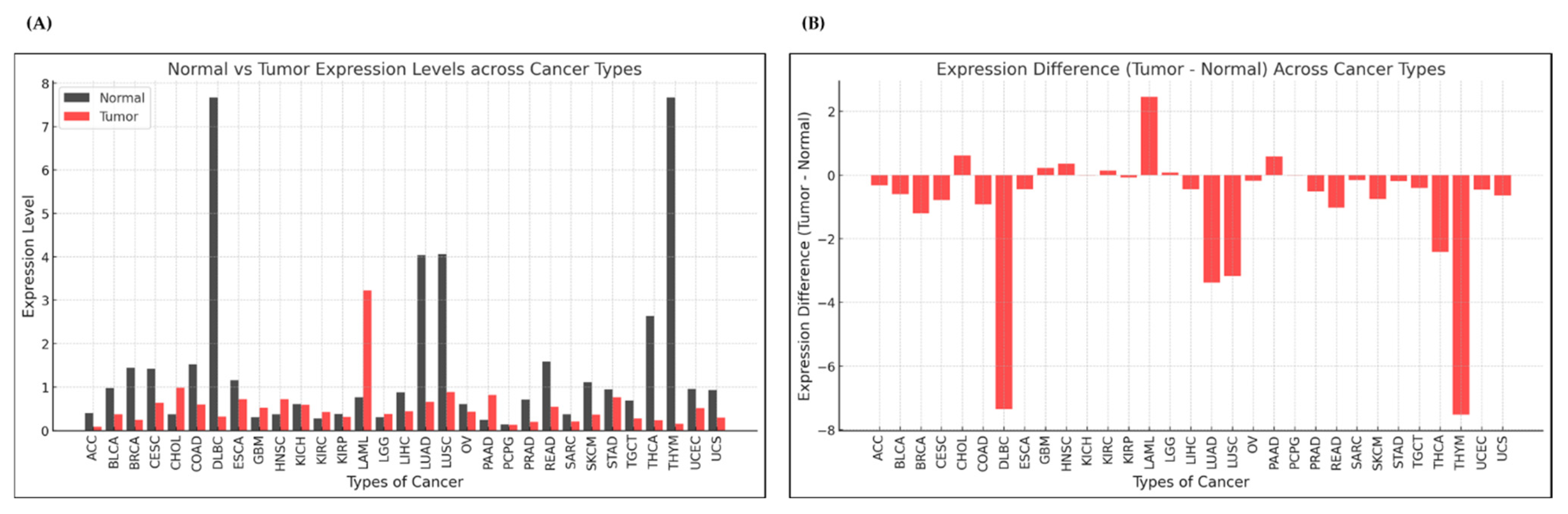
3.1. Expression Analysis of ADGRG3 Gene in Various Cancers

3.1.1. Pan-Cancer Analysis

Previous studies have highlighted the role of aGPCRs, including ADGRG3 (GPR97), in various cancers, particularly in hematological malignancies such as AML. The high expression of certain aGPCRs, including ADGRG3, has been linked to poor prognosis in AML, particularly through pathways such as interleukin-8 (IL8) signaling [22]. However, compared to other GPCR family members, ADGRG3 remains less studied, especially across a broader range of cancers. To expand on this, this study analyzed ADGRG3 expression using TCGA data across various cancer types to understand its potential involvement in tumorigenesis and immune modulation.
This TCGA analysis revealed the significant differential expression of ADGRG3 between tumor and normal tissues across multiple cancers (Figure 1A,B). Notably, ADGRG3 was upregulated in certain cancers, with the highest increase in expression in acute myeloid leukemia (LAML, +2.46); elevated levels were also seen in cholangiocarcinoma (CHOL, +0.61) and pancreatic adenocarcinoma (PAAD, +0.58). Conversely, ADGRG3 expression was markedly downregulated in other cancers, including thymoma (THYM, −7.52) and diffuse large B-cell lymphoma (DLBC, −7.35). These results underscore ADGRG3’s complex, context-dependent role in cancer biology. The high expression in leukemia and other immune-involved cancers suggests its potential as a therapeutic target. In contrast, its downregulation in certain cancers may indicate a different or even protective role in tumor progression.
To further examine ADGRG3 alterations across various cancers, analyzed mutation data using the cBioPortal platform. The oncoprint visualization (Figure S1A,B) illustrates the distribution of ADGRG3 mutations across different cancer types, with the most frequent alterations observed as missense mutations, amplifications, and deep deletions. This pattern suggests that ADGRG3 undergoes diverse genetic changes, which may impact its function across different malignancies.
In addition, amino acid mutations in ADGRG3 were mapped to specific protein regions (Figure S2). The analysis identified important mutation sites within functional domains, such as the GPS (GPCR proteolysis site) and 7tm_2 (7-transmembrane domain) regions. The fact that these mutations were predicted to be damaging by both the GPS and 7tm_2 algorithms suggests that these sites may have critical structural and functional roles in ADGRG3’s activity, potentially influencing its impact on cancer progression. The top four cancers with differentially upregulated expression of ADGRG3, namely, LAML, CHOL, HNSC, and PAAD, were further analyzed based on their clinical attributes, as described below.

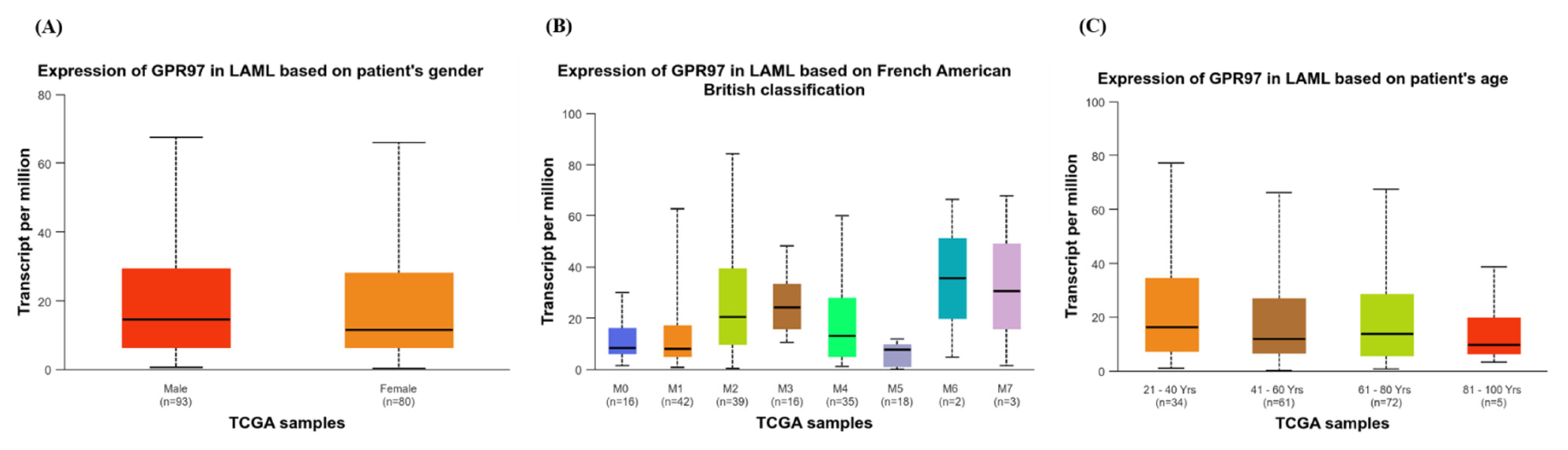
3.1.2. Expression of GPR97 in LAML Based on Clinical Attributes

AML is a complex cancer originating in the bone marrow, characterized by widespread distribution in the body rather than solid tumor formation. Unlike other cancers, AML prognosis depends on factors such as subtype, age, and genetic abnormalities, rather than staging. The French–American–British (FAB) classification divides AML into subtypes (M0–M7) based on cell type and maturity, each with distinct clinical implications. For instance, M3 (acute promyelocytic leukemia) requires specific treatment strategies. Prognosis is further influenced by genetic factors such as chromosomal translocations, including those between chromosomes 8 and 21 (common in M2) or 15 and 17 (seen in M3), along with other factors such as white blood cell count and residual disease after treatment.
In this study, ADGRG3 (GPR97) expression in LAML was analyzed across FAB subtypes to explore its potential variability. As shown in Figure 2A, higher GPR97 expression was observed in subtypes M2 (n = 39), M6 (n = 2), and M7 (n = 3), with moderate expression in M4 (n = 35). In contrast, subtypes M0 (n = 16), M1 (n = 42), and M5 (n = 18) showed lower expression. Gender-based analysis (Figure 2B) revealed similar expression levels in males (n = 93) and females (n = 80), while age-based analysis (Figure 2C) showed the highest expression in the 21–40-year-old group (n = 34), followed by the 61–80-year-old group (n = 72). These patterns suggest a potential subtype-specific role for ADGRG3, aligning with prior studies that link elevated ADGRG3 expression to poor prognosis and inflammatory pathway involvement in hematological malignancies [22].
These findings emphasize ADGRG3’s relevance in specific AML subtypes, particularly M2, M6, M7, and M4, and support its potential as a therapeutic target. Future research in other cancers with high ADGRG3 expression will help elucidate its broader role across tumor types.

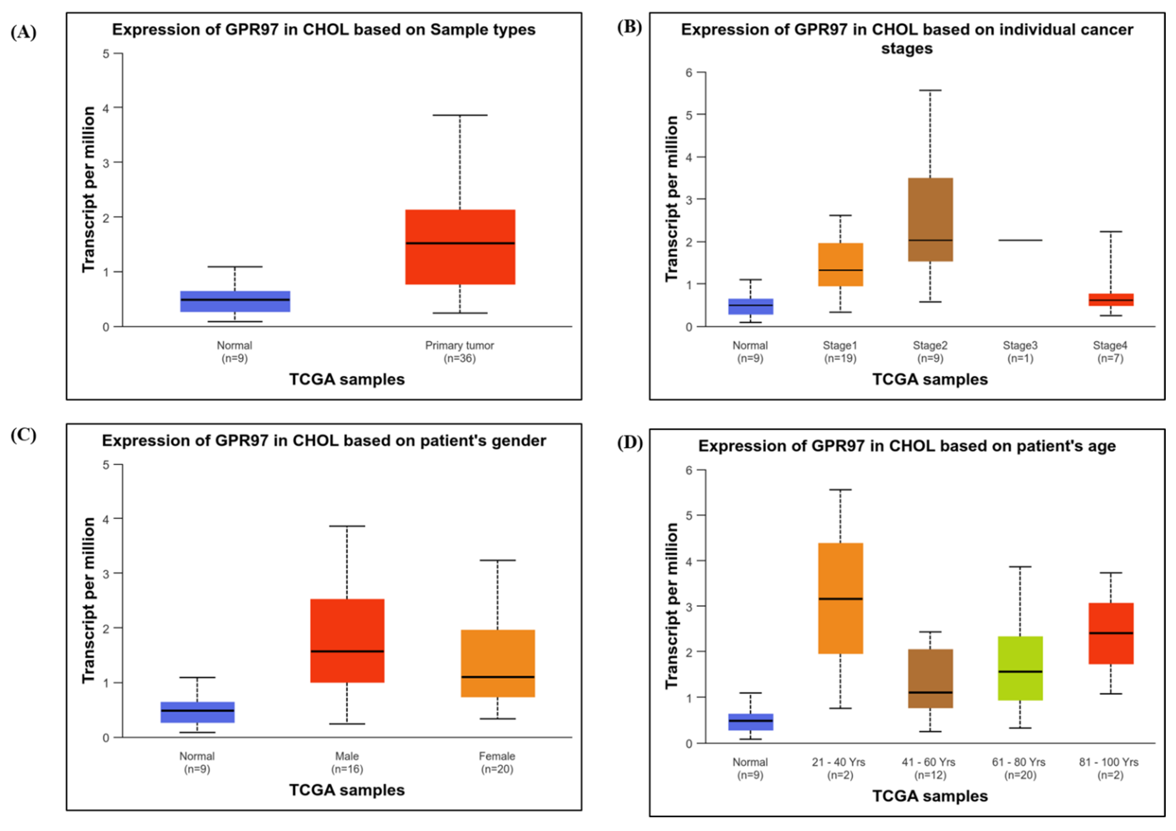
3.1.3. Expression of GPR97 in CHOL Based on Clinical Attributes

In cholangiocarcinoma (CHOL), ADGRG3 (GPR97) expression was evaluated across various clinical and demographic factors. Based on the sample types (Figure 3A), GPR97 expression was found to be significantly higher in primary tumor samples (n = 36) compared to normal samples (n = 9), indicating an upregulation of ADGRG3 in CHOL tumor tissues. When examined across individual cancer stages (Figure 3B), GPR97 expression peaked in stage 2 (n = 9), suggesting increased expression in early-to-mid stages of CHOL, with a decline observed in later stages.
Gender-based analysis (Figure 3C) revealed that both male (n = 16) and female (n = 20) patients had elevated GPR97 expression compared to patients with normal samples, with a slightly higher median expression in males. Age-based analysis (Figure 3D) showed the highest expression levels in younger patients (21–40 years, n = 2), with decreasing expression in older age groups, as follows: 41–60 years (n = 12) and 61–80 years (n = 20). The 81–100-year-old group (n = 2) showed slightly elevated GPR97 expression compared to patients with normal samples but lower expression than the younger age groups.
These findings indicate that GPR97 is upregulated in CHOL tumors, with variability across cancer stages, genders, and age groups. Elevated expression in early-to-mid stages and younger patients may point to the potential role of ADGRG3 in CHOL tumorigenesis and progression, warranting further investigation.

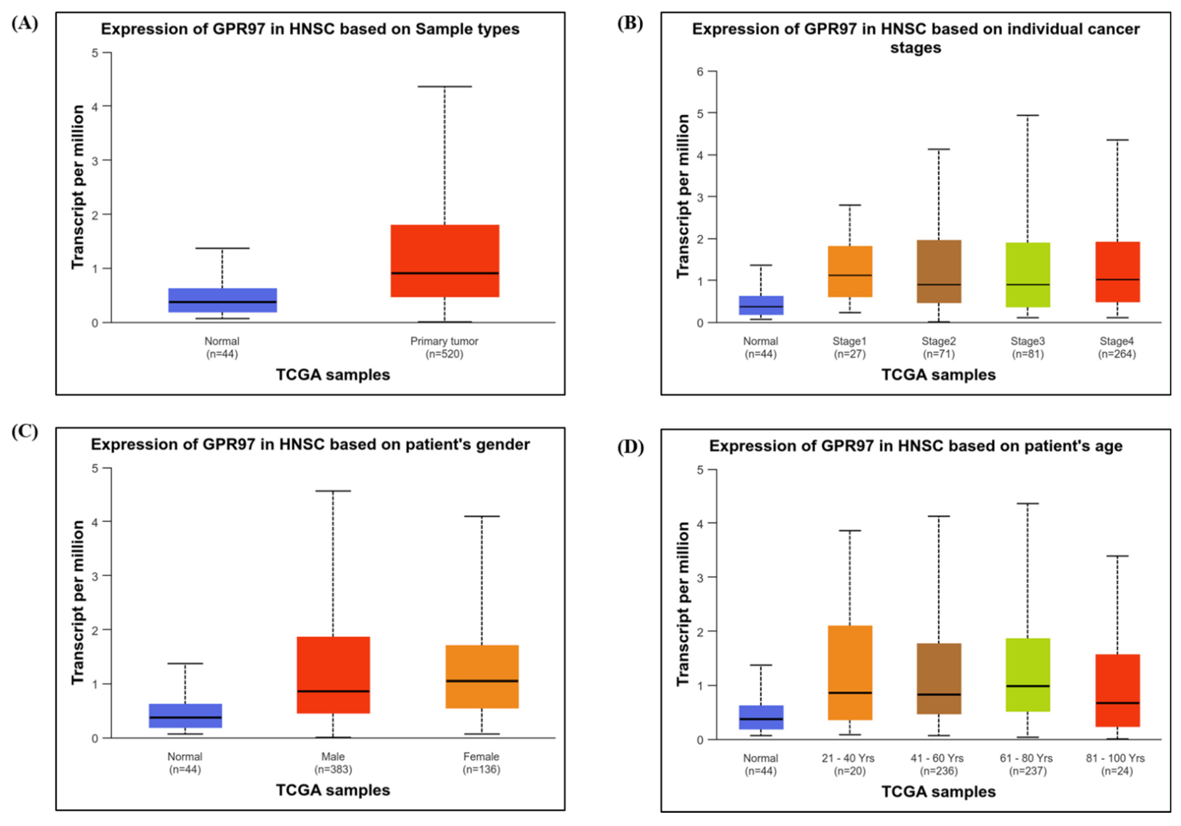
3.1.4. Expression of GPR97 in HNSC Based on Clinical Attributes

In head and neck squamous cell carcinoma (HNSC), ADGRG3 (GPR97) expression was assessed across different clinical and demographic factors. Based on sample types (Figure 4A), GPR97 expression was significantly higher in primary tumor samples (n = 520) compared to normal samples (n = 44), indicating notable upregulation in tumor tissues. An analysis across individual cancer stages (Figure 4B) showed consistent GPR97 expression in Stages 1 (n = 27), 2 (n = 71), 3 (n = 81), and 4 (n = 264), with all stages demonstrating elevated levels compared with normal samples.
Gender-based analysis (Figure 4C) revealed similar GPR97 expression levels in both male (n = 383) and female (n = 136) patients, suggesting no significant gender-based differences in expression. Age-based analysis (Figure 4D) showed elevated GPR97 expression across all age groups (21–100 years) compared to patients with normal samples, with no substantial age-related variation in expression levels.
These findings indicate that GPR97 is upregulated in HNSC tumor tissues across all stages, genders, and age groups, suggesting a broadly applicable role for ADGRG3 in HNSC tumorigenesis. Further research could explore the specific pathways by which ADGRG3 contributes to HNSC progression and its potential as a therapeutic target.

3.1.5. Expression of GPR97 in PAAD Based on Clinical Attributes

In pancreatic adenocarcinoma (PAAD), ADGRG3 (GPR97) expression was evaluated across different clinical and demographic factors. Based on sample types (Figure 5A), GPR97 expression was significantly lower in primary tumor samples (n = 178) compared to normal samples (n = 4), indicating a downregulation of ADGRG3 in PAAD tumor tissues. Across cancer stages (Figure 5B), Stage 3 (n = 4) showed the highest expression, followed by normal tissues. Stage 2 (n = 146) displayed moderate expression, while Stages 1 (n = 6) and 4 (n = 4) exhibited the lowest levels.
Gender-based analysis (Figure 5C) revealed similarly low GPR97 expression in both male (n = 97) and female (n = 80) patients compared to patients with normal samples, indicating no significant gender-based differences. Age-based analysis (Figure 5D) showed the lowest expression in the 21–40-year-old group (n = 9), with moderate expression in older age groups (41–100 years) but consistently lower expression in patients with normal samples.
These findings indicate that GPR97 is generally downregulated in PAAD tumor tissues, with some variation across cancer stages and age groups, but minimal difference across genders. This suggests that ADGRG3 may play a different role in PAAD compared with cancers where it is upregulated, warranting further investigation into its potential function in pancreatic cancer.

3.2. Survival and TME Analysis

Figure S3 presents the results of the Kaplan–Meier survival analyses examining the impact of GPR97 expression levels on patient’s survival in the following four cancer types: (A) LAML, (B) CHOL, (C) HNSC, and (D) PAAD. In LAML, the high GPR97 expression showed a trend toward lower survival, though this was not statistically significant (p = 0.19). In CHOL, there was no significant survival difference between the high and low/medium expression groups (p = 0.97). In HNSC, the higher GPR97 expression was associated with a near-significant reduction in survival probability (p = 0.067), suggesting a possible but inconclusive impact. Lastly, in PAAD, GPR97 expression did not significantly affect survival outcomes (p = 0.47). Overall, these findings suggest limited prognostic value for GPR97 expression across these cancers, with its potential role in HNSC requiring further investigation.
Figure S4 illustrates the results of the TIMER analysis for ADGRG3 (GPR97) in (A) CHOL, (B) PAAD, and (C) HNSC, which examined immune infiltration correlations and survival impacts. In CHOL, GPR97 showed weak, non-significant correlations with immune cell infiltration, with the highest correlation with B cells (partial.cor = 0.053, p = 0.761), and a mild, non-significant protective effect on survival (HR = 0.829). In PAAD, GPR97 significantly correlated with macrophages (partial.cor = 0.357, p = 1.67 × 10⁻⁶) and neutrophils (partial.cor = 0.461, p = 2.33 × 10⁻¹⁰), with high neutrophil infiltration linked to poor survival (HR = 5.78 × 10⁸, p = 0.002), while its correlation with CD4+ T cells showed a protective effect (HR = 0.000, p = 0.015). In HNSC, GPR97 showed significant correlations with CD8+ T cells (partial.cor = 0.232, p = 3.37 × 10⁷) and macrophages (partial.cor = 0.235, p = 1.67 × 10⁷), indicating an active immune environment. The Cox model suggests a non-significant increase in risk with higher GPR97 expression (HR = 1.183, p = 0.106), but there is an overall significance in the likelihood ratio (p = 0.010) and log-rank tests (p = 0.0226). These results suggest ADGRG3’s varied influence on immune infiltration and survival across these cancers.

3.3. Binding Affinity of Ginsenosides with ADGRG3
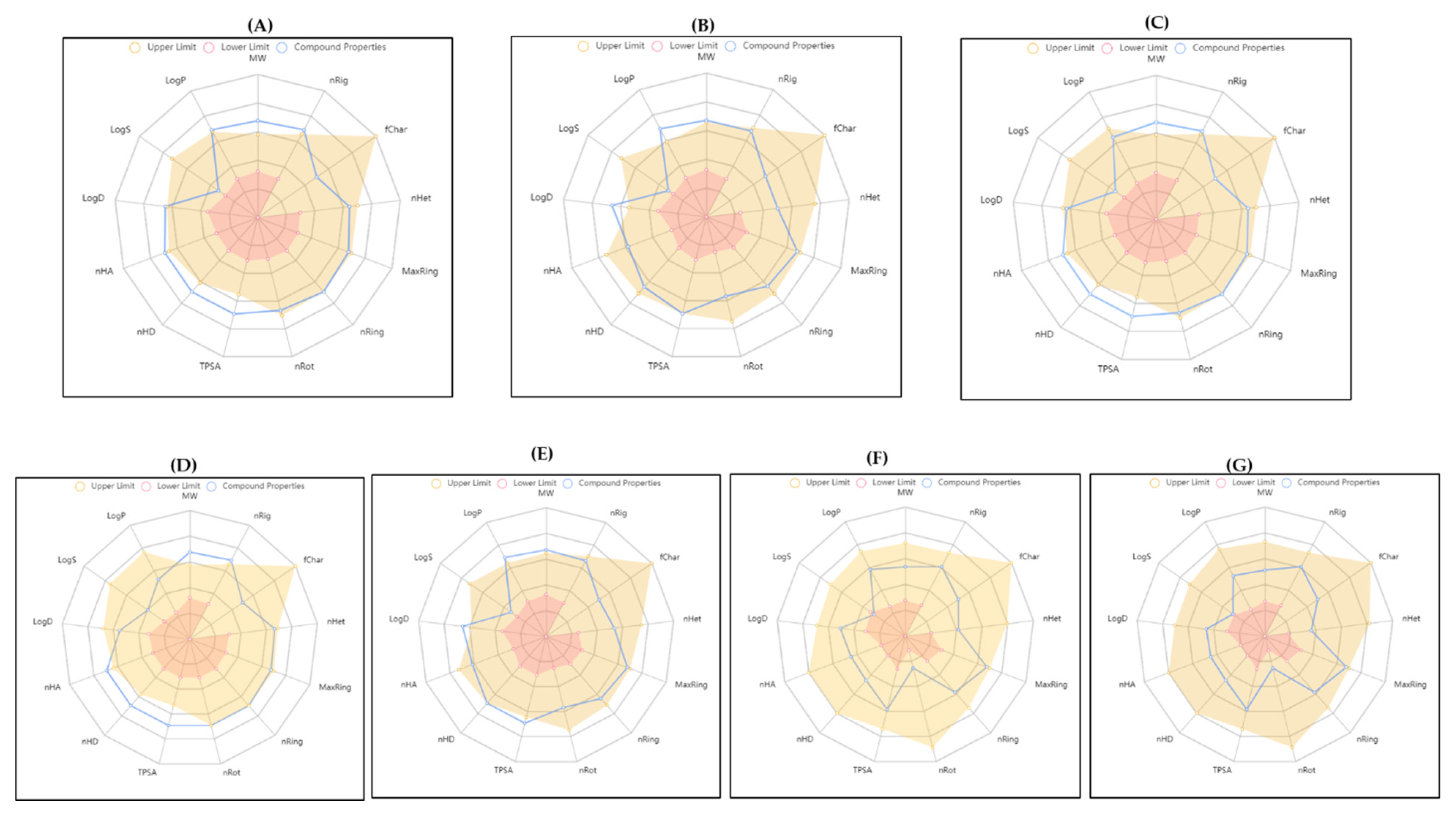
In the virtual screening of ginsenosides against ADGRG3, identified five top candidates ginsenosides Rg3, Rk3, F5, Rg7, and F1 based on their strong binding affinities and stable interactions within the ADGRG3 binding pocket, as shown in Table 1 and illustrated in Figure 6 and Supplementary Figure S5. Ginsenoside Rg3 had the highest binding affinity (−10.7 kcal/mol), with five hydrogen bonds involving key residues such as TYR 406 and ARG 409, along with additional hydrophobic contacts. Rk3 and F5 followed closely with binding energies of −10.6 and −10.5 kcal/mol, respectively. Rk3 formed one hydrogen bond with TYR 406 and several hydrophobic contacts, whereas F5 formed four hydrogen bonds and additional stabilizing interactions, which was comparable to HCY’s binding pattern.
Rg7 and F1 also showed strong binding, with binding energies of −10.4 and −10.3 kcal/mol, respectively. Both engaged key residues through hydrogen bonding, with Rg7 particularly aligning with ALA 493 and ASN 510, which was similar to the control ligands. Compared to the top two controls dexamethasone and HCY which showed binding energies of −10.3 and −10.4 kcal/mol, respectively, the selected ginsenosides displayed comparable or stronger binding and stable interactions, supporting their potential as ADGRG3-targeting agents. These top candidates demonstrate promising binding stability within the ADGRG3 site, warranting further MDS to assess their therapeutic potential in cancer.

3.4. ADMET

Further, the ADMET analysis of the top five ginsenosides (Rg3, Rk3, F5, Rg7, and F1) and the control drugs (dexamethasone and HCY) indicated favorable drug-likeness and pharmacokinetics for the ginsenosides, as summarized in Figure 7 and Tables S3–S8. All ginsenosides show favorable ADMET predictions, though these are slightly lower than those of the control. This warrants further experimental validation via in vitro and in vivo pharmacokinetic studies, such as Caco-2 permeability assays to validate absorption predictions, microsomal stability assays to assess metabolic degradation, and cytotoxicity assays to confirm toxicity predictions. In addition, further studies with MDS are needed to confirm the findings and validate the stability of ligand–ADGRG3 interactions with a more comprehensive analysis of the binding dynamics beyond the initial docking results.

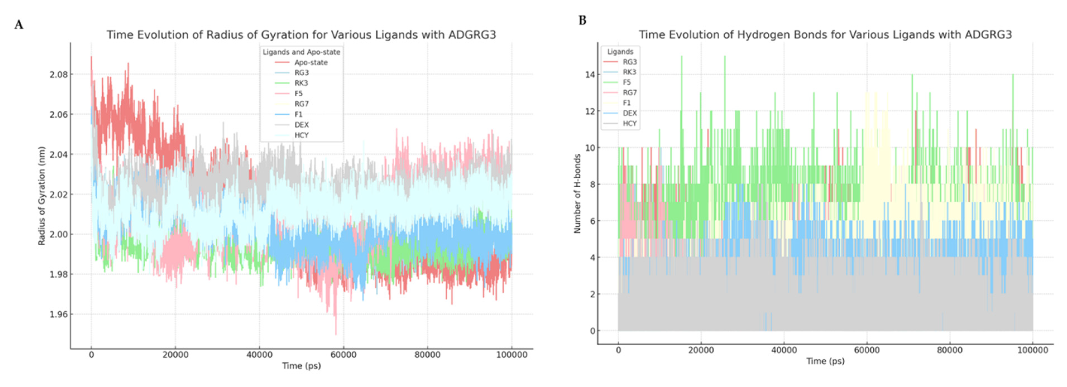
3.5. Molecular Interaction Dynamics of Ginsenosides with ADGRG3

In this study, MDSs were conducted to examine the stability, flexibility, compactness, and interaction dynamics of ADGRG3 in complex with several ginsenosides (Rg3, Rk3, F5, Rg7, and F1) and two valid control ligands—dexamethasone and hydrocortisone (HCY). Key structural parameters, including the RMSD, root mean square fluctuation (RMSF), radius of gyration (Rg), and hydrogen bonding patterns, were analyzed.
The RMSD analysis (Figure 8A) provided insights into the overall stability of the ADGRG3–ligand complexes throughout the simulation period. The RMSD profiles of ADGRG3 complexed with the ginsenosides and controls revealed varying degrees of stability. The Rg3 and Rk3 complexes showed relatively stable RMSD values over the simulation time, indicating strong and consistent binding to ADGRG3. In contrast, F5 and F1 displayed minor fluctuations, suggesting moderate flexibility in binding. The control ligands, dexamethasone and HCY, showed comparable RMSD patterns, with HCY presenting slightly higher RMSD values, indicating marginally lower stability compared to dexamethasone. The RMSF analysis (Figure 8B) highlighted the flexibility of individual residues in ADGRG3 when complexed with each ligand. Both Rg3 and Rk3 complexes showed lower RMSF values across most residues, indicating limited fluctuations and a generally stable conformation. In contrast, F5 and Rg7 complexes exhibited higher fluctuations in specific regions, suggesting areas of increased flexibility in the binding site. The control ligand HCY showed moderate fluctuations, whereas dexamethasone demonstrated relatively stable binding with limited residue flexibility, indicating that both control ligands engage the receptor with distinct but stable binding patterns.
The radius of gyration (Rg) analysis (Figure 9A) reflected the compactness of the ADGRG3–ligand complexes over the simulation period. Ginsenosides Rg3 and F5 showed lower Rg values, suggesting that ADGRG3 maintains a compact structure in these complexes. The higher Rg values observed with Rg7 and F1 indicated a slightly expanded structure, possibly due to the conformational adaptations required for optimal binding. Both dexamethasone and HCY displayed consistent Rg values, indicating stable complex compactness, with HCY showing marginally higher Rg values than dexamethasone. The hydrogen bonding patterns (Figure 9B) illustrated the binding strength and stability of the ADGRG3–ligand interactions. Ginsenoside Rg3 and Rk3 formed a higher average number of hydrogen bonds with ADGRG3, supporting their stable binding profiles observed in the RMSD and Rg analyses. In comparison, the RG7 and F5 complexes exhibited fewer hydrogen bonds, suggesting weaker interactions. Dexamethasone and HCY displayed moderate hydrogen bonding stability, with HCY forming slightly fewer bonds than dexamethasone, aligning with its somewhat higher RMSD and Rg values. Overall, the MDS analysis revealed that Rg3 and Rk3 exhibit the most stable binding profiles with ADGRG3, as evidenced by their low RMSD values, stable RMSF values, compact Rg, and strong hydrogen bonding interactions. These findings suggest that Rg3 and Rk3 may be promising candidates for targeting ADGRG3 after the validation of this outcome in preclinical cell-based assays. Further, binding free energy calculations for the seven complexes were conducted using the MM-PBSA method, leveraging the GROMACS software and MDS (MD) trajectories. The results obtained indicated that the binding energies were within a favorable range, suggesting robust interactions. As detailed in Table 2, the MM-PBSA analysis revealed that the ADGRG3–ginsenoside Rg3 complex exhibited the most significant binding affinity, with the lowest binding free energy recorded at −40.1 kJ/mol. Other complexes, including ADGRG3–Ginsenoside Rk3, ADGRG3–Ginsenoside F5, ADGRG3–Ginsenoside Rg7, ADGRG3–Ginsenoside F1, ADGRG3–dexamethasone, and ADGRG3–HCY, displayed binding free energies of −36.5, −35.6, −36.4, −39, −39.9, and −39.5 kJ/mol, respectively. These findings are consistent with the MD predictions, further confirming that these molecules effectively engage with the active site of ADGRG3.

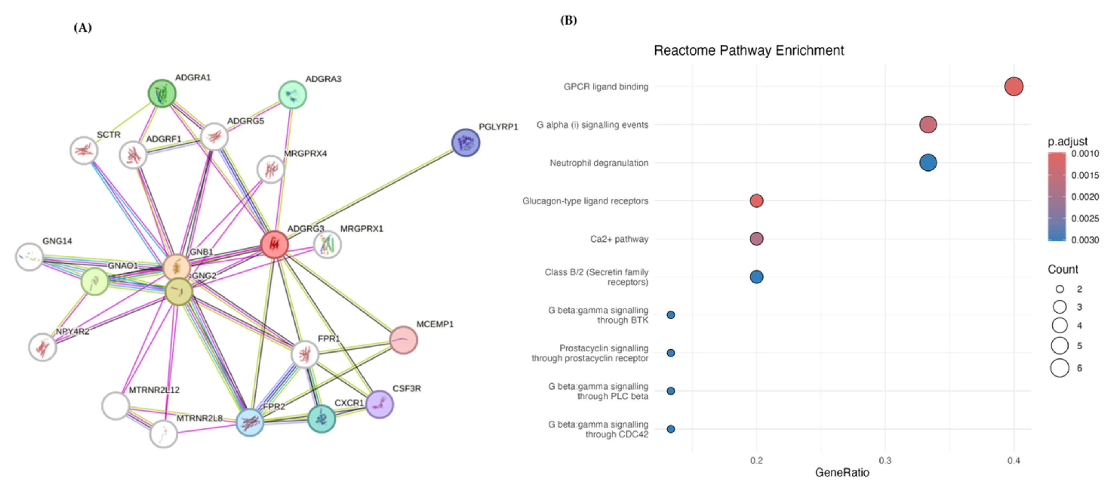
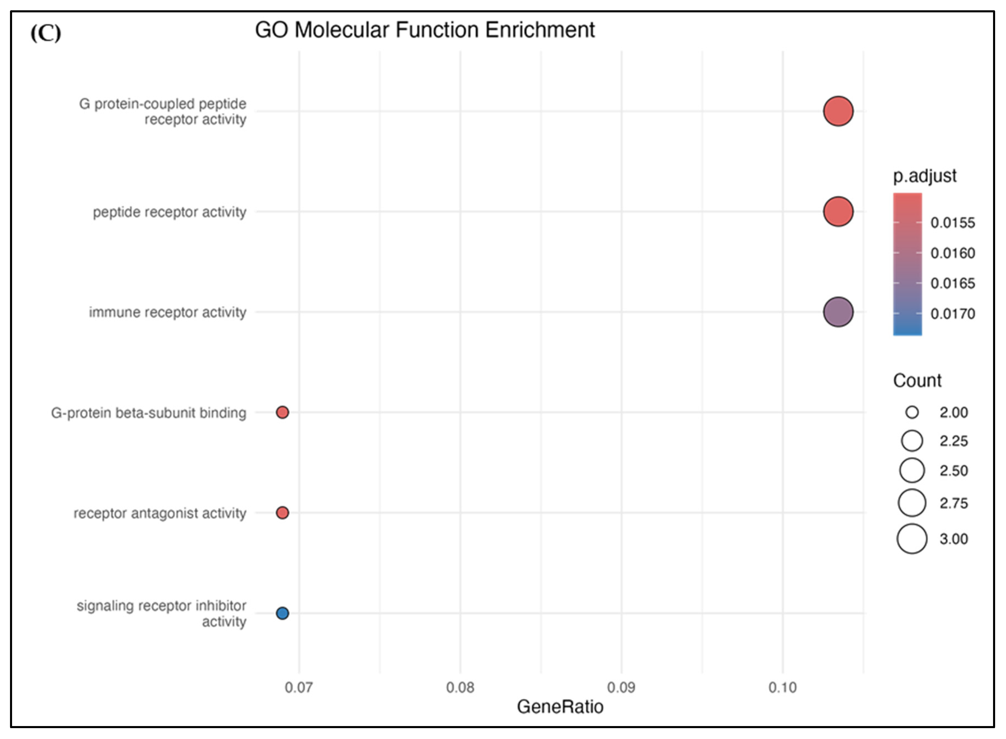
3.6. PPI and Pathway Analysis

To investigate ADGRG3’s role in cancer and immune pathways, performed PPI analysis using STRING (Figure 10A) and supplemented it with data from the Human Protein Atlas, gathering 29 interacting genes (Supplementary File S1: Information on PPI genes). The gene enrichment analysis via GO, KEGG, and Reactome identified key pathways that may link ADGRG3 to cancer and immune functions.
The Reactome analysis (Figure 10B) highlighted pathways such as GPCR ligand binding and G-alpha (i) signaling, involving proteins such as GNB1, GNG2, CXCR1, and FPR2, which are central to cell signaling in cancer and immune regulation. Additionally, neutrophil degranulation and Ca2+ signaling pathways suggest ADGRG3’s involvement in innate immune responses and inflammation, which are processes critical to both cancer progression and immune activity. These findings predict that ADGRG3 could possibly influence cancer and immune pathways through GPCR and cytokine signaling.
Figure 11 presents the top 10 enriched Gene Ontology (GO) terms for ADGRG3 interactors across the following three categories: biological process (BP), cellular component (CC), and molecular function [67]. Detailed GO terms are available in Supplementary File S1. For BP, the most enriched terms included adenylate cyclase-modulating GPCR signaling and dopamine receptor signaling, involving genes such as ADGRG3, GNB1, and GNG2. These pathways suggest ADGRG3’s involvement in GPCR-related signaling, which is important in cancer progression and immune modulation. In the CC category, enriched terms such as the heterotrimeric G-protein complex and specific granules highlight the localization of ADGRG3 interactors in structures relevant to signal transduction and immune responses, particularly those involving GNB1, GNG2, and GNAO1.
For MF, terms such as G-protein beta-subunit binding and G-protein-coupled peptide receptor activity were prominent, underscoring the functional role of ADGRG3 and its interactors in GPCR binding and receptor activity regulation. These findings support the role of ADGRG3 in GPCR signaling, immune regulation, and potentially in cancer-related pathways through its localization and interactions within critical cellular structures and signaling mechanisms.
The KEGG enrichment analysis of ADGRG3 and its interacting genes identified pathways primarily related to neurotransmitter signaling, including GABAergic synapse, morphine addiction, and circadian entrainment (Figure S6 and Supplementary File S1). Key genes such as GNB1, GNG2, and GNAO1 were involved across these pathways, suggesting ADGRG3’s potential role in modulating cellular signaling relevant to cancer and immune responses.

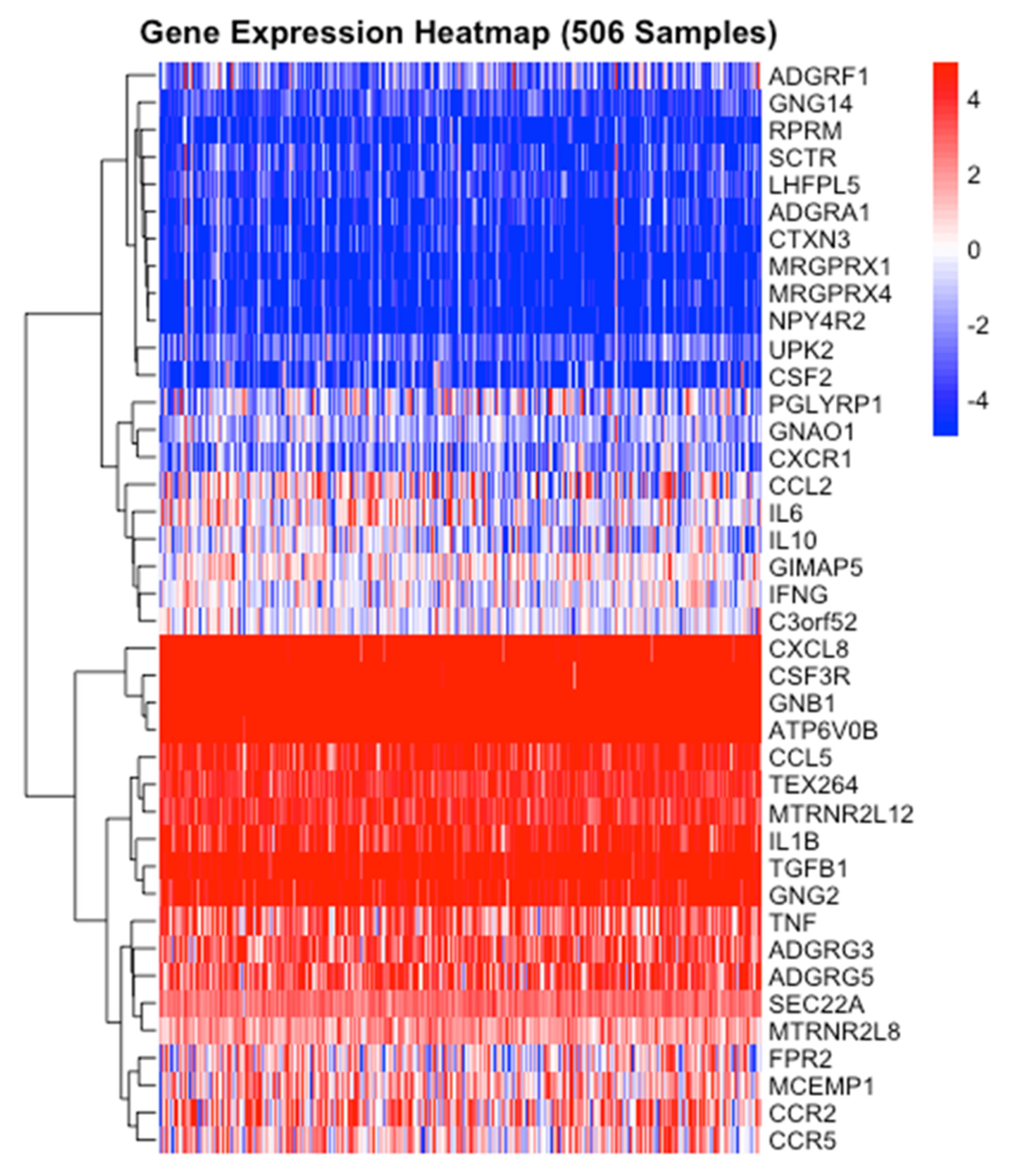
3.7. ADGRG3 Gene Expression and Other Interactions in Cancer and Inflammation

The gene expression analysis revealed distinct patterns across the 506 AML samples. ADGRG3 exhibited heterogeneous expression, with high levels in approximately 70~80% of the samples, moderate expression in some, and low levels in the remainder, reflecting the diverse clinical and biological profiles of AML patients. Genes such as GNB1, GNG2, CSF3R, and ATP6V0B were consistently highly expressed, highlighting their roles in GPCR signaling, immune modulation, and neutrophil activity, all of which are critical in AML progression. Cytokines and inflammation-related genes, including IL8, CXCL8, and CSF3R, showed elevated expression, reinforcing their involvement in inflammatory pathways and the cancer microenvironment. Other genes, such as IL10 and IFNG, demonstrated variable expression, potentially indicating differences in tumor–immune interactions. Hierarchical clustering revealed distinct co-expression patterns among GPCR-related genes (ADGRG3, GNB1, and GNG2) and inflammatory mediators (IL8, CSF3R, and CCL2), underscoring their functional interplay in AML. The results, visualized in Figure 12, highlight the complex and context-dependent roles of ADGRG3 and its interactors, particularly in the interplay between cancer and inflammation.

4. Discussion

This study provided a comprehensive computational analysis of ADGRG3 as a target in cancer and immune pathways, highlighting its differential expression across various cancers and its role in immune cell interactions. This study’s findings indicate elevated ADGRG3 expression in cancers such as AML, CHOL, and HNSC, suggesting an active role of ADGRG3 in tumor progression and immune-related pathways. The specificity of the high ADGRG3 expression in AML subtypes M2, M6, M7, and M4 points to its potential role in hematological malignancies, where ADGRG3 may influence inflammatory and immune pathways involved in cancer progression. A previous study highlighted ADGRG3 as a key adhesion GPCR in AML, associating its high expression-alongside that of ADGRE2 and other GPCRs—with poor clinical outcomes, reduced overall survival, and inflammation [17]. Strengthening this link, a recent in vitro and in vivo study reported ADGRE2’s role in AML by promoting leukemic stem cell survival and tumor progression via the MEK/AP1/DUSP1 axis. ADGRE2 knockdown impaired proliferation, induced apoptosis, and reduced LSC frequency, while xenograft models showed delayed leukemia progression and prolonged survival. Given the structural and functional similarities among adhesion GPCRs, this evidence highlights ADGRG3’s potential role in AML and supports the need for further experimental validation [23].
To increase the overall precision and specificity of the experimental results, in silico investigations were employed. The interaction between the targeted ginsenosides and the ADGRG3 biomolecule is better understood through the use of computational methods, such as physiological and ADMET characteristics and MD. Additionally, these techniques facilitate the prediction of possible toxicological and pharmacokinetic characteristics. In contrast to conventional methods, in silico projections for drug similarity, chemical descriptors, and ADMET characteristics help identify new lead compounds more quickly [70]. The MD technique has been employed to predict powerful therapeutic compounds that inhibit the proliferation of cancer stem cells [71]. MD results can help advance potential drug design strategies [72]. Computer-based drug design tools can help reduce costs and accelerate drug discovery processes, with molecular dynamics serving as a final filter following docking and optimizing hits (or leads) [73].
In the current study, the virtual screening identified five top ginsenosides—Rg3, Rk3, F5, Rg7, and F1—as promising ADGRG3 ligands, with Rg3 showing the strongest binding affinity. Rg3 has been emphasized for its anticancer effects, including inhibition of tumor growth, angiogenesis, and metastasis, while Rk3 has shown significant activity against non-small cell lung cancer through apoptosis induction and cell cycle arrest. Additionally, F5, Rg7, and F1 are known to regulate cancer cell metabolism, targeting pathways essential for tumor survival [74,75,76]. The strong binding of Rg3 and Rk3 to ADGRG3 suggests a probable dual role in modulating immune- and cancer-related pathways, which warrants further experimental validation. These ginsenosides bind with ADGRG3’s key binding pocket residues through hydrogen bonds and hydrophobic contacts. Studies have shown the efficient absorption and metabolism of ginsenosides such as Rg3 and Rk3, with Rg3 undergoing significant biotransformation and Rk3 targeting the PI3K/AKT pathway to regulate autophagy and apoptosis. Ginsenoside F1, while showing low brain permeability, can achieve enhanced delivery via intranasal administration with absorption enhancers.
The gene expression analysis of reported TCGA revealed that ADGRG3 is upregulated in several cancers, with the highest increase observed in AML. Supporting these findings, previous studies demonstrated that red ginseng extract, rich in ginsenosides Rg3, F5, Rg7, F1, and Rk3, exerts significant anticancer activity against leukemia cells by inducing apoptosis, suppressing proliferation, and inhibiting extramedullary infiltration [77,78,79]. This aligns with the docking results of the current study, reinforcing the therapeutic relevance of these ginsenosides in AML and other cancers, particularly in the context of inflammation and tumor progression. Furthermore, given the structural similarities between dexamethasone, a well-established anti-inflammatory and anticancer agent, and ginsenosides, these findings collectively highlight the need for further experimental validation to explore the therapeutic potential of predicted ginsenosides in AML and other malignancies.
The minimal CYP interaction and low toxicity, highlight that ginsenosides could be low-toxicity alternatives for cancers with elevated ADGRG3 expression [80,81,82,83,84]. The MDS and free binding energy calculations (MMPBSA) further supported these findings, demonstrating that the ginsenoside–ADGRG3 complexes are stable, with maintained flexibility and the formation of stable hydrogen bonds throughout the simulation. This stability suggests that these ginsenosides could possibly modulate ADGRG3. Notably, natural compounds such as curcumin have also been shown to modulate GPR97, further emphasizing the potential of natural products in targeting this receptor [85]. Future studies should incorporate experimental validation (e.g., SPR, ITC, or functional assays) to confirm the pharmacological relevance of ginsenosides in targeting ADGRG3.
The pathway and protein–protein interaction analyses further connected ADGRG3 to critical GPCR and immune pathways, such as neutrophil degranulation and Ca2+ signaling, both essential in cancer progression and immune modulation. ADGRG3’s associations with immune cell infiltration in cancers such as HNSC, where it correlated with CD8+ T cells and macrophages, suggest it plays a role in the tumor microenvironment and immune response. Ginsenosides, through their interactions with ADGRG3, may influence these pathways, potentially enhancing immune responses or altering the tumor microenvironment to improve outcomes. Notably, ginsenoside Rg3 has been shown to suppress neutrophil migration, as observed in COPD models, highlighting its potential to modulate neutrophil degranulation pathways. In cancer, Rg3 has demonstrated anti-leukemic effects by inhibiting HIF-1α and VEGF expression via the PI3K/Akt and ERK1/2 pathways and suppressing vasculogenic mimicry through the regulation of VE-cadherin/EphA2/MMP9/MMP2 expression. Similarly, ginsenoside compound K (CK) exhibits anticancer properties in acute myeloid leukemia and liver cancer, inducing ferroptosis via the FOXO pathway and exerting in vivo anti-leukemic effects. These findings provide understandings into the ability of ginsenosides to modulate key signaling pathways involved in tumor progression and immune regulation, further supporting their targeting potential for ADGRG3 [86,87,88,89,90,91,92].
The gene expression analysis of 506 AML samples revealed heterogeneous ADGRG3 expression, with high levels in 70–80% of samples, reflecting the diverse clinical and biological profiles of AML patients. Genes such as GNB1, GNG2, CSF3R, and ATP6V0B showed consistently high expression, highlighting their roles in GPCR signaling, immune modulation, and neutrophil activity. Previous studies have linked GNG2 to GPCR signaling and inflammation, as seen in Panax ginseng research, and CSF3R to neutrophil activity, with ginsenoside Rg3 reducing cancer drug toxicity. CXCL8, another inflammatory mediator, is highly expressed in AML, with studies showing that modified ginseng extract effectively blocks CXCL8-mediated Akt/NF-κB signaling in cancer models. Hierarchical clustering revealed co-expression of GPCR-related genes (ADGRG3, GNB1, and GNG2) and inflammatory mediators (IL8, CSF3R, and CCL2), underscoring their interplay in AML. These findings, combined with the findings of previous studies on ginsenosides, highlight their therapeutic relevance in modulating AML-specific pathways [93,94,95].
In summary, this study predicted that ginsenosides could be natural modulators of ADGRG3. Through integrative in silico analyses, including gene expression, MD, MDS, and pathway investigations, this study established a computational predicted framework for further ADGRG3-targeted assays. The findings encourage further exploration of ginsenosides for preclinical applications, offering a basis for future cancer treatments, especially in tumors with high ADGRG3 involvement. This early-stage drug discovery would help prioritize promising candidates by predicting their binding affinities, pharmacokinetic properties, and potential toxicity, thus reducing the number of compounds requiring experimental testing. This study serves as a foundation for further potential experimental follow-ups, such as biochemical binding assays and cell-based studies, to validate these predictions.

5. Conclusions

This study predicted that ADGRG3/GPR97 could be a target in cancer and immune modulation, which is supported by the results of integrative computational analyses. Virtual screening identified five ginsenosides—Rg3, Rk3, F5, Rg7, and F1—as probable ADGRG3-targeting ligands, with Rg3 showing the highest binding affinity and stability in MDS and free binding energy calculations. The favorable in silico ADMET profiles further highlighted these ginsenosides as safe, natural alternatives to synthetic drugs. Pathway and protein–protein interaction analyses linked ADGRG3 to critical GPCR and immune pathways, such as neutrophil degranulation and Ca2+ signaling, emphasizing its role in tumor microenvironment modulation. The gene expression analysis of 506 AML samples revealed high ADGRG3 expression in 70–80% of cases and co-expression with genes such as GNB1, GNG2, and CXCL8, further supporting its role in AML progression. These findings provide a robust basis for ADGRG3-targeted therapies, highlighting ginsenosides as possible modulators, particularly in elevated ADGRG3 expression. Further in vitro and in vivo preclinical and clinical studies are warranted to validate ginsenoside regulations in ADGRG3, as determined by these computational predictions.

Supplementary Materials

The following supporting information can be downloaded at: https://www.mdpi.com/article/10.3390/app15084332/s1, Figure S1: The genetic alterations of ADGRG3 and associations with mRNA expression. (A) Total genetic alterations of ADGRG3 genes based on cBioportal website. (B) Bubble map indicated the relationship between ADGRG3 mRNA expression and CNV. Red dots indicate that gene CNV levels were positively associated with mRNA expression, blue dots indicate the negative association; Figure S2: ADGRG3 mutation in pan-cancer based on cBioportal website. (A) Oncoprinter of ADGRG3 in different cancer types. (B) Amino acid mutation of ADGRG3. The important mutation sites that predicted as damaging in both GPS and 7tm_2 algorithms in the functional and structural importance of the protein sequence position; Figure S3: The effect of GPR97 expression level on (A) LAML, (B) CHOL, (C) HNSC, (D) PAAD patient survival; Figure S4: TME analysis for ADGRG3 gene in (A) CHOL, (B) PAAD and (C) HNSC cancers. The immune infiltration correlation provide insights into GPR97’s potential impact on immune microenvironments and patient survival; Figure S5: 2D interaction diagram of ADGRG3 with (A) Ginsenoside Rg3, (B) Ginsenoside Rk3, (C) Ginsenoside F5, (D) Ginsenoside Rg7, (E) Ginsenoside F1, (F) Dexamethasone, and (G) HCY; Figure S6: KEGG Enrichment Analysis of the top 20 genes in the PPI network of ADGRG3; Table S1: Grid box coordinates and size parameters used for molecular docking; Table S2: Active site prediction for ADGRG3 using DoGSiteSco; Table S3: Parameters evaluated for drug-likeness of ginsenosides and the control drugs; Table S4: Parameters evaluated for absorption of ginsenosides and the control drugs; Table S5: Parameters evaluated for distribution of ginsenosides and the control drugs; Table S6: Parameters evaluated for metabolism of ginsenosides and the control drugs; Table S7: Parameters evaluated for excretion of ginsenosides and the control drugs; Table S8: Parameters evaluated for toxicity of ginsenosides and the control drugs. File S1: List of ginsenosides used, docking scores, PPI genes, GO and KEGG analysis.

Funding

This Research was supported by Seokyeong University in 2023.

Institutional Review Board Statement

Not applicable.

Informed Consent Statement

Not applicable.

Data Availability Statement

All of the data generated or analyzed during this study are included in the article and Supplementary Materials.

Acknowledgments

This Research was supported by Seokyeong University in 2023. I thank Ramya and Vinothini for their valuable support and guidance.

Conflicts of Interest

The author declares no conflict of interest.

References

  1. Wisler, J.W.; Rockman, H.A.; Lefkowitz, R.J. Biased G Protein–Coupled Receptor Signaling. Circulation 2018, 137, 2315–2317. [Google Scholar] [CrossRef] [PubMed]
  2. Finan, C.; Gaulton, A.; Kruger, F.A.; Lumbers, R.T.; Shah, T.; Engmann, J.; Galver, L.; Kelley, R.; Karlsson, A.; Santos, R.; et al. The druggable genome and support for target identification and validation in drug development. Sci. Transl. Med. 2017, 9, eaag1166. [Google Scholar] [CrossRef] [PubMed]
  3. Jiang, J.; Yuan, J.; Hu, Z.; Zhang, Y.; Zhang, T.; Xu, M.; Long, M.; Fan, Y.; Tanyi, J.L.; Montone, K.T.; et al. Systematic illumination of druggable genes in cancer genomes. Cell Rep. 2022, 38, 110400. [Google Scholar] [CrossRef] [PubMed]
  4. Nussinov, R.; Zhang, M.; Liu, Y.; Jang, H. AlphaFold, allosteric, and orthosteric drug discovery: Ways forward. Drug Discov. Today 2023, 28, 103551. [Google Scholar] [CrossRef]
  5. Radoux, C.J.; Vianello, F.; McGreig, J.; Desai, N.; Bradley, A.R. The druggable genome: Twenty years later. Front. Bioinform. 2022, 2, 958378. [Google Scholar] [CrossRef]
  6. Surh, Y.J. Reverse pharmacology applicable for botanical drug development—Inspiration from the legacy of traditional wisdom. J. Tradit. Complement. Med. 2011, 1, 5–7. [Google Scholar] [CrossRef]
  7. Santhanam, B.; Sluter, M.; Babu, M.M. Exploring GPCR signaling pathway networks as cancer therapeutic targets. Cell Genom. 2024, 4, 100560. [Google Scholar] [CrossRef]
  8. Bernatavicius, A.; Šícho, M.; Janssen, A.P.A.; Hassen, A.K.; Preuss, M.; van Westen, G.J.P. AlphaFold Meets De Novo Drug Design: Leveraging Structural Protein Information in Multitarget Molecular Generative Models. J. Chem. Inf. Model. 2024, 64, 8113–8122. [Google Scholar] [CrossRef]
  9. Liessmann, F.; von Bredow, L.; Meiler, J.; Liebscher, I. Targeting adhesion G protein-coupled receptors. Current status and future perspectives. Structure 2024, 32, 2188–2205. [Google Scholar] [CrossRef]
  10. Lin, H.H. Functional partnerships between GPI-anchored proteins and adhesion GPCRs. Bioessays 2023, 45, e2300115. [Google Scholar] [CrossRef]
  11. Newman, D.J.; Cragg, G.M. Natural Products as Sources of New Drugs over the Nearly Four Decades from 01/1981 to 09/2019. J. Nat. Prod. 2020, 83, 770–803. [Google Scholar] [CrossRef] [PubMed]
  12. Rosenbaum, D.M.; Rasmussen, S.G.; Kobilka, B.K. The structure and function of G-protein-coupled receptors. Nature 2009, 459, 356–363. [Google Scholar] [CrossRef] [PubMed]
  13. Schottlender, G.; Prieto, J.M.; Palumbo, M.C.; Castello, F.A.; Serral, F.; Sosa, E.J.; Turjanski, A.G.; Martì, M.A.; Fernández Do Porto, D. From drugs to targets: Reverse engineering the virtual screening process on a proteomic scale. Front. Drug Discov. 2022, 2, 969983. [Google Scholar] [CrossRef]
  14. Serrano-Marín, J.; Reyes-Resina, I.; Martínez-Pinilla, E.; Navarro, G.; Franco, R. Natural Compounds as Guides for the Discovery of Drugs Targeting G-Protein-Coupled Receptors. Molecules 2020, 25, 5060. [Google Scholar] [CrossRef] [PubMed]
  15. Lei, P.; Wang, H.; Yu, L.; Xu, C.; Sun, H.; Lyu, Y.; Li, L.; Zhang, D.L. A correlation study of adhesion G protein-coupled receptors as potential therapeutic targets in Uterine Corpus Endometrial cancer. Int. Immunopharmacol. 2022, 108, 108743. [Google Scholar] [CrossRef]
  16. Wang, J.; Wang, X.; Chen, X.; Lu, S.; Kuang, Y.; Fei, J.; Wang, Z. Gpr97/Adgrg3 ameliorates experimental autoimmune encephalomyelitis by regulating cytokine expression. Acta Biochim. et Biophys. Sin. 2018, 50, 666–675. [Google Scholar] [CrossRef]
  17. Yang, J.; Wu, S.; Alachkar, H. Characterization of upregulated adhesion GPCRs in acute myeloid leukemia. Transl. Res. 2019, 212, 26–35. [Google Scholar] [CrossRef]
  18. Peng, Y.-M.; van de Garde, M.D.; Cheng, K.-F.; Baars, P.A.; Remmerswaal, E.B.; van Lier, R.A.; Mackay, C.R.; Lin, H.-H.; Hamann, J. Specific expression of GPR56 by human cytotoxic lymphocytes. J. Leukoc. Biol. 2011, 90, 735–740. [Google Scholar] [CrossRef]
  19. Shi, J.; Zhang, X.; Wang, S.; Wang, J.; Du, B.; Wang, Z.; Liu, M.; Jiang, W.; Qian, M.; Ren, H. Gpr97 is dispensable for metabolic syndrome but is involved in macrophage inflammation in high-fat diet-induced obesity in mice. Sci. Rep. 2016, 6, 24649. [Google Scholar] [CrossRef]
  20. Tischner, D.; Grimm, M.; Kaur, H.; Staudenraus, D.; Carvalho, J.; Looso, M.; Günther, S.; Wanke, F.; Moos, S.; Siller, N. Single-cell profiling reveals GPCR heterogeneity and functional patterning during neuroinflammation. JCI Insight 2017, 2, e95063. [Google Scholar] [CrossRef]
  21. Wang, J.; Zhang, L.; Zhang, H.-x.; Shen, C.; Lu, S.; Kuang, Y.; Wan, Y.; Wang, W.; Yan, H.; Dang, S. Gpr97 is essential for the follicular versus marginal zone B-lymphocyte fate decision. Cell Death Dis. 2013, 4, e853. [Google Scholar] [CrossRef] [PubMed]
  22. Rosa, M.; Noel, T.; Harris, M.; Ladds, G. Emerging roles of adhesion G protein-coupled receptors. Biochem. Soc. Trans. 2021, 49, 1695–1709. [Google Scholar] [CrossRef] [PubMed]
  23. Huang, D.; Yu, Z.; Lu, H.; Jiang, P.; Qian, X.; Han, Y.; Qian, P. Adhesion GPCR ADGRE2 Maintains Proteostasis to Promote Progression in Acute Myeloid Leukemia. Cancer Res. 2024, 84, 2090–2108. [Google Scholar] [CrossRef]
  24. Atanasov, A.G.; Zotchev, S.B.; Dirsch, V.M.; Orhan, I.E.; Banach, M.; Rollinger, J.M.; Barreca, D.; Weckwerth, W.; Bauer, R.; Bayer, E.A.; et al. Natural products in drug discovery: Advances and opportunities. Nat. Rev. Drug Discov. 2021, 20, 200–216. [Google Scholar] [CrossRef] [PubMed]
  25. Ji, H.F.; Li, X.J.; Zhang, H.Y. Natural products and drug discovery. Can thousands of years of ancient medical knowledge lead us to new and powerful drug combinations in the fight against cancer and dementia? EMBO Rep. 2009, 10, 194–200. [Google Scholar] [CrossRef]
  26. Nair, N.U.; Greninger, P.; Zhang, X.; Friedman, A.A.; Amzallag, A.; Cortez, E.; Sahu, A.D.; Lee, J.S.; Dastur, A.; Egan, R.K.; et al. A landscape of response to drug combinations in non-small cell lung cancer. Nat. Commun. 2023, 14, 3830. [Google Scholar] [CrossRef]
  27. Bishayee, A.; Ahmed, S.; Brankov, N.; Perloff, M. Triterpenoids as potential agents for the chemoprevention and therapy of breast cancer. Front. Biosci. 2011, 16, 980–996. [Google Scholar] [CrossRef]
  28. Aly, S.H.; Elbadry, A.M.M.; Doghish, A.S.; El-Nashar, H.A.S. Unveiling the pharmacological potential of plant triterpenoids in breast cancer management: An updated review. Naunyn-Schmiedeberg’s Arch. Pharmacol. 2024, 397, 5571–5596. [Google Scholar] [CrossRef]
  29. Câmara, J.S.; Perestrelo, R.; Ferreira, R.; Berenguer, C.V.; Pereira, J.A.M.; Castilho, P.C. Plant-Derived Terpenoids: A Plethora of Bioactive Compounds with Several Health Functions and Industrial Applications—A Comprehensive Overview. Molecules 2024, 29, 3861. [Google Scholar] [CrossRef]
  30. Li, Y.-X.; Kim, S.-K. Triterpenoids as Anticancer Drugs from Marine Sponges. In Handbook of Anticancer Drugs from Marine Origin; Kim, S.-K., Ed.; Springer International Publishing: Cham, Switzerland, 2015; pp. 15–27. [Google Scholar]
  31. Shin, B.K.; Kwon, S.W.; Park, J.H. Chemical diversity of ginseng saponins from Panax ginseng. J. Ginseng Res. 2015, 39, 287–298. [Google Scholar] [CrossRef]
  32. Wang, P.; Song, T.; Shi, R.; He, M.; Wang, R.; Lv, J.; Jiang, M. Triterpenoids From Alisma Species: Phytochemistry, Structure Modification, and Bioactivities. Front. Chem. 2020, 8, 363. [Google Scholar] [CrossRef]
  33. Goddard, Z.R.; Searcey, M.; Osbourn, A. Advances in triterpene drug discovery. Trends Pharmacol. Sci. 2024, 45, 964–968. [Google Scholar] [CrossRef]
  34. Strüh, C.M.; Jäger, S.; Kersten, A.; Schempp, C.M.; Scheffler, A.; Martin, S.F. Triterpenoids Amplify Anti-Tumoral Effects of Mistletoe Extracts on Murine B16.F10 Melanoma In Vivo. PLoS ONE 2013, 8, e62168. [Google Scholar] [CrossRef]
  35. Esmeeta, A.; Adhikary, S.; Dharshnaa, V.; Swarnamughi, P.; Ummul Maqsummiya, Z.; Banerjee, A.; Pathak, S.; Duttaroy, A.K. Plant-derived bioactive compounds in colon cancer treatment: An updated review. Biomed. Pharmacother. 2022, 153, 113384. [Google Scholar] [CrossRef] [PubMed]
  36. Huang, M.; Lu, J.-J.; Huang, M.-Q.; Bao, J.-L.; Chen, X.-P.; Wang, Y.-T. Terpenoids: Natural products for cancer therapy. Expert Opin. Investig. Drugs 2012, 21, 1801–1818. [Google Scholar] [CrossRef] [PubMed]
  37. Ahn, J.C.; Mathiyalagan, R.; Nahar, J.; Ramadhania, Z.M.; Kong, B.M.; Lee, D.-W.; Choi, S.K.; Lee, C.S.; Boopathi, V.; Yang, D.U.; et al. Transcriptome expression profile of compound-K-enriched red ginseng extract (DDK-401) in Korean volunteers and its apoptotic properties. Front. Pharmacol. 2022, 13, 999192. [Google Scholar] [CrossRef] [PubMed]
  38. Christensen, L.P. Ginsenosides chemistry, biosynthesis, analysis, and potential health effects. Adv. Food Nutr. Res. 2009, 55, 1–99. [Google Scholar] [CrossRef]
  39. Mathiyalagan, R.; Murugesan, M.; Ramadhania, Z.M.; Nahar, J.; Manivasagan, P.; Boopathi, V.; Jang, E.-S.; Yang, D.C.; Conde, J.; Thambi, T. Triterpenoid saponin-based supramolecular host-guest injectable hydrogels inhibit the growth of melanoma via ROS-mediated apoptosis. Mater. Sci. Eng. R Rep. 2024, 160, 100824. [Google Scholar] [CrossRef]
  40. Murugesan, M.; Mathiyalagan, R.; Boopathi, V.; Kong, B.M.; Choi, S.-K.; Lee, C.-S.; Yang, D.C.; Kang, S.C.; Thambi, T. Production of Minor Ginsenoside CK from Major Ginsenosides by Biotransformation and Its Advances in Targeted Delivery to Tumor Tissues Using Nanoformulations. Nanomaterials 2022, 12, 3427. [Google Scholar] [CrossRef]
  41. Im, D.S.; Nah, S.Y. Yin and Yang of ginseng pharmacology: Ginsenosides vs gintonin. Acta Pharmacol. Sin. 2013, 34, 1367–1373. [Google Scholar] [CrossRef]
  42. Leung, K.W.; Wong, A.S. Pharmacology of ginsenosides: A literature review. Chin. Med. 2010, 5, 20. [Google Scholar] [CrossRef] [PubMed]
  43. Morshed, M.N.; Akter, R.; Karim, M.R.; Iqbal, S.; Kang, S.C.; Yang, D.C. Bioconversion, Pharmacokinetics, and Therapeutic Mechanisms of Ginsenoside Compound K and Its Analogues for Treating Metabolic Diseases. Curr. Issues Mol. Biol. 2024, 46, 2320–2342. [Google Scholar] [CrossRef] [PubMed]
  44. Ouma, R.B.O.; Ngari, S.M.; Kibet, J.K. A review of the current trends in computational approaches in drug design and metabolism. Discov. Public Health 2024, 21, 108. [Google Scholar] [CrossRef]
  45. Yue, P.Y.K.; Mak, N.K.; Cheng, Y.K.; Leung, K.W.; Ng, T.B.; Fan, D.T.P.; Yeung, H.W.; Wong, R.N.S. Pharmacogenomics and the Yin/Yang actions of ginseng: Anti-tumor, angiomodulating and steroid-like activities of ginsenosides. Chin. Med. 2007, 2, 6. [Google Scholar] [CrossRef]
  46. Anuraga, G.; Tang, W.C.; Phan, N.N.; Ta, H.D.K.; Liu, Y.H.; Wu, Y.F.; Lee, K.H.; Wang, C.Y. Comprehensive Analysis of Prognostic and Genetic Signatures for General Transcription Factor III (GTF3) in Clinical Colorectal Cancer Patients Using Bioinformatics Approaches. Curr. Issues Mol. Biol. 2021, 43, 2–20. [Google Scholar] [CrossRef]
  47. Ewell, S.M.; Burton, H.; Mochona, B. In Silico Screening of 1,3,4-Thiadiazole Derivatives as Inhibitors of Vascular Endothelial Growth Factor Receptor-2 (VEGFR-2). Curr. Issues Mol. Biol. 2024, 46, 11220–11235. [Google Scholar] [CrossRef]
  48. Kritsi, E.; Christodoulou, P.; Tsiaka, T.; Georgiadis, P.; Zervou, M. A Computational Approach for the Discovery of Novel DNA Methyltransferase Inhibitors. Curr. Issues Mol. Biol. 2024, 46, 3394–3407. [Google Scholar] [CrossRef]
  49. Opo, F.; Alkarim, S.; Alrefaei, G.I.; Molla, M.H.R.; Alsubhi, N.H.; Alzahrani, F.; Ahammad, F. Pharmacophore-Model-Based Virtual-Screening Approaches Identified Novel Natural Molecular Candidates for Treating Human Neuroblastoma. Curr. Issues Mol. Biol. 2022, 44, 4838–4858. [Google Scholar] [CrossRef] [PubMed]
  50. Sadybekov, A.V.; Katritch, V. Computational approaches streamlining drug discovery. Nature 2023, 616, 673–685. [Google Scholar] [CrossRef]
  51. Morshed, M.N.; Karim, M.R.; Akter, R.; Iqbal, S.; Mathiyalagan, R.; Ahn, J.C.; Yang, D.C.; Song, J.H.; Kang, S.C.; Yang, D.U. Potential of Gut Microbial Metabolites in Treating Osteoporosis and Obesity: A Network Pharmacology and Bioinformatics Approach. Med. Sci. Monit. 2024, 30, e942899. [Google Scholar] [CrossRef]
  52. Tomczak, K.; Czerwińska, P.; Wiznerowicz, M. The Cancer Genome Atlas (TCGA): An immeasurable source of knowledge. Contemp. Oncol. Współczesna Onkol. 2015, 19, A68–A77. [Google Scholar] [CrossRef] [PubMed]
  53. Gao, J.; Aksoy, B.A.; Dogrusoz, U.; Dresdner, G.; Gross, B.; Sumer, S.O.; Sun, Y.; Jacobsen, A.; Sinha, R.; Larsson, E.; et al. Integrative analysis of complex cancer genomics and clinical profiles using the cBioPortal. Sci. Signal 2013, 6, pl1. [Google Scholar] [CrossRef]
  54. Chandrashekar, D.S.; Karthikeyan, S.K.; Korla, P.K.; Patel, H.; Shovon, A.R.; Athar, M.; Netto, G.J.; Qin, Z.S.; Kumar, S.; Manne, U.; et al. UALCAN: An update to the integrated cancer data analysis platform. Neoplasia 2022, 25, 18–27. [Google Scholar] [CrossRef]
  55. Li, T.; Fan, J.; Wang, B.; Traugh, N.; Chen, Q.; Liu, J.S.; Li, B.; Liu, X.S. TIMER: A Web Server for Comprehensive Analysis of Tumor-Infiltrating Immune Cells. Cancer Res. 2017, 77, e108–e110. [Google Scholar] [CrossRef]
  56. Ping, Y.-Q.; Mao, C.; Xiao, P.; Zhao, R.-J.; Jiang, Y.; Yang, Z.; An, W.-T.; Shen, D.-D.; Yang, F.; Zhang, H.; et al. Structures of the glucocorticoid-bound adhesion receptor GPR97–Go complex. Nature 2021, 589, 620–626. [Google Scholar] [CrossRef]
  57. Piao, X.; Zhang, H.; Kang, J.P.; Yang, D.U.; Li, Y.; Pang, S.; Jin, Y.; Yang, D.C.; Wang, Y. Advances in Saponin Diversity of Panax ginseng. Molecules 2020, 25, 3452. [Google Scholar] [CrossRef]
  58. Burley, S.K.; Berman, H.M.; Kleywegt, G.J.; Markley, J.L.; Nakamura, H.; Velankar, S. Protein Data Bank (PDB): The Single Global Macromolecular Structure Archive. Methods Mol. Biol. 2017, 1607, 627–641. [Google Scholar] [CrossRef] [PubMed]
  59. Kim, S.; Chen, J.; Cheng, T.; Gindulyte, A.; He, J.; He, S.; Li, Q.; Shoemaker, B.A.; Thiessen, P.A.; Yu, B.; et al. PubChem 2019 update: Improved access to chemical data. Nucleic Acids Res. 2018, 47, D1102–D1109. [Google Scholar] [CrossRef] [PubMed]
  60. Volkamer, A.; Kuhn, D.; Rippmann, F.; Rarey, M. DoGSiteScorer: A web server for automatic binding site prediction, analysis and druggability assessment. Bioinformatics 2012, 28, 2074–2075. [Google Scholar] [CrossRef]
  61. Trott, O.; Olson, A.J. AutoDock Vina: Improving the speed and accuracy of docking with a new scoring function, efficient optimization, and multithreading. J. Comput. Chem. 2010, 31, 455–461. [Google Scholar] [CrossRef]
  62. Fu, L.; Shi, S.; Yi, J.; Wang, N.; He, Y.; Wu, Z.; Peng, J.; Deng, Y.; Wang, W.; Wu, C.; et al. ADMETlab 3.0: An updated comprehensive online ADMET prediction platform enhanced with broader coverage, improved performance, API functionality and decision support. Nucleic Acids Res. 2024, 52, W422–W431. [Google Scholar] [CrossRef] [PubMed]
  63. Van Der Spoel, D.; Lindahl, E.; Hess, B.; Groenhof, G.; Mark, A.E.; Berendsen, H.J. GROMACS: Fast, flexible, and free. J. Comput. Chem. 2005, 26, 1701–1718. [Google Scholar] [CrossRef]
  64. Valdés-Tresanco, M.S.; Valdés-Tresanco, M.E.; Valiente, P.A.; Moreno, E. gmx_MMPBSA: A New Tool to Perform End-State Free Energy Calculations with GROMACS. J. Chem. Theory Comput. 2021, 17, 6281–6291. [Google Scholar] [CrossRef] [PubMed]
  65. Timalsina, P.; Charles, K.; Mondal, A.M. STRING PPI Score to Characterize Protein Subnetwork Biomarkers for Human Diseases and Pathways. In Proceedings of the 2014 IEEE International Conference on Bioinformatics and Bioengineering, Boca Raton, FL, USA, 10–12 November 2014; pp. 251–256. [Google Scholar]
  66. Pontén, F.; Jirström, K.; Uhlen, M. The Human Protein Atlas—A tool for pathology. J. Pathol. 2008, 216, 387–393. [Google Scholar] [CrossRef] [PubMed]
  67. Rashid, N.; Paul, A.; Islam, S.; Sajib, S.; Nasirujjaman, K.; Hoque, K.; Reza, M. Studies on antioxidant potential, phytochemical properties and toxicity of four popular medicinal plants of Bangladesh. J. Bio-Sci. 2017, 25, 27–37. [Google Scholar] [CrossRef]
  68. Barrett, T.; Suzek, T.O.; Troup, D.B.; Wilhite, S.E.; Ngau, W.C.; Ledoux, P.; Rudnev, D.; Lash, A.E.; Fujibuchi, W.; Edgar, R. NCBI GEO: Mining millions of expression profiles--database and tools. Nucleic Acids Res. 2005, 33, D562–D566. [Google Scholar] [CrossRef]
  69. Lasry, A.; Nadorp, B.; Fornerod, M.; Nicolet, D.; Wu, H.; Walker, C.J.; Sun, Z.; Witkowski, M.T.; Tikhonova, A.N.; Guillamot-Ruano, M.; et al. An inflammatory state remodels the immune microenvironment and improves risk stratification in acute myeloid leukemia. Nat. Cancer 2023, 4, 27–42. [Google Scholar] [CrossRef]
  70. Morshed, M.N.; Awais, M.; Akter, R.; Park, J.; Ling, L.; Kong, B.M.; Yang, D.C.; Yang, D.U.; Kang, S.C.; Jung, S.-K. Exploring the therapeutic potential of Terminalia ferdinandiana (Kakadu Plum) in targeting obesity-induced Type 2 diabetes and chronic inflammation: An in silico and experimental study. S. Afr. J. Bot. 2024, 171, 32–44. [Google Scholar] [CrossRef]
  71. Stark, J.L.; Powers, R. Application of NMR and MD in structure-based drug discovery. Top. Curr. Chem. 2012, 326, 1–34. [Google Scholar] [CrossRef]
  72. Balupuri, A.; Gadhe, C.G.; Balasubramanian, P.K.; Kothandan, G.; Cho, S.J. In silico study on indole derivatives as anti HIV-1 agents: A combined docking, molecular dynamics and 3D-QSAR study. Arch. Pharm. Res. 2014, 37, 1001–1015. [Google Scholar] [CrossRef]
  73. Zhao, H.; Caflisch, A. Molecular dynamics in drug design. Eur. J. Med. Chem. 2015, 91, 4–14. [Google Scholar] [CrossRef]
  74. Duan, Z.; Deng, J.; Dong, Y.; Zhu, C.; Li, W.; Fan, D. Anticancer effects of ginsenoside Rk3 on non-small cell lung cancer cells: In vitro and in vivo. Food Funct. 2017, 8, 3723–3736. [Google Scholar] [CrossRef] [PubMed]
  75. Sun, M.; Ye, Y.; Xiao, L.; Duan, X.; Zhang, Y.; Zhang, H. Anticancer effects of ginsenoside Rg3 (Review). Int. J. Mol. Med. 2017, 39, 507–518. [Google Scholar] [CrossRef] [PubMed]
  76. Yao, W.; Guan, Y. Ginsenosides in cancer: A focus on the regulation of cell metabolism. Biomed. Pharmacother. 2022, 156, 113756. [Google Scholar] [CrossRef] [PubMed]
  77. Dana, S.M.M.A.; Meghdadi, M.; Kakhki, S.K.; Khademi, R. Anti-leukemia effects of ginsenoside monomer: A narrative review of pharmacodynamics study. Curr. Ther. Res. 2024, 100, 100739. [Google Scholar] [CrossRef]
  78. Park, S.E.; Park, C.; Kim, S.H.; Hossain, M.A.; Kim, M.Y.; Chung, H.Y.; Son, W.S.; Kim, G.-Y.; Choi, Y.H.; Kim, N.D. Korean red ginseng extract induces apoptosis and decreases telomerase activity in human leukemia cells. J. Ethnopharmacol. 2009, 121, 304–312. [Google Scholar] [CrossRef]
  79. Ma, S.; Huang, Q.; Hu, Q.; Gao, R.; Lan, J.; Yu, X.; Zhao, Y.; Shen, F.; Mi, A.; Wang, B. Ginsenoside Rk3 Inhibits the Extramedullary Infiltration of Acute Monocytic Leukemia Cell via miR-3677-5p/CXCL12 Axis. Evid. Based Complement. Altern. Med. 2022, 2022, 3065464. [Google Scholar] [CrossRef]
  80. Liu, H.; Yang, J.; Du, F.; Gao, X.; Ma, X.; Huang, Y.; Xu, F.; Niu, W.; Wang, F.; Mao, Y.; et al. Absorption and disposition of ginsenosides after oral administration of Panax notoginseng extract to rats. Drug Metab. Dispos. 2009, 37, 2290–2298. [Google Scholar] [CrossRef]
  81. Kim, H.J.; Oh, T.K.; Kim, Y.H.; Lee, J.; Moon, J.M.; Park, Y.S.; Sung, C.M. Pharmacokinetics of Ginsenoside Rb1, Rg3, Rk1, Rg5, F2, and Compound K from Red Ginseng Extract in Healthy Korean Volunteers. Evid. Based Complement. Altern. Med. 2022, 2022, 8427519. [Google Scholar] [CrossRef]
  82. Xie, H.-T.; Wang, G.-J.; Zhao, X.-C.; Sun, J.-G. Study on uptake and metabolism of ginsenoside Rg3. Chin. J. Clin. Pharmacol. Ther. 2020, 9, 257–260. [Google Scholar]
  83. Qu, L.; Liu, Y.; Deng, J.; Ma, X.; Fan, D. Ginsenoside Rk3 is a novel PI3K/AKT-targeting therapeutics agent that regulates autophagy and apoptosis in hepatocellular carcinoma. J. Pharm. Anal. 2023, 13, 463–482. [Google Scholar] [CrossRef] [PubMed]
  84. Zhang, Y.; Yang, G.; Gao, Y.; Gao, L.; Kang, Y.; Zhao, Y.; Zhao, L.; Li, S. Total minor ginsenosides exert anti-fatigue effects via antioxidant, anti-inflammatory, regulating gut microbiota and serum metabolism. Life Sci. 2024, 359, 123231. [Google Scholar] [CrossRef]
  85. Harada, N.; Arahori, Y.; Okuyama, M.; Luis, P.B.; Joseph, A.I.; Kitakaze, T.; Goshima, N.; Schneider, C.; Inui, H.; Yamaji, R. Curcumin activates G protein-coupled receptor 97 (GPR97) in a manner different from glucocorticoid. Biochem. Biophys. Res. Commun. 2022, 595, 41–46. [Google Scholar] [CrossRef]
  86. Chen, J.; Wang, Z.; Fu, J.; Cai, Y.; Cheng, H.; Cui, X.; Sun, M.; Liu, M.; Zhang, X. Ginsenoside compound K induces ferroptosis via the FOXO pathway in liver cancer cells. BMC Complement. Med. Ther. 2024, 24, 174. [Google Scholar] [CrossRef]
  87. Chen, Y.; Xu, Y.; Zhu, Y.; Li, X. Anti-cancer effects of ginsenoside compound k on pediatric acute myeloid leukemia cells. Cancer Cell Int. 2013, 13, 24. [Google Scholar] [CrossRef]
  88. Guan, X.; Yuan, Y.; Wang, G.; Zheng, R.; Zhang, J.; Dong, B.; Ran, N.; Hsu, A.C.; Wang, C.; Wang, F. Ginsenoside Rg3 ameliorates acute exacerbation of COPD by suppressing neutrophil migration. Int. Immunopharmacol. 2020, 83, 106449. [Google Scholar] [CrossRef] [PubMed]
  89. Guo, J.Q.; Zheng, Q.H.; Chen, H.; Chen, L.; Xu, J.B.; Chen, M.Y.; Lu, D.; Wang, Z.H.; Tong, H.F.; Lin, S. Ginsenoside Rg3 inhibition of vasculogenic mimicry in pancreatic cancer through downregulation of VE-cadherin/EphA2/MMP9/MMP2 expression. Int. J. Oncol. 2014, 45, 1065–1072. [Google Scholar] [CrossRef]
  90. Hou, Y.; Meng, X.; Sun, K.; Zhao, M.; Liu, X.; Yang, T.; Zhang, Z.; Su, R. Anti-cancer effects of ginsenoside CK on acute myeloid leukemia in vitro and in vivo. Heliyon 2022, 8, e12106. [Google Scholar] [CrossRef] [PubMed]
  91. Jiang, J.; Yuan, Z.; Sun, Y.; Bu, Y.; Li, W.; Fei, Z. Ginsenoside Rg3 enhances the anti-proliferative activity of erlotinib in pancreatic cancer cell lines by downregulation of EGFR/PI3K/Akt signaling pathway. Biomed. Pharmacother. 2017, 96, 619–625. [Google Scholar] [CrossRef]
  92. Zeng, D.; Wang, J.; Kong, P.; Chang, C.; Li, J.; Li, J. Ginsenoside Rg3 inhibits HIF-1α and VEGF expression in patient with acute leukemia via inhibiting the activation of PI3K/Akt and ERK1/2 pathways. Int. J. Clin. Exp. Pathol. 2014, 7, 2172–2178. [Google Scholar]
  93. Cui, Z.Y.; Jo, E.; Jang, H.J.; Hwang, I.H.; Lee, K.B.; Yoo, H.S.; Park, S.J.; Jung, M.K.; Lee, Y.W.; Jang, I.S. Modified Ginseng Extract Induces Apoptosis in HepG2 Cancer Cells by Blocking the CXCL8-Mediated Akt/Nuclear Factor-κB Signaling Pathway. Am. J. Chin. Med. 2018, 46, 1645–1662. [Google Scholar] [CrossRef] [PubMed]
  94. Leem, K.H.; Kim, S.A.; Park, H.J. Antimania-Like Effect of Panax ginseng Regulating the Glutamatergic Neurotransmission in REM-Sleep Deprivation Rats. BioMed Res. Int. 2020, 2020, 3636874. [Google Scholar] [CrossRef] [PubMed]
  95. Rahimi, S.; van Leeuwen, D.; Roshanzamir, F.; Pandit, S.; Shi, L.; Sasanian, N.; Nielsen, J.; Esbjörner, E.K.; Mijakovic, I. Ginsenoside Rg3 Reduces the Toxicity of Graphene Oxide Used for pH-Responsive Delivery of Doxorubicin to Liver and Breast Cancer Cells. Pharmaceutics 2023, 15, 391. [Google Scholar] [CrossRef] [PubMed]
Figure 1. Bar plot illustrating the (A) gene expression levels of ADGRG3 across various cancer types and their paired normal tissues. (B) Expression difference in ADGRG3 (tumor–normal) across cancer types. Abbreviations: ACC, adenoid cystic carcinoma; BLCA, bladder urothelial carcinoma; BRCA, invasive breast carcinoma; CESC, cervical squamous cell carcinoma and endocervical adenocarcinoma; CHOL, cholangiocarcinoma; COAD, colon adenocarcinoma; DLBC, lymphoid neoplasm diffuse large B-cell lymphoma; ESCA, esophageal carcinoma; GBM, glioblastoma multiforme; HNSC, head and neck squamous cell carcinoma; KICH, kidney chromophobe; KIRC, kidney renal clear cell carcinoma; KIRP, kidney renal papillary cell carcinoma; LAML, acute myeloid leukemia; LGG, brain lower-grade glioma; LIHC, liver hepatocellular carcinoma; LUAD, lung adenocarcinoma; LUSC, lung squamous cell carcinoma; OV, ovarian cancer; PAAD, pancreatic adenocarcinoma; PCPG, pheochromocytoma and paraganglioma; PRAD, prostate adenocarcinoma; READ, rectum adenocarcinoma; SARC, sarcoma; SKCM, skin cutaneous melanoma; STAD, stomach adenocarcinoma; TGCT, tenosynovial giant cell tumor; THCA, thyroid carcinoma; THYM, thymoma; UCEC, uterine corpus endometrial carcinoma; and UCS, uterine carcinosarcoma.
Applsci 15 04332 g001
Figure 2. Expression of GPR97 in LAML based on (A) the French–American–British classification, (B) patients’ gender, and (C) patients’ age. The higher GPR97 expression was observed in subtypes M2 (n = 39), M6 (n = 2), and M7 (n = 3), than other subtypes and the highest expression in the 21–40-year-old group (n = 34).
Applsci 15 04332 g002
Figure 3. Expression of GPR97 in CHOL based on (A) sample types, (B) individual cancer stages, (C) patients’ gender, and (D) patients’ age. GPR97 expression was found to be significantly higher in primary tumor samples (n = 36) compared to normal samples (n = 9), expression peaked in stage 2 (n = 9), suggesting increased expression in early-to-mid stages of CHOL. CHOL: cholangiocarcinoma.
Applsci 15 04332 g003
Figure 4. Expression of GPR97 in HNSC analyzed based on (A) sample types, (B) individual cancer stages, (C) patients’ gender, and (D) patients’ age. GPR97 expression was significantly higher in primary tumor samples than normal in HSNC. HNSC: head and neck squamous cell carcinoma.
Applsci 15 04332 g004
Figure 5. Expression of GPR97 in PAAD based on (A) sample types, (B) individual cancer stages, (C) patients’ gender, and (D) patients’ age. GPR97 expression was significantly lower in primary tumor samples (n = 178) compared to normal samples (n = 4), and Stage 3 (n = 4) cancer stages showed the highest expression, followed by normal tissues in PAAD. PAAD: pancreatic adenocarcinoma.
Applsci 15 04332 g005
Figure 6. (A,B) Three-dimensional interaction diagrams of ADGRG3 with (a) ginsenoside Rg3 (green), (b) ginsenoside Rk3 (red), (c) ginsenoside F5 (blue), (d) ginsenoside Rg7 (yellow), (e) ginsenoside F1 (pink), (f) dexamethasone (cyan), and (g) HCY (hydrocortisone) (orange). (C) The predicted active site for ADGRG3 represents the high surface accessible area of active amino acids. (S1: Active site 1, S2: Active site 2, and S3: Active site 31 (Supplementary Table S2)).
Applsci 15 04332 g006
Figure 7. ADMET properties of (A) ginsenoside Rg3, (B) ginsenoside Rk3, (C) ginsenoside F5, (D) ginsenoside Rg7, (E) ginsenoside F1, (F) dexamethasone, and (G) HCY. Abbreviations: MW: molecular weight; nRig: number of rigid bonds; fChar: formal charge; nHet: number of heteroatoms; MaxRing: number of atoms in the biggest ring; nRing: number of rings; nRot: number of rotatable bonds; TPSA: topological polar surface area; nHD: number of hydrogen bond donors; nHA: number of hydrogen bond acceptors; LogD: logP at physiological pH of 7.4; logS: log of the aqueous solubility; and LogP: log of the octanol/water partition coefficient.
Applsci 15 04332 g007
Figure 8. (A) RMSD values of ginsenoside Rg3, ginsenoside Rk3, ginsenoside F5, ginsenoside Rg7, ginsenoside F1, and controls (dexamethasone and HCY) in complex with ADGRG3. (B) RMSF values of ginsenoside Rg3, ginsenoside Rk3, ginsenoside F5, ginsenoside Rg7, ginsenoside F1, and controls (dexamethasone and HCY) in complex with ADGRG3.
Applsci 15 04332 g008
Figure 9. (A) Radius of gyration plots of the MDS of ginsenoside Rg3, ginsenoside Rk3, ginsenoside F5, ginsenoside Rg7, ginsenoside F1, and controls (dexamethasone and HCY) in complex with ADGRG3. (B) Line plots of ligand–protein H bonds for ADGRG3 with ginsenoside Rg3, ginsenoside Rk3, ginsenoside F5, ginsenoside Rg7, ginsenoside F1, and controls (dexamethasone and HCY).
Applsci 15 04332 g009
Figure 10. (A) PPI network; (B) Reactome enrichment analysis.
Applsci 15 04332 g010
Figure 11. Gene Ontology enrichment analysis of the top 20 genes in the PPI network of ADGRG3: (A) biological process, (B) cellular component, and (C) molecular function.
Applsci 15 04332 g011aApplsci 15 04332 g011b
Figure 12. Gene expression in the AML dataset. AML: acute myeloid leukemia.
Applsci 15 04332 g012
Table 1. Interactions of the top five ginsenosides with the amino acid residue of ADGRG3.
ProteinCompoundBinding Energy
(kcal/mol)
Hydrogen Bond InteractionsOther InteractionsNo. of Hydrogen Bonds
ADGRG3Ginsenoside Rg3−10.7TYR 406, ARG 409, TYR 432, ALA 493, PHE 495HIS 436, TRP 4905
Ginsenoside Rk3−10.6TYR 406PHE 345, TRP 421, TRP 490, ALA 493, ASN 5101
Ginsenoside F5−10.5SER 272, CYS 276, ALA 493, ASN 510LEU 349, TYR 406, ILE 494, PHE 5254
Ginsenoside Rg7−10.4TYR 406, ARG 409, ALA 493, ASN 510LEU 349, ILE 4944
Ginsenoside F1−10.3GLY 357, TYR 438, THR 442VAL 385, LEU 389, PHE 3613
ControlDexamethasone−10.3PHE 506, ASN 510ALA 4932
HCY (hydrocortisone)−10.4ASN 510LEU 319, PHE 345, TRP 490, ALA 493, PHE 5061
The bold indicates the key amino acid residues engage in bonding as similar to control ligands.
Table 2. Summarized data of the MMPBSA-based free energy calculation of ligand–receptor interactions.
ComplexΔVdwaals (kcal/mol)ΔEEL (kcal/mol)ΔEPB (kcal/mol)ΔENPOLAR (kcal/mol)ΔEDISPER (kcal/mol)ΔGGas (kcal/mol)ΔGSolv (kcal/mol)ΔGTotal (kcal/mol)
ADGRG3–Ginsenoside Rg3−52.8947−14.105335−90.9−6726.9−40.1
ADGRG3–Ginsenoside Rk3−43.3333−8.6666720−50.5−5215.5−36.5
ADGRG3–Ginsenoside F5−60.0481−17.451952.7−121.2−77.541.9−35.6
ADGRG3–Ginsenoside Rg7−46.2609−9.7391325−60.6−5619.6−36.4
ADGRG3–Ginsenoside F1−58.0645−13.935540−81−7233−39
ADGRG3–Dexamethasone−45.6522−10.347821.5−60.6−5616.1−39.9
ADGRG3–HCY (hydrocortisone)−45.8333−9.1666720−50.5−5515.5−39.5
Abbreviation: ΔVdwaals = van der Waals energy; ΔEEL = electrostatic molecular energy; ΔEPB = polar contribution to the solvation energy; ΔENPOLAR = nonpolar contribution of repulsive solute–solvent interactions to the solvation energy; ΔEDISPER = nonpolar contribution of attractive solute–solvent interactions to the solvation energy; ΔGGas = total gas-phase molecular energy; ΔGSolv = total solvation energy; and ΔGTotal = total binding energy.
Disclaimer/Publisher’s Note: The statements, opinions and data contained in all publications are solely those of the individual author(s) and contributor(s) and not of MDPI and/or the editor(s). MDPI and/or the editor(s) disclaim responsibility for any injury to people or property resulting from any ideas, methods, instructions or products referred to in the content.

