Review

A Review on Flavonoids as Anti-Helicobacter pylori Agents

by
Aditya Tan
1,†,
Katia Castanho Scortecci
1,2,3,† and
Fabio Boylan
1,4,*
1
School of Pharmacy and Pharmaceutical Sciences, Trinity Biomedical Sciences Institute, Trinity College Dublin, D02 PN40 Dublin, Ireland
2
Laboratório de Transformação de Plantas e Análise em Microscopia (LTPAM), Departamento de Biologia Celular e Genética, Universidade Federal do Rio Grande do Norte (UFRN), Natal 59072-970, Brazil
3
Programa de Pós-Graduação em Bioquímica e Biologia Molecular, Centro de Biociências, Universidade Federal do Rio Grande do Norte (UFRN), Natal 59072-970, Brazil
4
Trinity Natural Products Research Centre, NatPro Centre, Trinity College Dublin, D02 PN40 Dublin, Ireland
*
Author to whom correspondence should be addressed.
These authors contributed equally to this work.
Appl. Sci. 2025, 15(7), 3936; https://doi.org/10.3390/app15073936
Submission received: 26 February 2025 / Revised: 28 March 2025 / Accepted: 28 March 2025 / Published: 3 April 2025
(This article belongs to the Section Chemical and Molecular Sciences)

Abstract

:
Helicobacter pylori is a Gram-negative bacterium from the Epsilonproteobacteria class, associated with various gastric diseases, including gastric cancer. It infects both adults and children, with a high prevalence in developing countries due to poor health conditions. The International Agency for Research on Cancer has classified H. pylori as a class I carcinogen, linked not only to gastric cancer but also to neurological disorders. Current treatment involves proton pump inhibitors combined with antibiotics for 10 to 14 days, but patient non-compliance can lead to increased antibiotic resistance. This review examines studies from the past decade that explore flavonoids as potential future treatments for H. pylori. Flavonoids like kaempferol, rutin, quercetin, myricetin, catechin, epicatechin, eupatilin, chrysin, apigenin, and hesperetin have been shown to regulate the expression of key H. pylori genes, alter cell membrane permeability, and affect proton efflux. These biomolecules, found in various plants, have demonstrated the potential to inhibit H. pylori, even in resistant strains. Gene expression and molecular docking studies reveal how these flavonoids interact with the membrane, bacterial genes, and proteins, affecting host cell transcription, translation, and bacterial adherence. While promising, clinical trials are needed to better understand their mechanisms and efficacy.

1. Introduction

Helicobacter pylori is a Gram-negative bacterium (member of Epsilonproteobacteria) associated with peptide ulcers, gastritis, and ulcer diseases and, in some cases, associated with gastric cancer [1,2]. This infection has been observed in adults and children [1,3]. Over 83% of chronic gastritis and dyspepsia patients were linked to H. pylori presence [4]. In addition, it has been verified that this bacterium may also induce dyspepsia, iron deficiency anaemia, and vitamin B12 deficiency [5].
It has been verified that a high frequency of H. pylori infection is prevalent in developing countries due to the economic and health situation of the population. In Africa, the frequency is approximately 80–90%, followed by Latin America and Asia, with 63.4 and 54.7%, respectively. The lowest frequency is observed in North America, with 37.1% [2,6]. Furthermore, various factors such as bacterial genotype, host genetic polymorphisms, and inflammatory response were attributed to the development of symptoms associated with infection by H. pylori [1,2,7]. Different studies have also shown that the expression of cytotoxin-associated gene A (CagA) and the more virulent vacuolating cytotoxin gene A (VacA) in specific strains of H. pylori can be associated with an increased risk for the development of severe gastritis, MALT lymphoma, and adenocarcinoma of the stomach [8,9,10,11,12,13].
Thus, the International Agency for Research on Cancer has classified H. pylori as a Class I carcinogen. In Africa, for example, H. pylori infection has been associated with a high frequency of colorectal cancer [2,14]. This suggests that this bacterium likely has a mutagenic effect on host DNA. Furthermore, in countries where H. pylori infection has been reduced or eradicated, the incidence of gastric and colorectal cancers has also decreased, demonstrating the correlation between this pathogen and these cancer types [1,2,3,4,5,6,7,9,14].
In addition, H. pylori may be linked to other diseases, such as those in dermatology (rosacea, urticaria), haematology, and neurology [15,16,17]. Beydoun et al. observed that the H. pylori infection may also be associated with Alzheimer’s disease (AD), Parkinson’s disease (PD), multiple sclerosis, and Guillain-Barré syndrome [18]. Furthermore, data from the meta-analysis showed that infection worsens PD clinic symptoms [16]. However, more research must be conducted to understand how this happens [2].
The current treatment for H. pylori infection is not infallible. The current regime fails for more than 20% of patients due to a combination of antimicrobial resistance and patient non-compliance, which is exacerbated by the lengthy treatment time of 10–14 days, depending on the treatment. In 2017, H. pylori was considered by the World Health Organization as one of 12 priority pathogens due to its antibiotic resistance, limited treatment options, problems with patient adherence to the treatment, and the disease severity [19]. All these points mentioned above help the bacteria develop resistance to antibiotics. In that sense, developing novel prevention and treatment options is crucial so plants and natural products can play a vital role [1,19,20].
In recent years, governmental agencies have recognised the importance of using medicinal plants as pharmacological aid. According to WHO [21] 109 countries created a regulatory framework for traditional medicines to be used on a large scale by the population. This recognition is important considering the efficacy, safety, and bioavailability of the active compounds of the medicinal plants. Most of their pharmacological activities are due to their antioxidant, anti-thrombosis, anti-hypertensive, anti-inflammatory, and antibacterial activities [22,23,24].
Based on this, the goal of this study is to review the literature from the past 10 years regarding the role of flavonoids in protecting against H. pylori infection.

2. Materials and Methods

This review was guided by the question, “Are flavonoids a promising alternative in treatments against Helicobacter pylori infection?” An initial search was carried out in Google Scholar, PubMed, ScienceDirect, and Scopus for published literature discussing flavonoids and Helicobacter pylori infection between May 2015 and March 2025. A total of 19,035 articles were found. Reference data of articles were compiled in an Excel spreadsheet, and duplicates and triplicates were removed, after which 16,736 articles remained (Figure 1).
The titles and abstracts of these papers were quickly skimmed. Papers that did not discuss H. pylori treatment or H. pylori infections were removed, and 15,488 articles were then excluded, resulting in 1288 articles. Then, 1255 studies were also eliminated for not describing an isolated or pure compound or for not having a reasonable link between the H. pylori inhibitory activity and a flavonoid. The remaining 33 articles were then supplemented with 28 new articles obtained from the citations of the original articles that were initially not included in the search criteria in the databases. The final figure discussed herein contains 61 articles (Figure 1).

3. Results

3.1. Review on H. pylori Infection and Treatment

3.1.1. Infection

For H. pylori to be able to infect its host, it must adhere to the stomach host cells in acidic pH. The bacterial strategy is to produce a cytoplasmatic urease enzyme. The presence of nickel ions helps the bacteria to metabolise molecules and survive in the stomach’s acidic pH. It has been verified that urease is also associated with reactive oxygen species (ROS), which help colonise the stomach mucosa [25]. It has been observed that the pro-inflammatory activity of urease promotes neuroinflammation and tau phosphorylation, which may be linked to AD [26]. On the other hand, if the urease is reduced, then colonisation would be difficult, which might be a strategy for treatment [2].
Adhesin proteins such as BabA, SabA, and HopQ (conserved outer membrane adhesin) are crucial for bacterial colonisation and survival, as they bind to mucins and receptors in mucosa cells.

3.1.2. Diagnosis and Treatment

The diagnostic accuracy for H. pylori will depend on the protocol used for each patient. There are non-invasive and invasive protocols, but the false negative frequency is important to consider, according to the protocol [2,20]. Although the endoscopic examination is an invasive protocol, it is a common method used as it allows for the detection of many abnormalities and allows the collection of a gastric biopsy to confirm H. pylori infection [2,27].
The options for treatment nowadays encompass the traditional use of proton pump inhibitors (PPIs) and two antibiotics. PPIs can be omeprazole, lansoprazole, pantoprazole, rabeprazole, and esomeprazole, while antibiotics commonly used are amoxicillin, clarithromycin, levofloxacin, and metronidazole. The treatment period usually is around 10–14 days. However, sometimes, the patient may have difficulties following all these days, which is a problem that may lead to increased antibiotic resistance [10,20]. The other protocol used is the bismuth quadruple therapy, which uses PPI plus bismuth and two antibiotics (e.g., metronidazole and tetracycline). Bismuth acts as a mucosal protective agent and inhibits some enzymes, such as urease, fumarase, alcohol dehydrogenase, and phospholipase, necessary for colonisation and infection [28]. This protocol is administered over 10 days and has an effective rate of 95% infection eradication in children. One problem that has been observed is the H. pylori resistance to clarithromycin and levofloxacin. Then, new drugs were searched to improve the eradication of bacteria. In the USA, there is a new treatment in clinical trial phase 3 for a drug approved by the FDA called RHB-105 (protocol ERADICATE Hp; NCT03198507 and ERADICATE Hp2; NCT01980095). Howden et al. evaluated the patients treated with RHB-105 in combination with PPIs, rifabutin, and amoxicillin versus placebo or dual therapy [5]. They verified an effectiveness of 63% in the treatment of 14 days. Suzuki et al. raised some points related to the ideal treatment needed to eradicate H. pylori without affecting the gut microbiota of patients [29]. Considering these aspects, they proposed a dual therapy using vanoprazan-amoxilin with PPIs that may facilitate the therapy for patients and have fewer adverse effects with a rate of 90% of H. pylori elimination. That is considered very good, as it depends on the bacteria’s genotypes and polymorphism in the patient.
Considering all these problems, there are also adjuvant therapies to be used, such as probiotics (Lactobacillus and Limosillactobacillus reuteri) that may secrete antimicrobial compounds to improve the mucosal barrier and compete with binding sites, consequently helping the eradication [30]. Therefore, the use of probiotics is a new protocol, and the results obtained are not clear enough. Then, more laboratory research and clinical trials are necessary when using these protocol combinations to improve these results [2]. In addition, there is some research about the use of inhibitors of biofilm formation, as well as the use of medicinal plants and plant extracts based on Traditional Chinese medicine (TCM) (e.g., Impatiens balsamina L., Chenopodium ambrosioides L., Adina pilulifera, Bryophyllum pinnatum) in addition to the use of pure natural products such as flavonoids (chrysin, apigenin, hesperidin, kaempferol) and other bioactive molecules and antimicrobial peptides (AMPs) [1,2,31]. It has been verified that the AMPs may be an efficient protocol to replace antibiotics in the treatment [1,32,33]. These peptides are produced by host cells as part of innate immunity, and they protect against different pathogens. Their size is less than 100 amino acids, and they usually have an amphipathic structure that allows them to interact with cell membranes. In the NCBI database, 22 peptides possess anti-H. pylori action. One disadvantage that has been raised is that it can be degraded by host cells; on the other hand, some in vitro data show that AMP synergistically affects the antibiotics typically used in the treatment. However, in vivo assays are necessary to understand their role in the disease [1,34].
All these adjuvant therapies may help improve the treatment and efficiency of elimination and may restrict the increase in antibiotic-resistant bacteria [1].
The data obtained to this point show the potential for different protocols and the importance of better understanding the different H. pylori genotypes, the infection process, the interaction with the host, and the host response [1,2].

3.1.3. Genome and Markers

H. pylori was first isolated in 1983 from a stomach biopsy and had the genome sequenced in 1997. In 1999, the bacteria had a comparative analysis using the genotypes 26,695 and J99. This comparative analysis helped to understand its evolution and the identification of its unique genes [35,36,37,38]. Considering its importance as a class I mutagenic agent, the number of genotypes sequenced has been increasing using different patients. These sequences are being deposited at NCBI (National Centre for Biotechnology Information) and PATRIC (Pathosystem Resource Integration Centre) [1,19,39]. H. pylori has also been sequenced from Iceman—a 5300-year-old European mummy [40]. The genome comparative analysis showed a high level of genomic diversity due to point mutations, recombination, slipped-strand mispairing, and inter-genomic recombination. All this information is essential for the identification of potential targets that can be used for developing new drugs using biotechnological tools [1]. These analyses show two important factors: a) bacterial genotype is important for disease severity, and b) host polymorphism in IL-1β and CYP2C19. These genes are important for PPI metabolization and might be associated with the risk of cancer development [1,41]. Furthermore, the genome analysis showed that H. pylori has a pan-genome (defined as the entire gene repertoire in each species). According to the genotypes analysed, the comparative analysis proposes that the core genome might have only 244 genes, and the virulent factors can be classified as part of the dispensable genome [42]. All this information helps to identify the potential targets for drug development and new treatment protocols. In addition, Alvarez et al. observed that samples from infected children and adults exhibited altered expression of GATA-5 and trefoil factor 1 (tff1) [43]. In their study, GATA-5 methylation was associated with infection, suggesting that epigenetic silencing in the gastric mucosa at earlier stages may contribute to cancer progression. They also detected an increase in the expression of miRNA-146a and miRNA-155, which may serve as indicators of chronic infection.
It was verified that homA and homB’s expression was modified in children with severe gastritis [3]. They proposed that these two genes may act synergistically and that they can be used as markers for the severity of the disease. Working with protein glycosylation, it was verified that proteins associated with adhesin glycosylation might be potential targets for H. pylori infection treatment [44].
H. pylori metabolism and host infection were analysed, and many genes/proteins that are important for bacteria metabolism, infection, and colonisation were identified [45]. A point raised by them, corroborated by other papers, is that these sequences can be used as markers for the development of new drugs and protocols for treatment (Table 1) [1,45,46]. One sequence is the urease gene, which is found exclusively in bacteria and is vital for their survival in the stomach’s acidic environment. Additionally, the chemotaxis system directs the bacteria toward the protective mucus layer of the gastric mucosa [45,47,48]. Urease protein is formed by two subunits, UreA and UreB. For its activity, nickel ions (Ni) with a nickel-responsive regulator (NikR) must be present to uptake Ni.
Then, it is transported to the cytoplasm, where the urease is present, by nickel-cobalt transporter (NixA). The outer membrane proteins (OMPs) are important for colonisation in the bacteria’s adhesion to gastric cells. The oipA gene encodes the OipA outer membrane protein, and the babA gene codifies the BabA protein, where both proteins are important for adhesion [1,45]. It has been observed that these genes/proteins are essential for the transition to a cancer development stage: the cytotoxin-associated protein A (CagA) encoded within the CagA pathogenicity island (CagPAI) is associated with membrane pore formation, cellular vacuolation, and inhibition of immune cells (Table 1). The T4SS pathway makes the CagA translocate into host epithelial cells, which is associated with cell transformation to metaplasia and neoplasia cells. The other protein important in this cell transformation is HopQ, which is associated with cell adhesion. However, it is associated with the human carcinoembryonic antigen-related cell adhesion molecules (CEACAMs) [1,45].
Moreover, the homeostasis stress regulator (HsrA) sequence plays an important role in regulating the expression of proteins associated with oxidative stress. This sequence is an orphan gene found exclusively in Epsilonproteobacteria [45].
Flavodoxin is also a potential marker for drug development due to its central role in H. pylori metabolic pathways (Table 1). Additionally, flavodoxin is structurally well-defined and absent in human cells. It is expressed in various gastrointestinal pathogens, suggesting flavodoxin inhibitors could offer broad-spectrum therapeutic potential. However, since it is also present in commensal bacteria, careful differentiation is necessary to minimise potential side effects on the human microbiota [45].
This self-regulating protein plays a critical role in controlling the expression of proteins involved in transcription, translation, metabolism, and protection against oxidative stress [45]. Flavodoxin, a small electron transfer protein, is considered an ideal marker for drug development due to its central role in H. pylori metabolic pathways (Table 1). Moreover, flavodoxin is structurally well-defined and is absent in human cells. Although it is also expressed in various gastrointestinal pathogens—suggesting that flavodoxin inhibitors might target multiple pathogens—its expression in commensal bacteria requires careful differentiation to minimise potential side effects on the human microbiota.
It was verified that H. pylori-positive children had a modified gut microbiota with more bacteria, and the presence of Proteobacteria, Clostridium, Firmicutes, and Prevotella was confirmed [49].

3.2. Medicinal Plants and Their Potential Application

Medicinal plants have been used for their health benefits since ancient times. Historical records from Traditional Chinese Medicine and other sources have verified that medicinal plants have long been documented for disease treatment and prevention [50]. However, the knowledge surrounding these plants is often poorly documented. In most cases, it has been passed down orally through generations, necessitating in situ observation and conversations with local populations to uncover their significance, particularly in inaccessible regions [51,52,53]. The processing of these herbs into medicines is usually rudimentary. It employs techniques limited by local technological advancements, including sun-drying, boiling, grinding, pounding, chewing, wrapping, pressing, or mixing with foods [51,52,53].
Despite advancements in modern medicine, a persistent demand remains for new compounds or compound combinations to treat longstanding and emerging diseases. These challenges have prompted the pharmaceutical industry to develop new drugs by characterising and innovating novel bioactive molecules [54,55,56]. As a result, the search for new and previously known molecules from medicinal plants continues to be a vital resource for the pharmaceutical industry and an alternative treatment option for individuals who prefer natural products. Basic and applied research on medicinal plants is essential for identifying new bioactive molecules and treatments for chronic and emerging diseases [54,55,56,57,58]. Remarkably, approximately one-quarter of all FDA- and EMA-approved drugs are plant-derived or utilise plant-based bioactive molecules, underscoring the critical role of medicinal plants in the pharmaceutical market [59].
Among the most significant classes of bioactive molecules synthesised by plants are polyphenols. This group includes phenolics, stilbenes, flavonoids, tannins, and lignoids, all characterised by phenolic groups. These compounds are widely distributed across various plant parts consumed in the human diet (e.g., fruits, vegetables, leaves, roots, seeds) or obtained through processed extracts (e.g., tea, coffee, pressed oils, or ethanol-based extracts). The prevalence of these bioactive compounds in dietary sources has driven research into their health benefits, particularly the correlation between plant-based diets and the high polyphenol content in these products [60,61].
After consumption, polyphenols undergo partial processing in the liver and small intestine before they enter systemic circulation. In the liver, they are subjected to Phase I and Phase II metabolic reactions facilitated by enzymes such as uridine-5-diphosphate glucuronosyltransferase (UGT), sulfotransferase (SULT), and catechol-O-methyltransferase (COMT). Meanwhile, certain polyphenols are hydrolysed into their aglycone forms in the small intestine before absorption (Figure 2). However, a significant portion of ingested flavonoids is metabolised by gut microbiota, with metabolites from Phase I and Phase II processes recycled through enterohepatic circulation [62].
Furthermore, the fraction of polyphenols that reaches the colon unaltered is metabolised by microbial enzymes, enhancing systemic bioavailability and promoting beneficial effects on gut health, thereby creating an ideal environment for probiotic bacteria (Figure 2) [63,64]. An example of the mutual relationship between gut microorganisms and polyphenols is the conversion of anthocyanins from red wine extracts into protocatechuic acid by colonic bacteria, which supports the growth of beneficial microbes such as Bifidobacterium and Lactobacillus [65,66].
The use of medicinal plants to prevent and treat Helicobacter pylori infection represents a promising alternative therapeutic strategy [67,68]. This approach is attributed mainly to the diverse mechanisms of action exhibited by flavonoids, which include binding to bacterial membranes, disrupting H. pylori adhesion to host cells, and inhibiting the transcription of essential virulence genes such as urease, CagA, and VacA. Additionally, flavonoids can induce DNA damage by increasing hydrogen peroxide levels and interfere with transcription and translation processes critical for bacterial survival [67,69,70].
Flavonoids are a diverse and ubiquitous group of phenolics present in plants. Usually, they are formed by three rings named A, B, and C (Figure 2). These compounds can be mainly divided into seven groups: anthocyanins, flavones, flavonols, flavanols, flavanones, isoflavones, and chalcones [70] (Figure 3). They are found in fruits, vegetables, and medicinal plants. They have different pharmacological properties, including but not limited to antioxidant, antitumour, and anti-inflammatory. Due to their chemical structure, these compounds reduce reactive oxidative species (ROS) [67,70].
In medicinal plants that contain flavonoids, naringenin—isolated from the flower of Hibiscus rosa-sinensis—also exhibited activity against H. pylori. A potent antibacterial effect against H. pylori was demonstrated, with a minimum inhibitory concentration (MIC) of 100 mg/L [71,72]. The flavonoids identified in almond skin extracts were epicatechin, naringenin, and protocatechuic acid, and the MIC50 values were 512, 256, and 128 µg/mL [73]. For extracts from Cannabis sativa L that were rich in naringenin, the MIC values ranged from 8 to 32 µg/mL. Other research on naringenin and its derivatives varies considerably, with MIC ranging from 8 to 1024 µg/L depending on the H. pylori strain tested [74]. An anti-biofilm effect was observed only for naringenin and myricetin. At a concentration of 200 µM, the naringenin derivative 7-O-butyl naringenin was found to be more than 10 times as potent as naringenin, exhibiting high effectiveness against H. pylori [75]. Meanwhile, the glycoside forms of naringenin—naringin and prunin—exhibited a weaker and stronger inhibitory effect compared to naringenin at 0.5 mM [76,77].
The importance of using plant extracts for H. pylori treatment has been reviewed [78]. Articles from 10 years (2015 to 2025) with in vitro assays were discussed in that study. The authors raised the importance of in vivo data, as those studies might provide more about mechanisms and be closer to human pharmacology. Some results showed that these extracts were significantly more efficient than antibiotic treatments, which could be very helpful to patients, as these extracts might not have so many side effects. The following plant extracts were identified by in vitro assays. Alchornea triplinervia (Spreng.) Müll.Arg., Euphorbia hirta L., Euphorbia umbellata (Pax) Bruyns, Jatropha podagrica Hook (leaves, stem, roots), Euphorbia retusa Forssk. Plants from the family Euphorbiaceae, which is considered a family rich in bioactive molecules (terpenes, alkaloids, phenolic compounds, flavonoids, and others), may have bioactive compounds that can inhibit the H. pylori growth, and the mechanism from some extracts was by the interaction with urease protein subunit A or subunit B.
Some genera, e.g., Euphorbia, Croton, and Plukenetia, had a further characterisation of the bioactive molecules [79,80,81,82]. Flavonoids such as myricetin, quercetin, luteolin, naringenin, hesperidin, kaempferol, apigenin, isorhamnetin, astragalin, and their derivatives, and some phenolic acids such as gallic acid, chlorogenic acid, vanillic acid, caffeic acid, and p-coumaric acid are among the bioactive molecules found in this genus [83,84,85,86,87].
The aqueous extract obtained from P. volubilis showed promising anti-H. pylori activity. Astragalin, a flavonoid isolated from this extract, can inhibit H. pylori growth in vitro, affecting sensitive and resistant strains. Furthermore, in silico analysis demonstrated its capacity to interact with specific genes/proteins (Table 1), likely interfering with their function [88].
The results presented herein show that flavonoids can be an excellent alternative source for treatment or combined with antibiotics or PPIs.

3.3. Flavonoids Usefulness in Treating H. pylori

Several hypotheses have been suggested to explain flavonoids’ antimicrobial properties. One theory is that the hydroxyl groups present in flavonoids act as free radical scavengers and chelate metal ions, contributing to their antioxidant activity [89,90]. Additionally, the hydroxyl and ketone groups in flavonoids may bind to nickel (Ni) ions or residues within the active site of the urease enzyme, thereby inhibiting its function in bacteria [91,92].
Table 2 summarises the pharmacological activity of selected flavonoids against H. pylori.
Similarly, 7-O-butyl naringenin also exhibits inhibitory effects against urease [75]. Hesperetin-7-rhamnoglucoside also produced a similar urease inhibitory activity via competitive inhibition [119]. Additionally, research by Saravanakumar et al. found that extracts from the pedicels of persimmon, containing flavonoids and other biomolecules, synergistically inhibited H. pylori urease (HPU) and peptide deformylase (HPPD), as confirmed by molecular docking results [120].
Moreover, hydrophilic flavonoids have been suggested to interact with proteins located on the cell membrane or in the cytosol by forming complexes, thereby modulating various cellular functions, including those of adhesins, cell envelope transporters, transcriptional regulators, enzymes, and toxins [89,121]. In contrast, flavonoids with higher lipophilicity can penetrate the cell membrane, affecting membrane fluidity and permeability [89,122].
(S)-sakuranetin exhibited inhibitory activity by binding to β-hydroxyacyl-acyl carrier protein dehydratase (HpFabZ) through hydrophobic interactions and hydrogen bonding. Molecular simulations revealed two potential mechanisms of action: either through a conformational change or by physically blocking the substrate from accessing the active site [123].
Quercetin administered to mouse models infected with H. pylori prevented the inflammation process in gastric cells. It also changed p38MAPK, BCL-2, and BAX protein levels. These proteins are associated with apoptosis to avoid cell mutation and tumour development. Such results indicate the potential targets on cells for quercetin modulation [96]. Furthermore, quercetin inhibited urease activity through hydrogen bonding with the 3-hydroxy, 5-hydroxy, or 3′,4′-dihydroxy groups [93]. Some studies have reported that the MBC of quercetin against specific H. pylori strains ranges between 64 and 128 µg/mL, indicating a relatively weak bactericidal activity [31,97,98].
To further explore the potential mechanism of action, an assay combining 1-N-phenylnaphthylamine (NPN) uptake and high-performance liquid chromatography (HPLC) demonstrated that quercetin does not affect outer membrane integrity but accumulates intracellularly, suggesting it is metabolised, although the specific metabolite remains unidentified. Another study showed that quercetin can bind to Ddl (d-Alanine:d-alanine ligase) with a MIC of 100–200 µg/mL, consistent with the MBC values [95]. However, a dose of 25 mg/kg body weight for in vivo quercetin administration failed to eradicate H. pylori infection, resulting in only a modest reduction in colony-forming units.
Another possible antimicrobial mechanism observed is forming a stable HsrA-flavonoid complex, effectively preventing HsrA from binding to transcriptional factors. This effect has been observed in flavonoids such as chrysin, apigenin, hesperetin, and kaempferol [31]. Apigenin also inhibited the DNA-binding activity of HsrA in H. pylori, resulting in a bactericidal effect at a concentration of 8 mg/L [31]. Additionally, apigenin has been reported to bind to Ddl, acting as a competitive and non-competitive inhibitor for its substrates ATP and D-alanine [95].
Furthermore, the administration of Leonotis nepetaefolia (L.) R. Br. hydroethanolic extract has been shown to disrupt the bacterial membrane. This extract contains nine flavonoid types that increase membrane permeability, leading to high potassium efflux and nucleotide leakage [124]. Another flavonoid that can influence H. pylori development is myricetin. Administration of sub-MIC doses of myricetin has been shown to delay the spiral-to-coccoid transition [69]. This delay in H. pylori development is significant, as it can prevent antibiotic tolerance [104].
Recently, a combined pharmacology network and structure-activity relationship analysis has given us an insight into the pharmacological activity of flavonoids. Different flavonoids preferred specific pathways or pathway groups, which is proposed to be caused by their difference in core scaffolds and side chain composition [125]. These in silico data have elucidated some general rules to predict the potential activity of flavonoids, in which they found that the parent structure, number, and positions of side chains such as hydroxyl, methoxy, and glycosylation affect their binding affinity with different receptors. This report was consistent with a previous finding regarding the effects of these factors, with an addition of planarity and degree of unsaturation of the flavonoid group [126].
Rutin has also been verified with moderate antibacterial activity against 11 strains of H. pylori. It was verified that the lowest MIC was 8 μg/mL and the highest was 125 μg/mL [98,106]. Rutin has an inhibitory potential comparable to clarithromycin and also produces a dose-dependent anti-biofilm effect [115,127]. Similarly, a dose of 50 μg/mL of rutin also matched 10 μg/mL of ampicillin in their anti-H. pylori effect [127,128]. In addition, luteolin and myricetin were found to produce an inhibition and bactericidal action towards H. pylori strains, with a MIC of 125 mg/L and MBC of 1250 mg/L [71]. Furthermore, the flavonoid orientin (8-C glucoside of luteolin) exhibited a stronger bacteriostatic action against H. pylori, with a MIC of 15.53 μg/mL, almost ten times higher than luteolin [107].
Further evidence supporting the antimicrobial potential of flavonoid-rich extracts has been observed in studies involving Monteverdia ilicifolia. Among the five tested extracts, two exhibited significant antimicrobial activity against H. pylori, with one showing a minimum inhibitory concentration (MIC) of 64 µg/mL. These two extracts, which demonstrated antimicrobial and antioxidant properties, were rich in tannins and glycosylated flavonoids. Molecular docking studies have suggested that these flavonoids, specifically kaempferol-galactoside-rhamnoside and quercetin-rhamnopyranosyl-glucopyranoside-rhamnoside, can interact with urease, a key enzyme associated with H. pylori virulence [68].
Myricetin has demonstrated considerable antimicrobial potential against H. pylori, exhibiting MIC values of 160 µg/mL and MBC values of 320 µg/mL. This flavonoid has also been shown to reduce the spiral-to-coccoid transformation of H. pylori, which is associated with bacterial persistence and antibiotic resistance. Then, when combined with antibiotics, myricetin exhibited synergistic effects, reducing MIC values by 4–16 times and enhancing the activity of antibiotics such as amoxicillin, clarithromycin, tetracycline, metronidazole, and levofloxacin (FICI = 0.31–0.5). Furthermore, myricetin modulates H. pylori morphology by affecting the expression of genes associated with muropeptide dimers (csd1, csd2, csd3) and monomers (csd3, csd6, csd4, amiA), while also inhibiting DNA and RNA polymerase activity, thereby blocking replication and transcription [69].
Additionally, quercetin derived from Ipomoea staphylina has demonstrated antimicrobial activity against H. pylori by significantly reducing bacterial growth, as evidenced by agar diffusion assays [129]. Isoquercetin (quercetin-3-O-β-D-glucoside) collected from Alchemilla monticola and A. viridiflora also showed some efficacy against H. pylori with a MIC of 480 μg/mL and MBC of 240 μg/mL [116].
Loureirin A, a novel flavonoid obtained from Sanguis Draconis, the gum resin of Dracaena plants, was found to act as a narrow-spectrum anti-H. pylori agent. Analysis by broth microdilution assay revealed that this flavonoid has an excellent bactericidal effect (MIC: 4 mg/mL) against H. pylori with no significant resistance after serial passaging over 60 days [117].
Moreover, some research data showed a strong correlation between catechins in tea samples (catechin, epicatechin, and epigallocatechin) and anti-H. pylori activity [108]. In vitro growth assay of H. pylori with catechin revealed a growth inhibition effect on doses as low as 0.4 mg/mL, and the potential of the reduction on bacterial proliferation rate was verified after incubation in 1 mg/mL catechin [109]. A similar activity was also observed for (−)-epicatechin, in which the MIC was found to be 0.6 mg/mL and full growth inhibition was achieved at concentrations above 0.8 mg/mL [110]. These results are supported by a different in vitro study that verified the MIC range between 128 and 1024 μg/mL, depending on the H. pylori tested strain and the catechins [73]. It has been observed that catechins and epicatechins also produce a gastroprotective effect against H. pylori via inhibition of urease and anti-adherent activities [111,112]. However, it has been observed that some extracts containing smaller amounts of catechins sometimes did not show any activity against H. pylori, which may suggest that catechins are not the sole effector against H. pylori inhibition. It raises the hypothesis that in the extracts that have an effect against H. pylori and have catechins, other compounds might work in combination and are not detected in HPLC analysis [108].
Another flavonoid identified for its activity against H. pylori is eupatilin. This compound has demonstrated dose-dependent anti-inflammatory effects, mainly through inhibiting pro-inflammatory cytokines. Studies involving both in vitro gastric cell models and in vivo rats have shown promising results. A clinical trial involving 512 patients with erosive gastritis reported high cure rates, suggesting eupatilin therapeutic potential. Its mechanisms of action include inhibition of the NF-κB inflammatory pathway, reduction of cytokine production, and suppression of the H. pylori virulence gene CagA, thereby mitigating inflammation [113].
Eupatilin has also been identified in Artemisia ludoviciana, where two promising extracts demonstrated MIC values ranging from 125 to 250 µg/mL. Evidence of modulation of NF-κB and IL-6 transcription further supported its anti-inflammatory mechanism, findings consistent with previous studies on eupatilin activity against H. pylori [114].
Furthermore, extracts from Cistus laurifolius L. flowers have shown promising activity against H. pylori. These extracts contain flavonoids such as quercetin-3-O-glucoside, kaempferol-3-glucoside, myricitrin-3-rhamnoside, and other compounds. Their activity was assessed by evaluating IL-8 inhibition in GES-1 cells, where they demonstrated a potent inhibitory effect on bacterial adhesion to host cells, which is crucial for bacterial colonisation. However, no significant modification in urease activity was observed, suggesting that their anti-H. pylori activity is primarily associated with adhesion rather than direct urease inhibition [130].
Additionally, extracts from Cleistocalyx operculatus floral buds, which contain high concentrations of flavonoids and saponins, have also exhibited activity against H. pylori. These extracts inhibited biofilm formation, likely through suppression of urease activity and enhanced membrane permeability. Such findings suggest that Cleistocalyx operculatus extracts may interfere with critical processes for H. pylori survival and colonisation [131].
The ethyl acetate fraction of Maackia amurensis was also found to contain three flavonoids—(-)-medicarpin, tectorigenin, and wistin—that were potentially anti-H. pylori active. Among these compounds, (-)-medicarpin has the most potent inhibitory activity with an MIC50 and MIC90 of 6.25 and 25 μM, respectively. In addition, administration of 50 μM allowed a total bactericidal activity to occur [132].
One area for further development lies in the combination and testing of antibiotics with flavonoids. Innovative approaches involving the co-crystallisation of antibiotics with flavonoids have shown promising results. This co-crystallisation of antibiotics such as levofloxacin and ciprofloxacin with flavonoids like quercetin or myricetin has been reported to enhance antibiotic efficacy. This improvement suggests that flavonoids may exhibit additive or synergistic effects when combined with conventional antibiotics, providing a potential strategy to improve antibiotic therapy against H. pylori [133].
These findings highlight the potential of flavonoid-rich medicinal plant extracts as promising therapeutic agents against H. pylori. Some insights on flavonoids’ pharmacological pathways against H. pylori are depicted in Figure 4. Further investigations are needed to confirm their efficacy and elucidate the precise mechanisms through which they exert their antimicrobial effects.

4. Conclusions

This review provides an overview of the current landscape regarding Helicobacter pylori infection, including the pathogenesis, current medical interventions, associated challenges, and potential genomic markers for future therapeutic targeting. Flavonoids demonstrate significant potential as therapeutic agents against H. pylori, targeting various mechanisms such as urease inhibition, membrane disruption, enzyme interference, and modulation of virulence factors. Quercetin, rutin, baicalin, hesperetin, chrysin, astragalin, and myricetin have shown antimicrobial and anti-inflammatory properties, with quercetin, rutin, and myricetin enhancing antibiotic efficacy. From a treatment perspective, flavonoids offer a compelling complementary approach to traditional antibiotic therapies. The synergistic effects observed when flavonoids are co-administered with antibiotics provide an avenue to enhance therapeutic efficacy and combat antibiotic resistance.
Additionally, developing flavonoid-based formulations, including flavonoid-rich extracts, nanoformulations, and co-crystallisation strategies, holds promise for improving bioavailability and targeting capabilities. Flavonoids are derived from natural sources, so their incorporation into dietary regimens or as nutraceuticals could provide a safe and accessible means of managing H. pylori infection. Future research should focus on optimising flavonoid formulations, understanding their precise mechanisms of action, and validating their efficacy through clinical trials.

Author Contributions

A.T.: Methodology, Writing—original draft, Writing—review and editing. K.C.S.: Methodology, Writing—original draft, Writing—review and editing. F.B.: Conceptualization, Methodology, Supervision, Writing—review and editing. All authors have read and agreed to the published version of the manuscript.

Funding

K.C.S. received a scholarship from PRINT (CAPES-PRINT—88887.877807/2023-00—Brazil); K.C.S. is a CNPq fellowship-honoured researcher (305520/2022-9—CNPq, Brazil).

Institutional Review Board Statement

Not applicable.

Informed Consent Statement

Not applicable.

Data Availability Statement

The data are presented in the article. Any other questions can be made to the corresponding author.

Acknowledgments

The authors thank Trinity College Dublin, CNPq, and CAPES.

Conflicts of Interest

The authors declare no conflicts of interest.

References

  1. Roszczenko-Jasinska, P.; Wojtys, M.I.; Jagusztyn-Krynicka, E.K. Helicobacter pylori treatment in the post-antibiotics era-searching for new drug targets. Appl. Microbiol. Biotechnol. 2020, 104, 9891–9905. [Google Scholar] [CrossRef] [PubMed]
  2. Sun, Q.; Yuan, C.; Zhou, S.; Lu, J.; Zeng, M.; Cai, X.; Song, H. Helicobacter pylori infection: A dynamic process from diagnosis to treatment. Front. Cell. Infect. Microbiol. 2023, 13, 1257817. [Google Scholar] [CrossRef] [PubMed]
  3. Mišak, Z.; Hojsak, I.; Homan, M. Review: Helicobacter pylori in pediatrics. Helicobacter 2019, 24 (Suppl. S1), e12639. [Google Scholar] [CrossRef]
  4. Fatema, J.; Khan, A.H.; Uddin, M.J.; Rahman, M.H.; Saha, M.; Safwath, S.A.; Alam, M.J.; Mamun, M.A. Chronic Gastritis and its Association with H. Pylori Infection. Mymensingh Med. J. 2015, 24, 717–722. [Google Scholar]
  5. Howden, C.W.; Sheldon, K.L.; Almenoff, J.S.; Chey, W.D. Pitfalls of Physician-Directed Treatment of Helicobacter pylori: Results from Two Phase 3 Clinical Trials and Real-World Prescribing Data. Dig. Dis. Sci. 2022, 67, 4382–4386. [Google Scholar] [CrossRef]
  6. Smith, S.; Jolaiya, T.; Fowora, M.; Palamides, P.; Ngoka, F.; Bamidele, M.; Lesi, O.; Onyekwere, C.; Ugiagbe, R.; Agbo, I.; et al. Clinical and Socio- Demographic Risk Factors for Acquisition of Helicobacter pylori Infection in Nigeria. Asian Pac. J. Cancer Prev. 2018, 19, 1851–1857. [Google Scholar] [CrossRef] [PubMed]
  7. Kim, J.; Wang, T.C. Helicobacter pylori and Gastric Cancer. Gastrointest. Endosc. Clin. N. Am. 2021, 31, 451–465. [Google Scholar] [CrossRef]
  8. Atrisco-Morales, J.; Martinez-Santos, V.I.; Roman-Roman, A.; Alarcon-Millan, J.; De Sampedro-Reyes, J.; Cruz-Del Carmen, I.; Martinez-Carrillo, D.N.; Fernandez-Tilapa, G. vacA s1m1 genotype and cagA EPIYA-ABC pattern are predominant among Helicobacter pylori strains isolated from Mexican patients with chronic gastritis. J. Med. Microbiol. 2018, 67, 314–324. [Google Scholar] [CrossRef] [PubMed]
  9. FitzGerald, R.; Smith, S.M. An Overview of Helicobacter pylori Infection. In Helicobacter Pylori; Smith, S.M., Ed.; Springer: New York, NY, USA, 2021; pp. 1–14. [Google Scholar]
  10. Malfertheiner, P.; Megraud, F.; Rokkas, T.; Gisbert, J.P.; Liou, J.M.; Schulz, C.; Gasbarrini, A.; Hunt, R.H.; Leja, M.; O’Morain, C.; et al. Management of Helicobacter pylori infection: The Maastricht VI/Florence consensus report. Gut 2022, 71, 1724–1762. [Google Scholar] [CrossRef]
  11. Matos, J.I.; de Sousa, H.A.C.; Marcos-Pinto, R.; Dinis-Ribeiro, M. Helicobacter pylori CagA and VacA genotypes and gastric phenotype: A meta-analysis. Eur. J. Gastroenterol. Hepatol. 2013, 25, 1431–1441. [Google Scholar] [CrossRef]
  12. Salimzadeh, L.; Bagheri, N.; Zamanzad, B.; Azadegan-Dehkordi, F.; Rahimian, G.; Hashemzadeh-Chaleshtori, M.; Rafieian-Kopaei, M.; Sanei, M.H.; Shirzad, H. Frequency of virulence factors in Helicobacter pylori-infected patients with gastritis. Microb. Pathog. 2015, 80, 67–72. [Google Scholar] [CrossRef] [PubMed]
  13. Umit, H.; Tezel, A.; Bukavaz, S.; Unsal, G.; Otkun, M.; Soylu, A.R.; Tucer, D.; Otkun, M.; Bilgi, S. The relationship between virulence factors of Helicobacter pylori and severity of gastritis in infected patients. Dig. Dis. Sci. 2009, 54, 103–110. [Google Scholar] [CrossRef]
  14. Guo, C.G.; Zhang, F.; Jiang, F.; Wang, L.; Chen, Y.; Zhang, W.; Zhou, A.; Zhang, S.; Leung, W.K. Long-term effect of Helicobacter pylori eradication on colorectal cancer incidences. Ther. Adv. Gastroenterol. 2023, 16, 17562848231170943. [Google Scholar] [CrossRef]
  15. Beydoun, M.A.; Beydoun, H.A.; Elbejjani, M.; Dore, G.A.; Zonderman, A.B. Helicobacter pylori seropositivity and its association with incident all-cause and Alzheimer’s disease dementia in large national surveys. Alzheimer’s Dement. 2018, 14, 1148–1158. [Google Scholar] [CrossRef]
  16. Dardiotis, E.; Tsouris, Z.; Mentis, A.A.; Siokas, V.; Michalopoulou, A.; Sokratous, M.; Dastamani, M.; Bogdanos, D.P.; Deretzi, G.; Kountouras, J.H. pylori and Parkinson’s disease: Meta-analyses including clinical severity. Clin. Neurol. Neurosurg. 2018, 175, 16–24. [Google Scholar] [CrossRef] [PubMed]
  17. AlBalbeesi, A.; Alsalman, H.; Alotaibi, H.; Halawani, M.; Almukhadeb, E.; Alsaif, F.; Azzam, N.; AlKaff, T.; Aldosari, M.; Shadid, A. Prevalence of Helicobacter pylori Infection Among Rosacea and Chronic Spontaneous Urticaria Patients in a Tertiary Hospital in Riyadh, Saudi Arabia. Cureus 2021, 13, e17617. [Google Scholar] [CrossRef]
  18. Beydoun, M.A.; Beydoun, H.A.; Weiss, J.; Hossain, S.; El-Hajj, Z.W.; Zonderman, A.B. Helicobacter pylori, periodontal pathogens, and their interactive association with incident all-cause and Alzheimer’s disease dementia in a large national survey. Mol. Psychiatry 2021, 26, 6038–6053. [Google Scholar] [CrossRef]
  19. World Health Organization. Prioritization of Pathogens to Guide Discovery, Research and Development of New Antibiotics for Drug-Resistant Bacterial Infections, Including Tuberculosis. Available online: https://www.who.int/publications/i/item/WHO-EMP-IAU-2017.12 (accessed on 4 June 2024).
  20. Liou, J.M.; Lee, Y.C.; Wu, M.S. Treatment of Helicobacter pylori infection and its long-term impacts on gut microbiota. J. Gastroenterol. Hepatol. 2020, 35, 1107–1116. [Google Scholar] [CrossRef]
  21. World Health Organization. WHO Global Report on Traditional and Complementary Medicine 2019. Available online: https://www.who.int/publications/i/item/978924151536 (accessed on 5 June 2024).
  22. Siritapetawee, J.; Limphirat, W.; Wongviriya, W.; Maneesan, J.; Samosornsuk, W. Isolation and characterization of a galactose-specific lectin (EantH) with antimicrobial activity from Euphorbia antiquorum L. latex. Int. J. Biol. Macromol. 2018, 120, 1846–1854. [Google Scholar] [CrossRef]
  23. Bahar, E.; Siddika, M.S.; Nath, B.; Yoon, H. Evaluation of In vitro Antioxidant and In vivo Antihyperlipidemic Activities of Methanol Extract of Aerial Part of Crassocephalum crepidioides (Asteraceae) Benth S Moore. Trop. J. Pharm. Res. 2016, 15, 481. [Google Scholar] [CrossRef]
  24. Gao, T.; Zhu, Z.Y.; Zhou, X.; Xie, M.L. Chrysanthemum morifolium extract improves hypertension-induced cardiac hypertrophy in rats by reduction of blood pressure and inhibition of myocardial hypoxia inducible factor-1alpha expression. Pharm. Biol. 2016, 54, 2895–2900. [Google Scholar] [CrossRef] [PubMed]
  25. de Souza, W.F.M.; Mariano, X.M.; Isnard, J.L.; de Souza, G.S.; de Souza Gomes, A.L.; de Carvalho, R.J.T.; Rocha, C.B.; Junior, C.L.S.; Moreira, R.F.A. Evaluation of the volatile composition, toxicological and antioxidant potentials of the essential oils and teas of commercial Chilean boldo samples. Food Res. Int. 2019, 124, 27–33. [Google Scholar] [CrossRef]
  26. Uberti, A.F.; Callai-Silva, N.; Grahl, M.V.C.; Piovesan, A.R.; Nachtigall, E.G.; Furini, C.R.G.; Carlini, C.R. Helicobacter pylori Urease: Potential Contributions to Alzheimer’s Disease. Int. J. Mol. Sci. 2022, 23, 3091. [Google Scholar] [CrossRef]
  27. Kato, T.; Yagi, N.; Kamada, T.; Shimbo, T.; Watanabe, H.; Ida, K.; Study Group for Establishing Endoscopic Diagnosis of Chronic Gastritis. Diagnosis of Helicobacter pylori infection in gastric mucosa by endoscopic features: A multicenter prospective study. Dig. Endosc. 2013, 25, 508–518. [Google Scholar] [CrossRef] [PubMed]
  28. Alkim, H.; Koksal, A.R.; Boga, S.; Sen, I.; Alkim, C. Role of Bismuth in the Eradication of Helicobacter pylori. Am. J. Ther. 2017, 24, e751–e757. [Google Scholar] [PubMed]
  29. Suzuki, S.; Kusano, C.; Horii, T.; Ichijima, R.; Ikehara, H. The Ideal Helicobacter pylori Treatment for the Present and the Future. Digestion 2022, 103, 62–68. [Google Scholar] [CrossRef]
  30. Ji, J.; Yang, H. Using Probiotics as Supplementation for Helicobacter pylori Antibiotic Therapy. Int. J. Mol. Sci. 2020, 21, 1136. [Google Scholar] [CrossRef]
  31. Gonzalez, A.; Salillas, S.; Velazquez-Campoy, A.; Espinosa Angarica, V.; Fillat, M.F.; Sancho, J.; Lanas, A. Identifying potential novel drugs against Helicobacter pylori by targeting the essential response regulator HsrA. Sci. Rep. 2019, 9, 11294. [Google Scholar] [CrossRef]
  32. Sierra, J.M.; Fuste, E.; Rabanal, F.; Vinuesa, T.; Vinas, M. An overview of antimicrobial peptides and the latest advances in their development. Expert Opin. Biol. Ther. 2017, 17, 663–676. [Google Scholar] [CrossRef]
  33. Xu, L.; Shao, C.; Li, G.; Shan, A.; Chou, S.; Wang, J.; Ma, Q.; Dong, N. Conversion of Broad-Spectrum Antimicrobial Peptides into Species-Specific Antimicrobials Capable of Precisely Targeting Pathogenic Bacteria. Sci. Rep. 2020, 10, 944. [Google Scholar] [CrossRef]
  34. Zharkova, M.S.; Orlov, D.S.; Golubeva, O.Y.; Chakchir, O.B.; Eliseev, I.E.; Grinchuk, T.M.; Shamova, O.V. Application of Antimicrobial Peptides of the Innate Immune System in Combination with Conventional Antibiotics-A Novel Way to Combat Antibiotic Resistance? Front. Cell. Infect. Microbiol. 2019, 9, 128. [Google Scholar] [CrossRef]
  35. Marshall, B.J.; Warren, J.R. Unidentified Curved Bacilli in the Stomach of Patients with Gastrities and Peptic Ulceration. Lancet 1984, 1, 1311–1315. [Google Scholar] [CrossRef]
  36. Tomb, J.-F.; White, O.; Kerlavage, A.R.; Clayton, R.A.; Sutton, G.G.; Fleischmann, R.D.; Ketchum, K.A.; Klenk, H.P.; Gill, S.; Dougherty, B.A.; et al. The complete genome sequence of the gastric pathogen Helicobacter pylori. Nature 1997, 388, 539–547. [Google Scholar] [CrossRef] [PubMed]
  37. Alm, R.A.; Ling, L.-S.L.; Moir, D.T.; King, B.L.; Brown, E.D.; Doig, P.C.; Smith, D.R.; Noonan, B.; Guild, B.C.; deJonge, B.L.; et al. Genomic-sequence comparison of two unrelated isolates of the human gastric pathogen Helicobacter pylori. Nature 1999, 397, 176–180. [Google Scholar] [CrossRef]
  38. Mobley, H.L.T.; Mendz, G.L.; Hazell, S.L. Helicobacter pylori: Physiology and Genetics; ASM Press: Washington, DC, USA, 2001. [Google Scholar]
  39. Wattam, A.R.; Gabbard, J.L.; Shukla, M.; Sobral, B.W. Comparative genomic analysis at the PATRIC, a bioinformatic resource center. Methods Mol. Biol. 2014, 1197, 287–308. [Google Scholar] [CrossRef]
  40. Maixner, F.; Krause-Kyora, B.; Turaev, D.; Herbig, A.; Hoopmann, M.R.; Hallows, J.L.; Kusebauch, U.; Vigl, E.E.; Malfertheiner, P.; Megraud, F.; et al. The 5300-year-old Helicobacter pylori genome of the Iceman. Science 2016, 351, 162–165. [Google Scholar] [CrossRef] [PubMed]
  41. Uotani, T.; Miftahussurur, M.; Yamaoka, Y. Effect of bacterial and host factors on Helicobacter pylori eradication therapy. Expert Opin. Ther. Targets 2015, 19, 1637–1650. [Google Scholar] [CrossRef]
  42. van Tonder, A.J.; Mistry, S.; Bray, J.E.; Hill, D.M.; Cody, A.J.; Farmer, C.L.; Klugman, K.P.; von Gottberg, A.; Bentley, S.D.; Parkhill, J.; et al. Defining the estimated core genome of bacterial populations using a Bayesian decision model. PLoS Comput. Biol. 2014, 10, e1003788. [Google Scholar] [CrossRef]
  43. Alvarez, M.C.; Fernandes, J.; Michel, V.; Touati, E.; Ribeiro, M.L. Effect of Helicobacter pylori Infection on GATA-5 and TFF1 Regulation, Comparison Between Pediatric and Adult Patients. Dig. Dis. Sci. 2018, 63, 2889–2897. [Google Scholar] [CrossRef]
  44. Teng, K.W.; Hsieh, K.S.; Hung, J.S.; Wang, C.J.; Liao, E.C.; Chen, P.C.; Lin, Y.H.; Wu, D.C.; Lin, C.H.; Wang, W.C.; et al. Helicobacter pylori employs a general protein glycosylation system for the modification of outer membrane adhesins. Gut Microbes 2022, 14, 2130650. [Google Scholar] [CrossRef]
  45. Salillas, S.; Sancho, J. Flavodoxins as Novel Therapeutic Targets against Helicobacter pylori and Other Gastric Pathogens. Int. J. Mol. Sci. 2020, 21, 1881. [Google Scholar] [CrossRef] [PubMed]
  46. Cover, T.L.; Lacy, D.B.; Ohi, M.D. The Helicobacter pylori Cag Type IV Secretion System. Trends Microbiol. 2020, 28, 682–695. [Google Scholar] [CrossRef] [PubMed]
  47. Lertsethtakarn, P.; Ottemann, K.M.; Hendrixson, D.R. Motility and chemotaxis in Campylobacter and Helicobacter. Annu. Rev. Microbiol. 2011, 65, 389–410. [Google Scholar] [CrossRef] [PubMed]
  48. Sycuro, L.K.; Wyckoff, T.J.; Biboy, J.; Born, P.; Pincus, Z.; Vollmer, W.; Salama, N.R. Multiple peptidoglycan modification networks modulate Helicobacter pylori’s cell shape, motility, and colonization potential. PLoS Pathog. 2012, 8, e1002603. [Google Scholar] [CrossRef]
  49. Benavides-Ward, A.; Vasquez-Achaya, F.; Silva-Caso, W.; Aguilar-Luis, M.A.; Mazulis, F.; Urteaga, N.; Del Valle-Mendoza, J. Helicobacter pylori and its relationship with variations of gut microbiota in asymptomatic children between 6 and 12 years. BMC Res. Notes 2018, 11, 468. [Google Scholar] [CrossRef]
  50. Petrovska, B.B. Historical review of medicinal plants’ usage. Pharmacogn. Rev. 2012, 6, 1–5. [Google Scholar] [CrossRef]
  51. Rajoo, K.S.; Lepun, P.; Alan, R.; Karam, D.S.; Abdu, A.; Rosli, Z.; Izani, N.; Gerusu, G.J. Ethnobotanical study of medicinal plants used by the Kenyah community of Borneo. J. Ethnopharmacol. 2023, 301, 115780. [Google Scholar] [CrossRef]
  52. Richard, K.; Andrae-Marobela, K.; Tietjen, I. An ethnopharmacological survey of medicinal plants traditionally used by the BaKalanga people of the Tutume subdistrict in Central Botswana to manage HIV/AIDS, HIV-associated conditions, and other health conditions. J. Ethnopharmacol. 2023, 316, 116759. [Google Scholar] [CrossRef]
  53. Mon, A.M.; Hein, P.P.; Zaw, M.; Kyaw, M.T.; Yang, Y.; Yang, X.; Shi, Y. Ethnobotanical surveys reveal the crucial role of medicinal plants in the primary healthcare system of the Shan people in Myanmar. J. Ethnopharmacol. 2024, 327, 117875. [Google Scholar] [CrossRef]
  54. Valli, M.; Bolzani, V.S. Natural Products: Perspectives and Challenges for use of Brazilian Plant Species in the Bioeconomy. Acad. Bras. Cienc. 2019, 91, e20190208. [Google Scholar] [CrossRef]
  55. Calixto, J.B. The role of natural products in modern drug discovery. Acad. Bras. Cienc. 2019, 91 (Suppl. S3), e20190105. [Google Scholar] [CrossRef] [PubMed]
  56. Barreiro, E.J. What is hidden in the biodiversity? The role of natural products and medicinal chemistry in the drug discovery process. Acad. Bras. Cienc. 2019, 91, e20190306. [Google Scholar] [CrossRef]
  57. Yang, C.S.; Chen, G.; Wu, Q. Recent scientific studies of a traditional chinese medicine, tea, on prevention of chronic diseases. J. Tradit. Complement. Med. 2014, 4, 17–23. [Google Scholar] [CrossRef] [PubMed]
  58. Welz, A.N.; Emberger-Klein, A.; Menrad, K. Why people use herbal medicine: Insights from a focus-group study in Germany. BMC Complement. Altern. Med. 2018, 18, 92. [Google Scholar] [CrossRef] [PubMed]
  59. Thomford, N.E.; Senthebane, D.A.; Rowe, A.; Munro, D.; Seele, P.; Maroyi, A.; Dzobo, K. Natural Products for Drug Discovery in the 21st Century: Innovations for Novel Drug Discovery. Int. J. Mol. Sci. 2018, 19, 1578. [Google Scholar] [CrossRef]
  60. Pandey, K.B.; Rizvi, S.I. Plant polyphenols as dietary antioxidants in human health and disease. Oxid. Med. Cell. Longev. 2009, 2, 270–278. [Google Scholar] [CrossRef]
  61. Spencer, J.P.; Abd El Mohsen, M.M.; Minihane, A.M.; Mathers, J.C. Biomarkers of the intake of dietary polyphenols: Strengths, limitations and application in nutrition research. Br. J. Nutr. 2008, 99, 12–22. [Google Scholar] [CrossRef]
  62. Cassidy, A.; Minihane, A.M. The role of metabolism (and the microbiome) in defining the clinical efficacy of dietary flavonoids. Am. J. Clin. Nutr. 2017, 105, 10–22. [Google Scholar] [CrossRef]
  63. Beslo, D.; Golubic, N.; Rastija, V.; Agic, D.; Karnas, M.; Subaric, D.; Lucic, B. Antioxidant Activity, Metabolism, and Bioavailability of Polyphenols in the Diet of Animals. Antioxidants 2023, 12, 1141. [Google Scholar] [CrossRef]
  64. Zhang, W.; Qi, S.; Xue, X.; Al Naggar, Y.; Wu, L.; Wang, K. Understanding the Gastrointestinal Protective Effects of Polyphenols using Foodomics-Based Approaches. Front. Immunol. 2021, 12, 671150. [Google Scholar] [CrossRef]
  65. Man, A.W.C.; Zhou, Y.; Xia, N.; Li, H. Involvement of Gut Microbiota, Microbial Metabolites and Interaction with Polyphenol in Host Immunometabolism. Nutrients 2020, 12, 3054. [Google Scholar] [CrossRef]
  66. Makarewicz, M.; Drozdz, I.; Tarko, T.; Duda-Chodak, A. The Interactions between Polyphenols and Microorganisms, Especially Gut Microbiota. Antioxidants 2021, 10, 188. [Google Scholar] [CrossRef]
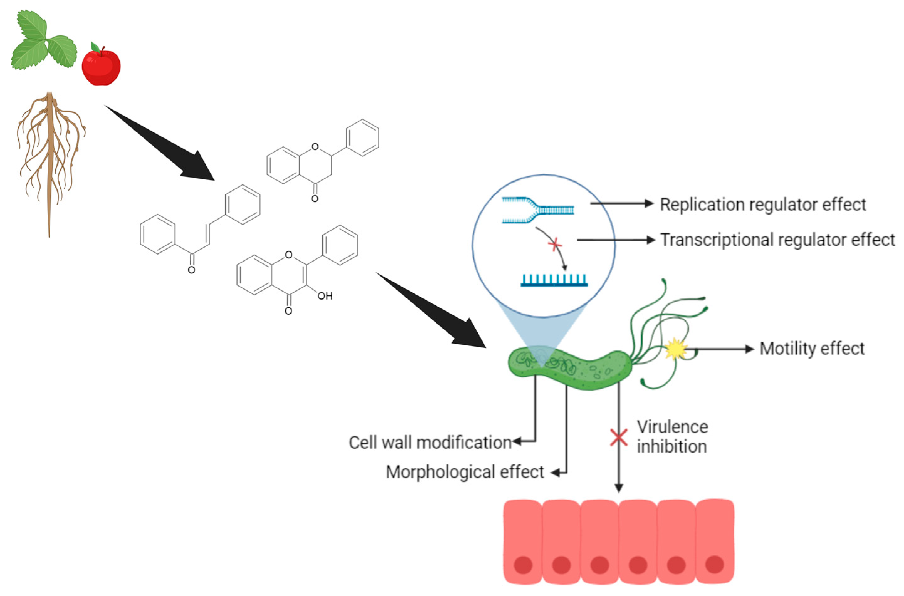
  67. Rego, R.I.d.A.; Silvestre, G.F.G.; de Melo, D.F.; Albino, S.L.; Pimentel, M.M.; Cruz, S.B.S.C.; Wurzba, S.D.S.; Rodrigues, W.F.; de Lima Damasceno, B.P.G.; Castellano, L.R.C. Flavonoids-Rich Plant Extracts Against Helicobacter pylori Infection as Prevention to Gastric Cancer. Front. Pharmacol. 2022, 13, 951125. [Google Scholar] [CrossRef]
  68. de Paula, M.N.; Ribeiro, T.; Isolani, R.; de Medeiros Araujo, D.C.; Borges, A.S.; Philippsen, G.S.; de Cassia Ribeiro Goncalves, R.; Kitagawa, R.R.; Seixas, F.A.V.; de Mello, J.C.P. An In Vitro and In Silico Investigation about Monteverdia ilicifolia Activity against Helicobacter pylori. Antioxidants 2022, 12, 46. [Google Scholar] [CrossRef]
  69. Krzyzek, P.; Migdal, P.; Paluch, E.; Karwanska, M.; Wieliczko, A.; Gosciniak, G. Myricetin as an Antivirulence Compound Interfering with a Morphological Transformation into Coccoid Forms and Potentiating Activity of Antibiotics against Helicobacter pylori. Int. J. Mol. Sci. 2021, 22, 2695. [Google Scholar] [CrossRef]
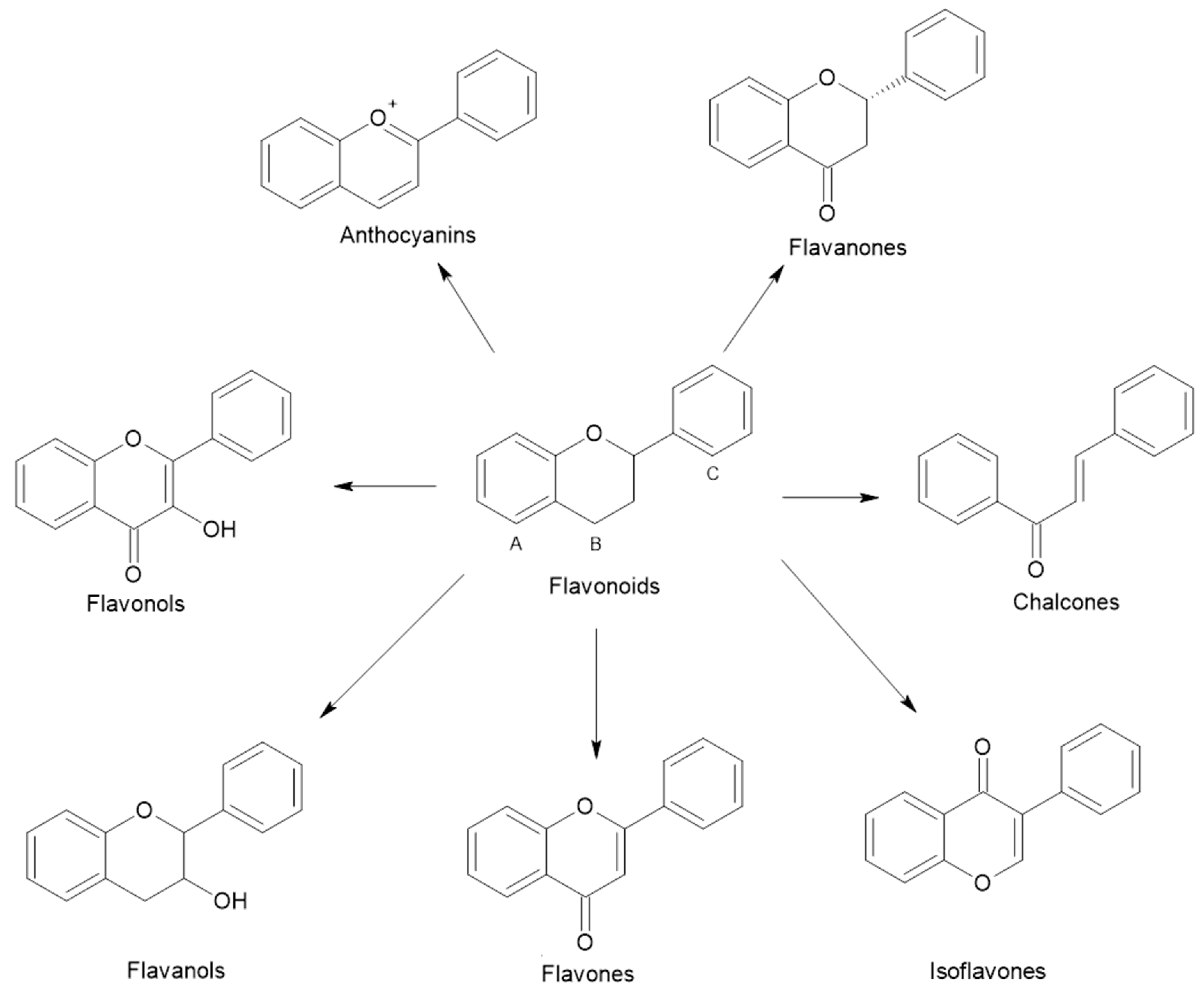
  70. Shen, N.; Wang, T.; Gan, Q.; Liu, S.; Wang, L.; Jin, B. Plant flavonoids: Classification, distribution, biosynthesis, and antioxidant activity. Food Chem. 2022, 383, 132531. [Google Scholar] [CrossRef]
  71. Tran Trung, H.; Truong Thi Huynh, H.; Nguyen Thi Thuy, L.; Nguyen Van Minh, H.; Thi Nguyen, M.N.; Luong Thi, M.N. Growth-Inhibiting, Bactericidal, Antibiofilm, and Urease Inhibitory Activities of Hibiscus rosa sinensis L. Flower Constituents toward Antibiotic Sensitive- and Resistant-Strains of Helicobacter pylori. ACS Omega 2020, 5, 20080–20089. [Google Scholar] [CrossRef] [PubMed]
  72. Liu, M.; Gao, H.; Miao, J.; Zhang, Z.; Zheng, L.; Li, F.; Zhou, S.; Zhang, Z.; Li, S.; Liu, H.; et al. Helicobacter pylori infection in humans and phytotherapy, probiotics, and emerging therapeutic interventions: A review. Front. Microbiol. 2024, 14, 1330029. [Google Scholar] [CrossRef]
  73. Bisignano, C.; Filocarno, A.; La Camera, E.; Zummo, S.; Fera, M.T.; Mandalari, G. Antibacterial activities of almond skins on cagA-positive and-negative clinical isolates of Helicobacter pylori. BMC Microbiol. 2013, 13, 103. [Google Scholar]
  74. Zengin, G.; Menghini, L.; Di Sotto, A.; Mancinelli, R.; Sisto, F.; Carradori, S.; Cesa, S.; Fraschetti, C.; Filippi, A.; Angiolella, L.; et al. Chromatographic Analyses, In Vitro Biological Activities, and Cytotoxicity of Cannabis sativa L. Essential Oil: A Multidisciplinary Study. Molecules 2018, 23, 3266. [Google Scholar] [CrossRef]
  75. Moon, S.H.; Lee, J.H.; Kim, K.T.; Park, Y.S.; Nah, S.Y.; Ahn, D.U.; Paik, H.D. Antimicrobial effect of 7-O-butylnaringenin, a novel flavonoid, and various natural flavonoids against Helicobacter pylori strains. Int. J. Environ. Res. Public Health 2013, 10, 5459–5469. [Google Scholar] [CrossRef] [PubMed]
  76. Lee, Y.S.; Huh, J.Y.; Nam, S.H.; Moon, S.K.; Lee, S.B. Enzymatic bioconversion of citrus hesperidin by Aspergillus sojae naringinase: Enhanced solubility of hesperetin-7-O-glucoside with in vitro inhibition of human intestinal maltase, HMG-CoA reductase, and growth of Helicobacter pylori. Food Chem. 2012, 135, 2253–2259. [Google Scholar] [CrossRef]
  77. de Cassia Dos Santos, R.; Bonamin, F.; Perico, L.L.; Rodrigues, V.P.; Zanatta, A.C.; Rodrigues, C.M.; Sannomiya, M.; Dos Santos Ramos, M.A.; Bonifacio, B.V.; Bauab, T.M.; et al. Byrsonima intermedia A. Juss partitions promote gastroprotection against peptic ulcers and improve healing through antioxidant and anti-inflammatory activities. Biomed. Pharmacother. 2019, 111, 1112–1123. [Google Scholar] [CrossRef]
  78. Salehi, B.; Sharopov, F.; Martorell, M.; Rajkovic, J.; Ademiluyi, A.O.; Sharifi-Rad, M.; Fokou, P.V.T.; Martins, N.; Iriti, M.; Sharifi-Rad, J. Phytochemicals in Helicobacter pylori Infections: What Are We Doing Now? Int. J. Mol. Sci. 2018, 19, 2361. [Google Scholar] [CrossRef] [PubMed]
  79. Abulizi, P.; Cong, Y.; Yakupu, M.; Wahepu, A. Chemical Constituents of Euphorbia sororia. Chem. Nat. Compd. 2014, 50, 908–909. [Google Scholar] [CrossRef]
  80. Novello, C.R.; Marques, L.C.; Pires, M.E.; Kutschenco, A.P.; Nakamura, C.V.; Nocchi, S.; Sarragiotto, M.H.; Mello, J.C.P. Bioactive Indole Alkaloids from Croton echioides. J. Braz. Chem. Soc. 2016, 27, 2203–2209. [Google Scholar] [CrossRef]
  81. Cayona, R.; Creencia, E. Phytochemicals of Euphorbia hirta L. and Their Inhibitory Potential Against SARS-CoV-2 Main Protease. Front. Mol. Biosci. 2021, 8, 801401. [Google Scholar] [CrossRef]
  82. Cardenas, D.M.; Gomez Rave, L.J.; Soto, J.A. Biological Activity of Sacha Inchi (Plukenetia volubilis Linneo) and Potential Uses in Human Health: A Review. Food Technol. Biotechnol. 2021, 59, 253–266. [Google Scholar] [CrossRef]
  83. Wang, S.; Zhu, F.; Kakuda, Y. Sacha inchi (Plukenetia volubilis L.): Nutritional composition, biological activity, and uses. Food Chem. 2018, 265, 316–328. [Google Scholar] [CrossRef]
  84. Goyal, A.; Tanwar, B.; Kumar Sihag, M.; Sharma, V. Sacha inchi (Plukenetia volubilis L.): An emerging source of nutrients, omega-3 fatty acid and phytochemicals. Food Chem. 2022, 373, 131459. [Google Scholar] [CrossRef]
  85. Kittibunchakul, S.; Kemsawasd, V.; Hudthagosol, C.; Sanporkha, P.; Sapwarobol, S.; Suttisansanee, U. The Effects of Different Roasting Methods on the Phenolic Contents, Antioxidant Potential, and In Vitro Inhibitory Activities of Sacha Inchi Seeds. Foods 2023, 12, 4178. [Google Scholar] [CrossRef] [PubMed]
  86. Mhd Rodzi, N.A.R.; Lee, L.K. Sacha Inchi (Plukenetia volubilis L.): Recent insight on phytochemistry, pharmacology, organoleptic, safety and toxicity perspectives. Heliyon 2022, 8, e10572. [Google Scholar] [CrossRef]
  87. Abd Rahman, I.Z.; Nor Hisam, N.S.; Aminuddin, A.; Hamid, A.A.; Kumar, J.; Ugusman, A. Evaluating the Potential of Plukenetia volubilis Linneo (Sacha Inchi) in Alleviating Cardiovascular Disease Risk Factors: A Mini Review. Pharmaceuticals 2023, 16, 1588. [Google Scholar] [CrossRef]
  88. Tan, A.; Scortecci, K.C.; Cabral De Medeiros, N.M.; Kukula-Koch, W.; Butler, T.J.; Smith, S.M.; Boylan, F. Plukenetia volubilis leaves as source of anti-Helicobacter pylori agents. Front. Pharmacol. 2024, 15, 1461447. [Google Scholar] [CrossRef] [PubMed]
  89. Gonzalez, A.; Casado, J.; Lanas, A. Fighting the Antibiotic Crisis: Flavonoids as Promising Antibacterial Drugs Against Helicobacter pylori Infection. Front. Cell. Infect. Microbiol. 2021, 11, 709749. [Google Scholar] [CrossRef]
  90. Hasnat, H.; Shompa, S.A.; Islam, M.M.; Alam, S.; Richi, F.T.; Emon, N.U.; Ashrafi, S.; Ahmed, N.U.; Chowdhury, M.N.R.; Fatema, N.; et al. Flavonoids: A treasure house of prospective pharmacological potentials. Heliyon 2024, 10, e27533. [Google Scholar] [CrossRef]
  91. Biglar, M.; Soltani, K.; Nabati, F.; Bazl, R.; Mojab, F.; Amanlou, M. A preliminary investigation of the jack-bean urease inhibition by randomly selected traditionally used herbal medicine. Iran. J. Pharm. Res. 2012, 11, 831–837. [Google Scholar] [PubMed]
  92. Uddin, G.; Ismail; Rauf, A.; Raza, M.; Khan, H.; Nasruddin; Khan, M.; Farooq, U.; Khan, A.; Arifullah. Urease inhibitory profile of extracts and chemical constituents of Pistacia atlantica ssp. cabulica Stocks. Nat. Prod. Res. 2016, 30, 1411–1416. [Google Scholar] [CrossRef]
  93. Xiao, Z.P.; Wang, X.D.; Peng, Z.Y.; Huang, S.; Yang, P.; Li, Q.S.; Zhou, L.H.; Hu, X.J.; Wu, L.J.; Zhou, Y.; et al. Molecular docking, kinetics study, and structure-activity relationship analysis of quercetin and its analogous as Helicobacter pylori urease inhibitors. J. Agric. Food Chem. 2012, 60, 10572–10577. [Google Scholar] [CrossRef]
  94. Egas, V.; Salazar-Cervantes, G.; Romero, I.; Mendez-Cuesta, C.A.; Rodriguez-Chavez, J.L.; Delgado, G. Anti-Helicobacter pylori metabolites from Heterotheca inuloides (Mexican arnica). Fitoterapia 2018, 127, 314–321. [Google Scholar] [CrossRef]
  95. Wu, D.; Kong, Y.; Han, C.; Chen, J.; Hu, L.; Jiang, H.; Shen, X. D-Alanine:D-alanine ligase as a new target for the flavonoids quercetin and apigenin. Int. J. Antimicrob. Agents 2008, 32, 421–426. [Google Scholar] [CrossRef]
  96. Zhang, S.; Huang, J.; Xie, X.; He, Y.; Mo, F.; Luo, Z. Quercetin from Polygonum capitatum Protects against Gastric Inflammation and Apoptosis Associated with Helicobacter pylori Infection by Affecting the Levels of p38MAPK, BCL-2 and BAX. Molecules 2017, 22, 744. [Google Scholar] [CrossRef] [PubMed]
  97. Brown, J.C.; Jiang, X. Activities of muscadine grape skin and polyphenolic constituents against Helicobacter pylori. J. Appl. Microbiol. 2013, 114, 982–991. [Google Scholar] [CrossRef] [PubMed]
  98. Qing, L.; Li, S.; Yan, S.; Wu, C.; Yan, X.; He, Z.; Chen, Q.; Huang, M.; Shen, C.; Wang, S.; et al. Anti-Helicobacter pylori activity of Fagopyrum tataricum (L.) Gaertn. Bran flavonoids extract and its effect on Helicobacter pylori-induced inflammatory response. Food Sci. Nutr. 2023, 11, 3394–3403. [Google Scholar] [CrossRef] [PubMed]
  99. Aranaz, P.; Navarro-Herrera, D.; Romo-Hualde, A.; Zabala, M.; López-Yoldi, M.; González-Ferrero, C.; Gil, A.G.; Martinez, J.A.; Vizmanos, J.L.; Milagro, F.I.; et al. Broccoli extract improves high fat diet-induced obesity, hepatic steatosis and glucose intolerance in Wistar rats. J. Funct. Foods 2019, 59, 319–328. [Google Scholar] [CrossRef]
  100. Yu, X.D.; Zheng, R.B.; Xie, J.H.; Su, J.Y.; Huang, X.Q.; Wang, Y.H.; Zheng, Y.F.; Mo, Z.Z.; Wu, X.L.; Wu, D.W.; et al. Biological evaluation and molecular docking of baicalin and scutellarin as Helicobacter pylori urease inhibitors. J. Ethnopharmacol. 2015, 162, 69–78. [Google Scholar] [CrossRef]
  101. Chen, M.E.; Su, C.H.; Yang, J.S.; Lu, C.C.; Hou, Y.C.; Wu, J.B.; Hsu, Y.M. Baicalin, Baicalein, and Lactobacillus Rhamnosus JB3 Alleviated Helicobacter pylori Infections in Vitro and in Vivo. J. Food Sci. 2018, 83, 3118–3125. [Google Scholar] [CrossRef]
  102. Dmitrieva, A.; Kozlova, O.; Atuchin, V.; Milentieva, I.; Vesnina, A.; Ivanova, S.; Asyakina, L.; Prosekov, A. Study of the Effect of Baicalin from Scutellaria baicalensis on the Gastrointestinal Tract Normoflora and Helicobacter pylori. Int. J. Mol. Sci. 2023, 24, 11906. [Google Scholar] [CrossRef]
  103. Kim, H.W.; Woo, H.J.; Yang, J.Y.; Kim, J.B.; Kim, S.H. Hesperetin Inhibits Expression of Virulence Factors and Growth of Helicobacter pylori. Int. J. Mol. Sci. 2021, 22, 10035. [Google Scholar] [CrossRef]
  104. Krzyzek, P.; Grande, R. Transformation of Helicobacter pylori into Coccoid Forms as a Challenge for Research Determining Activity of Antimicrobial Substances. Pathogens 2020, 9, 184. [Google Scholar] [CrossRef]
  105. Qing, W.; Haibing, H.; Na, Z.; Meitian, X.; Jing, Y. Exogenous spraying of sucrose improves sensory quality, bioactive components content and anti-obesity activity of Tieguanyin oolong tea. Ind. Crops Prod. 2023, 193, 116146. [Google Scholar] [CrossRef]
  106. Garro, M.F.; Salinas Ibanez, A.G.; Vega, A.E.; Arismendi Sosa, A.C.; Pelzer, L.; Saad, J.R.; Maria, A.O. Gastroprotective effects and antimicrobial activity of Lithraea molleoides and isolated compounds against Helicobacter pylori. J. Ethnopharmacol. 2015, 176, 469–474. [Google Scholar] [CrossRef]
  107. Abdel-Baki, P.M.; El-Sherei, M.M.; Khaleel, A.E.; Abdel-Aziz, M.M.; Okba, M.M. Irigenin, a novel lead from Iris confusa for management of Helicobacter pylori infection with selective COX-2 and HpIMPDH inhibitory potential. Sci. Rep. 2022, 12, 11457. [Google Scholar] [CrossRef]
  108. Ankolekar, C.; Johnson, D.; Pinto Mda, S.; Johnson, K.; Labbe, R.; Shetty, K. Inhibitory potential of tea polyphenolics and influence of extraction time against Helicobacter pylori and lack of inhibition of beneficial lactic acid bacteria. J. Med. Food 2011, 14, 1321–1329. [Google Scholar] [CrossRef]
  109. Díaz-Gómez, R.; López-Solís, R.; Obreque-Slier, E.; Toledo-Araya, H. Comparative antibacterial effect of gallic acid and catechin against Helicobacter pylori. LWT Food Sci. Technol. 2013, 54, 331–335. [Google Scholar] [CrossRef]
  110. Escandón, R.A.; del Campo, M.; López-Solis, R.; Obreque-Slier, E.; Toledo, H. Antibacterial effect of kaempferol and (−)-epicatechin on Helicobacter pylori. Eur. Food Res. Technol. 2016, 242, 1495–1502. [Google Scholar] [CrossRef]
  111. Pastene, E.; Parada, V.; Avello, M.; Ruiz, A.; Garcia, A. Catechin-based procyanidins from Peumus boldus Mol. aqueous extract inhibit Helicobacter pylori urease and adherence to adenocarcinoma gastric cells. Phytother. Res. 2014, 28, 1637–1645. [Google Scholar] [CrossRef] [PubMed]
  112. Santos, R.C.; Kushima, H.; Rodrigues, C.M.; Sannomiya, M.; Rocha, L.R.; Bauab, T.M.; Tamashiro, J.; Vilegas, W.; Hiruma-Lima, C.A. Byrsonima intermedia A. Juss.: Gastric and duodenal anti-ulcer, antimicrobial and antidiarrheal effects in experimental rodent models. J. Ethnopharmacol. 2012, 140, 203–212. [Google Scholar] [CrossRef]
  113. Lee, B.E.; Park, S.J.; Kim, G.H.; Joo, D.C.; Lee, M.W. Anti-inflammatory effects of eupatilin on Helicobacter pylori CagA-induced gastric inflammation. PLoS ONE 2024, 19, e0313251. [Google Scholar] [CrossRef]
  114. Palacios-Espinosa, J.F.; Nunez-Aragon, P.N.; Gomez-Chang, E.; Linares, E.; Bye, R.; Romero, I. Anti-Helicobacter pylori Activity of Artemisia ludoviciana subsp. mexicana and Two of Its Bioactive Components, Estafiatin and Eupatilin. Molecules 2021, 26, 3654. [Google Scholar] [CrossRef]
  115. Qanash, H.; Al-Rajhi, A.M.H.; Almashjary, M.N.; Basabrain, A.A.; Hazzazi, M.S.; Abdelghany, T.M. Inhibitory potential of rutin and rutin nano-crystals against Helicobacter pylori, colon cancer, hemolysis and Butyrylcholinesterase in vitro and in silico. Appl. Biol. Chem. 2023, 66, 79. [Google Scholar] [CrossRef]
  116. Krivokuća, M.; Niketić, M.; Milenković, M.; Golić, N.; Masia, C.; Scaltrito, M.M.; Sisto, F.; Kundaković, T. Anti-Helicobacter pylori Activity of Four Alchemilla Species (Rosaceae). Nat. Prod. Commun. 2015, 10, 1369–1371. [Google Scholar] [CrossRef] [PubMed]
  117. Tong, Q.; Yin, C.; Hang, X.; Bai, Y.; Zhang, C.; Xu, J.; Huang, Y.; Ge, Y.; Chen, T.; Zeng, L.; et al. Loureirin A is a narrow-spectrum antimicrobial agent against Helicobacter pylori. Antimicrob. Agents Chemother. 2024, 68. [Google Scholar] [CrossRef]
  118. Yeon, M.J.; Lee, M.H.; Kim, D.H.; Yang, J.Y.; Woo, H.J.; Kwon, H.J.; Moon, C.; Kim, S.H.; Kim, J.B. Anti-inflammatory effects of Kaempferol on Helicobacter pylori-induced inflammation. Biosci. Biotechnol. Biochem. 2019, 83, 166–173. [Google Scholar] [CrossRef] [PubMed]
  119. Sharaf, M.; Arif, M.; Hamouda, H.I.; Khan, S.; Abdalla, M.; Shabana, S.; Rozan, H.E.; Khan, T.U.; Chi, Z.; Liu, C. Preparation, urease inhibition mechanisms, and anti-Helicobacter pylori activities of hesperetin-7-rhamnoglucoside. Curr. Res. Microb. Sci. 2022, 3, 100103. [Google Scholar] [CrossRef]
  120. Saravanakumar, K.; Chellia, R.; Hu, X.; Kathiresan, K.; Oh, D.H.; Wang, M.H. Eradication of Helicobacter pylori through the inhibition of urease and peptide deformylase: Computational and biological studies. Microb. Pathog. 2019, 128, 236–244. [Google Scholar] [CrossRef]
  121. Górniak, I.; Bartoszewski, R.; Króliczewski, J. Comprehensive review of antimicrobial activities of plant flavonoids. Phytochem. Rev. 2018, 18, 241–272. [Google Scholar] [CrossRef]
  122. Tarahovsky, Y.S.; Kim, Y.A.; Yagolnik, E.A.; Muzafarov, E.N. Flavonoid-membrane interactions: Involvement of flavonoid-metal complexes in raft signaling. Biochim. Biophys. Acta 2014, 1838, 1235–1246. [Google Scholar] [CrossRef]
  123. Zhang, L.; Kong, Y.; Wu, D.; Zhang, H.; Wu, J.; Chen, J.; Ding, J.; Hu, L.; Jiang, H.; Shen, X. Three flavonoids targeting the beta-hydroxyacyl-acyl carrier protein dehydratase from Helicobacter pylori: Crystal structure characterization with enzymatic inhibition assay. Protein Sci. 2008, 17, 1971–1978. [Google Scholar] [CrossRef]
  124. Oliveira, D.M.; Melo, F.G.; Balogun, S.O.; Flach, A.; de Souza, E.C.; de Souza, G.P.; Rocha Ido, N.; da Costa, L.A.; Soares, I.M.; da Silva, L.I.; et al. Antibacterial mode of action of the hydroethanolic extract of Leonotis nepetifolia (L.) R. Br. involves bacterial membrane perturbations. J. Ethnopharmacol. 2015, 172, 356–363. [Google Scholar] [CrossRef]
  125. Qiu, T.; Wu, D.; Yang, L.; Ye, H.; Wang, Q.; Cao, Z.; Tang, K. Exploring the Mechanism of Flavonoids Through Systematic Bioinformatics Analysis. Front. Pharmacol. 2018, 9, 918. [Google Scholar] [CrossRef]
  126. Gonzalez-Aspajo, G.; Belkhelfa, H.; Haddioui-Hbabi, L.; Bourdy, G.; Deharo, E. Sacha Inchi Oil (Plukenetia volubilis L.), effect on adherence of Staphylococus aureus to human skin explant and keratinocytes in vitro. J. Ethnopharmacol. 2015, 171, 330–334. [Google Scholar] [CrossRef] [PubMed]
  127. Akash, S.R.; Tabassum, A.; Aditee, L.M.; Rahman, A.; Hossain, M.I.; Hannan, M.A.; Uddin, M.J. Pharmacological insight of rutin as a potential candidate against peptic ulcer. Biomed. Pharmacother. 2024, 177, 116961. [Google Scholar] [CrossRef]
  128. Jeong, C.-S. Evaluation for Protective Effect of Rutin, a Natural Flavonoid, against HCl/Ethanol-Induced Gastric Lesions. Biomol. Ther. 2009, 17, 199–204. [Google Scholar] [CrossRef]
  129. Narayanan, L.; Sr, S. Pharmacological insights into Ipomoea staphylina: Therapeutic activities and the isolated bioactive metabolic compounds. Mong. J. Chem. 2024, 25, 1–9. [Google Scholar] [CrossRef]
  130. Martinelli, G.; Fumagalli, M.; Pozzoli, C.; Nicotra, G.; Vicentini, S.F.; Maranta, N.; Sangiovanni, E.; Dell’Agli, M.; Piazza, S. Exploring In Vitro the Combination of Cistus x incanus L. and Castanea sativa Mill. Extracts as Food Supplement Ingredients against H. pylori Infection. Foods 2023, 13, 40. [Google Scholar] [CrossRef]
  131. Thanh, D.T.; Tan, M.T.; Thu, N.T.M.; Trinh, P.N.P.; Thuong, P.T.H.; Tuyet, P.T.G.; Ngan, L.T.M.; Hieu, T.T. Phytochemical Composition, Antioxidant, Anti-Helicobacter pylori, and Enzyme Inhibitory Evaluations of Cleistocalyx operculatus Flower Bud and Leaf Fractions. BioTech 2024, 13, 42. [Google Scholar] [CrossRef]
  132. Park, W.S.; Bae, J.-Y.; Kim, H.J.; Kim, M.G.; Lee, W.-K.; Kang, H.-L.; Baik, S.-C.; Lim, K.M.; Lee, M.K.; Ahn, M.J. Anti-Helicobacter pylori Compounds from Maackia amurensis. Nat. Prod. Sci. 2015, 21, 49–53. [Google Scholar]
  133. Fiore, C.; Antoniciello, F.; Roncarati, D.; Scarlato, V.; Grepioni, F.; Braga, D. Levofloxacin and Ciprofloxacin Co-Crystals with Flavonoids: Solid-State Investigation for a Multitarget Strategy against Helicobacter pylori. Pharmaceutics 2024, 16, 203. [Google Scholar] [CrossRef]
Figure 1. Flow diagram for the systematic review of the anti-H. pylori action of flavonoids. The diagram describes the screening process undertaken for all articles retrieved from databases, which underwent title and abstract screening, full-text analysis, and inclusion of relevant references not originally included in the search criteria.
Applsci 15 03936 g001
Figure 2. Polyphenol metabolism in the body. Schematic representation showing how polyphenols are used by our body. Created in BioRender. Tan, A. (2025) https://BioRender.com/m42e871, accessed on 19 March 2025.
Applsci 15 03936 g002
Figure 3. The general structure and seven groups of flavonoids. Schematic representation from different groups. Created in ACD/ChemSketch Freeware version.
Applsci 15 03936 g003
Figure 4. Flavonoid pharmacological pathways against H. pylori. Schematic representation of the potential effect of flavonoids on cells and H. pylori infection. Created in BioRender. Tan, A. (2025) https://BioRender.com/z25s605, accessed on 18 March 2025.
Applsci 15 03936 g004
Table 1. Genes/Protein and Potential Markers for Biotechnological Development.
Gene/ProteinGenomeFunctionReference
CYP2C19HostDrug metabolisation[1,41]
NikRBacteriaNickel-responsive regulator[45]
HsrABacteriaMetabolism and stress defence[45]
OipABacteriaOuter membrane protein[1,45]
CagABacteriaCell transformation to cancer cells[1,45]
BabABacteriaAdhesion to mucosa cells[1,45]
HopQBacteriaAdhesion to CEACAMs[1,45]
UreaseBacteriaProtein involved in helping the colonization in the acid pH in stomach[45,48]
FlavodoxinBacteriaSmall electron transfer protein involved in pyruvate metabolism[45]
Table 2. Pharmacological Activity of Flavonoids Against H. pylori.
FlavonoidAction MechanismMICIn Vitro DataIn Vivo DataSynergistic EffectScientific Reference
QuercetinUrease inhibition via binding with zinc cation; binding to Ddl; lipid peroxidation of bacterial membrane.64–128 µg/mL; 100–200 µg/mLMolecular docking; inhibits urease by forming ionic bonds with zinc; binding to Ddl.Prevents inflammation in gastric cells, affects p38MAPK, BCL-2, and BAX protein levels in mouse animal model.Co-crystallisation with antibiotics enhances efficacy.[31,67,71,93,94,95,96,97,98,99]
Baicalin and BaicaleinBinding to urease active site reducing virulence by decreasing vacA gene expression.≥1 mM (Baicalin); 0.125–1 mM (Baicalein)Reduces virulence by lowering vacA expression.Baicalein has enhanced bactericidal effects.N/A[100,101,102]
HesperetinDownregulates virulence factors UreA and UreB; prevents bacterial infection by gene expression reduction.8 mg/LReduces gene expression essential for H. pylori development, inhibition of urease.N/ASynergistic with metronidazole and clarithromycin.[31,75,103]
ChrysinForms a stable HsrA-flavonoid complex, inhibiting HsrA.≤8 mg/LInhibits HsrA in H. pylori.N/AN/A[31]
MyricetinDisrupts transcription of virulence-associated genes; delays spiral-to-coccid transformation.160 µg/mLAffects gene expression related to muropeptide dimers and monomers.Delays morphological transformation, improving antimicrobial potential.Synergistic effects with antibiotics (FICI = 0.31–0.5).[69,104]
NaringeninUrease inhibitionN/AInhibitis effect of ureaseN/AN/A[75]
RutinImpairs biofilm formation; moderate antibacterial activity.8–125 µg/mLBiofilm formation inhibition.N/AN/A[105,106]
ApigeninBinds to Ddl as competitive and non-competitive inhibitor; inhibits DNA-binding activity of HsrA.8 mg/LInhibits HsrA and Ddl activities.N/AN/A[31,95]
LuteolinInhibits H. pylori strains; possible action on adhesion to host cells.125 mg/LInhibition and bactericidal action.N/AN/A[71,107]
Catechins and EpicatechinBinding to cell membranes; urease inhibition and anti-adherent activities.N/AGrowth inhibition at low concentrations; potential gastroprotective effects.N/ASynergistic activity with antibiotics likely present but not fully understood.[67,71,108,109,110,111,112]
EupatilinInhibits NF-κB inflammatory pathway; suppresses CagA gene.125–250 µg/mLAnti-inflammatory effects; modulation of NF-κB and IL-6 transcription.Clinical trial show high cure rates in erosive gastritis patients.N/A[113,114]
OrientinBacteriostatic action15.53 µg/mLBacteriostatic effectN/AN/A[115]
Isoquercetin(quercetin-3-O-β-D-glucoside)Inhibitis H. pylori growth480 μg/mL N/AN/AN/A[116]
Loureirin A (from Sanguis Draconis) bactericidal effect4 mg/mLN/AN/AN/A[117]
KaempferolPrevents secretion of virulence factors; prevention of transcription; forms complexes with the cell wall.N/ADecreases mRNA levels of T4SS components; modifies modulation of cagA and vacA genes; prevention of transcription fractors binding.N/AN/A[31,98,118]
Disclaimer/Publisher’s Note: The statements, opinions and data contained in all publications are solely those of the individual author(s) and contributor(s) and not of MDPI and/or the editor(s). MDPI and/or the editor(s) disclaim responsibility for any injury to people or property resulting from any ideas, methods, instructions or products referred to in the content.

