Article

Effects of Frost Mulberry Leaf Superfine Powder on the Hypoglycemic and Gut Microbiota of High-Fat Diet/Streptozotocin-Induced Type 2 Diabetes Mellitus Mice

1
School of Food Science and Engineering, Qilu University of Technology (Shandong Academy of Sciences), Jinan 250353, China
2
College of Life Sciences, Shandong Normal University, Jinan 250300, China
3
Economic Forest Institute, Shandong Academy of Forestry Sciences, Jinan 250014, China
*
Authors to whom correspondence should be addressed.
Appl. Sci. 2025, 15(7), 3766; https://doi.org/10.3390/app15073766
Submission received: 6 March 2025 / Revised: 27 March 2025 / Accepted: 28 March 2025 / Published: 29 March 2025

Abstract

:
Frost mulberry leaves possess significant medicinal and nutritional values and feature extensive resource availability and convenient acquisition. The study investigated the physicochemical structure and functional properties of frost mulberry leaf superfine powder (FMLSP) and the effects of FMLSP on the hypoglycemic activity and gut microbiota of type 2 diabetes mellitus (T2DM) mice. The results indicated that the total flavonoid content of FMLSP reached 91.30 mg/g, with significant inhibitory effects on both α-glucosidase and α-amylase activities. Animal experimental data showed that FMLSP could significantly reduce insulin content, improve insulin resistance, and protect liver and pancreatic tissues in T2DM mice. Meanwhile, FMLSP showed significant effects on lipid metabolism, especially the low-density lipoprotein cholesterol (LDL-C) content in T2DM mice was significantly reduced by 76.22%. In addition, FMLSP has excellent antioxidant effects, which greatly alleviated the oxidative stress phenomenon in T2DM mice, especially the malondialdehyde (MDA) content was significantly reduced by 72.17%. FMLSP also restored the diversity and structure of the gut microbiota, significantly increasing the abundance of beneficial bacteria such as Akkermansia, Lachnospiraceae_NK4A136_group, Alloprevotella, and Lactobacillus in T2DM mice and significantly decreasing the abundance of abundance of harmful bacteria such as Rikenellaceae_RC9_gut_group, Enterorhabdus. These results indicate that FMLSP may serve as a potential dietary intervention for the prevention and treatment of T2DM.

1. Introduction

Diabetes mellitus has become an increasing challenge in global public health. The main cause of diabetes mellitus is defective insulin secretion, leading to insulin resistance (IR) in tissues and organs and impaired pancreatic β-cell function, which induces disorders of glucolipid metabolism and subsequent development of a pathological state characterized by hyperglycemia and dyslipidemia [1,2]. Among the subtypes of diabetes mellitus, type 2 diabetes mellitus (T2DM) dominates, with epidemiologic data showing accounts for up to 90% of cases in the total prevalent population [3]. Studies have shown that conventional diabetes medication can control the disease but has side effects associated with long-term use, such as bloating and diarrhea. Therefore, dietary interventions with safety, economy, and effectiveness are more appropriate for the long-term management of diabetes [4,5]. Meanwhile, studies have disclosed the intimate association between the pathogenesis of T2DM and gut microbiota [6,7]. When the gut microbiota is in a state of homeostatic imbalance, its metabolites and abnormal immunoregulatory function may become the causative factors of metabolic syndrome-like diseases, such as obesity, T2DM, and dyslipidemia [8,9]. Based on this, it is essential to explore dietary interventions that can synchronously regulate the abnormalities of glucolipid metabolism and optimize the function of the gut microbiota.
Mulberry leaf is a medicinal and food catalog ingredient with high medicinal and culinary value [10]. Research has found that mulberry leaves are full of vitamins, minerals, protein and other nutrients and flavonoids, alkaloids, phenols, polysaccharides, and other functional components, with hypoglycemic, hypolipidemic, inhibition of drug toxicity, antioxidant, and other functional effects [11,12]. Mulberry leaves harvested after the frost are usually used for medicinal purposes [13]. In numerous ancient Chinese medical books, mulberry leaves were mostly collected after being exposed to frost, and mulberry leaves included in the previous editions of the Chinese Pharmacopoeia were also frost mulberry leaves, which indicates that frost-covered mulberry leaves have higher medicinal value [14]. It has been demonstrated that there are seasonal variations in the amount of functional components found in mulberry leaves, with the amount of flavonoid components dramatically increasing following frost [13,15]. Xu et al. hypothesized that low temperature induces gene expression of enzymes critical in the pathway of flavonoid synthesis and promotes an increase in flavonoid content [14]. Therefore, frosted mulberry leaves have the potential as a functional food ingredient.
Currently, most mulberry leaves are still used as silkworm feed or discarded, resulting in a waste of resources. Consequently, it is imperative to investigate the logical utilization of functional components found in mulberry leaves. Mulberry leaf extract pharmacological studies for the treatment of diabetes mellitus are the primary focus of the current antidiabetic research on mulberry leaves [16]. In T2DM mice, Bae et al. and Jung et al. confirmed that mulberry leaf aqueous extracts had a hypoglycemic impact [17,18]. Meng et al. isolated the flavonoid components of mulberry leaves and examined their mode of action and hypoglycemia potential [19]. It should be noted that the majority of research has been conducted using conventional methods, including alcohol and water extraction, to extract the useful components. These extraction methods will cause some waste of functional components in mulberry leaves and will also increase the production cost [20]. Consequently, the primary goal of this research is to fully utilize bioactive substances in frosted mulberry leaves and fill the research gap of frosted mulberry leaves as a functional food ingredient in practical applications.
Studies have shown that superfine grinding technology can enhance the organoleptic sentimental value of powders and promote the dissolution and in vivo absorption of functional ingredients [21,22]. As an inexpensive and eco-friendly superfine grinding method, planetary ball milling has a lot of promise for producing wholesome and useful meals. The study of Zhang et al. illustrated that planetary ball milling facilitated the release of phytochemicals present in Lycium ruthenicum Murray powder and improved its antioxidant capacity as well [23]. The study by Pablo Martín Palavecino et al. showed that planetary ball milling significantly altered the antioxidant capacity of sorghum powders [24]. It can be seen that the planetary ball mill has a good prospect for the practical application of frosted mulberry leaves.
Thus, as shown in Scheme 1, the physicochemical and functional characteristics of frosted mulberry leaf superfine powder (FMLSP) were examined in this experiment after it was made using planetary ball milling technology. High-fat diet (HFD) and streptozotocin (STZ)-induced T2DM mouse models were also used to evaluate the antidiabetic impact of FMLSP. Furthermore, the effect of FMLSP on the structure of the gut microbiota in T2DM mice was investigated. This study presents a rationale for the development of glucose-lowering health food raw materials from plants with broad market prospects and provides a solution to the problem of the existence of mulberry leaf resource waste.

2. Materials and Methods

2.1. Preparation of FMLSP

The dried frosted mulberry leaves (Ancient Mulberry Research Institute, Xiajin, Shandong, China) were simply pulverized with a high-speed herbal grinder (FW135, Tianjin Tester Instrument Co., Ltd., Tianjin, China), and 80-mesh sieve was passed to obtain the coarse powder. And then, the coarse powder was superfine crushed by planetary ball mill (F-P4000E, Focucy Experimental Instrument Co., Ltd., Hunan, China). Together with the powder sample, 10 mm diameter zirconium dioxide (ZrO2) balls were put in a grinding jar; the weight of the ZrO2 balls was seven times that of the sample. The FMLSP was obtained by pulverizing the material for 10 h at 500 rpm in a planetary ball mill.

2.2. Determination of Physicochemical Properties and Components of FMLSP

2.2.1. Particle Size

Determinations were made using a laser particle size analyzer (90plus PALS, Brookhaven Instruments, Nashua, NH, USA).

2.2.2. Structural Characterization Analysis

Refer to Wang et al. [25] method and make slight modifications. The microstructure of FMLSP was observed with a magnification of 1000× and 5000× (ZEISS GEMINI 500, Zeiss AG, Oberkochen, Germany).

2.2.3. Determination of Basic Nutrients

Ash, fat, protein, and dietary fiber in FMLSP were determined according to the methods in the Chinese Standard 5009 series of national food safety standards [26].

2.2.4. Analysis of Bioactive Substances

Flavonoid and Polyphenol

According to Tu et al. [27], 200 mg of FMLSP was accurately weighed, 75% ethanol (20 mL) was added, and then extracted for 60 min at 70 °C. To extract the supernatant, centrifuge for 20 min at 3000 rpm. Repeat the operation twice. The supernatant was diluted to 50 mL with 75% ethanol to obtain the extraction solution. A 1.0 mL amount of the extracted solution was added to 0.5 mL 5% NaNO2 and left for 6 min. Next, 0.5 mL Al(NO3)3 (10%) and 4 mL NaOH (4%) were added after 6 min. Then, 60% ethanol was used to dilute the solution to 10.0 mL. The absorbance at 510 nm was tested ten minutes later. The equivalent milligrams of rutin (mg GAE/g dw) were calculated using a calibration curve (0.2–1.0 mg/mL, R2 = 0.9997).
According to Tu et al. [27], 2 mL of the extract was measured, and distilled water (30 mL) and Folin-Ciocalteu reagent (2.5 mL) were added and mixed immediately. After 1.0 min, 7.5 mL Na2CO3 (20%) solution was added, and volume to 50 mL, then 75 °C for 10 min. At 760 nm, measure the absorbance. The results are displayed using a calibration curve with an R2 of 0.9965 and a range of 0.05 to 0.25 mg/mL in milligram equivalents of gallic acid (mg GAE/g dw).

Polysaccharides

As stated by Zeng and colleagues [28], in each test tube, 5 mL vitriol, 1 mL extract, and 1 mL 5% (v/v) phenol solution were mixed together. Absorbance readings were recorded at 490 nm. A calibration curve (0.02–0.10 mg/mL, R2 = 0.9928) was prepared with anhydrous glucose standards, and the polysaccharide content was presented in milligram equivalents of glucose (mg GAE/g dw).

1-Deoxynojirimycin (1-DNJ)

HPLC (LC-20AT, Agilent Technologies Inc., Santa Clara, CA, USA) determined the 1-DNJ content with pre-column derivatization as described by Vichasilp et al. [29]. After thoroughly mixing 10 μL extract, 10 μL K2BO3, and 20 μL 5 mmol/L FMOC-Cl, which reacted at 20 °C (20 min), add 950 μL of 0.1% acetic acid and 0.1 mol/L glycine (10 μL) to the reaction system. It was run at 254 nm UV for 30 min using C18 column. The results of the research were presented in milligram equivalents of 1-DNJ (mg CE/g dw), R2 = 0.9994 for calibration curve of 0.02–0.10 mg/mL. Chromatograms associated with the standards are given in the Supplementary Material.

2.3. Determination of FMLSP Functional Properties

2.3.1. In Vitro Antioxidant Activity

Determination of FMLSP scavenging activity against DPPH radicals and ABTS radicals by the method of Adel Faidi et al. [30]. The 2 mL volumes of extracts with varying mass concentrations were meticulously combined with equal volume of DPPH solution (0.05 mg/mL) and incubated for half an hour in the dark. At 517 nm, absorbance values were observed. A positive control is vitamin C.
According to Adel Faidi et al. [30], for six minutes, the combination of 1.0 mL sample solution and 4 mL ABTS working solution was cultivated at 37 °C in the dark, then spectrophotometric analysis at 734 nm. Vitamin C is positive control.

2.3.2. In Vitro Digestive Simulation

For in vitro digestion, simulated gastric fluid (SGF) and simulated gut fluid (SIF) were devised following procedure outlined by Yu et al. [31]. FMLSP (1.0 g) was solubilized in a 10 mL solution of 0.9% NaCl, followed by 10 mL SGF was added, mixed well, and incubated at 37 °C for 2 h. Subsequent sampling occurred at intervals of 5, 10, 15, 30, 60, and 120 min, with enzyme deactivation taking place at 70 °C (10 min), cooling, and subsequent centrifugation at 10,000× g (10 min), prior to analysis of the supernatant. The simulated gastric digestion products were neutralized with 1 mol/L NaHCO3. In order to replicate gut digestion, SIF and the byproducts of simulated stomach digestion were mixed in a 3:10 ratio and cultivated at 37 °C (2 h). Aliquots were extracted at specified intervals, followed by immediate analytical processing.

2.3.3. In Vitro Hypoglycemic Analysis

The α-glucosidase inhibition assay was conducted following Awosaka et al. [32]. A mixture was prepared by combining 50 μL extract with 100 μL solution containing α-glucosidase and subjected to a 10 min preincubation at 37 °C. Subsequently, enzymatic activity was initiated by introducing 50 μL 5 mM 4-nitrophenyl-α-D-glucopyranoside (PNPG) into reaction system. Following a 30 min incubation period, the enzymatic reaction was terminated by using 80 μL Na2CO3. Measured absorbance at 405 nm and the inhibition efficiency determined by using the formula:
α-Glucosidase inhibition % = [1 − (Aw − Ax)/(Ay − Az)] × 100%
where Aw: the sample’s absorbance, Ax: represents background absorbance of enzyme-free sample, Ay: positive control absorbance, and Az: control blank absorbance.
Referring to Awosika et al. method [32], measured the absorbance at 540 nm. The Formula (2) for calculating enzymatic inhibition efficiency was used.
α-Amylase inhibition % = [1 − Ax/Ay] × 100%
Ax: optical density measurements of experimental samples containing α-amylase, and Ay: control absorbance values from aqueous blanks (distilled water substituted for extract).

2.4. Animal Experimental

Male Kunming mice, aged four weeks, weighing between 17 and 23 g, were purchased from Beijing Vital River Laboratory Animal Technology Co., Ltd. (Beijing, China) as specific pathogen-free (SPF) mice. Mice were kept under a 12 h light/dark cycle in a particular pathogen-free environment with a constant temperature (23 ± 2 °C) and humidity (50 ± 10%). Following a week of acclimatization, twenty-four mice were given an HFD and six animals were randomized to the normal control (NC) group, which was fed a standard diet. Mice fed HFD for two weeks were given an intraperitoneal injection of STZ at 45 mg/kg over a period of two days after fasting overnight. NC group received the same amount of citrate buffer treatment. Three days after the STZ injection, mice were given a blood sample from the tip of their tails to measure their fasting blood glucose (FBG). The T2DM mouse model was effectively created if FBG was more than or equal to 11.1 mmol/L. As shown in Table 1, 24 mice with T2DM were allocated into four groups (6 mice per group) in a randomized manner, including a model group (T2DM), a medication group (Met, 0.5 g/kg metformin hydrochloride), an FMLSP low-dose group (FMLSPL, 1.0 g/kg FMLSP), and an FMLSP high-dose group (FMLSPH, 2.0 g/kg FMLSP). For 4 weeks, FMLSP and metformin hydrochloride were given orally by gavage every day after being dissolved in 0.4 mL of sterile water. Gavages of the same amount of sterile water were provided to the NC as well as Met groups.
During the experimental period, daily monitoring was conducted to evaluate the increase in body weight and the intake of food, alongside the assessment of the mice’s FBG levels using blood glucose meter (Yuyue Medical Equipment and Supply Co., Ltd., Shanghai, China) once a week. During the 26 days of therapy, an oral glucose tolerance test (OGTT) was administered to the mice, in which they were all given 2.0 g glucose/kg BW verbally after a 12 h fast. The next steps were to monitor blood glucose at 0, 30, 60, 90, and 120 min.
After the experimental technique was finished, mice underwent a 12 h fasting period, received anesthesia, and were subjected to cardiac blood collection prior to euthanasia via cervical dislocation. Serum was isolated through centrifuge at 1000× g for 15 min and cryopreserved at −60 °C for biochemical assays. Mouse liver, colon as well as pancreas samples should be promptly gathered for additional research. The Animal Experiment Ethics Review Committee of Shandong Normal University granted ethical permission for this animal experiment (AEECSDNU2024006).

2.5. Biochemical Analysis

Serum biochemical parameters in mice were monitored using commercial test kits (Nanjing Jiancheng Bioengineering Institute, Nanjing, China), such as total cholesterol (TC), total triglyceride (TG), glycated serum protein (GSP), low-density lipoprotein cholesterol (LDL-C), high-density lipoprotein cholesterol (HDL-C), insulin, and aspartate transaminase (AST), alanine transaminase (ALT), catalase (CAT), pyruvate kinase (PK), and hexokinase (HK) activities. At the same time, standardized kits from the same manufacturer were used to measure the liver’s glutathione peroxidase (GSH-PX), superoxide dismutase (SOD) and activity, glycogen, and malondialdehyde (MDA) levels.

2.6. Analysis of Tissue Sections

Liver and pancreatic tissues from mice were preserved in 4% paraformaldehyde, processed through paraffin embedding, and divided into slices of 5 μm for examination. Staining using hematoxylin–eosin (HE) was conducted, and subsequent histopathological alterations were visualized through light microscopy [33].

2.7. Examination of the Gut Microbiota

Mice’s colon tissue was used to extract DNA from their stomach contents. Using particular primers 515F and 806R, the 16S rDNA V4 region was amplified by PCR after qualification. PCR products were detected, and target fragments were recovered using 2% agarose gel electrophoresis. The Illumina TruSeq Kit (Illumina Inc., San Diego, CA, USA) was used to build the library, and the Illumina NovaSeq 6000 system (Illumina Inc., San Diego, CA, USA) was employed for quality control and sequencing. The QIIME2 program (Version-QIIME2-202202, Illumina Inc., San Diego, CA, USA) was used to evaluate the variations in the groups’ gut microbiota content.

2.8. Data Analysis

Standard deviation (SD) ± mean was used to represent all data. One-way analysis of variance (ANOVA) and Tukey’s test were performed for all statistics using SPSS software (IBM SPSS Statistics 26, Chicago, IL, USA), with p < 0.05 being deemed statistically significant.

3. Results

3.1. Physicochemical Properties and Compositional Analysis of FMLSP

Particles with diameters less than 25 μm are normally considered superfine powders [34]. It can be seen that the volume-weighted average diameter (D [4,3]) of the prepared FMLSP can reach 8.52 μm from Table 2. Therefore, the coarse powder of frost mulberry leaf can be crushed by planetary ball milling for 10 h to reach the grade of superfine powder.
The basic nutrients and bioactive substances content of FMLSP are shown in Table 3. The flavonoid of FMLSP could be as high as 91.30 ± 1.92 mg/g and the polyphenol could be up to 50.41 ± 1.04 mg/g. The polysaccharide content and 1-DNJ content of FMLSP are 58.41 ± 1.39 mg/g and 0.89 ± 0.008 mg/g, which suggests that there are a lot of hypoglycemic compounds in FMLLSP.
As illustrated in Figure 1A,B, with different magnifications, the SEM images of FMLSP particles revealed excellent uniformity. The particle distribution showed a dense state, and some degree of agglomeration occurred. It bears resemblance to the discoveries made by Chen et al. [35]. This could be because the FMLSP particles’ surface electrostatic attraction changes as a result of their size decrease after the ball milling process.

3.2. Functional Properties of FMLSP

The scavenging of DPPH and ABTS radicals by FMLSP and VC initially increased and subsequently leveled off with increasing concentration, as shown in Figure 1C,D. Significant antioxidant activity was shown by FMLSP, according to the IC50 values derived from the curves. The IC50 values of VC (0.07 mg/mL and 0.14 mg/mL) were similar to the IC50 values of FMLSP for DPPH and ABTS radical scavenging, which were 0.36 mg/mL and 0.19 mg/mL, respectively.
As can be seen from Figure 1E,F, the release of total flavonoids as well as total phenols from FMLSP during the simulated gastric digestion phase was not much different from that before digestion and decreased rapidly within 15 min from the beginning of the simulated gut digestion phase. This indicates that the total flavonoids and total phenols of FMLSP are stable in an acidic environment but easily oxidized under alkaline, light, and humid conditions. Therefore, the release was stable in the simulated gastric digestion stage, while it was partially oxidized in the simulated enteric digestion environment, leading to a decrease in the release.
The inhibition rate of FMLSP on α-amylase and α-glucosidase activities showed a trend of increasing (Figure 1G and then leveling off in the experimental concentration range. The inhibition rate of FMLSP on α-amylase peaked at 63.60% when the concentration hit 3 mg/mL. The inhibition rate of FMLSP on α-glucosidase peaked at 86.15% when the concentration hit 4 mg/mL. The results indicated that FMLSP has the potential for hypoglycemia.

3.3. Effect of FMLSP on Body Weight, FBG, and Organ Index in T2DM Mice

Patients with T2DM will show significant changes in diet, body weight, and blood glucose [36]. Therefore, we monitored the mice’s food intake, body weight, FBG, OGTT, and other indexes during the experimental period to assess FMLSP’s therapeutic impact in T2DM mice. According to Figure 2A,B, the mice in the T2DM group significantly increased their food consumption, but conversely, their body weight tended to decrease when compared to the mice in the NC group. Mice treated with FMLSP and Met for 4 weeks showed better hyperphagia and weight loss symptoms when compared to the T2DM group (p < 0.05).
The variations in mice’s FBG levels during the course of the experiment are shown in Figure 2C. In the NC group, the FBG levels remained constant. The FBG levels in the T2DM group were considerably greater (p < 0.01) than those in the NC group, indicating that the T2DM mouse model had been successfully established. Following FMLSP and Met treatments, mice’s FBG levels were considerably lower than those of the T2DM group (p < 0.01). In the T2DM group, FBG was maintained at 25.03 mmol/L at week 4, while in the FMLSPL, FMLSPH, and Met groups, it dropped to 19.51, 17.92, and 16.87 mmol/L, respectively. This suggested that FMLSP might provide a more favorable hypoglycemic impact.
The OGTT test showed the results as shown in Figure 2D. The overall trend showed an increase and then a decrease. The mice in each experimental group exhibited their highest blood glucose values at the 30 min time point, with the T2DM group showing markedly elevated blood glucose in comparison to the NC group. These findings point toward a significant deterioration in glucose tolerance among the mice in the T2DM group. At 120 min, blood glucose concentrations in the T2DM group remained high compared to those in the 0 min cohort; however, blood glucose concentrations in the FMLSPL, FMLSPH, and Met cohorts decreased significantly and eventually returned to their initial values. Similar conclusions were obtained by calculating the results of AUC. According to Figure 2E, the AUC values observed in the T2DM cohort exhibited a statistically significant elevation compared to the NC cohort. Moreover, the AUC values recorded in the FMLSPL, FMLSPH, and Met cohorts were notably lower than those in the T2DM cohort, demonstrating reductions of 16.14%, 24.25%, and 22.83%, respectively. The findings showed that the high dosage of FMLSP considerably enhanced the diabetic mice’s glucose tolerance.
It is common to use organ indices to assess the physiological and pathological conditions of animals. According to Figure 2F,G, in mice in the T2DM group, the liver and pancreatic indices showed a significant increase of 33.24% and 38.57%, respectively, in comparison to the NC group. This suggested that mice’s liver and pancreas experienced some impacts from a diet rich in fat and sugar as well as an injection of STZ solution. After intervention with FMLSP, the liver index decreased by 11.94%, 19.19%, and 19.83% in the FMLSPL, FMLSPH, and Met cohorts, respectively, in comparison to the T2DM group. Pancreatic index, on the other hand, decreased by 16.46%, 22.27%, and 25.70%, respectively. The findings indicated that FMLSP exhibited a protective influence on the liver and pancreas of mice with T2DM.

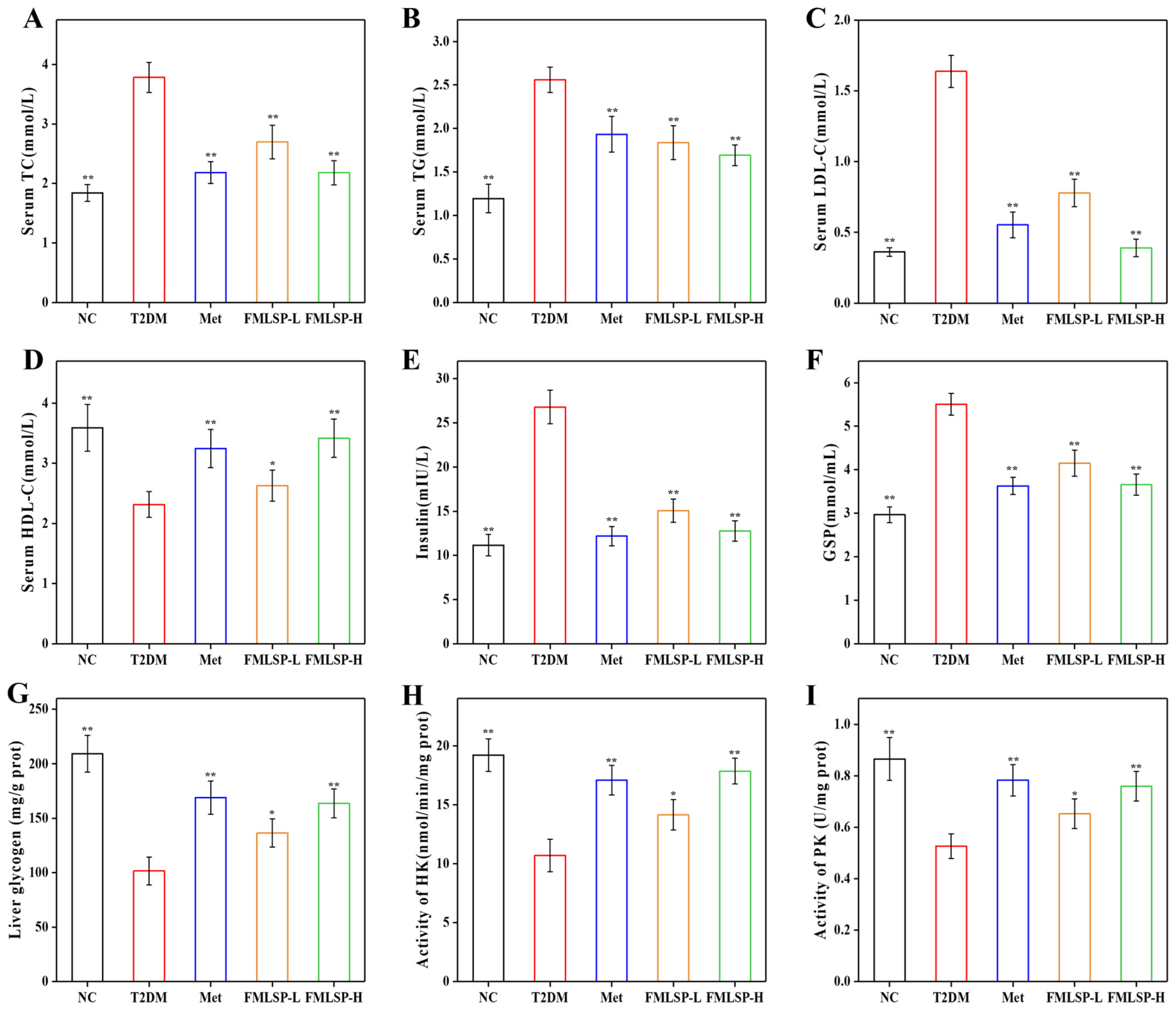
3.4. Effect of FMLSP on Lipid Metabolism

As seen in Figure 3A–D, T2DM mice had considerably greater serum levels of TC, TG, and LDL-C than NC mice, whereas T2DM mice had significantly lower serum levels of HDL-C (p < 0.01), suggesting that significant hyperlipidemia was present in T2DM mice.
In contrast to the T2DM group, while HDL-C increased by 47.45% (p < 0.01), FMLSP-H therapy decreased blood TC, TG, and LDL-C concentrations by 42.34%, 33.88%, and 76.22% (p < 0.01). It indicated that FMLSP treatment had a significant hypolipidemic effect.

3.5. Effect of FMLSP on Glucose Metabolism in Mice

With a relative rise of 139.98%, the insulin content of mice in the T2DM group was significantly greater (p < 0.01) than that of the NC group, as shown in Figure 3E. This was due to the diabetic mice developing insulin resistance (IR), which made it difficult for insulin to function resulting in accumulation [37]. Mice in the FMLSPL, FMLSPH, and Met groups had lower insulin contents after Met and FMLSP treatments than those in the T2DM group by 43.78%, 52.34%, and 54.53%, respectively. FMLSP has a moderating impact on IR symptoms, as seen by the reduction in insulin.
The mouse body’s average blood glucose level during the previous two to three weeks is reflected in GSP [38]. Mice in the T2DM group had a GSP level that was 1.86 times greater than the NC group, however, after the FMLSPH and Met treatments, the GSP level decreased by 33.58% and 34.12%, respectively, indicating that FMLSP effectively ameliorated the hyperglycemia in T2DM mice (Figure 3F). The liver glycogen content in the NC group was 2.06 times higher than that in the T2DM group, as shown in Figure 3G. Treatment with FMLSP and Met resulted in a significant increase in glycogen levels (p < 0.01). When compared to the T2DM group, the liver glycogen content rose by 61.08% and 66.36%, respectively, indicating that the effects of FMLSPH and Met were comparable.
HK and PK activities in T2DM mice demonstrated a marked reduction compared to normoglycemic controls (p < 0.01), reflecting impaired glycolysis and compromised blood glucose regulation in the diabetic cohort (Figure 3H,I). FMLSP administration upregulated HK and PK catalytic capacities in a way that depends on dosage (p < 0.05). This illustrates how FMLSP intervention might somewhat increase the activity of enzymes linked to glycolysis, which in turn encourages glycolysis to reduce blood glucose.

3.6. Impact of FMLSP on the Mice’s Antioxidant System

The research examined the levels of lipid peroxidation products (MDA) and the activity of antioxidant enzymes (CAT, GSH-PX, and SOD) in diabetic mice. As shown in Figure 4A–D, CAT, GSH-PX, and SOD activities were reduced by 49.78%, 38.33%, and 36.17% (p < 0.01), and the T2DM group’s MDA content was substantially higher than that of the NC group by 72.05% (p < 0.01). The findings are consistent with earlier research showing that T2DM mice exhibit higher oxidative stress [39]. Following FMLSPH therapy, the content of MDA dramatically decreased by 72.17%, while the activities of CAT, GSH-PX, and SOD rose significantly by 46.23%, 47.79%, and 36.02%, respectively, in comparison to the T2DM group. Consequently, FMLSP significantly reduces oxidative stress and thus improves diabetes.

3.7. Protective Effect of FMLSP on Mouse Liver Tissues

Mice in the T2DM group had substantially greater levels of ALT and AST than mice in the NC group (p < 0.01), as seen in Figure 4E,F. There was a 2.39-fold increase in ALT and a 1.54-fold increase in AST, which indicated that the hepatocytes were severely damaged. The serum levels of ALT in the mice in the FMLSPL, FMLSPH, and Met groups were reduced by 24.95%, 33.71%, and 31.20%, respectively, and the AST was reduced by 13.56%, 23.88%, and 15.78%, respectively, when compared with those in the T2DM group. The hepatocytes in the NC group were neatly arranged, and the liver was in a normal state (Figure 4G). The T2DM group had blurred hepatocyte interstitial spaces, exhibited hepatic steatosis and inflammatory cell infiltration, and had a high number of lipid droplets. Following FMLSP administration, the mice’s hepatocyte arrangement tended to be regular, and as the dosage increased, fewer lipid droplets were seen, and the inflammatory infiltration and severity of the injury were improved effectively. Both the FMLSPH and Met groups recovered to a state similar to that of the NC group.

3.8. Protective Effect of FMLSP on Mouse Pancreatic Tissues

STZ destroys pancreatic islet tissue in mice, thereby interfering with the normal production of insulin within the conductor of elevated blood glucose [40]. Figure 4H shows that the morphology and structure of pancreatic islets in the NC group were intact and full, the cells were closely arranged, and there was no abnormality in size or number. In contrast, the pancreatic islets of mice in the T2DM group exhibited severe atrophy, irregular structure as well as a reduced number of islets, and a small number of fat vacuoles, and other symptoms were observed at the same time. This demonstrated that T2DM mice’s pancreatic tissues were seriously harmed, which was also proved in the study of Huang et al. [41]. After the intervention of FMLSP and Met, the pancreatic islets increased significantly in size and recovered their morphology. Consequently, FMLSP can effectively reduce the damage to pancreatic islet structure and restore insulin production in diabetic mice.

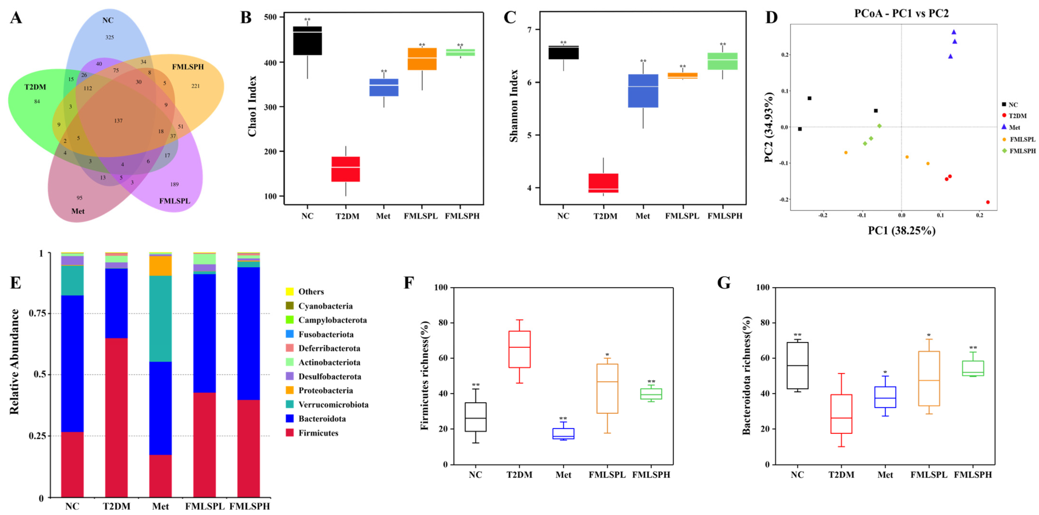
3.9. FMLSP’s Impact on the Taxonomic Diversity of Mice’s Gut Microbiota

As seen in Figure 5A, there were 137 OTUs common to all groups, and 325, 189, and 221 unique OTUs in the NC, FMLSPL, and FMLSPH groups, respectively. With just 84 distinct OTUs, the gut microbiota diversity in the T2DM group was significantly reduced. This implies that each mouse group’s gut microbiota varies more than the others. In addition, Figure 5B,C show the alpha diversity indices, including the Chao1 and Shannon indexes, which are positively correlated with community diversity [42]. The T2DM group’s Chao1 and Shannon indices were significantly lower than those of the NC group, while the FMLSP treatment significantly increased the Chao1 and Shannon diversity indexes. According to the findings of the β-diversity analysis in Figure 5D, PC1 and PC2 were responsible for 38.25% and 34.93%, respectively, of the total variation in the microbiota as measured by principal coordinate analysis (PcoA). The T2DM group’s mouse gut microbiota distribution was very different from that of the NC and FMLSPH groups, and it was more similar to that of the NC and FMLSPH groups. In summary, FMLSP restored the abundance and diversity of gut microbiota in diabetic mice to some extent.
Figure 5E displays changes in the relative abundance of gut microbiota at the phylum level for each group. Firmicutes, Bacteroidota, Verrucomicrobiota, Proteobacteria, and Actinobacteriota make up the majority of the gut microbiota in mice. At the phylum level, Firmicutes and Bacteroidota are the two most prevalent bacteria among them. Comparing the gut microbiota of mice in the T2DM group to that of the NC group, Figure 5F,G demonstrates that the quantity of Firmicutes was much greater, and the number of Bacteroidota was significantly lower. This is in line with Zhao et al. [43], who demonstrated that diabetic mice had fundamentally unbalanced gut flora. FMLSP treatment, conversely, ameliorated gut microbiota disruption in T2DM mice.

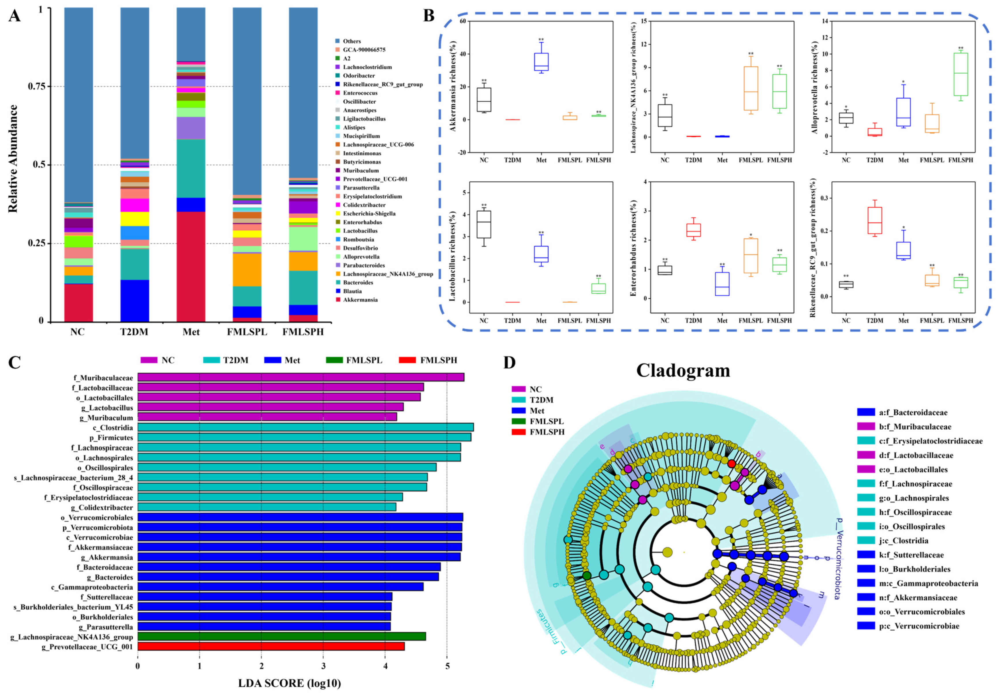
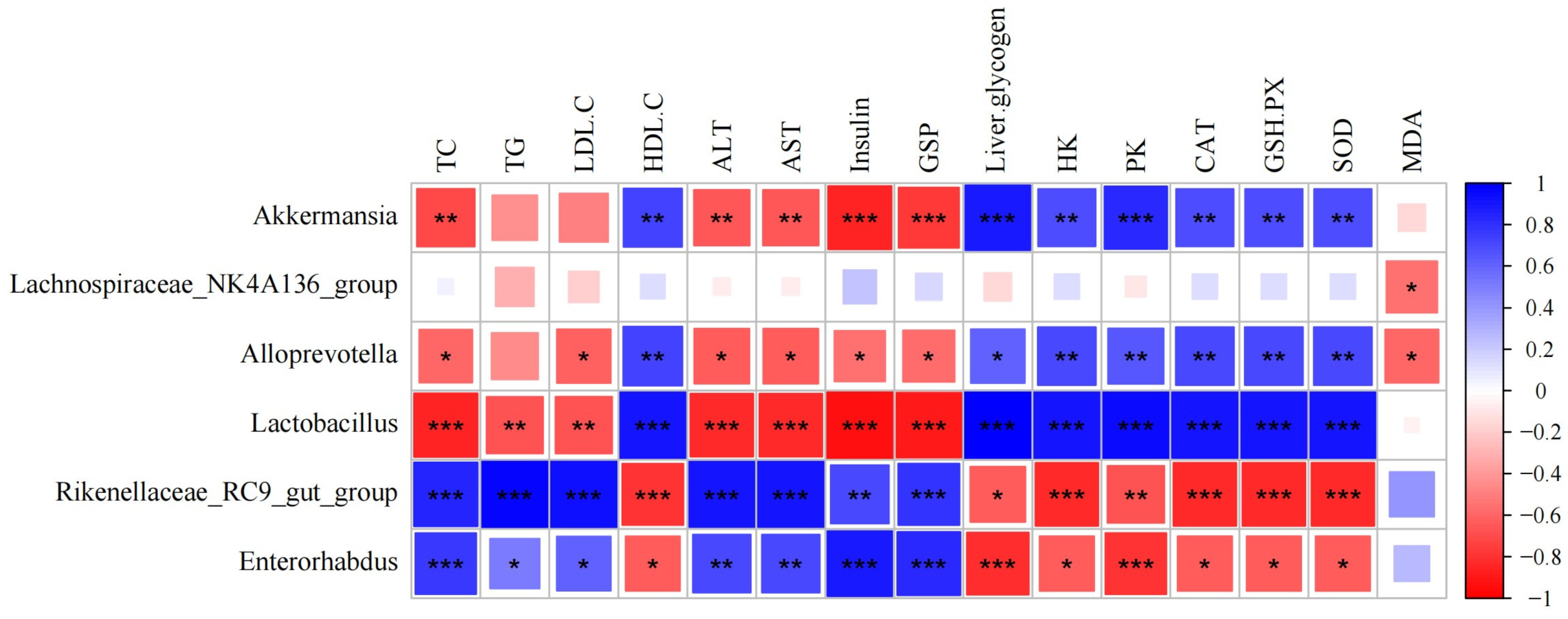
3.10. FMLSP Restores Gut Microbial Composition in Mice

We also noticed alterations in the gut microbiota’s composition at the genus level (Figure 6A). We found that FMLSP intervention significantly increased the abundance of beneficial bacteria such as Akkermansia, Lachnospiraceae_NK4A136_group, Alloprevotella, and Lactobacillus and markedly decreased the abundance of harmful bacteria such as Rikenellaceae_RC9_gut_group in T2DM mice (Figure 6B). The results suggest that FMLSP intervention increases the beneficial microorganisms that protect the body, decreases the presence of harmful microorganisms, and restores the balance of the gut microbiota.
According to Figure 6C,D from the LDA effect size (LEfSe), for the NC, T2DM, Met, FMLSPL, and FMLSPH treatment groups, 5, 9, 12, 1, and 1 bacterial clades showed statistically significant differences, respectively. Some of the bacterial taxa with large differences in abundance belonged to Oscillospiraceae and Lachnospiraceae. Meanwhile, the relationship between gut microbiota and physiological markers of diabetes mellitus is examined in Figure 7. Three beneficial bacteria, Akkermansia, Alloprevotella, and Lactobacillus, showed a significantly negative correlation with TC, TG, LDL-C, ALT, AST, insulin, and GSP and a significantly positive correlation with HDL-C, liver glycogen, HK, PK, CAT, GSH-PX, and SOD. However, Lachnospiraceae_NK4A136_group only showed a significantly negative correlation with the content of MDA. Rikenellaceae_RC9_gut_group, and Enterorhabdus, two harmful bacteria, were the opposite of beneficial bacteria. Consistent with previous experimental data, it indicates that FMLSP can ameliorate symptoms of IR, disorders of glucolipid metabolism, liver injury, and oxidative stress by regulating the abundance of gut microbiota. In summary, mice with diabetes have an imbalanced gut flora. Through the promotion of gut metabolism, FMLSP may mitigate the effects of diabetes on the body.

4. Discussion

In this work, frosted mulberry leaves were ground into a fine powder using a planetary ball mill. Additionally, the interventional therapeutic effects of FMLSP on T2DM mice were examined, along with the physicochemical and functional characteristics of the compound. The particle size of FMLSP was determined to be 8.52 μm, which meets the requirement of ultrafine powder. SEM results showed that the ultrafine crushed frosted mulberry leaf had better dispersion. The reduction in particle size led to agglomeration, but a higher degree of fragmentation was seen, which contributed to the release of bioactive components and bioavailability. Determination of the chemical content in FMLSP revealed a high flavonoid content of 91.30 mg/g, which was slightly higher than the levels of common mulberry leaves given in other reports [5,44]. Meanwhile, in vitro antioxidant experiments revealed that FMLSP had a good scavenging effect on DPPH as well as ABTS free radicals, which was close to the antioxidant effect of VC. This indicates that FMLSP has potential functional food properties. As catalysts in the process of digestion, α-glucosidase and α-amylase exhibit the ability to decompose carbohydrates into glucose that can be absorbed [32]. Inhibitors of α-amylase and α-glucosidase, which directly affect blood glucose levels through stepwise digestion of carbohydrates, have great value in the management of diabetes. In the current investigation, FMLSP was found to be effective in inhibiting the activities of α-glucosidase and α-amylase. This may be attributed to the main active ingredient in frosted mulberry leaves, 1-DNJ, which has the strong inhibitory activity of α-glucosidase and has been well-documented to have an antidiabetic effect [45].
T2DM is a metabolic disease characterized by chronic abnormally elevated blood glucose, and effective regulation of glucose homeostasis has become its core therapeutic goal [46]. Typical symptoms in T2DM mice are polyphagia, weight loss, and elevated blood glucose, which is consistent with the findings of Han et al. [47]. FMLSP improved symptoms of hyperphagia and weight loss in mice in this study while significantly reducing blood glucose in T2DM. The OGTT test is commonly used in clinics to rapidly diagnose diabetic conditions [48]. The results indicated that the high dose of FMLSP significantly improved glucose tolerance in diabetic mice. The reason may be that FMLSPs are rich in dietary fiber. Dietary fiber in the body can significantly increase the viscosity of the contents of the small intestine, forming a mesh structure that prevents the digestive juices from making full contact with the food. It reduces the rate of gastric emptying, delays the diffusion of glucose, and slows down the absorption of glucose in the intestines, thereby decreasing blood glucose levels [49,50,51].
Glucose metabolism and lipid metabolism are interrelated in the body, and glucose abnormalities in diabetic patients often cause abnormalities in lipid metabolism [52]. In this study, TC, TG, and LDL-C levels were elevated, HDL-C levels were decreased in T2DM mice, and FMLSP intervention significantly improved lipid metabolism disorders and reduced the risk of cardiovascular disease in T2DM mice. Meanwhile, the study found that FMLSP had an excellent effect on the removal of LDL-C in T2DM mice. This may be attributed to the high flavonoid content in FMLSP. It has been indicated that flavonoids in mulberry leaves attenuate the development of atherosclerotic lesions by inhibiting the oxidative modification of LDL [53]. Persistent hyperglycemia triggers progressive damage to pancreatic β-cell function with enhanced IR, leading to significant metabolic disturbances in insulin target organs. The metabolic disturbances result in abnormally high blood glucose levels, creating a vicious cycle [54]. In the study, insulin secretion was increased in T2DM mice, but the GSP remained at a high level, suggesting that T2DM mice had severe IR. After FMLSP intervention, insulin and GSP levels were decreased, and the islet volume and morphology were also improved. Based on previous reports, it was hypothesized that the active ingredients (flavonoids, 1-DNJ, polysaccharides, etc.) in FMLSP could ameliorate IR by protecting the structure of pancreatic islet cells and enhancing insulin sensitivity [55]. Meng et al. found that flavonoids in mulberry leaves could ameliorate IR by improving mitochondrial function in skeletal muscle [19].
Diabetic hyperglycemia boosts free radical generation in the body, resulting in oxidative stress. Conversely, it has been shown that oxidative stress plays a major role in the development of insulin resistance [56,57]. It was reported that antioxidant enzymes (SOD, GSH-Px, and CAT) defend against the formation of reactive oxygen species in response to oxidative stress, while MDA production is a marker of oxidative stress leading to liver injury [33]. Zhang et al. discovered that the multi-components in mulberry leaves could relieve oxidative stress. This is because they can enhance the activity of antioxidant enzymes and reduce MDA accumulation. Consequently, IR is improved, and hypoglycemic effects are achieved [58]. The results of the present study are in agreement with the findings of Zhang et al. [58]. It was hypothesized that it was because the frosted mulberry leaves were superfinely crushed to expose a large number of active substances such as phenols, free polyphenols, and flavonoids bound to dietary fibers, which allowed them to be fully utilized, thus decreasing reactive oxygen species levels and exerting an antioxidant effect in T2DM mice [59,60].
The liver serves as a major organ involved in glycolipid metabolism, and disorders of glycolipid metabolism due to IR can impair liver function [47]. Notably, circulating ALT and AST maintain basal concentrations in physiological conditions yet exhibit marked elevation upon hepatocyte membrane integrity compromise, establishing these enzymes as sensitive biomarkers for quantifying hepatocellular injury severity [61]. Elevated ALT and AST levels combined with the morphologic structure of liver tissues in T2DM mice suggested severe liver injury in T2DM mice, similar to previous studies [62]. In the present study, FMLSP had a protective effect on liver tissue. Glycogen is the storage form of glucose, and a decrease in glycogen stores is associated with disturbances in glucolipid metabolism due to IR [63]. And glycolysis is a key stage that organisms must undergo to metabolize glucose, and hexokinase and pyruvate kinase are key enzymes in the glycolytic pathway, and they play an important role in maintaining blood glucose homeostasis [47]. In the current study, T2DM impairs hepatic glycogen synthesis and attenuates glycolysis in mice. This means that glucose cannot be properly catabolized and metabolized, and therefore blood glucose levels are elevated [58]. FMLSP intervention improves glycolysis in T2DM mice, reduces hepatic glucose output, promotes hepatic glycogen synthesis, and thus reduces blood glucose levels, which ultimately facilitates the restoration of glucose homeostasis in T2DM mice.
Emerging evidence delineates a pathological cascade wherein hyperglycemia induces gut microbiota dysbiosis, subsequently driving metabolic dysregulation and chronic low-grade inflammation that collectively exacerbate diabetes mellitus progression [64]. It has been shown in previous studies that the ratio of Firmicutes to Bacteroidota increases as the severity of diabetic conditions changes [65]. In the current research, the T2DM group of mice had considerably higher Firmicute abundance and significantly lower Bacteroidota abundance, and this disruption of the intestinal-derived metabolic regulatory network accelerates the process of β-cell de-differentiation. Notably, the FMLSP treatment significantly ameliorated this intestinal flora disruption in T2DM mice. This may be due to the fact that FMLSP is rich in phenolics, which were previously shown to alter the abundance of critical gut microbiota at the phylum level by Yuan et al. [66]. Rodrigues et al. showed that Akkermansia in the gut has the potential to promote metabolic function in diabetic patients, and low concentrations of Akkermansia indicate a weakened gut barrier function [67]. In the present study, FMLSP treatment significantly increased the abundance of Akkermansia. Studies have demonstrated that Lachnospiraceae_NK4A136_group is associated with favorable metabolism and a healthy diet, which contributes to the degradation of dietary fiber for the prevention and treatment of diabetes [51]. FMLSP is rich in dietary fiber, which increases the abundance of Lachnospiraceae_NK4A136_group and promotes intestinal metabolism. Meanwhile it has been shown that Alloprevotella is associated with IR and is a key gut microbe involved in the hypoglycemic effect [68]. In addition, lactobacilli have been shown to repair the gut mucosal barrier to prevent the passage of harmful bacteria and regulate dyslipidemia due to long-term blood sugar metabolism disorders [69,70]. This research demonstrated that the FMLSP intervention improved T2DM by dramatically increasing the number of these two beneficial bacteria. The beneficial bacteria in the intestines of T2DM mice are reduced, but the abundance of some bacteria increases, such as Rikenellaceae_RC9_gut_group, and Enterorhabdus. It is reported that the Rikenellaceae_RC9_gut_group, Enterorhabdus as harmful bacteria are proven to cause colorectal cancer, gut inflammation, obesity, and diabetes [71]. In the research, FMLSP restored the equilibrium of the intestinal flora in T2DM mice and dramatically decreased the number of Enterorhabdus and the dangerous bacteria Rikenellaceae_RC9_gut_group. In conclusion, by influencing the quantity of both good and bad bacteria in the gut flora, FMLSP may help people with type 2 diabetes.

5. Conclusions

In summary, the results indicate that FMLSP possesses a protective effect on HFD/STZ-induced T2DM by improving disorders of glucolipid metabolism, alleviating oxidative stress phenomena, and regulating the balance of the gut microbiota. This study may reveal biological mechanisms by which FMLSP has a protective effect against T2DM, thus enhancing its therapeutic potential as a plant-based functional food ingredient in the treatment of T2DM. Furthermore, FMLSP exhibits potential as a functional food matrix for industrial applications, effectively enhancing both nutritional profiles and bioactive functionalities in food products. Consequently, FMLSP is expected to exert antidiabetic functional activity as a novel dietary supplement.

Supplementary Materials

The following supporting information can be downloaded at: https://www.mdpi.com/article/10.3390/app15073766/s1, Figure S1: Chromatogram.

Author Contributions

Conceptualization, J.W.; methodology, J.W.; software, J.W., Q.W. and G.Z.; validation, J.W.; formal analysis, J.W.; investigation, J.W.; data curation, J.W., Q.W. and G.Z.; writing—original draft preparation, J.W.; writing—review and editing, J.W. and R.S.; resources, J.L., L.S., M.J. and M.Y.; visualization, Q.W. and M.Y.; supervision, R.S.; project administration, R.S.; funding acquisition, R.S. All authors have read and agreed to the published version of the manuscript.

Funding

This research was funded by the Jinan 20 Rules of High School (No. 2020GXRC024) and the Qilu University of Technology 2022 major innovation project of the pilot of integration of science, education, and industry (No. 2022JBZ01-08).

Institutional Review Board Statement

The animal experiment was approved by the Animal Experiment Ethics Review Committee of Shandong Normal University (AEECSDNU2024006).

Informed Consent Statement

Not applicable.

Data Availability Statement

The original contributions presented in the study are included in the article, further inquiries can be directed to the corresponding author.

Conflicts of Interest

The authors declare no conflicts of interest.

References

  1. Taheri, S. Defining type 2 diabetes remission: KISS goodbye to confusion? Lancet Diabetes Endocrinol. 2021, 9, 806–808. [Google Scholar] [CrossRef] [PubMed]
  2. Rachdaoui, N. Insulin: The Friend and the Foe in the Development of Type 2 Diabetes Mellitus. Int. J. Mol. Sci. 2020, 21, 1770. [Google Scholar] [CrossRef]
  3. Atlas, D. International Diabetes Federation, 7th ed.; International Diabetes Federation: Brussels, Belgium, 2015; Volume 33. [Google Scholar]
  4. Jouad, H.; Haloui, M.; Rhiouani, H.; El Hilaly, J.; Eddouks, M. Ethnobotanical survey of medicinal plants used for the treatment of diabetes, cardiac and renal diseases in the North centre region of Morocco (Fez-Boulemane). J. Ethnopharmacol. 2001, 77, 175–182. [Google Scholar] [CrossRef] [PubMed]
  5. Sheng, Y.; Zheng, S.; Ma, T.; Zhang, C.; Ou, X.; He, X.; Xu, W.; Huang, K. Mulberry leaf alleviates streptozotocin-induced diabetic rats by attenuating NEFA signaling and modulating intestinal microflora. Sci. Rep. 2017, 7, 12041. [Google Scholar] [CrossRef]
  6. Allin, K.H.; Tremaroli, V.; Caesar, R.; Jensen, B.A.H.; Damgaard, M.T.F.; Bahl, M.I.; Licht, T.R.; Hansen, T.H.; Nielsen, T.; Dantoft, T.M.; et al. Aberrant intestinal microbiota in individuals with prediabetes. Diabetologia 2018, 61, 810–820. [Google Scholar] [CrossRef]
  7. Navab-Moghadam, F.; Sedighi, M.; Khamseh, M.E.; Alaei-Shahmiri, F.; Talebi, M.; Razavi, S.; Amirmozafari, N. The association of type II diabetes with gut microbiota composition. Microb. Pathog. 2017, 110, 630–636. [Google Scholar] [CrossRef] [PubMed]
  8. Cunningham, A.L.; Stephens, J.W.; Harris, D.A. Gut microbiota influence in type 2 diabetes mellitus (T2DM). Gut Pathog. 2021, 13, 50. [Google Scholar] [CrossRef]
  9. Sikalidis, A.K.; Maykish, A. The Gut Microbiome and Type 2 Diabetes Mellitus: Discussing A Complex Relationship. Biomedicines 2020, 8, 8. [Google Scholar] [CrossRef]
  10. Ling, X.; Hu, Y.; Hu, Y.; Meng, J. Analysis of chlorogenic acid and two flavonoids in mulberry leaves of different harvest periods and origins and HPLC fingerprint study for quality control. J. Food Compos. Anal. 2024, 132, 106284. [Google Scholar] [CrossRef]
  11. Chan, E.W.; Lye, P.Y.; Wong, S.K. Phytochemistry, pharmacology, and clinical trials of Morus alba. Chin. J. Nat. Med. 2016, 14, 17–30. [Google Scholar] [CrossRef]
  12. Ma, G.; Chai, X.; Hou, G.; Zhao, F.; Meng, Q. Phytochemistry, bioactivities and future prospects of mulberry leaves: A review. Food Chem 2022, 372, 131335. [Google Scholar] [CrossRef] [PubMed]
  13. Sugiyama, M.; Katsube, T.; Koyama, A.; Itamura, H. Seasonal Changes in Functional Component Contents in Mulberry (Morus alba L.) Leaves. Hortic. J. 2017, 86, 534–542. [Google Scholar] [CrossRef]
  14. Xu, D.-Q.; Cheng, S.-Y.; Zhang, J.-Q.; Lin, H.-F.; Chen, Y.-Y.; Yue, S.-J.; Tian, M.; Tang, Y.-P.; Zhao, Y.-C. Morus alba L. Leaves—Integration of Their Transcriptome and Metabolomics Dataset: Investigating Potential Genes Involved in Flavonoid Biosynthesis at Different Harvest Times. Front. Plant Sci. 2021, 12, 736332. [Google Scholar]
  15. Yu, X.; Zhu, Y.; Fan, J.; Wang, D.; Gong, X.; Ouyang, Z. Accumulation of Flavonoid Glycosides and UFGT Gene Expression in Mulberry Leaves (Morus alba L.) before and after Frost. Chem. Biodivers. 2017, 14, e1600496. [Google Scholar] [CrossRef] [PubMed]
  16. Liu, C.-H.; Liu, F.; Xiong, L. Medicinal parts of mulberry (leaf, twig, root bark, and fruit) and compounds thereof are excellent traditional Chinese medicines and foods for diabetes mellitus. J. Funct. Foods 2023, 106, 105619. [Google Scholar] [CrossRef]
  17. Bae, U.J.; Jung, E.S.; Jung, S.J.; Chae, S.W.; Park, B.H. Mulberry leaf extract displays antidiabetic activity in db/db mice via Akt and AMP-activated protein kinase phosphorylation. Food Nutr. Res. 2018, 62, 1473. [Google Scholar] [CrossRef]
  18. Jung, S.-H.; Han, J.-H.; Park, H.-S.; Lee, D.-H.; Kim, S.J.; Cho, H.S.; Kang, J.S.; Myung, C.-S. Effects of unaltered and bioconverted mulberry leaf extracts on cellular glucose uptake and antidiabetic action in animals. BMC Complement. Altern. Med. 2019, 19, 55. [Google Scholar] [CrossRef]
  19. Meng, Q.; Qi, X.; Fu, Y.; Chen, Q.; Cheng, P.; Yu, X.; Sun, X.; Wu, J.; Li, W.; Zhang, Q.; et al. Flavonoids extracted from mulberry (Morus alba L.) leaf improve skeletal muscle mitochondrial function by activating AMPK in type 2 diabetes. J. Ethnopharmacol. 2020, 248, 112326. [Google Scholar] [CrossRef]
  20. Usman, I.; Muzzamal, H.; Ali, I.; Muhammad, A.; Farhan, S.; Mehak, J.; Atka, A.; Iqra, A.; Entessar, A.J.; Saewan, S.A. Traditional and innovative approaches for the extraction of bioactive compounds. Int. J. Food Prop. 2022, 25, 1215–1233. [Google Scholar] [CrossRef]
  21. Huang, X.; Liang, K.-h.; Liu, Q.; Qiu, J.; Wang, J.; Zhu, H. Superfine grinding affects physicochemical, thermal and structural properties of Moringa Oleifera leaf powders. Ind. Crops Prod. 2020, 151, 112472. [Google Scholar] [CrossRef]
  22. Gao, W.; Chen, F.; Wang, X.; Meng, Q. Recent advances in processing food powders by using superfine grinding techniques: A review. Compr. Rev. Food Sci. Food Saf. 2020, 19, 2222–2255. [Google Scholar] [CrossRef] [PubMed]
  23. Zhang, J.; Dong, Y.; Nisar, T.; Fang, Z.; Wang, Z.-C.; Guo, Y. Effect of superfine-grinding on the physicochemical and antioxidant properties of Lycium ruthenicum Murray powders. Powder Technol. 2020, 372, 68–75. [Google Scholar] [CrossRef]
  24. Palavecino, P.M.; Penci, M.C.; Ribotta, P.D. Effect of planetary ball milling on physicochemical and morphological properties of sorghum flour. J. Food Eng. 2019, 262, 22–28. [Google Scholar] [CrossRef]
  25. Wang, F.; Zeng, J.; Gao, H.; Sukmanov, V. Effects of different physical technology on compositions and characteristics of bean dregs. Innov. Food Sci. Emerg. Technol. 2021, 73, 102789. [Google Scholar] [CrossRef]
  26. Ma, X.; Sang, X.; Yan, C.; Zhang, Y.; Bi, J.; Zhang, G.; Hao, H.; Hou, H. Dynamics of Bacterial Composition and Association with Quality Formation and Biogenic Amines Accumulation during Fish Sauce Spontaneous Fermentation. Appl. Environ. Microbiol. 2022, 88, e00690-00622. [Google Scholar] [CrossRef]
  27. Tu, J.; Liu, G.; Jin, Y.; Tang, C.; Yao, T.; Zhuo, J.; Li, Q.; Liu, L.; Wang, J. Enrichment of γ-aminobutyric acid in mulberry leaves and the inhibitory effects of the water extract on ACE and α-glucosidase activity. Ind. Crops Prod. 2022, 177, 114485. [Google Scholar] [CrossRef]
  28. Zeng, C.; Ye, G.; Li, G.; Cao, H.; Wang, Z.; Ji, S. RID serve as a more appropriate measure than phenol sulfuric acid method for natural water-soluble polysaccharides quantification. Carbohydr. Polym. 2022, 278, 118928. [Google Scholar] [CrossRef] [PubMed]
  29. Vichasilp, C.; Nakagawa, K.; Sookwong, P.; Higuchi, O.; Luemunkong, S.; Miyazawa, T. Development of high 1-deoxynojirimycin (DNJ) content mulberry tea and use of response surface methodology to optimize tea-making conditions for highest DNJ extraction. LWT—Food Sci. Technol. 2012, 45, 226–232. [Google Scholar] [CrossRef]
  30. Faidi, A.; Becheikh, M.E.H.; Lassoued, M.A.; Stumbé, J.F.; Safta, F.; Sfar, S. Isolation of sodium alginate-like polysaccharide from Padina pavonica: Optimization, characterization and antioxidant properties. J. Mol. Struct. 2025, 1321, 139737. [Google Scholar] [CrossRef]
  31. Yu, W.; Wang, J.; Xiong, Y.; Liu, J.; Baranenko, D.; Zhang, Y.; Lu, W. In vivo absorption, in vitro simulated digestion, and fecal fermentation properties of Imperata cylindrica polysaccharides and their effects on gut microbiota. Food Chem. 2024, 461, 140773. [Google Scholar] [CrossRef]
  32. Awosika, T.O.; Aluko, R.E. Inhibition of the in vitro activities of α-amylase, α-glucosidase and pancreatic lipase by yellow field pea (Pisum sativum L.) protein hydrolysates. Int. J. Food Sci. Technol. 2019, 54, 2021–2034. [Google Scholar] [CrossRef]
  33. Yang, M.; Lv, J.; Yang, J.; Yang, S.; Wang, F.; Wang, Y.; Zhang, C. Effects of Codonopsis pilosula crude polysaccharides by hypoglycemic and modulating gut microbiome in a high-fat diet and streptozotocin-induced mouse model of T2DM. J. Funct. Foods 2023, 111, 105893. [Google Scholar] [CrossRef]
  34. Lin, S.; Jin, X.; Gao, J.; Qiu, Z.; Ying, J.; Wang, Y.; Dong, Z.; Zhou, W. Impact of wheat bran micronization on dough properties and bread quality: Part I—Bran functionality and dough properties. Food Chem. 2021, 353, 129407. [Google Scholar] [CrossRef] [PubMed]
  35. Chen, X.; Guo, M.; Sang, Y.; Sun, J. Effect of ball-milling treatment on the structure, physicochemical properties and allergenicity of proteins from oyster (Crassostrea gigas). LWT 2022, 166, 113803. [Google Scholar] [CrossRef]
  36. Xu, S.; Li, X.; Podio, N.S.; Han, Y.; Wang, X.-Y.; Gong, E.S. Black rice phenolics alleviate T2DM through comprehensive regulation of oxidative stress, metabolic pathways and gut microbiota. Food Biosci. 2024, 62, 105494. [Google Scholar] [CrossRef]
  37. Yang, C.; Ma, L.; Ma, J.; Liu, S.; Fu, J.; Fan, Y.; Liu, Y. Lycium barbarum leaf flavonoids ameliorate high fructose induced insulin resistance in mice by regulating blood glucose and gut microbiota composition. Food Biosci. 2024, 62, 105087. [Google Scholar] [CrossRef]
  38. Yang, H.-B.; Song, J.-Y.; Xu, C.; Li, J.; Zhang, C.; Xie, S.; Teng, C.-l. Interventional effects of Pueraria oral liquid on T2DM rats and metabolomics analysis. Biomed. Pharmacother. 2024, 175, 116780. [Google Scholar] [CrossRef]
  39. Semaming, Y.; Kukongviriyapan, U.; Kongyingyoes, B.; Thukhammee, W.; Pannangpetch, P. Protocatechuic Acid Restores Vascular Responses in Rats With Chronic Diabetes Induced by Streptozotocin. Phytother. Res. 2016, 30, 227–233. [Google Scholar] [CrossRef]
  40. Furman, B.L. Streptozotocin-Induced Diabetic Models in Mice and Rats. Curr. Protoc. 2021, 1, e78. [Google Scholar] [CrossRef]
  41. Huang, W.; Huang, G.-P.; Zhang, L.-X.; da Yu, E.; Yang, W.-K.; Ye, M.; Zou, S.-Q.; Ni, L.; He, H.-Q. Lignan-rich extract from Cinnamomum camphora leaf attenuates metabolic syndrome by modulating glycolipid metabolism and gut microbiota in T2DM mice. Phytomedicine 2024, 135, 156118. [Google Scholar] [CrossRef]
  42. Zaheer, R.; Noyes, N.; Ortega Polo, R.; Cook, S.R.; Marinier, E.; Van Domselaar, G.; Belk, K.E.; Morley, P.S.; McAllister, T.A. Impact of sequencing depth on the characterization of the microbiome and resistome. Sci. Rep. 2018, 8, 5890. [Google Scholar] [CrossRef]
  43. Zhao, L.; Lou, H.; Peng, Y.; Chen, S.; Zhang, Y.; Li, X. Comprehensive relationships between gut microbiome and faecal metabolome in individuals with type 2 diabetes and its complications. Endocrine 2019, 66, 526–537. [Google Scholar] [CrossRef]
  44. Hao, J.-Y.; Wan, Y.; Yao, X.-H.; Zhao, W.-G.; Hu, R.-Z.; Chen, C.; Li, L.; Zhang, D.-Y.; Wu, G.-H. Effect of different planting areas on the chemical compositions and hypoglycemic and antioxidant activities of mulberry leaf extracts in Southern China. PLoS ONE 2018, 13, e0198072. [Google Scholar] [CrossRef]
  45. Li, Y.; Zhong, S.; Yu, J.; Sun, Y.; Zhu, J.; Ji, D.; Wu, C. The mulberry-derived 1-deoxynojirimycin (DNJ) inhibits high-fat diet (HFD)-induced hypercholesteremia and modulates the gut microbiota in a gender-specific manner. J. Funct. Foods 2019, 52, 63–72. [Google Scholar] [CrossRef]
  46. He, X.; Wang, C.e.; Zhu, Y.; Jiang, X.; Qiu, Y.; Yin, F.; Xiong, W.; Liu, B.; Huang, Y. Spirulina compounds show hypoglycemic activity and intestinal flora regulation in type 2 diabetes mellitus mice. Algal Res. 2022, 66, 102791. [Google Scholar] [CrossRef]
  47. Han, C.; Kong, X.; Xia, X.; Huang, X.; Mao, Z.; Han, J.; Shi, F.; Liang, Y.; Wang, A.; Zhang, F. Effects of ginseng peptides on the hypoglycemic activity and gut microbiota of a type 2 diabetes mellitus mice model. J. Funct. Foods 2023, 111, 105897. [Google Scholar] [CrossRef]
  48. Virtue, S.; Vidal-Puig, A. GTTs and ITTs in mice: Simple tests, complex answers. Nat. Metab. 2021, 3, 883–886. [Google Scholar] [CrossRef]
  49. Li, M.; Ma, S. A review of healthy role of dietary fiber in modulating chronic diseases. Food Res. Int. 2024, 191, 114682. [Google Scholar] [CrossRef]
  50. Gill, S.K.; Rossi, M.; Bajka, B.; Whelan, K. Dietary fibre in gastrointestinal health and disease. Nat. Rev. Gastroenterol. Hepatol. 2021, 18, 101–116. [Google Scholar] [CrossRef]
  51. Mo, Z.; Zhan, M.; Yang, X.; Xie, P.; Xiao, J.; Cao, Y.; Xiao, H.; Song, M. Fermented dietary fiber from soy sauce residue exerts antidiabetic effects through regulating the PI3K/AKT signaling pathway and gut microbiota-SCFAs-GPRs axis in type 2 diabetic mellitus mice. Int. J. Biol. Macromol. 2024, 270, 132251. [Google Scholar] [CrossRef]
  52. Jiang, S.; Young, J.L.; Wang, K.; Qian, Y.; Cai, L. Diabetic-induced alterations in hepatic glucose and lipid metabolism: The role of type 1 and type 2 diabetes mellitus (Review). Mol. Med. Rep. 2020, 22, 603–611. [Google Scholar] [CrossRef] [PubMed]
  53. Enkhmaa, B.; Shiwaku, K.; Katsube, T.; Kitajima, K.; Anuurad, E.; Yamasaki, M.; Yamane, Y. Mulberry (Morus alba L.) Leaves and Their Major Flavonol Quercetin 3-(6-Malonylglucoside) Attenuate Atherosclerotic Lesion Development in LDL Receptor-Deficient Mice1. J. Nutr. 2005, 135, 729–734. [Google Scholar] [CrossRef]
  54. Chen, J.; Meng, X. Aronia melanocarpa Anthocyanin Extracts Improve Hepatic Structure and Function in High-Fat Diet-/Streptozotocin-Induced T2DM Mice. J. Agric. Food Chem. 2022, 70, 11531–11543. [Google Scholar] [CrossRef]
  55. Zhang, L.; Su, S.; Zhu, Y.; Guo, J.; Guo, S.; Qian, D.; Ouyang, Z.; Duan, J.-A. Mulberry leaf active components alleviate type 2 diabetes and its liver and kidney injury in db/db mice through insulin receptor and TGF-β/Smads signaling pathway. Biomed. Pharmacother. 2019, 112, 108675. [Google Scholar] [CrossRef] [PubMed]
  56. Qi, M.Y.; Wang, X.T.; Xu, H.L.; Yang, Z.L.; Cheng, Y.; Zhou, B. Protective effect of ferulic acid on STZ-induced diabetic nephropathy in rats. Food Funct. 2020, 11, 3706–3718. [Google Scholar] [CrossRef]
  57. Fatima, F.; Zakaria, M. Chapter 8—Oxidative stress and insulin resistance. In Fundamental Principles of Oxidative Stress in Metabolism and Reproduction; Alam, F., Rehman, R., Eds.; Academic Press: Cambridge, MA, USA, 2024; pp. 123–137. [Google Scholar]
  58. Zhang, Y.; Li, L.; Chai, T.; Xu, H.; Du, H.-y.; Jiang, Y. Mulberry leaf multi-components exert hypoglycemic effects through regulation of the PI-3K/Akt insulin signaling pathway in type 2 diabetic rats. J. Ethnopharmacol. 2024, 319, 117307. [Google Scholar] [CrossRef]
  59. Hu, T.-G.; Wen, P.; Liu, J.; Long, X.-S.; Liao, S.-T.; Wu, H.; Zou, Y.-X. Combination of mulberry leaf and oat bran possessed greater hypoglycemic effect on diabetic mice than mulberry leaf or oat bran alone. J. Funct. Foods 2019, 61, 103503. [Google Scholar] [CrossRef]
  60. Wang, R.; Wang, L.; Wu, H.; Zhang, L.; Hu, X.; Li, C.; Liu, S. Noni (Morinda citrifolia L.) fruit phenolic extract supplementation ameliorates NAFLD by modulating insulin resistance, oxidative stress, inflammation, liver metabolism and gut microbiota. Food Res. Int. 2022, 160, 111732. [Google Scholar] [CrossRef]
  61. Carobene, A.; Braga, F.; Roraas, T.; Sandberg, S.; Bartlett, W.A. A systematic review of data on biological variation for alanine aminotransferase, aspartate aminotransferase and γ-glutamyl transferase. Clin. Chem. Lab. Med. 2013, 51, 1997–2007. [Google Scholar] [CrossRef]
  62. Khajuria, P.; Raghuwanshi, P.; Rastogi, A.; Koul, A.L.; Zargar, R.; Kour, S. Hepatoprotective effect of seabuckthorn leaf extract in streptozotocin induced diabetes mellitus in Wistar rats. Indian J. Anim. Res. 2018, 52, 1745–1750. [Google Scholar] [CrossRef]
  63. Zhong, R.-F.; Liu, C.-J.; Hao, K.-X.; Fan, X.-D.; Jiang, J.-G. Polysaccharides from Flos Sophorae Immaturus ameliorates insulin resistance in IR-HepG2 cells by co-regulating signaling pathways of AMPK and IRS-1/PI3K/AKT. Int. J. Biol. Macromol. 2024, 280, 136088. [Google Scholar] [CrossRef]
  64. Tanase, D.M.; Gosav, E.M.; Neculae, E.; Costea, C.F.; Ciocoiu, M.; Hurjui, L.L.; Tarniceriu, C.C.; Maranduca, M.A.; Lacatusu, C.M.; Floria, M.; et al. Role of Gut Microbiota on Onset and Progression of Microvascular Complications of Type 2 Diabetes (T2DM). Nutrients 2020, 12, 3719. [Google Scholar] [CrossRef]
  65. Ley, R.E.; Bäckhed, F.; Turnbaugh, P.; Lozupone, C.A.; Knight, R.D.; Gordon, J.I. Obesity alters gut microbial ecology. Proc. Natl. Acad. Sci. USA 2005, 102, 11070–11075. [Google Scholar] [CrossRef] [PubMed]
  66. Yuan, Y.; Zheng, Y.; Zhou, J.; Geng, Y.; Zou, P.; Li, Y.; Zhang, C. Polyphenol-Rich Extracts from Brown Macroalgae Lessonia trabeculate Attenuate Hyperglycemia and Modulate Gut Microbiota in High-Fat Diet and Streptozotocin-Induced Diabetic Rats. J. Agric. Food Chem. 2019, 67, 12472–12480. [Google Scholar] [CrossRef]
  67. Rodrigues, V.F.; Elias-Oliveira, J.; Pereira, Í.S.; Pereira, J.A.; Barbosa, S.C.; Machado, M.S.G.; Carlos, D. Akkermansia muciniphila and Gut Immune System: A Good Friendship That Attenuates Inflammatory Bowel Disease, Obesity, and Diabetes. Front. Immunol. 2022, 13, 934695. [Google Scholar] [CrossRef]
  68. Guo, P.; Zeng, M.; Zhang, Y.; Zhang, Z.; Wu, Y.; Ye, K.; Chang, F.; Wang, Y.; Zheng, X.; Feng, W. Integration strategies involving 16S rDNA sequencing combined with untargeted metabolomics revealed the mechanism of Selaginella tamariscina (Beauv.) Spring in db/db diabetic mice. Biomed. Pharmacother. 2024, 180, 117546. [Google Scholar] [CrossRef]
  69. Robles-Vera, I.; Toral, M.; de la Visitación, N.; Sánchez, M.; Romero, M.; Olivares, M.; Jiménez, R.; Duarte, J. The Probiotic Lactobacillus fermentum Prevents Dysbiosis and Vascular Oxidative Stress in Rats with Hypertension Induced by Chronic Nitric Oxide Blockade. Mol. Nutr. Food Res. 2018, 62, 1800298. [Google Scholar] [CrossRef]
  70. Zhang, X.; Chen, S.; Zhang, M.; Ren, F.; Ren, Y.; Li, Y.; Liu, N.; Zhang, Y.; Zhang, Q.; Wang, R. Effects of Fermented Milk Containing Lacticaseibacillus paracasei Strain Shirota on Constipation in Patients with Depression: A Randomized, Double-Blind, Placebo-Controlled Trial. Nutrients 2021, 13, 2238. [Google Scholar] [CrossRef]
  71. Xia, T.; Zhang, Z.; Zhao, Y.; Kang, C.; Zhang, X.; Tian, Y.; Yu, J.; Cao, H.; Wang, M. The anti-diabetic activity of polyphenols-rich vinegar extract in mice via regulating gut microbiota and liver inflammation. Food Chem. 2022, 393, 133443. [Google Scholar] [CrossRef]
Scheme 1. Illustration of the preparation of frosted mulberry leaves superfine powder (FMLSP) and investigation of hypoglycemic activity.
Applsci 15 03766 sch001
Figure 1. Structural characterization and functional properties of FMLSP. (A) SEM results for FMLSP (×1000); (B) SEM results for FMLSP (×5000); scavenging rate of DPPH (C) and ABTS (D) radicals by different concentrations of FMLSP; release of total flavonoids (E) and total polyphenols (F) from FMLSP during in vitro digestion; (G) rate at which varying doses of FMLSP inhibit the activities of α-amylase and α-glucosidase.
Applsci 15 03766 g001
Figure 2. Effect of FMLSP on body weight, FBG and organ index in T2DM mice. (A) Body weight curves; (B) food intake; (C) FBG; (D) OGTT; (E) AUC of OGTT; (F) liver index; (G) pancreas index. (*) p < 0.05 and (**), p < 0.01 vs. T2DM group.
Applsci 15 03766 g002
Figure 3. Effect of FMLSP on glycolipid metabolism in T2DM mice. (A) Serum of TC; (B) serum of TG; (C) serum of LDL-C; (D) serum of HDL-C; (E) insulin content; (F) GSP content; (G) liver glycogen content; (H) activity of HK; (I) activity of PK. (*) p < 0.05 and (**), p < 0.01 vs. T2DM group.
Applsci 15 03766 g003
Figure 4. Effects of FMLSP on the antioxidant system and on the liver and pancreas of T2DM mice. (A) Activity of CAT; (B) activity of GSH-PX; (C) activity of SOD; (D) MDA content; (E) activity of ALT; (F) activity of AST; (G) pictures of H&E-stained liver tissue (×200); and (H) images of pancreas tissue H&E staining (×200). (*) p < 0.05 and (**), p < 0.01 vs. T2DM group.
Applsci 15 03766 g004
Figure 5. Effect of FMLSP on the taxonomic diversity of gut microflora in T2DM mice (A) OUT; (B) Chao1 index; (C) Shannon index; (D) PCoA; (E) abundance at the phylum level of gut microbiota; (F) Firmicute richness; and (G) Bacteroidota richness. (*) p < 0.05 and (**), p < 0.01 vs. T2DM group.
Applsci 15 03766 g005
Figure 6. Effect of FMLSP on gut microbial composition of T2DM mice. (A) Abundance in genus level; (B) six bacterial species at the genus level; and (C,D) linear discriminative analysis (LDA) effect size (LEfSe) analyses. (*) p < 0.05 and (**), p < 0.01 vs. T2DM group.
Applsci 15 03766 g006
Figure 7. Spearman correlation analysis of gut bacterial communities and the diabetes indices. * p < 0.05, ** p < 0.01, *** p < 0.001.
Applsci 15 03766 g007
Table 1. Animal experimental grouping and drug administration.
GroupFeed TypeGavage DrugDose
NCNormal dietSterile water0.4 mL
T2DMHFDSterile water0.4 mL
MetHFDMetformin hydrochloride0.5 g/kg
FMLSPLHFDFMLSP1.0 g/kg
FMLSPHHFDFMLSP2.0 g/kg
Table 2. FMLSP particle size distribution.
D10%/μmD50%/μmD90%/μmD [4,3]/μm
4.21 ± 0.168.09 ± 0.2018.52 ± 0.578.52 ± 0.33
Table 3. The chemical composition content of FMLSP.
CompoundContent (mg/g)CompoundContent (mg GAE/g dw)
Ash82.28 ± 0.04Polyphenol50.41 ± 1.04
Protein227.52 ± 0.07Flavonoid91.30 ± 2.92
Fat31.93 ± 0.08Polysaccharides58.41 ± 1.39
Dietary fiber305.18 ± 5.281-DNJ0.89 ± 0.008
Disclaimer/Publisher’s Note: The statements, opinions and data contained in all publications are solely those of the individual author(s) and contributor(s) and not of MDPI and/or the editor(s). MDPI and/or the editor(s) disclaim responsibility for any injury to people or property resulting from any ideas, methods, instructions or products referred to in the content.

