Article

In Vitro Protective Effects of Resveratrol-Loaded Pluronic Micelles Against Hydrogen Peroxide-Induced Oxidative Damage in U87MG Glioblastoma Cells

1
Institute of Experimental Morphology, Pathology and Anthropology with Museum, Bulgarian Academy of Sciences, 1113 Sofia, Bulgaria
2
Institute of Neurobiology, Bulgarian Academy of Sciences, 1113 Sofia, Bulgaria
3
Institute of Molecular Biology “Acad. R. Tsanev”, Bulgarian Academy of Sciences, 1113 Sofia, Bulgaria
4
Faculty of Pharmacy, Medical University of Sofia, 1000 Sofia, Bulgaria
*
Authors to whom correspondence should be addressed.
Appl. Sci. 2025, 15(6), 2995; https://doi.org/10.3390/app15062995
Submission received: 31 January 2025 / Revised: 21 February 2025 / Accepted: 7 March 2025 / Published: 10 March 2025

Abstract

:
Numerous studies reported that resveratrol (RVT) exhibits strong antioxidant and cytoprotective effects in brain pathologies, but its low solubility and bioavailability limit its therapeutic potential. Encapsulation of RVT in nanoparticles offers a promising strategy to enhance its effectiveness. The aim of this study was to evaluate the in vitro cytoprotective, DNA protective, and antioxidant capacity of resveratrol-loaded Pluronic (P123/F127) micelles. The effects of micellar (mRVT, water dispersion) and pure RVT (30% hydroethanolic solution) were compared in glioblastoma U87MG cells with H2O2-induced oxidative damage. The cells were pretreated with mRVT or pure RVT (1, 3, 10, and 30 µM) for 24 h before cell damage. The cell viability, DNA damage, acetylcholine esterase (AChE) inhibitory activity, and some biomarkers for oxidative stress like lipid peroxidation (LPO), superoxide dismutase (SOD), and catalase (CAT) were evaluated. In addition, the cellular uptake efficiency of the micelles (50 nm) was tracked using red fluorescent rhodamine B as a marker. Our findings revealed that the micelles were localized in the cytoplasm of U87MG cells within 1 h of incubation. Empty micelles, mRVT, and RVT did not reduce the viability of U87MG cells after 24 h incubation and protect them from H2O2 exposure. The most effective treatment was with mRVT (1 and 3 µM), which significantly reduced the DNA damage index, maintained LPO levels close to the control, and normalized the activities of AChE, SOD, and CAT that were disrupted by H2O2 treatment. These promising results highlight the feasibility and advantages of using resveratrol-loaded nanoparticles for therapeutic applications.

1. Introduction

Oxidative stress (OS) in biological systems is characterized by a set of alterations at molecular, cellular, and tissue levels and is part of the pathogenesis of acute or chronic neurodegenerative processes [1,2]. It arises from an imbalance between oxidants and antioxidants, caused either by elevated levels of reactive oxygen species (ROS) and reactive nitrogen species (RNS) or by dysfunction of the antioxidant system [3,4,5]. At physiological concentrations, ROS play an important regulatory role in different biological processes as gene expression, apoptosis, and signal transduction [6]. However, an uncontrolled increase in the levels of reactive species triggers a chain of radical reactions, disrupting cellular redox homeostasis and causing serious damage to cellular macromolecules. In one non-specific way, ROS can cause nucleic acid and lipid oxidation, protein misfolding, and aggregation, ultimately compromising cellular integrity and function and leading to apoptosis [7,8,9]. Hydrogen peroxide, along with superoxide anions and hydroxyl radicals, are among the most harmful ROS. Processes such as amyloid aggregation [10,11], dopamine oxidation [12], and brain ischemia/reperfusion injury [13] are closely related to excessive hydrogen peroxide production, while an overabundance of superoxide anions is associated with N-methyl-D-aspartate (NMDA)-induced neurotoxicity [1,2].
The brain’s high oxygen consumption and lipid content make it particularly vulnerable to damage from ROS and RNS. Markers of lipid peroxidation, such as malondialdehyde and 4-hydroxynonenal, have been detected in the brain of patients diagnosed with neurodegenerative disorders as a Parkinson’s or Alzheimer’s diseases [14]. Antioxidants are molecules that play a crucial role in managing cellular protection against oxidative stress by either preventing free radical formation or neutralizing them during their propagation phase [15].
From a scientific standpoint, natural antioxidants show great promise in aiding the prevention and treatment of neurodegenerative diseases [16].
Resveratrol (RVT, trans-3,5,4′-trihydroxystilbene) was isolated for the first time from Takaoka in 1940 from the root of Veratrum grandiflorum [17]. Since then it has been discovered in more than 70 plant species, especially in highly pigmented vegetables and fruits as a grape, mulberries, bilberries, etc. [18]. As a polyphenolic compound and phytoalexin, it has demonstrated several biological activities, including anti-inflammatory, vasorelaxing, anticancer, antiaging, and antiallergenic properties [19,20,21,22]. Resveratrol is among the most extensively studied antioxidant compounds [22,23]. Its strong antioxidant properties have been demonstrated in numerous in vitro and in vivo studies. The cytoprotective effects of resveratrol (RVT) against H2O2-induced apoptosis in Caco-2 cells are associated with decreased malondialdehyde (MDA) levels and upregulated expression of superoxide dismutase (SOD) and heme oxygenase-1 (HO-1), contributing to intracellular ROS scavenging and reduction [24]. Additionally, RVT (at concentrations of 5–25 µM) has been shown to mitigate Aβ-induced oxidative damage and inhibit apoptosis in various in vitro oxidative stress models, including PC12 cells, human umbilical vein endothelial cells (HUVECs), SH-SY5Y neuroblastoma cells, and hippocampal neuronal cultures [25,26,27,28,29]. In diabetic patients, RVT treatment reduces advanced glycation end product (AGE)-induced oxidative stress and apoptosis [30]. In rats with ischemic spinal cord damage, RVT reduces plasma MDA levels while enhancing the enzymatic activity of superoxide dismutase (SOD) and catalase (CAT) [31].
The limited bioavailability and poor stability of RVT significantly constrain its practical application, despite its broad spectrum of pharmacological effects [32,33,34]. Nanoformulations, as advanced drug delivery systems, have been developed to address these challenges by enhancing RVT solubility and preserving its biological activity [35].
Pluronics® are triblock copolymers recognized by the US Food and Drug Administration as safe and biocompatible compounds [36]. Composed of propylene oxide (PO) and ethylene oxide (EO), they can self-assemble into micellar structures, enabling the encapsulation of hydrophobic drugs and enhancing their therapeutic efficacy [37,38,39,40]. Yordanov et al. [41] achieved high encapsulation efficiency (84%) and enhanced neuroprotective effect of cannabidiol in neuroblastoma cells (SH-SY5Y and Neuro-2a) by its loading in Pluronic micelles. The small size and the narrow size distribution of such systems could enable their penetration across the blood–brain barrier [42]. Mixed Pluronic micelles have also been studied as a resveratrol carrier system in our previous research [43]. The referenced study demonstrated that resveratrol loaded micelles (mRVT) exhibited greater pharmacological efficacy than the pure compound in a rat model of experimental dementia. This was evidenced by its enhanced AChE-inhibitory activity and increased noradrenaline neurotransmission. Furthermore, mRVT activated the BDNF/CREB signaling pathway and exerted significant anti-apoptotic effects in the cortical and hippocampal regions. These findings highlight the superior neuroprotective potential of mRVT and its improved effectiveness in supporting cognitive function, including learning and memory.
The current study aimed to evaluate and compare the in vitro protective effects of resveratrol-loaded Pluronic (P123/F127) micelles and pure resveratrol against H2O2-induced oxidative damage in the U87MG glioblastoma cell line. For this purpose, cell viability, DNA protection, acetylcholinesterase activity, and antioxidant capacity of RVT and mRVT were examined. Additionally, the intracellular accumulation of mRVT was assessed using fluorescence microscopy.

2. Materials and Methods

2.1. Materials

Trans-resveratrol was purchased from Sigma Chemical Co. (Schnelldorf, Germany). Pluronic® F127 (PEO101PPO56PEO101) and Pluronic® P123 (PEO20PPO70PEO20) were purchased from BASF (Ludwigshafen, Germany). The Dulbecco’s modified eagle’s medium (DMEM), 3-(4,5-Dimethylthiazol-2-yl)-2,5-diphenyltetrazolium bromide (MTT), phosphate-buffered saline (PBS), Trypsin-EDTA solution, hydrogen peroxide, rhodamine B, and dimethyl sulfoxide (DMSO) were purchased from Sigma-Aldrich (Merck KGaA, Darmstadt, Germany), fetal bovine serum (FBS) from (Gibso/BRL, Grand Island, NY, USA). Penicillin and streptomycin, L-glutamine, and 4′,6-diamidino-2-phenylindole (DAPI) were purchased from AppliChem, Darmstadt, Germany. The plastic disposable consumables were purchased from Orange Scientific (Braine-l’Alleud, Belgium).

2.2. Formulation of Resveratrol and Rhodamine B-Loaded Micelles

Resveratrol or rhodamine B were loaded in Pluronic micelles via the film hydration method as described previously [43]. In brief, a 1:10 (w/w) ratio between resveratrol/rhodamine B and the polymers (Pluronic F127:Pluronic P123, 1:1 (w/w)) was applied. Briefly, the polymers were dissolved in methanol and the antioxidant or the dye was added to the mixture. Thereafter, the methanol was evaporated at room temperature until the formation of a thin film. The film was then redispersed in water and filtered (0.2 µm).

2.3. Cell Line and Cultivation Condition
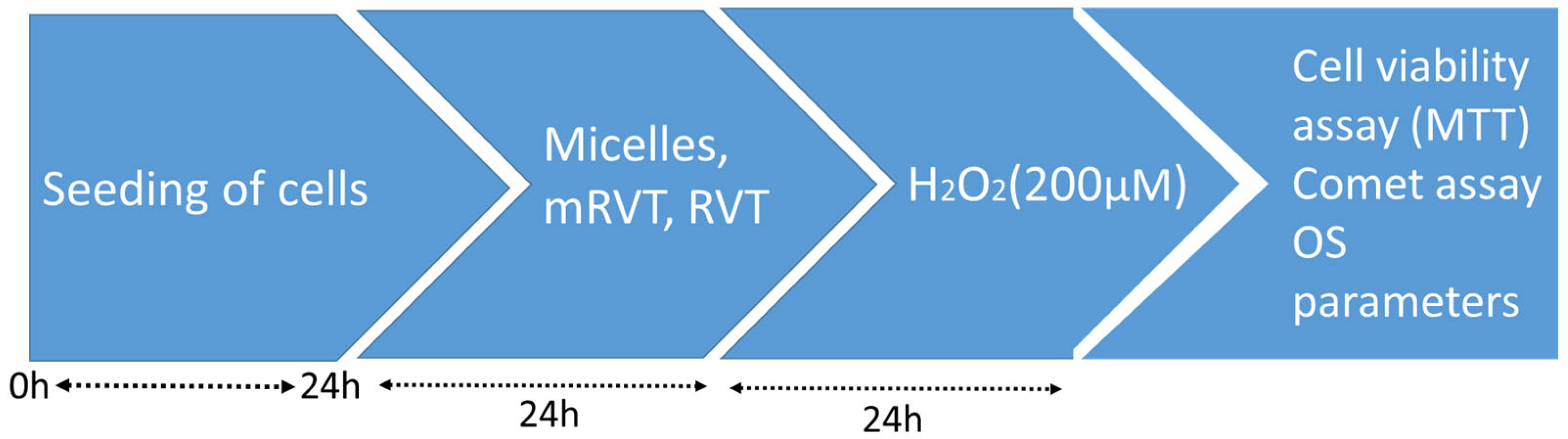
The U87MG human glioblastoma cell line (CLS Cell Lines Service, Eppelheim, Germany) was cultivated in complete DMEM supplemented with L-glutamine (2 mM), penicillin (100 U/mL), streptomycin (100 µg/mL), and 10% FBS. The cells were maintained in 75 cm2 flasks in a CO2 incubator (Thermo Electron Corporation, Stockport, OH, USA) at 37 °C.

2.4. Cell Treatment

The cells were seeded in a 96- or 24-well plate in а density of 10,000 cells/well or 40,000 cells/well, respectively, and incubated for 24 h. The toxicity of empty micelles (0.2–30 μg/mL), RVT, and mRVT (1, 3, 10, or 30 μM) was evaluated via an MTT test after 24 h of incubation. For the experiments in the model of oxidative damage, the cells were pretreated with resveratrol and resveratrol-loaded micelles for 24 h in concentrations of 1, 3, 10, and 30 µM. The culture medium was replaced with medium supplemented with H2O2 (200 µM) for another 24 h. Thereafter, different assays were performed as described in Figure 1.

2.5. Cell Viability

Cell viability was determined by the standard colorimetric MTT assay, in which living cells produced a dark blue formazan product, while no staining was observed in dead cells [44]. After 48 h of incubation with micelles, mRVT, RVT (24 h), and H2O2 (24 h), the cells were washed with PBS. Medium containing MTT solution (100 μL) was then added, followed by incubation for 2 h at 37 °C. A lysing solution (DMSO/ethanol) was subsequently added. The optical density of the samples was measured using an ELISA plate reader (TECAN, Sunrise™; Grödig/Salzburg, Austria). Cell viability was expressed as a percentage relative to untreated control cells.

2.6. Cells Homogenates Preparation

Cells were homogenized mechanically using a TissueRuptor II homogenizer (Qiagen GmbH, Hilden, Germany) in ice-cold PBS containing 5 mM dithiothreitol (to protect and preserve enzyme activity). Homogenates were centrifuged at 4 °C, 3000 rpm for 20 min, and supernatant was retained for the enzyme activity assay. The protein concentration of the cell homogenates was measured using an ND-1000 spectrophotometer (NanoDrop Technologies, Wilmington, DE, USA).

2.7. Oxidative Stress Parameters Measurement

2.7.1. Lipid Peroxidation Activity

Lipid peroxidation (LPO) levels were detected with an MDA Assay Kit (Cat. № MAK085, Sigma-Aldrich Co. LLC, St. Louis, MO, USA). As an end product of lipid peroxidation, MDA reacts with thiobarbituric acid that forms a colorimetric (532 nm) substance, the intensity of which is proportional to the extent of lipid peroxidation. The values were expressed in nmoles MDA/mg protein, with a molar extinction coefficient of 1.56 × 10⁵ M−1 cm−1.

2.7.2. Superoxide Dismutase Assay

SOD activity was measured with a Superoxide Dismutase Assay Kit (Kit-WST 19160, Sigma-Aldrich Co. LLC, USA). The formed blue-colored formazan was determined at λ = 440 nm. One unit of SOD activity was defined as the amount of enzyme required to achieve a 50% reduction in water-soluble tetrazolium salt (WST). Enzyme activity was expressed in U/mg protein.

2.7.3. Catalase Assay

Catalase activity was determined using a Catalase Activity Assay Kit (Cat. № CAT 100, Sigma-Aldrich Co. LLC, USA). As a measure of catalase activity, the decomposition of hydrogen peroxide (H2O2) was read at λ = 240 nm. The enzyme activity was expressed as U/mg protein.

2.8. AChE Activity in U87MG Glioblastoma Cell Homogenates

Acetylcholine esterase activity was determined by Ellman’s method [45] as previously described [43]. After the incubation time, the resulting yellow colored compound was detected at λ = 405 nm.

2.9. Single-Cell Gel Electrophoresis (The Comet Assay)

The assay was performed according to the classical alkaline comet assay with some modification as described in our previous article [46]. Briefly, the cell suspension in PBS (~1 × 10⁶ cells/mL) was mixed with low-melting point agarose and transferred to pre-coated microscope slides. This was followed by a step involving the lysis of cells for at least 1 h and incubated for 20–40 min in electrophoresis solution (0.3 M NaOH and 1 mM Na2EDTA, pH > 13). Electrophoresis was performed for 30 min at 25 V (~1 V/cm), pH > 13 at 4 °C. The slides were stained with silver nitrate and photographed using a Leica DM 5000B microscope with a Leica DMC 4500 camera (Wetzlar, Germany). Comets were analyzed using CometScore image software v2.0.0.38 (TriTec, McRae-Helena, GA, USA). One hundred comets per slide were visually scored according to the amount of DNA present in the tail. The index of DNA damages was calculated using the formula proposed by Azqueta et al. [47]:
DNA damage index = 0x*(n) + 1*(n) + 2*(n) + 3*(n) + 4*(n),
where n is the number of cells in each category.
To determine a DNA damage index, we used a classification based on the DNA percentage in the tail according to the categorization suggested by Noroozi et al. [48].

2.10. Nanoparticles Uptake by U87MG Cells

For micellar uptake evaluation, U87MG cells were cultured in a 24-well plate on 13 mm coverslips and treated with 1 μg/mL rhodamine B-loaded micelles for 1 h. After incubation, the medium was removed, and the cells were fixed with 4% paraformaldehyde and counterstained with the nuclear dye DAPI (1 µg/mL). A fluorescence microscope (Leica DM 5000B, Wetzlar, Germany) with red fluorescence indicating rhodamine B and blue fluorescence indicating DAPI was used to observe and capture images of the cells.

2.11. Statistical Analysis

Data are presented as the mean ± SEM from three independent experiments and analyzed using one-way analysis of variance (ANOVA) with Tukey’s post hoc test in GraphPad Prism v8.0 (GraphPad Software, San Diego, CA, USA). Statistical significance was set at p < 0.05, p < 0.01, and p < 0.001

3. Results

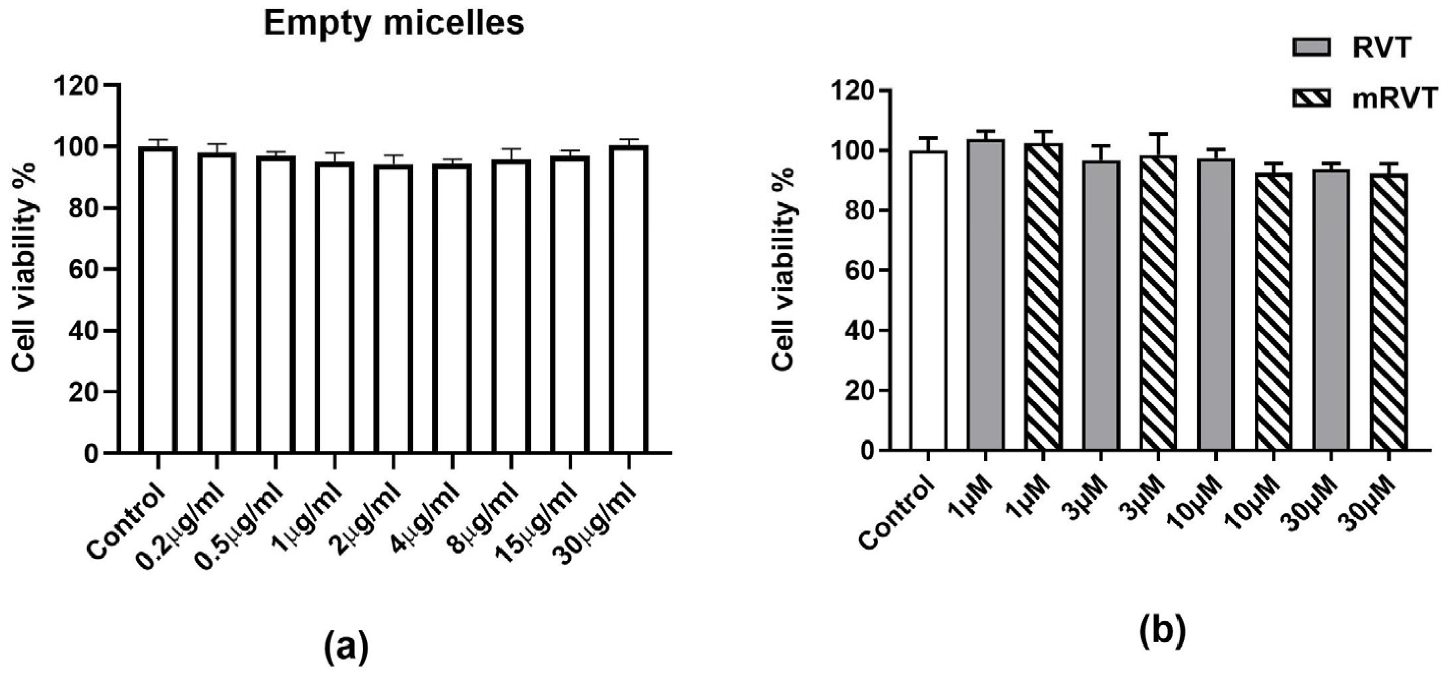
3.1. Effects of Empty Micelles, RVT, and mRVT Treatment on Cell Viability (MTT Assay)

Cell viability was assessed after treatment with empty micelles, RVT, and mRVT. Our results showed that a 24 h incubation of U87MG cells with empty micelles at concentrations of 0.2–30 μg/mL (Figure 2a), drug-loaded micelles (mRVT), and pure resveratrol (RVT) at concentrations of 1, 3, 10, and 30 µM (Figure 2b) did not affect cell viability. These findings indicate the safety of the treatment.

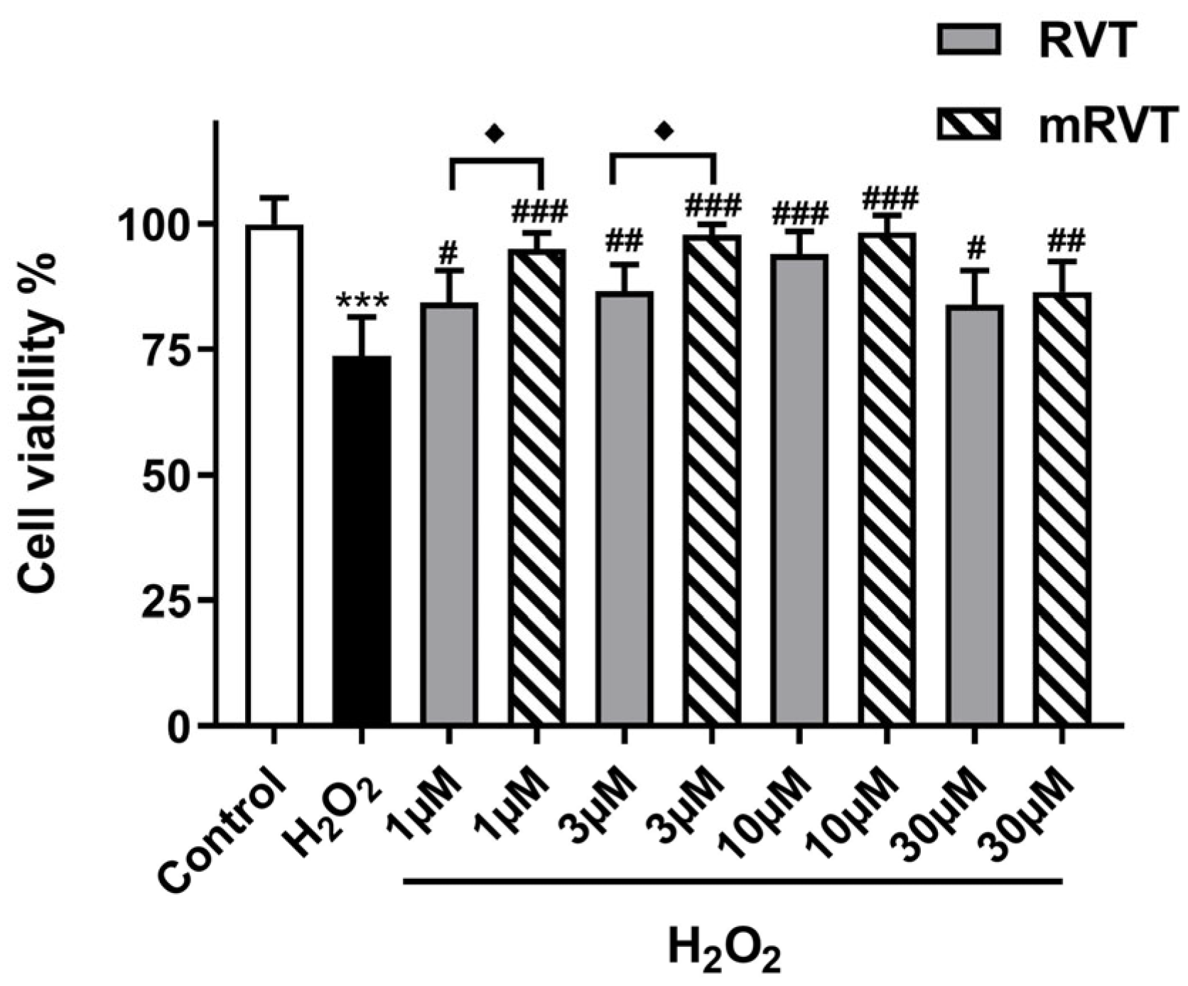
3.2. Cell Viability and Protection in the Model of H2O2-Induced Oxidative Damage

Cell viability, measured by the MTT assay, was reduced by 26.3% (p < 0.001, n = 3) compared to the control after 24 h incubation of U87MG cells with H2O2 (200 µM) (Figure 3). The mRVT (1, 3, and 10 μM) and RVT (10 μM) pretreatment for 24 h restored cell viability near to the control levels. According to the post hoc test the mRVT treatment in concentrations of 1 and 3 μM increased cell viability by the 28.9% (p < 0.001, n = 3) and 32.7% (p < 0.001, n = 3), respectively, compared to the H2O2 treated group and by 14.4% (p < 0.05, n = 3) and 17.6% (p < 0.05, n = 3), respectively, compared to the pure RVT in the equivalent concentrations.

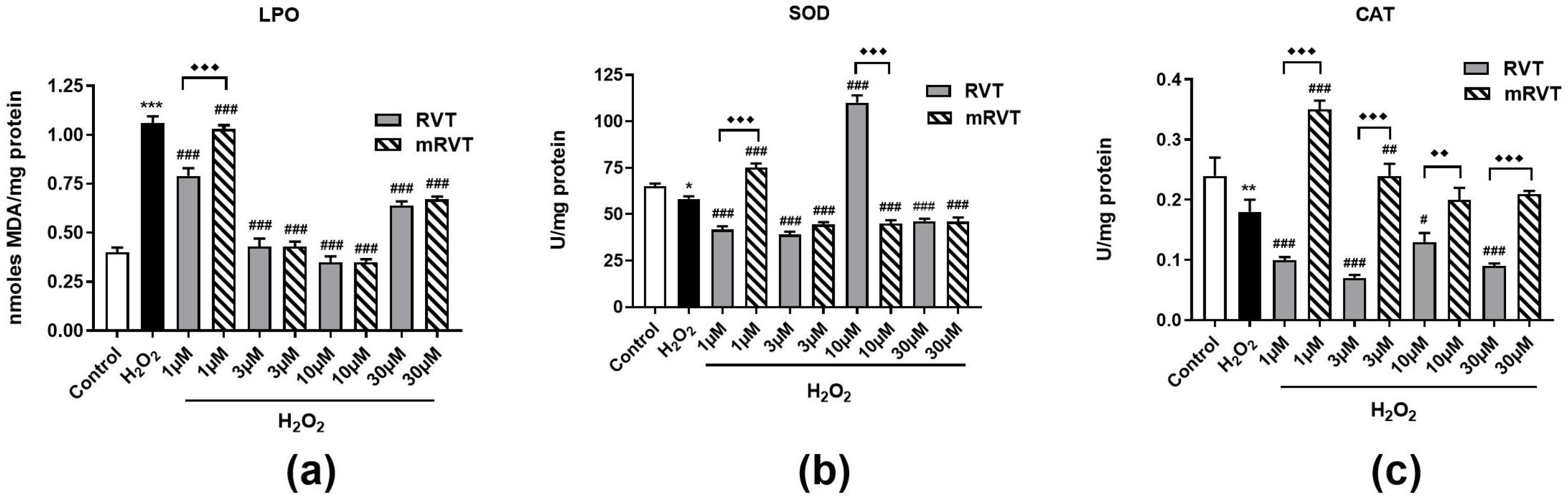
3.3. Effects of RVT and mRVT Treatment Against H2O2-Induced Oxidative Stress in U87MG Cells

Our results showed that the H2O2 treatment of U87MG glioma cells increased LPO levels by 165% (p < 0.001, n = 3) and decreased SOD and CAT activity by 11% (p < 0.05, n = 3) and 25% (p < 0.01, n = 3), respectively, compared to the control (Figure 4a–c). The treatment with pure and micellar resveratrol decreased MDA levels in the samples, which indicates an antioxidant effect. The most effective concentration for both forms of resveratrol was 10 µM, at which the control levels of LPO were preserved. According to SOD activity analysis, the enzyme activity was reversed after mRVT treatment in concentration 1 µM (by 28%, p < 0.001, n = 3) and after pure RVT in concentration 10 µM (by 88.58%, p < 0.001, n = 3), compared to the H2O2-treated group. CAT activity was reversed only after mRVT treatment with the strongest effect at concentration 1 µM (by 94.44%, p < 0.001, n = 3), compared to the H2O2-treated group.

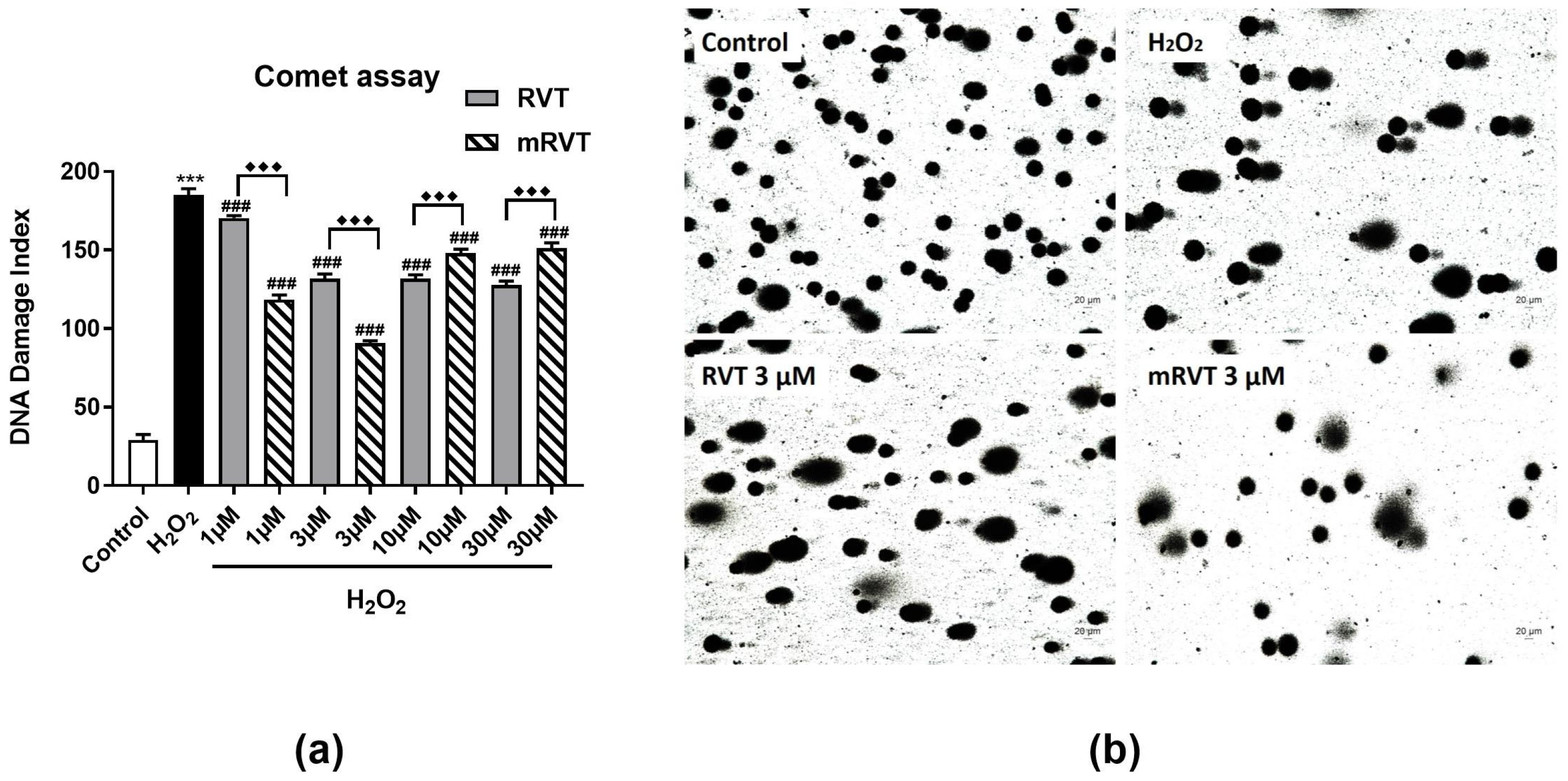
3.4. Effects of RVT and mRVT on DNA Damge

The protective effect of mRVT and RVT against single-strand and double-strand DNA breaks was investigated by alkaline comet assay. Figure 5a,b shows the distribution of the DNA damage index of H2O2 and tested compounds. The obtained results showed an 84% (p < 0.001, n = 3) increase in the DNA damage index in U87MG cells after 24 h exposure to H2O2 (200 μM) compared to the control (non-treated cells). RVT and mRVT pretreatment of the cells showed protective DNA damage capacity. The best protective effect was observed after mRVT treatment at lower concentrations (1 μM and 3 μM), where the DNA damage index decreased by 36% (p < 0.001, n = 3) and 51% (p < 0.001, n = 3), respectively, compared to the H2O2-treated group. In comparison, the DNA damage index was decreased by 8% (p < 0.001, n = 3) and by 29% (p < 0.001, n = 3) after RVT treatment in concentrations of 1 μM and 3 μM, respectively (Figure 5a). It was confirmed by the images of the cells in Figure 5b, where increased DNA migration was observed in the H2O2-treated cells, but in the cell group treated with 3 μM mRVT and RVT, it was reduced.

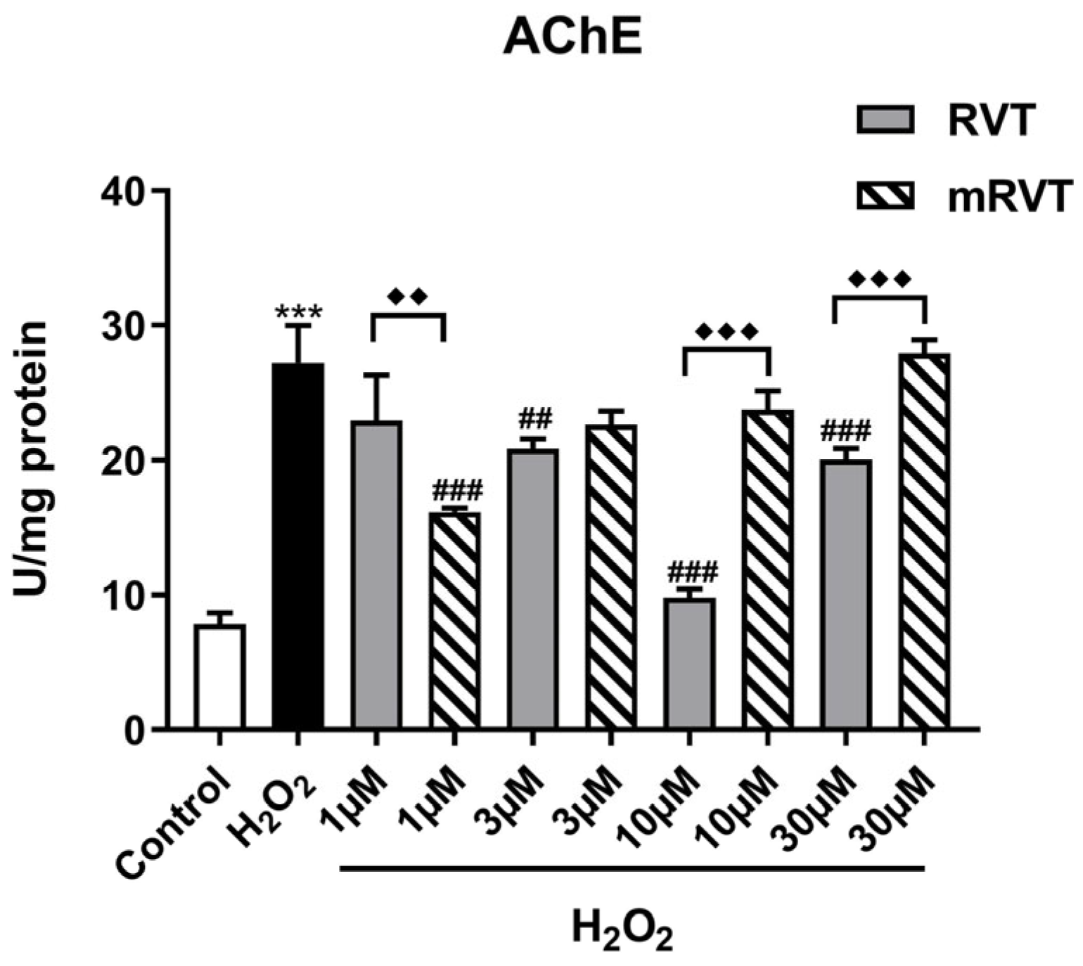
3.5. Effects of RVT and mRVT Treatments on H2O2-Induced Increase in Acetylcholinesterase Activity in U87MG Glioma Cells Homogenate

H2O2 (200 µM) treatment of U87MG glioma cells increased the AChE activity by 247.32% (p < 0.001, n = 3) compared to the control (Figure 6). The treatment with both pure and micellar resveratrol decreased enzyme activity. The best effect was observed after mRVT pretreatment in the concentration of 1 µM (by 40.65%, p < 0.001, n = 3) and for pure RVT in the concentration of 10 µM (by 63.94%, p < 0.001, n = 3) compared to the H2O2-treated cells.

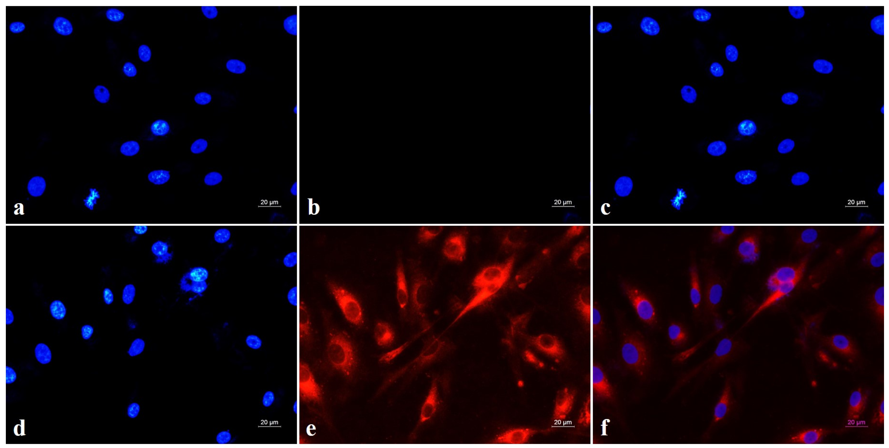
3.6. Intracellular Micellar Penetration

The cellular internalization of the micelles was studied in human U87MG glioblastoma cells via fluorescence microscopy. The fluorescence images of the uptake behavior of the cell lines showed the preferential internalization of rhodamine-B marked micelles 1 h after the treatment (Figure 7). The observed morphological alterations demonstrate that rhodamine-B-loaded micelles were localized in the cytoplasm.

4. Discussion

The present research explored the cytoprotective, genoprotective, and antioxidant capacities of resveratrol-loaded Pluronic (P123/F127) micelles as an approach to improve the low water solubility and poor bioavailability of pure resveratrol. The in vitro effects of mRVT (an aqueous dispersion) were compared with those of pure RVT (30% hydroethanolic solution) in the human U87MG glioblastoma cell line with H2O2-induced oxidative damage.
Oxidative stress as an imbalance in ROS generation and deactivation has been linked to several neurodegenerative diseases [3,7,8,49,50]. The central nervous system (CNS) is especially susceptible to oxidative damage because of its elevated oxygen consumption, weak antioxidant defenses, and limited capacity for cellular regeneration [8,14]. Antioxidants of natural origin have been proposed as preventive and therapeutic agents for neurodegenerative diseases due to their potential to mitigate oxidative stress-induced damage in neuronal cells [16,50,51,52,53,54,55,56,57]. RVT is a natural polyphenol known for its diverse pharmacological properties, including significant antioxidant activity [22,23]. Its low bioavailability poses significant challenges for clinical use and one possible way to address this limitation is encapsulation of resveratrol in polymeric nanomicelles as a drug delivery system.
As the first step in our research, the cellular internalization of rhodamine-B-marked Pluronic (P123/F127) micelles was evaluated. As shown in Figure 7, the obtained morphological alterations of the cells demonstrate that the rhodamine-B-loaded micelles were localized in the cytoplasm 1 h after treatment.
As the next step cytotoxicity of empty micelles (in the range of 0.2–30 μg/mL), RVT and mRVT (in the range of 1 to 30 μM) was assessed in human U87MG glioblastoma cells via an MTT assay. Our results showed that 24 h incubation with empty micelles, RVT, and mRVT caused no significant changes in cell viability, indicating a lack of toxicity. This is consistent with previous findings showing that several hours of incubation with RVT at concentrations below 50 µM does not cause cell toxicity in PC12 cells [25,58]. The lack of toxicity of P123/F127 micelles loaded with cannabidiol in SH-SY5Y and Neuro-2a cells line was also reported [41].
H2O2 is a major redox-signaling molecule in cells under physiological conditions and widely used to induce oxidative stress-related damage, particularly in neuronal cell models at supraphysiological concentrations [5,59,60,61,62]. Due to its small size and neutral charge, H2O2 easily crosses cell membranes and contributes to the generation of highly reactive hydroxyl radicals through Fenton’s reaction. The byproducts of this reaction, along with H2O2 itself, can directly interact with cellular macromolecules, including proteins, lipids, and DNA [63]. These changes in cellular metabolism can potentially lead to cell death through apoptosis [64,65,66,67,68,69]. In this study, H2O2 at cytotoxic concentration of 200 µM [70] was used to create a model of oxidative stress-induced cell damage where the protective effects of RVT and mRVT were compared after 24 h of preincubation. Under our experimental conditions, both the pure and encapsulated forms of resveratrol reversed the cytotoxic effects of H2O2 at all tested concentrations. However, mRVT at low concentrations (1 and 3 μM) was more effective than the pure drug at equivalent concentrations. These results are consistent with a previous report indicating that the controlled release of resveratrol from nanoparticles can reverse Aβ (1–−42) toxicity in PC12 cells within the concentration range of 1 to 10 μM [71].
The toxic effects of high H2O2 concentrations are measured by the MDA levels and the activity of SOD and CAT in the cells used as markers for oxidative stress in our study. Treatment of the human U87MG glioblastoma cells with 200 μM H2O2 for 24 h increased the MDA levels and decreased SOD and CAT enzyme activity. These changes in the redox status of the cells indicate generated oxidative stress. Our observations align with previously reported findings on the modulatory role of H2O2 in oxidative stress parameters in the U87MG cell line [70,72]. The preincubation for 24 h with RVT and mRVT (in the range 1–30 μM) ameliorated H2O2-induced oxidative stress. It should be noted that mRVT at low concentrations (1 and 3 μM) demonstrated stronger and more significant antioxidant activity than the pure drug at equivalent concentrations. Moreover, the CAT activity, altered by H2O2 (200 μM), was only affected by the encapsulated formulation, with more effective results at lower concentrations.
Oxidative stress causes also genomic instability [73], making it essential to investigate potential protective agents. The influence of mRVT and RVT on DNA damage was therefore assessed using a comet assay. This technique is widely used to investigate primary genotoxic effects in the cells’ culture. It is applied to assess damage caused by acute and chronic oxidative stress, DNA crosslinks, apoptosis, and genotoxicity induced by chemical compounds [74,75]. The genoprotective effects of resveratrol in a concentration range from 10 to 250 μM against hydrogen peroxide-induced DNA damage in C6 glioma cells was reported by Quincozes-Santos et al. [74]. Comet assay results from this research also suggest a gene-protective effect of resveratrol treatment. In our study, the hydrogen peroxide-induced nuclear DNA damage observed in the U87MG glioblastoma cell line was significantly reduced after treatment with pure and micellar resveratrol. Notably, the mRVT formulation demonstrated superior protective effects at lower concentrations compared to the pure drug, with the best protection observed at concentration of 3 μM.
Furthermore, the effects of RVT and mRVT were compared in regard to their ability to alter AChE activity in cells’ homogenates. AChE is an enzyme responsible for the hydrolysis of acetylcholine (ACh) in the synaptic cleft [76]. The low levels of ACh are one of the main features of neurodegenerative diseases such as Alzheimer’s disease. Therefore, one of the primary strategies for the treatment of such diseases is the use of AChE inhibitors [76,77]. Data for in vitro and in vivo AChE-inhibitory activity of RVT were already reported [43,78]. Our results showed that H2O2 increased the AChE activity after 24 h of incubation. More importantly, mRVT demonstrated the strongest inhibitory effect on the enzyme activity at a low concentration of 1 μM, whereas the pure drug exhibited its maximum effect at a higher concentration of 10 μM. This is consistent with our previously reported results demonstrating enhanced AChE inhibitory activity of RVT-loaded Pluronic micelles in the in vivo model of scopolamine-induced dementia [43]. This result confirmed that the encapsulation of the hydrophobic antioxidant in the polymeric micelles could potentiate its effects.
According to the Biopharmaceutics Classification System, RVT is classified as a Class II drug, which means it has poor water solubility and high cell membrane permeability [79]. However, there are data that its highly lipophilic nature leads to cell membrane accumulation rather than penetration [80,81,82,83,84,85]. The results from this study showed better effectiveness of mRVT at lower concentrations compared to the pure RVT which expressed its antioxidant activity in higher concentrations. This phenomenon may be explained by the better and faster penetration of mRVT through cell membranes. According to our results, one-hour incubation was sufficient for cytoplasmic accumulation of the micelles. Moreover, the intracellular concentration of RVT appears to be appropriate for cell protection due to the controlled release from the nanoparticle, as we already reported [43].

5. Conclusions

Our results demonstrated that mRVT at low concentrations (1 and 3 μM) had a stronger protective effect than the pure drug at equivalent concentrations against H2O2-induced cytotoxicity, oxidative stress, nuclear DNA damage, and increased AChE activity. In light of these findings, and taking into account that empty Pluronic micelles did not exert cytotoxic effects on the glioblastoma cell line, we can conclude that the encapsulation of resveratrol in Pluronic (P123/F127) micelles may represent a promising platform for therapeutic interventions against neurodegenerative processes involving oxidative stress.

Author Contributions

Conceptualization, M.L.; methodology, I.S., E.T., A.G., V.D., L.R. and K.Y.; software, M.L., I.S. and E.T.; validation, I.S., E.T. and V.D.; formal analysis, M.L., E.T. and I.S.; investigation, I.S. and E.T.; resources, M.L.; data curation, M.L., L.R. and K.Y.; writing—original draft preparation, M.L.; writing—review and editing, I.S., K.Y., E.T. and L.R.; visualization, M.L. and I.S.; supervision, M.L.; project administration, M.L.; funding acquisition, M.L. All authors have read and agreed to the published version of the manuscript.

Funding

This research was funded by the Bulgarian National Science Fund; grant number KP-06-N73/10 from 15 December 2023.

Institutional Review Board Statement

Not applicable.

Informed Consent Statement

Not applicable.

Data Availability Statement

The original contributions presented in this study are included in the article. Further inquiries can be directed to the corresponding author.

Conflicts of Interest

The authors declare no conflicts of interest.

References

  1. Coyle, J.T.; Puttfarcken, P. Oxidative stress, glutamate, and neurodegenerative disorders. Science 1993, 262, 689–695. [Google Scholar] [CrossRef] [PubMed]
  2. Dringen, R. Metabolism and functions of glutathione in brain. Prog. Neurobiol. 2000, 62, 649–671. [Google Scholar] [CrossRef] [PubMed]
  3. Singh, A.; Kukreti, R.; Saso, L.; Kukreti, S. Oxidative stress: A key modulator in neurodegenerative diseases. Molecules 2019, 24, 1583. [Google Scholar] [CrossRef] [PubMed]
  4. Lushchak, V.I.; Storey, K.B. Oxidative stress concept updated: Definitions, classifications, and regulatory pathways implicated. EXCLI J. 2021, 20, 956–967. [Google Scholar]
  5. Sies, H. Hydrogen peroxide as a central redox signaling molecule in physiological oxidative stress: Oxidative eustress. Redox Biol. 2017, 11, 613–619. [Google Scholar] [CrossRef]
  6. Finkel, T. Signal transduction by reactive oxygen species. J. Cell Biol. 2011, 194, 7–15. [Google Scholar] [CrossRef]
  7. Scarian, E.; Viola, C.; Dragoni, F.; Di Gerlando, R.; Rizzo, B.; Diamanti, L.; Gagliardi, S.; Bordoni, M.; Pansarasa, O. New insights into oxidative stress and inflammatory response in neurodegenerative diseases. Int. J. Mol. Sci. 2024, 25, 2698. [Google Scholar] [CrossRef]
  8. Li, J.; Wuliji, O.; Li, W.; Jiang, Z.-G.; Ghanbari, H.A. Oxidative stress and neurodegenerative disorders. Int. J. Mol. Sci. 2013, 14, 24438–24475. [Google Scholar] [CrossRef]
  9. Zhao, X.; Fang, J.; Li, S.; Gaur, U.; Xing, X.; Wang, H.; Zheng, W. Artemisinin attenuated hydrogen peroxide (H2O2)-induced oxidative injury in SH-SY5Y and hippocampal neurons via the activation of AMPK Pathway. Int. J. Mol. Sci. 2019, 20, 2680. [Google Scholar] [CrossRef]
  10. Dickens, M.G.; Franz, K.J. A prochelator activated by hydrogen peroxide prevents metal-induced amyloid-aggregation. Chem. Biochem. 2010, 11, 59–62. [Google Scholar] [CrossRef]
  11. Yamato, M.; Kudo, W.; Shiba, T.; Yamada, K.I.; Watanabe, T.; Utsumi, H. Determination of ROS associated with the degeneration of dopaminergic neurons during dopamine metabolism. Free Radic. Res. 2010, 44, 249–257. [Google Scholar] [CrossRef] [PubMed]
  12. Fernández-Ferreiro, A.; Gil-Longo, J. Vascular pro-oxidant effects related to the autoxidation of dopamine. Free Radic. Res. 2009, 43, 295–303. [Google Scholar] [CrossRef]
  13. Arthur, P.G.; Lim, S.C.; Meloni, B.P.; Munns, S.E.; Chan, A.; Knuckey, N.W. The protective effect of hypoxic preconditioning on cortical neuronal cultures is associated with increases in the activity of several antioxidant enzymes. Brain Res. 2004, 1017, 146–154. [Google Scholar] [CrossRef] [PubMed]
  14. Bhat, A.H.; Dar, K.B.; Anees, S.; Zargar, M.A.; Masood, A.; Sofi, M.A.; Ganie, S.A. Oxidative stress, mitochondrial dysfunction and neurodegenerative diseases; a mechanistic insight. Biomed. Pharmacother. 2015, 74, 101–110. [Google Scholar] [CrossRef] [PubMed]
  15. Georgieva, E.; Ivanova, D.; Zhelev, Z.; Bakalova, R.; Gulubova, M.; Aoki, I. Mitochondrial Dysfunction and Redox Imbalance as a Diagnostic Marker of “Free Radical Diseases”. Anticancer Res. 2017, 37, 5373–5381. [Google Scholar]
  16. Nguyen-Thi, P.T.; Vo, T.K.; Pham, T.H.T.; Nguyen, T.T.; Van Vo, G. Natural flavonoids as potential therapeutics in the management of Alzheimer’s disease: A review. 3 Biotech 2024, 14, 68. [Google Scholar] [CrossRef]
  17. Takaoka, M.J. Of the phenolic substances of white hellebore (Veratrum grandiflorum Loes. fil.). J. Fac. Sci. Hokkaido Imp. Univ. 1940, 3, 1–16. [Google Scholar] [CrossRef]
  18. Milardi, G.L.; Stringaro, A.; Colone, M.; Bonicontro, A.; Risuleo, G. The cell membrane is the main target of resveratrol as shown by interdisciplinary biomolecular/cellular and biophysical approaches. J. Membr. Biol. 2014, 247, 1–8. [Google Scholar] [CrossRef]
  19. Gambini, J.; Ingles, M.; Olaso, G.; Lopez-Grueso, R.; Bonet-Costa, V.; Gimeno-Mallench, L.; Mas-Bargues, C.; Abdelaziz, K.M.; Gomez-Cabrera, M.C.; Vina, J.; et al. Properties of Resveratrol: In Vitro and In Vivo Studies about Metabolism, Bioavailability, and Biological Effects in Animal Models and Humans. Oxid. Med. Cell. Longev. 2015, 2015, 837042. [Google Scholar] [CrossRef]
  20. Cristòfol, R.; Porquet, D.; Corpas, R.; Coto-Montes, A.; Serret, J.; Camins, A.; Pallàs, M.; Sanfeliu, C. Neurons from senescence-accelerated SAMP8 mice are protected against frailty by the sirtuin 1 promoting agents melatonin and resveratrol. J. Pineal Res. 2012, 52, 271–281. [Google Scholar] [CrossRef]
  21. Bai, Y.; Mao, Q.-Q.; Qin, J.; Zheng, X.Y.; Wang, Y.B.; Yang, K.; Shen, H.F.; Xie, L.P. Resveratrol induces apoptosis and cell cycle arrest of human T24 bladder cancer cells in vitro and inhibits tumor growth in vivo. Cancer Sci. 2010, 101, 488–493. [Google Scholar] [CrossRef] [PubMed]
  22. Fremont, L. Minireview: Biological effects of resveratrol. Life Sci. 2020, 66, 663–673. [Google Scholar] [CrossRef] [PubMed]
  23. Carrizzo, A.; Forte, M.; Damato, A.; Trimarco, V.; Salzano, F.; Bartolo, M.; Maciag, A.; Puca, A.A.; Vecchione, C. Antioxidant effects of resveratrol in cardiovascular, cerebral and metabolic diseases. Food Chem. Toxicol. 2013, 61, 215–226. [Google Scholar] [CrossRef] [PubMed]
  24. Wang, N.; Han, Q.; Wang, G.; Ma, W.-P.; Wang, J.; Wu, W.-X.; Guo, Y.; Liu, L.; Jiang, X.-Y.; Xie, X.-L.; et al. Resveratrol Protects Oxidative Stress-Induced Intestinal Epithelial Barrier Dysfunction by Upregulating Heme Oxygenase-1 Expression. Dig. Dis. Sci. 2016, 61, 2522–2534. [Google Scholar] [CrossRef]
  25. Jang, J.H.; Surh, Y.J. Protective effects of resveratrol on β-amyloid-induced oxidative PC12 cell death. Free Radic. Biol. Med. 2003, 34, 1100–1110. [Google Scholar] [CrossRef]
  26. Russo, A.; Palumbo, M.; Aliano, C.; Lempereur, L.; Scoto, G.; Renis, M. Red wine micronutrients as protective agents in Alzheimer-like induced insult. Life Sci. 2003, 72, 2369–2379. [Google Scholar] [CrossRef]
  27. Sharma, M.; Gupta, Y.K. Chronic treatment with trans resveratrol prevents intracerebroventricular streptozotocin induced cognitive impairment and oxidative stress in rats. Life Sci. 2002, 71, 2489–2498. [Google Scholar] [CrossRef]
  28. Savaskan, E.; Olivieri, G.; Meier, F.; Seifritz, E.; Wirz-Justice, A.; Muller-Spahn, F. Red wine ingredient resveratrol protects frombeta-amyloid neurotoxicity. Gerontology 2003, 49, 380–383. [Google Scholar] [CrossRef]
  29. Han, Y.S.; Zheng, W.H.; Bastianetto, S.; Chabot, J.G.; Quirion, R. Neuroprotective effects of resveratrol against beta-amyloid-induced neurotoxicity in rat hippo-campal neurons: Involvement of protein kinase C. Br. J. Pharmacol. 2004, 141, 997–1005. [Google Scholar] [CrossRef]
  30. Ginés, C.; Cuesta, S.; Kireev, R.; García, C.; Rancan, L.; Paredes, S.D.; Vara, E.; Tresguerres, J.A. Protective effect of resveratrol against inflammation, oxidative stress and apoptosis in pancreas of aged SAMP8 mice. Exp. Gerontol. 2017, 90, 61–70. [Google Scholar] [CrossRef]
  31. Fu, S.; Lv, R.; Wang, L.; Hou, H.; Liu, H.; Shao, S. Resveratrol, an antioxidant, protects spinal cord injury in rats by suppressing MAPK pathway. Saudi J. Biol. Sci. 2018, 25, 259–266. [Google Scholar] [CrossRef] [PubMed]
  32. Francioso, A.; Mastromarino, P.; Masci, A.; d’Erme, M.; Mosca, L. Chemistry, stability and bioavailability of resveratrol. Med. Chem. 2014, 10, 237–245. [Google Scholar] [CrossRef] [PubMed]
  33. Amri, A.; Chaumeil, J.C.; Sfar, S.; Charrueau, C. Administration of resveratrol: What formulation solutions to bioavailability limitations? J. Control. Release 2012, 158, 182–193. [Google Scholar] [CrossRef] [PubMed]
  34. Zupancic, S.; Lavric, Z.; Kristl, J. Stability and solubility of trans-resveratrol are strongly influenced by pH and temperature. Eur. J. Pharm. Biopharm. 2015, 93, 196–204. [Google Scholar] [CrossRef]
  35. Peterle, L.; Sanfilippo, S.; Borgia, F.; Li Pomi, F.; Vadalà, R.; Costa, R.; Cicero, N.; Gangemi, S. The Role of Nutraceuticals and Functional Foods in Skin Cancer: Mechanisms and Therapeutic Potential. Foods 2023, 12, 2629. [Google Scholar] [CrossRef]
  36. Oerlemans, C.; Bult, W.; Bos, M.; Storm, G.; Nijsen, J.F.; Hennink, W.E. Polymeric micelles in anticancer therapy: Targeting, imaging and triggered release. Pharm. Res. 2010, 27, 2569–2589. [Google Scholar] [CrossRef]
  37. Kabanov, A.V.; Alakhov, V.Y. Pluronic® block copolymers in drug delivery: From micellar nanocontainers to biological response modifiers. Crit. Rev. Ther. Drug 2002, 19, 1–73. [Google Scholar] [CrossRef]
  38. Yoncheva, K.; Calleja, P.; Agueros, M.; Petrov, P.; Miladinova, I.; Tsvetanov, C.; Irache, J.M. Stabilized Micelles as Delivery Vehicles for Paclitaxel. Int. J. Pharm. 2012, 436, 258–264. [Google Scholar] [CrossRef]
  39. Taha, E.I.; Badran, M.M.; El-Anazi, M.H.; Bayomi, M.A.; El-Bagory, I.M. Role of Pluronic F127 Micelles in Enhancing Ocular Delivery of Ciprofloxacin. J. Mol. Liq. 2014, 199, 251–256. [Google Scholar] [CrossRef]
  40. Niu, J.; Yuan, M.; Chen, C.; Wang, L.; Tang, Z.; Fan, Y.; Liu, X.; Ma, Y.J.; Gan, Y. Berberine-Loaded Thiolated Pluronic F127 Polymeric Micelles for Improving Skin Permeation and Retention. Int. J. Nanomed. 2020, 15, 9987–10005. [Google Scholar] [CrossRef]
  41. Yordanov, Y.; Stefanova, D.; Spassova, I.; Kovacheva, D.; Tzankova, V.; Konstantinov, S.; Yoncheva, K. Formulation of Nanomicelles Loaded with Cannabidiol as a Platform for Neuroprotective Therapy. Pharmaceutics 2022, 14, 2625. [Google Scholar] [CrossRef] [PubMed]
  42. Hersh, A.M.; Alomari, S.; Tyler, B.M. Crossing the Blood-Brain Barrier: Advances in Nanoparticle Technology for Drug Delivery in Neuro-Oncology. Int. J. Mol. Sci. 2022, 23, 4153. [Google Scholar] [CrossRef] [PubMed]
  43. Lazarova, M.; Stefanova, M.; Tsvetanova, E.; Georgieva, A.; Tasheva, K.; Radeva, L.; Yoncheva, K. Resveratrol-Loaded Pluronic Micelles Ameliorate Scopolamine-Induced Cognitive Dysfunction Targeting Acetylcholinesterase Activity and Programmed Cell Death. Int. J. Mol. Sci. 2024, 25, 12777. [Google Scholar] [CrossRef] [PubMed]
  44. Mosmann, T. Rapid colorimetric assay for cellular growth and survival: Application to proliferation and cytotoxicity assays. J. Immunol. Methods 1983, 65, 55–63. [Google Scholar] [CrossRef]
  45. Ellman, G.L.; Courtney, K.D.; Andres, J.V.; Featherstone, R.M. Anew and rapid colorimetric determination of acetylcholinesterase activity. Biochem. Pharmacol. 1969, 7, 88–90. [Google Scholar] [CrossRef]
  46. Sulikovska, I.; Djeliova, V.; Kirazov, L.; Ivanov, I.; Dimitrova, M. Evaluation of different staining methods and image analyses of the results from a comet assay in human colorectal cancer cells treated with hydrogen peroxide. Acta Morphol. Anthropol. 2023, 30, 5–13. [Google Scholar] [CrossRef]
  47. Azqueta, A.; Langie, S.A.S.; Boutet-Robinet, E.; Duthie, S.; Ladeira, C.; Møller, P.; Collins, A.R.; Godschalk, R.W.L. DNA repair as a human biomonitoring tool: Comet assay approaches. Mutat. Res. Rev. Mutat. Res. 2019, 781, 71–87. [Google Scholar] [CrossRef]
  48. Noroozi, M.; Angerson, W.J.; Lean, M.E.J. Effects of flavonoids and Vitamin C on oxidative DNA damage to human lymphocytes. Am. J. Clin. Nutr. 1998, 67, 1210–1218. [Google Scholar] [CrossRef]
  49. Mayne, K.; White, J.A.; McMurran, C.E.; Rivera, F.J.; de la Fuente, A.G. Aging and neurodegenerative disease: Is the adaptive immune system a friend or foe? Front. Aging Neurosci. 2020, 12, 572090. [Google Scholar] [CrossRef]
  50. Olufunmilayo, E.O.; Gerke-Duncan, M.B.; Holsinger, R.M.D. Oxidative stress and antioxidants in neurodegenerative disorders. Antioxidants 2023, 12, 517. [Google Scholar] [CrossRef]
  51. He, W.J.; Lv, C.H.; Chen, Z.; Shi, M.; Zeng, C.X.; Hou, D.X.; Qin, S. The regulatory effect of phytochemicals on chronic diseases by targeting Nrf2-ARE signaling pathway. Antioxidants 2023, 12, 236. [Google Scholar] [CrossRef] [PubMed]
  52. Deshpande, P.; Gogia, N.; Singh, A. Exploring the efficacy of natural products in alleviating Alzheimer’s disease. Neural. Regen. Res. 2019, 14, 1321–1329. [Google Scholar] [PubMed]
  53. Venkatesan, R.; Ji, E.; Kim, S.Y. Phytochemicals that regulate neurodegenerative disease by targeting neurotrophins: A comprehensive review. BioMed Res. Int. 2015, 2015, 814068. [Google Scholar] [CrossRef] [PubMed]
  54. Khan, A.; Jahan, S.; Alshahrani, S.; Alshehri, B.M.; Sameer, A.S.; Arafah, A.; Ahmad, A.; Rehman, M.U. Phytotherapeutic agents for neurodegenerative disorders: A neuropharmacological review. In Phytomedicine; Academic Press: Cambridge, MA, USA, 2021; pp. 581–620. [Google Scholar]
  55. Chen, B.; Zhao, J.; Zhang, R.; Zhang, L.; Zhang, Q.; Yang, H.; An, J. Neuroprotective effects of natural compounds on neurotoxininduced oxidative stress and cell apoptosis. Nutr. Neurosci. 2022, 25, 1078–1099. [Google Scholar] [CrossRef]
  56. Tie, F.; Fu, Y.; Hu, N.; Wang, H. Silibinin protects against H2O2-induced oxidative damage in SH-SY5Y cells by improving mitochondrial function. Antioxidants 2022, 11, 1101. [Google Scholar] [CrossRef]
  57. Wang, C.M.; Yang, C.Q.; Cheng, B.H.; Chen, J.; Bai, B. Orexin-A protects SH-SY5Y cells against H2O2-induced oxidative damage via the PI3K/MEK1/2/ERK1/2 signaling pathway. Int. J. Immunopathol. Pharmacol. 2018, 32, 2058738418785739. [Google Scholar] [CrossRef]
  58. Jang, M.H.; Piao, X.L.; Young, K.H.; Cho, E.J. Resveratrol oligomers from vitis amurensis attenuate -amyloid-induced oxidative stress in PC12 cells. Biol. Pharm. Bull. 2007, 30, 1130–1134. [Google Scholar] [CrossRef]
  59. Loh, K.P.; Huang, S.H.; Silva, R.D.; Tan, B.K.H.; Zhu, Y.Z. Oxidative stress: Apoptosis in neuronal injury. Curr. Alzheimer Res. 2006, 3, 327–337. [Google Scholar] [CrossRef]
  60. Sies, H. Oxidative stress: Concept and some practical aspects. Antioxidants 2020, 9, 852. [Google Scholar] [CrossRef]
  61. Konno, T.; Melo, E.P.; Chambers, J.E.; Avezov, E. Intracellular sources of ROS/H2O2 in health and neurodegeneration: Spotlight on endoplasmic reticulum. Cells 2021, 10, 233. [Google Scholar] [CrossRef]
  62. Halliwell, B. Reactive oxygen species and the central nervous system. J. Neurochem. 1992, 59, 1609–1623. [Google Scholar] [CrossRef] [PubMed]
  63. Jang, J.H.; Surh, Y.J. Possible role of NF-κB in Bcl-XL protection against hydrogen peroxide-induced PC12 cell death. Redox Rep. 2014, 9, 343–348. [Google Scholar] [CrossRef] [PubMed]
  64. Halliwell, B.; Aruoma, O.I. DNA damage by oxygen-derived species. Itsmechanism and measurement in mammalian systems. FEBS Lett. 1991, 28, 9–19. [Google Scholar] [CrossRef] [PubMed]
  65. Zhang, L.; Yu, H.; Sun, Y.; Lin, X.; Chen, B.; Tan, C.; Cao, G.; Wang, Z. Protective effects of salidroside on hydrogen peroxide-induced apoptosis in SH-SY5Y human neuroblastoma cells. Eur. J. Pharmacol. 2007, 564, 18–25. [Google Scholar] [CrossRef]
  66. Asada, K. Ascorbate peroxidase-a hydrogen peroxide-scavenging enzyme in plants. Physiol. Plant. 1992, 85, 235–241. [Google Scholar] [CrossRef]
  67. Turrens, J.F. Mitochondrial formation of reactive oxygen species. J. Physiol. 2003, 552, 335–344. [Google Scholar] [CrossRef]
  68. Yoshikawa, A.; Saito, Y.; Maruyama, K. Lignan compounds and 4,4′-dihydroxybiphenyl protect C2C12 cells against damage from oxidative stress. Biochem. Biophys. Res. Commun. 2006, 344, 394–399. [Google Scholar] [CrossRef]
  69. El Gamal, A.A.; AlSaid, M.S.; Raish, M.; Al-Sohaibani, M.; Al-Massarani, S.M.; Ahmad, A.; Rafatullah, S. Beetroot (Beta vulgaris L.) extract ameliorates gentamicin-induced nephrotoxicity associated oxidative stress, inflammation, and apoptosis in rodent model. Mediat. Inflamm. 2014, 2014, 983952. [Google Scholar] [CrossRef]
  70. Gopalan, M.; Jadhav, A.S. Protective effect beetroot Beta vulgaris extract against H2O2 Induced Oxidative stress in U87MG gliomacells. South Asian J. Exp. Biol. 2021, 11, 266–274. [Google Scholar] [CrossRef]
  71. Lu, X.; Ji, C.; Xu, H.; Li, X.; Ding, H.; Ye, M.; Zhu, Z.; Ding, D.; Jiang, X.; Ding, X.; et al. Resveratrol-loaded polymeric micelles protect cells from Abeta-induced oxidative stress. Int. J. Pharm. 2009, 375, 89–96. [Google Scholar] [CrossRef]
  72. Raj, A.; Dey, S.; Rao, S.; Madhunapantula, V.; Manjula, S.N. Neuroprotective And Cognitive Enhancing Effect Of Methanolic Morus Alba Leaf Fraction In U87mg Cell Lines And Experimental Rat Model. Asian J. Pharm. Clin. Res. 2019, 12, 238–243. [Google Scholar]
  73. Martemucci, G.; Portincasa, P.; Di Ciaula, A.; Mariano, M.; Centonze, V.; D’Alessandro, A.G. Oxidative stress, aging, antioxidant supplementation and their impact on human health: An overview. Mech. Ageing Dev. 2022, 206, 111707. [Google Scholar] [CrossRef]
  74. Quincozes-Santos, A.; Andreazza, A.C.; Nardin, P.; Funchal, C.; Gonçalves, C.A.; Gottfried, C. Resveratrol attenuates oxidative-induced DNA damage in C6 Glioma cells. Neurotoxicology 2007, 28, 886–891. [Google Scholar] [CrossRef] [PubMed]
  75. Hirrlinger, J.; Resch, A.; Gutterer, J.M.; Dringen, R. Oligodendroglial cells in culture effectively dispose of exogenous hydrogen peroxide: Comparison with cultured neurones, astroglial and microglial cells. J. Neurochem. 2002, 82, 635–644. [Google Scholar] [CrossRef] [PubMed]
  76. Ballard, C.; Greig, N.; Guillozet-Bongaarts, A.; Enz, A.; Darvesh, S. Cholinesterases: Roles in the brain during health and disease. Curr. Alzheimer Res. 2005, 2, 307–318. [Google Scholar] [CrossRef]
  77. Francis, P.T.; Palmer, A.M.; Snape, M.; Wilcock, G.K. The cholinergic hypothesis of Alzheimer’s disease: A review of progress. J. Neurol. Neurosurg. Psychiatry 1999, 66, 137–147. [Google Scholar] [CrossRef]
  78. Foudah, A.I.; Devi, S.; Alam, A.; Salkini, M.A.; Ross, S.A. Anticholinergic Effect of Resveratrol with Vitamin E on Scopolamine-Induced Alzheimer’s Disease in Rats: Mechanistic Approach to Prevent Inflammation. Front. Pharmacol. 2023, 14, 1115721. [Google Scholar] [CrossRef]
  79. Deng, Z.; Li, Y.; Liu, H.; Xiao, S.; Li, L.; Tian, J.; Cheng, C.; Zhang, G.; Zhang, F. The role of sirtuin 1 and its activator, resveratrol in osteoarthritis. Biosci. Rep. 2019, 39, BSR20190189. [Google Scholar] [CrossRef]
  80. Athar, M.; Back, J.H.; Tang, X.; Kim, A. Resveratrol: A reviewof preclinical studies for human cancer prevention. Toxicol. Appl. Pharmacol. 2007, 224, 274–283. [Google Scholar] [CrossRef]
  81. Szende, B.; Tyihбk, E.; Kirбly-Vйghely, Z. Dose-dependent effect of resveratrol on proliferation and apoptosis in endothelial and tumor cell cultures. Exp. Mol. Med. 2000, 32, 88–92. [Google Scholar] [CrossRef]
  82. Sotomatsu, A.; Nakano, M.; Hirai, S. Phospholipid peroxidation induced by the catechol-Fe31(Cu21) complex: A possible mechanism of nigrostriatal cell damage. Arch. Biochem. Biophys. 1990, 283, 334–341. [Google Scholar] [CrossRef] [PubMed]
  83. Rice-Evans, C.; Burdon, R. Free radical–Lipid interactions and their pathological consequences. Prog. Lipid Res. 1993, 32, 71–110. [Google Scholar] [CrossRef] [PubMed]
  84. Galati, G.; Sabzevari, O.; Wilson, J.X.; O’Brien, P.J. Prooxidant activity and cellular effects of the phenoxyl radicals of dietary flavonoids and other polyphenolics. Toxicology 2002, 177, 91–104. [Google Scholar] [CrossRef] [PubMed]
  85. Minotti, G.; Aust, S.D. The role of iron in oxygen radical mediated lipid peroxidation. Chem. Biol. Interact. 1989, 71, 1–19. [Google Scholar] [CrossRef]
Figure 1. Cell treatment paradigm for the investigation.
Applsci 15 02995 g001
Figure 2. Viability of U87MG cells after 24 h of incubation with (a) empty micelles and (b) resveratrol-loaded micelles (mRVT) and pure resveratrol (RVT). Values are mean ± SEM (n = 3).
Applsci 15 02995 g002
Figure 3. Cell viability after 24 h pre-incubation with RVT or mRVT against 200 μM H2O2-induced cell damage. Compared with the control: *** p < 0.001; compared with H2O2 group: # p < 0.05, ## p < 0.01, ### p < 0.001; compared between RVT and mRVT groups: ♦ p < 0.05.
Applsci 15 02995 g003
Figure 4. Effects of RVT and mRVT treatment on LPO levels (a), SOD (b), and CAT (c) activities in H2O2-induced oxidative stress in U87MG glioma cells. Compared with the control: * p < 0.05, ** p < 0.01, *** p < 0.001; compared with the H2O2-tgroup: # p < 0.05, ## p < 0.01, ### p < 0.001; compared between the RVT and mRVT groups: ♦♦ p < 0.01, ♦♦♦ p < 0.001.
Applsci 15 02995 g004
Figure 5. Protection of H2O2-induced DNA damage by mRVT and RVT in U87MG cells. (a) Graph of the DNA damage index. (b) Microphotographs of the cells treated with H2O2 (200 μM) or cells pretreated with RVT and mRVT in 3 μM concentration. Objective 10×. Compared with the control: *** p < 0.001; compared with the H2O2 group: ### p < 0.001; compared between the RVT and mRVT groups: ♦♦♦ p < 0.001.
Applsci 15 02995 g005
Figure 6. Effects of RVT and mRVT treatment on acetylcholine esterase activity in H2O2-induced oxidative stress in U87MG glioma cells. Compared with the control: *** p < 0.001; compared with the H2O2 group: ### p < 0.001, ## p < 0.01; compared between the RVT and mRVT groups: ♦♦ p < 0.01, ♦♦♦ p < 0.001.
Applsci 15 02995 g006
Figure 7. The fluorescence microscopic images of U87MG cells; (ac) non-treated (control group) and (df) incubated with rhodamine B-loaded micelles for 1 h. Nuclei were stained with DAPI (blue), cells incubated with rhodamine-B micelles (red), and the two images were merged. Objective 40×.
Applsci 15 02995 g007
Disclaimer/Publisher’s Note: The statements, opinions and data contained in all publications are solely those of the individual author(s) and contributor(s) and not of MDPI and/or the editor(s). MDPI and/or the editor(s) disclaim responsibility for any injury to people or property resulting from any ideas, methods, instructions or products referred to in the content.

