Review

Superconductivity and Cryogenics in Medical Diagnostics and Treatment: An Overview of Selected Applications

by
Oleksandr Boiko
and
Henryka Danuta Stryczewska
*
Department of Electrical Engineering and Superconductivity Technologies, Lublin University of Technology, 38A Nadbystrzycka Street, 20-618 Lublin, Poland
*
Author to whom correspondence should be addressed.
Appl. Sci. 2025, 15(23), 12579; https://doi.org/10.3390/app152312579
Submission received: 27 October 2025 / Revised: 17 November 2025 / Accepted: 25 November 2025 / Published: 27 November 2025
(This article belongs to the Section Electrical, Electronics and Communications Engineering)

Abstract

This article presents a comprehensive overview of the current and emerging roles of cryogenics and superconductivity in medical diagnostics, imaging, and therapy. Beginning with the historical foundations of both fields and their technological maturation, this review emphasizes how cryogenic engineering and superconducting materials have become indispensable to modern medical systems. Cryogenic technologies are highlighted in applications such as cryosurgery, cryotherapy, cryostimulation, and cryopreservation, all of which rely on controlled exposure to extremely low temperatures for therapeutic or biological preservation purposes. This article outlines the operating principles of cryomedical devices, the refrigerants and cooling methods used, and the technological barriers. This paper reviews the latest applications of superconductivity phenomena in medicine and identifies those that could be used in the future. These include cryogenic therapy, radiotherapy (cyclotrons, particle accelerators, synchrotron radiation generation, isotope production, and proton and ion beam delivery), magnetic resonance imaging (MRI), nuclear magnetic resonance spectroscopy (NMR), positron emission tomography (PET), and ultra-sensitive magnetic signal transducers based on SQUIDs for detecting ultra-low bio-signals emitted by human body organs. CT, MRI/NMR, and PET features are compared using the operation principle, specific applications, safety, contraindications for patients, examination time, and additional valued peculiarities. This article outlines the prospects for the development of superconducting and cryogenic materials and technologies in medical applications. Advances in diagnostic imaging are reviewed, with particular attention on the progression from conventional MRI scanners to ultra-high-field (UHF) systems exceeding 7–10.5 T, culminating in the 11.7 T Iseult whole-body MRI magnet. Another important application area described in this article includes biofunctionalized magnetic nanoparticles and superconducting quantum interference devices (SQUIDs), which enable the ultrasensitive detection of biomagnetic fields and targeted cancer diagnostics. Finally, this article identifies future directions of development in superconducting and cryogenic technologies for medicine.

1. Introduction

Cryogenics and superconductivity have been closely linked since their discovery by Heike Kamerlingh-Onnes (author of the term “cryogenics”), who liquefied helium in 1908 and then discovered the phenomenon of superconductivity in mercury in 1911. Currently, cryogenics is a distinct field that involves researching and developing methods for obtaining and using temperatures below 120 K [1]. Table 1 presents a selection of the research milestones in the area of cryogenics and superconductivity that gave impetus to their contemporary applications. The use of cryogenic methods in the technical gas industry dates back to the turn of the 20th century.
Advances in technologies for liquefying, transferring, and storing liquefied gases are making cryogenic technologies more widely used in industry, medicine, agriculture, food processing, aviation, energy, telecommunications, etc. Liquefied gases, mainly nitrogen and helium, are present in all research laboratories related to life sciences. The volumes of cryogenic liquids produced in air rectification facilities can reach tens of thousands of tons of liquefied gases per day, and the largest liquid methane tanks have volume of the order of 200,000 m3, equivalent to about 60 million Nm3 of gas [24]. In research laboratories, the quantities of cryogenic gases can range from a few liters to hundreds of tons, as is the case in high-energy physics laboratories with superconducting particle accelerators. The amount of helium that fills the LHC accelerator facility at CERN is 120 tons.
In medicine, cryogenics is present in several areas. Many industrial achievements would not have been possible without the use of superconductivity and the associated electrical resistivity vanishing effect. Magnets utilizing low-temperature type II superconductors have transformed spectroscopic analysis. They are primarily used in medical diagnostics through magnetic resonance imaging and in industrial research via high-field nuclear magnetic resonance, particularly in the pharmaceutical and molecular biology sectors. Currently, there are tens of thousands of these devices in use globally, and the number continues to rise [25,26]. The mastery of the technology for producing superconducting niobium–titanium alloy has the obvious purpose of simply making magnets, which has made it possible to construct and disseminate tomographs using NMR. Coils of magnets made of NbTi alloy require cooling to a temperature of several K. The critical temperature of NbTi is 9.6 K, but, due to the need to provide a sufficiently high magnetic field, the operating temperature of such magnets does not exceed 5 K. The most common way to cooling of magnets is to flood them with liquid helium at normal pressure, which requires constant monitoring of its level in the cryostat and periodic replenishment. The development of cryogenic coolers (Gifford-McMahon, pulse tubes) allows for the construction of magnets cooled only by thermal contact with the coolant, and thus makes it possible to build NMR instruments that will not require the periodic replenishment of liquid helium [27]. Medical diagnostics is the field in which low-temperature superconductivity finds the widest application. The discovery in 1986 of high-temperature superconductivity in ceramic perovskite materials [18] and the development of technology for manufacturing cables from these superconductors gives hope that NMR tomographs can be cooled with liquid nitrogen. This is very important, as the operating costs of refrigerators increase significantly when lowering the cooler’s temperature.
Table 2 presents critical parameters for type I and II superconductors. With regard to the nature of the changes in the properties of a superconductor in a magnetic field, a distinction is made between type I and II superconductors, and with regard to the value of the critical temperature, a distinction is made between low-temperature superconductors (LTSs) and high-temperature superconductors (HTSs).
The conventional boundary separating low-temperature superconductors from high-temperature superconductors is a temperature of Tc = 25 K, resulting from the BCS microscopic theory. In a real superconductor, the transition from one state to another is very sharp, but not abrupt. The parameter is assumed to reach a critical value when the critical current of the superconductor or the electric field in the superconductor reaches the accepted criterion value. For low-temperature superconductors, the following resistive criteria or field criteria are used, 10−14 Ω·m and 10−13 Ω·m or 10 μV/m and 100 μV/m, while for high-temperature superconductors, the resistive and field criteria are 2·10−13 Ω·m and 10−12 Ω·m or 100 μV/m and 500 μV/m [28]. Superconducting materials and the winding wires made from them make it possible to build electrical equipment, machines, and apparatuses for a wide range of applications. In addition to applications in high-current devices such as accelerators, MRI and NMR magnets, and synchrotrons, superconducting materials have found applications in electronics and metrology, computer technology, and spintronics. All of these both weak- and strong-current superconductors find medical applications in diagnostics and treatment. The main differences between MRI and NMR are that NMR uses the frequency of the radiation to generate information while MRI uses the intensity of the radiation, and the aim of MRI is to generate detailed images of the body while in NMR spectroscopy, the chemical structure of matter is determined.
For industrial cables, a key requirement is the capacity to carry high electrical currents in the superconducting state, even when exposed to strong magnetic fields. After discovering superconductivity in mercury, Kamerlingh Onnes found that what we now call Type I superconductors were unsuitable for industrial use due to their limited current capacity and tolerance to magnetic fields. This realization led to reduced interest in practical superconducting applications for several decades. However, the discovery of Type II superconductors in 1935, followed by the development of their theoretical framework in 1950, marked a turning point and laid the foundation for all modern superconducting technologies.
The main objective of this review is to present the current state and prospects for the development of research on superconductivity and cryogenics applied in medical imaging, diagnostics, treatment, and rehabilitation, as well as to indicate potential directions for future research. We also want to interest biomedical engineering students in research on the applications of these technologies in medicine, hoping that this will encourage them to undertake research in this rapidly developing field of research and development as part of their master’s and doctoral theses.
The following chapters present the current applications of cryogenics and superconductivity in medicine (Section 2 and Section 3) and the prospects for their future applications (Section 4).

2. Cryogenics Applications in Medicine

Beyond its significance in superconductivity, cryogenic technology is also utilized for preserving biological tissues, such as heart valves prepared for transplantation. It is further employed in the cryostorage of gametes, embryos, and various types of stem cells. These applications support fertility preservation, biodiversity conservation, and the treatment of various diseases including muscular dystrophy, cardiovascular and liver conditions, diabetes, and Parkinson’s disease [24,29,30].
The direct application of low temperatures to the patient’s body for the purpose of destroying diseased tissues (cryosurgery) or stimulating the body’s response to a low-temperature is the widest area of cryogenics application. Since the low temperatures directly applied to the patient’s body are not below the temperature of liquid nitrogen, the working agents of cryomedical devices are mostly nitrogen. They can also be other gases and mixtures of gases with boiling points close to nitrogen, in particular argon, nitrogen mixtures, hydrocarbons, and nitrous oxide.
Cryomedical devices can be used for tissue destruction, as is the case with cryosurgery, or for local or total stimulation of the body without causing tissue freezing or frostbite. In the case of cryosurgical devices, it is important to have direct contact between the tissue to be frozen and the tip of the cryo-applicator, which is cooled from the inside with expanded liquid nitrogen (less commonly expanded nitrous oxide) or directly with liquid nitrogen applied to the diseased tissue by spraying. There are also refrigerators designed for cryosurgical procedures using the Peltier effect. Their advantage is that they are independent of the need to supply liquid nitrogen or compressed nitrous oxide, while their disadvantages are the high temperature of the cryoapplicator and low cooling capacity. With cryotherapy, the skin comes into contact with air or nitrogen vapor at a low temperature of 100–150 K. As there is virtually no moisture in such a low-temperature atmosphere, there is very little heat transfer between the gas and the patient’s body, and only a slight cooling of the skin occurs.
The classification of cryomedical equipment based on the nature of the procedure (cryotherapy, cryosurgery) and the type of device used is presented in Figure 1. The classification of cryomedical equipment based on the nature of the procedure (cryotherapy, cryosurgery) and the type of device used is presented in the form of a flowchart.
With the exception of total body therapeutic cryochambers, cryomedical equipment is usually portable and powered by liquid nitrogen, which can be stored in Dewar vessels of varying capacities. For hygienic reasons, the method of direct vaporization of nitrogen applied to the skin via a tampon soaked in liquid nitrogen is not used.

2.1. Site Cryotherapy

This type of therapy involves applying dry nitrogen vapors at 100–150 K to a selected area on the patient’s body. The equipment used in a site cryotherapy consists of a tank of liquid nitrogen in which a heater is placed to create slightly superheated and dry nitrogen vapors, which are fed through a flexible tube into a nozzle from where they are blown directly onto the patient’s body.

2.2. Cryotherapy and Total Body Cryostimulation

Total body cryotherapy and cryostimulation is carried out in cryochambers that can be cooled with liquid nitrogen, a mixture of liquid nitrogen and oxygen in the ratio of 79% N2 and 21% O2 (synthetic air), or by using cascade compressor coolers. Cryochambers cooled with liquid nitrogen are the most common due to the very good availability of this refrigerant and the lack of contraindications due to the risk of oxygen deprivation in the chamber. The gas that the patient breathes in a liquid nitrogen-fueled cryochamber is atmospheric air, pre-dried, and then cooled by liquid nitrogen evaporating in heat exchangers located in the chamber.

2.3. Equipment Used in Cryosurgery

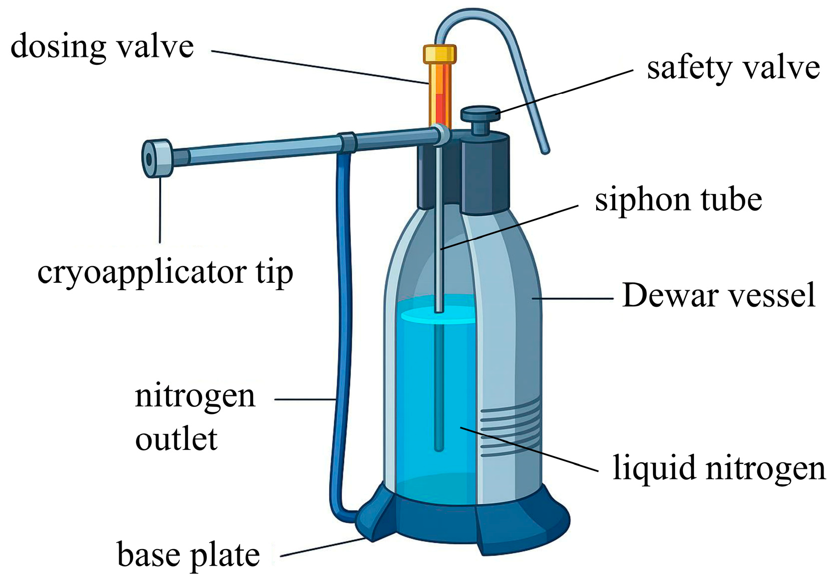
The aim of cryosurgery is to achieve necrosis of diseased tissue by freezing it through direct contact with a metal cryoapplicator. The cryo-applicator can be cooled by phase transformation of the evaporating nitrogen inside, supplied from a dewar vessel, or nitrous oxide N2O supplied from a high-pressure cylinder and throttled using a valve. The use of liquid nitrogen allows for the cryo-applicator to cool to approximately 83 K (−190 °C). A schematic of a portable cryosurgical unit powered by liquid nitrogen that allows for liquid nitrogen to be sprayed directly onto the diseased tissue is depicted in Figure 2.
One obstacle to the widespread use of cryotherapy is the need to supply liquid nitrogen. Small cryosurgery units use around 10 L of nitrogen per day, portable cryotherapy units around 100 kg, while whole-body cryochambers require supplies of up to 1000 kg of liquid nitrogen per day. Technologies currently under development enable the local production of liquid nitrogen in sufficient quantities to meet the needs of medical practices using single cryosurgery or topical cryotherapy units. Liquid nitrogen can be produced in small quantities (i.e., up to a few tens of liters per day) by combining membrane air–nitrogen separators with Joule–Thomson chillers powered by gas mixtures. The ideal cryomedical unit should achieve performance analogous to liquid nitrogen-powered units and be powered solely by electricity.
Both closed-circuit Joule–Thomson chillers powered by gas mixtures and cascade systems are interesting solutions that are finding application in cryomedicine. The uptake of cryomedicine will enable the use of pulsed tubes in this field, which are currently too expensive due to their limited applications [24,30,31]. Similarly to superconductivity, where medical diagnostics using NMR scanners have created the largest global market for LTS magnets, cryomedicine could lead to the development of pulse tube-based cooling systems enable to construct cryotherapeutic and cryosurgical devices that achieve sufficiently low temperatures without a liquid nitrogen supply.

3. Superconductivity Applications in Medicine

In the medical field, the most significant use of superconductivity involves high-field energy-efficient magnets. These are integral to cyclotrons used in radiotherapy (RT) for the generation of isotopes and creation of therapeutic proton/ion beams. Most importantly, they are essential components of currently the largest commercial application of superconductivity—MRI systems [32]. In this context, superconducting magnets greatly enhance the quality of diagnostic imaging and contribute to the exploration of biological system functions and biochemical mechanisms [25,33,34].
Deases imaging, diagnosis, and treatment are the main medical areas of superconductivity application, and include the following: (a) RT-cyclotrons and accelerators, (b) synchrotron radiation (the scope of its use is shown in Figure 3), (c) isotope production, (d) proton and ion beam delivery, (e) magnetic resonance imaging MRI, (f) nuclear magnetic resonance spectroscopy NMR, (g) positron emission tomography PET, and (h) ultrasensitive electro-magnetic signal pickups and transducers.
The latter are based on Superconducting Quantum Interference Devices (SQUIDs), which are utilized in biomagnetism to detect magnetic fields produced by ionic currents in the brain, heart, nerves, fetus, and muscles. SQUIDs are capable of measuring extremely weak magnetic fields (in the nano- to femtotesla range), enabling significant progress in biological imaging. Another notable area of superconductivity, particularly active in accelerator technology but not yet applied in medicine, involves superconducting radio-frequency (SCRF) magnets. Although SCRF accelerator structures currently exceed the performance needs of medical technologies, they may become relevant in future medical applications. The following subsections present selected applications of superconductivity in medical diagnostics, imaging, and cancer treatment.

3.1. Cyclotrons for Radiotherapy

The first model of cyclotron was developed 85 years ago by the University of California at Berkeley, and its originator was Ernest Orlando Lawrence, who, in the 1930s, was involved in research into nuclear reactions involving neutrons and artificial radioactivity. In 1939, in collaboration with William Brobeck, Lawrence built a cyclotron capable of accelerating deuterons to energies of 19 MeV. This device was installed in the Crocker Laboratory, where researchers achieved several groundbreaking milestones, including the first transmutation of certain elements, the discovery of multiple transuranic elements, and the production of hundreds of radioisotopes derived from known elements. The Crocker Laboratory is recognized as the birthplace of nuclear medicine, a field that harnesses radioisotopes for the diagnosis and treatment of human diseases. In 1936, Lawrence’s brother, John Lawrence, ran a clinic treating leukemia and melanoma patients with radioactive phosphorus produced in Crocker’s laboratory [35,36].
In 1944, the principle of phase stability, which occurs in cyclotrons accelerated by radio-frequency electric fields, was presented by the American scientist E.M. McMillan and the Russian researcher V.I. Veksler [37]. This principle made it possible to convert conventional cyclotrons into synchrotrons, also known as synchrocyclotrons, which are widely used in biomedical engineering today. Synchrotrons can achieve almost unlimited acceleration energy, making them a vital part of heavy-ion RT for cancer treatment. Spectral RT using high-power synchrotron beams, for example, is particularly effective with a low risk of complications in the treatment of tumors located in the central nervous system (gliomas) [25,38,39,40,41]. Such accelerators emits protons with an energy of 235 MeV at a tissue penetration of 32 cm. As for the lower-energy photon beam synchrotrons, they have been recently used in biomedical research aimed at studying tissues, cells, and subcellular structures [42,43].
Currently, performing an RT diagnostic involving living organisms requires beams of the highest possible intensity, which can be provided using superconducting synchrotrons known as “wigglers”. Multipole superconducting wigglers are integrated into synchrotron radiation (SR) sources to enhance radiation characteristics by boosting both the intensity and spectral hardness of the emitted SR. These devices consist of a series of superconducting dipole magnets arranged to produce an alternating transverse magnetic field. As an electron beam travels through this magnetic structure, it emits SR at each magnet, with the total radiation intensity effectively combining from all sources within nearly the same solid angle. This configuration offers a cost-effective method for significantly increasing the strength and quality of the radiation output [44].

3.2. Radiation Oncology

Radiation used in the treatment of cancer and other diseases can originate from external sources positioned outside the patient’s body or from radioactive materials implanted internally. Prior to the 1970s, many hospitals relied on gamma radiation from isotopes such as 60Co or megavoltage X-rays generated by betatrons. Later, medical facilities began adopting linear accelerators (LINACs), which produce photons through interactions between high-energy electrons and a target material. Currently, this form of radiation generation (involving both photons and electrons) is considered conventional for RT applications.
The main objectives of effective cancer RT are as follows: (1) the dose localization effect (DL, the relation between the distribution of the radiation dose and the volume of the tumor lesion), (2) the intensification of the relative biological effectiveness (RBE) of the radiation beam, and (3) linear energy transfer (LET)—the attenuation of the radiation beam energy per unit length, also as a measure of the biological damage to healthy cells, surrounding the tumor lesion, caused by ionizing radiation. These goals are based on the study of different types of radiation, particularly nuclear and subatomic particles such as hydrogen, helium, carbon, neon, and argon, as well as charged particles (protons, which are the nuclei of hydrogen atoms and the lightest nuclear particles) and neutral particles (neutron beams). Nuclear particles that are heavier than protons are known as heavy particles (i.e., ions of carbon, helium and oxygen).
Comparative analysis of the relative dose percentages for different therapeutic beams (including protons, neutrons, and carbon ions) versus X-rays, as a function of tissue penetration depth, reveals that both protons and carbon ions exhibit a pronounced dose increase near their stopping points, unlike the exponential attenuation observed with X-rays [25,45]. High-energy neutrons (~60 MeV) possess energy levels similar to photons produced by a 7–8 MeV source. Carbon ions display a sharper Bragg peak due to reduced multiple scattering. The residual dose beyond the range of carbon ions results from the lighter nuclear fragments generated during the deceleration of the carbon particles. Figure 4 presents a comparison of X-rays, protons, neutrons, and carbon ions in terms of relative therapeutic effectiveness. The following observations can be made:
  • X-rays have low LET and limited DL;
  • Protons also exhibit low LET, but achieve good DL;
  • Neutrons offer higher LET, but poor DL;
  • Carbon ions combine both high LET and high DL.
In the case of neutron therapy, the therapeutic dose is delivered through nuclear collisions that transfer energy to protons [46,47]. These secondary low-energy protons subsequently break molecular bonds, causing biological damage—including to healthy tissues. As illustrated in Figure 4, neutrons, compared to charged particles, demonstrate higher biological effectiveness. However, their dose distribution closely resembles that of X-rays. As clinical studies show, the use of neutron RT has two significant drawbacks: a) it exhibits poor DL, and b) it provides extensive damage to surrounding healthy tissues [48].
An additional factor driving the adoption of superconductivity in RT is the requirement to deliver therapeutic beams from virtually any angle around the patient, covering the full 4π steradian range. Modern therapeutic X-rays are generated using compact electron accelerators that can be mounted on rotating gantries. Electron beams with energies up to 20 MeV can be steered using conventional magnets with bending radii of approximately 10 cm.
While isocentric delivery of electron beams is relatively simple from a technological standpoint, it becomes more challenging with proton beams. For example, a 250 MeV proton beam has a magnetic induction Bp of around 2.5 T·m, requiring much larger gantry (such as shown in Figure 5) systems when using conventional magnets. The challenge is even greater with 400 MeV carbon ion beams, which have a Bp of approximately 6.6 T·m, making beam delivery significantly more complex [25].
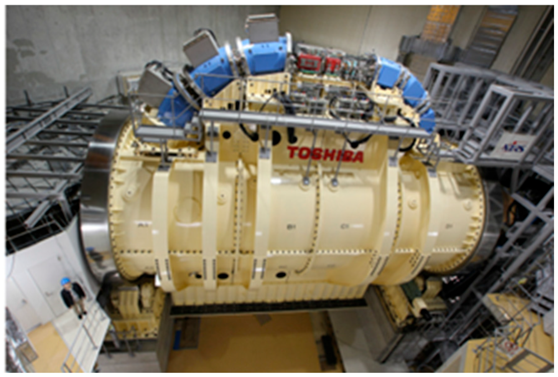
Figure 5 shows a photograph of a superconducting rotating gantry for carbon ion RT (CIRT), taken at the Heavy Ion Medical Accelerator (HIMAC, Chiba, Japan). The superconducting magnets and scanning magnets are shown in blue and red, respectively [48]. The use of superconducting magnets may allow for the greater availability of gantries in medical centers with smaller spaces. In addition, the use of a combination of fixed horizontal and non-horizontal beams and a therapy table that rotates with a certain range of pitch, yaw, and roll can be an effective method of expanding the range of beam angles [36].

3.3. Magnetic Resonance Imaging

The quality of medical imaging has made great developments in recent decades of the 20th and first decades of the 21st century, especially in the field of oncology diagnosis. X-ray computed tomography (CT), MRI/NMR, and PET have made it possible to obtain spatial and functional information about the human body and its organs, which has revolutionized diagnostics [25,49]. Each of these diagnostic methods offers distinct advantages, and the highest diagnostic accuracy is achieved by integrating data from all three. The primary challenge lies in aligning or merging the images (a process known as “fusion”), which is typically carried out within the imaging system itself.
Ongoing research in MRI aims to improve diagnostic power by increasing sensitivity, resolution, and accessibility. The development of low-cost portable MRI systems could open the door to novel point-of-care and monitoring applications, which are currently unfeasible with traditional, centralized MRI setups [50,51,52]. While affordability and accessibility have long been important, the most notable advancements have occurred in scan speed, image resolution, and patient comfort—including shorter magnet lengths and wider bore diameters. Despite technological progress, dedicated MRI systems (e.g., for limbs, brain, or breast imaging) have seen limited commercial success. Portable MRI scanners comparable in function to ultrasound or CT devices have yet to emerge, and MR monitoring tools remain largely confined to research settings [53,54]. However, expanding MRI markets in developing regions and recent advances in hardware and computing are reigniting interest in affordable, widely accessible MRI solutions.
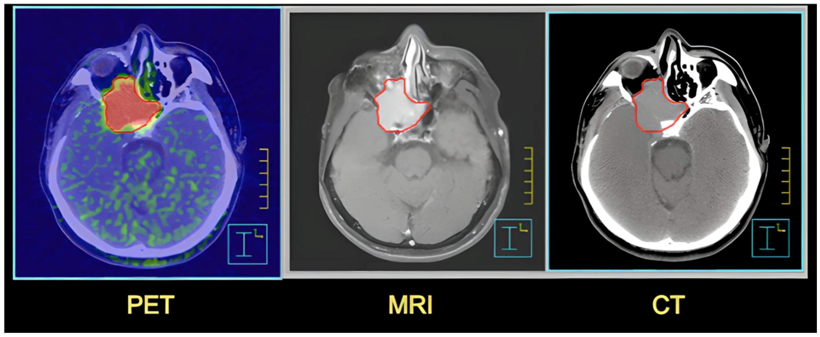
One of the most important medical applications of imaging technologies is determining the volume of tumor lesions to which therapeutic doses (produced by the particles described in Section 3.2) should be delivered [36]. Traditionally, the detection and assessment of tumors relied on physical examinations, tissue biopsies, and imaging techniques such as X-rays, CT, and MRI. In study [49], the authors explored the use of PET/CT with Fluorine-18-labeled prostate-specific membrane antigen-1007 (18F-PSMA-1007) to achieve three-dimensional spatial localization and volume estimation of prostate tumors. The results obtained were better than for MRI and support the use of 18F-PSMA-1007 PET/CT before treatment of localized prostate cancer. PET provides more sensitive identification of abnormal tissues of the tumor lesion, as shown in Figure 6 [36,49,55].
To summarize the applications of CT, MRI/NMR, and PET features, Table 3 presents a comparison of them in terms of operation principle, specific applications, safety, contraindications for patients, examination time, and additional valued peculiarities.
MRI generates highly detailed images of internal body structures by combining strong magnetic fields, radiofrequency waves, and advanced computer processing. The traditional MRI system currently uses 1.5 T or 3.0 T electromagnetic fields. Several MRI scanners with a superconducting magnet of 4 T, 7 T, 8 T, and 9.4 T MR systems, the first whole-body 10.5 T scanner is already operational [25,56,57]. Moreover, 11.7 T magnets intended for human MRI are currently being introduced at NeuroSpin (Paris France), at the National Institutes of Health (Bethesda, MD, USA), and at Gachon Medical University (Incheon, South Korea) [58].

3.4. Ultra-High-Field MRI

MRI with UHF is a promising technology for studying the human brain in previously unattainable detail due to the increase in signal-to-noise ratio (SNR) and contrast-to-noise ratio (CNR) with the value of magnetic field induction. The first UHF magnet was an 8 T induction, 8 m diameter system developed in 1998 by Magnex Scientific Ltd. (Yarnton, UK) for Ohio State University. One year later, a 7 T magnetic induction, 0.90 m diameter magnet was installed at the University of Minnesota. In 18 years, between 1997 and 2015, Magnex Scientific built approximately 72 UHF magnets [41,59].
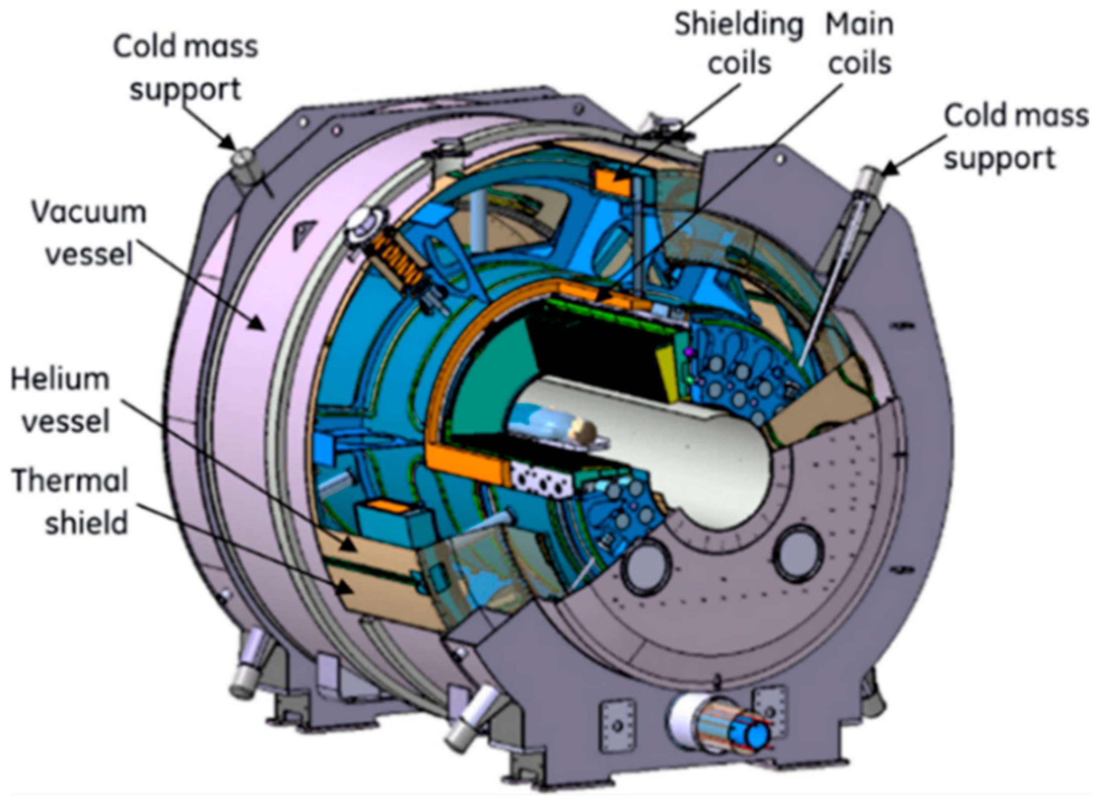
In the early years of this century, the French Atomic and Alternative Energy Commission (CEA) [60] launched a program funded by France and Germany to develop and build a “human brain researcher”. The intention was to develop a whole-body (WB) MRI scanner operating at a magnetic induction density of 11.7 T. The magnet was part of a larger effort to develop molecular imaging. At that time, the idea was regarded as highly ambitious. However, the CEA, leveraging its expertise in constructing magnets for fusion reactors and particle detectors, accepted the challenge of developing an 11.7 T WB MRI magnet. Following a feasibility assessment, the Iseult-Inumac project received funding to move forward. Figure 7 depicts the Iseult magnet and cryostat with the following specifications: current = 1470 A, operating temperature = 1.8 K, central field = 11.72 T, inductance = 308 H, stored energy 338 MJ, and mass = 132 tons.
Prior to the launch of the Iseult-Inumac project in 2018, an MRI with a 10.5 T magnetic field, the largest ever used for human scanning, started to operate at the University of Minnesota [61]. After almost two decades of R&D, prototyping, production, and commissioning in the frame of the project, the magnet first reached its target induction in 2019 and the first images were obtained in 2021 [59].

3.5. Low- and Ultra-Low-Field MRI

ULF MRI offers several potential advantages, including the possibility of significantly reducing equipment costs and achieving higher image contrast than conventional MRI, which opens the door to novel diagnostic techniques. These may include cancer screening without the need for contrast agents, imaging of traumatic brain injuries, detection of neurodegenerative conditions such as Alzheimer’s disease, and estimating the time since the onset of a stroke [62]. The primary challenge facing ULF MRI is its relatively low SNR compared to high-field systems. However, recent developments have introduced viable solutions, such as sensors capable of operating with noise levels as 0.1 fT·Hz [38].
Designing ULF-MRI systems for functional neural current imaging (NCI) of the brain with adequate SNR poses a complex optimization challenge involving multiple constraints. Enhancing SNR can be approached in two main ways: by minimizing system noise and/or increasing the prepolarization field to boost tissue magnetization. However, applying high currents to a resistive prepolarization coil necessitates either active cooling or a reduced duty cycle to prevent overheating—especially since signal-averaging sequences, used to enhance SNR, require multiple repetitions [63]. If a type II superconductor is used for the prepolarizing coil, the magnetic field applied must remain below the lower critical field (Hc1) to prevent flux penetration, which could lead to remanent field disturbances and affect image quality.
Early clinical low-field MRI systems employed permanent magnet dipole designs with iron yokes, as seen in models like the GE Signa Profile (0.2 T), Siemens Magnetom C! (0.35 T), and Hitachi AIRIS (0.3 T). Although marketed as ‘open’ systems, the use of iron yokes and rare earth magnetic materials made them heavier than 1.5 T superconducting solenoid magnets. Nonetheless, with targeted design optimizations, the overall weight of such systems can be significantly reduced—even to a portable level. For instance, a 0.2 T permanent dipole magnet with an iron yoke weighing approximately 200 kg was recently installed in a minivan for on-site imaging of elbow injuries in baseball players [64].

3.6. Biofunctionalized Magnetic Nanoparticles for Immunoassay

Magnetic particle imaging (MPI) is an emerging imaging technique designed to visualize the spatial distribution of magnetic nanoparticles (MNPs). It generates images based on harmonic signals produced by the nonlinear magnetization behavior of MNPs when exposed to an alternating magnetic field. The effectiveness of MPI is therefore closely tied to the magnetic characteristics of the nanoparticles, including core size, relaxation time, and their distribution. As a result, significant research has focused on developing MNPs optimized for use as tracers in MPI applications [65,66].
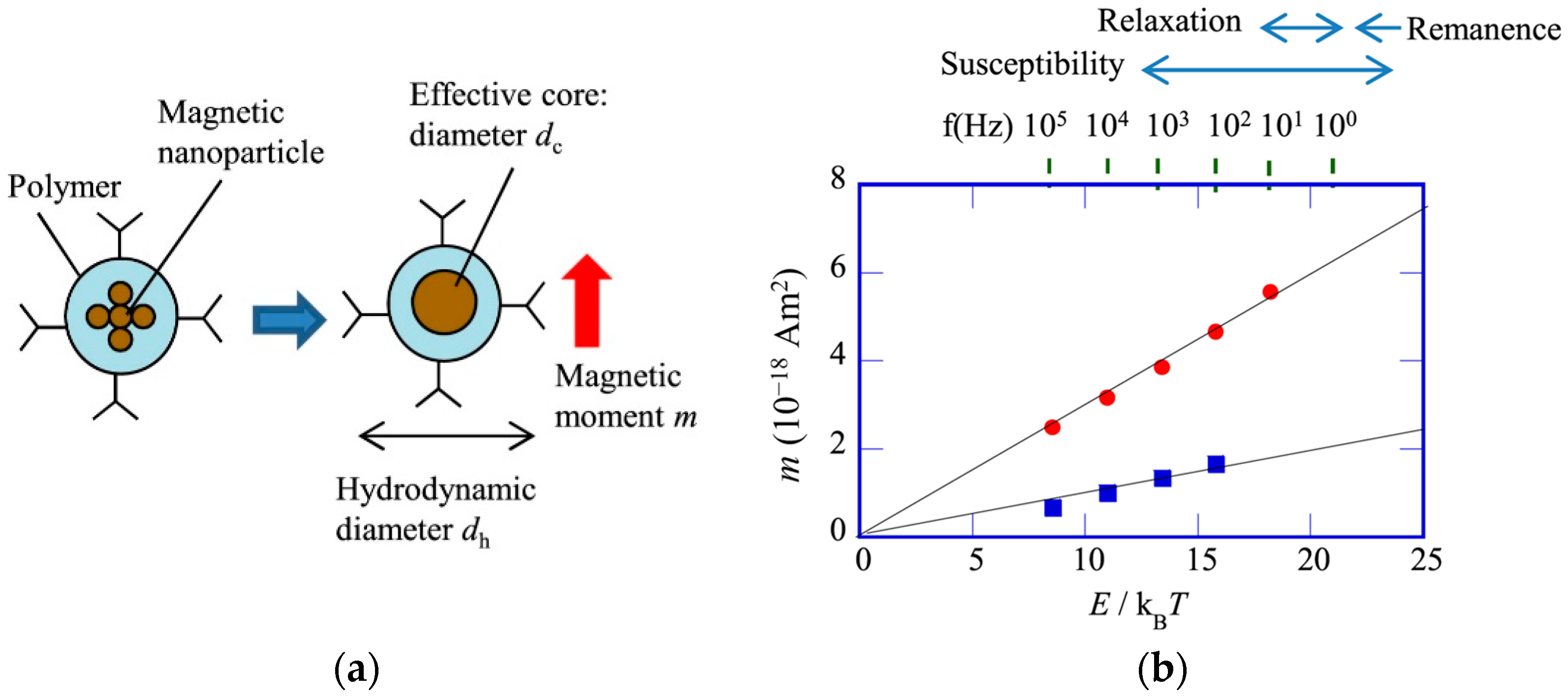
Commercially available cancer diagnostic methods based on bio-functionalized magnetic markers are increasingly used in practice. Figure 8 shows how MPNs are magnetically fractionated. Optimizing cancer cell detection methods across various organs relies on measuring (1) AC magnetic susceptibility, (2) magnetic relaxation time, and (3) remanence in nanoparticles NPs with differing magnetic characteristics. MNPs typically consist of aggregated nanostructures, as illustrated schematically in Figure 9a. When these particles are used as markers in immunoassays, two critical parameters are their magnetic moment m and relaxation time t. A high magnetic moment is desirable to generate strong detection signals, while the appropriate relaxation time depends on the specific detection technique employed. Figure 9b illustrates the relationship between m and the ratio of magnetic anisotropy energy to thermal energy (E/kBT) for two different commercially available markers [65]. The upper horizontal axis indicates the frequency associated with the Néel relaxation time, expressed as f = 1/(2…N). Arrows indicate the typical frequency ranges applied in various detection techniques.
Figure 9b highlights several fundamental properties of the markers: (1) both m and E are distributed across the particle population, meaning that only those particles with suitable m and E values contribute effectively to the magnetic signal. (2) Each detection method requires a specific range of E values, so it is important to choose markers with a significant proportion of particles falling within this optimal range. (3) The moment m is directly proportional to E, and, since the signal from an individual marker depends on its m value, this parameter ultimately defines the sensitivity of the detection system.

3.7. High-Sensitivity Magnetic Relaxometry Using SQUIDs

Magnetic relaxometry, particularly when using SQUID technology, is an emerging diagnostic approach that offers rapid and potentially more specific tumor detection than mammography, which can miss 10–25% of tumors. In theory, magnetic relaxometry also provides greater specificity than MRI, as it detects only those nanoparticles that are bound to the target [25,33,34,38,67]. Highly sensitive magnetometer systems based on low-temperature SQUID detectors have been developed for applications in cardiology (e.g., magnetocardiography) and for tracking the distribution of magnetic nanoparticles in biological systems. These SQUID-based systems are notable for their resistance to electromagnetic interference, allowing them to be used even in unshielded environments.
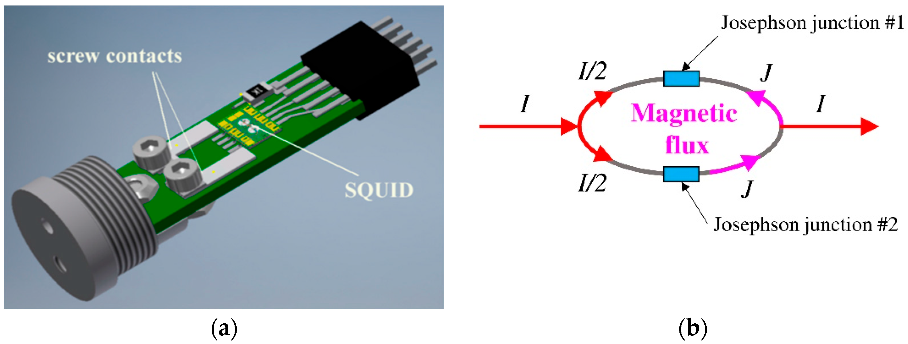
The SQUID-based magnetic field detection method relies on interference effects generated by power transmission and induced currents within the sensor. An exemplary Nb-based low-Tc DC SQUID sensor construction is shown in Figure 10a. Each SQUID sensor consists of two Josephson junctions connected in parallel. A Josephson junction is formed by two weakly coupled superconductors separated by a thin insulating barrier or another type of weak link (as shown in Figure 10b). In this configuration, a small supercurrent can flow between the superconductors without producing a voltage. A constant bias current is applied to the system and maintained in a closed loop, with the current evenly split between the two branches of the loop. When the bio-magnetic flux passes through the loop, the flux-induced current is added to half of the initial current on one side of the loop, while on the opposite side it subtracts from the initial half. This produces a phase shift between the junctions, so that one side reaches the current earlier than the other. The quantization of magnetic flux, characteristic of superconductivity, results in a voltage that oscillates between the maximum and minimum quantum values, with the maxima appearing at integer multiples of the flux quantum and the minima at half-integer multiples. By monitoring these oscillations or voltage variations, it is possible to evaluate the changes in BMF [67].
Since the above is a very detailed explanation of how the SQUID sensor works, it can be simplified to two basic issues:
(a).
It works by using the quantum mechanical properties of superconductors to detect tiny changes in magnetic fields;
(b).
It measures the magnetic flux through a superconducting loop, which is converted into a measurable voltage or current, allowing for extremely sensitive measurements of magnetic fields.
At present, both low- and high-temperature critical Tc detectors are applied in research and clinical settings for BMF diagnosis. Earlier investigations and commercially available instruments relied on SQUID systems cooled with costly liquid helium to approximately 4 K. More recent studies [34,69,70,71] have explored SQUID detectors functioning at liquid nitrogen temperatures near 77 K in order to reduce expenses. However, this cost advantage comes with the drawback of a roughly tenfold lower signal-to-noise ratio, since high-Tc SQUIDs generally exhibit inferior noise characteristics compared with their low-Tc counterparts, with noise factors approximately an order of magnitude higher.
The human body is made up of more than 200 cell types and four different tissue types, through which electrical impulses propagate in the form of action potentials. Electric fields from ionic currents have long been used, e.g., in the diagnosis of the heart electrocardiography (ECG) and brain electroencephalography (EEG). The values of the electric signals depend on the electrical permeability and conductivity of tissues, often leading to inaccurate and limited diagnostic results. In addition, the such procedures and devices use electrodes that are in direct contact with the skin, making them invasive.
Another approach involves measuring the magnetic fields generated by ionic currents. Characteristics of the magnetic field, such as spatial localization and detection of eddy currents, can accelerate diagnostic procedures and improve diagnostic accuracy. Since biological tissues exhibit magnetic permeability close to that of a vacuum, biomagnetic fields (BMF) pass through the skin’s surface without distortion and can be recorded in a non-contact manner. This avoids skin irritation and potential allergic reactions that may occur when electrical activity is monitored using electrodes [72,73]. Diagnostic applications of BMF include muscle magnetomyography (MMG), cardiac magnetocardiography (MCG), brain magnetoencephalography (MEG), nerve magnetoneurography (MNG), and spinal magnetospinography (MSG) [74]. The primary difficulty in capturing these BMF lies in their extremely low field strength and wide bandwidth. As shown in Table 4, their frequency ranges from a few hertz to several kilohertz, while their magnitudes span from 10−10 T down to approximately 10−15 T [69,70,71].
The use of SQUIDs and magnetic shielding techniques has allowed for magnetography (MG)-based diagnostics to be introduced into clinical practice. In order to obtain the required sensitivity and a high signal-to-noise ratio, the majority of MG devices necessitate shielding. This may involve partial shielding of a specific region or full shielding, implemented as a dedicated shielded room where the measurements are performed. MG devices operating in screened rooms are huge and expensive. They require liquid helium cooling, which increases complexity and cost. The use of cryogens (nitrogen) and cryochambers can partially overcome these limitations. Recently, encouraging outcomes have been reported for BMF recordings conducted in unshielded environments, employing advanced digital signal processing methods. These include, among others, band-pass filtering, window averaging, moving average filtering, and feedback mechanisms aimed at reducing magnetic noise in the processed data [33,34,67,68,75,76].
Highly sensitive magnetometer systems based on low-temperature SQUID detectors have been developed for cardiology research (magnetocardiography) and for investigating the distribution of magnetic nanoparticles in biological systems. These SQUID magnetometers are characterized by strong resistance to external interference, which makes it possible to conduct studies in unshielded environments. Their high reproducibility of measurement results has been confirmed [75,76]. The application of magnetocardiographic systems has facilitated the creation of a novel screening technology for the early diagnosis of cardiac diseases. Magnetic imaging of cardiac activity currents provides an optimal method for analyzing local electrical inhomogeneities within the myocardium. Magnetocardiography has demonstrated substantial potential both for advancing the fundamental understanding of cardiac biosignals and for clinical cardiology [76].
In contrast to heart- and brain-related diseases, which pose a greater risk of mortality, conditions affecting bones, muscles, and nerves have historically received comparatively limited attention. As living standards have risen, disability-inducing disorders have gained increasing significance. Musculoskeletal diseases, if left untreated (despite being preventable or manageable when addressed in time), can result in irreversible changes, with the absence of effective diagnostics and therapies further aggravating the issue. A variety of diagnostic techniques are employed in clinical practice, including electromyography (EMG), surface and needle EMG, quantitative EMG, ultrasound, and MRI, among others [77].
Experiments reported in [71] have demonstrated that neuromuscular magnetic fields may offer significant advantages in disease diagnosis, condition monitoring, and rehabilitation. Compared with EMG, MMG provides a higher signal-to-noise ratio, is non-invasive, delivers more accurate signals, and is less affected by surrounding muscle tissues, while also enabling simultaneous localization of BMF. Magnetic diagnostics of spinal nerves show certain benefits over conventional clinical methods, and MMG additionally holds potential for elucidating details of skeletal muscle contraction mechanisms. Consequently, BMF detection in nerves and muscles is anticipated to evolve into an important auxiliary technique, with substantial relevance for clinical disease diagnosis and studies of kinematic mechanisms.
Considerable attention is directed toward rapid processes occurring in the human brain. The SQUID system, with its millisecond response time to brain magnetic fields, has proven highly valuable for investigating brain function and activity, particularly in deep regions. Studies using a SQUID system within a superconducting magnetic shield, as reported in [69], revealed that only the right hippocampus responds to random somatosensory stimuli. These investigations also showed that the sensitivity of neuromagnetic measurements is limited by the noise temperature of the SQUID hardware rather than by so-called “brain noise.” The application of a SQUID system with a high-Tc superconducting magnetic shield provides novel insights into deep brain structures, representing an important contribution of both LTS and HTS technologies.

4. Prospects for Future Applications of Cryogenics and Superconductivity in Medicine

Theranostics, which we understand as the combination of diagnostics and therapy to select targeted treatment for the individual patient—at the same time safe and effective—is the future of medicine, including cryogenic, superconducting, and nuclear medicine.
The uptake of cryomedicine will enable the use of pulse tubes in this field, which are currently too expensive due to their limited applications [24,30,31,35,77,78,79].
Future significant improvement in ion beam therapy is strongly depends on the new generation of cyclotrons capable of generating both carbon ions and protons (e.g., C400 cyclotron developed by Ion Beam Applications in Belgium). In conjunction with superconducting gantries, it will help decrease the size and cost of facilities required for heavy ion treatments, making them comparable to current proton therapy centers.
Supercompact synchrotrons used to produce PET and other radioisotopes can incur very high manufacturing and operational costs due to the use of HTS. This has limited their deployment in medical applications, resulting in a current undersupply. Progress in this area could be achieved by using materials for magnet windings that operate at higher temperatures, such as ferroelectric superconductors and MgB2.
The development of pulse tube-based cooling systems, in which heat is transferred away from the superconducting magnets, makes it possible to construct cryotherapeutic and cryosurgical devices that can reach sufficiently low temperatures without the need for a liquid nitrogen supply.
For particle-beam based RT facilities to operate effectively, it is crucial to develop reliable and cost-efficient instrumentation capable of regulating different important parameters such as beam-energy variability, energy step size and switching time, positional and angular accuracy, temporal beam intensity control, robust control systems to ensure patient safety, etc. The direction of development in this area may be three-dimensional dynamic conformal therapy with new engineering ideas, such as pencil-beam scanning technology, allowing for the meeting of the majority of economic, energy, and health safety expectations.
In case of MNPs prospects in biomedical application, the following directions of development can be determined:
(a).
Targeted drug delivery: MNPs can be functionalized with drugs to be guided to specific sites, like tumors, using external magnetic fields, which increases drug accumulation in the target tissue and reduces side effects.
(b).
Controlled release: Releasing the therapeutic agent from the MNPs can be triggered remotely by a magnetic field at the precise time and location.
(c).
Hyperthermial properties: MNPs can be used to locally heat and ablate diseased tissue, such as cancer cells, when an alternating magnetic field is applied.
(d).
Gene therapy (magnetofection): MNPs can act as carriers to deliver therapeutic genes to target cells under the external magnetic field.
The future generation of high-field superconducting magnets that will contribute to the development of MRI and NMR technologies should exhibit exceptionally strong critical current densities and outstanding tolerance to high magnetic fields at low temperatures (around 15 K). Among the various promising candidates, the conductors Nb3Ge and Nb3(Al,Ge) stand out, with research on these materials only now beginning to gain significant momentum.
SQUID-based biomagnetic sensing continues to reveal new possibilities for the non-invasive diagnosis of cardiac, neural, and neuromuscular activity. It offers greater sensitivity and provides more information than conventional electrical methods. Developments in this area are crucial for advanced materials applications in biomedicine.

5. Conclusions

This review shows that cryogenics and superconductivity are two closely linked technological areas that continue to have a growing impact on modern medicine. Their current applications in cryogenic therapy, radiotherapy, diagnostic and imaging devices (MRI, PET), synchrotron radiation devices (isotope generation, proton and ion beam delivery), and ultra-sensitive SQUID magnetic signal transducers for detecting ultra-low biosignals emitted by human body organs are presented. This shows that both fields have moved from experimental concepts to essential components of diagnostic and therapeutic practice. Despite this progress, however, there is still significant room for improvement in each area, particularly with regard to efficiency, accessibility, and cost reduction.
Cryogenic technologies, including localized and whole-body therapeutic cooling as well as cryopreservation, are poised for transformation through the development of compact, electricity-powered cooling systems. Such systems are based on pulse-tube cryocoolers, Joule–Thomson units, and cascade refrigeration. This could reduce dependence on large-scale liquid nitrogen supplies, making cryomedical procedures more affordable and easier to implement in a variety of clinical settings.
Superconductivity, particularly in high-field magnet technology, remains central to advanced medical imaging and radiotherapy. The continuing evolution of superconducting magnets has already enabled ultra-high-field MRI scanners exceeding 7–11.7 T, which are expected to enhance imaging resolution, functional analysis, and the early detection of neurological disorders. In radiotherapy, superconducting gantries and compact accelerator designs can shrink the footprint and cost of proton and heavy-ion therapy facilities, potentially making these highly effective treatments available to a broader population.
A particularly dynamic area of development concerns the use of SQUID-based biomagnetic sensing and biofunctionalized magnetic nanoparticles (MNPs). SQUID magnetometers—capable of detecting magnetic fields in the femto- to picotesla range—are steadily progressing from laboratory environments toward broader clinical adoption. Their ability to record biomagnetic signals without physical contact and without distortion from biological tissues offers significant advantages over conventional electrophysiological methods. In parallel, MNP-based technologies are advancing rapidly toward clinical translation, enabling targeted cancer diagnostics and high-specificity immunoassays. They also demonstrate a huge potential for future realization in controlled drug delivery, magnetically triggered therapeutic release, hyperthermia treatments, and gene-delivery methods. Together, SQUID sensors and engineered magnetic nanoparticles form a powerful technological platform with the potential to transform non-invasive diagnostics, personalized theranostics, and real-time physiological monitoring.

Author Contributions

Conceptualization, O.B. and H.D.S.; methodology, H.D.S.; software, O.B.; validation, O.B.; formal analysis, O.B. and H.D.S.; investigation, H.D.S.; software, O.B.; resources, H.D.S.; writing—original draft preparation, O.B. and H.D.S.; writing—review and editing, O.B.; visualization, O.B.; supervision, H.D.S.; funding acquisition, O.B. and H.D.S. All authors have read and agreed to the published version of the manuscript.

Funding

This research was funded by Lublin University of Technology grants numbers FD 20/EE-2/416 and FD-20/EE-2/401 intended for research activities within the “Automatics, Electronics and Electrical Engineering” scientific discipline.

Data Availability Statement

Not applicable.

Acknowledgments

The authors teach Applications of Superconducting and Plasma Technologies in Medicine to graduate students in Biomedical Engineering, to whom they dedicate this review, believing that they will become interested in these issues and want to put them into practice in their future work as biomedical engineers.

Conflicts of Interest

The authors declare no conflicts of interest.

References

  1. Stryczewska, H.D.; Stępień, M.; Boiko, O. Plasma and Superconductivity for the Sustainable Development of Energy and the Environment. Energies 2022, 15, 4092. [Google Scholar] [CrossRef]
  2. Assunçao, R.; Eckl, F.; Ramos, C.P.; Correia, C.B.; Neto, R.C. Oxygen liquefaction economical value in the development of the hydrogen economy. Int. J. Hydrogen Energy 2024, 62, 109–118. [Google Scholar] [CrossRef]
  3. Weisend, J.G., II. Superfluid. In How a Quantum Fluid Revolutionized Modern Science; Springer Nature: London, UK, 2023; pp. 3–10. [Google Scholar]
  4. Banaszkiewicz, T.; Chorowski, M.; Gizicki, W.; Jedrusyna, A.; Kielar, J.; Malecha, Z.; Piotrowska, A.; Polinski, J.; Rogala, Z.; Sierpowski, K.; et al. Liquefied Natural Gas in Mobile Applications—Opportunities and Challenges. Energies 2020, 13, 5673. [Google Scholar] [CrossRef]
  5. Chen, B.S.; Zeng, Y.P.; Luo, E.R.; Peng, N.; Zou, A.H. An innovative method for helium refrigeration liquefaction utilizing transonic nozzle. Energy 2025, 314, 134283. [Google Scholar] [CrossRef]
  6. Sharafi, M.; Borghei-Rad, S.M.; Hezavehei, M.; Shahverdi, A.; Benson, J.D. Cryopreservation of Semen in Domestic Animals: A Review of Current Challenges, Applications, and Prospective Strategies. Animals 2022, 12, 3271. [Google Scholar] [CrossRef]
  7. Rezaie, H.; Ziabasharhagh, M.; Mafi, M. A review of hydrogen liquefaction, current situation and its future. In Proceedings of the International Conference on Engineering & Applied Sciences, Dubai, United Arab Emirates, 10 March 2016. [Google Scholar]
  8. Omori, K.M.; Mazon-Cartagena, R.; Fernández-Torres, M.J.; Caballero, J.A.; Ravagnani, M.A.S.S.; Pavão, L.V.; Costa, C.B.B. Sensitivity Analysis in Simple Cycles for Hydrogen Liquefaction. Processes 2025, 13, 3076. [Google Scholar] [CrossRef]
  9. Li, Z.A.; Xu, M.J.; Wang, J.H.; Zhang, F. Recent Advances in Cryogenic 3D Printing Technologies. Adv. Eng. Mater. 2022, 24, 2200245. [Google Scholar] [CrossRef]
  10. Tuoriniemi, J.T.; Knuuttila, T.A. Nuclear cooling and spin properties of rhodium down to picokelvin temperatures. Phys. B Condens. Matter 2000, 280, 474–478. [Google Scholar] [CrossRef]
  11. Mizunami, T.; Tatehata, H.; Kawashima, H. High-sensitivity cryogenic fibre-Bragg-grating temperature sensors using Teflon substrates. Meas. Sci. Technol. 2001, 12, 914–917. [Google Scholar]
  12. Wu, M.C.; DeHaven, S.L. High-sensitivity cryogenic temperature sensors using pressurized fiber Bragg gratings. In Proceedings of the SPIE Photonics Europe, Strasbourg, France, 3–7 April 2006; Volume 6189, p. 61890O. [Google Scholar]
  13. Deppner, C.; Herr, W.; Cornelius, M.; Stromberger, P.; Sternke, T.; Grzeschik, C.; Grote, A.; Rudolph, R.; Krutzik, M.; Wenzlawski, A.; et al. Collective-Mode Enhanced Matter-Wave Optics. Phys. Rev. Lett. 2021, 127, 100401. [Google Scholar] [CrossRef]
  14. Rjabinin, J.N.; Shubnikow, L.W. Magnetic Properties and Critical Currents of Supra-conducting Alloys. Nature 1935, 135, 581–582. [Google Scholar] [CrossRef]
  15. Ginzburg, V.L.; Landau, L.D. On the Theory of Superconductivity. J. Exptl. Theoret. Phys. 1950, 20, 138–167. [Google Scholar]
  16. Bardeen, J.; Cooper, L.N.; Schrieffer, J.R. Theory of Superconductivity. Phys. Rev. 1957, 108, 1175–1204. [Google Scholar] [CrossRef]
  17. Braginski, A.I. Consequences of Brian Josephson’s Theoretical Discovery. J. Supercond. Nov. Magn. 2021, 34, 1597–1600. [Google Scholar] [CrossRef]
  18. Bednorz, J.G.; Muller, K.A. The new approach to high-Tc superconductivity. Rev. Mod. Phys. 1988, 60, 585–600. [Google Scholar] [CrossRef]
  19. Nagamatsu, J.; Nakagawa, N.; Muranaka, T.; Zenitani, Y.; Akimitsu, J. Superconductivity at 39 K in magnesium diboride. Nature 2001, 410, 63–64. [Google Scholar] [CrossRef]
  20. Takahashi, H.; Igawa, K.; Arii, K.; Kamihara, Y.; Hirano, M.; Hosono, H. Superconductivity at 43 K in an iron-based layered compound LaO1-xFxFeAs. Nature 2008, 53, 376–378. [Google Scholar] [CrossRef] [PubMed]
  21. Physics of Iron-Based High Temperature Superconductors. Available online: https://boulderschool.yale.edu/sites/default/files/files/MatsudaNote.pdf (accessed on 17 November 2025).
  22. Cao, Y.; Fatemi, V.; Fang, S.; Watanabe, K.; Taniguchi, T.; Kaxiras, E.; Jarillo-Herrero, P. Unconventional superconductivity in magic-angle graphene superlattices. Nature 2018, 556, 43–50. [Google Scholar] [CrossRef] [PubMed]
  23. Eremets, M.I.; Minkov, V.S.; Drozdov, A.P.; Kong, P.P.; Ksenofontov, V.; Shylin, S.I.; Bud’ko, S.L.; Prozorov, R.; Balakirev, F.F.; Sun, D.; et al. High-Temperature Superconductivity in Hydrides: Experimental Evidence and Details. J. Supercond. Nov. Magn. 2022, 35, 965–977. [Google Scholar] [CrossRef]
  24. Podbielska, A.; Skrzek, A. Application of Low Temperatures in Biomedicine/Zastosowanie Niskich Temperatur w Biomedycynie, 1st ed.; Wrocław University of Technology Publishing House: Wrocław, Poland, 2012. [Google Scholar]
  25. Alonso, J.R.; Antaya, T.A. Superconductivity in Medicine. Rev. Accel. Sci. Technol. 2012, 5, 227–263. [Google Scholar] [CrossRef]
  26. Flukiger, R. Overview of Superconductivity and Challenges in Applications. Rev. Accel. Sci. Technol. 2012, 5, 1–23. [Google Scholar] [CrossRef]
  27. Cipria, K.; Lopez, E.; Naso, V. Investigation of the use of Pulse Tube in cell cryopreservation systems. Cryobiology 2010, 61, 225–230. [Google Scholar] [CrossRef]
  28. Janowski, T.; Stryczewska, H.D.; Wac-Włodarczyk, A. Superconducting and Plasma Technologies in Energy/Technologie Nadprzewodnikowe i Plazmowe w Energetyce, 1st ed.; Lublin Scientific Society Publishing House: Lublin, Poland, 2009. (In Polish) [Google Scholar]
  29. Kriogenika w Zastosowaniach Przemysłowych, Medycznych i Badawczych. Cryogenics in Industrial, Medical and Research Applications. Available online: https://www.klimatyzacja.pl/chlodnictwo/artykuly/kriogenika/kriogenika-w-zastosowaniach-przemyslowych-medycznych-i-badawczych (accessed on 16 November 2025). (In Polish).
  30. Chorowski, M. Cryogenics, Basics and Applications/Kriogenika, Podstawy i Zastosowania; IPPU Masta: Gdańsk, Poland, 2007. (In Polish) [Google Scholar]
  31. Tomaru, T.; Suzuki, T.; Haruyama, T.; Shintomi, T.; Yamamoto, A.; Koyama, T.; Li, R. Vibration analysis of cryocoolers. Cryogenics 2004, 44, 309–317. [Google Scholar] [CrossRef]
  32. Szkliniarz, K. Cyclotron Production and Study of Radioisotopes Used in Medical Diagnostics and Therapy/Cyklotronowa Produkcja i Badanie Radioizotopów Stosowanych w Diagnostyce i Terapii Medycznej. Ph.D. Thesis, Silesian University, Katowice, Poland, 2016. [Google Scholar]
  33. Körber, R.; Storm, J.H.; Seton, H.; Mäkelä, J.P.; Paetau, R.; Parkkonen, L.; Pfeiffer, C.; Riaz, B.; Schneiderman, J.F.; Dong, H.; et al. SQUIDs in biomagnetism: A roadmap towards improved healthcare. Supercond. Sci. Technol. 2016, 29, 113001. [Google Scholar] [CrossRef]
  34. Oladapo, B.I.; Zahedi, S.A.; Chaluvadi, S.C.; Bollapalli, S.S.; Ismail, M. Model design of a superconducting quantum interference device of magnetic field sensors for magnetocardiography. Biomed. Signal Process. Control. 2018, 46, 116–120. [Google Scholar] [CrossRef]
  35. Chu, W.T. Accelerators and Medicine. J. Korean Phys. Soc. 2007, 50, 1385–1389. [Google Scholar] [CrossRef]
  36. Levy, R.P.; Blakely, E.A.; Chu, W.T.; Coutrakon, G.B.; Hug, E.B.; Kraft, G.; Tsujii, H. The Current Status and Future Directions of Heavy Charged Particle Therapy in Medicine. AIP Conf. Proc. 2009, 1099, 410–425. [Google Scholar]
  37. Iwata, Y.; Noda, K. 8.10—Ion Linac and Synchrotron. Compr. Biomed. Phys. 2014, 8, 153–168. [Google Scholar]
  38. Hathaway, H.J.; Butler, K.S.; Adolphi, N.L.; Lovato, D.M.; Belfon, R.; Fegan, D.; Monson, T.C.; Trujillo, J.E.; Tessier, T.E.; Bryant, H.C.; et al. Detection of breast cancer cells using targeted magnetic nanoparticles and ultra-sensitive magnetic field sensors. Breast Cancer Res. 2011, 13, R108. [Google Scholar] [CrossRef] [PubMed]
  39. Nawrocki, W. Introduction to Quantum Metrology: Quantum Standards and Instrumentation, 1st ed.; Springer: Berlin/Heidelberg, Germany, 2015. [Google Scholar]
  40. Iwata, Y.; Noda, K.; Shirai, T.; Murakami, T.; Furukawa, T.; Mori, S.; Fujita, T.; Itano, A.; Shouda, K.; Mizushima, K.; et al. Design of a superconducting rotating gantry for heavy-ion therapy. Phys. Rev. ST Accel. Beams 2012, 15, 044701. [Google Scholar] [CrossRef]
  41. Iwata, Y.; Fujimoto, T.; Matsuba, S.; Fujita, T.; Sato, S.; Furukawa, T.; Hara, Y.; Mizushima, K.; Saraya, Y.; Tansho, R.; et al. Beam commissioning of a superconducting rotating-gantry for carbon-ion radiotherapy. Nucl. Instrum. Methods Phys. Res. A 2016, 834, 71–80. [Google Scholar] [CrossRef]
  42. Pełka, B. Synchrotron radiation in biology and medicine. Synchrotron Radiat. Nat. Sci. 2007, 6, 99–107. [Google Scholar]
  43. Kisiel, A. Synchrotron as a tool: Applications of synchrotron radiation in solid-state spectroscopy. Synchrotron Radiat. Nat. Sci. 2006, 5, 145–167. [Google Scholar]
  44. Sun, X.J.; Chen, F.S.; Yang, X.C.; Chen, W.; Bian, X.J.; Li, M.X.; Ge, R.; Xu, M.F.; Gao, Y.; Wang, J.C.; et al. Superconducting multipole wiggler with large magnetic gap for HEPS-TF. Nucl. Sci. Tech. 2022, 33, 16. [Google Scholar] [CrossRef]
  45. Wilkinson, B.; Hill, M.A.; Parsons, J.L. The Cellular Response to Complex DNA Damage Induced by Ionising Radiation. Int. J. Mol. Sci. 2023, 24, 4920. [Google Scholar] [CrossRef]
  46. Catterall, M. The assessment of the results of neutron therapy. Int. J. Radiat. Oncol. Biol. Phys. 1982, 8, 1573–1580. [Google Scholar] [CrossRef]
  47. Laramore, G.E.; Krall, J.M.; Griffin, T.W.; Duncan, W.; Richter, M.P.; Saroja, K.R.; Maor, M.H.; Davis, L.W. Neutron versus photon irradiation for unresectable salivary gland tumors: Final report of an RTOG-MRC randomized clinical trial. Int. J. Radiat. Oncol. Biol. Phys. 1993, 27, 235–240. [Google Scholar] [CrossRef] [PubMed]
  48. Iwata, Y.; Fujita, T.; Furukawa, T.; Hara, Y.; Matsuba, S.; Mizushima, K.; Murakami, T.; Noda, K.; Saotome, N.; Saraya, Y.; et al. Superconducting gantry for carbon-ion radiotherapy. In Proceedings of the 9th International Particle Accelerator Conference IPAC2018, Vancouver, BC, Canada, 29 April–4 May 2018. [Google Scholar]
  49. Huang, G.; Albers, P.; Mookerji, N.; Pfanner, T.; Hui, A.; Mittal, R.; Broomfield, S.; Dean, L.; Martin, B.S.; Jacobsen, N.-E.; et al. Three-dimensional spatial localization and volume estimation of prostate tumors using 18F-PSMA-1007 PET/CT versus multiparametric MRI. Eur. J. Nucl. Med. Mol. Imaging 2025, 52, 1642–1648. [Google Scholar] [CrossRef] [PubMed]
  50. Kimberly, W.T.; Sorby-Adams, A.J.; Webb, A.G.; Beekman, R.; Bowry, R.; Schiff, S.J.; de Havenon, A.; Shen, F.X.; Sze, G.; Schaefer, P.; et al. Brain imaging with portable low-field MRI. Nat. Rev. Bioeng. 2023, 1, 617–630. [Google Scholar] [CrossRef]
  51. Sorby-Adams, A.J.; Guo, J.N.F.; Laso, P.; Kirsch, J.E.; Zabinska, J.; Guarniz, A.L.G.; Schaefer, P.W.; Payabvash, S.; de Havenon, A.; Rosen, M.S.; et al. Portable, low-field magnetic resonance imaging for evaluation of Alzheimer’s disease. Nat. Commun. 2024, 15, 10488. [Google Scholar] [CrossRef]
  52. Glowacki, B.A. Hydrogen Cryomagnetic a Common Solution for Metallic and Oxide Superconductors. Materials 2025, 18, 3665. [Google Scholar] [CrossRef]
  53. Zabinska, J.; de Havenon, A.; Sheth, K.N. Recent advances in portable, low-field magnetic resonance imaging in cerebrovascular disease. Curr. Opin. Neurol. 2025, 38, 35–39. [Google Scholar] [CrossRef]
  54. Shoghli, A.; Chow, D.; Kuoy, E.; Yaghmai, V. Current role of portable MRI in diagnosis of acute neurological conditions. Front. Neurol. 2023, 14, 1255858. [Google Scholar] [CrossRef]
  55. Vijayakumar, S.; Yang, J.; Nittala, M.R.; Velazquez, A.E.; Huddleston, B.L.; Rugnath, N.A.; Adari, N.; Yajurvedi, A.K.; Komanduri, A.; Yang, C.C.; et al. Changing Role of PET/CT in Cancer Care with a Focus on Radiotherapy. Cureus 2022, 14, e32840. [Google Scholar] [CrossRef]
  56. Özütemiz, C.; White, M.; Elvendahl, W.; Eryaman, Y.; Marjanska, M.; Metzger, G.J.; Patriat, R.; Kulesa, J.; Harel, N.; Watanabe, Y.; et al. Use of a Commercial 7-T MRI Scanner for Clinical Brain Imaging: Indications, Protocols, Challenges, and Solutions-A Single-Center Experience. Am. J. Roentgenol. 2023, 221, 788–805. [Google Scholar] [CrossRef] [PubMed]
  57. Feinberg, D.A.; Beckett, A.J.S.; Vu, A.T.; Stockmann, J.; Huber, L.; Ma, S.M.T.; Ahn, S.; Setsompop, K.; Cao, X.Z.; Park, S.; et al. Next-generation MRI scanner designed for ultra-high-resolution human brain imaging at 7 Tesla. Nat. Methods 2023, 20, 1833. [Google Scholar] [CrossRef] [PubMed]
  58. Platt, T.; Ladd, M.E.; Paech, D. 7 Tesla and Beyond: Advanced Methods and Clinical Applications in Magnetic Resonance Imaging. Investig. Radiol. 2021, 56, 705–725. [Google Scholar] [CrossRef] [PubMed]
  59. Boulant, N.; Quettier, L.; The Iseult Consortium. Commissioning of the Iseult CEA 11.7 T whole-body MRI: Current status, gradient–magnet interaction tests and first imaging experience. Magn. Reson. Mater. Phy. 2023, 36, 175–189. [Google Scholar] [CrossRef]
  60. TSAR—Topological Solitons in AntifeRroics. Available online: https://www.tsar-fetopen.eu/#consortium (accessed on 8 February 2025).
  61. U Scientists Scan World’s First 10.5-Tesla Human MRI Image|RIO. Available online: https://research.umn.edu/news/u-scientists-scan-worlds-first-105-tesla-human-mri-image (accessed on 8 February 2025).
  62. Clarke, J.; Hatridge, M.; Mößle, M. SQUID-Detected Magnetic Resonance Imaging in Microtesla Fields. Annu. Rev. Biomed. Eng. 2007, 9, 383–413. [Google Scholar] [CrossRef]
  63. Bandettini, P.A.; Petridou, N.; Bodurka, J. Direct Detection of Neuronal Activity with MRI: Fantasy, Possibility, or Reality? Appl. Magn. Reson. 2005, 29, 65–88. [Google Scholar] [CrossRef]
  64. Wald, L.L.; McDaniel, P.C.; Witzel, T.; Stockmann, J.P.; Zimmerman-Cooley, C. Low-Cost and Portable MRI. Magn. Reason. Imaging 2020, 52, 686–696. [Google Scholar] [CrossRef] [PubMed]
  65. Yoshida, T.; Nakamura, T.; Higashi, O.; Enpuku, K. Magnetic fractionation and characterization of magnetic nanoparticles for magnetic particle imaging. Jpn. J. Appl. Phys. 2018, 57, 080302. [Google Scholar] [CrossRef]
  66. Enpuku, K.; Yang, S.Y.; Chieh, J.J. Magnetic nanoparticles for immunoassay. Supercond. Sci. Technol. 2016, 29, 113001. [Google Scholar]
  67. Rogalla, H.; Kes, P.H. 100 Years of Superconductivity, 1st ed.; CRC Press: Boca Raton, FL, USA, 2011. [Google Scholar]
  68. Faley, M.I.; Kostyurina, E.A.; Kalashnikov, K.V.; Maslennikov, Y.V.; Koshelets, V.P.; Dunin-Borkowski, R.E. Superconducting Quantum Interferometers for Non-destructive Evaluation. Sensors 2017, 17, 2798. [Google Scholar] [CrossRef] [PubMed]
  69. Ohta, H.; Matsui, T.; Uchikawa, Y. Studies on response of human hippocampus to random somatosensory stimuli by a SQUID system in a superconducting magnetic shield. IEEE Trans. Appl. Supercond. 2011, 21, 469–472. [Google Scholar] [CrossRef]
  70. Escalona-Vargas, D.; Siegel, E.R.; Oliphant, S.; Eswaran, H. Evaluation of pelvic floor muscles in pregnancy and postpartum with non-invasive magnetomyography. IEEE J. Transl. Eng. Health Med. 2022, 10, 1800106. [Google Scholar] [CrossRef]
  71. Zhang, Z.; He, A.; Xu, Z.; Yang, K.; Kong, X. Neuromuscular Magnetic Field Measurement Based on Superconducting Bio-Sensors. Micromachines 2023, 14, 1768. [Google Scholar] [CrossRef]
  72. Krause, H.J.; Dong, H. Biomagnetic Sensing. In Label-Free Biosensing; Schöning, M., Poghossian, A., Eds.; Springer International Publishing: Cham, Switzerland, 2017. [Google Scholar]
  73. Malmivuo, J.; Plonsey, R. Principles and Applications of Bioelectric and Biomagnetic Fields, 1st ed.; Oxford University Press: Oxford, UK, 1995. [Google Scholar]
  74. Ruffieux, S. High-Temperature Superconducting Magnetometers for on-Scalp MEG. Ph.D. Thesis, Chalmers University of Technology, Göteborg, Sweden, 2020. [Google Scholar]
  75. Zhu, K.; Kiourti, A. A Review of Magnetic Field Emissions From the Human Body: Sources, Sensors, and Uses. IEEE Open J. Antennas Propag. 2022, 3, 732–744. [Google Scholar] [CrossRef]
  76. Voitovych, I.D.; Primin, M.A.; Sosnytskyy, V.N. Application of SQUIDs for registration of biomagnetic signals. Low Temp. Phys. 2012, 38, 311–320. [Google Scholar] [CrossRef]
  77. Ohta, H.; Matsui, T.; Uchikawa, Y. A Whole-Head SQUID System in a Superconducting Magnetic Shield. IEEE Trans. Appl. Supercon. 2007, 17, 730–733. [Google Scholar] [CrossRef]
  78. Iqbal, S.; Saleem, S. A Perspective on Medical Applications of High Temperature Superconductors. J. Bioeng. Biomed. Sci. 2014, 4, e119. [Google Scholar]
  79. Maciejczyk, A.; Sztuder, A. Proton therapy should be developed in Poland. Nowotw. J. Oncol. 2017, 2, 65–70. [Google Scholar] [CrossRef]
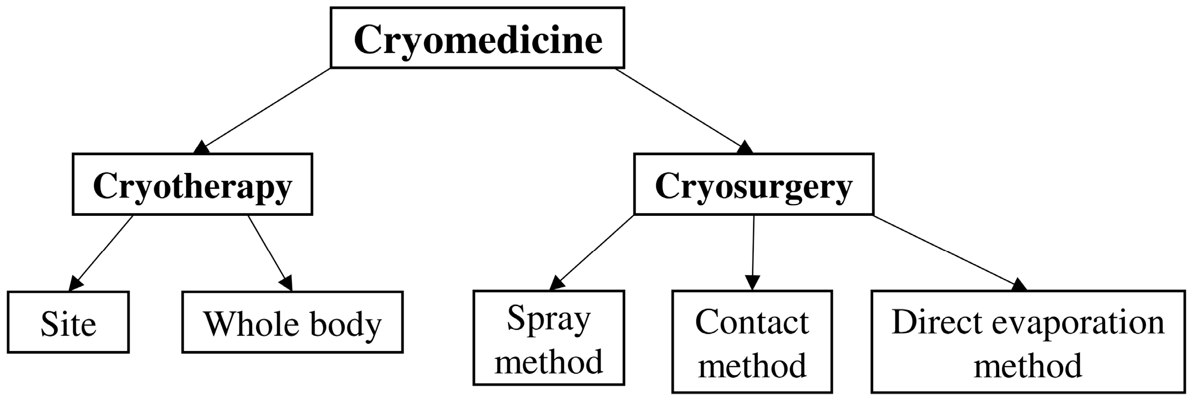
Figure 1. Cryomedical methods and devices. Developed on the basis of [24].
Applsci 15 12579 g001
Figure 2. Schematic diagram of portable cryosurgical device operated with liquid nitrogen.
Applsci 15 12579 g002
Figure 3. Synchrotron radiation use area.
Applsci 15 12579 g003
Figure 4. Radiation beam therapy characteristics [45]: relative percentage dose of various therapy beam distributions: proton, neutron, and carbon in comparison to X-ray versus penetration depth in tissue.
Applsci 15 12579 g004
Figure 5. Superconducting rotating-gantry HIMAC (Chiba, Japan) [48].
Applsci 15 12579 g005
Figure 6. PET, MRI, and CT image fusion for RT planning [36].
Applsci 15 12579 g006
Figure 7. View of the Iseult magnet and cryostat [59].
Applsci 15 12579 g007
Figure 8. Schematic figure of magnetic fractionation of MNPs [65].
Applsci 15 12579 g008
Figure 9. Schematic of the magnetic marker (a); relationship between magnetic moment m and anisotropy energy E for two different markers (b) [66].
Applsci 15 12579 g009
Figure 10. Josephson junction: (a) schematic representation of Nb-based low-Tc DC SQUID sensor [68]; (b) concept of operation.
Applsci 15 12579 g010
Table 1. Phenomena in cryogenics and superconductivity.
P *ResearchersDiscoveries
CryogenicsLouis-Paul Cailletet, French physicist and industrialist, and Raoul-Pierre Pictet, Swiss physicist.1877—first attempts to condense oxygen to 90 K [2].
Karol Olszewski, Polish chemist, mathematician, and physicist, together with Zygmunt Wróblewski, chemist and physicist, Cracow Laboratory.1883—the world’s first successful liquefaction of oxygen, nitrogen, and carbon dioxide from atmospheric air in a stable state carried out at the Cracow Laboratory [3].
1884—Olszewski was the pioneer in liquefying hydrogen under dynamic conditions, reaching a then-unprecedented low temperature of −225 °C (48 K). In 1895, he also succeeded in liquefying argon [4].
H. Kamerlingh Onnes, Dutch physicist, University of Leiden1908—Helium liquefaction [5].
Christopher Polge, British biologist, is considered the father of modern cryopreservation.1949—C. Polge discovered the cryoprotective properties of glycerol, which enabled the first effective freezing of semen [6].
Carl von Linde, German engineer and inventor and Georges Claude, French engineer and inventor.1985—C. von Linde was the first to apply the methods of air condensation on an industrial scale, followed by air rectification in 1902. In the process of air liquefaction, Linde used the Joule-Thomson effect for pre-cooled air in a recuperative heat exchanger [7].
1902—G. Claude used an expander cycle to condense the air [8].
Ming C. Leu et al. from Missouri University of Science and Technology, Rolla, MI, USA. 1999—the investigators employed an inkjet-based dispensing system to deposit water in sequential layers, each of which was subjected to rapid cryogenic solidification. This approach was subsequently designated as the RFP technique [9].
Scientists of Helsinki University of Technology’s Low-Temperature Lab in Espoo, Finland.2000—nuclear spin temperatures in picokelvin range (below 100 pK) were reported for an experiment [10].
Mizunami K. et al. and Wu M.C. et al., one of the first developed high-sensitivity cryogenic fiber Bragg grating temperature sensors.2001—Mizunami K. investigated low-temperature sensing using a fiber Bragg grating fixed on Teflon substrate [11].
2006—Wu M.C. studied cryogenic temperature sensing using a pressurized fiber Bragg grating [12].
Markus K. et al., German and French teams from Hannover, Bremen, Mainz, Berlin, Orsay and Toulouse. 2021—setting the current world record for effective temperatures at 38 pK through matter-wave lensing of rubidium [13].
Superc.H. Kamerlingh Onnes, Dutch physicist, University of Leiden.1911—phenomena of current flow without resistance discovery. Mercury cooled by liquid helium to 4.2 K becomes a superconductor [5].
JN Rjabinin i Lev Shubnikov, Ukrainian researchers from the USSR.1935—experimentally discovered type II superconductors, which they published in [14].
SuperconductivityLev Landau and Vitaly Ginzburg, Soviet physicists.1950—discovered and developed the theory of two types of superconductors I and II, which they published in their paper [15].
J. Bardeen (IL, USA), C. Cooper (OH, USA) and J.R. Schiffer (Birmingham, England).1957—BCS Theory—the basis of the theory is the Cooper pair: two electrons with opposite spin and momentum, bound together in a system with resultant spin and momentum equal to zero [16].
Brian D. Josephson, University of Cambridge, UK.1962—Josephson stationary phenomenon. System consisting of two superconductors separated by a layer of insulator that plays the role of a barrier to the flowing current—macroscopic quantum phenomenon [17].
J. Georg Bednorz and K. Alex Müllerz, IBM Zurich.1986—HTS Superconducting properties at temperatures higher than helium—ceramic composites [18].
J. Nagamatsu et al., Japanese Scientists2001—discovery of MgB2 (Tc = 39 K) [19].
Takahashi H, Igawa K, Arii K, Kamihara Y, Hirano M, Hosono H., Japanese Scientists.2008—first superconducting Fe-based pnictides, with Tc values up to 56 K [20,21].
Cao Y. et al., scientists from universities and institutes of MIT, Harvard, National Institute for Materials Science of Japan.2018—reported the emergence of intrinsic, unconventional superconductivity—phenomena not accounted for by weak electron–phonon coupling—in a two-dimensional superlattice formed by stacking two graphene layers rotated with respect to one another by a small twist angle [22].
Eremets M.I. et al. together with Swedish and American scientists. 2022—described in detail the experiments revealing high-temperature superconductivity in hydrides under high pressures [23].
* Phenomenon name.
Table 2. Critical parameters of type I and II superconductors.
ParameterSymbolValue
critical
temperature
TcTemperature below which a superconductor exhibits superconductivity at zero critical field strength and zero critical current.
lower critical magnetic field strengthHc1Magnetic field strength at which the first fluxion enters the volume of a type II superconductor, causing a departure from ideal diamagnetism.
upper critical magnetic strengthHc2Maximum magnetic field strength below which a type II superconductor is in the mixed state.
critical currentIcMaximum direct current that can be considered to flow in a superconductor without resistance.
critical current densityJcElectric current density at the critical current, determined for the whole superconducting cross-section or the unstable part thereof, when a stabilizer is present.
Table 3. Comparison of the CT, MRI/NMR, and PET features.
FeatureCTMRI and NMRPET
Operation principleX-raysMagnetic field and radio waves Radioactive tracer and detection of emitted positrons
ApplicationsImaging of bone structures, injuries, in emergency situationsImaging of soft tissues, organs, nervous system, joints (MRI); lab chemical analysis techniques (NMR)Detection and assessment of metabolic activity in cells, e.g., tumors
SafetyExposure to ionizing radiation Safer-does not use ionizing radiationDepends on the tracer used, but generally safe
ContraindicationsPregnancy, frequent exposure to radiation bodies Medical implants, metallic foreign, pacemakersSpecific to the tracer used
Examination timeShort (a few to several minutes)Longer (several to several dozen minutes)Variable (depends on the examination)
Additional commentsContrast agents: Iodine-based contrast agents are used in CT scans Gadolinium-based compounds are used in MRI scansParticularly useful in the diagnosis of oncological diseases, examinations are combined, PET/CT or PET/MRI, to obtain a more complete clinical picture
Table 4. Biomagnetic signal features from different sources [75].
SourceRangeFrequencyBandwidth
Brain (MEG)100 fT–1 pT0.5-500 Hz
(clinically revelant < 70 Hz)
~500 Hz/70 Hz
Heart (MCG)500–100 pT<75 Hz75 Hz
Nerve (MNG)5 fT–8 pT6–500 Hz494 Hz
Spine (MSG)1–100 fT100–5000 Hz4900 Hz
Hand/Leg/Head/Muscle (MMG)1 fT–1 pT1–300 Hz300 Hz
Disclaimer/Publisher’s Note: The statements, opinions and data contained in all publications are solely those of the individual author(s) and contributor(s) and not of MDPI and/or the editor(s). MDPI and/or the editor(s) disclaim responsibility for any injury to people or property resulting from any ideas, methods, instructions or products referred to in the content.