Article

Evaluation of the Effects of Carvacrol on Gram-Negative Bacilli Isolated from Wound Infections

1
Department of Laboratory Medicine, Pomeranian Medical University, 70-111 Szczecin, Poland
2
Department of Pharmaceutical Microbiology and Microbiological Diagnostics, Medical University of Lodz, 90-151 Lodz, Poland
3
Department of Clinical Microbiology, Pomeranian Medical University in Szczecin, 70-111 Szczecin, Poland
4
Department of Diagnostic Immunology, Pomeranian Medical University in Szczecin, 70-111 Szczecin, Poland
5
Department of Industrial Microbiology and Biotechnology, Faculty of Biology and Environmental Protection, University of Lodz, 90-237 Lodz, Poland
*
Author to whom correspondence should be addressed.
Appl. Sci. 2025, 15(21), 11309; https://doi.org/10.3390/app152111309
Submission received: 14 September 2025 / Revised: 17 October 2025 / Accepted: 20 October 2025 / Published: 22 October 2025

Featured Application

The present study demonstrates the potential use of carvacrol as a component of formulations or active dressings for the treatment of chronic wounds caused by bacteria exhibiting resistance to recommended antibiotics.

Abstract

Wound infections pose a significant challenge in modern medicine, driven by multimorbidity, weakened immunity, microbial virulence factors, and resistance to antibiotics and antiseptics. This study aims to evaluate the antibacterial properties of carvacrol (CAR), its impact on biofilm formation, and its capacity to trigger oxidative stress in clinical strains of Pseudomonas aeruginosa, Klebsiella pneumoniae, and Enterobacter cloacae. Carbapenemases in the studied bacteria were detected using culture on CarbaId agar. The presence of genes encoding bacterial virulence factors and carbapenemase production was confirmed using the PCR method. The antimicrobial activity of carvacrol was evaluated using the broth microdilution method. The ability of strains to form biofilm was determined using a modified crystal violet assay. Oxidative stress levels in bacterial cells in response to carvacrol treatment were measured using 2′,7′-dichlorofluorescein diacetate. Real-TimePCR was used to confirm the presence of NDM family carbapenemase genes in K. pneumoniae strains, KPC genes in E. cloacae strains, and VIM genes in P. aeruginosa strains. CAR exhibited a broad spectrum of antibacterial activity against the tested bacteria, with MIC values ranging from 125 to 1000 μg/mL. Treatment with 1/2 MIC of CAR did not significantly influence biofilm formation, except in a K. pneumoniae isolate. At 1/2 MIC, CAR induced an increase in intracellular ROS in most tested strains, with the exception of P. aeruginosa 25521221. This study provides insights into the antimicrobial efficacy of carvacrol against carbapenemase-producing pathogens isolated from wound infections—specifically P. aeruginosa, K. pneumoniae, and E. cloacae. CAR demonstrated promising bactericidal properties, likely mediated through the induction of oxidative stress, as evidenced by increased ROS generation in most studied isolates.

1. Introduction

Chronic wound infections represent a significant clinical challenge, largely due to the involvement of opportunistic Gram-negative pathogens capable of forming resilient biofilms and expressing multiple virulence factors. Among these pathogens, P. aeruginosa, Klebsiella spp., and Enterobacter spp. are frequently isolated from infected wounds and are characterized by their intricate pathogenic mechanisms and strong antimicrobial resistance.
P. aeruginosa is a ubiquitous environmental bacterium and a leading cause of nosocomial infections. Its pathogenic success is largely attributed to a diverse array of virulence determinants, including type III secretion system (TTSS) effectors (exoT, exoS, exoU, exoY), pyocyanin biosynthesis genes (phzM, phzS), alkaline protease (aprA), hemolytic phospholipase C (plcH), and motility-related proteins such as type IV pili (pilA, pilB) and flagellin (fliC) [1]. These factors collectively contribute to tissue invasion, immune evasion, biofilm development, and oxidative stress induction in host tissues.
Klebsiella species, particularly K. pneumoniae, are opportunistic pathogens increasingly linked to wound infections, with a rising prevalence of hypervirulent and multidrug-resistant strains. Their virulence factors include adhesins such as fimH, which facilitate bacterial adhesion; lipopolysaccharide (LPS)-related genes (wabG, uge) involved in immune evasion; iron acquisition systems (kfu, iroN); and capsule regulatory genes (magA, rmpA) that promote a mucoid phenotype and biofilm development [2]. These characteristics enhance bacterial survival in hostile environments and markedly hinder treatment efforts.
Enterobacter spp. are emerging opportunistic pathogens with virulence profiles similar to other members of the Enterobacteriaceae family. Key virulence factors include adhesion proteins such as fimH, iron acquisition systems (kfu, iroN), and toxins like hemolysin (hlyA) [3]. However, compared to Pseudomonas and Klebsiella, Enterobacter strains typically form weaker biofilms, which may influence their pathogenic profile and susceptibility to antiseptics.
Multidrug-resistant Enterobacteriaceae, including Enterobacter spp., represent a major public health concern due to their wide range of resistance mechanisms, notably linked with production of extended-spectrum β-lactamases (ESBLs) and carbapenemase production. Carbapenemase-producing Gram-negative rods pose a severe threat in hospital settings, particularly among patients with multiple comorbidities, as these enzymes confer resistance to nearly all β-lactam antibiotics. Compounding the problem, carbapenemase genes are often located on plasmids, facilitating horizontal transfer to other bacterial species and even different genera within Enterobacterales [4].
The most common carbapenemases in K. pneumoniae include K. pneumoniae carbapenemase (KPC) and New Delhi metallo-β-lactamase (NDM) [5]. Among non-fermenting Gram-negative bacilli, such as Pseudomonas, metallo-β-lactamases (MBLs) predominate, particularly Verona integron-encoded metallo-β-lactamases (VIM), which hydrolyzenearly all β-lactams except monobactams and cefiderocol [6].
Resistance to synthetic antimicrobials is both time- and dose-dependent, making it a common and inevitable phenomenon. Consequently, there is an ongoing search for alternative agents among natural compounds, which frequently pose a greater challenge for microorganisms to resist [7].
Carvacrol (Figure 1), a monoterpenoid derived from the essential oils of oregano and thyme, exhibits notable antimicrobial and antibiofilm activities. Its mechanisms of action include disruption of bacterial membranes, modulation of quorum sensing, and induction of reactive oxygen species (ROS), leading to oxidative damage in microbial cells. Carvacrol demonstrates efficacy against multidrug-resistant Gram-negative bacteria and the ability to disrupt mature biofilms, making it a promising candidate for integration into antiseptic regimens [7,8,9].
Silva et al. [7] reported that carvacrol exhibited minimum inhibitory concentration (MIC) values of 81 μg/mL against Staphylococcus aureus, 161 μg/mL against P. aeruginosa, and 128 μg/mL against a multidrug-resistant strain of P. aeruginosa.
Given the rising prevalence of antimicrobial resistance and the persistence of biofilm-forming pathogens in chronic wounds, assessing the effects of carvacrol on clinically relevant Pseudomonas, Klebsiella, and Enterobacter strains is crucial. This study aims to elucidate the antimicrobial efficacy of carvacrol and its influence on virulence factor expression, biofilm production, and oxidative stress induction, thereby informing improved therapeutic strategies for recalcitrant wound infections.

2. Materials and Methods

2.1. Bacterial Strains and Growth Conditions

This study included six strains of Gram-negative bacteria isolated from wound swabs during routine microbiological diagnosis, from patients treated at various hospitals. The study did not require approval from the Bioethics Committee. The collection consisted of two strains each of Pseudomonas spp. (3411122 and25521221), Klebsiella spp. (473/24 and 23520/23), and Enterobacter spp. (25269 and 24985), representing clinically relevant pathogens commonly associated with wound infections. All strains were cultivated for 18 h at 37 °C in an aerobic atmosphere on Columbia agar with 5% sheep blood and MacConkey agar (bioMérieux, Craponne, France). Strains were identified using Maldi-Tof (Bruker, Berlin, Germany).

2.2. Carbapenemases Detection

Carbapenemases were detected by culture on CarbaId agar (bioMerieux, Ceaponne, France) and confirmation of the presence of carbapenemases was also carried out by real-time PCR (GeneXpert, Cepheid, Solna, Sweden).

2.3. DNA Extraction

Genes encoding bacterial virulence factors were detected by PCR method. Isolation of genomic DNA was carried out using the Gene MATRIX Bacterial&Yeast Genomic DNA Purification Kit column kit (EURx, Gdańsk, Poland). The Gene MATRIX Plasmid Miniprep DNA Purification Kit (EURx, Poland) was used to perform the plasmid DNA isolation procedure. The PCR reaction was performed using the gene-specific primer sequences (Table 1 and Table 2) [10,11].

2.4. DNA Amplification

The amplification reaction, which consisted of 30 cycles, was carried out in an Applied BiosystemsVeriti 96 Well-ThermalCycler (Applied Biosystems, Foster City, CA, USA) according to the parameters: initial activation at 95 °C for 15 min, followed by 30 cycles at 94 °C for 30 s; annealing at 60 °C for 90 s, followed by extension at 72 °C for 60 s and a final extension at 72 °C for 10 min. Electrophoresis was performed using a 1.5% agarose gel (DNA, Gdansk, Poland) mixed with ethidium bromide (Sigma-Aldrich, Schnelldorf, Germany) at a concentration of 0.5 μg/mL. Positive controls were standard strains suitable for specific genes. Electrophoresis was performed at 80 V for 80 min. The result was read under UV light using a GelDoc-It2 Imager system (Upland, CA, USA).
In Pseudomonas spp., the following virulence genes were targeted: phzM and phzS, involved in the biosynthesis of pyocyanin; exoT, exoY, exoS, and exoU, encoding effector proteins of the type III secretion system (TTSS); pilA and pilB, coding for type IV pilus structural and biogenesis proteins, respectively; aprA, encoding alkaline protease; and plcH, encoding the precursor of hemolytic phospholipase C.
In Klebsiella spp., the following genes were examined: fimH, associated with fimbrial adhesion; wabG and uge, involved in LPS biosynthesis; kfu and iroN, related to iron acquisition systems; hlyA, encoding hemolysin; and magA and rmpA, responsible for the production and regulation of a mucoid capsule.
In Enterobacter spp., the detection of fimH, kfu, iroN, and hlyA was performed to assess adhesion, iron acquisition capability, and cytotoxic potential.

2.5. Determination of Minimum Inhibitory Concentration

The antimicrobial activity of carvacrol (CAR) was assessed using a broth microdilution method in accordance with Clinical and Laboratory Standards Institute (CLSI) guidelines. Serial two-fold dilutions of carvacrol (Pol-Aura Sp. z o.o., Morąg, Poland, nr CAS 499-75-2) were prepared in 96-well microplates. Carvacrol solutions of varying concentrations were prepared, and MICs were established for each strain to assess both bacteriostatic effect and minimum bactericidal concentration (MBC).

2.6. Antibiofilm Activity

The ability of all strains to form biofilms was determined by the modified method with crystal violet (Chempur, PiekaryŚląskie, Poland) [12,13,14]. Cultures of bacteria were incubated with (1/2 MIC) or without CAR into 96-well plates (24 h at 37 °C). Following incubation, wells were washed and stained with crystal violet. Crystal violet was dissolved in 99.8% ethanol (POCH). Absorbances were measured using spectrophotometer (Biotek Epoch™, Santa Clara, CA, USA) at 600 nm.

2.7. Reactive Oxygen Species Measurements

In this study, the generation of endogenous ROS in response to treatment with carvacrolwas measured. 2′,7′-dichlorofluorescein diacetate (DCFH-DA) (Merck Life Science Sp. z o.o., Darmstadt, Germany) was used to detect oxidative stress levels in bacterial cells. Fluorescence intensity was quantified using the spectrofluorometer (FLUOstar® Omega BMG LABTECH, Ortenberg, Germany) at λex = 485 nm and λem = 530 nm (software version 5.10). The cell suspensions were incubated with DCFH-DA (10 μM) for 30 min at 37 °C in the dark. Results were expressed as fluorescence intensity 2′,7′-dichlorofluorescein (DCF) in the treated cells compared to the untreated controls [15,16,17].

3. Results

3.1. Detection of Carbapenemases

On CarbaId agar, K. pneumoniae and E. cloacae strains grew green colonies, which meant that all isolates were carbapenemase producers. Real-TimePCR-based tests confirmed the presence of genes encoding NDM carbapenemase in K. pneumoniae strains, KPC genes in E. cloacae and VIM genes in P. aeruginosa strains.

3.2. Detection of Virulence Genes

PCR analysis revealed the presence of selected virulence-associated genes in the tested Gram-negative isolates. Among the P. aeruginosa strains, one isolate harbored the exoT, aprA, phzS, and plcH genes, indicating the presence of type III secretion system effectors, alkaline protease, pyocyanin biosynthesis, and phospholipase C activity, respectively. In K. pneumoniae, one isolate was positive for five key virulence genes: fimH, wabG, uge, kfu, and rmpA. These genes are associated with adhesion, LPS synthesis, iron acquisition, and hypermucoviscosity phenotype regulation. Both E. cloacae strains carried the fimH, kfu, and iroN genes, indicating their potential for adhesion and iron uptake.
The presence of virulence genes in examined bacterial strains is presented in Table 3.

3.3. Carvacrol Susceptibility

Carvacrol exhibited a broad spectrum of antibacterial activity against Gram-negative bacteria (MIC values between 125 and 1000 μg/mL) (Table 4). The highest MIC and MBC value was found for P. aeruginosa 25521221. The MIC of carvacrol for P. aeruginosa 25521221 was 1000 µg/mL, whereas its MBC was 2000 µg/mL. E. cloacae 24985 was the most sensitive Gram-negative bacterium to carvacrol, with MIC value of 125 μg/mL and MBC value of 250 μg/mL, respectively, while E. cloacae 25269 was the leastsusceptible with MIC value of 250 μg/mL.

3.4. Biofilm Formation

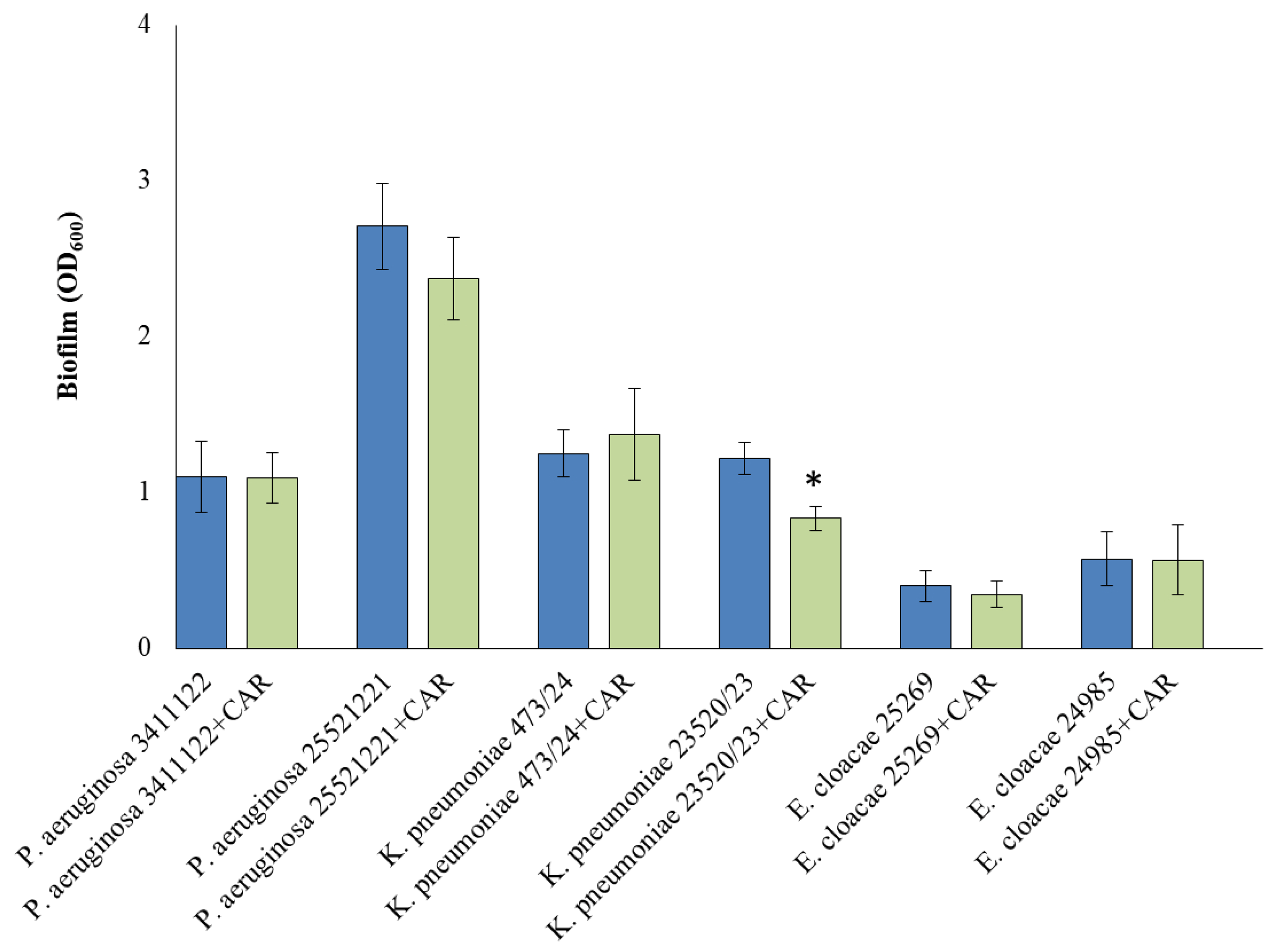
Biofilm production varied between species. P. aeruginosa and K. pneumoniae demonstrated strong biofilm-forming capabilities as measured by crystal violet staining, while both E. coloacae isolates showed weak biofilm production under the same experimental conditions. The influence of CAR on biofilm production is shown in Figure 2. It has been observed that the 1/2 MIC of CAR did not significantly influence biofilm formation of the five strains. The only significant difference was observed in K. pneumoniae 23520/23, where the formation of biofilm was smaller than in control.

3.5. Detection of ROS

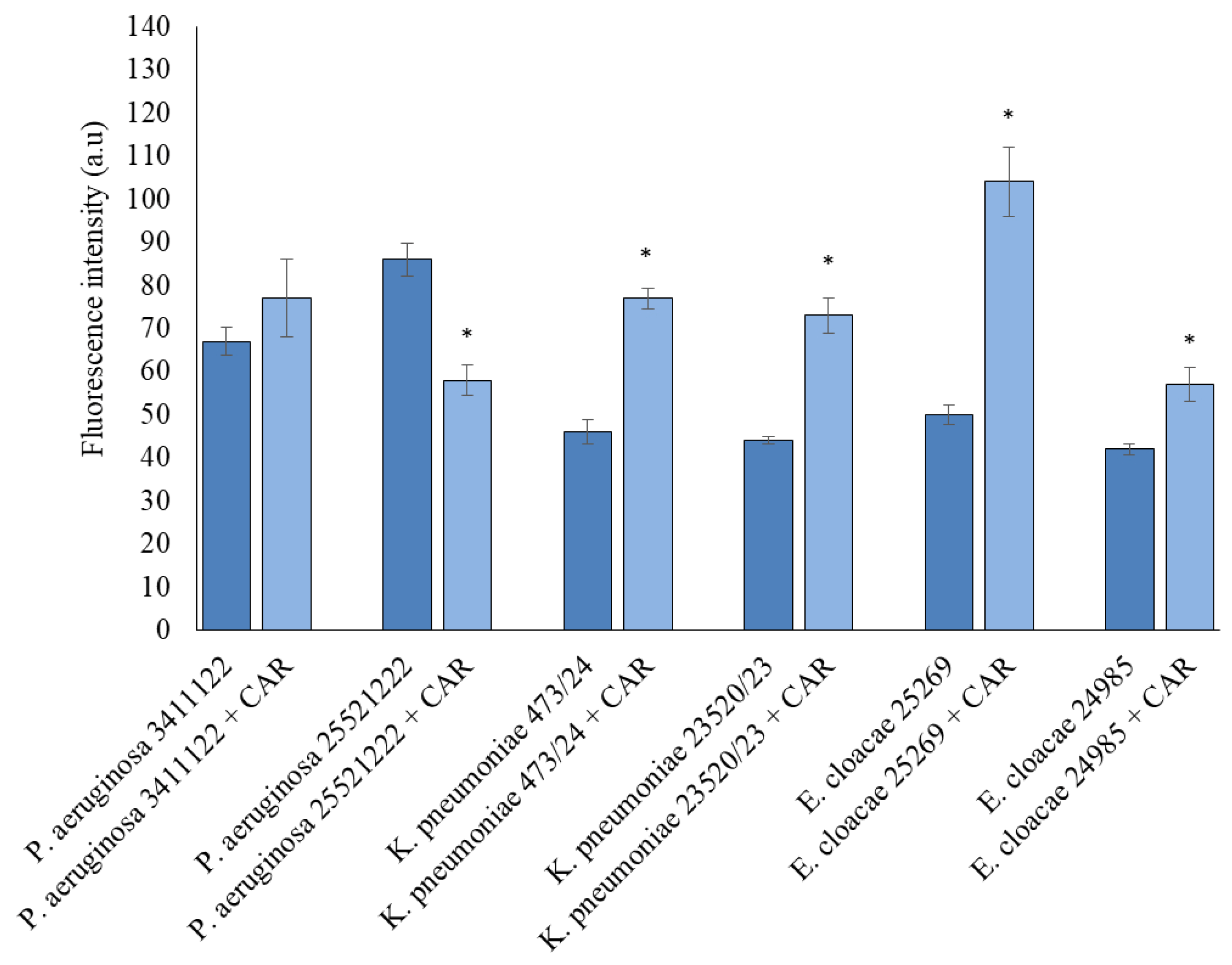
As illustrated in Figure 3, treatment with carvacrol at 1/2 MIC induced an increase in intracellular ROS in four of the tested strains, as determined by the fluorescence intensity of DCF. An exception was P. aeruginosa 25521221, which demonstrated a decrease in ROS levels following exposure to carvacrol. Additionally, there was no significant difference in ROS production between the control and carvacrol-treated samples in P. aeruginosa 3411122, suggesting limited oxidative stress induction in this strain.

4. Discussion

According to numerous studies, natural compounds, including secondary plant metabolites, exhibit strong antimicrobial activity, even against multidrug-resistant bacteria [18,19,20]. In environments where resistant strains are selected, such as hospitals, effective control of pathogen transmission is crucial. Given the side effects and the growing resistance to synthetic biocides, natural antimicrobial compounds—widely available in nature and used for centuries—offer a promising alternative. Advances in scientific research and methodology have allowed for a more detailed understanding of their properties, mechanisms of action, and safety profiles [21].
One of the most serious problems in modern medicine is the management of difficult-to-heal wounds and ulcers, the treatment of which is often complicated by underlying systemic conditions. Multidrug-resistant pathogens such as Gram-negative Escherichia coli, K. pneumoniae, Acinetobacter baumannii, P. aeruginosa, and the Gram-positive S. aureus persist in chronic wounds through the formation of biofilms, which prolong inflammation and delay healing. Polymicrobial biofilms in chronic wound infections are characterized by a high tolerance to antibiotics, thereby reducing the efficacy of many conventional antimicrobial agents [22].
In this study, we aimed to analyze the effects of the natural plant-derived compound carvacrol on various strains of Gram-negative bacilli isolated from wound infections. The clinical P. aeruginosa, K. pneumoniae, and E. cloacae isolates examined in this work display a complex interplay of virulence potential, biofilm formation, and antiseptic resistance, underscoring the challenges of chronic wound management.
The P. aeruginosa strain harboring exoT, aprA, phzS, and plcH genes exhibits a highly virulent phenotype. These genes collectively promote toxin secretion, enzymatic tissue degradation, pyocyanin-induced oxidative stress, and phospholipase-driven membrane disruption—mechanisms that support biofilm formation and persistence in chronic wounds, as described by Malanović et al. [1].
Similarly, the K. pneumoniae isolate expressing fimH, wabG, uge, kfu, and rmpA genes is consistent with hypervirulent phenotypes, combining strong adhesion capacity, lipopolysaccharide synthesis, iron acquisition, and mucoid capsule production. These genomic profiles are strongly linked to enhanced biofilm formation and effective immune evasion in clinical settings [2].
In contrast, Enterobacter strains carrying fimH, kfu, and iroN exhibited only weak biofilm formation. The absence of capsule regulatory genes such as rmpA likely limits their biofilm-forming potential, emphasizing that extensive exopolysaccharide synthesis is crucial for mature biofilm architecture [3].
The results of Hasavand et al. showed that carvacrol exhibited antibacterial activity against S. aureus and P. aeruginosa and could be used as a herbal and natural alternative to glutaraldehyde in the sterilization of hospital equipment [23].
Mączka et al. [24] reported that carvacrol has shown significant antibacterial activity against biofilm produced by P. aeruginosa and S. aureus. However, the authors noted a high MIC value for carvacrol (625 µg/mL) for the reference strain P.aeruginosa ATCC 15442. For the tested clinical strains of P. aeruginosa, MIC values were within the range of 500–1000 µg/mL.
In our study, clinical E. cloacae was the most sensitive Gram-negative bacterium to carvacrol with MIC value of 125 μg/mL and MBC value of 250 μg/mL, respectively, while the highest MIC (1000 µg/mL) and MBC (2000 µg/mL) valueswerefound for P. aeruginosa.
It is well established that bacteria within biofilm structures develop resistance to antibacterial agents, including antibiotics and biocides. In our study, P. aeruginosa and K. pneumoniae formed robust biofilms, in contrast to E. cloacae, which produced weaker biofilms. Treatment with carvacrol at 1/2 MIC significantly inhibited biofilm formation in the K. pneumoniae 23520/23 strain. Future studies on clinical isolates should consider increasing the number of tested clinical strains from various clinical materials with different susceptibility to antimicrobial drugs prolonged incubation times of up to 72 h to better assess biofilm dynamics.
Liu et al. [25] demonstrated that the biofilm-forming ability of E. cloacae isolated from food was significantly reduced by carvacrol oil at concentrations of 64 and 128 µg/mL during 72 h cultivation.
Farhadi et al. [26] further demonstrated that carvacrol not only inhibits biofilm formation but can also eliminate established biofilms in carbapenem-resistant Gram-negative bacilli.
Additional studies have demonstrated the ability of carvacrol to disrupt biofilm production in selected bacterial strains. Under the influence of carvacrol, there was a significant reduction in biofilm of 91–100% in P. aeruginosa and 95–100% in S. aureus [9]. These findings emphasize the potential use of carvacrol in wound care to target biofilm-embedded pathogens.
It has been proven that during the infection process, bacteria are exposed to ROS produced by the host’s phagocytic cells. ROS can easily damage bacterial cell membranes, DNA, and proteins. It appears that oxidative stress triggers antioxidant defense reactions, but it turns out that nutrient deprivation also triggers antioxidant reactions [27]. Our tests confirmed increased intracellular ROS levels after treatment with carvacrol, detected using the DCFH-DA dye.
Mechanistically, carvacrol compromises membrane integrity, induces membrane depolarization and ATP leakage, and promotes ROS accumulation [28]. In our study, four isolates showed increased ROS levels when treated with half the MIC of carvacrol, supporting a ROS-mediated mechanism of bacterial killing.
However, the resistant P. aeruginosa strain showed suppressed ROS levels, suggesting the activation of antioxidant defenses—such as increased catalase and superoxide dismutase expression.
The antimicrobial mechanisms of carvacrol highlight its potential as a therapeutic agent. A comparison of bacterial cells grown in normal biofilms with biofilm bacteria grown in the presence of carvacrol oil showed that they were much more easily inactivated by other antimicrobial agents [25].
Bacteria have developed several adaptive responses to oxidative stress.Quorum sensing plays an important role in the oxidative stress survival of P. aeruginosa. Moreover, the pigments of P. aeruginosa provide protection against oxidative stress [29]. P. aeruginosa can also alleviate oxidative damage by increasing the activity of enzymes (catalases, etc.). Considering the problems in treating difficult infections with multidrug-resistant bacterial strains, the data from literature reports on the bactericidal activity of essential oils are highly promising [30,31].
Despite the robustness of our molecular and functional data, several limitations of this study should be acknowledged. The analysis was performed on a limited number of clinical strains and has not been validated in wound infection models. Future investigations should incorporate larger isolate collections, detailed flow-cell biofilm eradication assays, molecular monitoring of ROS responses, cytotoxicity assessments in human skin models, and systematic combination studies (e.g., checkerboard assays) to optimize carvacrol dosing regimens.
In summary, our findings highlight a strategic approach to combating Gram-negative wound infections through targeted induction of oxidative stress and the use of biofilm-disrupting agents such as carvacrol. These strategies offer considerable potential for the development of advanced antiseptic and therapeutic regimens capable of addressing both biofilm persistence and microbial resistance mechanisms.

5. Conclusions

Carvacrol demonstrated notable antimicrobial activity against Gram-negative, carbapenemase-producing pathogens isolated from wound infections, including P. aeruginosa (VIM), K. pneumoniae (NDM), and E. cloacae (KPC). Differences in virulence gene profiles were associated with variations in biofilm formation and susceptibility to carvacrol. Strong biofilm production by P. aeruginosa and K. pneumoniae supports their roles in chronic wound persistence, while weaker biofilm formation by Enterobacter isolates suggests distinct pathogenic traits. Carvacrol exhibited strong bactericidal effects at higher concentrations, mainly through oxidative stress induction, though some strains may possess antioxidant defenses that limit its activity. Overall, carvacrol shows promise as a natural antimicrobial capable of disrupting biofilms and reducing bacterial viability. Future studies should expand to more wound-associated bacteria and explore theireffects on heat shock protein expression, cell envelope structure, and in vivo efficacy and safety.

Author Contributions

Conceptualization, A.P. and A.L.; methodology, A.P., A.L. and M.S. (Mirosława Słaba); formal analysis, A.P., A.L., M.S. (Monika Sienkiewicz), H.M. and P.K.; investigation, A.P., A.L. and M.S. (Mirosława Słaba); resources, A.P.; data curation, A.P., A.L. and M.S. (Monika Sienkiewicz); writing—original draft preparation, A.P., A.L. and M.S. (Monika Sienkiewicz); writing—review and editing, A.P., A.L., H.M. and P.K.; visualization, A.P. and A.L.; supervision, B.D.; project administration, A.P.; funding acquisition, B.D. and M.S. (Monika Sienkiewicz). All authors have read and agreed to the published version of the manuscript.

Funding

This research received no external funding.

Institutional Review Board Statement

Our research is not a medical experiment and does not require the approval of the Bioethics Committee, as confirmed by the Chairman of the Bioethics Committee of the Pomeranian Medical University in Szczecin, Poland.

Informed Consent Statement

Patient consent was waived due to the bacteria were provided by the Department of Medicine.

Data Availability Statement

Data supporting reported results can be found in Department of Laboratory Medicine, Pomeranian Medical University, and Department of Pharmaceutical Microbiology and Microbiological Diagnostics, Medical University of Lodz, Poland.

Acknowledgments

During the preparation of this study, the authors used media and equipment prepared by employee of Department of Pharmaceutical Microbiology and Microbiological Diagnostics, Medical University of Lodz, Poland Agata Marciniak.

Conflicts of Interest

The authors declare no conflicts of interest.

Abbreviations

The following abbreviations are used in this manuscript:
ATPAdenosine triphosphate
CARCarvacrol
CLSIClinical and Laboratory Standards Institute
DCF2′,7′-dichlorofluorescein
DCFH-DA2′,7′-dichlorofluorescein diacetate
DNADeoxyribonucleic acid
ESBLExtended-spectrum beta-lactamases
H2DCFDA2′,7′-dichlorodihydrofluorescein diacetate
KPCKlebsiella pneumoniae carbapenemase
LPSLipopolysaccharide
MBCMinimum bactericidal concentration
MBLMetallo-beta-lactamases
MICMinimum inhibitory concentration
NDMNew Delhi metallo-beta-lactamases
PCRPolymerase chain reaction
ROSReactive oxygen species
TTSSThe type III secretion system
VIMVerona integron-encoded metallo-beta-lactamase

References

  1. Malanovic, N.; Ön, A.; Pabst, G.; Zellner, A.; Lohner, K. Octenidine: Novel insights into the detailed killing mechanism of Gram-negative bacteria at a cellular and molecular level. Int. J. Antimicrob. Agents 2020, 56, 106146. [Google Scholar] [CrossRef] [PubMed]
  2. Nahar, N.; Rashid, R.B. Phylogenetic Analysis of Antibiotic Resistance Genes and Virulence Genes of Klebsiella species in silico. Dhaka Univ. J. Pharm. Sci. 2017, 16, 119–127. [Google Scholar] [CrossRef]
  3. Bujňáková, D.; Puvača, N.; Ćirković, I. Virulence Factors and Antibiotic Resistance of Enterobacterales. Microorganisms 2022, 10, 1588. [Google Scholar] [CrossRef] [PubMed]
  4. Lazar, D.S.; Nica, M.; Dascalu, A.; Oprisan, C.; Albu, O.; Codreanu, D.R.; Kosa, A.G.; Popescu, C.P.; Florescu, S.A. Carbapenem-Resistant NDM and OXA-48-like Producing K. pneumoniae: From Menacing Superbug to a Mundane Bacteria; A Retrospective Study in a Romanian Tertiary Hospital. Antibiotics 2024, 13, 435. [Google Scholar] [CrossRef]
  5. Xiang, T.; Chen, C.; Wen, J.; Liu, Y.; Zhang, Q.; Cheng, N.; Wu, X.; Zhang, W. Resistance of Klebsiella pneumoniae Strains Carrying blaNDM–1 Gene and the Genetic Environment of blaNDM–1. Front. Microbiol. 2020, 11, 700. [Google Scholar] [CrossRef] [PubMed]
  6. De Freitas, G. Verona Integron-Encoded Metallo-β-Lactamase (VIM)-Producing Pseudomonas aeruginosa Pyelonephritis in a Young Adult: A Case Report. Cureus 2025, 17, e78932. [Google Scholar] [CrossRef]
  7. da Silva, A.R.P.; do Socorro Costa, M.; Araújo, N.J.S.; de Freitas, T.S.; dos Santos, A.T.L.; Gonçalves, S.A.; da Silva, V.B.; Andrade-Pinheiro, J.C.; Tahim, C.M.; Lucetti, E.C.P.; et al. Antibacterial activity and antibiotic-modifying action of carvacrol against multidrug-resistant bacteria. Adv. Sample Prep. 2023, 7, 100072. [Google Scholar] [CrossRef]
  8. Cacciatore, F.A.; Maders, C.; Alexandre, B.; Barreto Pinilla, C.M.; Brandelli, A.; da Silva Malheiros, P. Carvacrol encapsulation into nanoparticles produced from chia and flaxseed mucilage: Characterization, stability and antimicrobial activity against Salmonella and Listeria monocytogenes. Food Microbiol. 2022, 108, 104116. [Google Scholar] [CrossRef]
  9. Walczak, M.; Michalska-Sionkowska, M.; Olkiewicz, D.; Tarnawska, P.; Warżyńska, O. Potential of Carvacrol and Thymol in Reducing Biofilm Formation on Technical Surfaces. Molecules 2021, 26, 2723. [Google Scholar] [CrossRef]
  10. Ghorbani, G.; Rahimi, E.; Shakerian, A. Antibiotic resistance’s Genotypic and Phenotypic Characteristics and the Frequency of Virulence Factors in P. aeruginosa Isolates Isolated from Water Samples in Iran. Biomed. Res. Int. 2022, 2022, 7076433. [Google Scholar] [CrossRef]
  11. Wahl, A.; Fischer, M.A.; Klaper, K.; Müller, A.; Borgmann, S.; Friesen, J.; Hunfeld, K.P.; Ilmberger, A.; Kolbe-Busch, S.; Kresken, M.; et al. Presence of hypervirulence-associated determinants in Klebsiella pneumoniae from hospitalised patients in Germany. Int. J. Med. Microbiol. 2024, 314, 151601. [Google Scholar] [CrossRef]
  12. Shi, S.-F.; Jia, J.; Guo, X.; Zhao, Y.; Chen, D.; Guo, Y.; Zhang, X. Reduced Staphylococcus aureus biofilm formation in the presence of chitosan-coated iron oxide nanoparticles. Int. J. Nanomed. 2016, 11, 6499–6506. [Google Scholar] [CrossRef]
  13. Wilson, C.; Lukowicz, R.; Merchant, S.; Valquier-Flynn, H.; Caballero, J.; Sandoval, J.; Okuom, M.; Huber, C.; Brooks, T.D.; Wilson, E.; et al. Quantitative and Qualitative Assessment Methods for Biofilm Growth: A Mini-review. Res. Rev. J. Eng. Technol. 2017, 6, 1–25. [Google Scholar]
  14. Coles, V.E.; Puri, L.; Bhandari, M.; Wood, T.J.; Burrows, L.L. The effects of chlorhexidine, povidone-iodine and vancomycin on growth and biofilms of pathogens that cause prosthetic joint infections: An in-vitro model. J. Hosp. Infect. 2024, 151, 99–108. [Google Scholar] [CrossRef]
  15. Pourhajibagher, M.; Bahador, A. Periodontal ligament stem cell-derived exosome-loaded Emodin mediated antimicrobial photodynamic therapy against cariogenic bacteria. BMC Oral Health 2024, 24, 311. [Google Scholar] [CrossRef]
  16. Zhang, K.; Yang, N.; Teng, D.; Mao, R.; Hao, Y.; Wang, J. Expression and characterization of the new antimicrobial peptide AP138L-arg26 anti Staphylococcus aureus. Appl. Microbiol. Biotechnol. 2024, 108, 111. [Google Scholar] [CrossRef] [PubMed]
  17. Wang, Y.; Haqmal, M.A.; Liang, Y.; Muhammad, I.; Zhao, X.; Elken, E.M.; Gao, Y.; Jia, Y.; He, C.; Wang, Y.; et al. Antibacterial activity and cytotoxicity of a novel bacteriocin isolated from Pseudomonas sp. strain 166. Microb. Biotechnol. 2022, 15, 2337–2350. [Google Scholar] [CrossRef]
  18. Liu, X.; Liu, R.; Zhao, R.; Wang, J.; Cheng, Y.; Liu, Q.; Wang, Y.; Yang, S. Synergistic Interaction Between Paired Combinations of Natural Antimicrobials Against Poultry-Borne Pathogens. Front. Microbiol. 2022, 13, 811784. [Google Scholar] [CrossRef]
  19. Vasireddy, L.; Bingle, L.E.H.; Davies, M.S. Antimicrobial activity of essential oils against multidrug-resistant clinical isolates of the Burkholderia cepacia complex. PLoS ONE 2018, 13, e0201835. [Google Scholar] [CrossRef]
  20. Faleiro, M.L.; Miguel, M.G. Chapter 6-Use of Essential Oils and Their Components against Multidrug-Resistant Bacteria. In Fighting Multidrug Resistance with Herbal Extracts, Essential Oils and Their Components, 1st ed.; Kumar Rai, M., Volodymyrivna Kon, K., Eds.; Academic Press: Cambridge, MA, USA, 2013; pp. 65–94. [Google Scholar]
  21. Reichling, J. Anti-biofilm and Virulence Factor-Reducing Activities of Essential Oils and Oil Components as a Possible Option for Bacterial Infection Control. Planta Med. 2020, 86, 520–537. [Google Scholar] [CrossRef] [PubMed]
  22. Li, Y.; Wu, M.X. Visualization and elimination of polymicrobial biofilms by a combination of ALA-carvacrol-blue light. J. Photochem. Photobiol. B Biol. 2022, 234, 112525. [Google Scholar] [CrossRef]
  23. Hasanvand, T.; Mohammadi, M.; Abdollahpour, F.; Kamarehie, B.; Jafari, A.; Ghaderpoori, A.; Karami, M.A. A comparative study on antibacterial activity of carvacrol and glutaraldehyde on Pseudomonas aeruginosa and Staphylococcus aureus isolates: An in vitro study. J. Environ. Health Sci. Eng. 2021, 19, 475–482. [Google Scholar] [CrossRef]
  24. Mączka, W.; Twardawska, M.; Grabarczyk, M.; Wińska, K. Carvacrol—A Natural Phenolic Compound with Antimicrobial Properties. Antibiotics 2023, 12, 824. [Google Scholar] [CrossRef]
  25. Liu, F.; Jin, P.; Sun, Z.; Du, L.; Wang, D.; Zhao, T.; Doyle, M.P. Carvacrol oil inhibits biofilm formation and exopolysaccharide production of Enterobacter cloacae. Food Control 2021, 119, 107473. [Google Scholar] [CrossRef]
  26. Farhadi, K.; Rajabi, E.; Varpaei, H.A.; Iranzadasl, M.; Khodaparast, S.; Salehi, M. Thymol andcarvacrol against Klebsiella: Anti-bacterial, antibiofilm, and synergistic activities—Asystematic review. Front. Pharmacol. 2024, 15, 1487083. [Google Scholar] [CrossRef] [PubMed]
  27. Khakimova, M.; Ahlgren, H.G.; Harrison, J.J.; English, A.M.; Nguyen, D. The stringent response controls catalases in Pseudomonas aeruginosa and is required for hydrogen peroxide and antibiotic tolerance. J. Bacteriol. 2013, 195, 2011–2020. [Google Scholar] [CrossRef] [PubMed]
  28. Khan, I.; Bahuguna, A.; Shukla, S.; Aziz, F.; Chauhan, A.K.; Ansari, M.B.; Bajpai, V.K.; Huh, Y.S.; Kang, S.C. Antimicrobial potential of the food-grade additive carvacrol against uropathogenic E. coli based on membrane depolarization, reactive oxygen species generation, and molecular docking analysis. Microb. Pathog. 2020, 142, 104046. [Google Scholar] [CrossRef]
  29. da Cruz Nizer, W.S.; Inkovskiy, V.; Versey, Z.; Strempel, N.; Cassol, E.; Overhage, J. Oxidative Stress Response in Pseudomonas aeruginosa. Pathogens 2021, 10, 1187. [Google Scholar] [CrossRef]
  30. Kachur, K.; Suntres, Z. The antibacterial properties of phenolic isomers, carvacrol and thymol. Crit. Rev. Food Sci. Nutr. 2020, 60, 3042–3053. [Google Scholar] [CrossRef]
  31. Kwiatkowski, P.; Pruss, A.; Grygorcewicz, B.; Wojciuk, B.; Dołęgowska, B.; Giedrys-Kalemba, S.; Kochan, E.; Sienkiewicz, M. Preliminary Study on the Antibacterial Activity of Essential Oils Alone and in Combination with Gentamicin Against Extended-Spectrum β-Lactamase-Producing and New Delhi Metallo-β-Lactamase-1-Producing Klebsiella pneumoniae Isolates. Microb. Drug Resist. 2018, 24, 1368–1375. [Google Scholar] [CrossRef]
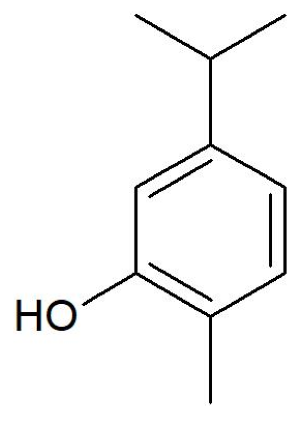
Figure 1. Chemical structure of carvacrol.
Applsci 15 11309 g001
Figure 2. The effects of CAR (1/2 MIC) on bacterial cells’biofilm formation. Mean absorbance isshown as bar graphs with mean and the standard deviations (n = 5–8); * p < 0.05 compared with control sample (without CAR).
Applsci 15 11309 g002
Figure 3. Determination of the production of ROS using the DCFH-DA probe. The bacterial cells treated with 1/2 MIC carvacrol. Mean fluorescence intensities in cells are shown as bar graphs with mean and the standard deviations (n = 4–8); * p < 0.05.
Applsci 15 11309 g003
Table 1. List of primers used to detect virulence factors of P. aeruginosa.
Virulence GenesSequence (5′–3′)Size of Product
(bp)
phzMF: CGTCGTGTTCAAGCAGATGGTGCTG
R: CCGAACCGCTTCACCAGGC
875
phzSF: CAATCATCTCAGCAGAACCC
R: TGTCGTAGAGGATCTCCTG
1752
exoTF: CATCGTCTACGCCATGAG
R: AGCAGCACCTCGGAATAG
1159
exoYF: GGAATGAACGAAGCGTTCTCCGAC
R: TGGCGTCGACGAACACCTCG
1035
exoSF: GTGTGCTTTATGCCATGAG
R: GGTTTCCTTTTCCAGGTC
444
exoUF: CTGCGCGGGTCTATGTGCC
R: GATGCTGGACGGGTCGAG
3308
pilAF: ATGGAGAGCGGGATCGACAG
R: ATGCGGGTTTCCATCGGCAG
1675
pilBF: TCGCCATGACCGATACGCTC
R: ACAACCTGAGCCAGCCTTCC
408
aprF: GCACGTGGTCATCCTGATGC
R: TCCGTAGGCGTCGACGTAC
1017
plcHF: CACACGGAAGGTTAATTCTGA
R: CGGTTARACGGCTGAACCTG
608
Table 2. List of primers used to detect virulence factors of K. pneumoniae and E. cloacae.
Virulence GenesSequence (5′–3′)Size of Product
(bp)
ugeF: GAT CAT CCG GTC TCC CTG TA
R: TCT TCA CGC CTT CCT TCA CT
534
kfuF: GAA GTG ACG CTG TTT CTG GC
R: TTT CGT GTG GCC AGT GAC TC
797
wabGF: CGG ACT GGC AGA TCC ATA TC
R: ACC ATC GGC CAT TTG ATA GA
683
fimHF: ATG AAC GCC TGG TCC TTT GC
R: GCT GAA CGC CTA TCC CCT GC
688
iroNF: AAG TCA AAG CAG GGG TTG CCC G
R: GAC GCC GAC ATT AAG ACG CAG
665
hlyAF: AAC AAG GAT AAG CAC TGT TCT GGC T
R: ACC ATA TAA GCG GTC ATT CCC GTC A
1177
rmpAF: ACT GGG CTA CCT CTG CTT CA
R: CTT GCA TGA GCC ATC TTT CA
535
magAF: GGT GCT CTT TAC ATC ATT GC
R: GCA ATG GCC ATT TGC GTT AG
1280
Table 3. Presence of virulence genes in examined bacterial strains.
PresenceAbsence
P. aeruginosa 3411122exoT, aprA, phzS, plcHphzM, exoY, exoS, exoU, pilA, pilB, aprA
P. aeruginosa 25521221exoT, phzS, plcHphzM, exoY, exoS, exoU, pilA, pilB, aprA
K. pneumoniae 473/24fimH, wabG, uge, iroNkfu, rmpA, magA, hlyA
K. pneumoniae 23520/23fimH, wabG, uge, kfu, rmpAmagA, hlyA
E. cloacae 25269fimH, kfu, iroNhlyA
E. cloacae 24985fimH, kfu, iroNhlyA
Table 4. Antibacterial activity of carvacrol.
P. aeruginosa 3411122P. aeruginosa 25521221K. pneumoniae 473/24K. pneumoniae 23520/23E. cloacae 25269E. cloacae 24985
MIC
[μg/mL]
5001000250250250125
MBC
[μg/mL]
5002000250250250250
Disclaimer/Publisher’s Note: The statements, opinions and data contained in all publications are solely those of the individual author(s) and contributor(s) and not of MDPI and/or the editor(s). MDPI and/or the editor(s) disclaim responsibility for any injury to people or property resulting from any ideas, methods, instructions or products referred to in the content.