Article

Hispidulin Protects C6 Astroglial Cells Against H2O2-Induced Injury by Attenuating Oxidative Stress, Inflammation, and Apoptosis

1
Department of Food Science and Nutrition, Pusan National University, Busan 46241, Republic of Korea
2
BK21 FOUR Program: Precision Nutrition Program for Future Global Leaders, Pusan National University, Busan 46241, Republic of Korea
3
Department of Nutrition and Food Hygiene, Guangxi Key Laboratory of Environmental Exposureomics and Entire Lifecycle Health, School of Public Health, Guilin Medical University, No.1 Zhiyuan Road, Guilin 541100, China
*
Author to whom correspondence should be addressed.
These authors contributed equally to this work.
Appl. Sci. 2025, 15(20), 11069; https://doi.org/10.3390/app152011069
Submission received: 20 September 2025 / Revised: 9 October 2025 / Accepted: 10 October 2025 / Published: 15 October 2025
(This article belongs to the Special Issue Dietary Bioactive Compounds and Their Neuroprotective Potential)

Abstract

Oxidative stress occurs when excessive production of reactive oxygen species (ROS) disrupts the redox balance between oxidants and antioxidants. The brain is particularly vulnerable to oxidative stress due to its high metabolic rate. Astrocytes, the key homeostatic cells in the brain, play a crucial role in maintaining physiological function, including the regulation of oxidative stress. In the present study, we investigated whether hispidulin can mitigate oxidative damage by regulating redox imbalance, inflammatory signaling and apoptotic response in hydrogen peroxide (H2O2)-treated C6 astroglial cells. The cells were exposed to hispidulin at various concentrations for 24 h and then challenged with H2O2 for another 24 h. Hispidulin treatment significantly increased the viability in all concentrations and attenuated H2O2-induced increases in ROS production, lactate dehydrogenase release, and nitric oxide levels. Furthermore, it significantly downregulated proinflammatory markers, including tumor necrosis factor α, interleukin-6 (IL-6), and IL-1β. Western blot analysis exhibited that hispidulin significantly increased the antioxidant defense system-related proteins such as nuclear factor erythroid 2-related factor 2, glutathione peroxidase 1, and superoxide dismutase. In addition, hispidulin decreased the pro-apoptotic Bax and cytochrome C, while increasing the levels of anti-apoptotic Bcl-2. In conclusion, hispidulin showed a protective effect against H2O2-induced injury in C6 astroglial cells by suppressing oxidative stress, inflammation, and apoptosis.

1. Introduction

Astrocytes, the most abundant glial cells in the central nervous system (CNS), play an essential role in maintaining neuronal homeostasis. The cells regulate extracellular ion concentrations, assist synaptic transmission, supply metabolic substrates to neurons, maintain blood–brain barrier (BBB) integrity, and facilitate glutamate uptake to prevent excitotoxicity [1]. However, under pathological states, astrocytes are highly susceptible to oxidative stress. Neurological insults such as ischemic stroke, traumatic brain injury, Alzheimer’s disease, and Parkinson’s disease are often accompanied by an excessive generation of reactive oxygen species (ROS), derived from both damaged neurons and activated glia [2]. In addition, astrocytes themselves contribute to ROS production through mitochondrial dysfunction and the activation of NADPH oxidase, resulting in oxidative damage, impaired morphology, dysregulated glutamate uptake, and the increased release of pro-inflammatory cytokines, ultimately exacerbating neuronal injury [3].
Recent mechanistic investigations emphasize the significance of the nuclear factor erythroid 2-related factor 2 (Nrf2) pathway in the redox regulation of astrocytes. Activation of Nrf2 enhances transcription of antioxidant enzymes such as glutathione peroxidase (GPx1) and superoxide dismutase (SOD), promotes glutathione biosynthesis, diminishes ROS accumulation, and downregulates pro-inflammatory signaling cascades, thereby protecting both astrocytes and adjacent neurons [4]. Both in vitro and in vivo studies have demonstrated that astrocyte-specific activation of Nrf2 provides neuroprotection in models of spinal cord injury and neurodegeneration, while the loss of astrocytic Nrf2 function increases susceptibility to oxidative stress [5].
Given the interplay between oxidative stress, neuroinflammation, and astrocyte dysfunction, restoring astrocytic redox balance has emerged as a promising therapeutic strategy in various CNS disorders. However, pharmacological approaches specifically targeting astrocyte proteins under oxidative conditions remain underexplored. Most studies focus on neuronal survival or overall whole-brain tissue responses; in contrast, astrocyte-specific mechanisms underlying oxidative damage and their modulation remain poorly defined. In particular, recent studies have shown that astrocytic dysfunction can precede neuronal degeneration in neurodegenerative diseases, emphasizing the need to evaluate individual flavonoids such as hispidulin for their astrocyte-centered protective mechanisms [5,6].
Hispidulin (4′,5,7-trihydroxy-6-methoxyflavone), a naturally occurring flavone found in numerous medicinal plants, vegetables, and fruits, has been reported to exhibit a broad spectrum of bioactivities, including antioxidant, antifungal, antimutagenic, antitumor, anti-osteoporotic, anti-inflammatory, and neuroprotective effects [7,8,9]. Notably, hispidulin modulated multiple intracellular signaling pathways, i.e., NF-κB/NLRP3, AMPK/GSK3β, JAK/STAT, and the activation of Nrf2, which are critically involved in oxidative stress and neuroinflammation. In models of cerebral ischemia–reperfusion injury, hispidulin has been shown to reduce brain edema and pro-inflammatory cytokine levels, partially suppressing NLRP3 inflammasome-mediated pyroptosis in astrocytes, thereby improving neurological outcomes [10,11,12]. These findings suggest that hispidulin may have protective effects on astrocytes under pathological conditions through multiple mechanisms, including the activation of Nrf2. Given its antioxidant, anti-inflammatory, and cytoprotective potential, hispidulin represents a promising candidate for glial-targeted therapies. Although hispidulin has been investigated in other cell types, including neurons and cancer cells [11,12], its astrocyte-specific effects remain largely unexplored, despite the central role of astrocytes in regulating oxidative stress and neuroinflammation. Therefore, the present study aimed to investigate the protective effects of hispidulin on hydrogen peroxide (H2O2)-induced oxidative stress in C6 astrocytes, with a focus on its potential role in regulatory mechanisms that enhance astrocyte resilience under oxidative stress conditions.

2. Materials and Methods

2.1. Reagents

Hispidulin (Figure 1) was supplied by the Natural Product Institute of Science and Technology, Chung-Ang University (Anseong, Republic of Korea). Culture reagents, including Dulbecco’s modified Eagle medium (DMEM), fetal bovine serum (FBS), trypsin–ethylenediaminetetraacetic acid (EDTA), and penicillin–streptomycin, were obtained from Welgene (Daegu, Republic of Korea). H2O2 and sodium pentacyanonitrosylferrate (III) dihydrate (SNP) were purchased from Junsei (Tokyo, Japan). The 3-(4,5-dimethylthiazol-2-yl)-2,5-diphenyltetrazolium bromide (MTT) reagent was provided by Bio Pure (Kitchener, ON, Canada), and dimethyl sulfoxide (DMSO) was from Daejung (Shi/heung, Gyeonggi-do, Republic of Korea). For ROS detection, 2′,7′-dichlorodihydrofluorescein diacetate (DCF-DA) was obtained from Sigma (St. Louis, MO, USA). The lactate dehydrogenase (LDH) cytotoxicity kit was purchased from Takara Bio (Shiga, Japan), while enzyme-linked immunosorbent assay (ELISA) kits for tumor necrosis factor-alpha (TNF-α), interleukin-6 (IL-6), and interleukin-1β (IL-1β) were from R&D Systems (Minneapolis, MN, USA). Polyvinylidene difluoride (PVDF) membranes were supplied by Millipore (Burlington, MA, USA), Radioimmunoprecipitation assay (RIPA) buffer was purchased from Elpis Biotech (Daejeon, Republic of Korea), and the enhanced chemiluminescence (ECL) detection kit was obtained from Bio-Rad (Hercules, CA, USA).

2.2. Cell Culture and Treatment

C6 rat glial cells (ATCC, Manassas, VA, USA) were maintained in DMEM supplemented with 10% heat-inactivated FBS and 1% penicillin–streptomycin at 37 °C in a humidified incubator with 5% CO2. The cells were plated for the experiments when the confluency reached 80%. Hispidulin (0.1, 0.5, and 1 μM) was pretreated for 4 h, followed by 300 μM of H2O2 treatment for 24 h to induce oxidative stress.

2.3. Measurement of Cell Viability

Cell viability was evaluated using the MTT assay. Briefly, 20 μL of MTT solution (5 mg/mL) was added to each well containing 100 μL of culture medium, and the mixture was incubated at 37 °C in the dark for 4 h. Following incubation, the supernatant was carefully removed, and the resulting formazan crystals were solubilized in 200 μL of DMSO and incubated for an additional 30 min at room temperature in the dark with gentle shaking. The absorbance was measured at 540 nm using a microplate reader (Thermo Fisher Scientific, Waltham, MA, USA).

2.4. Measurement of LDH

Cytotoxicity of C6 cells was evaluated by measuring the release of lactate dehydrogenase (LDH) using a commercial LDH cytotoxicity detection kit (Takara Bio Inc., Kusatsu, Japan). Following treatment, 100 μL of culture supernatant from each well was mixed with 100 μL of the LDH reaction mixture provided in the kit. The samples were incubated for 15 min at room temperature in the dark. The absorbance was measured at 490 nm using a microplate reader (Thermo Fisher Scientific, Waltham, MA, USA).

2.5. Measurement of Cellular ROS Production

The generation of the ROS was evaluated using the DCF-DA probe (St Louis, MO, USA). Following treatment, the cells were rinsed twice with PBS to eliminate residual medium and then incubated with 80 μM DCFH-DA prepared in DMEM for 30 min at 37 °C in the dark. Fluorescence was subsequently recorded at 480 nm for excitation and 535 nm for emission using a FLUOstar OPTIMA microplate reader (BMG Labtech, Ortenberg, Germany).

2.6. Measurement of NO Production

NO production in cells was assessed using the Griess reaction. The supernatants of the cells were collected and centrifuged at 1000 rpm for 5 min to remove debris. Equal volumes of clarified supernatant and Griess reagent (1% sulfanilamide and 0.1% N-(1-naphthyl) ethylenediamine dihydrochloride in 2.5% phosphoric acid) were mixed and incubated at room temperature for 30 min in the dark. The absorbance was then measured at 540 nm using a microplate reader (Thermo Fisher Scientific, Waltham, MA, USA). Sodium nitrite (NaNO2) was used to generate a standard curve for quantification.

2.7. Proinflammatory Cytokine Measurements

The levels of the pro-inflammatory cytokines TNF-α, IL-6, and IL-1β in the conditioned medium of hispidulin-treated cells were quantified using ELISA kits according to the manufacturer’s instructions. In brief, 100 μL of each collected supernatant sample was dispensed into wells pre-coated with the corresponding capture antibodies, and the plates were incubated at 37 °C for 2 h. After washing to remove unbound materials, detection antibodies were added, followed by the application of enzyme conjugates and the substrate solution. The enzymatic reaction was terminated with 2 N sulfuric acid, and the absorbance of each well was recorded at 450 nm using a microplate reader (Thermo Fisher Scientific, Waltham, MA, USA). A standard calibration curve was established using serial dilutions of recombinant cytokine standards provided in the kits, and the cytokine concentrations in the samples were subsequently calculated based on this curve.

2.8. Western Blot Analysis

Cells were lysed in RIPA buffer supplemented with a protease inhibitor cocktail. Protein concentrations were determined using the Bio-Rad Protein Assay Kit (Bio-Rad, Hercules, CA, USA). Equal amounts of protein (20 μg) were separated by 8–13% SDS-PAGE and transferred onto PVDF membranes. Membranes were blocked with 5% non-fat dry milk containing 0.1% Tween-20 for 1 h at room temperature and subsequently incubated overnight at 4 °C with the following primary antibodies: Nrf2 (1:500, Cell Signaling Technology, Danvers, MA, USA), Gpx1 (1:500, Santa Cruz Biotechnology, Dallas, TX, USA), SOD (1:500, Santa Cruz Bio-technology, Dallas, TX, USA), Bax (1:500, Santa Cruz Biotechnology, Dallas, TX, USA), Bcl-2 (1:500, Santa Cruz Biotechnology, Dallas, TX, USA), β-actin (1:1000, Cell Signaling Technology, Danvers, MA, USA). The membranes were then incubated for 1 h at room temperature with horseradish peroxidase-conjugated secondary antibodies. Protein bands were visualized using an ECL detection kit, and images were captured using a chemiluminescence imaging system (Davinch-ChemiTM, Davinch-K, Seoul, Republic of Korea).

2.9. Statistical Analysis

Statistical analyses were carried out using GraphPad Prism 10 (GraphPad Software, La Jolla, CA, USA). The significant differences among the groups were evaluated by one-way analysis of variance (ANOVA) with Tukey’s post hoc test. All data are presented as the mean ± standard deviation (SD), and a p-value of less than 0.05 was considered statistically significant.

3. Results

3.1. Hispidulin Protects C6 Astroglial Cells Against H2O2-Induced Cytotoxicity

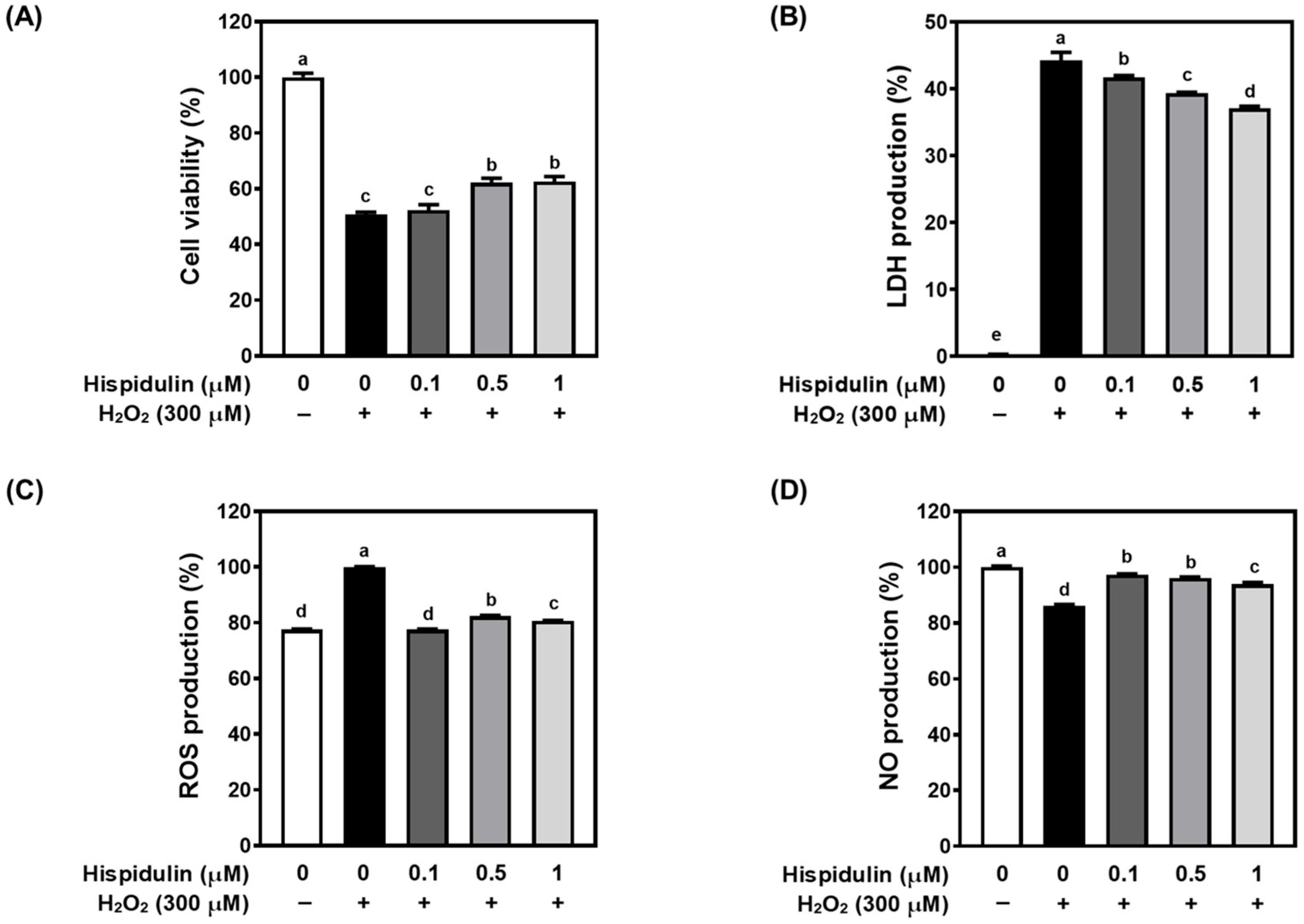
When C6 astroglial cells were exposed to 300 μM H2O2, a marked decline in cell viability was observed along with signs of oxidative injury, including increased LDH release, intracellular ROS, and NO production. In Figure 2A, pretreatment with hispidulin markedly improved the survival of C6 cells challenged with H2O2. LDH release, an indicator of cell membrane integrity loss, was markedly elevated in H2O2-treated cells (Figure 2B). Pretreatment of hispidulin significantly attenuated LDH release in a concentration-dependent manner, with 1 μM concentration showing the most protective effect (p < 0.05). The increase in intracellular ROS levels by H2O2 was attenuated by hispidulin in a dose-dependent manner (Figure 2C). The marked increase in NO production by oxidative stress was effectively suppressed by hispidulin treatment (Figure 2D). Collectively, these results demonstrate that hispidulin alleviates H2O2-induced oxidative damage by reducing ROS and NO production, limiting LDH release, and preserving astrocyte viability under oxidative stress conditions.

3.2. Hispidulin Inhibits Proinflammatory Cytokine Secretion in H2O2-Treated C6 Astroglial Cells

To evaluate the anti-inflammatory properties of hispidulin, the levels of proinflammatory cytokines TNF-α, IL-6, and IL-1β were measured in H2O2-stimulated C6 astroglial cells. As shown in Figure 3, H2O2 treatment significantly elevated the secretion of all of the cytokines relative to the control (p < 0.05), indicating an inflammatory response under oxidative stress. Hispidulin treatment significantly attenuated TNF-α levels at all tested concentrations (Figure 3A). The increased levels of IL-6 (Figure 3B) and IL-1β (Figure 3C) in H2O2-treated cells were markedly reduced in a concentration-dependent manner by hispidulin treatment. These results indicate that hispidulin effectively suppresses H2O2-stimulated inflammatory response in C6 astroglial cells and may contribute to anti-inflammatory regulation under oxidative conditions.

3.3. Hispidulin Increases the Expression of Nrf2 and Downstream Antioxidant Enzymes

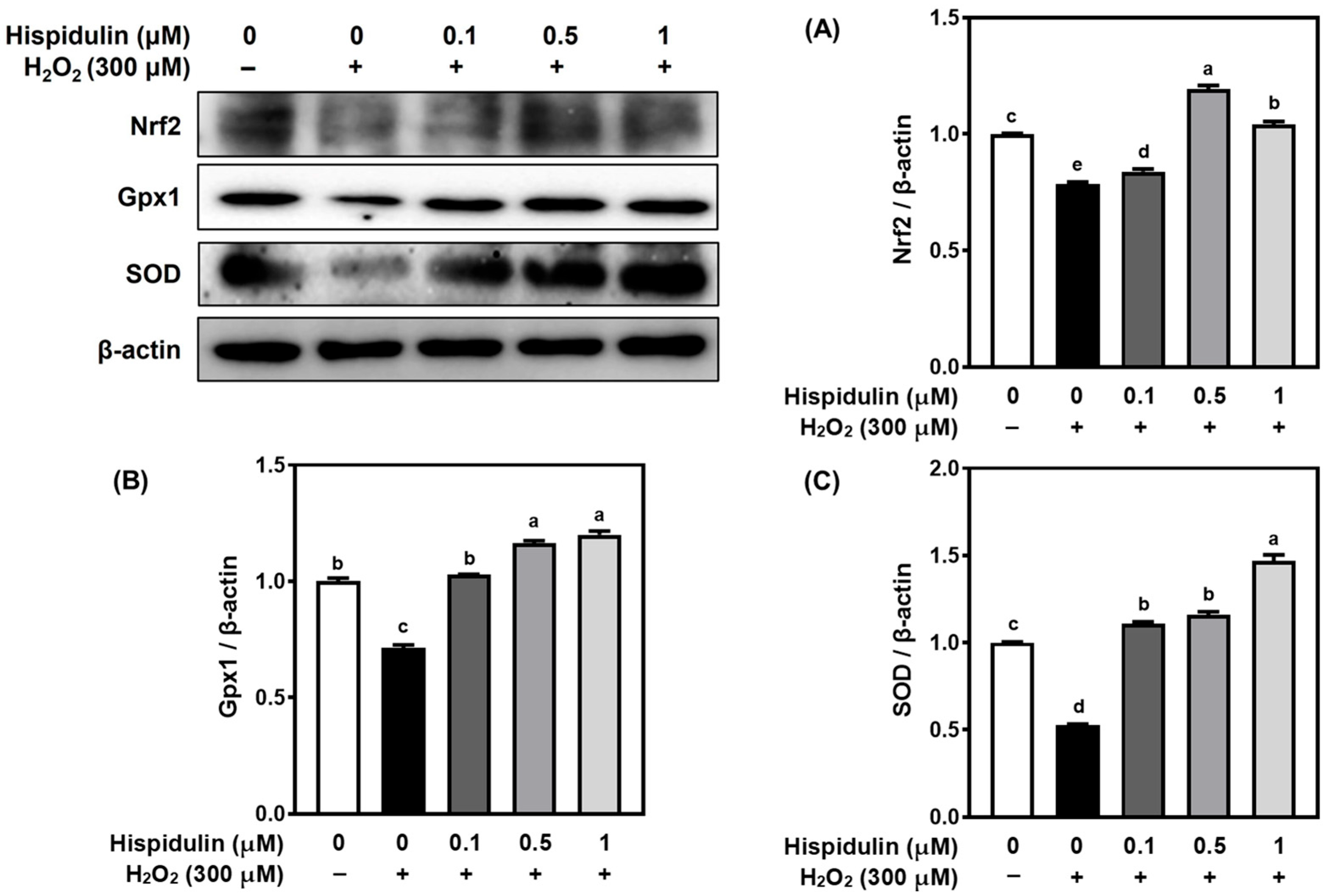
To explore the mechanisms that may account for the antioxidant action of hispidulin, we examined Nrf2 and its downstream antioxidant enzymes, Gpx1 and SOD. Exposure to H2O2 markedly decreased the protein levels of Nrf2, Gpx1, and SOD relative to the control group. Hispidulin treatment restored and increased the expression of Nrf2 (Figure 4A), suggesting a potential role of the Nrf2 pathway. Furthermore, protein levels of Gpx1 (Figure 4B) and SOD (Figure 4C) were also significantly elevated in hispidulin-treated cells, indicating that hispidulin may enhance endogenous antioxidant capacity, potentially through modulation of the Nrf2-associated pathway.

3.4. Hispidulin Modulates Apoptosis-Related Proteins in H2O2-Stimulated C6 Astroglial Cells

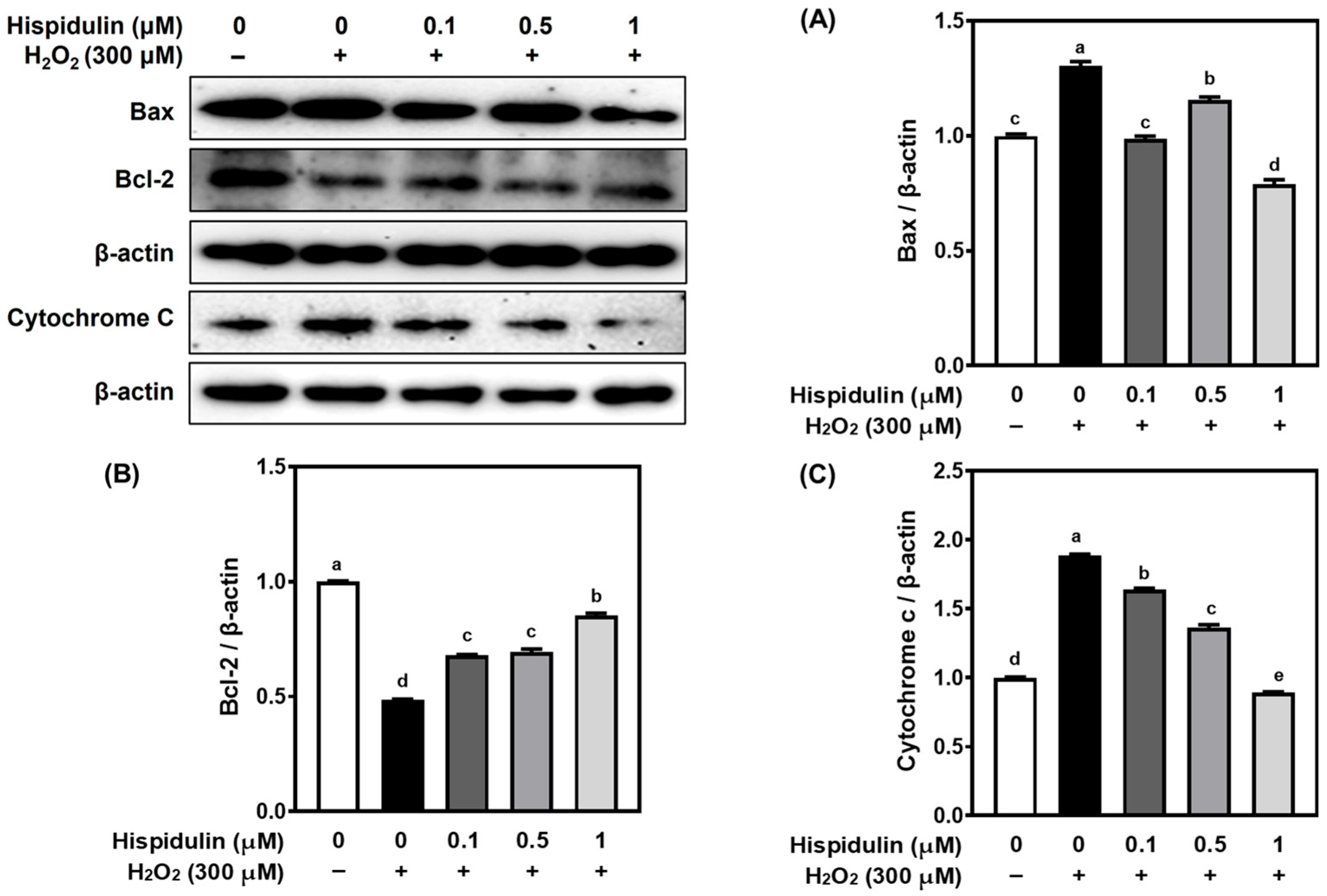
To evaluate the anti-apoptotic activity of hispidulin, we analyzed the expression of critical proteins involved in apoptosis, i.e., Bcl-2, Bax, and cytochrome C. Figure 5 demonstrates that H2O2 treatment significantly decreased the expression of Bcl-2, while increasing Bax and cytochrome C levels in comparison to the untreated group (p < 0.05). Hispidulin reversed these alterations induced by H2O2 in C6 cells. Specifically, 1 μM of hispidulin significantly restored Bcl-2 expression and markedly suppressed Bax and cytochrome c levels to values comparable to those of the control group (p < 0.05). These results suggest that hispidulin mitigates oxidative stress-induced apoptosis by modulating the apoptotic signaling pathway.

4. Discussion

Astrocytes in the CNS play essential roles in maintaining neuronal function and brain homeostasis. Historically viewed as passive support cells, astrocytes are now increasingly recognized as active contributors in physiological and pathological metabolisms within the CNS. The cells regulate critical functions, including maintenance of the blood–brain barrier, modulation of synaptic transmission, regulation of neurotransmitter and extracellular ion levels, and support of neurons. Emerging evidence suggests that astrocytes undergo a range of phenotypes and functional changes, which can be both protective and deleterious depending on the neurodegenerative disease stages. Notably, astrocytic dysfunction has been shown to precede neuronal degeneration and may directly contribute to disease onset and progression. Pathological mechanisms of astrocytic dysfunction include impaired glutamate clearance due to downregulation of excitatory amino acid transporters, disruption of astrocyte-neuron metabolic coupling, and the adoption of a pro-inflammatory, neurotoxic phenotype characterized by elevated cytokine production and ROS generation [6]. These pathological changes compromise the intrinsic neuroprotective capacity of astrocytes, thereby exacerbating neuronal vulnerability to oxidative, inflammatory, and excitotoxic damage. Given their prominent vulnerability to oxidative stress, astrocytes are considered crucial biological targets in therapeutic approaches aimed at mitigating oxidative damage and inflammation in neurodegenerative disorders. In line with this, growing evidence has highlighted the promise of natural compounds as potential interventions for such diseases. Many bioactive components derived from plants, such as polyphenols, flavonoids, and alkaloids, have demonstrated antioxidant, anti-inflammatory, and neuroprotective properties in both in vitro and in vivo models. Consequently, natural products are being actively investigated as promising candidates for the prevention of neurodegeneration.
In the present study, we investigate whether hispidulin has neuroprotective potential in astrocyte-mediated mechanisms under oxidative stress conditions using C6 astroglial cells. The C6 cell line serves as a well-established and reproducible in vitro model for investigating oxidative, inflammatory, and apoptotic signaling pathways. The cells consistently respond to H2O2 exposure while retaining key astrocytic characteristics, making them suitable for controlled evaluation of the protective effects of candidate compounds [13,14]. Therefore, the C6 model remains a valuable tool for initial mechanistic investigation due to its reproducibility and well-characterized responses. Hispidulin pretreatment significantly preserved the viability of C6 cells under H2O2-induced oxidative stress. The results were confirmed by MTT assays and LDH release measurements. Aligned with these results, flavonoids counteracted oxidative insults through antioxidant enzyme induction and stabilization of cellular membranes in C6 glioma cells [15,16]. In cortical neurons, hispidulin reduced excitotoxic cell death by limiting calcium influx and stabilizing mitochondrial integrity.
The observed enhancement in cell viability by hispidulin was further supported by a marked decrease in intracellular levels of ROS and NO. Although the inhibitory effects of hispidulin on ROS and NO production did not follow a strict dose-dependent manner, similar patterns have often been reported for natural compounds, which may exert threshold or saturation effects at certain concentrations [17]. Such variability could also be attributed to differential cellular uptake or metabolism of flavonoids. Nevertheless, all concentrations of hispidulin consistently reduced ROS and NO levels compared with H2O2 treatment alone, supporting its overall protective role under oxidative stress conditions. Given that oxidative stress plays a central role in the pathophysiology of neurodegenerative diseases by disrupting astrocytic function and promoting neuronal injury, the ability of hispidulin to reduce free radicals underscores its potential role for neuroprotection [12]. Hispidulin not only acts as a free radical scavenger but also modulates cellular redox signaling pathways to restore homeostasis. The dual mechanisms of hispidulin have been reported by in vivo studies demonstrating neuroprotection against anesthesia-induced neurotoxicity. Notably, both in vitro and in vivo studies have shown that hispidulin pre-treatment attenuates sevoflurane-induced neurological impairment, suggesting its antioxidant effect within the CNS [18]. The redox-modulating activity of hispidulin was shown to improve hepatic redox status by increasing catalase and SOD activities in high-fat diet-induced obese mice [19]. These findings suggest hispidulin modulates oxidative stress across multiple organ systems by a conserved and adaptable mechanism of action that could be beneficial in multifactorial disorders like neurodegeneration. The consistency across experimental models enhances the translational relevance of hispidulin in neurodegenerative disorders.
Hispidulin markedly reduced the release of major pro-inflammatory cytokines such as TNF-α, IL-6, and IL-1β. The observed anti-inflammatory effects are consistent with previous research showing that hispidulin modulates critical inflammatory signaling pathways, most notably the NF-κB/NLRP3 axis and the JAK/STAT cascade [12]. Considering the role of reactive astrocytes in perpetuating chronic inflammation that exacerbates neuronal damage, suppression of cytokine release by hispidulin highlights its potential role in alleviating neuroinflammation [19]. Reduced astrocytic cytokine release has been associated with decreased neuronal apoptosis and improved behavioral outcomes in preclinical models [20]. Importantly, elevated concentrations of TNF-α and IL-1β are known to drive microglial activation and leukocyte infiltration, amplifying inflammation in the CNS, whereas IL-6 promotes the transition of astrocytes into a reactive state that contributes to synaptic dysfunction. Therefore, the coordinated reduction in these cytokines by hispidulin may interrupt multiple inflammatory feedback mechanisms within the CNS. In particular, IL-1β is a critical activator of the inflammasome and a key mediator of BBB disruption, while IL-6 exerts systemic effects that prolong neuroinflammation and impair neurogenesis. TNF-α, in addition to its immunomodulatory role, directly alters synaptic plasticity and has been implicated in depression-like behaviors [21,22,23]. Collectively, the suppression of these cytokines by hispidulin may affect astrocytic inflammation and neuromodulatory benefits, contributing to overall CNS resilience.
Parallel to its anti-inflammatory and antioxidant activities, hispidulin also upregulated the expression of the pivotal components of the intrinsic antioxidant defense system, Gpx1, SOD, and Nrf2. The activation of Nrf2, the transcription factor that orchestrates the cellular antioxidant response, induces genes that restore redox balance and protect against oxidative damage [24,25]. Hispidulin enhanced Nrf2 signaling and reinforced its multifunctional mechanism of action, mirroring similar observations in models of cerebral ischemia and Alzheimer’s disease [10,11,18]. SOD converts superoxide radicals into hydrogen peroxide, and GPx1 further breaks down hydrogen peroxide into water. Together, these enzymes establish a cooperative defense mechanism against ROS [26]. Modest enhancement of Nrf2 signaling can shift the intracellular environment toward a more reducing state, thereby preserving astrocytic metabolism and supporting neuronal function under oxidative stress [27]. In addition, Nrf2 activation also downregulates pro-inflammatory mediators by competing with NF-κB for transcriptional co-activators, thereby linking redox control to the suppression of inflammation. SOD and GPx1 work synergistically with catalase and glutathione reductase to sustain glutathione homeostasis, one of the most critical anti-oxidant defense systems in astrocytes [28,29]. Deficiency in these enzymes is associated with accelerated cognitive decline and increased vulnerability to excitotoxic injury, highlighting the importance of hispidulin on oxidative stress defense. The bioactivity of flavones is closely linked to their structural substituents. Hydroxyl groups generally enhance antioxidant and anti-inflammatory potential, while methoxy groups contribute to lipophilicity and membrane permeability [30,31]. In the case of hispidulin, the 6-methoxy and 7-hydroxyl groups may underlie its astrocyte-protective effects. Although no in silico studies of hispidulin have been reported to date, such approaches represent a valuable avenue for future investigation. Taken together with previous reports, our results suggest that hispidulin targets multiple molecular pathways in glial cells, including activation of the AMPK/GSK3β–Nrf2 axis and suppression of NF-κB and NLRP3 signaling, which together likely underlie its combined antioxidant and anti-inflammatory actions [10,11,12].
Astrocyte survival has been known to directly affect neuronal homeostasis, and mitochondria-dependent apoptosis is a predominant cell death pathway in oxidative stress conditions [32,33]. Mitochondrial-mediated apoptosis was effectively suppressed by hispidulin, as indicated by modulation of Bcl-2, Bax, and cytochrome C levels. Similar to the results, hispidulin preserved mitochondrial integrity in hippocampal neurons subjected to oxidative damage, supporting its protective effects extend across CNS cell types [18]. Mechanistically, Bax facilitates mitochondrial outer membrane permeabilization and the subsequent release of cytochrome C, initiating caspase activation and apoptosis. In contrast, Bcl-2 acts as a counter-regulator that maintains membrane stability [33]. By restoring the Bcl-2/Bax ratio and preventing cytochrome c leakage, hispidulin effectively preserves mitochondrial function, thereby ensuring adenosine triphosphate production and limiting caspase-dependent apoptosis in astrocytes.
In the intrinsic pathway, the release of cytochrome C activates apoptosome formation and initiates caspase-9–caspase-3 cascades, culminating in DNA fragmentation and cell death [33]. Inhibition of cytochrome c leakage by hispidulin may halt this apoptotic reaction at an early stage. Maintaining mitochondrial integrity is essential for calcium homeostasis and metabolic coupling between astrocytes and neurons, the processes that are critical for CNS stability [34]. Given that astrocytic apoptosis can trigger secondary neuronal loss through impaired trophic support and dysregulation of glutamate, the anti-apoptotic action of hispidulin may offer comprehensive protective effects within the CNS network [35].
These data collectively support a polymechanistic model in which hispidulin simultaneously modulates oxidative stress, inflammation, and apoptosis to preserve astrocytic function. The multi-targeted properties of bioactive compounds may be particularly advantageous in complex CNS disorders, which are typically involved in complex and multi-cellular pathogenesis. The restoration of astrocytic homeostasis by hispidulin suggests that its neuroprotective effect may occur at an early stage of cellular dysfunction, potentially preventing the onset of irreversible neuronal damage. By targeting astrocytes, a key type of glial cell responsible for maintaining the neuronal environment, hispidulin may interrupt the pathological cascade that leads to neuronal injury. This supports a glia-centric therapeutic strategy, emphasizing preservation or restoration of glial cell function as a primary approach to prevent neurodegeneration [36].
Notably, the present findings highlight the therapeutic potential of hispidulin in mitigating astrocytic damage under oxidative stress conditions. Furthermore, previous reports of its high oral bioavailability and low toxicity in rodent models provide a strong rationale for further translational research [37]. In addition to oral bioavailability, recent reports synthesize evidence that hispidulin shows central nervous system (CNS)-relevant actions and BBB permeability within neuroprotective contexts [38,39]. Quantitative pharmacokinetic studies also indicate an oral bioavailability of around 17–18% in rats [40]. Together with in vivo protection in cerebral ischemia/reperfusion models via NLRP3 and AMPK/GSK3β/Nrf2 signaling [11], these data support the translational plausibility of hispidulin for CNS indications.
Collectively, our results suggest that hispidulin exerts multifaceted protective effects, likely involving antioxidant, anti-inflammatory, and anti-apoptotic mechanisms, that contribute to the preservation of astrocytic function. These properties support hispidulin as a functional bioactive compound that targets astrocyte dysfunction, underscoring its potential application in therapeutic strategies for neurological disorders. A limitation of the present work is that we only examined the preventive effects of hispidulin against oxidative injury. Future studies are warranted to investigate whether hispidulin can also promote recovery and repair when administered after cellular damage.

5. Conclusions

In the present study, hispidulin exerted multifaceted protective effects against H2O2-induced oxidative damage in C6 astroglial cells. Hispidulin significantly enhanced cell viability by mitigating ROS and NO accumulation, attenuated pro-inflammatory cytokine release, and modulated the expression of mitochondrial apoptosis-related proteins under oxidative stress. Importantly, the regulation of the Nrf2 signaling pathway and its downstream antioxidant enzymes, such as GPx1 and SOD, highlights the mechanistic basis of its action in reinforcing intrinsic redox defense systems. Beyond these findings, our results underscore the therapeutic potential of hispidulin as a glia-targeted bioactive compound, suggesting that astrocytes may represent a critical cellular target for early intervention in neurodegenerative diseases. Considering its high oral bioavailability and low toxicity reported in preclinical models, hispidulin may hold translational value for future development as a dietary supplement or pharmacological agent. Further in vivo studies and clinical investigations are warranted to validate its neuroprotective efficacy, clarify pharmacokinetic profiles, and explore synergistic effects with other therapeutic strategies. Collectively, these findings support hispidulin as a promising candidate for the development of functional foods and drugs targeting astrocyte dysfunction and neuroinflammation.

Author Contributions

Conceptualization, J.-H.K. and E.J.C.; methodology, J.-H.K. and Q.Q.P.; formal analysis, J.-H.K. and B.K.; investigation, J.-H.K. and Q.Q.P.; writing—original draft preparation, J.-H.K. and B.K.; writing—review and editing, B.K.; supervision, B.K. and E.J.C.; project administration, B.K. and E.J.C.; funding acquisition, B.K. and E.J.C. All authors have read and agreed to the published version of the manuscript.

Funding

This study was financially supported by the 2024 Post-Doc. Development Program of Pusan National University.

Data Availability Statement

The original contributions presented in the study are included in the article; further inquiries can be directed to the corresponding authors.

Acknowledgments

The authors would like to express sincere gratitude to Eun Ju Cho, who passed away during the preparation of this manuscript. We honor her memory and gratefully acknowledge her valuable contribution to this work.

Conflicts of Interest

The authors declare no conflicts of interest to report regarding the present study.

Abbreviations

The following abbreviations are used in this manuscript:
CNSCentral Nervous System
BBBBlood–Brain Barrier
ROSReactive Oxygen Species
Nrf2Nuclear factor erythroid 2–related factor 2
GPx1Glutathione Peroxidase 1
SODSuperoxide Dismutase
NF-κBNuclear Factor kappa-light-chain-enhancer of activated B cells
NLRP3NOD-, LRR- and pyrin domain-containing protein 3
AMPKAMP-activated Protein Kinase
GSK3βGlycogen Synthase Kinase 3 beta
JAK/STATJanus Kinase/Signal Transducer and Activator of Transcription
DMEMDulbecco’s Modified Eagle Medium
FBSFetal Bovine Serum
EDTAEthylenediaminetetraacetic Acid
H2O2Hydrogen Peroxide
SNPSodium Pentacyanonitrosylferrate (III) dihydrate
MTT3-(4,5-dimethylthiazol-2-yl)-2,5-diphenyltetrazolium bromide
DMSODimethyl Sulfoxide
DCF-DA2′,7′-Dichlorodihydrofluorescein Diacetate
LDHLactate Dehydrogenase
ELISAEnzyme-Linked Immunosorbent Assay
PVDFPolyvinylidene Difluoride
RIPARadioimmunoprecipitation Assay
ECLEnhanced Chemiluminescence

References

  1. Valles, S.L.; Signh, S.K.; Campos-Campos, J.; Colmena, C.; Campo-Palacio, I.; Alvarez-Gamez, K.; Caballero, O.; Jorda, A. Functions of astrocytes under normal conditions and after a brain disease. Int. J. Mol. Sci. 2023, 24, 8434. [Google Scholar] [CrossRef] [PubMed]
  2. Trofin, D.-M.; Sardaru, D.-P.; Trofin, D.; Onu, I.; Tutu, A.; Onu, A.; Onita, C.; Galaction, A.I.; Matei, D.V. Oxidative stress in brain function. Antioxidnats 2025, 14, 297. [Google Scholar] [CrossRef]
  3. Chen, Y.; Qin, C.; Huang, J.; Tang, X.; Liu, C.; Huang, K.; Xu, J.; Guo, G.; Tong, A.; Zhou, L. The role of astrocytes in oxidative stress of central nervous system: A mixed blessing. Cell Prolif. 2020, 53, e12781. [Google Scholar] [CrossRef]
  4. Park, J.-E.; Leem, Y.-H.; Park, J.-S.; Kim, S.-E.; Kim, H.-S. Astrocytic Nrf2 mediates the neuroprotective and anti-inflammatory effects of nootkatone in an MPTP-induced Parkinson’s disease mouse model. Antioxidants 2023, 12, 1999. [Google Scholar] [CrossRef]
  5. Zhao, W.; Gasterich, N.; Clarner, T.; Voelz, C.; Behrens, V.; Beyer, C.; Fragoulis, A.; Zendedel, A. Astrocytic Nrf2 expression protects spinal cord from oxidative stress following spinal cord injury in a male mouse model. J. Neuroinflamm. 2022, 19, 134. [Google Scholar] [CrossRef] [PubMed]
  6. Brandebura, A.N.; Paumier, A.; Onur, T.S.; Allen, N.J. Astrocyte contribution to dysfunction, risk and progression in neurodegenerative disorder. Nat. Rev. Neurosci. 2022, 24, 23–39. [Google Scholar] [CrossRef]
  7. Patel, K.; Patel, D.K. Medicinal importance, pharmacological activities, and analytical aspects of hispidulin: A concise report. J. Tradit. Complement. Med. 2017, 7, 360–366. [Google Scholar] [CrossRef]
  8. Liu, K.; Zhao, F.; Yan, J.; Xia, Z.; Jiang, D.; Ma, P. Hispidulin: A promising flavonoid with diverse anti-cancer properties. Life Sci. 2020, 259, 118395. [Google Scholar] [CrossRef]
  9. Ashaq, A.; Maqbool, M.F.; Maryam, A.; Khan, M.; Shakir, H.A.; Irfan, M.; Qazi, J.I.; Li, Y.; Ma, T. Hispidulin: A novel natural compound with therapeutic potential against human cancers. Phytother. Res. 2021, 35, 771–789. [Google Scholar] [CrossRef]
  10. An, P.; Wu, T.; Yu, H.; Fang, K.; Ren, Z.; Tang, M. Hispidulin protects against focal cerebral ischemia reperfusion injury in rats. J. Mol. Neurosci. 2018, 65, 203–212. [Google Scholar] [CrossRef] [PubMed]
  11. An, P.; Xie, J.; Qiu, S.; Liu, Y.; Wang, J.; Xiu, X.; Li, L.; Tang, M. Hispidulin exhibits neuroprotective activities against cerebral ischemia reperfusion injury through suppressing NLRP3-mediated pyroptosis. Life Sci. 2019, 232, 116599. [Google Scholar] [CrossRef]
  12. Yu, C.-I.; Cheng, C.-I.; Kang, Y.-F.; Chang, P.-C.; Lin, I.-P.; Kuo, Y.-H.; Jhou, A.-J.; Lin, M.-Y.; Chen, C.-Y.; Lee, C.-H. Hispidulin inhibits neuroinflammation in lipopolysaccharide-activated BV2 microglia and attenuates the activation of Akt, NF-kB, and STAT3 pathway. Neurotox. Res. 2020, 38, 163–174. [Google Scholar] [CrossRef] [PubMed]
  13. Giakoumettis, D.; Kritis, A.; Foroglou, N. C6 cell line: The gold standard in glioma research. Hippokratia 2018, 22, 105–112. [Google Scholar]
  14. Quincozes-Santos, A.; Bobermin, L.D.; Latini, A.; Wajner, M.; Souza, D.O.; Goncalves, C.-A.; Gottfried, C. Reveratrol protects C6 astrocyte cell line against hydrogen peroxide-induced oxidative stress through heme oxygenase 1. PLoS ONE 2013, 8, e64372. [Google Scholar] [CrossRef] [PubMed]
  15. Choi, E.-O.; Jeong, J.-W.; Park, C.; Hong, S.H.; Kim, G.-Y.; Hwang, H.-J.; Cho, E.-J.; Choi, Y.H. Baicalein protects C6 glial cells against hydrogen peroxide-induced oxidative stress and apoptosis through regulation of the Nrf2 signaling pathway. Int. J. Mol. Med. 2016, 37, 798–806. [Google Scholar] [CrossRef] [PubMed]
  16. Polster, B.M.; Mark, K.A.; Arze, R.; Hudson, D. Calpain-independent intracellular protease activity is elevated in excitotoxic cortical neurons prior to delayed calcium deregulation and mitochondrial dysfunction. Biomolecules 2022, 12, 1004. [Google Scholar] [CrossRef]
  17. Xi, X.; Han, J.; Zhang, J.; Li, C.; Sun, Y.; Guo, Y.; Xu, Y. The biphasic effect of flavonoids on oxidative stress and cell proliferation. Antioxidants 2022, 11, 1975. [Google Scholar] [CrossRef]
  18. Huang, L.; Huang, K.; Ning, H. Hispidulin prevents sevoflurane-induced memory dysfunction in aged rats. Biomed. Pharmacother. 2018, 97, 412–422. [Google Scholar] [CrossRef]
  19. Lawrence, J.M.; Schardien, K.; Wigdahl, B.; Nonnemacher, M.R. Roles of neuropathology-associated reactive astrocytes: A systemic review. Acta Neuropathol. Commun. 2023, 11, 42. [Google Scholar] [CrossRef]
  20. Kitazawa, M.; Cheng, D.; Tsukamoto, M.R.; Koike, M.A.; Wes, P.D.; Vasilevko, V.; Cribbs, D.H.; LaFerla, F.M. Blocking IL-1 signaling rescues cognition, attenuates tau pathology, and restores neuronal b-catenin pathway function in an Alzheimer’s disease model. J. Immunol. 2011, 187, 6539–6549. [Google Scholar] [CrossRef]
  21. Yang, J.; Ran, M.; Li, H.; Lin, Y.; Ma, K.; Yang, Y.; Fu, X.; Yang, S. New insight into neurological degeneration: Inflammatory cytokines and blood-brain barrier. Front. Mol. Neurosci. 2022, 15, 1013933. [Google Scholar] [CrossRef]
  22. Gruol, D.L. IL-6 regulation of synaptic function in the CNS. Neuropharmacology 2015, 96, 42–52. [Google Scholar] [CrossRef]
  23. Lyra e Silva, N.M.; Gonçalves, R.A.; Pascoal, T.A.; Lima-Filho, R.A.S.; França Resende, E.P.; Vieira, E.L.M.; Teixeira, A.L.; de Souza, L.C.; Peny, J.A.; Fortuna, J.T.S.; et al. Pro-inflammatory interleukin-6 signaling links cognitive impairments and peripheral metabolic alterations in Alzheimer’s disease. Transl. Psychiatr. 2021, 11, 251. [Google Scholar] [CrossRef] [PubMed]
  24. Yamamoto, M.; Kensler, T.W.; Motohashi, H. The KEAP1-NRF2 system: A thiol-based sensor-effector apparatus for maintaining redox homeostasis. Physiol. Rev. 2018, 98, 1169–1203. [Google Scholar] [CrossRef] [PubMed]
  25. Ngo, V.; Duennwald, M.L. Nrf2 and oxidative stress: A general overview of mechanisms and implications in human disease. Antioxidants 2022, 11, 2345. [Google Scholar] [CrossRef]
  26. Jomova, K.; Alomar, S.Y.; Alwasel, S.H.; Nepovimova, E.; Kuca, K.; Valko, M. Several lines of antioxidant defense against oxidative stress: Antioxidant enzymes, nanomaterials with multiple enzyme-mimicking activities, and low-molecular-weight antioxidants. Arch. Toxicol. 2024, 98, 1323–1367. [Google Scholar] [CrossRef] [PubMed]
  27. Esteras, N.; Blacker, T.S.; Zherebtsov, E.A.; Stelmashuk, O.A.; Zhang, Y.; Wigley, W.C.; Duchen, M.R.; Dinkova-Kostova, A.T.; Abramov, A.Y. Nrf2 regulates glucose uptake and metabolism in neurons and astrocytes. Redox. Biol. 2023, 62, 102672. [Google Scholar] [CrossRef]
  28. Zgorzynska, E.; Dziedzic, B.; Walczewska, A. An overview of the Nrf2/ARE pathway and its role in neurodegenerative disease. Int. J. Mol. Sci. 2021, 22, 9592. [Google Scholar] [CrossRef]
  29. Dringen, R.; Arend, C. Glutathione metabolism of the brain-the role of astrocytes. J. Neurochem. 2025, 169, e70073. [Google Scholar] [CrossRef]
  30. Preet, G.; Hasan, A.H.; Ramlagan, P.; Fawdar, S.; Boulle, F.; Jaspars, M. Anti-neurodegenerating activity: Structure-activity relationship analysis of flavonoids. Molecules 2023, 28, 7188. [Google Scholar] [CrossRef]
  31. Patigo, A.; Hengphasatporn, K.; Cao, V.; Paunrat, W.; Vijara, N.; Chokmahasarn, T.; Maitarad, P.; Rungrotmongkol, T.; Shigeta, Y.; Boonyasuppayakorn, S.; et al. Design, synthesis, in vitro, in silico, and SAR studies of flavone analogs towards anti-dengue activity. Sci. Rep. 2022, 12, 21646. [Google Scholar] [CrossRef] [PubMed]
  32. Fiebig, C.; Keiner, S.; Ebert, B.; Schaffner, I.; Jagasia, R.; Lie, D.C.; Beckervordersandforth, R. Mitochondrial dysfunction in astrocytes impairs the generation of reactive astrocytes and enhances neuronal cell death in the cortex upon photothrombotic lesion. Front. Mol. Neurosci. 2019, 12, 40. [Google Scholar] [CrossRef]
  33. Nguyen, T.T.; Wei, S.; Nguyen, T.H.; Jo, Y.; Zhang, Y.; Park, W.; Gariani, K.; Oh, C.-M.; Kim, H.H.; Ha, K.-T.; et al. Mitochondria-associated programmed cell death as a therapeutic target for age-related disease. Exp. Mol. Med. 2023, 55, 1595–1619. [Google Scholar] [CrossRef]
  34. Verkhratsky, A.; Nedergaard, M. Physiology of astrolia. Physiol. Rev. 2018, 98, 239–389. [Google Scholar] [CrossRef]
  35. Verkhratsky, A.; Butt, A.; Li, B.; Illes, P.; Zorec, R.; Semyanov, A.; Tang, Y.; Sofroniew, M.V. Astrocytes in human central nervous system disease: A frontier for new therapies. Signal Transduct. Target. Ther. 2023, 8, 396. [Google Scholar] [CrossRef]
  36. Parenti, I.; Rabaneda, L.G.; Schoen, H.; Novarino, G. Neurodevelopmental disorders: From genetics to functional pathways. Trends Neurosci. 2020, 43, 608–621. [Google Scholar] [CrossRef]
  37. Zhang, Y.; Wang, M.-Y.; Huang, Q.-Y.; Zhu, M.-Z.; Ren, J.; Cao, X.; Xiong, W.-C.; Xiao, X.-D.; Zhu, X.-H. An improved ultra-high performance liquid chromatography-tandem mass spectrometry method for simultaneous quantitation of cytochrome P450 metabolites of arachidonic acid in human plasma. J. Chromatogr. A 2018, 1563, 144–153. [Google Scholar] [CrossRef]
  38. Mustapha, S.; Magaji, R.A.; Magaji, M.G.; Gaya, I.B.; Umar, B.; Yusha’u, Y.; Daku, A.B.; Chiroma, S.M.; Jaafar, A.; Mehat, M.Z.; et al. Neuroprotective roles of flavonoid “hispidulin” in the central nervous system: A review. Iron J. Basic Med. Sci. 2024, 27, 1077–1084. [Google Scholar]
  39. Singh, A.; Singh, L.; Dalal, D. Neuroprotective potential of hispidulin and diosmin: A review of molecular mechanisms. Metab. Brain Dis. 2025, 40, 188. [Google Scholar] [CrossRef] [PubMed]
  40. Zhang, E.; Chen, J.; Li, X.; Luo, L.; Ma, Y.; Zhang, Q.; Wang, X. An improved ultra-high performance liquid chromatography–tandem mass spectrometry method determining hispidulin and homoplantaginin in rat plasma and associated pharmacokinetic studies. Acta Chromatogr. 2023, 35, 331–337. [Google Scholar] [CrossRef]
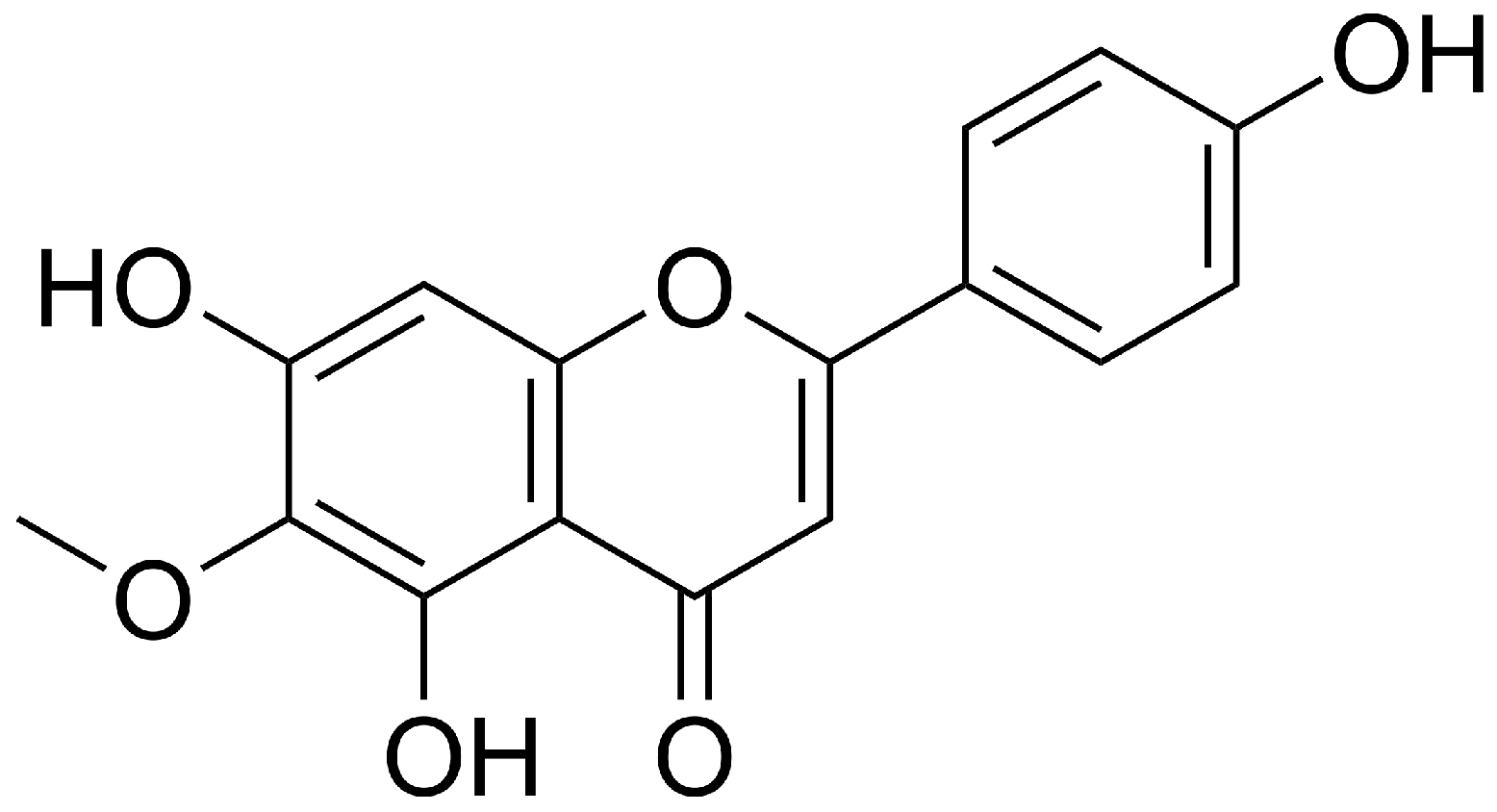
Figure 1. Chemical structure of hispidulin.
Applsci 15 11069 g001
Figure 2. Effects of hispidulin on cell viability, cytotoxicity, ROS generation, and NO production in H2O2-treated C6 cells. Different concentrations of hispidulin were pretreated for 4 h, followed by 300 μM of H2O2 treatment. (A) Cell viability was assessed using the MTT assay. (B) Cytotoxicity was evaluated by measuring LDH release. (C) Intracellular ROS levels were determined using DCF-DA staining. (D) NO production was measured by the Griess reaction. Data are expressed as mean ± SD. Bars with different letters (a, b, c, d and e) indicate statistically significant differences among groups (p < 0.05), whereas bars sharing the same letter are not significantly different, according to Duncan’s multiple range test.
Applsci 15 11069 g002
Figure 3. Effects of hispidulin on pro-inflammatory cytokine production in H2O2-stimulated C6 cells. C6 cells were preincubated with hispidulin for 24 h and subsequently exposed to 300 μM H2O2 for another 24 h. The conditioned media were then collected and assessed for cytokine production. (A) TNF-α, (B) IL-6, and (C) IL-1β were quantified using ELISA kits. Data are shown as mean ± SD. Bars with different letters (a, b, c, and d) indicate statistically significant differences among groups (p < 0.05), whereas bars sharing the same letter are not significantly different, according to Duncan’s multiple range test.
Applsci 15 11069 g003
Figure 4. Effects of hispidulin on antioxidant protein expression in H2O2-treated C6 cells. Western Blot analysis was performed to assess the protein expression levels after pretreatment with hispidulin 24 h followed by 300 μM H2O2 exposure. The protein expression of (A) Nrf2, (B) Gpx1, and (C) SOD was evaluated and adjusted relative to β-actin. Data are shown as fold change compared with the normal group and are expressed as mean ± SD. Bars with different letters (a, b, c, d, and e) indicate statistically significant differences among groups (p < 0.05), whereas bars sharing the same letter are not significantly different, according to Duncan’s multiple range test.
Applsci 15 11069 g004
Figure 5. Effects of hispidulin on apoptosis-related proteins in H2O2-exposed C6 cells. Western Blot analysis was performed to assess the protein expression levels after pretreatment with hispidulin for 24 h, followed by 300 μM H2O2 exposure. The protein levels of (A) Bax, (B) Bcl-2, and (C) cytochrome C were quantified and normalized to β-actin. Data are presented as fold changes relative to the normal group and expressed as mean ± SD. Bars with different letters (a, b, c, d, and e) indicate statistically significant differences among groups (p < 0.05), whereas bars sharing the same letter are not significantly different, according to Duncan’s multiple range test.
Applsci 15 11069 g005
Disclaimer/Publisher’s Note: The statements, opinions and data contained in all publications are solely those of the individual author(s) and contributor(s) and not of MDPI and/or the editor(s). MDPI and/or the editor(s) disclaim responsibility for any injury to people or property resulting from any ideas, methods, instructions or products referred to in the content.