Article

Surface Engineering of Enamel with Sodium Hypochlorite: Effects on Bond Strength and Etching Microstructure in Adhesive Applications

by
Mar Torrella-Girbes
1,
Santiago Arias-Luxán
1,
Clara Guinot-Barona
2,
Laura Marqués-Martínez
2,3,
Esther García-Miralles
3,* and
Juan Ignacio Aura-Tormos
3
1
Dentistry Department, Health Sciences Faculty, Cardenal Herrera CEU University, 46115 Valencia, Spain
2
Dentistry Department, Medicine and Health Science Faculty, Catholic University of Valencia San Vicente Mártir, 46001 Valencia, Spain
3
Stomatology Department, Medicine and Dentistry Faculty, University of Valencia, 46010 Valencia, Spain
*
Author to whom correspondence should be addressed.
Appl. Sci. 2025, 15(20), 10952; https://doi.org/10.3390/app152010952 (registering DOI)
Submission received: 1 September 2025 / Revised: 5 October 2025 / Accepted: 10 October 2025 / Published: 12 October 2025

Abstract

Featured Application

This study demonstrates that a 30-s 5% sodium hypochlorite pretreatment can be used as a simple, cost-effective surface engineering approach to enhance enamel bonding, with applications in orthodontic and restorative dentistry.

Abstract

Background: Adhesion to enamel is influenced by surface preparation, which affects the micromechanical retention of resin-based materials. Sodium hypochlorite (NaOCl) deproteinization has been proposed as a pretreatment to improve acid etching efficacy, but the optimal application time remains unclear. Methods: This in vitro study evaluated the effect of 5% NaOCl pretreatment at three exposure times (15, 30, and 60 s) on shear bond strength (SBS), the adhesive remnant index (ARI), and enamel etching patterns. Extracted human premolars (n = 140) were divided into four groups: the control (acid etching only) and three experimental groups. SBS was tested per ISO 11405, while ARI scores were assessed under stereomicroscopy, and surface morphology was examined by scanning electron microscopy (SEM). Results: The 30-s NaOCl group exhibited the highest SBS (20.9 MPa) compared with the control (15.9 MPa, p < 0.05) and 15-s (14.9 MPa, p < 0.05) groups. SEM analysis showed predominantly Type I–II etching patterns for the 30-s group, irregular Type III for 15 s, and overetched Type IV with loss of prism definition for 60 s, compromising the adhesive interface. ARI scores indicated 86.7% of samples in the 30-s group retained all adhesive on enamel (score 3). Conclusions: A 30-s 5% NaOCl pretreatment before acid etching improved enamel micromorphology and bonding performance compared to shorter or longer exposures. The intermediate duration provided effective deproteinization without structural damage, whereas prolonged exposure degraded the enamel microstructure. This protocol may offer a simple, cost-effective method to enhance clinical adhesive procedures, though prolonged exposure (60 s) should be avoided due to structural degradation of the enamel microstructure.

1. Introduction

The adhesion of restorative and orthodontic biomaterials to dental tissues is a cornerstone of modern dentistry, relying on complex physical and chemical interactions at the substrate interface. The durability of this interface is critically influenced by surface preparation, which determines the quality of micromechanical retention and the long-term stability of adhesive systems [1,2,3]. From a materials science perspective, dental enamel behaves as a highly mineralized bioceramic, and any modification of its surface energy and microtopography directly affects the bonding efficacy of resin-based materials [4,5,6].
Phosphoric acid etching, the gold standard for enamel conditioning, aims to create a microretentive surface by selectively dissolving the mineral phase. However, the presence of a tenacious organic pellicle and the aprismatic nature of the outer enamel layer often result in incomplete, non-uniform etching patterns, compromising bond strength and leading to clinical failures [7,8,9,10]. To overcome these limitations, chemical deproteinization strategies have been proposed to selectively remove the organic component prior to acid etching, thereby optimizing the substrate [11,12].
Sodium hypochlorite (NaOCl), a potent oxidizing agent, has emerged as a promising deproteinizing agent due to its ability to dissolve organic matter and expose the underlying mineralized prism structure, potentially enhancing the penetration and interlocking of adhesive monomers [13,14,15,16,17]. However, the existing body of evidence presents conflicting results. While some studies report significant improvements in bond strength following NaOCl pretreatment [9,10,15], others show negligible or even detrimental effects [18,19]. This inconsistency can be largely attributed to a critical lack of standardization, with significant variations in NaOCl concentration (ranging from 1% to 10%) and, most notably, application time—a parameter whose optimization is fundamental for clinical translation. Some authors have suggested that short application times may be insufficient for effective deproteinization [11,20], whereas prolonged exposure could induce structural alterations in the enamel, degrading its bonding potential [21,22,23,24]. This lack of a consensus protocol underscores a significant gap in the literature.
Building on the heterogeneous literature regarding NaOCl concentration and application time, we posited a time-dependent ‘therapeutic window’ at 5% capable of maximizing deproteinization benefits while avoiding over-etching.
The aim of this study was to evaluate the effect of a 5% sodium hypochlorite pretreatment, applied for different durations, on enamel shear bond strength (SBS), adhesive remnant index (ARI), and etching morphology, to identify the optimal protocol for clinical adhesive procedures.

2. Materials and Methods

A total of 140 extracted human premolars (70 maxillary and 70 mandibular) meeting strict inclusion criteria (caries-free, no restorations, fractures, or fluorosis) were initially selected. From this pool,
-
120 teeth were randomly allocated into four experimental groups of 30 teeth each for shear bond strength (SBS) testing.
-
The remaining 20 teeth (5 per group) were designated exclusively for scanning electron microscopy (SEM) evaluation of enamel morphology.
Following extraction, the teeth were stored in sterile saline solution at 4 °C for a maximum period of one month prior to experimentation, in order to minimize structural alterations and preserve enamel characteristics. This storage duration has been shown to preserve enamel properties without significant alterations [25].
The sample size of 30 specimens per group was determined based on precedents in the literature. Previous in vitro shear bond strength studies typically employed 12 to 30 samples per group—for example, Iglesias et al. used six groups of 12 samples (72 teeth total), and other investigations have reported needing at least 15 samples per group to reach a power > 80% [25,26]. Therefore, we chose 30 specimens per group to ensure adequate statistical power and comparability with existing research.

2.1. Experimental Groups

Group assignment was as follows:
-
Group I (Control): Phosphoric acid 36% etching (n = 30);
-
Group II: 5% NaOCl (15 s) + phosphoric acid etching (n = 30);
-
Group III: 5% NaOCl (30 s) + phosphoric acid etching (n = 30);
-
Group IV: 5% NaOCl (60 s) + phosphoric acid etching (n = 30).
In all experimental groups, 36% phosphoric acid was applied immediately after rinsing off the NaOCl, for 15 s. A schematic diagram of the experimental protocol is presented in Figure 1 to illustrate the study groups, treatments, and evaluation methods.

2.2. Specialized Subgroup

SEM analysis was performed on 5 additional teeth per group (n = 20 total), which underwent identical surface treatments but were not subjected to bracket bonding or mechanical testing.

2.3. Materials Used

  • Etching agent: 36% orthophosphoric acid (Solventum, St. Paul, MN, USA).
  • Deproteinizing agent: 5% sodium hypochlorite (NaOCl), freshly prepared by diluting commercial-grade NaOCl (Panreac, Spain) in distilled water.
  • Adhesive system: Transbond™ XT primer and light-cured composite resin (Solventum, Monrovia, CA, USA).
The use of a single, widely accepted orthodontic adhesive system (Transbond™ XT) was chosen to standardize the bonding protocol, thereby allowing for a clearer interpretation of the isolated effects of NaOCl pretreatment time.
  • Brackets: Stainless steel MBT brackets (Victory Series™, Solventum) with a base area of 10.5 mm2.
  • Light-curing unit: LED curing device (Ortholux™ XT, Solventum).
The 5% NaOCl concentration was selected for this investigation as it represents a clinically available and frequently reported concentration in the dental literature for enamel deproteinization studies [13,15]. While lower concentrations exist, 5% has been shown to provide a potent deproteinizing effect within a clinically feasible application time, allowing for a clear observation of its time-dependent effects. This study aimed to establish a proof of concept for the optimal application time using this common concentration, acknowledging that future research should explore the efficacy and safety profile of lower concentrations. The materials employed in the study are summarized in Table 1.

2.4. Surface Preparation and Bonding Protocol

All bonding procedures were conducted in accordance with ISO/TS 11405:2015 [27].
Teeth were cleaned with pumice and rinsed. In Groups II–IV, 5% NaOCl was applied with a microbrush for the assigned time, rinsed with distilled water (10 s), and air-dried (5 s). All samples were etched with 36% phosphoric acid (15 s), rinsed (5 s), and dried. Subsequently, the Transbond™ XT primer was applied with a microbrush, gently blown with air for 5 s to homogenize the layer and light-cured for 20 s. Brackets were then bonded, excess removed, and adhesive light-cured (20 s mesial/distal).
The etching time of 15 s with 36% phosphoric acid was selected based on established clinical protocols and supported by previous literature [28,29], which recommend this duration to achieve optimal etching without excessive enamel loss.
The rinsing (10 s) and drying (5 s) times following NaOCl application were selected based on common clinical practice and preliminary tests to ensure the complete removal of NaOCl residues, which could otherwise neutralize the acid etchant and interfere with the subsequent adhesion process.

2.5. Storage Before Testing

The 120 SBS-tested samples were stored in distilled water at 37 °C for 24 h before testing, per ISO/TS 11405:2015 guidelines [27].
SBS was measured with a universal testing machine (Autograph AGS-1KND, Shimadzu Corporation, Kyoto, Japan) using a 1 kN load cell. A chisel-shaped blade applied load at 1 mm/min in the occluso-gingival direction, angled at 30°. Debonding force was recorded in Newtons and converted to MPa by dividing by the bracket base area (10.5 mm2).
We evaluated multiple thresholds reported in the literature (9, 11, 13, and 15 MPa). For logistic regression, we predefined <15 MPa as failure to reflect a conservative clinical benchmark.

2.6. Adhesive Remnant Index (ARI)

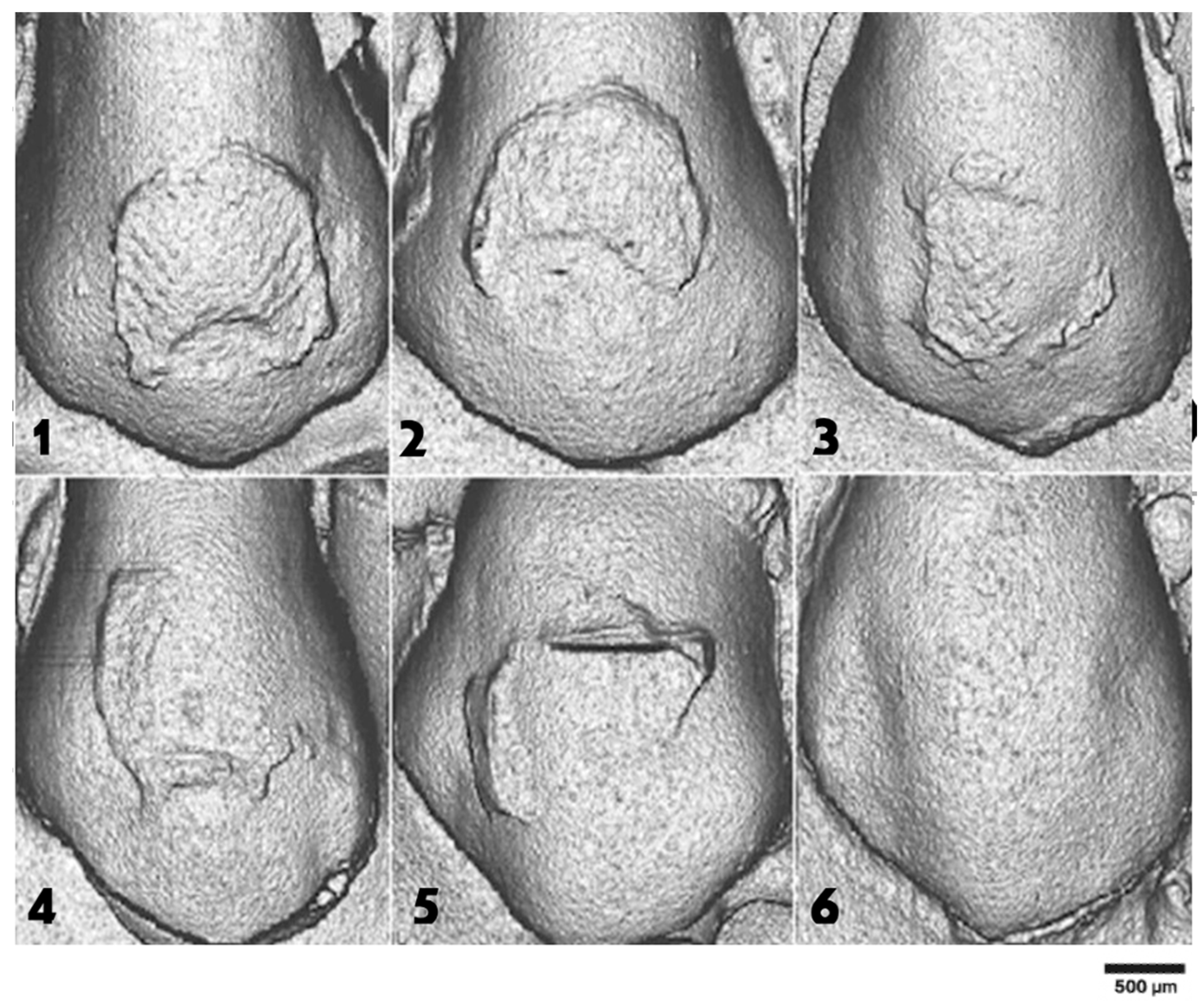
ARI scores were assessed in a randomly selected subset of 15 specimens per group (n = 60 total), drawn from the original 30 samples per group subjected to SBS testing. After debonding, enamel surfaces were examined under a stereomicroscope (Leica M165C, Leica Microsystems, Wetzlar, Germany) 20× magnification with digital imaging. Residual adhesive was scored using the ARI scale:
Score 0: No adhesive (failure at enamel–adhesive interface)
Score 1: <50% adhesive
Score 2: >50% adhesive
Score 3: All adhesive (failure at bracket–adhesive interface)
This subsampling approach followed ISO 11405 recommendations [27] (Figure 2).

2.7. Scanning Electron Microscopy (SEM)

Twenty teeth (five per group) were analysed by SEM to assess enamel surface morphology. Samples received the same pretreatments as in SBS testing, except no brackets were bonded. After gold-palladium coating, specimens were examined under a Hitachi S-4100 scanning electron microscope (Hitachi High-Technologies, Tokyo, Japan).
Etching patterns were classified per Silverstone et al. [30]:
Type I: Prism cores dissolved (honeycomb); optimal retention;
Type II: Prism peripheries dissolved;
Type III: Irregular, unclear prism structure;
Type IV: Minimal or no prism exposure.
Types I and II are ideal for micromechanical adhesion.
Etching patterns were classified according to Silverstone’s criteria [30] by two independent examiners who were calibrated and blinded to the experimental group assignments. The percentage distribution of etching patterns was calculated as the consensus evaluation after independent analysis of five representative areas from each specimen. In cases of disagreement, a third experienced examiner was consulted to reach a final consensus.

2.8. Statistical Analysis

SBS values were recorded in MPa. A threshold of 9–11 MPa was considered clinically relevant for failure. Descriptive statistics (mean, SD, min, max, median, 95% CI) were calculated. A logistic regression model evaluated the odds of SBS < threshold across groups.
Normality was assessed with Kolmogorov–Smirnov, and homogeneity with Levene’s test. Box–Cox transformations were applied as needed. One-way ANOVA with Tukey’s post hoc test was used for pairwise comparisons.
ARI scores (n = 60; 15/group) were analysed using the Kruskal–Wallis test. Pairwise comparisons were done with Mann–Whitney U and Bonferroni correction.

2.9. Intra-Examiner Reliability

To assess reproducibility, the examiner re-evaluated ARI scores 15 days later. Overall agreement was 91.7%, with full concordance in 55 of 60 cases. The weighted Kappa index confirmed high consistency.
Three groups showed very high Kappa values. The 15-s group had slightly lower agreement due to score concentration in “all adhesive remaining” (score 3), but raw agreement remained high at 86.7%, supporting reliability.

2.10. Ethical Considerations

This study was conducted in accordance with the Declaration of Helsinki. The extracted premolars were obtained from patients undergoing orthodontic treatment at the Dental Clinics of the Cardenal Herrera CEU University (Valencia, Spain). Written informed consent was obtained from all patients or their legal guardians for the extraction, use, and transfer of the teeth for research purposes. According to Spanish national regulations on biomedical research (Law 14/2007, BOE-A-2007-12945), and institutional policies, in vitro studies using anonymised extracted teeth obtained for therapeutic purposes do not require formal ethics committee approval.

3. Results

3.1. Shear Bond Strength

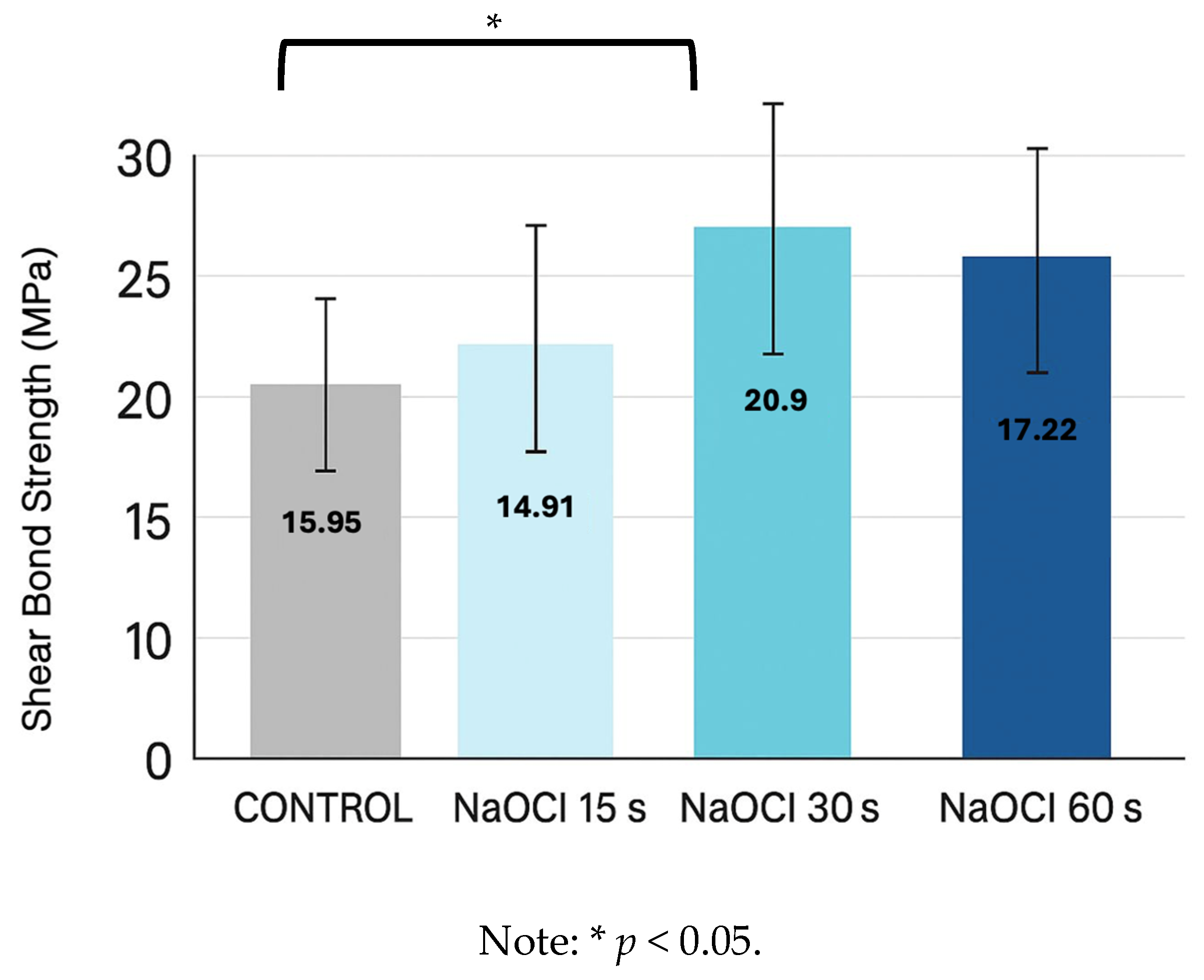
SBS differed significantly among groups. The 30-s NaOCl group showed the highest mean (20.9 MPa, 95% CI [19.9, 21.9]), significantly greater than control (15.95 MPa, 95% CI [14.8, 17.1]) and 15-s group (14.91 MPa, 95% CI [13.7, 16.2]). The 60-s group showed intermediate strength (17.22 MPa, 95% CI [16.1, 18.3]), not significantly different from control.
Logistic regression revealed a 68% lower failure risk (<15 MPa) in the 30-s group (p = 0.038). The 15- and 60-s groups did not show significant reductions.
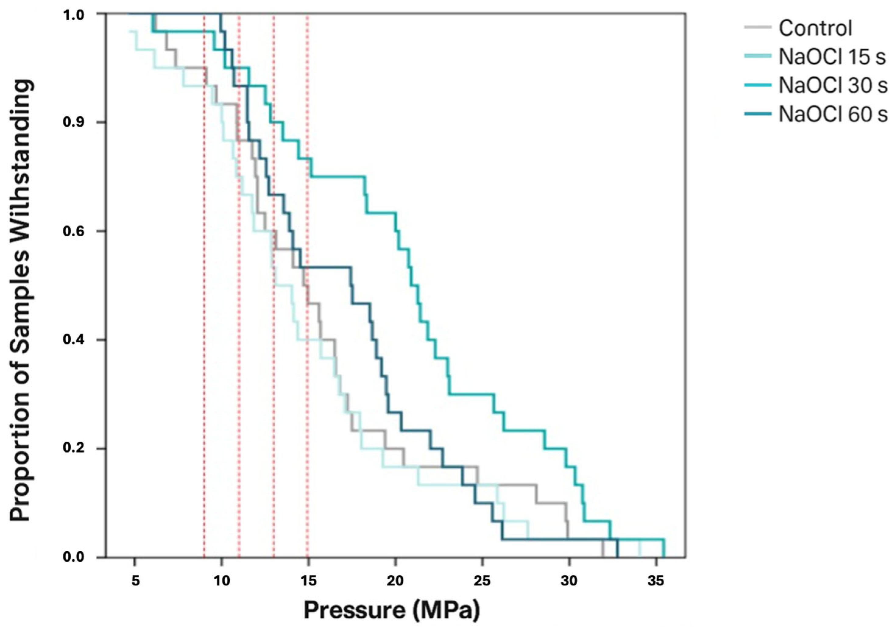
The distribution of samples exceeding different clinical thresholds (9, 11, 13, and 15 MPa) is shown in Figure 3. The control group displayed lower percentages of specimens above the higher thresholds, particularly at 13 and 15 MPa, where less than half of the samples resisted these levels. The 15-s NaOCl group had the weakest performance, with the majority of samples failing to surpass 11 MPa. In contrast, the 30-s NaOCl group demonstrated the most favorable results, with over 80% of specimens surpassing 13 MPa and more than two-thirds exceeding 15 MPa, indicating a significant improvement in bond strength reliability. The 60-s NaOCl group showed intermediate results, with performance superior to the control and 15-s groups but inferior to the 30-s group. These distributions confirm that a 30-s NaOCl pretreatment not only increases mean bond strength but also ensures a greater proportion of clinically acceptable outcomes.
The distribution of samples surpassing various clinical thresholds (9, 11, 13, and 15 MPa) is depicted in Figure 4.

3.2. Adhesive Remnant Index (ARI)

Analysis of ARI scores (assessed in 15 randomly selected specimens per group from SBS-tested samples) revealed significant differences across treatments. In the 30-s NaOCl group, 86.7% of samples exhibited score 3 (all adhesive retained on enamel), compared to 66.7% in the 60-s group and 53.3% in the control. The 15-s group showed the lowest frequency of score 3 (6.7%).
Kruskal–Wallis test confirmed substantial differences between NaOCl-treated groups and control (p < 0.0004), though variations between NaOCl exposure durations were not statistically significant. Pairwise comparisons using Mann–Whitney U test with Bonferroni correction are detailed in Table 2.

3.3. Scanning Electron Microscopy (SEM)

SEM images showed clear differences in etching morphology. SEM analysis revealed that the control group exhibited partial dissolution of prism cores and peripheries, corresponding to mixed Type I–II patterns. The 15 s NaOCl group showed irregular prism outlines and areas of incomplete deproteinization (Type III). The 30 s group displayed homogeneous prism exposure and clear interprismatic regions (Types I–II), while the 60 s group presented overetched areas with loss of prism definition (Type IV) (Figure 5).

3.4. Etching Pattern Distribution

Table 3 summarises etching patterns by group. The 30-s NaOCl group had the highest frequency of Type I and II patterns. The 60-s group showed increased Type IV patterns, less favourable for bonding. NaOCl-treated groups also showed more residual adhesive than control.

4. Discussion

Adhesion to enamel is fundamentally a surface engineering problem, in which the preparation of the substrate determines the quality of the adhesive interface and its resistance to mechanical stresses during clinical function [31]. Conventional acid etching alone frequently results in non-uniform dissolution of the aprismatic layer, leaving a significant proportion of enamel areas insufficiently conditioned [32,33]. Inadequate surface modification leads to weak bonding and a higher incidence of bracket failure [34,35,36].
Sodium hypochlorite (NaOCl) acts as a selective organic solvent capable of removing the proteinaceous pellicle that masks mineral prisms, allowing a deeper and more homogeneous etching pattern [37,38,39,40]. From a materials science perspective, this controlled chemical conditioning changes the surface energy and exposes a microtopography more favourable to micromechanical interlocking with adhesive systems.
The present study provides clear evidence that a 30-s pretreatment with 5% NaOCl prior to acid etching creates the most favourable conditions for bonding, resulting in a significant 31% increase in SBS compared to the control group. This finding is consistent with several previous studies. Justus et al. [9] and Pereira et al. [11] similarly reported enhanced bond strength following NaOCl deproteinization, attributing it to improved resin infiltration. Our results offer a direct microstructural explanation for their findings. The SEM analysis revealed that this specific treatment duration promoted predominantly Type I and II etching patterns, which generate the ideal honeycomb morphology for mechanical retention. This aligns with the work of Ahuja et al. [36], who also observed more defined prism patterns after NaOCl application.
However, the scientific literature on NaOCl pretreatment is not unanimous. Our results help clarify the contradictions by demonstrating that application time is a critical, and previously under-standardized, variable. The inferior performance of the 15-s group (SBS of 14.91 MPa) is directly explained by our SEM findings, which revealed irregular Type III etching patterns. This morphology is indicative of incomplete deproteinization, where residual organic matter impeded uniform acid penetration and compromised the formation of a retentive microtopography.
Conversely, the decline in etching quality and the intermediate SBS value (17.22 MPa) in the 60-s group underscore the potential adverse effects of prolonged exposure. The appearance of overetched areas with disorganised microstructure (Type IV) is consistent with previous reports describing the aggressive nature of NaOCl [41,42,43]. López-Luján et al. [35] also noted that while NaOCl improved bonding, the effect was time-dependent. Our study systematically maps this dependency, identifying 30 s as the optimal balance for a 5% concentration, effectively removing the organic mask without causing excessive demineralization.

Adhesive Remnant Index (ARI)

Although the significant bond strength (20.9 MPa) achieved in the 30-s group represents a notable improvement in adhesive performance, the clinical implications of excessively high SBS values warrant consideration. Elevated bond strengths may potentially increase the risk of enamel fracture or cohesive failure within the enamel during debonding procedures [33]. However, the predominant ARI score of 3 (86.7%) observed in this group indicates that failure occurred preferentially at the bracket-adhesive interface rather than at the enamel-adhesive interface. This failure mode suggests that while the bond to enamel was strengthened, the clinical risk of enamel damage may be mitigated as the adhesive interface remains the weakest point, thus protecting the underlying enamel substrate during mechanical debonding.
In the present study, the 30-s NaOCl group exhibited the highest proportion of ARI score 3, indicating that all adhesive remained on the enamel surface after debonding. From a clinical standpoint, this finding has both advantages and drawbacks. On one hand, adhesive failure at the bracket–adhesive interface preserves the enamel and reduces the risk of iatrogenic damage during bracket removal. On the other hand, the complete retention of adhesive on enamel requires additional chairside time for removal, which can be technically demanding and may increase the risk of enamel scratches or surface roughness if inappropriate finishing protocols are used. Ideally, bonding protocols should achieve high bond strength while leaving minimal residual adhesive, thereby facilitating debonding procedures. Further research should investigate modifications to NaOCl pretreatment or etching parameters that could optimize this balance between mechanical performance and clinical efficiency.
The significant bond strength improvement afforded by the 30-s 5% NaOCl pretreatment must be balanced against its potential clinical risks. Sodium hypochlorite is a potent oxidizing agent with known caustic effects on soft tissues. Therefore, the clinical translation of this protocol mandates stringent safety measures to prevent iatrogenic injury. The use of absolute isolation, preferably with a rubber dam, is considered essential to protect the gingiva and oral mucosa. Furthermore, controlled application using microbrushes directed solely at the enamel surface and followed by thorough rinsing with copious water are non-negotiable steps to mitigate risk. It is also crucial to highlight that the positive findings of this study are strictly confined to enamel substrates. The application of NaOCl on dentin is controversial and generally contraindicated for bonding purposes, as it dissolves the collagen fibrils essential for the formation of the hybrid layer in etch-and-rinse adhesives. Therefore, the proposed pretreatment should not be extrapolated to restorative procedures involving dentin surfaces. While this study demonstrates a clear laboratory benefit, its clinical adoption should be preceded by in-situ or clinical trials that confirm its efficacy and safety in the dynamic oral environment.
Furthermore, while not assessed in this study, other potential adverse effects such as enamel discoloration or the impact of NaOCl on hypomineralized or fluorotic enamel warrant investigation in future research to fully establish the safety profile of this pretreatment.
Nevertheless, the study has limitations. As an in vitro investigation, it does not reproduce intraoral variables such as saliva, occlusal loading, and long-term ageing. Future studies should include artificial ageing methods, extended follow-up periods, and different adhesive systems to validate the clinical effectiveness of this approach [43]. Another limitation of the present study is that the teeth were stored for up to one month prior to experimentation. This protocol was chosen to minimize structural alterations and preserve enamel characteristics; however, it cannot completely rule out the possibility of morphological or chemical changes in dental tissues over time. Such alterations may influence the etching pattern and, consequently, bonding performance. Recent evidence suggests that storage media and duration can affect enamel and dentin differently, with prolonged storage—especially in certain disinfectant solutions—associated with variations in bond strength and microleakage outcomes [39].
In addition, the absence of a control group using freshly extracted teeth represents a methodological limitation. Including such a group would provide valuable insight into whether immediate bonding differs from delayed bonding after storage. Future investigations incorporating this comparison are recommended to better establish the impact of storage duration on adhesive performance and to strengthen the external validity of in vitro findings.
A limitation of this study is the qualitative nature of the SEM analysis. While SEM provided valuable qualitative insights into etching patterns, quantitative surface topography data, such as surface roughness parameters (Ra, Rq) or the percentage of prism area exposed through image analysis software, were not obtained. The classification of etching patterns, while performed by blinded examiners, remains inherently subjective. Future studies should incorporate quantitative surface metrology techniques such as atomic force microscopy (AFM) or optical profilometry to provide objective measurements of the topographical changes induced by NaOCl pretreatment.
Furthermore, the findings are specific to the adhesive system employed (Transbond™ XT). Future research is warranted to investigate the interaction of NaOCl pretreatment with the chemistry of different adhesive systems, such as self-etch or universal adhesives.

5. Conclusions

This in vitro study demonstrates that a 30-s pretreatment with 5% sodium hypochlorite prior to acid etching represents an optimal protocol for enamel surface engineering under controlled conditions. This specific duration significantly enhanced shear bond strength and promoted the formation of Type I, II etching patterns, indicative of superior micromechanical retention, by achieving an ideal balance between effective deproteinization and structural preservation. Therefore, incorporating a 30-s 5% NaOCl pretreatment shows great potential as a simple and cost-effective strategy to improve the reliability of adhesive procedures in orthodontics and restorative dentistry. However, these promising in vitro findings require validation through clinical trials to confirm their efficacy, safety, and durability in the dynamic oral environment before routine clinical application can be recommended.

Author Contributions

Conceptualization, E.G.-M., M.T.-G. and J.I.A.-T.; methodology, E.G.-M.; validation, E.G.-M., C.G.-B. and L.M.-M.; formal analysis, E.G.-M.; investigation, M.T.-G., S.A.-L. and E.G.-M.; resources, M.T.-G., S.A.-L. and E.G.-M.; data curation, M.T.-G. and E.G.-M.; writing—original draft preparation, E.G.-M.; writing—review and editing, E.G.-M., L.M.-M., M.T.-G., S.A.-L. and C.G.-B.; visualization, E.G.-M.; supervision, J.I.A.-T.; project administration, J.I.A.-T. All authors have read and agreed to the published version of the manuscript.

Funding

This research received no external funding.

Institutional Review Board Statement

The study was conducted in accordance with the Declaration of Helsinki. All teeth used were extracted for orthodontic purposes as part of routine dental treatment at the Dental Clinics of the Cardenal Herrera CEU University (Valencia, Spain). In accordance with institutional policy and Spanish national regulations on biomedical research (Law 14/2007 on Biomedical Research, BOE-A-2007-12945), in vitro studies with anonymized extracted teeth obtained for therapeutic purposes do not require formal approval from an ethics committee. Therefore, Institutional Ethics Committee (IEC) approval was not required for this research.

Informed Consent Statement

Written informed consent for extraction, use, and transfer of the teeth for research purposes was obtained from all patients or their legal guardians prior to the procedures.

Data Availability Statement

The original contributions presented in the study are included in the article.

Acknowledgments

The authors express their gratitude to Eliseo Plasencia (†), whose teachings and dedication to orthodontic research continue to inspire this work. The authors thank the Catholic University of Valencia San Vicente Martir for their contribution and help in paying for the open access publication fee.

Conflicts of Interest

The authors declare no conflicts of interest.

References

  1. Soares, F.Z.; Rocha, R.O.; Raggio, D.P.; Sadek, F.T.; Cardoso, P.E. Microtensile bond strength of different adhesive systems to primary and permanent dentin. Pediatr. Dent. 2005, 27, 457–462. [Google Scholar]
  2. Armstrong, S.; Geraldeli, S.; Maia, R.; Raposo, L.H.; Soares, C.J.; Yamagawa, J. Adhesion to tooth structure: A critical review of “micro” bond strength test methods. Dent. Mater. 2010, 26, e50–e62. [Google Scholar] [CrossRef]
  3. Dos Santos, P.A.; Garcia, P.P.; Palma-Dibb, R.G. Shear bond strength of adhesive systems to enamel and dentin. J. Mater. Sci. Mater. Med. 2005, 16, 727–732. [Google Scholar] [CrossRef] [PubMed]
  4. Scougall-Vilchis, R.J.; Zárate-Díaz, C.; Kusakabe, S.; Yamamoto, K. Bond strengths of different orthodontic adhesives after enamel conditioning with the same self-etching primer. Aust. Orthod. J. 2010, 26, 84–89. [Google Scholar] [CrossRef] [PubMed]
  5. Gwinnett, A.J. The effect of various surface treatments on the enamel of human teeth. J. Dent. Res. 1967, 46, 1401–1404. [Google Scholar]
  6. Espinosa, R.; Valencia, R.; Uribe, M.; Ceja, I.; Cruz, J.; Saadia, M. Resin replica in enamel deproteinization and its effect on acid etching. J. Clin. Pediatr. Dent. 2010, 35, 47–51. [Google Scholar] [CrossRef]
  7. Pithan, S.A.; Hallgren, A.; Cordeiro, R.; Pithan, J.C.; Carlsson, J.; Pithan, E.G.T. The effect of sodium hypochlorite on the bond strength of an adhesive system to dental enamel. Pesqui. Odontol. Bras. 2003, 17, 349–353. [Google Scholar]
  8. Estrela, C.; Estrela, C.R.; Barbin, E.L.; Spanó, J.C.; Marchesan, M.A.; Pécora, J.D. Mechanism of action of sodium hypochlorite. Braz. Dent. J. 2002, 13, 113–117. [Google Scholar] [CrossRef]
  9. Justus, R.; Cubero, T.; Ondarza, R.; Morales, F. A new technique with sodium hypochlorite to increase bracket shear bond strength. Semin. Orthod. 2010, 16, 66–75. [Google Scholar] [CrossRef]
  10. Kiryk, S.; Kiryk, J.; Matys, J.; Dobrzyński, M. The Influence of Resin Infiltration on the Shear Bond Strength of Orthodontic Brackets: A Systematic Review and Meta-Analysis. Biomater. Investig. Dent. 2025, 16, 32. [Google Scholar] [CrossRef]
  11. Pereira, T.B.; Jansen, W.C.; Pithon, M.M.; Souki, B.Q.; Tanaka, O.M.; Oliveira, D.D. Effects of enamel deproteinization on bracket bonding. Eur. J. Orthod. 2013, 35, 442–446. [Google Scholar] [CrossRef]
  12. Elnafar, A.A.; Alam, M.K.; Hasan, R. The impact of surface preparation on shear bond strength. J. Orthod. 2014, 41, 201–207. [Google Scholar] [CrossRef]
  13. Peloso, R.M.; Cotrin, P.; Oliveira, R.C.G.; Valarelli, F.P.; Freitas, K.M.S. Evaluation of enamel deproteinization in bond strength. Am. J. Orthod. Dentofac. Orthop. 2022, 162, 443–450. [Google Scholar] [CrossRef]
  14. Sun, Z.; You, X.; Xu, J.; Chen, L.; Li, S.; Zhang, Z.; Guo, L. Effectiveness of sodium hypochlorite treatment. Dent. Mater. J. 2022, 41, 660–667. [Google Scholar] [CrossRef]
  15. Alrai, S.; Al-Madi, A.; Al-Khalifa, K.; Al-Ghamdi, H.; Bukhary, S.; Bawazir, S.; Alqahtani, A.; Alharbi, A.; Alotaibi, R.; Alghamdi, F. Effect of Enamel Deproteinization on Bonding Orthodontic Brackets: A systematic review and meta-analysis. J. Clin. Exp. Dent. 2025, 17, e122–e129. [Google Scholar] [CrossRef]
  16. Ni, Y.; Zhang, Y.; Wang, H.; Li, X.; Huang, Y.; Chen, L. Effect of Sodium Hypochlorite and EDTA Pretreatment on Enamel Surface for Resin Infiltration. Front. Dent. Med. 2024, 5, 11600858. [Google Scholar]
  17. Basalamah, A.; Maher, A.; Whba, A.H.; Scribante, A.; Sfondrini, M.F.; Montasser, M.A. Effects of fluorosed enamel on orthodontic bracket bonding. J. Orofac. Orthop. 2023, 84, 88–99. [Google Scholar] [CrossRef] [PubMed]
  18. Daou, C.; Akl, R.; Mati, M.; Kassis, A.; Ghoubril, J.; Khoury, E. Effects of enamel deproteinization with different application times on the shear bond strength of a self-etching primer: An in vitro study. Int. Orthod. 2021, 19, 505–511. [Google Scholar] [CrossRef]
  19. Alharbi, A.; Smith, J.; Johnson, L. Effect of sodium hypochlorite deproteinization on bond strength in fluorotic enamel. Angle Orthod. 2023, 93, 421–428. [Google Scholar]
  20. Cordeiro, P.; Neves, J.G.; Tsuzuki, M.; Facury, A.G.F.; Correr-Sobrinho, L.; Costa, A.R. Enamel deproteinization with sodium hypochlorite: Effects on ceramic bracket bond strength. Braz. Dent. J. 2025, 36, e246106. [Google Scholar] [CrossRef] [PubMed]
  21. De Lima, R.B.; Souza, R.A.; Mendes, J.F.; Araújo, M.V.; Figueiredo, D.S.; Pithon, M.M.; Oliveira, D.D. Clinical evaluation of sodium hypochlorite pretreatment on bracket failure. Am. J. Orthod. Dentofac. Orthop. 2023, 165, 45–52. [Google Scholar]
  22. Poggio, C.; Beltrami, R.; Colombo, M.; Chiesa, M.; Scribante, A. Influence of dentin pretreatment on bond strength. Acta Biomater. Odontol. Scand. 2017, 3, 30–35. [Google Scholar] [CrossRef] [PubMed]
  23. Siqueira, F.S.F.; Armas-Vega, A.; Izquierdo-Bucheli, A.; Pinto, T.F.; Hanzen, T.A.; Bauer, J.; Cardenas, A.F.M.; Loguercio, A.D. Does the Conditioning Mode and Duration of Universal Adhesives Affect the Bonding Effectiveness to Fluorotic Enamel? J. Adhes. Dent. 2019, 21, 525–536. [Google Scholar] [PubMed]
  24. Mas-López, A.C.; Robles-Ruiz, J.; Arriola, L.E. Effect of sodium hypochlorite as a deproteinizing agent on the bond strength of metal brackets. J. Clin. Pediatr. Dent. 2024, 16, e947–e952. [Google Scholar] [CrossRef]
  25. Eliades, T.; Hiskia, A.; Eliades, G.; Athanasiou, A.E. Surface characterization of retrieved orthodontic brackets. Eur. J. Orthod. 2007, 29, 350–357. [Google Scholar]
  26. Nilgun, A.; Usumez, A.; Ozturk, B.; Usumez, S. Influence of different light sources on microleakage. J. Oral Rehabil. 2004, 31, 500–504. [Google Scholar] [CrossRef]
  27. ISO/TS 11405:2015; Dental Materials-Testing of Adhesion to Tooth Structure. International Organization for Standardization: Geneva, Switzerland, 2015.
  28. Van Meerbeek, B.; De Munck, J.; Yoshida, Y.; Inoue, S.; Vargas, M.; Vijay, P.; Van Landuyt, K.; Lambrechts, P.; Vanherle, G. Adhesion to enamel and dentin: Current status and future challenges. Oper. Dent. 2003, 28, 215–235. [Google Scholar]
  29. Pashley, D.H.; Tay, F.R.; Breschi, L.; Tjäderhane, L.; Carvalho, R.M.; Carrilho, M.; Tezvergil-Mutluay, A. State of the art etch-and-rinse adhesives. Dent. Mater. 2011, 27, 1–16. [Google Scholar] [CrossRef]
  30. Silverstone, L.M.; Saxton, C.A.; Dogon, I.L.; Fejerskov, O. Variation in the pattern of acid etching of enamel surfaces: An electron microscopic study. Br. Dent J. 1975, 138, 69–76. [Google Scholar]
  31. Iglesias, A.; Flores, T.; Moyano, J.; Artés, M.; Gil, F.J.; Puigdollers, A. In vitro study of shear bond strength in direct and indirect bonding with three types of adhesive systems. Materials 2020, 13, 2644. [Google Scholar] [CrossRef]
  32. Blöcher, S.; Frankenberger, R.; Hellak, A.; Schauseil, M.; Roggendorf, M.J.; Korbmacher-Steiner, H.M. Effect on enamel shear bond strength of adding microsilver and nanosilver particles to the primer of an orthodontic adhesive. BMC Oral Health 2015, 15, 42. [Google Scholar] [CrossRef]
  33. Da Rocha, J.M.; Gravina, M.A.; da Silva Campos, M.J.; Quintão, C.C.; Elias, C.N.; Vitral, R.W. Shear bond resistance and enamel surface comparison after the bonding and debonding of ceramic and metallic brackets. Dent. Press J. Orthod. 2014, 19, 77–85. [Google Scholar] [CrossRef]
  34. Ayman, M.; Bakry, N.; El-Kest, A.S. Effects of sodium hypochlorite enamel deproteinization on bonding efficacy. Dent. Med. Probl. 2022, 59, 223–229. [Google Scholar]
  35. López-Luján, N.A.; Munayco-Pantoja, E.R.; Torres-Ramos, G.; Blanco-Victorio, D.J.; Siccha-Macassi, A.; López-Ramos, R.P. Deproteinization of primary enamel with sodium hypochlorite before etching. Acta Odontol. Latinoam. 2019, 32, 29–35. [Google Scholar]
  36. Ahuja, B.; Yeluri, R.; Baliga, S.; Munshi, A.K. Enamel deproteinization before acid etching: A scanning electron microscopic observation. J. Clin. Pediatr. Dent. 2010, 35, 169–172. [Google Scholar] [CrossRef] [PubMed]
  37. Söderholm, K.J.; Roberts, M.J. Variables influencing the bond strength of dentin adhesives. J. Dent. Res. 1990, 69, 844–851. [Google Scholar]
  38. Al-Daher, M.S.; Sultan, K.; Hajeer, M.Y.; Burhan, A.S. Enamel deproteinization or sandblasting for enamel reconditioning. Cureus 2024, 16, e66210. [Google Scholar] [CrossRef]
  39. Perdigão, J.; Gomes, G.; Duarte, S., Jr.; Lopes, M.M. Enamel bond strengths of pairs of adhesives from the same manufacturer. Oper. Dent. 2005, 30, 492–499. [Google Scholar] [PubMed]
  40. Cardoso, M.V.; De Almeida Neves, A.; Mine, A.; Coutinho, E.; Van Landuyt, K.; De Munck, J.; Van Meerbeek, B. Current aspects on bonding effectiveness and stability in adhesive dentistry. Aust. Dent. J. 2011, 56 (Suppl. S1), 31–44. [Google Scholar] [CrossRef]
  41. Wang, Y.; Spencer, P. Hybridization efficiency of the adhesive/dentin interface. J. Dent. Res. 2003, 82, 141–145. [Google Scholar] [CrossRef]
  42. Billoo, M.S.; Khan, M.A.K.; Khalid, T.; Kazmi, S.M.R. Effect of storage medium of extracted human teeth on the result of in vitro studies—A scoping review. Biomater Investig Dent. 2025, 12, 44183. [Google Scholar] [CrossRef] [PubMed] [PubMed Central]
  43. Mohammadi, A.; Nili, M.; Yazdi, F.M.; Taghavi, R. The Effect of Sodium Hypochlorite Enamel Pretreatment on Improving the Bond Strength of Resin-Modified Glass Ionomer Cement. Dent. Res. J. 2021, 18, 13. [Google Scholar] [CrossRef]
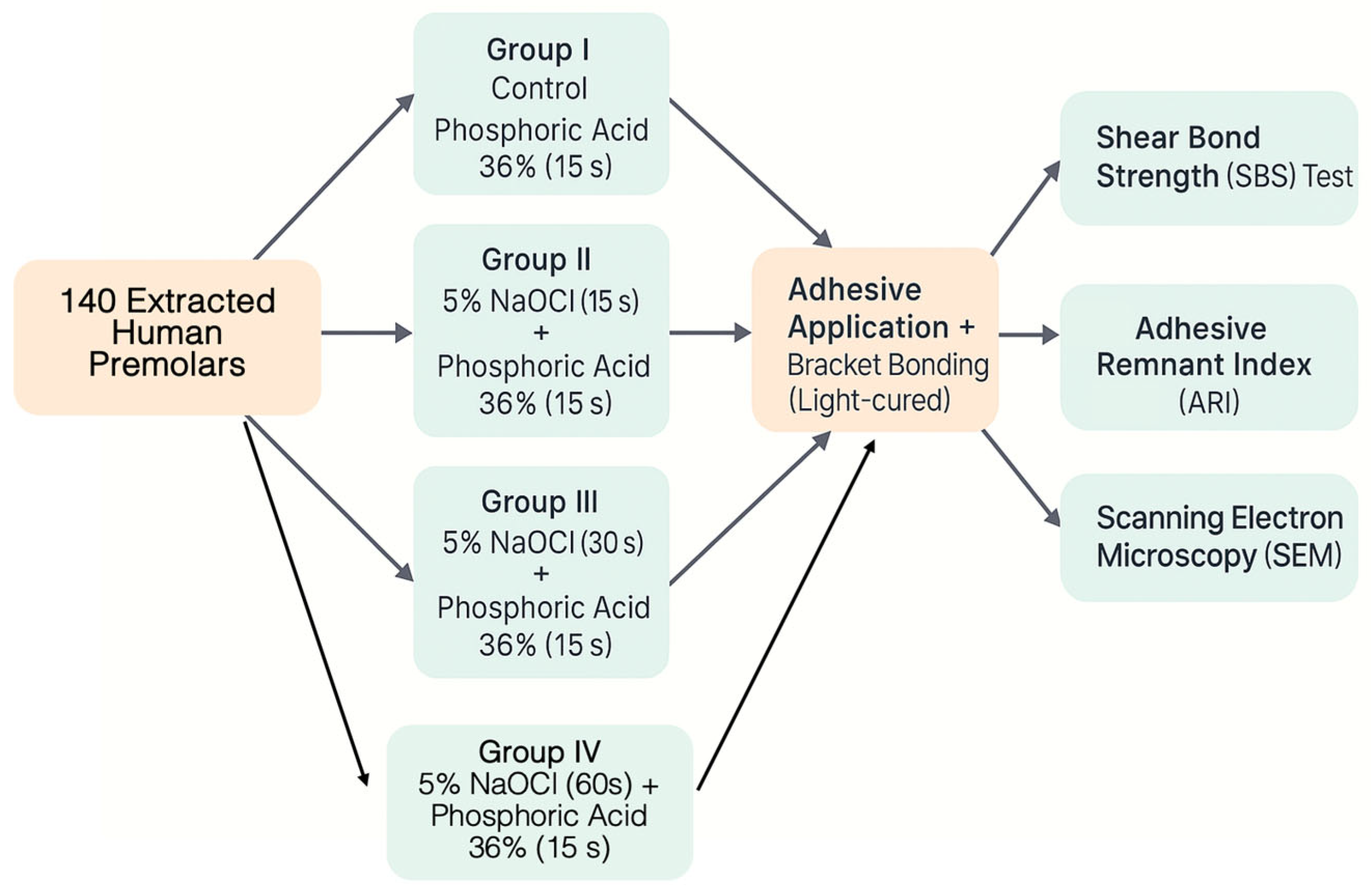
Figure 1. Schematic representation of the experimental protocol. Extracted human premolars were divided into four groups: control (phosphoric acid only) and experimental groups pretreated with 5% sodium hypochlorite for 15, 30, or 60 s, followed by phosphoric acid etching. All samples underwent adhesive application, bracket bonding, and subsequent evaluation by shear bond strength (SBS), adhesive remnant index (ARI), and scanning electron microscopy (SEM).
Applsci 15 10952 g001
Figure 2. Representative stereomicroscopic images illustrating ARI scoring system: (1) Score 3; (2) Score 2; (35) Score 1; (6) Score 0. Images obtained at 20× magnification. Scale bars = 500 µm.
Applsci 15 10952 g002
Figure 3. Shear bond strength at debonding (MPa) for all groups (mean ± SD). Different letters indicate significant between-group differences (one-way ANOVA with Tukey’s post hoc, α = 0.05).
Applsci 15 10952 g003
Figure 4. Proportion of specimens meeting SBS thresholds (≥9, ≥11, ≥13, ≥15 MPa) by group.
Applsci 15 10952 g004
Figure 5. Representative SEM micrographs of etched enamel per group at 3000×, 7000× and 15,000× (scale bars shown). Control: mixed Type I–II; 15 s NaOCl: irregular Type III; 30 s NaOCl: predominantly Type I–II; 60 s NaOCl: Type IV with loss of prism definition. Scale bars = 10 µm, 5 µm, and 2 µm.
Applsci 15 10952 g005
Table 1. Materials used in the study.
MaterialCommercial NameManufacturer (City, Country)Purpose in Study
Phosphoric acid 36%Scotchbond™Solventum, St. Paul, MN, USAEtching agent for enamel conditioning
Sodium hypochlorite 5%Prepared from commercial-grade NaOClPanreac, Barcelona, SpainDeproteinising agent prior to etching
Adhesive primerTransbond™ XT PrimerSolventum, Monrovia, CA, USAPrimer for enhancing resin bonding to enamel
Light-cured composite resinTransbond™ XT CompositeSolventum, Monrovia, CA, USAResin material for bracket bonding
Stainless steel MBT bracketsVictory Series™Solventum, Monrovia, CA, USAOrthodontic brackets for SBS testing
LED curing unitOrtholux™ XTSolventum, Monrovia, CA, USALight source for curing adhesive materials
Note: All materials were used according to the manufacturers’ instructions.
Table 2. Pairwise comparisons of ARI scores between experimental groups (Mann–Whitney U test with Bonferroni correction).
ControlNaOCl 15 sNaOCl 30 sNaOCl 60 s
Control
NaOCl 15 s0.00001 ***
NaOCl 30 s0.0003 **0.137
NaOCl 60 s0.00001 ***0.4120.412
Note: *** p < 0.001; ** p < 0.01.
Table 3. Frequency of etching pattern types observed via SEM in each group.
Etching Pattern TypeControl Group (H3PO4)NaOCl 15 sNaOCl 30 sNaOCl 60 s
Type I25%25%75%
Type II25%50%20%25%
Type III25%25%5%75%
Type IV25%
Disclaimer/Publisher’s Note: The statements, opinions and data contained in all publications are solely those of the individual author(s) and contributor(s) and not of MDPI and/or the editor(s). MDPI and/or the editor(s) disclaim responsibility for any injury to people or property resulting from any ideas, methods, instructions or products referred to in the content.