Article

Oxyanion Recovery from Wastewater with Special Reference to Selenium Uptake by Marine Macroalgae

1
“Cristofor Simionescu” Faculty of Chemical Engineering and Environmental Protection, “Gheorghe Asachi” Technical University of Iasi, Bd D. Mangeron 73, 700050 Iași, Romania
2
Faculty of Hydrotechnics, Geodesy & Environmental Engineering, “Gheorghe Asachi” Technical University of Iasi, Bd. D. Mangeron 65, 700050 Iași, Romania
*
Author to whom correspondence should be addressed.
Appl. Sci. 2025, 15(20), 10946; https://doi.org/10.3390/app152010946 (registering DOI)
Submission received: 25 July 2025 / Revised: 15 September 2025 / Accepted: 3 October 2025 / Published: 12 October 2025
(This article belongs to the Special Issue Water Pollution and Wastewater Treatment Chemistry)

Abstract

This study investigates the capacity of green and brown algae to sustainably remove oxyanions from contaminated waters, highlighting their cost-effectiveness. Often considered biomass waste and contributors to organic contamination, these algae can be used as effective biosorbents, aligning with circular economy principles and sustainable waste management. Various pre-treatments were tested to enhance adsorption capacity, with mixed results regarding their effectiveness. The focus then shifted to the use of Cladophora sericea algae for the uptake and removal of selenium species, specifically selenite (Se(IV)) and selenate (Se(VI)). The effects of different operational parameters on oxyanion uptake by algae were studied in batch mode. The assessments were conducted on a single-component and a multi-component synthetic matrix. The results indicate that pH significantly impacts biosorption, with equilibrium achieved in 90 min. Both pseudo-first-order and pseudo-second-order models provided a good fit to the experimental data. The algae’s retention capacity for selenium remained largely unaffected by the presence of other anions, a key advantage for application in complex real effluent matrices. Kinetic studies performed under different values of initial pollutant concentration and biosorbent mass indicate a biosorbed amount at an equilibrium of 570 µg g−1.

1. Introduction

Water is one of the most critical resources for humanity since it is essential for sustaining life. However, rapid industrial and urban development has significantly altered the quality of water, both underground and terrestrial. Consequently, regulations establishing maximum contaminant thresholds, based on their toxicity, have been imposed. Finding solutions to remove these contaminants and restore aqueous effluents to safe quality conditions is crucial.
Among the highly toxic contaminants, arsenic, selenium, and antimony have significant carcinogenic potential, being identified as such by the International Agency for Research on Cancer [1]. These oxyanions can be found in aqueous environments from natural or anthropogenic sources such as charcoal combustion, mining, agriculture, oil refining, insecticide production and use, glass manufacturing, and photocells [2,3,4,5]. In body waters, arsenic concentrations have various values and can reach up to 40,000 μg L−1 [6,7,8,9], while the guideline value for arsenic in drinking water is 10 μg L−1 [10]. Antimony is present in polyethylene terephthalate (PET), plastics, textiles, and rubber [11]—materials that, in one way or another, come into direct contact with humans, which makes them particularly dangerous. The highest concentrations of antimony (Sb) are found in rivers in the neighborhood of mining areas, such as the Xikuangshan area of Hunan, China, where the antimony level was found to be 6384 μg L−1 [12]. USEPA has marked 6 μg L−1 as the maximum contaminant level for antimony, while the EU’s maximum admissible concentration for drinking water is 5 μg L−1 [13].
Mining activity has turned selenium into an emerging global contaminant, releasing significant amounts of this element into the environment [14,15]. Usually, in mining wastewaters, the selenium concentration ranges from 3 µg L−1 to 12 µg L−1 [16], while the level of selenium pollution in wastewater from uranium mines can reach up to 1600 µg L−1 [17]. Crude oil can have a selenium content of between 500 and 2200 µg L−1, while the selenium concentration in oil refinery wastewater is around 15–75 µg L−1 [18]. Seawaters have a higher selenium concentration of almost 0.08 µg L−1 [19] compared to 0.02 µg L−1 in fresh waters. Thus, the oceans, through seafood, play an important role in human exposure to selenium. Industrial wastewater effluents from insecticide production, glass manufacturing, and oil refining have been shown to have concentrations as high as 0.1 and 20 mg L−1 [20].
The boundary between the level at which selenium has positive biological effects for humans and animals, and the level when it becomes toxic, is a very narrow one [21,22]. Selenium is a critical micronutrient as well as an important trace element with antioxidant properties: it contributes to the regulation of thyroid function and the immune system, its intake helping in issues related to reproduction, cardiovascular diseases, and mood disorders [23,24,25]. Several studies report that selenium reduces the risk of cancer [23,24]. Moreover, due to its mutual antagonism with mercury, selenium can also be used for detoxification [26,27]. Selenium deficiency is connected to certain diseases, and in this case, a selenium supplement is required [28,29]. However, selenium can become harmful at levels slightly above homeostatic requirements [30,31,32] and the effects of its toxicity are extremely severe with implications in carcinogenesis (prostate, liver), cytotoxicity (stopping the cell cycle and inhibition of cell growth), and genotoxicity effects (affecting DNA) [33]. The concentration of selenium is only one determinant for its toxicity; its oxidation state also has a crucial importance in its toxicity, considering that the chemical properties change with the oxidation state. The inorganic selenium, from selenide and selenate, present in wastewater usually has up to 40 times higher toxicity compared with the organic forms [34,35]. For its part, Se(IV) specie is more toxic than Se(VI) ones [36,37,38]. Beneficial concentrations for the proper functioning of the human body lie between 63 and 135 μg L−1 [39] while the tolerable upper intake level for adults is 400 μg per day [40]. The symptoms of selenium poisoning begin to appear upon selenium concentration intake above 800 μg per day [41]. The legal limit of selenium concentration in drinking water in the European Union (EU) regulation is 10 μg L−1, while American legislation is more permissive, setting the legal limit at 50 μg L−1 [42].
The options to remove arsenic, selenium, and antimony from aqueous effluents are through physical, chemical, and biological methods. Adsorption is an efficient, inexpensive, and simple to operate method of water decontamination, successfully removing low concentrations of pollutants. The process results in a reduced amount of sludge, and most of the time the adsorbent can be successfully regenerated and reused in several adsorption–desorption cycles [43]. Biosorption (adsorption using non-living biomass) is a promising technology for the removal of heavy metals from aqueous effluents. Macroalgae represent a renewable resource, available in sea and fresh water, widely distributed worldwide, reusable, and able to uptake high metal pollutants amounts [44]. The functional groups present on the surface of the algae (amino, hydroxyl, carboxyl, and sulfate), due to polysaccharides, proteins, or lipids in the algal structure, explain the high metal binding capacity of seaweeds [44]. The removal of arsenic and antimony through biosorption using seaweed is well-documented in the literature, with detailed accounts of suitable biosorbents, optimal reaction conditions, and specific studies highlighting the use of green and brown algae for oxyanion removal [45,46,47].
Selenium biosorption by algae is a topic quite scarce in the literature. There are a few studies reporting data related to the oxyanions, especially selenium [48], adsorption capacity of algae, kinetic analysis, possible pre-treatments applied to seaweeds in order to improve their retention capacity, as well as the identification of the most suitable types of algae (green, red, or brown) [49].
This study presents extensive research on the application of seaweed for the removal of oxyanions from aqueous environments and aims to: (i) point out various pre-treatments applied to algae to enhance their biosorption capacity; (ii) provide new insights into the adsorption of selenide using the green macroalga Cladophora sericea; and (iii) highlight the influence of competing anions in a complex aqueous matrix. Additionally, the study offers a systematic characterization of the macroalgae used as biosorbent, establishes the optimal conditions for the adsorption process, and describes a possible mechanism for selenium sequestration, correlating it with the structural features of the biosorbent.

2. Materials and Methods

2.1. Chemical Substances and Analytical Methods

Adsorbate solutions were prepared by diluting commercial analytical standards. Arsenic solutions were prepared by diluting an appropriate volume of AAS standard solution (1000 ± 3 mg L−1 As(III)) in 4% HNO3 (SCP Science—Baie-d’Urfé, QC, Canada) and As(V) from HAsNa2O4·7H2O salt in 2% HCl (Sigma Aldrich—Schnelldorf, Germany). Antimony solutions were prepared as follows: Sb(III) from a KSbOC4H4O6 solution (1000 ± 2 mg L−1 Sb(III)) in 2–5% HCl (Carlo Erba—Milano, Italy), and Sb(V) from 1000 ± 2 mg L−1 Sb(V) in 2–5% HNO3 (Chem Lab—Zedelgem, Belgium). The biosorption of selenium was examined in two oxidation states, Se4+ and Se6+. The Se(IV) solution was prepared by diluting a 1000 mg L−1 AAS standard solution (Carlo Erba—Milano, Italy), while the Se(VI) solution was obtained by dissolving sodium selenate (Na2SeO4, analytical grade, Acros Organics—Geel, Belgium). Adjustments to pH were made as needed using diluted solutions of NaOH, HCl, and HNO3 prepared from analytical grade reagents, including NaOH pellets (purity >99.0% from Merck—Darmstadt, Germany, concentrated HNO3 (65%, Merck—Darmstadt, Germany), and concentrated HCl (37%). Additional solutions, such as FeCl3 (≥98%, Merck), HDTMA (hexadecyltrimethylammonium, a cationic surfactant) (Sigma-Aldrich—Schnelldorf, Germany), and ammonia (NH3, 25%, Merck—Darmstadt, Germany), were prepared by diluting commercial analytical grade products. Ultrapure water was used as the solvent throughout.
The uptake of As, Sb, and Se was quantified using flame atomic absorption spectroscopy (AAS, GBC 932 plus—Keysborough, Australia) with deuterium background correction and triplicate readings. Dilutions were performed as necessary to maintain concentrations within the linear range of detection. Calibration was performed daily, with acceptable linearity confirmed for correlation coefficients (R2 > 0.995). Prior to AAS analysis, samples were filtered through cellulose acetate filters with a 0.45 µm pore size. All plastic and glassware used in the experiments were pre-soaked for 24 h in a 20% HNO3 solution and then rinsed with distilled water.

2.2. Preparation of Biomass

Two brown algae, Sargassum muticum and Ascophyllum nodosum, were harvested from Viana do Castelo beach in Portugal, while two green seaweeds, Ulva rigida and Cladophora sericea, were gathered from the Romanian coast of the Black Sea, specifically from the Costinesti and Mangalia seaside locations. These algae were selected due to their abundance in natural environments. In Portugal, algae have various applications, such as natural fertilizer, food additives, and in the medical and cosmetic industries [50]. In Romania, these seaweeds are largely harvested without designated applications, except for minimal utilization in cosmetic industries [51]. The seaweeds were initially washed with tap water to eliminate sand and visible particles, followed by rinsing with distilled water until the conductivity of the rinse water matched that of distilled water. Conductivity was monitored using a calibrated HI 8733 conductivity meter. According to the literature, drying temperatures between 50 °C and 65 °C are recommended for seaweed processing [52,53,54,55]. Accordingly, in this study, the seaweeds were dried in an oven at 60 °C for 24 h. The algal biomass was ground to a smaller, non-uniform particle size (approximately 0.5 cm) using an electric grinder, the GM-100 Retsch Knife Mill Grindomix (Haan, Germany). The processed algae were then re-dried and stored in a desiccator. In this study, these untreated seaweeds are labeled as follows: Sargassum muticum (MV), Ascophyllum nodosum (NV), Ulva rigida (UV), and Cladophora sericea (CV).
Three distinct pre-treatments were applied to the seaweeds, focusing on simple chemical modifications to ensure cost-effectiveness. Expensive pre-treatments that require significant energy or chemical inputs are economically and environmentally impractical and were therefore avoided. Figure 1 provides a summary of the pre-treatments applied to the algae in this study, along with the specific designations assigned to each type of pre-treated algae. This overview is followed by a detailed description of the procedure, including inputs, conditions, and processing steps.
(i)
Arsenic and selenium exhibit a high affinity for iron, and numerous studies in the literature have explored the efficacy of activated carbons and biomaterials pre-treated with iron for the adsorption of these elements [56,57,58,59,60,61]. For the iron pre-treatment in this study, an in-house method was applied, using 10 g L−1 of untreated algae contacted with a 0.05 mol L−1 FeCl3 solution for 24 h under agitation (150–200 rpm) at a pH range of 2.8–3.5. Following treatment, the algae were rinsed with distilled water in multiple cycles until the rinse water reached a neutral pH. The resulting iron-loaded algae, designated as M-Fe (S. muticum) and N-Fe (A. nodosum), were then dried at 60 °C and stored in a desiccator until further use.
(ii)
For protonation, the virgin seaweed was treated with an 8 g L−1 concentration of algae in a 1 mol L−1 HNO3 solution for 6 h under agitation at 150–200 rpm. After treatment, the algae were rinsed with distilled water through several cycles until the wash water reached a pH of approximately 4–4.5 [62]. The protonated algae were then dried in an oven at 60 °C and stored in a desiccator. The acid-treated algae were designated as protonated seaweeds: S. muticum (MP), A. nodosum (NP), U. rigida (UP), and C. sericea (CP).
(iii)
The chemical modification of the surfaces of Sargassum muticum, Cladophora sericea with ammonium followed the same procedure as described by Filote et al. [63]. The pre-treatment involved stirring the algae (at a dosage of 10 g L−1) with a 0.1 mol L−1 ammonia solution, which was prepared by diluting a commercial 25% NH3 solution (analytical grade), for a period of 24 h. After solid–liquid separation through filtration and washing, the ammonia-treated algae, designated as MA and CA, were dried at 60 °C. This pre-treatment aims to create a positively charged surface, which may enhance the retention of oxyanions.

2.3. Seaweeds Characterization

Proximate and ultimate analyses were performed to delineate the seaweeds’ chemical composition using the Laboratory Analytical Procedures outlined by the National Renewable Energy Laboratory (NREL) [64]. Moisture content was determined for post-drying samples in a BINDER oven (Tuttlingen, Germany) at 105 ± 3 °C, and ash content analysis was conducted in a Nabertherm oven (Lilienthal, Germany) at 600 °C. Ultimate analysis facilitates the determination of elemental composition and content within a specific raw material sample. This technique offers crucial insights into the presence and proportions of essential elements like carbon, hydrogen, oxygen, nitrogen, and sulfur. Elemental analysis was performed by applying the Pregl method [65] for carbon and hydrogen, the Kjeldahl method [66] for nitrogen, respectively, the Schöniger method [67,68] for sulfur.
The macroelements analysis of raw algae was performed by digesting 1.5 g of biomass in glass tubes at 150 °C for 2 h. The digestion process utilized 5.0 mL of distilled water, 12 mL of hydrochloric acid (HCl, Merck), and 4 mL of nitric acid (HNO3, Merck). Following digestion, the resulting solutions were filtered using cellulose acetate membrane filters (Sartorius Stedim—Göttingen, Germany). Duplicate digestions were performed for each sample, along with a blank digestion to control for background contamination. Metal concentrations in the filtrates were measured Via flame atomic absorption spectroscopy (AAS).
Fourier Transform Infrared Spectroscopy (FTIR) analyses were conducted to identify the functional groups available on the surface of the pre-treated algae. The assessments were performed in duplicate using a Shimadzu FTIR spectrometer (Kyoto, Japan), model IRAffinity-1 by ATR module (diamond reflection attenuated total reflectance). The spectral data were recorded in the wavenumber range of 400–4000 cm−1 with 50 scans per sample and a resolution of 8.0 cm−1. Prior to analysis, the samples were dried and ground into fine, homogeneous particles. Infrared spectra were obtained for all four untreated algae samples (MV, NV, CV, and UV).

2.4. Effect of pH

After establishing the biosorbent characteristics, a set of experiments was conducted using As(III) and As(V), Sb(III) and Sb(V), and Se(IV) and Se(VI) as adsorbates with both raw and treated algae as biosorbents. Preliminary assays were performed to study the effect of the pH on the removal of the tested oxyanions. Batch-mode adsorption experiments were conducted using seaweed samples in both raw and pre-treated forms to assess their adsorption capacities for arsenic (As), antimony (Sb), and selenium (Se). The assays were carried out in Erlenmeyer flasks containing adsorbate solutions and adsorbent materials, with a contact time of 4 h under orbital stirring at a controlled temperature of 23 ± 1 °C. The pH was continuously monitored and adjusted as necessary to maintain stability within ±0.5 units. For arsenic adsorption assays, initial concentrations of 10 mg L−1 or 25 mg L−1 As were used, while antimony assays employed an initial concentration of 25 mg L−1 Sb.
The biosorption of selenium in its Se(IV) and Se(VI) forms by C. sericea was evaluated across a pH range of 2 to 7, in duplicate assays. For each pH level, duplicate assays were conducted by mixing 25.0 mL of adsorbate solution (initial concentration C0 = 25 mg L−1) with 0.25 g of algae in Erlenmeyer flasks for 4 h at 120 rpm, using a thermostatic orbital shaker (GFL 3031—Burgwedel, Germany) at 23 ± 1 °C [46]. The pH was controlled by the addition of HNO3 for Se(VI), or HCl for Se(IV), along with NaOH. All samples were filtered through cellulose acetate membrane filters (0.45 µm pore size) and subsequently analyzed to determine the remaining metal concentration (C). The removal efficiency (% removal) was calculated using Equation (1).
%   r e m o v a l = C 0 C C 0 100 ,
where C0 represents the initial concentration of the adsorbate in the liquid phase (mg L−1), while C denotes the adsorbate concentration remaining in the liquid phase after the adsorption (mg L−1).

2.5. Biosorption Kinetics and Equilibrium

The effect of contact time on Se(IV) biosorption by C. sericea was investigated in batch mode at a controlled temperature of 23 ± 1 °C and pH 2. Experimental conditions were selected to examine both the influence of initial adsorbate concentration (1, 10, and 25 mg L−1, with an algae dosage of 10 g L−1) and the impact of the solid-to-liquid ratio (5, 10, and 20 g L−1 of adsorbent dosage at a fixed initial Se(IV) concentration of 25 mg L−1). Throughout the contact period, samples were collected, filtered, and analyzed to determine the residual selenium concentration in the liquid phase.
The equilibrium adsorption capacity per unit mass of adsorbent (q, mg g−1) was calculated using the mass balance equation, provided in Equation (2).
q = C 0 C m V ,
where C0 represents the initial concentration of the adsorbate in the liquid phase (mg L−1), C is the concentration remaining after the adsorption period (mg L−1), m denotes the mass of the biosorbent (g), and V is the volume of the solution (L).

2.6. Competing Ions Impact

Certain anionic species may compete with selenium oxyanions for accessible adsorption sites on the algae surface. To assess potential interference with selenide uptake by C. sericea, four solutions containing known concentrations of different anions were tested: chloride (50 mg L−1) from NaCl (Merck), nitrate (50 mg L−1) from KNO3 (Merck), sulfate (100 mg L−1) from Na2SO4 (Merck), and phosphate (10 mg L−1) from Na2HPO4 (Merck). All salts were of analytical grade. Adsorption assays were conducted in batch mode, using an initial Se(IV) concentration of 25 mg L−1, an algae dosage of 10 g L−1, a pH of 2, and a contact time of 4 h. The selected anion concentrations reflect typical ranges found in groundwater and water influenced by mining activities [69,70,71,72].

3. Results and Discussion

3.1. Biosorbent Characterization

The biosorption capacity of seaweeds is closely linked to their compositional characteristics, necessitating detailed investigation and analysis. A comprehensive understanding of the main physical and chemical properties of algal biomass is essential for identifying optimal pre-treatment methods and establishing effective conditions for the adsorption process. Additionally, the chemical composition of algae varies significantly among species and is influenced by factors such as growth conditions and water properties [73]. Characterizing the physicochemical properties of algae is essential for assessing their potential to adsorb contaminants from liquid effluents.
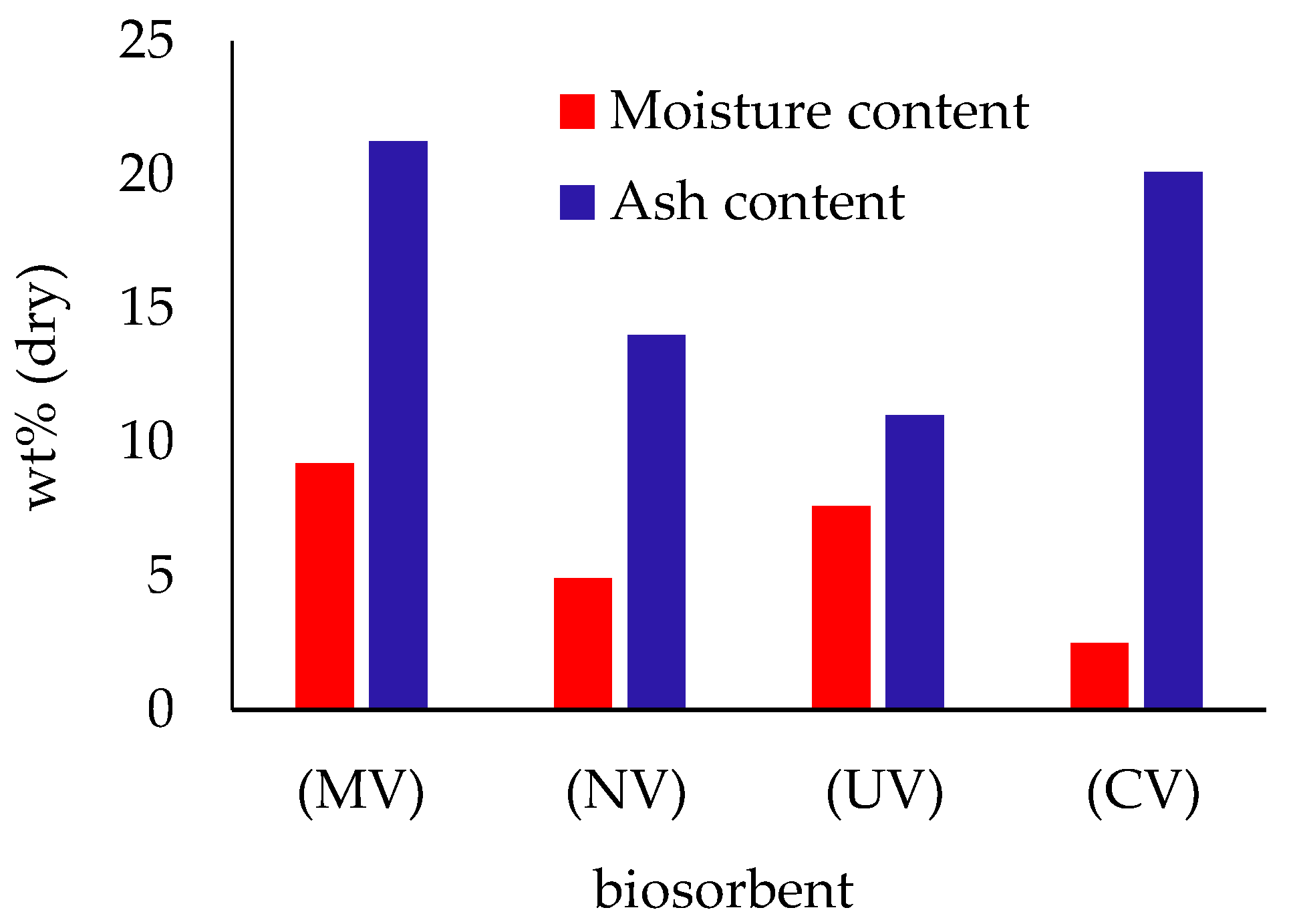
The results of the proximate analysis of macroalgae obtained from this research, together with the ultimate analysis outcomes, are presented in Figure 2. The brown seaweed Sargassum muticum demonstrated the highest ash content among the species examined (21.24%), while the lowest was measured for Ulva rigida (11.0%), a green alga. Comparable results have been reported in other published studies as well [74,75].
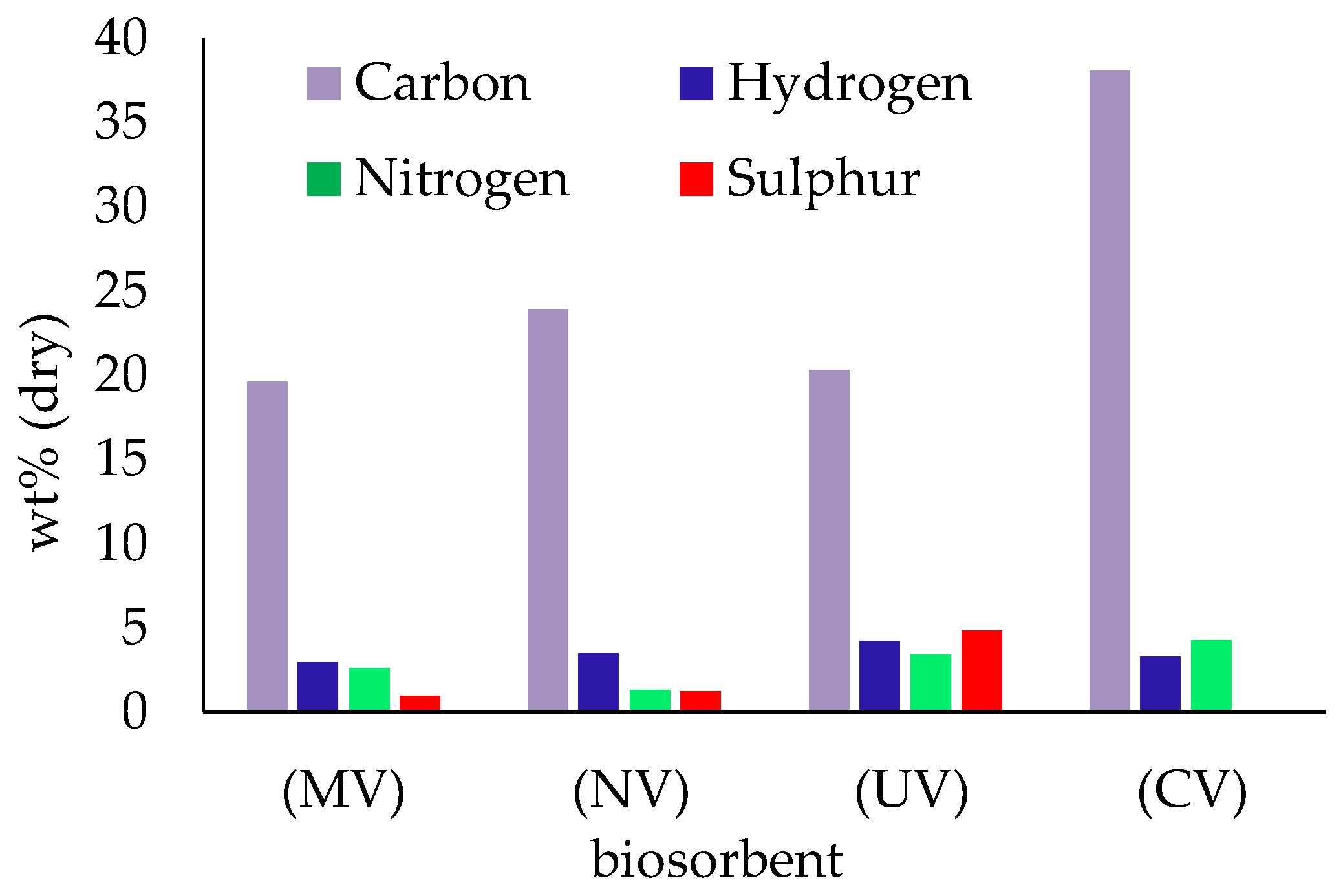
The ultimate analysis results depicted in Figure 3 reveal that carbon is the main organic constituent of algae, with its percentage ranging from 20.31% to over 38%. Hydrogen, nitrogen, and sulfur are also present in all examined macroalgae biomass.
The major microelements present in all algal species are sodium and potassium, followed by calcium and magnesium, which are the dominant constituents of seawater (Figure 4). Based on these findings, brown algae exhibited higher concentrations of sodium (Na), calcium (Ca), magnesium (Mg), and potassium (K) compared to green seaweeds. These results are in concordance with previous data published in the literature [76,77]. Specific information has been published about Ascophyllum nodosum and another species of Ulva (Ulva fasciata) [78] and Cladophora (Cladophora glomerata) [79], and these results align with the data obtained in this study.
The chemical composition of macroalgae is affected by several factors, including water characteristics, algal structure, harvesting time, and washing procedures. It is noteworthy that, with the exception of Cladophora sericea, the concentrations of alkali metals (sodium and potassium) in the seaweeds examined are considerably higher than those of alkaline earth metals (calcium and magnesium), which aligns with the prevalence of sodium in marine environments. A comprehensive cleaning process was applied to the harvested algae to prevent pigmentation and secondary contamination from organic matter when employing the algae as biosorbents. This meticulous procedure likely results in the low concentrations of Na+ and K+ observed in C. sericea, attributed to the high-water solubility of alkali metals, leading to a subsequent decrease in sodium and potassium content.
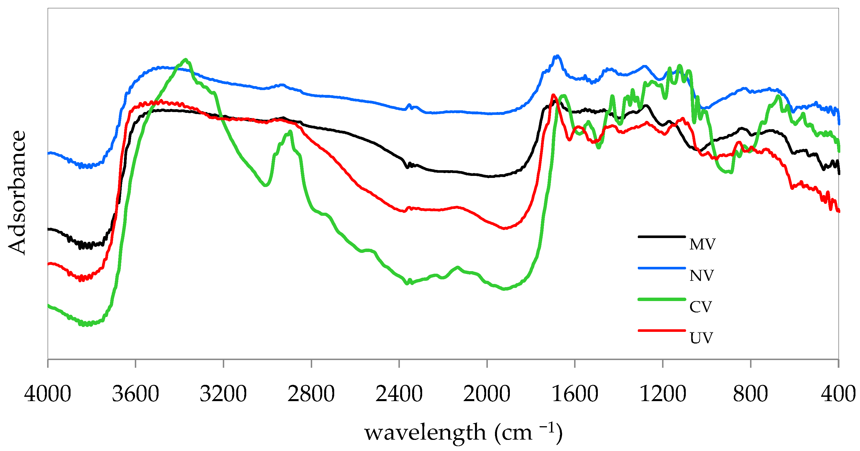
Biosorption is predominantly related to the wall cell composition, which includes functional groups like amino, hydroxyl, carboxyl, and sulfate [45]. To qualitatively identify these functional groups, as well as others possibly present on the surface of the algae, the FTIR spectrum was obtained. Infrared spectra of MV, NV, CV, and UV are presented in Figure 5.
The infrared spectra reveal multiple absorption bands, reflecting the complex composition of the studied biomass, predominantly characterized by carboxylic and hydroxyl functional groups. In all spectra, a hydrogen-bonded stretching band is observed in the range of 3700–3000 cm−1, exhibiting strong and broad intensity, indicative of the O-H group associated with carboxylic acids and alcohols. For CV, the band appears sharper compared to UV, MV and NV, suggesting a notable presence of alcohols within the protein content of the algae. Within a similar wavenumber range (3500–3300 cm−1), the N-H stretching bond of amine groups is observed, displaying medium intensity for MV, NV and UV. The presence of O-H and N-H functional groups confirm the presence of proteins and polysaccharides on the cell wall what is characteristic for the algal cell wall structure [80,81]. Bands observed between 2980 and 2850 cm−1 are assigned to C-H stretching of alkyl groups or to the stretching vibrations of C-H bonds associated with methyl, methylene, and methoxy groups [80]. Absorption bands observed in the range of 1800–1600 cm−1 correspond to C=O stretching vibrations of carbonyl groups in carboxyl functional groups. Bands at 1500–1400 cm−1 are associated with the asymmetric and symmetric stretching vibrations of carboxylate groups [82]. Additionally, the bands detected in the range of 1300–1200 cm−1 are attributed to sulfate ester groups (S=O), which are components of fucoidan, a polysaccharide present in the cell walls of brown seaweeds [82].
FTIR spectra previously published in the literature for unloaded and oxyanion-loaded algae indicates a potential chemical interaction involving hydrogen atoms from carboxyl (-COOH), hydroxyl (-OH), and amine (-NH) groups in the biomass and the adsorbate [46,47,63]. Thus, the most likely retention mechanisms are attributed to hydrogen bonding or surface complexation [45,83].

3.2. Results Attained After the Tested Pre-Treatments

Preliminary assessments were conducted by the research team utilizing all four seaweed species: Cladophora sericea, Ulva rigida, Sargassum muticum, and Ascophyllum nodosum, in both their raw forms and following various pre-treatments. Complete data of all pre-treatments applied, algae tested and results obtained from adsorption capacity testing are presented in detail in Table 1.
The results indicated no biosorption of arsenide under the predefined conditions for MV, NV, UV, and CV. For arsenate (As(V)), only slight removal was observed at pH 5 by CV, with a biosorption capacity of 0.11 mg g−1. These findings clearly suggest that green and brown seaweeds lack a natural capacity for arsenic uptake from wastewater. A. nodosum pre-treated with iron (N-Fe) was tested across a pH range of 2 to 4, as higher pH values led to significant iron precipitation above pH 4. However, under these conditions, no biosorption of As(III) was observed. Additional tests using protonated algae for both As(III) and As(V) also demonstrated negligible uptake. Acid pre-treatment of algae has been documented in the literature as a potential method for enhancing biosorption capacity for cations, facilitating ion exchange between surface H+ ions and metal ions [84,85]. Nevertheless, the positive effects of acid pre-treatment have not been observed consistently [86]. Depending on the structure of the algae and the specific treatment conditions, it is also essential to consider the potential dissolution of certain components within the algal matrix, as this can alter its structure and affect biosorption capacity. This may help explain why acid treatment does not consistently yield positive or negative outcomes for the removal of cationic metals. In the case of neutral or anionic adsorbates, protonation can increase the positive surface charge of the adsorbent, thereby enhancing electrostatic attraction toward soluble neutral or anionic species. No adsorption of As(III) and As(V) was detected for NH3–loaded S. muticum (MA) and NH3–loaded C. sericea (CA).
Under the same experimental conditions, both brown seaweed species demonstrated the ability to adsorb Sb(III) from aqueous solutions: S. muticum achieved 80% removal of Sb, while A. nodosum removed 22% of the soluble Sb(III). Biologically derived materials have been documented to participate in the absorption of antimony through carboxyl, amine, and hydroxyl groups [87]. It appears that a high number of total basic groups in S. muticum, coupled with potential greater heterogeneity, may be associated with the observed higher adsorption capacity. The superior performance observed for CV compared to UV may be attributed to the higher concentration of hydroxyl sites in CV, which was found to be 36% greater than that in UV [63]. Sb(III) uptake for N-Fe exhibited significantly higher biosorbed amounts compared to NV, although leaching and precipitation of iron in solution had occurred. At pH 5, after 2 h of contact time, suspended iron particles were detected.
Seaweeds in protonated form exhibited slightly improved performance compared to their original state, and S. muticum proves to be a more effective biosorbent for Sb(III) than A. nodosum under any circumstances. Nonetheless, the protonation process involves treating seaweeds with a potent acid, leading to substantial processing and cost implications. Considering that the increase in adsorption capacity is not substantial, protonation could not be recommended as a pre-treatment for practical applications.
In selenium assays, results indicated that untreated green and brown algae exhibited low or negligible affinity for the removal of Se(VI) from aqueous solutions. In comparison, CV removed a greater amount of Se(IV), likely due to the speciation of Se(IV) in solution.

3.3. Cladophora Sericea and Selenium Removal

3.3.1. Effect of pH and Removal Mechanism

The biosorption process is generally affected by various factors, with pH commonly recognized as a primary determinant in the uptake of metals. In this study, experiments were conducted within an acidic to neutral pH range, aligning with typical conditions anticipated in most contaminated water sources [46]. Figure 6 illustrates the impact of pH on the biosorption of Se(IV) and Se(VI) using CV.
The data indicate that the algal surface exhibits a greater affinity for Se(IV) compared to Se(VI) across the entire pH range studied. Results from potentiometric titrations [63] suggest that a pH range of 3 to 5 is more favorable for achieving a higher positive charge on C. sericea. In addition, the changes observed in the infrared spectra of raw seaweed compared to that of seaweed-loaded Se [63] in the bands corresponding to the carboxylic and phenolic groups demonstrate the importance of these functional groups in the adsorption of Se(IV).
The highest quantities of adsorbed Se(IV) were observed under acidic pH conditions (pH 2, 3, and 4), with removal efficiencies exceeding 17%. As the pH increased, the amount of adsorbed Se(IV) decreased by approximately half, coinciding with an increase in the fraction of the SeO32− species relative to HSeO3 (Table 2).
The decrease in Se(IV) adsorption with increasing pH can be explained by the interaction between the speciation of selenium and the surface charge of the adsorbent. At pH 2, Se(IV) mainly occurs as H2SeO3, with a smaller percentage present as HSeO3 [88]. However, as the pH increases, HSeO3 and SeO32− become the primary forms of Se(IV), with their relative abundances influenced by the pH of the solution. At lower pH values, the surface of the algae biosorbent is likely positively charged due to protonation of functional groups. This promotes electrostatic attraction between the positively charged carboxylic and hydroxyl groups on the biosorbent and the negatively charged HSeO3 species, or alternatively, through hydrogen bonding mechanisms, enhancing adsorption. As pH increases, the biosorbent’s surface charge becomes more negative due to deprotonation of these functional groups. This leads to electrostatic repulsion between the negatively charged surface and the Se(IV) oxyanions (HSeO3 and SeO32−), thereby reducing the adsorption capacity. Furthermore, the reduction in the HSeO3 fraction diminishes the likelihood of hydrogen bonding, thereby limiting adsorption capacity.
The FTIR analysis reveals that C. sericea contains significantly more hydroxyl groups compared to the rest of the analyzed algae. This might indicate that the (-OH) groups could contribute to a better retention of Se(IV) Via hydrogen bonding and/or weak coordination with low to moderate binding strength.
Similar findings have been reported for selenide adsorption on peanut shells [89] or rice husk [90] where adsorbed quantities declined with increasing pH. Conversely, another study identified optimal Se(IV) removal by Cladophora hutchinsiae at pH 5 [4]. The influence of pH on the adsorption of Se(VI) was similar to that detected for Se(IV); more precisely it is observed a decrease in the adsorbed amount was observed when increasing pH, although the pH effect is more drastic in the case of selenide, which is also confirmed by the published literature [91,92,93]. As shown in Figure 4, Cladophora sericea demonstrated the capacity to adsorb both selenide and selenate within the tested pH range, though to a limited extent. Adsorption was more effective for selenide (Se(IV)), the most toxic form of selenium, which was consequently selected for further assessments.

3.3.2. Kinetics and Modeling of Biosorption

The biosorption kinetics of Se(IV) onto C. sericea seaweed were investigated at a constant pH of 2, identified as the optimal pH condition, under varying initial concentrations of adsorbate and different quantities of algal biomass. Figure 7 illustrates the influence of contact time (t, min) on the adsorption capacity, expressed as the amount of Se(IV) adsorbed per unit mass of adsorbent (q, mg g−1).
As depicted in Figure 7, selenide adsorption proceeds rapidly, reaching equilibrium within approximately 2 h for initial adsorbate concentrations of 10 and 25 mg L−1. For water with low selenium contamination (1 mg L−1), no significant change in the concentration of Se in the liquid phase was observed after 15 min.
The experimental data were analyzed using non-linear regression to fit both the Lagergren pseudo-first-order and pseudo-second-order models [94] as represented by Equations (3) and (4), respectively.
q = q e   ( 1 e k 1   t ) ,
q = k 2 q e 2 t 1 + k 2 q e t ,
In both equations, q (mg g−1) represents the biosorbed amount at a given contact time t (min), qe (mg g−1) denotes the biosorbed amount at equilibrium, k1 (min−1) and k2 (g mg−1 min−1) are the kinetic constants associated with each model.
The model parameters derived from the mathematical fittings are provided in Table 3. Both models effectively describe the experimental data, as indicated by low standard error (SE) values (≤0.03). A comparison between the predicted equilibrium adsorption capacities (qe) and the experimentally observed values reveals no significant differences in the performance of the two models. Figure 7 presents the pseudo-first-order and pseudo-second-order model predictions alongside the experimental data for visual comparison.
A good fit for both models may imply that the biosorbent has versatile properties, allowing it to interact with the adsorbate in various ways, which can be attributed to physical adsorption (represented by the pseudo-first-order model) and chemical interactions (represented by the pseudo-second-order model). Consequently, the biosorption mechanism may be complex, potentially involving multiple processes. Also, it may indicate that the rate of adsorption is influenced by both the availability of active sites on the biosorbent and the concentration of the adsorbate.
The biosorbed amount (qe) exhibited an increasing trend with higher initial selenide concentrations, while it decreased with the increase in biosorbent dosages. This behavior is typical, as a higher concentration gradient (increased C0) enhances mass transfer between the liquid and solid phases. Additionally, although higher adsorbent dosages provide a greater number of available surface sites for accommodating Se(IV) species, the use of excessive biomass can result in inefficient utilization of the algal surface area, leading to a lower adsorption capacity at equilibrium (Table 3). From the perspective of treatment efficiency regarding Se(IV) removal from liquid solutions, utilizing lower algal dosages yields more favorable removal efficiencies at equilibrium, although these efficiencies remain relatively modest. For initial Se(IV) concentrations of 25 mg L−1 and algal dosages of 5, 10, and 20 g L−1, the respective Se-removal rates were 11%, 15%, and 20%.
The observed increase in Se(IV) removal rates with higher algal dosages can be attributed to the greater availability of active adsorption sites as the biomass concentration increases. At a fixed initial selenium concentration (25 mg L−1), increasing the amount of algal biomass enhances the total surface area and binding sites available for interaction with selenium ions. This allows for a higher proportion of the selenium in the solution to be adsorbed, leading to increased removal rates. However, the removal efficiency does not increase proportionally with the algal dosage, as seen in the modest incremental gains (from 11% to 20%). This may be due to limitations such as the saturation of accessible adsorption sites, mass transfer resistance, or possible aggregation of algae at higher dosages, which can reduce the effective surface area. Thus, while higher dosages improve Se(IV) removal up to a point, the efficiency is influenced by physical and chemical factors that constrain the adsorption process.

3.3.3. Equilibrium Study and Comparative Approach

Results on equilibrium study of Se(IV) removal onto C. sericea were previously reported, and Langmuir model estimated a peak of C. sericea adsorption capacity for selenium Se(IV) of 4 mg g−1 [63]. However, the evaluation of a biosorbent must take into account multiple criteria. The first indicator that is discussed is the maximum adsorption capacity resulted from the mathematical modeling of the experimental data. The results obtained in the mentioned study, using CV algae for Se(IV) removal, are remarkable if compare with the performance obtained by testing iron-coated granular activated carbon for the adsorption of the same selenium ion, the maximum removal capacity being 2.58 mg g−1, on pH 6 and 303 K [95]. It should be emphasized that C. sericea used could be considered a waste with no acquisition costs and whose conversion into a biosorbent can be complete by cost-effective procedures, while iron-coated granular activated carbon is a synthesized material which requires high temperatures to be obtained, as well as a subsequent stage to promote the iron coating. Another adsorbent tested for the removal of Se(IV) in initial selenium concentrations similar to the values tested in the present study was magnetite with a maximum adsorption potential of 0.22 mg g−1, at pH 4 [96], in this case also, C. sericea proving a superior performance. Studies with better results in terms of maximum adsorbed amounts have also been published, namely the application of a commercial FeOOH with a capacity of 26 mg g−1 [92]. Tuzen and Sari [48] investigated the uptake of Se(IV) by Cladophora hutchinsiae, also a species of green algae. Despite the use of exceptionally high Se concentrations in that study (Ce ≈ 0–300 mg L−1), which prevents a direct and reliable comparison with the maximum adsorption capacities observed in the present work, the reported adsorption capacity (74.9 mg g−1) is unexpectedly higher than the values obtained in this study.
The results obtained for selenium removal using natural adsorbents were compared with results reported by using synthetic tailored ones. Thus, the use of iron oxy-hydroxides as adsorbent led to a retention capacity of 10 μg Se(VI) g−1) and 4.3 mg Se(IV) g−1, a composite material hematite-coated magnetic nanoparticles lead to an adsorption capacity of 25.0 mg Se(IV) g−1, a bimetallic diatom composite retained 227 mg Se(IV) g−1, while activated alumina 9.02 µg Se(VI) g−1 and 5.38 µg Se(IV) g−1 [97]. Regardless of the type of biosorbent used the data reported confirm that Se(VI) is much more difficult to remove from aqueous solutions.

3.3.4. Studies on Interfering Ions Influence

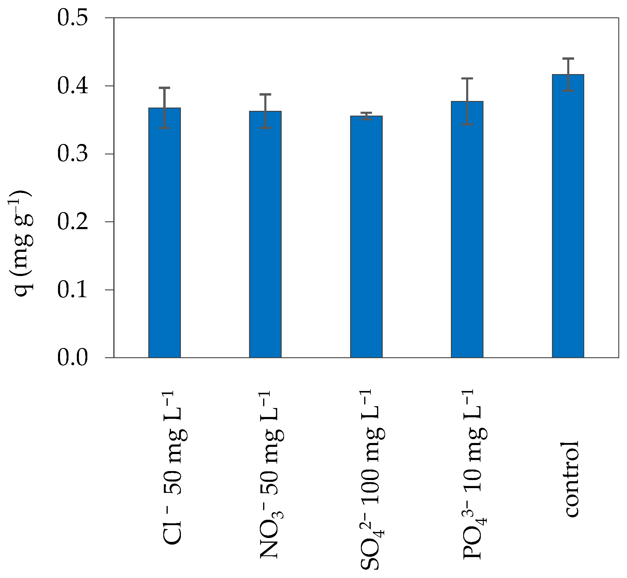
Natural and mining-influenced waters typically contain a mixture of ions that can interfere with the adsorption process. The effect of commonly coexisting anions-chloride, nitrate, sulfate, and phosphate-on the efficiency of C. sericea in Se(IV) uptake was assessed, with the results illustrated in Figure 8. The control assay represents the adsorption experiment conducted in the absence of any competing ions, with only Se(IV) present.
As illustrated in Figure 8, Se(IV) removal is only minimally impacted by the presence of coexisting anions in solution. Compared to the qe value observed in the control experiment, the adsorbed amounts decreased by only 10–14% in the presence of Cl, NO3, SO42−, and PO43−. The results for Se(IV) removal at an initial concentration of 100 mg L−1 using Eucalyptus camaldulensis bark indicated that the presence of sulfate and phosphate anions at a concentration of 50 mg L−1 led to reductions in the adsorbed amounts by 22% and 44%, respectively [98]. Johansson et al. [99] studied the impact of sulfate and nitrate on the adsorption of Se(VI) at an initial concentration of 108 µg L−1 using Gracilaria Modified Bio-char (GMB). The authors concluded that the biosorption capacity of GMB for Se(VI) was significantly influenced by increasing concentrations of SO42−, with no removal of Se(VI) observed at a Se(VI):SO42− molar ratio of 1:10,000. In contrast, the presence of NO3 did not affect Se(VI) adsorption. The Se(IV) biosorption could be negatively influenced by the presence of chlorine ions in solution due to the capacity of Cl to act like an oxidizing agent that convert Se(IV) in Se(VI), a less bioavailable form. However, the interfering ions tests proved that the Se(IV) removal capacity of C. Sericea is not influenced by the presence of chlorine ions.
Despite the limited biosorption capacity of C. sericea for Se, the results indicate that this capacity was not significantly affected by the presence of other anions in solution, including sulfate and phosphate, which typically serve as stronger competitors. This characteristic represents a significant advantage for C. sericea, particularly in regard to its potential applications.
Given this advantage and considering the studies reporting chemical modifications aimed at improving the selenium retention capacity of C. sericea, the alternative of a more effective thermochemical treatment can be suggested.

4. Conclusions

In this study, two green algae and two brown algae were evaluated for their efficiency in removing three oxyanions—selenium, antimony, and arsenic—from aqueous media. The seaweeds were assessed in their natural forms and following various pre-treatments aimed at enhancing their bioremediation capacities; however, these pre-treatments did not improve biosorption properties. Further investigation focused on Cladophora sericea (CV), a green macroalga commonly found on beaches worldwide. This alga was converted into a cost-effective biosorbent and tested for its ability to remove selenium, a hazardous pollutant, from contaminated water. The experiments were conducted with a low incipient concentration of selenium, 25 mg L−1, specifically to highlight the affinity of the bio-sorbent tested to this element. The uptake of selenide and selenate by Cladophora sericea was studied under varying pH conditions. Optimal results were observed at pH 2–4 for Se(IV) and at pH 2–3 for Se(VI). A decline in the adsorbed amounts was noted at higher pH values. From the data collected, it is possible to assume that the likely adsorption mechanism is by hydrogen bonding or by electrostatic attraction between the positively charged carboxylic and hydroxyl groups present on the surface of the adsorbent and the negative HSeO3-. The biosorption kinetics were rapid and adequately described by both pseudo-first-order and pseudo-second-order models. Furthermore, a notable advantage of the biosorption performance of C. sericea was its relative insensitivity to the presence of nitrate, chloride, sulfate, and phosphate anions in solution. The limitation of the present study is the relatively low retention capacity of green seaweed for selenium, but future challenges are open considering the possibility of activation and functionalization on this waste-based adsorbent.

Author Contributions

Conceptualization, G.U. and I.V.; methodology, G.U.; software, V.L.P.; validation, G.U. and I.V.; formal analysis, G.U. and V.L.P.; investigation, G.U.; resources, I.V.; data curation, G.U. and I.V.; writing—original draft preparation, G.U. and V.L.P.; writing—review and editing, G.U. and I.V.; visualization, I.V.; supervision, I.V.; funding acquisition, I.V. All authors have read and agreed to the published version of the manuscript.

Funding

This research received no external funding.

Institutional Review Board Statement

Not applicable.

Informed Consent Statement

Not applicable.

Data Availability Statement

The original contributions presented in the study are included in the article; further inquiries can be directed to the corresponding author.

Conflicts of Interest

The authors declare no conflicts of interest.

References

  1. IARC. Agents Classifed by the IARC Monographs; International Agency for Research on Cancer: Lyon, France, 2024. [Google Scholar]
  2. Fernandez-Martinez, A.; Charlet, L. Selenium Environmental Cycling and Bioavailability: A Structural Chemist Point of View. Rev. Environ. Sci. Bio Technol. 2009, 8, 81–110. [Google Scholar] [CrossRef]
  3. Hamilton, S.J. Review of selenium toxicity in the aquatic food chain. Sci. Total Environ. 2004, 326, 1–31. [Google Scholar] [CrossRef]
  4. Sarı, A.; Çıtak, D.; Tuzen, M. Equilibrium, thermodynamic and kinetic studies on adsorption of Sb(III) from aqueous solution using low-cost natural diatomite. Chem. Eng. J. 2010, 162, 521–527. [Google Scholar] [CrossRef]
  5. Jha, P.K.; Tripathi, P. Arsenic and fluoride contamination in groundwater: A review of global scenarios with special reference to India. Groundw. Sustain. Dev. 2021, 13, 100576. [Google Scholar] [CrossRef]
  6. Smedley, P.L.; Kinniburgh, D.G. A review of the source, behaviour and distribution of arsenic in natural waters. Appl. Geochem. 2002, 17, 517–568. [Google Scholar] [CrossRef]
  7. Nath, B.; Stüben, D.; Mallik, S.B.; Chatterjee, D.; Charlet, L. Mobility of arsenic in West Bengal aquifers conducting low and high groundwater arsenic. Part I: Comparative hydrochemical and hydrogeological characteristics. Appl. Geochem. 2008, 23, 977–995. [Google Scholar] [CrossRef]
  8. Sánchez-Rodas, D.; Luis Gómez-Ariza, J.; Giráldez, I.; Velasco, A.; Morales, E. Arsenic speciation in river and estuarine waters from southwest Spain. Sci. Total Environ. 2005, 345, 207–217. [Google Scholar] [CrossRef]
  9. Modabberi, S.; Moore, F. Environmental geochemistry of Zarshuran Au-As deposit, NW Iran. Environ. Geol. 2004, 46, 796–807. [Google Scholar] [CrossRef]
  10. Issanov, A.; Adewusi, B.; Saint-Jacques, N.; Dummer, T.J.B. Arsenic in drinking water and lung cancer: A systematic review of 35 years of evidence. Toxicol. Appl. Pharmacol. 2024, 483, 116808. [Google Scholar] [CrossRef]
  11. Reimann, C.; Matschullat, J.; Birke, M.; Salminen, R. Antimony in the environment: Lessons from geochemical mapping. Appl. Geochem. 2010, 25, 175–198. [Google Scholar] [CrossRef]
  12. Wang, X.; He, M.; Xi, J.; Lu, X. Antimony distribution and mobility in rivers around the world’s largest antimony mine of Xikuangshan, Hunan Province, China. Microchem. J. 2011, 97, 4–11. [Google Scholar] [CrossRef]
  13. World Health Organization. Guidelines for Drinking—Water Quality, 4th ed.; WHO: Geneva, Switzerland, 2011. [Google Scholar]
  14. Etteieb, S.; Magdouli, S.; Zolfaghari, M.; Brar, S. Monitoring and analysis of selenium as an emerging contaminant in mining industry: A critical review. Sci. Total Environ. 2020, 698, 134339. [Google Scholar] [CrossRef] [PubMed]
  15. Khamkhash, A.; Srivastava, V.; Ghosh, T.; Akdogan, G.; Ganguli, R.; Aggarwal, S. Mining-Related Selenium Contamination in Alaska, and the State of Current Knowledge. Minerals 2017, 7, 46. [Google Scholar] [CrossRef]
  16. Wasewar, K.L.; Prasad, B.; Gulipalli, S. Adsorption of Selenium Using Bagasse Fly Ash. CLEAN Soil Air Water 2009, 37, 534–543. [Google Scholar] [CrossRef]
  17. Twidwell, L.; McCloskey, J.; Joyce, H.; Dahlgren, E.; Hadden, A. Removal of Selenium Oxyanions from Mine Waters Utilizing Elemental Iron and Galvanically Coupled Metals. In Innovations in Natural Resoure Processing—Proceedings of the Jan. D. Miller Symposium, SME; Society for Mining, Metallurgy, and Exploration, Incorporated: Englewood, CO, USA, 2005; Volume 2005. [Google Scholar]
  18. Lemly, A.D. Aquatic selenium pollution is a global environmental safety issue. Ecotoxicol. Environ. Saf. 2004, 59, 44–56. [Google Scholar] [CrossRef] [PubMed]
  19. Mitchell, K.; Mason, P.R.D.; Van Cappellen, P.; Johnson, T.M.; Gill, B.C.; Owens, J.D.; Diaz, J.; Ingall, E.D.; Reichart, G.-J.; Lyons, T.W. Selenium as paleo-oceanographic proxy: A first assessment. Geochim. Cosmochim. Acta 2012, 89, 302–317. [Google Scholar] [CrossRef]
  20. Stefaniak, J.; Dutta, A.; Verbinnen, B.; Shakya, M.; Rene, E. Selenium removal from mining and process wastewater: A systematic review of available technologies. J. Water Supply Res. Technol. AQUA 2018, 67, 903–918. [Google Scholar] [CrossRef]
  21. Thiry, C.; Ruttens, A.; De Temmerman, L.; Schneider, Y.-J.; Pussemier, L. Current knowledge in species-related bioavailability of selenium in food. Food Chem. 2012, 130, 767–784. [Google Scholar] [CrossRef]
  22. Pommier, A.L.; Simon, S.; Buzier, R.; Guibaud, G. Evaluation of a mercapto-functionalized silica binding phase for the selective sampling of Se(IV) by Diffusive Gradients in Thin films. Talanta 2019, 199, 590–595. [Google Scholar] [CrossRef]
  23. Combs, G.F., Jr.; Gray, W.P. Chemopreventive agents: Selenium. Pharmacol. Ther. 1998, 79, 179–192. [Google Scholar] [CrossRef]
  24. Rayman, M.P. The importance of selenium to human health. Lancet 2000, 356, 233–241. [Google Scholar] [CrossRef]
  25. Taylor, J.B.; Reynolds, L.P.; Redmer, D.A.; Caton, J.S. Maternal and fetal tissue selenium loads in nulliparous ewes fed supranutritional and excessive selenium during mid- to late pregnancy1,2. J. Anim. Sci. 2009, 87, 1828–1834. [Google Scholar] [CrossRef]
  26. Burger, J.; Gochfeld, M. Selenium and mercury molar ratios in saltwater fish from New Jersey: Individual and species variability complicate use in human health fish consumption advisories. Environ. Res. 2012, 114, 12–23. [Google Scholar] [CrossRef] [PubMed]
  27. Zhao, Y.; Ni, S.; Pei, C.; Sun, L.; Wu, L.; Xu, A.; Nie, Y.; Liu, Y. Parental treatment with selenium protects Caenorhabditis elegans and their offspring against the reproductive toxicity of mercury. Sci. Total Environ. 2024, 912, 169461. [Google Scholar] [CrossRef]
  28. Saliba, W.; El Fakih, R.; Shaheen, W. Heart failure secondary to selenium deficiency, reversible after supplementation. Int. J. Cardiol. 2010, 141, e26–e27. [Google Scholar] [CrossRef]
  29. Li, H.; Jia, L.; Deng, Z.; Sun, X.; Zhang, H.; Li, H. The effects of selenium on the growth and bone development in the weaned rats. Food Biosci. 2023, 55, 103018. [Google Scholar] [CrossRef]
  30. Zhang, H.; Feng, X.; Chan, H.M.; Larssen, T. New insights into traditional health risk assessments of mercury exposure: Implications of selenium. Environ. Sci. Technol. 2014, 48, 1206–1212. [Google Scholar] [CrossRef]
  31. Hammouh, F.; Zein, S.; Amr, R.; Ghazzawi, H.; Muharib, D.; Saad, D.; Subih, H. Assessment of dietary selenium intake of Jordanian adults in Madaba: A cross sectional study. Nutr. Food Sci. 2020, 51, 494–506. [Google Scholar] [CrossRef]
  32. Rayman, M.P.; Winther, K.H.; Pastor-Barriuso, R.; Cold, F.; Thvilum, M.; Stranges, S.; Guallar, E.; Cold, S. Effect of long-term selenium supplementation on mortality: Results from a multiple-dose, randomised controlled trial. Free Radical Biol. Med. 2018, 127, 46–54. [Google Scholar] [CrossRef] [PubMed]
  33. Sun, H.-J.; Rathinasabapathi, B.; Wu, B.; Luo, J.; Pu, L.-P.; Ma, L.Q. Arsenic and selenium toxicity and their interactive effects in humans. Environ. Int. 2014, 69, 148–158. [Google Scholar] [CrossRef] [PubMed]
  34. Herrero Latorre, C.; Barciela García, J.; García Martín, S.; Peña Crecente, R.M. Solid phase extraction for the speciation and preconcentration of inorganic selenium in water samples: A review. Anal. Chim. Acta 2013, 804, 37–49. [Google Scholar] [CrossRef]
  35. Okonji, S.O.; Achari, G.; Pernitsky, D. Environmental Impacts of Selenium Contamination: A Review on Current-Issues and Remediation Strategies in an Aqueous System. Water 2021, 13, 1473. [Google Scholar] [CrossRef]
  36. Khakpour, H.; Younesi, H.; Mohammadhosseini, M. Two-stage biosorption of selenium from aqueous solution using dried biomass of the baker’s yeast Saccharomyces cerevisiae. J. Environ. Chem. Eng. 2014, 2, 532–542. [Google Scholar] [CrossRef]
  37. Somogyi, Z.; Kádár, I.; Kiss, I.; Juríková, T.; Szekeres, L.; Balla, Š.; Nagy, P.; Bakonyi, G. Comparative toxicity of the selenate and selenite to the potworm Enchytraeus albidus (Annelida: Enchytraeidae) under laboratory conditions. Eur. J. Soil Biol. 2012, 50, 159–164. [Google Scholar] [CrossRef]
  38. Somogyi, Z.; Kiss, I.; Kádár, I.; Bakonyi, G. Toxicity of selenate and selenite to the potworm Enchytraeus albidus (Annelida: Enchytraeidae): A laboratory test. Ecotoxicology 2007, 16, 379–384. [Google Scholar] [CrossRef]
  39. Albert, M.; Demesmay, C.; Rocca, J.L. Analysis of organic and non-organic arsenious or selenious compounds by capillary electrophoresis. Fresenius’ J. Anal. Chem. 1995, 351, 426–432. [Google Scholar] [CrossRef]
  40. Amoako, P.O.; Uden, P.C.; Tyson, J.F. Speciation of selenium dietary supplements; formation of S-(methylseleno)cysteine and other selenium compounds. Anal. Chim. Acta 2009, 652, 315–323. [Google Scholar] [CrossRef]
  41. Ebert, R.; Jakob, F. Selenium deficiency as a putative risk factor for osteoporosis. Int. Congr. Ser. 2007, 1297, 158–164. [Google Scholar] [CrossRef]
  42. He, Y.; Xiang, Y.; Zhou, Y.; Yang, Y.; Zhang, J.; Huang, H.; Shang, C.; Luo, L.; Gao, J.; Tang, L. Selenium contamination, consequences and remediation techniques in water and soils: A review. Environ. Res. 2018, 164, 288–301. [Google Scholar] [CrossRef]
  43. Zoroufchi Benis, K.; Motalebi Damuchali, A.; McPhedran, K.N.; Soltan, J. Treatment of aqueous arsenic—A review of biosorbent preparation methods. J. Environ. Manag. 2020, 273, 111126. [Google Scholar] [CrossRef]
  44. He, J.; Chen, J.P. A comprehensive review on biosorption of heavy metals by algal biomass: Materials, performances, chemistry, and modeling simulation tools. Bioresour. Technol. 2014, 160, 67–78. [Google Scholar] [CrossRef] [PubMed]
  45. Ungureanu, G.; Santos, S.; Boaventura, R.; Botelho, C. Arsenic and antimony in water and wastewater: Overview of removal techniques with special reference to latest advances in adsorption. J. Environ. Manag. 2015, 151, 326–342. [Google Scholar] [CrossRef] [PubMed]
  46. Ungureanu, G.; Filote, C.; Santos, S.C.R.; Boaventura, R.A.R.; Volf, I.; Botelho, C.M.S. Antimony oxyanions uptake by green marine macroalgae. J. Environ. Chem. Eng. 2016, 4, 3441–3450. [Google Scholar] [CrossRef]
  47. Ungureanu, G.; Santos, S.C.R.; Volf, I.; Boaventura, R.A.R.; Botelho, C.M.S. Biosorption of antimony oxyanions by brown seaweeds: Batch and column studies. J. Environ. Chem. Eng. 2017, 5, 3463–3471. [Google Scholar] [CrossRef]
  48. Tuzen, M.; Sarı, A. Biosorption of selenium from aqueous solution by green algae (Cladophora hutchinsiae) biomass: Equilibrium, thermodynamic and kinetic studies. Chem. Eng. J. 2010, 158, 200–206. [Google Scholar] [CrossRef]
  49. Liu, F.; Huang, J.-C.; Zhou, C.; Gao, W.; Xia, S.; He, S.; Zhou, W. Development of an algal treatment system for selenium removal: Effects of environmental factors and post-treatment processing of Se-laden algae. J. Hazard. Mater. 2019, 365, 546–554. [Google Scholar] [CrossRef]
  50. Pereira, L. As Algas Marinhas e Respectivas Utilidades; Universidade de Coimbra: Coimbra, Portugal, 2010. [Google Scholar]
  51. Petrescu, R. Aurul Verde din Marea Neagra. Green Report. 2010. Available online: http://www.green-report.ro/aurul-verde-din-marea-neagra/ (accessed on 28 February 2025).
  52. Akbari, M.; Hallajisani, A.; Keshtkar, A.R.; Shahbeig, H.; Ali Ghorbanian, S. Equilibrium and kinetic study and modeling of Cu(II) and Co(II) synergistic biosorption from Cu(II)-Co(II) single and binary mixtures on brown algae C. indica. J. Environ. Chem. Eng. 2015, 3, 140–149. [Google Scholar] [CrossRef]
  53. Bakatula, E.N.; Cukrowska, E.M.; Weiersbye, I.M.; Mihaly-Cozmuta, L.; Peter, A.; Tutu, H. Biosorption of trace elements from aqueous systems in gold mining sites by the filamentous green algae (Oedogonium sp.). J. Geochem. Explor. 2014, 144, 492–503. [Google Scholar] [CrossRef]
  54. Brouers, F.; Al-Musawi, T.J. On the optimal use of isotherm models for the characterization of biosorption of lead onto algae. J. Mol. Liq. 2015, 212, 46–51. [Google Scholar] [CrossRef]
  55. Pahlavanzadeh, H.; Keshtkar, A.R.; Safdari, J.; Abadi, Z. Biosorption of nickel(II) from aqueous solution by brown algae: Equilibrium, dynamic and thermodynamic studies. J. Hazard. Mater. 2010, 175, 304–310. [Google Scholar] [CrossRef] [PubMed]
  56. Yu, P.; Xing, J.; Tang, J.; Wang, Z.; Zhang, C.; Wang, Q.; Xiao, X.; Huang, W. Polyethyleneimine-modified iron-doped birnessite as a highly stable adsorbent for efficient arsenic removal. J. Colloid Interface Sci. 2024, 661, 164–174. [Google Scholar] [CrossRef]
  57. Dechdacho, P.; Howard, S.; Hershey, R.L.; Parashar, R.; Perez, L.J. Effective removal of arsenic from contaminated groundwater using an iron-based metal-organic framework. Environ. Technol. Innov. 2023, 32, 103406. [Google Scholar] [CrossRef]
  58. Zeng, H.; Zhao, W.; Sun, S.; Sun, X.; Zeng, Y.; Hao, R.; Zhang, J.; Li, D. Facile preparation of maghemite based on iron sludge for arsenic removal from water. Sci. Total Environ. 2024, 906, 167575. [Google Scholar] [CrossRef]
  59. Zeng, H.; Liu, C.; Wang, F.; Zhang, J.; Li, D. Disposal of iron-manganese sludge from waterworks and its potential for Arsenic removal. J. Environ. Chem. Eng. 2022, 10, 108480. [Google Scholar] [CrossRef]
  60. Gupta, A.R.; Mondal, M.; Bapat, P.S.; Joshi, V.C.; Popat, K.M.; Indurkar, P.D.; Sharma, S. Construction of arsenic selective chelating resin with iron precursor for removal of low-concentration arsenic: Breakthrough modeling and field deployment. J. Hazard. Mater. 2023, 459, 132000. [Google Scholar] [CrossRef]
  61. Yoon, K.; Kwon, G.; Kim, E.; Rinklebe, J.; Song, H. Production of Fe-biochar from paper-mill sludge and its application to Se(VI) and Se(IV) removal. Chem. Eng. J. 2024, 484, 149470. [Google Scholar] [CrossRef]
  62. Ungureanu, G.; Santos, S.; Boaventura, R.; Botelho, C. Biosorption of antimony by brown algae S. muticum and A. nodosum. Environ. Eng. Manag. J. 2015, 14, 455–463. [Google Scholar] [CrossRef]
  63. Filote, C.; Ungureanu, G.; Boaventura, R.; Santos, S.; Volf, I.; Botelho, C. Green macroalgae from the Romanian coast of Black Sea: Physico-chemical characterization and future perspectives on their use as metal anions biosorbents. Process Saf. Environ. Prot. 2017, 108, 34–43. [Google Scholar] [CrossRef]
  64. NREL. Biomass Compositional Analysis Laboratory Providing Detailed and Accurate Characterization; National Renewable Energy Laboratory, Office of Energy Efficiency and Renewable Energy: Golden, CO, USA, 2014.
  65. Pregl, F.; Roth, H. Quantitative Organische Mikroanalyse; Springer: Berlin/Heidelberg, Germany, 1949. [Google Scholar]
  66. Kjeldahl, J. Neue Methode zur Bestimmung des Stickstoffs in organischen Körpern. Z. Für Anal. Chem. 1883, 22, 366–382. [Google Scholar] [CrossRef]
  67. Schöniger, W. Eine mikroanalytische Schnellbestimmung von Halogen in organischen Substanzen. Microchim. Acta 1955, 43, 123–129. [Google Scholar] [CrossRef]
  68. Schöniger, W. Die mikroanalytische Schnellbestimmung von Halogenen und Schwefel in organischen Verbindungen. Microchim. Acta 1956, 44, 869–876. [Google Scholar] [CrossRef]
  69. Bordoloi, S.; Nath, S.K.; Gogoi, S.; Dutta, R.K. Arsenic and iron removal from groundwater by oxidation–coagulation at optimized pH: Laboratory and field studies. J. Hazard. Mater. 2013, 260, 618–626. [Google Scholar] [CrossRef]
  70. Wu, Z.; He, M.; Guo, X.; Zhou, R. Removal of antimony (III) and antimony (V) from drinking water by ferric chloride coagulation: Competing ion effect and the mechanism analysis. Sep. Purif. Technol. 2010, 76, 184–190. [Google Scholar] [CrossRef]
  71. Kazapoe, R.W.; Addai, M.O.; Amuah, E.E.Y.; Dankwa, P. Characterization of groundwater in southwest Ghana: Implications for sustainable agriculture and safe water supply in a mining-dominated zone. Environ. Sustain. Indic. 2024, 22, 100341. [Google Scholar] [CrossRef]
  72. Chen, X.; Tang, Z.; Li, G.; Zhang, J.; Xie, F.; Zheng, L. Tracing sulfate sources and transformations of surface water using multiple isotopes in a mining-rural-urban agglomeration area. Ecotoxicol. Environ. Saf. 2024, 269, 115805. [Google Scholar] [CrossRef]
  73. Li, Y.; Jiang, J.; Zhang, R.; Qie, W.; Shao, J.; Zhu, W.; Xu, N. Effects of photoperiod on the growth and physiological responses in Ulva prolifera under constant and diurnal temperature difference conditions. Mar. Environ. Res. 2024, 197, 106477. [Google Scholar] [CrossRef]
  74. Samarasinghe, M.B.; van der Heide, M.E.; Weisbjerg, M.R.; Sehested, J.; Sloth, J.J.; Bruhn, A.; Vestergaard, M.; Nørgaard, J.V.; Hernández-Castellano, L.E. A descriptive chemical analysis of seaweeds, Ulva sp., Saccharina latissima and Ascophyllum nodosum harvested from Danish and Icelandic waters. Anim. Feed. Sci. Technol. 2021, 278, 115005. [Google Scholar] [CrossRef]
  75. Rohani-Ghadikolaei, K.; Abdulalian, E.; Ng, W.-K. Evaluation of the proximate, fatty acid and mineral composition of representative green, brown and red seaweeds from the Persian Gulf of Iran as potential food and feed resources. J. Food Sci. Technol. 2012, 49, 774–780. [Google Scholar] [CrossRef]
  76. Shen, W.; He, S.; Mu, M.; Cao, B.; Wang, S.; Naqvi, S.R.; Hanelt, D.; Abomohra, A. A comprehensive review on the intricate processes involved in algae pyrolysis mechanism and possible migration of undesirable chemical elements. J. Anal. Appl. Pyrolysis 2024, 177, 106365. [Google Scholar] [CrossRef]
  77. Kulikova, Y.; Sukhikh, S.; Kalashnikova, O.; Chupakhin, E.; Ivanova, S.; Chubarenko, B.; Gorbunova, J.; Babich, O. Assessment of the Resource Potential of Baltic Sea Macroalgae. Appl. Sci. 2022, 12, 3599. [Google Scholar] [CrossRef]
  78. Pourkarimi, S.; Sadeh, M.S.; Hallajisani, A.; Hajikhani, M.; Moradi, M.; Alizadeh, O.; Nouralishahi, A. Investigation of catalytic pyrolysis of Azolla filiculoides and Ulva fasciata for bio-oil production. Biochem. Eng. J. 2022, 178, 108278. [Google Scholar] [CrossRef]
  79. Parsa, M.; Jalilzadeh, H.; Pazoki, M.; Ghasemzadeh, R.; Abdoli, M. Hydrothermal Liquefaction of Gracilaria gracilis and Cladophora glomerata macro-algae for biocrude production. Bioresour. Technol. 2017, 250, 26–34. [Google Scholar] [CrossRef]
  80. Ghoneim, M.M.; El-Desoky, H.S.; El-Moselhy, K.M.; Amer, A.; Abou El-Naga, E.H.; Mohamedein, L.I.; Al-Prol, A.E. Removal of cadmium from aqueous solution using marine green algae, Ulva lactuca. Egypt. J. Aquat. Res. 2014, 40, 235–242. [Google Scholar] [CrossRef]
  81. Deng, L.; Su, Y.; Su, H.; Wang, X.; Zhu, X. Sorption and desorption of lead (II) from wastewater by green algae Cladophora fascicularis. J. Hazard. Mater. 2007, 143, 220–225. [Google Scholar] [CrossRef] [PubMed]
  82. Blanco-Pascual, N.; Montero, M.P.; Gómez-Guillén, M.C. Antioxidant film development from unrefined extracts of brown seaweeds Laminaria digitata and Ascophyllum nodosum. Food Hydrocoll. 2014, 37, 100–110. [Google Scholar] [CrossRef]
  83. Santos, S.; Ungureanu, G.; Boaventura, R.; Botelho, C. Selenium contaminated waters: An overview of analytical methods, treatment options and recent advances in sorption methods. Sci. Total Environ. 2015, 521–522, 246–260. [Google Scholar] [CrossRef] [PubMed]
  84. Pozdniakova, T.A.; Mazur, L.P.; Boaventura, R.A.R.; Vilar, V.J.P. Brown macro-algae as natural cation exchangers for the treatment of zinc containing wastewaters generated in the galvanizing process. J. Clean. Prod. 2016, 119, 38–49. [Google Scholar] [CrossRef]
  85. Mazur, L.P.; Pozdniakova, T.A.; Mayer, D.A.; de Souza, S.M.A.G.U.; Boaventura, R.A.R.; Vilar, V.J.P. Cation exchange prediction model for copper binding onto raw brown marine macro-algae Ascophyllum nodosum: Batch and fixed-bed studies. Chem. Eng. J. 2017, 316, 255–276. [Google Scholar] [CrossRef]
  86. Gupta, V.K.; Rastogi, A. Equilibrium and kinetic modelling of cadmium(II) biosorption by nonliving algal biomass Oedogonium sp. from aqueous phase. J. Hazard. Mater. 2008, 153, 759–766. [Google Scholar] [CrossRef]
  87. Wu, F.; Sun, F.; Wu, S.; Yan, Y.; Xing, B. Removal of antimony(III) from aqueous solution by freshwater cyanobacteria Microcystis biomass. Chem. Eng. J. 2012, 183, 172–179. [Google Scholar] [CrossRef]
  88. Neal, R.H.; Sposito, G.; Holtzclaw, K.M.; Traina, S.J. Selenite adsorption on alluvial soils. I. Soil composition and pH effects. Soil Sci. Soc. Am. J. 1987, 51, 1161–1165. [Google Scholar] [CrossRef]
  89. El-Shafey, E.-S. Removal of Se(IV) from Aqueous Solution Using Sulfuric Acid-Treated Peanut Shell. J. Environ. Manag. 2007, 84, 620–627. [Google Scholar] [CrossRef] [PubMed]
  90. El-Shafey, E.-S. Sorption of Cd(II) and Se(IV) from Aqueous Solution Using Modified Rice Husk. J. Hazard. Mater. 2007, 147, 546–555. [Google Scholar] [CrossRef]
  91. Jordan, N.; Marmier, N.; Lomenech, C.; Giffaut, E.; Ehrhardt, J.-J. Competition between selenium (IV) and silicic acid on the hematite surface. Chemosphere 2009, 75, 129–134. [Google Scholar] [CrossRef]
  92. Sharrad, M.O.M.; Liu, H.; Fan, M. Evaluation of FeOOH performance on selenium reduction. Sep. Purif. Technol. 2012, 84, 29–34. [Google Scholar] [CrossRef]
  93. Zelmanov, G.; Semiat, R. Selenium removal from water and its recovery using iron (Fe3+) oxide/hydroxide-based nanoparticles sol (NanoFe) as an adsorbent. Sep. Purif. Technol. 2013, 103, 167–172. [Google Scholar] [CrossRef]
  94. Lagergren, S. About the Theory of So-Called Adsorption of Soluble Substances. K. Sven. Vetenskapsakademiens Handl. 1898, 24, 1–39. [Google Scholar]
  95. Zhang, N.; Lin, L.-S.; Gang, D. Adsorptive selenite removal from water using iron-coated GAC adsorbents. Water Res. 2008, 42, 3809–3816. [Google Scholar] [CrossRef]
  96. Martinez, M.; Giménez, J.; Pablo, J.; Rovira, M.; Duro, L. Sorption of Selenium (IV) and Selenium(VI) onto Magnetite. Appl. Surf. Sci. 2006, 252, 3767–3773. [Google Scholar] [CrossRef]
  97. Fadaei, A.; Mohammadian-Hafshejani, A. Selenium Removal from Water and Wastewater by Different Technologies: A Systematic Review. Iran. J. Public Health 2023, 52, 64–77. [Google Scholar] [CrossRef]
  98. Rajamohan, N.; Rajasimman, M. Biosorption of Selenium using activated plant based sorbent—Effect of variables, isotherm and kinetic modeling. Biocatal. Agric. Biotechnol. 2015, 4, 795–800. [Google Scholar] [CrossRef]
  99. Johansson, C.L.; Paul, N.A.; de Nys, R.; Roberts, D.A. Simultaneous biosorption of selenium, arsenic and molybdenum with modified algal-based biochars. J. Environ. Manag. 2016, 165, 117–123. [Google Scholar] [CrossRef] [PubMed]
Figure 1. Pre-treatments applied to the seaweeds and the designations assigned to each type of pre-treated algae.
Applsci 15 10946 g001
Figure 2. Raw algae proximate analysis.
Applsci 15 10946 g002
Figure 3. Raw algae organic elements.
Applsci 15 10946 g003
Figure 4. Raw algae macro elements.
Applsci 15 10946 g004
Figure 5. Infrared spectra of MV (black line), NV (blue line), CV (green line) and UV (red line).
Applsci 15 10946 g005
Figure 6. Influence of pH on the adsorption of Se(IV) and Se(VI) by C. sericea and standard deviation (C0 = 25 mg L−1, Cs = 10 g L−1, T = 23 ± 1 °C, 4 h contact time).
Applsci 15 10946 g006
Figure 7. Biosorption kinetics of Se(IV) on C. sericea, at pH 2, 23 ± 1 °C, and using (a) different initial algae dosages and (b) different initial selenium concentrations. Experimental conditions: (1) C0 = 25 mg L−1, Cs = 10 g L−1; (2) C0 = 25 mg L−1, Cs = 5 g L−1; (3) C0 = 25 mg L−1, Cs = 20 g L−1; (4) C0 = 10 mg L−1, Cs = 10 g L−1; (5) C0 = 1 mg L−1, Cs = 10 g L−1. Experimental data and modeling: pseudo-first-order (-----) and pseudo-second-order model (- - - -).
Applsci 15 10946 g007
Figure 8. Influence of different anions in aqueous solution over Se(IV) biosorbed amounts (pH 2).
Applsci 15 10946 g008
Table 1. Experimental conditions and results of preliminary adsorption tests of As, Sb, and Se with various raw and pre-treated algae (q presented is the amount adsorbed for C = C0).
AdsorbateAdsorbentC0 (mg L−1)CS (g L−1)pHq (mg g−1)
As (III)NV25105; 7≈0
MV25105; 7≈0
CV25105; 7≈0
UV25105; 7≈0
UP25103≈0
CP25103≈0
N-Fe1022; 2.5; 3; 4≈0
As (V)NV1026≈0
MV1026≈0
UV25105; 7≈0
MP10240.09 ± 0.01
NP1024≈0
UP25103≈0
CP25103≈0
MA25107≈0
CA25107≈0
Sb (III)NV251040.55 ± 0.02
N-Fe1021.5; 2; 3; 41.4(±0.1); 2.25(±0.04)
MP251022.21 ± 0.02
NP251021.27 ± 0.03
Sb (V)MV251052.005 ± 0.001
UV251050.66 ± 0.01
UP251020.93 ± 0.01
CP251020.95 ± 0.02
Se (VI)NV25105≈0
MV25105≈0
CV251050.12 ± 0.04
UV25105≈0
MP1022; 3; 4≈0
NP102030.124 ± 0.004
N-Fe1022; 3; 40.3–0.6
M-Fe1022; 3; 40.5–0.7
Table 2. Selenium speciation Eh-pH in aqueous solution (at 25 °C)–adapted [88].
pH RangeEh Range (mV)Predominant Selenium Species
<3≈500<H2SeO3/+4 (Se (IV))
3–7≈500–700HSeO3/+4 (Se (IV))
7–14≈300–500SeO32−/+4 (Se (IV))
4–10≈700<SeO42−/+6 (Se (VI))
<2≈1000<HSeO4/+6 (Se (VI))
3–10≈200–400Se (elemental)
2–4<−200H2Se/−2 (Se(-II))
4<<−200HSe/−2 (Se(-II))
Table 3. Parameters attained from kinetic model fittings (value ± interval for 95% confidence).
Pseudo-First OrderPseudo-Second Order
C0
(mg L−1)
Cs
(g L−1)
k1
(min−1)
qe
(mg g−1)
SE
(mg g−1)
k2
(g mg−1 min−1)
qe
(mg g−1)
SE
(mg g−1)
2550.03 ± 0.010.50 ± 0.060.030.07 ± 0.030.57 ± 0.050.02
25100.07 ± 0.020.34 ± 0.030.020.2 ± 0.10.38 ± 0.040.02
25200.016 ± 0.0060.21 ± 0.030.010.07 ± 0.050.25 ± 0.050.01
10100.03 ± 0.010.12 ± 0.010.0090.3 ± 0.10.13 ± 0.010.005
1100.13 ± 0.080.037 ± 0.0030.0018 ± 80.039 ± 0.0030.001
Disclaimer/Publisher’s Note: The statements, opinions and data contained in all publications are solely those of the individual author(s) and contributor(s) and not of MDPI and/or the editor(s). MDPI and/or the editor(s) disclaim responsibility for any injury to people or property resulting from any ideas, methods, instructions or products referred to in the content.