Article

Gut Microbiota of Peruvian Anchovy (Engraulis ringens) as a Novel Source of Lipase-Producing Bacteria with Biocatalytic Potential

by
Margaret Huarcaya
,
Antony Barrientos
,
Jhonathan S. Benites Pariente
,
Luis Gabriel Gutierrez Mesias
,
Ilanit Samolski
,
Yvette Ludeña
*,† and
Gretty K. Villena
*,†
Laboratorio de Micología y Biotecnología “Marcel Gutiérrez-Correa” (LMB-MGC), Universidad Nacional Agraria La Molina, Lima 15024, Peru
*
Authors to whom correspondence should be addressed.
These authors contributed equally to this work.
Appl. Sci. 2025, 15(20), 10930; https://doi.org/10.3390/app152010930
Submission received: 16 September 2025 / Revised: 4 October 2025 / Accepted: 8 October 2025 / Published: 11 October 2025
(This article belongs to the Section Applied Microbiology)

Abstract

Featured Application

Lipases from Staphylococcus ureilyticus strains isolated from Peruvian anchovy gut hold potential for future industrial applications in the enzymatic processing of natural and recycled vegetable oils, contributing to the sustainable production of oleochemical derivatives, such as biodiesel, and other value-added products.

Abstract

The search for novel microbial lipases with robust and versatile biochemical properties remains a priority in industrial biotechnology. In this study, the gut microbiota of the Peruvian anchovy (Engraulis ringens) was explored as a potential source of lipase-producing bacteria. A total of 31 distinct bacterial strains were isolated, among which 10 exhibited extracellular lipase activity in qualitative plate assays. Molecular identification revealed representatives of the genera Staphylococcus, Serratia, and Aeromonas. Two promising strains, Staphylococcus ureilyticus LMB-06 and LMB-Ju02, were selected based on their superior lipase productivity and were further subjected to partial biochemical characterization. Their lipase-containing secretomes displayed activity across a broad temperature range, retained stability under mildly acidic conditions (pH 5.0–6.0), tolerated several organic solvents, and exhibited enhanced activity in the presence of Ca2+. Notably, the lipase activity of LMB-06 was positively influenced by Mg2+ and K+—a response not previously reported for Staphylococcus lipases—suggesting unique enzymatic properties. In addition, LMB-06 retained activity in the presence of H2O2, highlighting its suitability for biodiesel production from recycled oils. Furthermore, hydrolysis assays using various natural oils as substrates revealed a marked preference for plant-based oils, particularly olive oil. Altogether, these findings highlight the value of S. ureilyticus strains from anchovy gut microbiota as novel biocatalyst sources for sustainable oil bioprocessing and oleochemical applications.

1. Introduction

Lipases (triacylglycerol acyl hydrolases, EC 3.1.1.3) are highly versatile biocatalysts widely used in biotechnological applications. Hydrolysis of acylglycerols typically occurs in aqueous environments, releasing free fatty acids (FFA) and glycerol, whereas low water activity conditions (e.g., organic solvents or solvent-free systems) favor ester bond formation through various reactions such as classical esterification and transesterification [1,2,3].
The global enzyme market is projected to grow significantly between 2025 and 2035, and is expected to reach approximately USD 2.6 billion for the lipase sector [4]. The expanding demand for microbial lipases, which currently comprise around 65% of the lipase market, reflects their extensive use across diverse sectors such as food processing, pharmaceuticals, cosmetics, biofuels, detergents, polymers, waste management, bioremediation, and biosensing [1,2,3,5,6,7].
Microbial lipases, generally preferred over those of animal or plant origin for their cost-effective production, catalytic versatility, and high stability under diverse conditions [1,6,8], have attracted increasing attention over the last decades as biocatalysts for the sustainable transformation of lipid-based feedstocks. Their promising applications in biodiesel production and the synthesis of high-value oleochemicals result from their substrate specificity, efficiency under mild operational conditions, and lower environmental impact compared to conventional chemical catalysts [6,7,9,10,11,12,13]. These advantages have positioned microbial lipases as key drivers in the transition toward greener industrial processes, especially when coupled with waste valorization strategies. Despite their broad utility, the current repertoire of commercially available lipases does not always meet the specific demands of industrial processes, thereby underscoring the importance of prospecting novel microorganisms capable of producing enzymes with enhanced properties.
Marine environments, characterized by diverse abiotic pressures (e.g., nutrient limitation, high salinity, and temperature fluctuations), harbor a vast reservoir of microorganisms with unique metabolic adaptations and, hence, considerable biotechnological potential [14,15,16]. Part of this microbial diversity resides in the microbiota associated with the fish gastrointestinal tract, which is primarily shaped by host-related factors such as habitat and feeding behavior [17,18]. This microbiota plays a crucial role in host digestion through the secretion of extracellular hydrolytic enzymes, representing a promising source of novel biocatalysts with robust activity and high stability under harsh conditions [19,20]. Among lipolytic bacteria isolated from this niche are species belonging to the genera Acinetobacter, Aeromonas, Bacillus, Exiguobacterium, Pseudomonas, Providencia, and Staphylococcus [20,21,22,23].
Engraulis ringens (Peruvian anchovy) is a small pelagic fish abundant in the nutrient-rich Humboldt Current System, with significant ecological and economic importance [24]. It is considered a cornerstone species in the fishing industry, primarily harvested to produce fishmeal and fish oil. However, the lipolytic potential of gut-associated bacteria in this marine species remains largely unexplored, representing an untapped source of novel lipases. Therefore, this study aims to isolate lipase-producing bacteria from the gut of E. ringens and partially characterize the lipase activity of their secretome.

2. Materials and Methods

2.1. Isolation of Bacteria from Anchovy Guts

Twenty fresh anchovies were randomly collected from Chancay Bay (Lima, Peru), and aseptic gut dissections were performed following the protocol adapted from Sanchez et al. (2012) [25]. Five grams of gut tissue, including both luminal contents and mucosa, were transferred into 25 mL of sterile double-strength phosphate-buffered saline (PBS; 2.5% [w/v] Na2HPO4, 3% [w/v] NaH2PO4, 1.5% [w/v] NaCl). The samples were cut into small pieces with sterile scissors and homogenized for 1 min using a vortex. After homogenization, the suspension was allowed to be settled. Serial dilutions (10−1 to 10−3) of the supernatant were plated onto nutrient agar and incubated at 37 °C. Isolated bacterial colonies were subsequently evaluated morphologically by Gram staining [26] and examined under a light microscope.

2.2. Screening of Lipase-Producing Bacterial Isolates

Bacterial isolates were selected by screening for their lipolytic activity using three agar-based assays: Tributyrin Agar (TrA), Tween 80 Agar (TA), and Rhodamine B Agar (RA). The TrA and TA media were prepared following the formulations described by Ilesanmi et al. (2020) [27]. TrA consisted of 5 g/L peptone, 3 g/L yeast extract, 15 g/L agar, and 1% [v/v] tributyrin (pH 7.5); after 48 h of incubation at 37 °C, lipase activity was indicated by clear halos around the colonies due to tributyrin hydrolysis. TA contained 10 g/L peptone, 5 g/L NaCl, 0.1 g/L CaCl2·2H2O, 20 g/L agar, and 1% [v/v] Tween 80 (pH 7.5); lipase activity was evaluated after 72 h at 37 °C based on precipitation around colonies resulting from Tween 80 hydrolysis. For the RA assay, the medium was adapted from Ramnath et al. (2017) [28], consisting of 8 g/L nutrient broth, 4 g/L NaCl, 20 g/L agar, 31.25 mL/L olive oil, and 0.001% [w/v] Rhodamine B (pH 7.0); after 48 h of incubation at 37 °C, plates were examined under UV light (365 nm) and lipase activity was indicated by fluorescence in or around the bacterial colonies. Staphylococcus aureus ATCC 25923 and Escherichia coli ATCC 25922 were used as positive and negative controls, respectively.

2.3. Molecular Identification and Phylogenetic Analysis of Lipolytic Strains

Selected bacterial strains were identified by amplifying the 16S rRNA gene using the universal primers 27F (5′-AGAGTTTGATCCTGGCTCAG-3′) and 1492R (5′-CGGTTACCTTGTTACGACTT-3′). DNA amplification was performed by colony PCR in a T100 Thermal Cycler (Bio-Rad, Hercules, CA, USA) using the GoTaq® G2 Flexi DNA Polymerase kit (Promega, Madison, WI, USA), following the manufacturer’s protocol. Briefly, a single isolated colony was resuspended in 1 mL of TE buffer (10 mM Tris, 1 mM EDTA) and heated at 95 °C for 10 min to achieve cell lysis. After centrifugation at 13,000× g for 5 min, the supernatant containing genomic DNA was used as the PCR template. PCR products were purified and sequenced by Macrogen Inc. (Seoul, Korea). Raw sequences were manually edited using BioEdit v7.2.5, assembled with the Contig Assembly Program (CAP) tool, and compared against the GenBank database using the BLAST v2.16.0 (Basic Local Alignment Search Tool) algorithm from NCBI. Sequences showing over 99% identity were aligned using Clustal Omega v1.2.4. Phylogenetic trees were constructed in MEGA X v10.2.6 using the Maximum Likelihood method with the Tamura–Nei model and 1000 bootstrap replicates.

2.4. Lipase Production

Selected strains were cultured by triplicate with two different media for lipase production, both adjusted to pH 7.0. Production medium A was prepared following Veerapagu et al. (2013) [29], and contained 5 g/L peptone, 3 g/L yeast extract, 1 g/L NH4H2PO4, 2.5 g/L NaCl, 0.4 g/L MgSO4·7H2O, 0.4 g/L CaCl2·H2O, and 2% [v/v] filter-sterilized olive oil. Production medium B, based on Esakkiraj et al. (2010) [30], contained 10 g/L peptone, 1 g/L yeast extract, 10 g/L NaCl, 1 g/L MgSO4·7H2O, 1 g/L K2HPO4, 5 g/L meat extract, and 2% [v/v] filter-sterilized olive oil. For inoculum preparation, each selected strain was cultured in 5 mL of nutrient broth at 37 °C and 130 rpm. After 20 h, bacterial cultures were diluted to a turbidity equivalent to the 0.5 McFarland standard. For lipase production, 2.5 mL of these bacterial suspensions (5% [v/v]) were used to inoculate 250 mL flasks containing 50 mL of production medium. Inoculated flasks were incubated at 37 °C in a shaking incubator at 170 rpm. After 20 and/or 40 h of incubation, each flask was centrifuged at 10,000 rpm for 5 min at 4 °C. The resulting supernatants (secretomes) were filtered through 0.22 µm Polyvinylidene Fluoride (PVDF) filters (Millipore, Burlington, MA, USA) and stored at 4 °C until further analysis. For biomass determination, the resulting pellet was dried for dry weight measurement.

2.5. Determination of Soluble Protein Content

The filtered secretomes from each treatment were used for soluble protein precipitation by addition of trichloroacetic acid (TCA), following the protocol described by Link & LaBaer (2011) [31] with some modifications. The resulting protein pellet was resuspended in a 1% [w/v] sodium dodecyl sulfate (SDS) solution. Protein concentration was determined at 550 nm using the Lowry colorimetric method with Folin–Ciocalteu (Merck, Darmstadt, Germany) reagent, employing bovine serum albumin (BSA) as the standard protein [32].

2.6. Determination of Lipase Activity

The filtered secretomes from each treatment were used as crude enzymatic extracts for lipase activity assays employing p-nitrophenyl laurate (pNPL) (Sigma-Aldrich, St. Louis, MO, USA) as the substrate, following Tripathi et al. (2014) [33] with some modifications. The reaction mixture contained 800 µL of 100 mM Tris-HCl buffer (pH 7.0), 20 µL of 10 mM pNPL solution (dissolved in a 1:1 mixture of acetonitrile and 2-propanol), and 20 µL of enzymatic extract. Reactions were incubated for 20 min at 37 °C and stopped by adding 200 µL of 1 M Na2CO3. Samples were then centrifuged at 13,000 rpm for 15 min, and absorbance was measured at 410 nm using an Epoch 2 microplate reader (BioTek Instruments, Winooski, VT, USA). One unit of enzyme activity (1 U) was defined as the amount of enzyme that released 1 µmol of p-nitrophenol per minute under the assay conditions (ε = 1500 L·mol−1·cm−1). A calibration curve (R2 = 0.99) was prepared using p-nitrophenol standards ranging from 20 to 140 µM in 100 mM Tris-HCl buffer (pH 7.0).

2.7. Effect of pH and Temperature on Lipase Activity

The optimal pH for lipase activity of the secretomes was determined at 37 °C by incubating the reaction mixture for 20 min across a pH range using appropriate buffers: 100 mM citrate buffer (pH 4–6), 100 mM Tris-HCl buffer (pH 7.0–8.0), and 100 mM glycine-NaOH buffer (pH 9–10). Similarly, the optimal temperature was evaluated at pH 7.0 by incubating the reaction mixture at various temperatures between 20 and 60 °C. Enzyme activity was measured using pNPL as previously described. Thermostability was assessed by pre-incubating aliquots of the secretomes mixed 1:1 with 100 mM Tris-HCl buffer (pH 7.0) at different temperatures (30–60 °C). In parallel, pH stability was evaluated by pre-incubating samples at 30 °C in buffers of varying pH (4.0–10.0), maintaining the same 1:1 ratio. Both assays were conducted for up to 18 h. Residual enzyme activities were then determined under optimal pH and temperature conditions using the pNPL assay as described above.

2.8. Effect of Different Ions, Solvents and Other Factors on Lipase Activity

The effect of metal ions on lipase activity in the secretome was assessed by incubating the reaction mixture with 10 mM of each ion, using the following salts: NaCl, KCl, CaCl2·H2O, MgCl2·6H2O, ZnCl2, and CuSO4·5H2O. To evaluate the influence of solvents, the secretomes were pre-incubated with 30% [v/v] of n-hexane, cyclohexane, ethanol, isopropanol, acetonitrile, or glycerol at 30 °C for 1 h. To investigate enzyme inhibition, the effects of two potential inhibitors, EDTA and H2O2, were tested. Secretome reactions contained either 0.1% or 1% [v/v] H2O2, or 1 mM or 10 mM EDTA. Following incubation, residual lipase activities were determined under optimal pH and temperature conditions using the pNPL assay as previously described.

2.9. Triglyceride Hydrolysis Assay

Titrimetric determination of triglyceride hydrolysis was carried out using concentrated secretome as crude enzymatic extracts and four different natural substrates (olive, sunflower, palm, and crude anchovy oils). For secretome concentration, the previously filtered supernatants from triplicate cultures of each strain were concentrated by ultrafiltration using a Labscale™ TFF System (Millipore, Burlington, MA, USA) equipped with a 10 kDa molecular weight cut-off (MWCO) membrane (Millipore Pellicon XL, Burlington, MA, USA). Final concentration factors were 7.14X for strain LMB-06 and 7.55X for strain LMB-Ju02. The hydrolysis assay was based on the emulsion method described by Mustranta (1992) [34], with minor modifications. Briefly, emulsions were prepared by mixing 3.5 mL of emulsification solution (17.9 g/L NaCl, 0.41 g/L KH2PO4, 540 mL/L glycerol, 10 g/L gum arabic, and distilled water), 4 mL of 0.2 M phosphate buffer (pH 7.0), 0.1 mL of 1 M CaCl2·H2O, and 1.5 mL of the corresponding oil. The mixtures were sonicated using a Q125 Sonicator (QSONICA, Newtown, CT, USA) at 50% amplitude in four 30 s pulses. Each emulsion was then incubated with 0.1 U of crude enzyme for 18 h at the optimal temperature previously determined for the lipases from each bacterial strain, with shaking at 130 rpm. The same amount of commercial lipase (DF “Amano” 15 from Rhizopus oryzae) was used as a positive control under the optimal pH and temperature specified in the manufacturer’s datasheet (pH 7.0, 37 °C). A heat-inactivated (90 °C for 15 min) concentrated secretome was used as a negative control. All reactions were performed in triplicate and stopped by adding 10 mL of a 1:1 mixture of acetone and 96% ethanol. Released free fatty acids (FFA) were titrated with 0.05 N KOH using a potentiometric titrator (Titrino Plus, METROHM AG, Herisau, Switzerland). The percentage of hydrolysis and FFA content was calculated according to Yan et al., 2011 [35].

2.10. Statistical Analysis

Statistical analyses were performed using Minitab v22.1.0. An analysis of variance (ANOVA) at a 95% confidence level was used to assess significant differences among the experimental groups. Prior to the analysis, data were tested for normality using the Shapiro–Wilk test and for homogeneity of variances using Levene’s test. When significant effects were detected, post hoc tests were applied: Tukey’s Honest Significant Difference (HSD) test for pairwise comparisons, and Dunnett’s test for comparisons against a single control group.

3. Results and Discussion

3.1. Isolation and Identification of Lipase-Producing Bacterial Strains from Anchovy Gut

From 2.02 × 10⁵ CFUs per gram of intestine, 31 colonies exhibiting distinct characteristics based on macroscopic and microscopic observations were isolated (Figure 1).
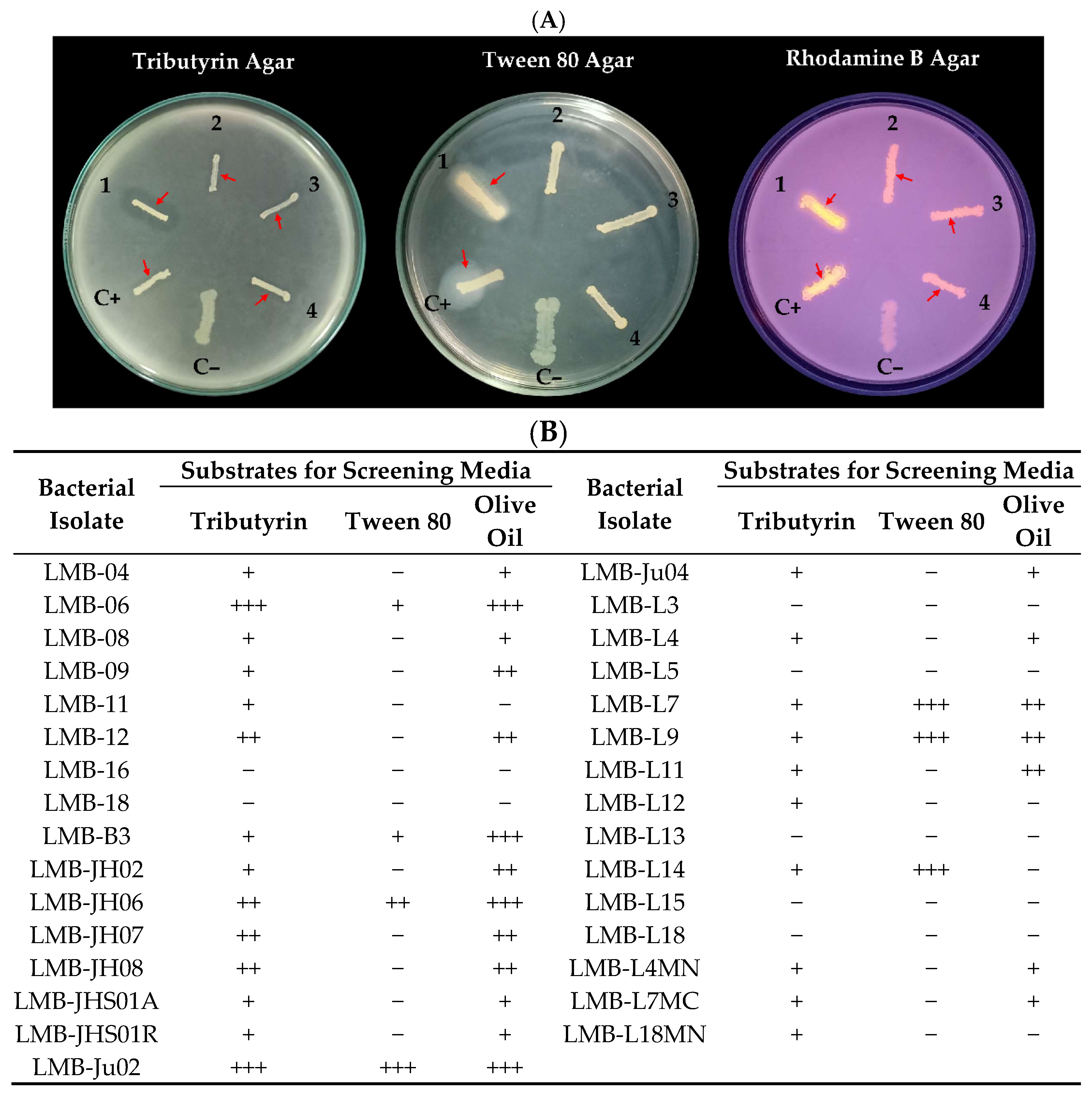
As shown in Figure 2, the preliminary qualitative screening for lipolytic activity using fatty acid esters as substrates (tributyrin, Tween 80, and olive oil) revealed a greater number of isolates (twenty-four) producing hydrolysis halos on tributyrin agar (TrA) plates, with isolates LMB-06 and LMB-Ju02 displaying the most pronounced halos. This outcome can be attributed to the ability of lipases, and some esterases, to hydrolyze tributyrin due to the short chain length of butyric acid (C4) and its high solubility in aqueous media [36]. Likewise, although Tween 80 contains a long-chain fatty acid (oleic acid; C18) in its structure, it is not a substrate specific to lipases, as some esterases can hydrolyze it as well [37]. However, only seven isolates exhibited hydrolytic activity on Tween 80 agar (TA) plates, with isolate LMB-Ju02 showing one of the largest precipitation halos, alongside isolates LMB-L7, LMB-L9 and LMB-L14.
The reduced halos developed by some strains on Tween 80 agar (TA) plates compared to tributyrin agar (TrA) plates, including isolates LMB-06 and LMB-Ju02, suggest that Tween 80 may act more as an inhibitor than as an inducer of lipase production. This effect could be attributed to its synthetic nature and surfactant properties, which differ from natural substrates such as triglycerides. A dual effect of Tween 80 on lipase production has been reported previously in various bacterial species, acting either as an inducer or an inhibitor depending on the microbial strain and culture conditions [27,38,39,40].
Regarding rhodamine agar (RA) plates, twenty isolates exhibited fluorescence under UV light, indicating their ability to hydrolyze olive oil. In this assay, isolates LMB-06, LMB-B3, LMB-JH06 and LMB-Ju02 exhibited the highest hydrolytic activity (Figure 2). Olive oil is considered an inducer of lipase production in various microorganisms and a robust substrate for lipase detection due to its high triglyceride content, primarily composed of oleic acid and other long-chain fatty acids [41]. However, its lower sensitivity and poor emulsification in aqueous media make it less commonly used than tributyrin in routine screening assays [42].
Based on these results, ten isolates exhibiting higher lipolytic activity on both tributyrin and olive oil were preselected for further characterization. These isolates were molecularly identified as members of the genera Serratia, Aeromonas, and Staphylococcus (Table 1).
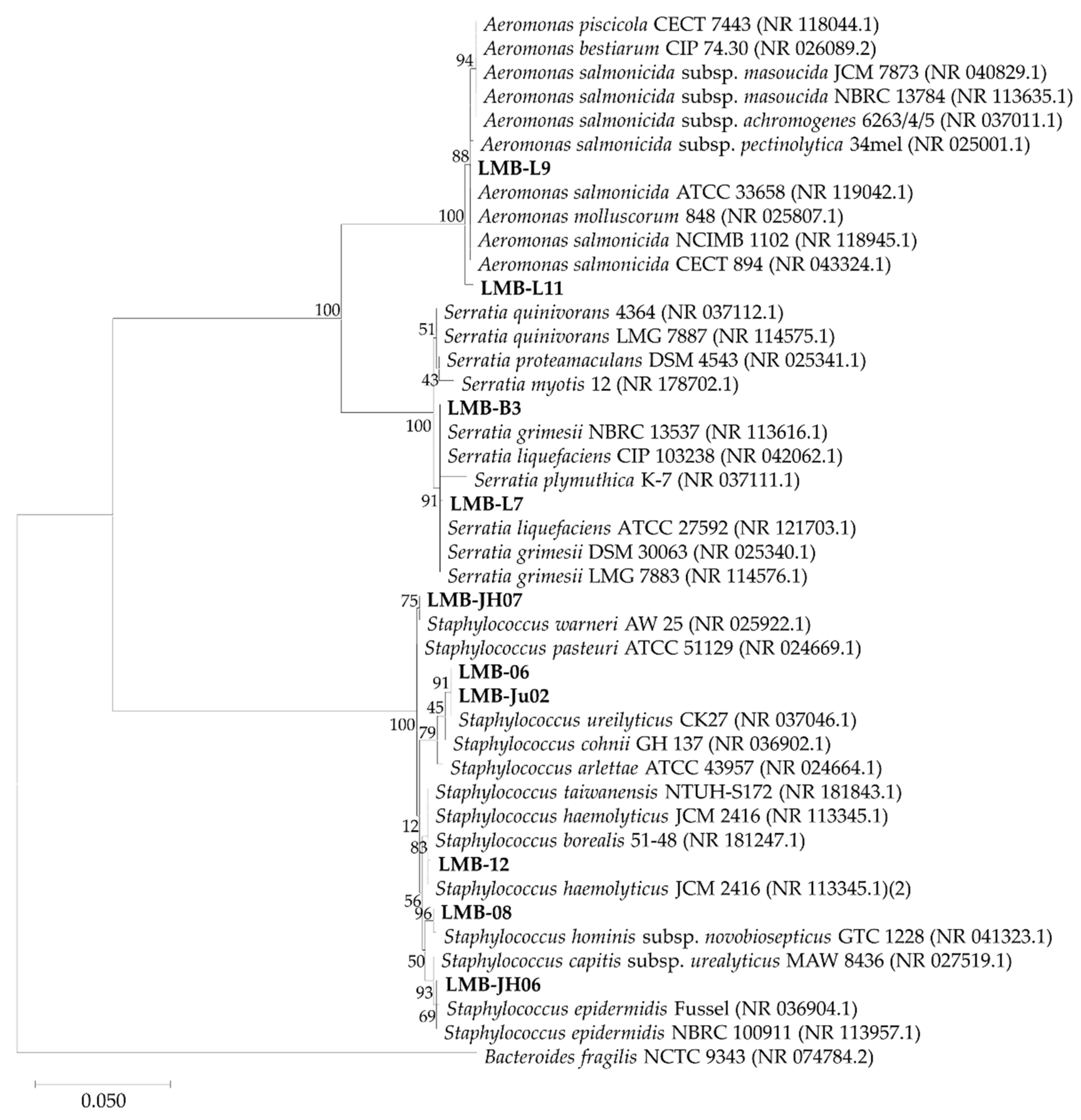
Phylogenetic analysis revealed that isolates LMB-L7 and LMB-B3 belong to the genus Serratia, showing the highest similarity to Serratia grimesii and Serratia liquefaciens, respectively (Figure 3). The genus Serratia has been previously reported in the intestinal microbiota of Mediterranean fish [43], Antarctic fish [44], rainbow trout (Oncorhynchus mykiss) [45], and rohu (Labeo rohita) [46]. Bacteria of this genus are known to produce nucleases, lipases, chitinases, and proteases, and are often selected using Tween 80. This is consistent with the precipitation halos observed on Tween 80 agar (TA) plates by isolates LMB-B3 and LMB-L7 (Figure 2).
Isolates LMB-L9 and LMB-L11 showed the highest similarity to various species within the genus Aeromonas, with A. salmonicida being the most frequently matched species (Figure 3). It has been reported that phylogenetic analysis based on the 16S rRNA gene is not sufficiently reliable for species-level identification within Aeromonas, since a single 16S sequence can align with multiple species, often exhibiting a high similarity percentage (99.8–100%). Therefore, alternative genes such as gyrB and rpoD are recommended for accurate species identification [47]. Aeromonas is considered an indigenous genus in aquatic environments, predominant in the microbiota of marine species, and is characterized by its capacity to secrete a wide range of extracellular enzymes, including lipases, proteases, amylases, chitinases, and nucleases [47,48].
The remaining isolates were identified as members of the genus Staphylococcus (Figure 3), which is also known for producing extracellular enzymes such as proteases and lipases [49]. Likewise, the presence of Staphylococcus spp. in different marine fish species has been previously reported. For instance, S. warneri and S. epidermidis, among other staphylococcal species, were isolated from the intestines of sub-tropical fish [50]; S. hominis from Mediterranean fish isolates [43,47]; S. equorum from Atlantic salmon and rainbow trout fed with vegetable oils [51,52]; and S. cohnii from the intestines of Sardinella longiceps, where lipase production was optimized through solid-state fermentation using anchovy processing wastes [53]. Additionally, the isolation of lipolytic S. epidermidis and S. cohnii subsp. urealyticus (HS S. ureilyticus) has been reported from shrimp gut and paste, respectively [30,54].

3.2. Yield and Productivity of Staphylococcus Lipases

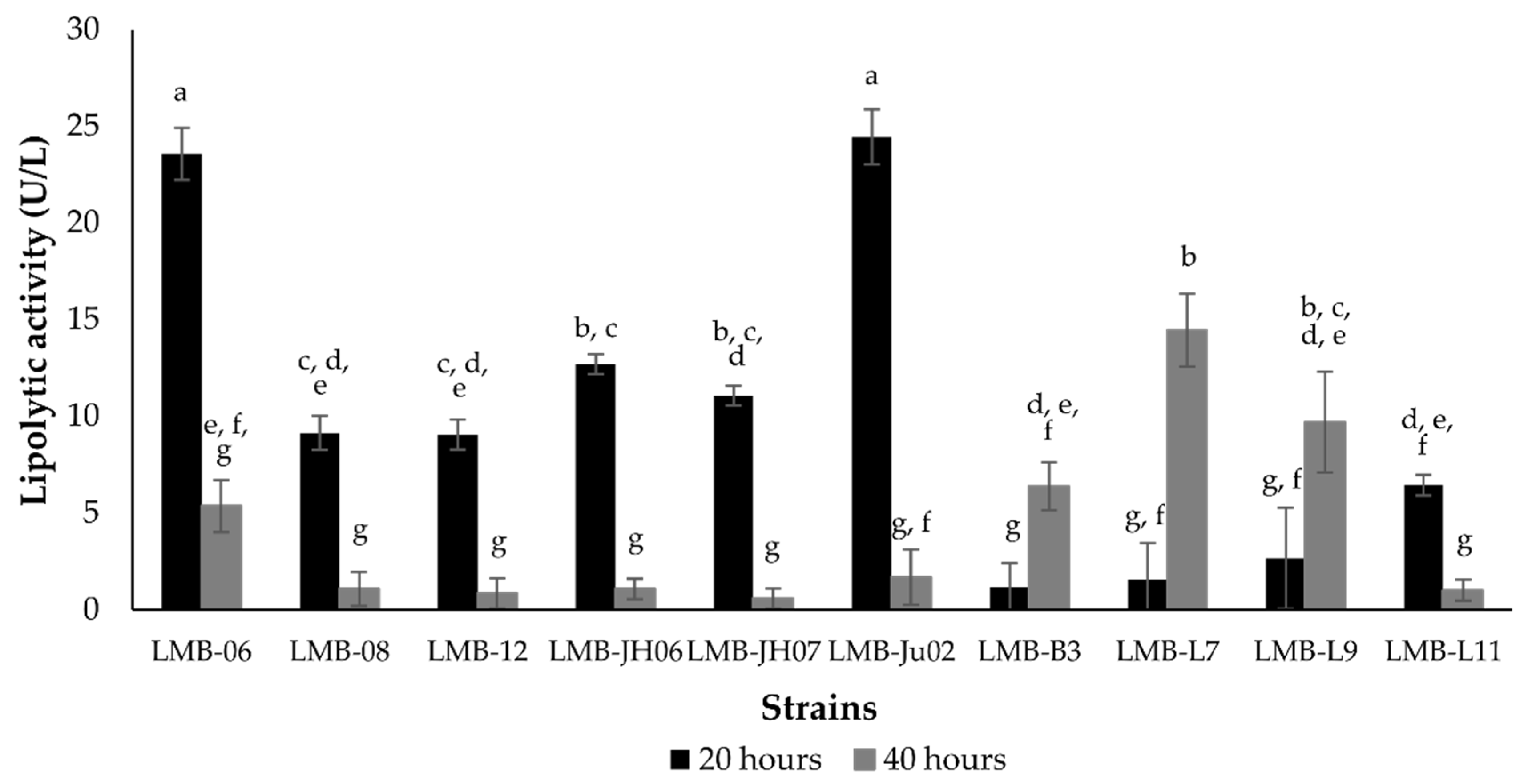
Additionally, a quantitative screening was performed to identify Staphylococcus isolates with the highest lipolytic activities. The results revealed that both incubation time and isolate identity had a significant effect on lipolytic activity (p < 0.05). Secreted enzymes from seven isolates (LMB-06, LMB-08, LMB-12, LMB-JH06, LMB-Ju02 and LMB-11) exhibited higher activity after 20 h of culture compared to 40 h (Figure 4). Consistent with previous results, isolates LMB-06 and LMB-Ju02 stood out, displaying the highest activity at 20 h (23.56 and 24.45 U/L, respectively). In contrast, isolates LMB-B3, LMB-L7, and LMB-L9 showed significantly lower levels at the same time point. Based on their superior mean activity levels, isolates LMB-06 and LMB-Ju02, along with LMB-JH06 and LMB-JH07, were selected for further evaluation of enzymatic yields and productivity.
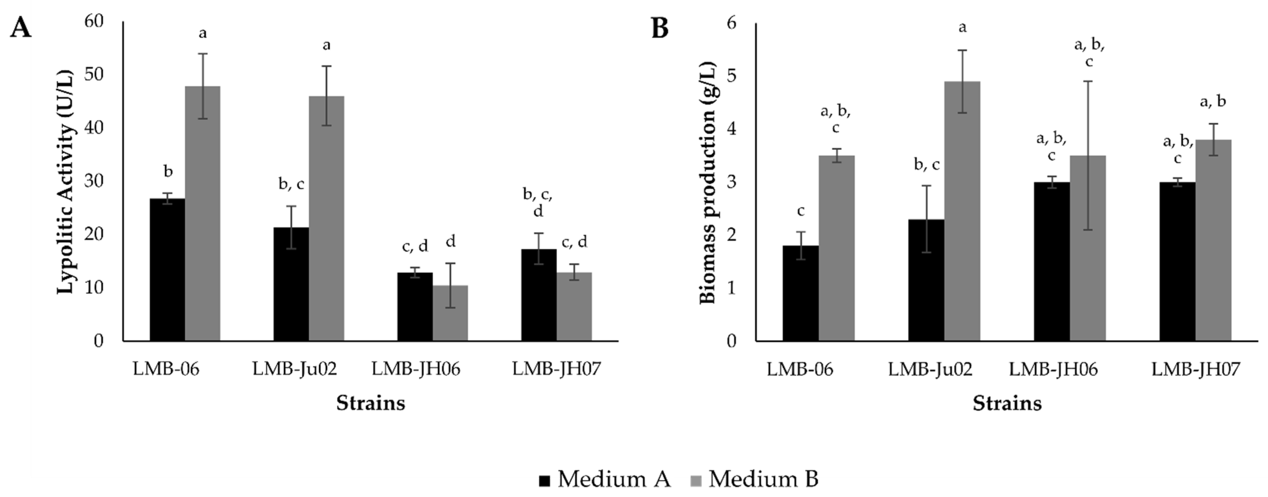
As depicted in Figure 5, the performance of selected Staphylococcus isolates was evaluated in two different production media. Results indicated that both isolate identity and culture medium significantly influenced lipolytic activity and biomass formation (p < 0.05). Strains LMB-06 and LMB-Ju02 exhibited higher lipolytic activity in both media being 1.7 and 2.2 more productive in Medium B (47.8 and 45.9 U/L, respectively). In contrast, strains LMB-JH06 and LMB-JH07 showed no significant differences between the two media, producing 12.8 and 17.3 U/L in Medium A, and 10.4 and 12.9 U/L in Medium B, respectively. Moreover, all four strains demonstrated improved growth in Medium B, with LMB-06 and LMB-Ju02 generating nearly twice the biomass compared to Medium A. Thus, S. ureilyticus strains LMB-06 and LMB-Ju02 were selected for subsequent lipase production assays in Medium B to further assess their lipolytic potential.
The media used differ both in the quantity and type of nitrogen sources. Medium B contains mainly organic nitrogen: twice as much peptone, one-third the amount of yeast extract, and an additional 0.5% [w/v] meat extract. In contrast, Medium A relies predominantly on 0.1% [w/v] ammonium phosphate as its inorganic nitrogen source. It is well established that the use of inorganic nitrogen sources generally leads to reduced lipase production. For example, Pseudomonas aeruginosa KM110 produced approximately 80% less lipase when cultivated in a medium containing ammonium phosphate compared to one supplemented with peptone [55]. Consequently, the higher concentration of organic nitrogen in Medium B likely contributed to the elevated lipase levels observed in strains LMB-06 and LMB-Ju02. Nonetheless, although nitrogen is critical for enzyme biosynthesis, excessive organic nitrogen may suppress lipolytic activity, possibly due to toxic effects or a metabolic shift that favors biomass accumulation over enzyme secretion [56].
Regarding the salt composition, Medium B contains four times more NaCl (1% [w/v]) and 2.5 times more MgSO4 (0.1% [w/v]) than Medium A. The influence of sodium salts on lipase production is known to vary depending on the microorganism. For instance, 2.3% [w/v] NaCl reduced lipolytic activity in S. aureus 111, while simultaneously enhancing biomass formation [38]. Conversely, NaCl stimulated lipase activity in S. epidermidis CMST-Pi 2 (isolated from shrimp gut) and S. cohnii AP-CMST (from Indian oil sardine), although activity declined significantly at concentrations above 4% [w/v] [30,53]. Likewise, a S. epidermidis strain obtained from spoiled frozen marine fish exhibited a 2.6-fold increase in lipolytic activity at 0.9% [w/v] NaCl [57]. In the halophilic species Alkalibacillus salilacus, both lipase production and biomass increased markedly, peaking at 17.5% [w/v] NaCl [58]. These observations suggest that the enhanced lipolytic activity in LMB-06 and LMB-Ju02 may also be influenced by the higher NaCl content in Medium B. Magnesium ions, on the other hand, play a fundamental role in ribosomal function and serve as cofactors for several key microbial enzymes. As with sodium, their impact on lipase production depends on both concentration and microbial species. For example, the addition of 0.02% [w/v] MgSO4 stimulated lipolytic activity in S. epidermidis CMST-Pi 1 (shrimp gut isolate) and S. cohnii AP-CMST [30,53]. Similarly, the previously mentioned marine-derived S. epidermidis strain responded positively to 0.9% [w/v] MgSO4, whereas in Bacillus coagulans, 0.1% [w/v] MgSO4 led to a reduction in enzyme output [59].
Additionally, Medium A includes CaCl2 (0.04% [w/v]), while this salt is absent in Medium B. Divalent cations such as Ca2+ have been shown to exert strain-specific effects on lipase production, either enhancing or inhibiting it [58]. This variability is evident in the case of the S. epidermidis strain from marine fish, which exhibited reduced lipolytic activity upon the addition of 0.9% [w/v] CaCl2 [57]. In contrast, both S. epidermidis CMST-Pi 1 and S. cohnii AP-CMST demonstrated increased enzyme production when 0.02% [w/v] CaCl2 was added to the basal medium [30,53].
Variations in enzymatic performance were also reflected in the lipase productivity parameters of the evaluated Staphylococcus strains. As shown in Table 2, strains LMB-06 and LMB-Ju02 exhibited the highest volumetric and specific productivity values, with no significant differences observed between the two media. In contrast, strains LMB-JH06 and LMB-JH07 showed improved yield and specific productivity in Medium A, suggesting a possible metabolic shift favoring enzyme synthesis over cellular growth under these conditions. For all strains, specific activity was consistently higher in Medium A, with S. warneri LMB-JH07 reaching the maximum value (556.4 U/g), indicating that the composition of this medium, particularly its salt and nitrogen content, may promote lipase production over other protein outputs. Overall, these results underscore the opportunity to enhance production through bioprocess optimization, paving the way for further analyses.

3.3. Partial Characterization of S. ureilitycus Secretomes

3.3.1. Effect of pH and Temperature on Enzymatic Activity and Stability

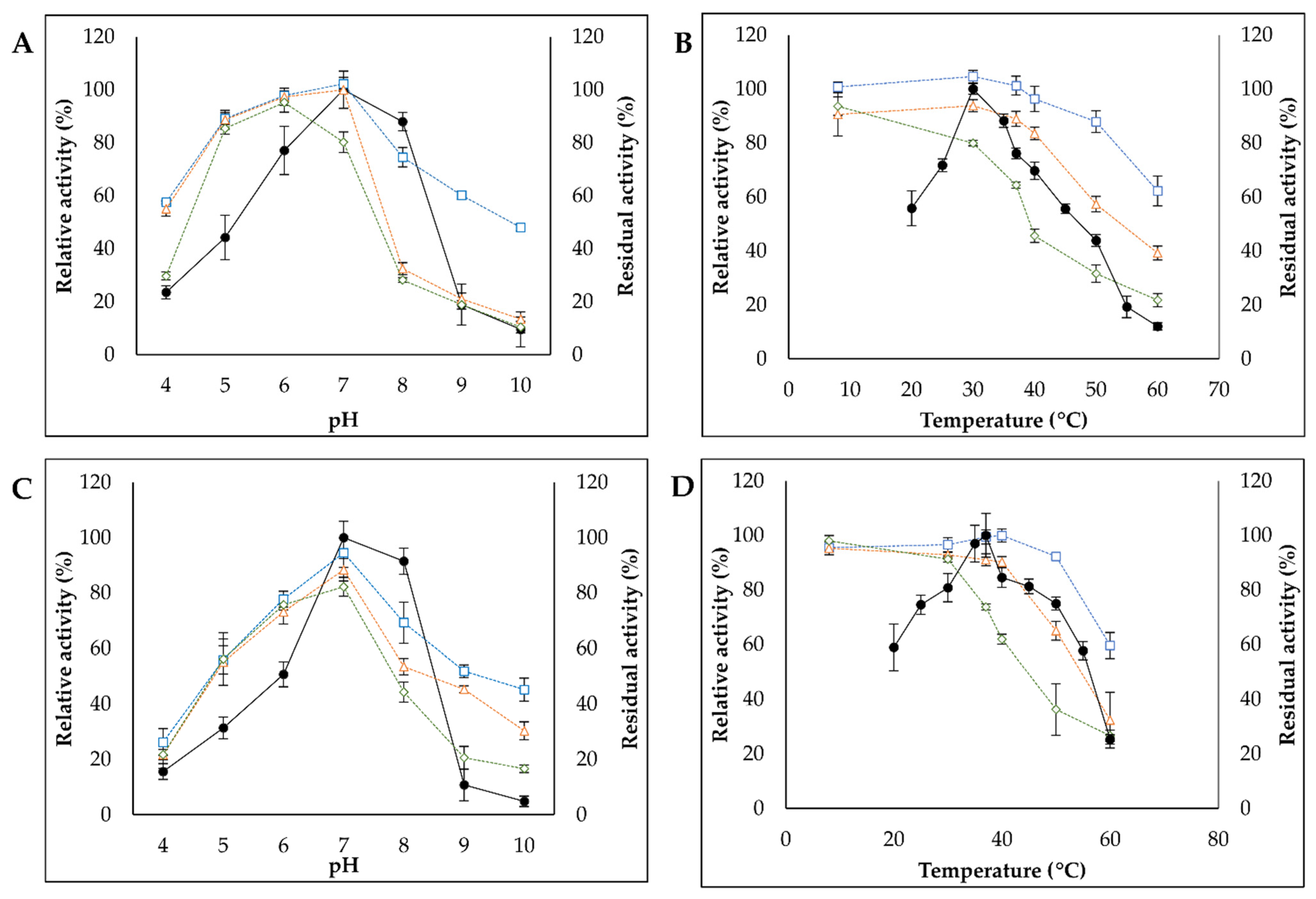
As shown in Figure 6, the effects of pH and temperature on both the activity and stability of S. ureilyticus secretomes were evaluated. For both strains (LMB-06 and LMB-Ju02), optimal lipase activity was observed between pH 7.0 and 8.0 (63.6 and 61.9 U/L, respectively), with more than 50% residual activity retained across the pH range of 6.0 to 8.0. Strain LMB-06 exhibited slightly higher activity than LMB-Ju02 between pH 4.0 and 6.0 (Figure 6A,C). Based on these results, pH 7.0 was selected as the optimal condition for subsequent assays. These findings are consistent with Rosenstein & Götz (2000) [60], who reported that optimal activity of Staphylococcus lipases generally falls within the pH range of 5.0 to 8.0. Similar behavior has been reported for lipases produced by S. lipolyticus sp. nov., isolated from marine waters and sediments [61], and by S. epidermidis strains from shrimp gut and marine fish [30,57], both showing optimal activity at pH 7.0.
Regarding the optimal temperature, lipases in the secretome of LMB-06 exhibited maximum activity at 30 °C (67.7 U/L), retaining more than 50% residual activity between 20 and 45 °C (Figure 6B). In contrast, the secretome of LMB-Ju02 showed optimal activity at 35–37 °C (57.7–59.5 U/L), maintaining over 50% residual activity across a broader temperature range of 20 to 55 °C (Figure 6D). These temperature optima are consistent with the range reported for lipases from various Staphylococcus species, including S. lipolyticus [61], S. arlettae, S. aureus and S. epidermidis [62,63], which typically exhibit optimal activity around 30 °C [30,61,62].
In terms of stability, the maximum activity of LMB-06 secretome was observed after up to 4 h of preincubation at pH 7.0. In contrast, preincubation under acidic (pH 4.0) and alkaline (pH 8.0–10.0) conditions, particularly at 4 and 18 h, significantly reduced lipolytic activity (Figure 6A). Conversely, the LMB-Ju02 secretome exhibited stability at pH 7.0 only during the first hour of preincubation, reaching activity levels above 100%. Significant decreases in residual activity were observed at pH values both below and above 7.0 (Figure 6C). These pH effects were more pronounced after 18 h of preincubation for both strains. These results indicate, on the one hand, that lipases present in the LMB-Ju02 secretome exhibit lower stability over time compared to those from strain LMB-06. On the other hand, while the pH stability profile of LMB-Ju02 lipases suggests a predominantly neutral behavior, those from LMB-06 demonstrate greater stability under mildly acidic conditions (pH 5.0–6.0). This characteristic contrasts with the common preference of bacterial lipases for slightly alkaline environments [6]. However, it is important to note that lipase behavior at different pH values may vary depending on the substrate used. For instance, Rhizomucor miehei has been reported to hydrolyze short-chain fatty acids at low pH, while at high pH its specificity increases for triglycerides with long-chain fatty acids [64].
In relation to the effects of temperature on enzyme stability, lipases present in the secretome of strain LMB-06 remained active over a broad temperature range (8–60 °C). Maximum hydrolytic activity was observed at 30 °C after the first hour of preincubation, with residual activity remaining above 60% even after pretreatment at 60 °C. Similarly, lipases produced by strain LMB-Ju02 were active across a wide temperature range, exhibiting maximum hydrolytic activity at 37 and 40 °C after one hour of preincubation and, as observed for LMB-06, maintaining residual activity above 60% following pretreatment at 60 °C. However, a gradual decline in lipolytic activity was recorded for both strains after 18 h of preincubation at temperatures above 30 °C. For LMB-06, residual activity dropped to 20% after 18 h of incubation at 60 °C (Figure 6B), whereas for LMB-Ju02, it decreased to approximately 25% after both 4 and 18 h of incubation at the same temperature (Figure 6D).

3.3.2. Effect of Metal Ions, Organic Solvents and Inhibitors on Enzymatic Activity and Stability

As shown in Figure 7A, the activity of lipases secreted by strain LMB-06 was significantly enhanced in the presence of Ca2+, Mg2+, and K+, with increases of 86.9%, 71.5%, and 51.2%, respectively. In contrast, Zn2+ completely inhibited enzymatic activity, while Cu2+ reduced it by 95.7%. For lipases produced by strain LMB-Ju02, a significant increase in activity (87.8%) was observed only in the presence of Ca2+. Conversely, Zn2+ and Cu2+ decreased activity by 75.5% and 73.6%, respectively, whereas Mg2+, Na+, and K+ had no significant effect on lipase activity.
These results are consistent with previous studies highlighting the importance of calcium as a cofactor for the activity of lipases produced by Staphylococcus strains. For example, the enhanced activity of lipases from S. caprae in the presence of Ca2+ has been mainly attributed to the stabilization of the enzyme’s active site [65]. Additionally, it has been suggested that calcium ions may bind to the fatty acids released after hydrolysis, leading to the formation of insoluble salts that could prevent active site saturation and medium acidification [66].
In the case of other metal ions such as Mg2+, Na+, and K+, variable effects on lipase activity have been reported. For instance, the addition of Mg2+ had no significant effect on the lipases produced by S. lipolyticus sp. nov. or S. caprae NCU S6 [61,65]. Conversely, Na+ enhanced the lipolytic activity of S. caprae NCU S6 lipases, while K+ had no effect [65]. In contrast, for the lipases from S. arlettae JPBW-1, both ions inhibited enzymatic activity [62]. Notably, the enhancing effect of Mg2+ and K+ on the lipase activity of LMB-06 has not been previously reported in other Staphylococcus lipases, suggesting that these enzymes possess distinct properties compared to those from other species. Finally, Zn2+ and Cu2+ ions have also been reported to exert varying effects on Staphylococcus lipases, from no detectable impact to significant inhibition, resulting in activity reductions of up to 70% [65,67], as observed for the lipases from strains LMB-06 and LMB-Ju02.
Solvent tolerance is a desirable characteristic of lipases, as it enhances their applicability in industrial processes such as ester synthesis and biotransformations involving poorly water-soluble substrates, including those encountered in biodiesel production, which involves hydrophobic triglycerides. In general, lipases tend to be less stable in polar, water-miscible solvents because these solvents disrupt the enzyme’s hydration shell, leading to conformational changes and loss of catalytic function [68,69]. Lipases from Rhizopus oryzae, for instance, are valued in industry due to their stability in water-immiscible solvents, such as hexane and long-chain alcohols, whereas water-miscible solvents, such as acetone and short-chain alcohols, often have detrimental effects on enzyme activity [70].
As shown in Figure 7B, the lipases secreted by strains LMB-06 and LMB-Ju02 exhibited residual activities of around 50% or less in the presence of 2-propanol and acetonitrile. In contrast, the activity of both enzymes was enhanced by ethanol, reaching relative activities of approximately 132% and 115%, respectively—values higher than those reported for S. caprae NCU S6 [65]. The activation of lipases by polar solvents has been attributed to the disruption of aggregates formed between enzyme molecules or between enzymes and residual lipids present in the fermentation medium [69]. Similar behavior has been reported for Bacillus megaterium lipases, which exhibited relative activities of 166% and 111% after incubation in 25% isopropanol and ethanol, respectively [68]. Notably, the ethanol-induced activation observed in the lipases from strains LMB-06 and LMB-Ju02 highlights their potential for applications in the synthesis of ethyl esters, both for nutritional uses and for biodiesel production.
Remarkably, a significant increase in relative activity was also observed for the lipases from LMB-06 after incubation with n-hexane and cyclohexane, reaching 154.24% and 159.37%, respectively. Meanwhile, the lipases from LMB-Ju02 showed increased activity only in cyclohexane, with a relative activity of 118.29% (Figure 7B). In some cases, enhanced activity in non-polar solvents has been associated with a conformational shift from the closed to the open form of the lipase [69]. The tolerance and activation observed in the presence of these non-polar solvents clearly exceed the solvent stability reported for other staphylococcal lipases, such as those from S. saprophyticus M36 and S. caprae NCU S6 [65,71].
Other lipases reported to be stable or activated in organic solvents include those from Burkholderia cepacia ST-200 (stable in cyclohexane), thermotolerant lipases from Pseudomonas cepacia (active in ethanol and acetonitrile), and those from Bacillus sphaericus 205y, which nearly triple their activity after incubation in p-xylene and n-hexane [72,73,74].
Hydrogen peroxide is a compound that frequently coexists with lipases in oleochemical reaction systems. It induces oxidative modification of amino acid residues such as cysteine, methionine, and tryptophan, which can lead to the disruption of disulfide bonds. These structural changes result in protein aggregation and consequent loss of enzymatic activity [65,75]. As shown in Figure 7C, the addition of 1% [v/v] H2O2 almost completely inhibited the activity of the lipases from strain LMB-Ju02, while significantly reducing the activity of those from strain LMB-06 by 59.8%. At a lower concentration (0.1% [v/v]), both strains maintained about 60% of their activity. These values indicate greater sensitivity to H2O2 when compared to the lipase SCNL from S. caprae NCU S6, which has been reported to retain higher activity under similar conditions [65].
Regarding the effect of EDTA, a known metal ion chelator, a concentration of 1 mM significantly inhibited the activity of the lipases secreted by strain LMB-06, with a reduction of over 60%. In contrast, the lipases from LMB-Ju02 exhibited only a 13.5% decrease, indicating greater stability in the presence of EDTA (Figure 7C). Notably, this resistance was superior not only to the lipases from LMB-06 but also to the SCNL lipase from S. caprae [65]. At 10 mM EDTA, the activity of the lipases decreased further, reaching 65% inhibition for LMB-06 and 51% for LMB-Ju02.
The significant enhancement of activity in the presence of Ca2+ and Mg2+, along with the inhibition observed upon EDTA treatment, suggests that the lipases from both S. ureilyticus strains may function as metalloenzymes. The stronger sensitivity of LMB-06-derived enzymes to EDTA also indicates a greater dependence on metal ions compared to those from LMB-Ju02. However, further studies using purified enzymes are needed to clarify the precise nature of these metal ion interactions.

3.4. Enzymatic Hydrolysis of Triglycerides by Lipases from S. ureilyticus Secretomes

Hydrolysis assays were conducted using four types of oils (olive, sunflower, palm, and crude fish oil), chosen for their distinct fatty acid compositions and degrees of saturation. Olive oil is commonly used for evaluating lipolytic activity due to its high content of monounsaturated fatty acids (MUFAs), particularly oleic acid (C18:1), which accounts for approximately 68% of its total fatty acids [76]. Sunflower oil is rich in polyunsaturated fatty acids (PUFAs), with linoleic acid (C18:2) representing about 61% [76]. Palm oil contains a high proportion of saturated fatty acids (~50%), mainly palmitic acid (C16:0) [77]. In contrast, anchovy oil is characterized by a high content of long-chain ω-3 PUFAs, particularly eicosapentaenoic acid (EPA, C20:5) and docosahexaenoic acid (DHA, C22:6), each comprising approximately 26%, followed by 22% palmitic acid and 17% oleic acid [78]. The physicochemical characteristics of the oils used were determined experimentally and are reported in Table 3.
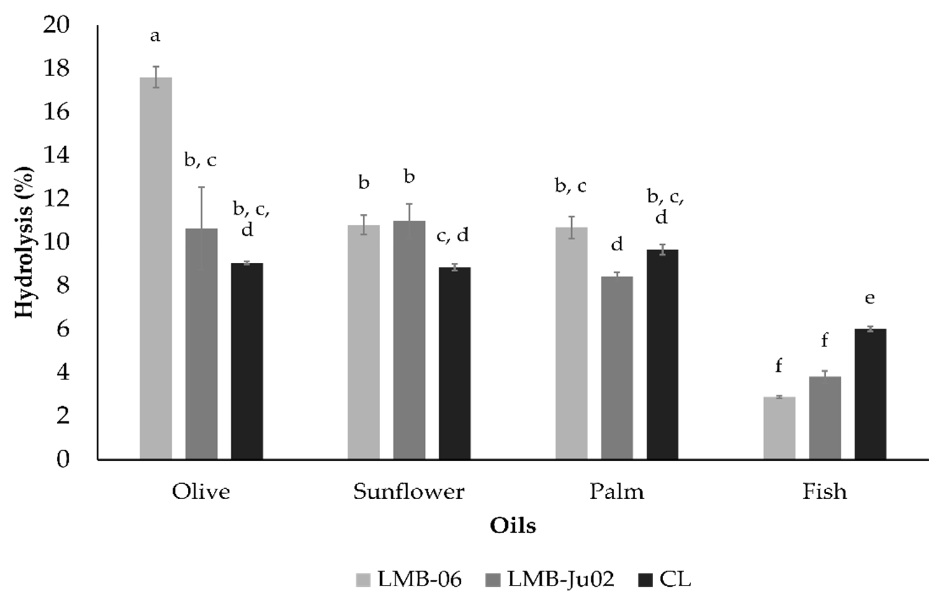
As shown in Figure 8, the lipases secreted by the S. ureilyticus strains LMB-06 and LMB-Ju02 hydrolyzed the vegetable oils to a considerably greater extent compared to the fish oil. In contrast, Xie (2012) [63] reported that lipases from S. epidermidis and S. aureus exhibited higher hydrolysis rates for fish oil than for olive oil, which may be attributed to differences in substrate specificity, as well as other factors such as oil quality. The lipases from strain LMB-06 achieved the highest hydrolysis rate with olive oil (17.6%), followed by sunflower oil (10.8%) and palm oil (10.7%), with no significant difference between the latter two, while the lowest hydrolysis level was observed with crude fish oil (2.9%). The lipases from strain LMB-Ju02 exhibited the highest hydrolysis rates with sunflower oil (11.0%) and olive oil (10.6%), with no statistically significant difference between them, whereas lower hydrolysis levels were obtained with palm oil (8.4%) and crude fish oil (3.8%). The hydrolytic performance of the commercial lipase, used as a positive control, was consistent across the three vegetable oils, with hydrolysis rates ranging from 8.8% to 9.7%. In the case of anchovy oil, although the hydrolysis rate was lower compared to the vegetable oils, the commercial lipase exhibited higher activity (6.0%) than the lipases secreted by both S. ureilyticus strains.
Although the relatively low hydrolysis yields observed do not reflect the full catalytic potential of the enzymes—mainly due to the limited amount of enzyme used (0.1 U)—the assay results nonetheless provided valuable insight into the oil selectivity of the lipases secreted by S. ureilyticus. The notably higher hydrolysis rate observed for olive oil indicates a differential efficiency depending on the oil composition. Lipases from the LMB-06 strain showed greater activity against substrates with a lower degree of saturation and simpler triacylglycerol profiles, such as those found in olive oil. However, to precisely determine specificity in terms of fatty acid composition, structural arrangement of the acylglycerols, and positional selectivity, it is necessary to perform hydrolysis assays using purified lipases and substrates with varying chain lengths and degrees of saturation, as well as to analyze the lipid profiles of the hydrolyzed oils in order to identify the fatty acids released in greater proportions. Overall, S. ureilyticus-derived lipases demonstrate promising catalytic performance on selected plant-based oils, supporting their potential application in sectors such as food processing and oleochemical modification [11,79].
The lower hydrolysis rates observed for crude fish oil in all enzymatic treatments, compared to the vegetable oils, may be partially explained by the higher content of free fatty acids present in the anchovy oil prior to enzymatic hydrolysis (Table 3). Unlike refined vegetable oils, which typically contain low levels of free fatty acids due to industrial processing, crude fish oils tend to exhibit a significantly higher acid value, indicating a substantial degree of prior triglyceride breakdown. As a result, the availability of intact ester bonds for enzymatic cleavage is reduced, leading to lower apparent hydrolysis. Additionally, the fish oil’s fatty acid composition— rich in long-chain ω-3 PUFAs—may also contribute to this outcome, as these complex substrates are more difficult for some microbial lipases to efficiently hydrolyze.

4. Conclusions

In conclusion, the lipases secreted by S. ureilyticus strains LMB-06 and LMB-Ju02, isolated from the E. ringens gut, exhibit desirable potential. Their stability of secretomes under mildly acidic conditions, combined with a broad temperature activity range, underscores their versatility for diverse applications. Their stability in the presence of hydrogen peroxide further supports their suitability for processing oils with high peroxide values, such as those encountered in waste oil recycling for biodiesel production [80]. In addition, their tolerance to solvents such as n-hexane, cyclohexane, and ethanol reinforces their applicability in the synthesis of ethyl and aromatic esters [81]. The positive influence of calcium ions observed in both strains suggests potential for promoting a more favorable enzyme conformation, thereby enhancing structural stability and catalytic efficiency. Interestingly, the beneficial effect of magnesium on the lipases from one strain, unreported in other Staphylococcus-derived lipases, may indicate distinctive structural features worthy of further investigation. Moreover, substrate preference towards triglycerides with lower saturation and simpler acylglycerol structures could guide targeted applications in industries that rely on plant-based oils. Nevertheless, to fully assess their suitability for specific industrial applications, further purification and detailed biochemical characterization of these enzymes are required. Finally, optimizing their production and reaction conditions will be essential to maximizing their functional and economic potential at an industrial scale.

Author Contributions

Conceptualization, G.K.V. and Y.L.; methodology and validation, M.H., A.B. and L.G.G.M.; formal analysis, M.H., J.S.B.P., I.S., Y.L. and G.K.V.; resources, I.S., Y.L. and G.K.V.; data curation, M.H.; writing—original draft preparation, M.H., I.S. and J.S.B.P.; writing—review and editing, Y.L. and G.K.V.; visualization, M.H., J.S.B.P., I.S., Y.L. and G.K.V.; supervision, J.S.B.P., Y.L., I.S. and G.K.V.; project administration, Y.L.; funding acquisition; I.S., Y.L. and G.K.V. All authors have read and agreed to the published version of the manuscript.

Funding

This research was funded by CONCYTEC (National Council of Science, Technology and Technological Innovation of Peru) through the grant No 371-2019-FONDECYT.

Institutional Review Board Statement

Not applicable.

Informed Consent Statement

Not applicable.

Data Availability Statement

The original data presented in the study are openly available in GeneBank (NCBI) at https://www.ncbi.nlm.nih.gov (accessed on 15 September 2025).

Acknowledgments

The authors would like to express their gratitude to Angélica López and Mary Pasmiño for their invaluable technical assistance and support during the course of this research.

Conflicts of Interest

The authors declare no conflicts of interest.

References

  1. Chandra, P.; Enespa; Singh, R.; Arora, P.K. Microbial Lipases and Their Industrial Applications: A Comprehensive Review. Microb. Cell Fact. 2020, 19, 169. [Google Scholar] [CrossRef] [PubMed]
  2. Mehta, A.; Guleria, S.; Sharma, R.; Gupta, R. The Lipases and Their Applications with Emphasis on Food Industry. In Microbial Biotechnology in Food and Health; Academic Press: Cambridge, MA, USA, 2021; pp. 143–164. [Google Scholar] [CrossRef]
  3. Casas-Godoy, L.; Gasteazoro, F.; Duquesne, S.; Bordes, F.; Marty, A.; Sandoval, G. Lipases: An Overview. In Lipases and Phospholipases; Sandoval, G., Ed.; Methods in Molecular Biology; Humana Press: New York, NY, USA, 2018; Volume 1835, pp. 1–28. [Google Scholar] [CrossRef]
  4. Future Market Insights. Lipase Market Outlook (2025–2035): Global Industry Analysis and Opportunity Assessment. 2024. Available online: https://www.futuremarketinsights.com/reports/lipase-market (accessed on 15 September 2025).
  5. Guerrand, D. Lipases Industrial Applications: Focus on Food and Agroindustries. OCL Oilseeds Fats Crops Lipids 2017, 24, D403. [Google Scholar] [CrossRef]
  6. Ali, S.; Khan, S.A.; Hamayun, M.; Lee, I.J. The Recent Advances in the Utility of Microbial Lipases: A Review. Microorganisms 2023, 11, 510. [Google Scholar] [CrossRef]
  7. Kumar, A.; Gudiukaite, R.; Gricajeva, A.; Sadauskas, M.; Malunavicius, V.; Kamyab, H.; Sharma, S.; Sharma, T.; Pant, D. Microbial Lipolytic Enzymes—Promising Energy-Efficient Biocatalysts in Bioremediation. Energy 2020, 192, 116674. [Google Scholar] [CrossRef]
  8. Javed, S.; Azeem, F.; Hussain, S.; Rasul, I.; Siddique, M.H.; Riaz, M.; Afzal, M.; Kouser, A.; Nadeem, H. Bacterial Lipases: A Review on Purification and Characterization. Prog. Biophys. Mol. Biol. 2018, 132, 23–34. [Google Scholar] [CrossRef] [PubMed]
  9. Vargas, M.; Niehus, X.; Casas-Godoy, L.; Sandoval, G. Lipases as Biocatalyst for Biodiesel Production. In Lipases and Phospholipases; Sandoval, G., Ed.; Methods in Molecular Biology; Humana Press: New York, NY, USA, 2018; Volume 1835, pp. 377–390. [Google Scholar] [CrossRef]
  10. Santos, S.; Puna, J.; Gomes, J. A Review on Bio-Based Catalysts (Immobilized Enzymes) Used for Biodiesel Production. Energies 2020, 13, 3013. [Google Scholar] [CrossRef]
  11. Biermann, U.; Bornscheuer, U.T.; Feussner, I.; Meier, M.A.; Metzger, J.O. Fatty Acids and Their Derivatives as Renewable Platform Molecules for the Chemical Industry. Angew. Chem. Int. Ed. 2021, 60, 20144–20165. [Google Scholar] [CrossRef]
  12. Zambare, V.; Patankar, R.; Bhusare, B.; Christopher, L. Recent Advances in Feedstock and Lipase Research and Development towards Commercialization of Enzymatic Biodiesel. Processes 2021, 9, 1743. [Google Scholar] [CrossRef]
  13. Rodrigues, J.G.C.; Cardoso, F.V.; Santos, C.C.d.; Matias, R.R.; Machado, N.T.; Duvoisin Junior, S.; Albuquerque, P.M. Biocatalyzed Transesterification of Waste Cooking Oil for Biodiesel Production Using Lipase from the Amazonian Fungus Endomelanconiopsis endophytica. Energies 2023, 16, 6937. [Google Scholar] [CrossRef]
  14. Rodrigues, C.J.C.; de Carvalho, C.C.C.R. Marine Bioprospecting, Biocatalysis and Process Development. Microorganisms 2022, 10, 1965. [Google Scholar] [CrossRef] [PubMed]
  15. Barzkar, N.; Sukhikh, S.; Babich, O. Study of Marine Microorganism Metabolites: New Resources for Bioactive Natural Products. Front. Microbiol. 2024, 14, 1285902. [Google Scholar] [CrossRef]
  16. Ragozzino, C.; Casella, V.; Coppola, A.; Scarpato, S.; Buonocore, C.; Consiglio, A.; Palma Esposito, F.; Galasso, C.; Tedesco, P.; Della Sala, G.; et al. Last Decade Insights in Exploiting Marine Microorganisms as Sources of New Bioactive Natural Products. Mar. Drugs 2025, 23, 116. [Google Scholar] [CrossRef]
  17. Huang, Q.; Sham, R.C.; Deng, Y.; Mao, Y.; Wang, C.; Zhang, T.; Leung, K.M.Y. Diversity of Gut Microbiomes in Marine Fishes Is Shaped by Host-Related Factors. Mol. Ecol. 2020, 29, 5019–5034. [Google Scholar] [CrossRef]
  18. Kim, P.S.; Shin, N.R.; Lee, J.B.; Kim, M.S.; Whon, T.W.; Hyun, D.W.; Yun, J.H.; Jung, M.J.; Kim, J.Y.; Bae, J.W. Host Habitat Is the Major Determinant of the Gut Microbiome of Fish. Microbiome 2021, 9, 166. [Google Scholar] [CrossRef]
  19. Ray, A.K.; Ghosh, K.; Ringø, E. Enzyme-Producing Bacteria Isolated from Fish Gut: A Review. Aquac. Nutr. 2012, 18, 465–492. [Google Scholar] [CrossRef]
  20. Hossain, T.J.; Chowdhury, S.I.; Mozumder, H.A.; Chowdhury, M.N.A.; Ali, F.; Rahman, N.; Dey, S. Hydrolytic Exoenzymes Produced by Bacteria Isolated and Identified from the Gastrointestinal Tract of Bombay Duck. Front. Microbiol. 2020, 11, 2097. [Google Scholar] [CrossRef] [PubMed]
  21. Hapsari, R.T.; Susilowati, A.; Pangastuti, A. Isolation and Identification of Lipolytic Bacteria from the Digestive Tract of Eel (Anguilla bicolor bicolor). Int. J. Bonorowo Wetl. 2020, 10, 44–50. [Google Scholar] [CrossRef]
  22. Uniacke-Lowe, S.; Stanton, C.; Hill, C.; Ross, R.P. The Marine Fish Gut Microbiome as a Source of Novel Bacteriocins. Microorganisms 2024, 12, 1346. [Google Scholar] [CrossRef] [PubMed]
  23. Dhayalan, A.; Jayanthi, K.; Manoharan, S.; Nadeem, A.; Govindasamy, B.; Pachiappan, P.; Vasudhevan, P. Fish Gut Symbiotic Bacterium Bacillus thuringiensis: RSM Optimization for Its Extracellular Lipase Enzyme Production, Lipase-Protein Purification, Characterization, and Docking Analysis. Int. J. Biol. Macromol. 2025, 301, 140428. [Google Scholar] [CrossRef]
  24. Espinoza, P.; Bertrand, A. Revisiting Peruvian Anchovy (Engraulis ringens) Trophodynamics Provides a New Vision of the Humboldt Current System. Prog. Oceanogr. 2008, 79, 215–227. [Google Scholar] [CrossRef]
  25. Sanchez, L.M.; Wong, W.R.; Riener, R.M.; Schulze, C.J.; Linington, R.G. Examining the Fish Microbiome: Vertebrate-Derived Bacteria as an Environmental Niche for the Discovery of Unique Marine Natural Products. PLoS ONE 2012, 7, e35398. [Google Scholar] [CrossRef]
  26. Coico, R. Gram Staining. Curr. Protoc. Microbiol. 2006, 1, A.3C.1–A.3C.2. [Google Scholar] [CrossRef] [PubMed]
  27. Ilesanmi, O.I.; Adekunle, A.E.; Omolaiye, J.A.; Olorode, E.M.; Ogunkanmi, A.L. Isolation, Optimization and Molecular Characterization of Lipase Producing Bacteria from Contaminated Soil. Sci. Afr. 2020, 8, e00279. [Google Scholar] [CrossRef]
  28. Ramnath, L.; Sithole, B.; Govinden, R. Identification of Lipolytic Enzymes Isolated from Bacteria Indigenous to Eucalyptus Wood Species for Application in the Pulping Industry. Biotechnol. Rep. 2017, 15, 114–124. [Google Scholar] [CrossRef]
  29. Veerapagu, M.; Sankara Narayan, A.; Ponmurugan, K.; Jeya, K.R. Screening Selection Identification Production and Optimization of Bacterial Lipase from Oil Spilled Soil. Asian J. Pharm. Clin. Res. 2013, 6, 62–67. [Google Scholar]
  30. Esakkiraj, P.; Rajkumarbharathi, M.; Palavesam, A.; Immanuel, G. Lipase Production by Staphylococcus epidermidis CMST-Pi 1 Isolated from the Gut of Shrimp Penaeus indicus. Ann. Microbiol. 2010, 60, 37–42. [Google Scholar] [CrossRef]
  31. Link, A.J.; LaBaer, J. Trichloroacetic Acid (TCA) Precipitation of Proteins. Cold Spring Harb. Protoc. 2011, 2011, 993–994. [Google Scholar] [CrossRef] [PubMed]
  32. Lowry, O.H.; Rosebrough, N.J.; Farr, A.L.; Randall, R.J. Protein Measurement with the Folin Phenol Reagent. J. Biol. Chem. 1951, 193, 265–275. [Google Scholar] [CrossRef]
  33. Tripathi, R.; Singh, J.; Bharti, R.K.; Thakur, I.S. Isolation, Purification and Characterization of Lipase from Microbacterium sp. and Its Application in Biodiesel Production. Energy Procedia 2014, 54, 518–529. [Google Scholar] [CrossRef]
  34. Mustranta, A. Use of Lipases in the Resolution of Racemic Ibuprofen. Appl. Microbiol. Biotechnol. 1992, 38, 61–66. [Google Scholar] [CrossRef]
  35. Yan, J.; Liu, S.; Hu, J.; Gui, X.; Wang, G.; Yan, Y. Enzymatic Enrichment of Polyunsaturated Fatty Acids Using Novel Lipase Preparations Modified by Combination of Immobilization and Fish Oil Treatment. Bioresour. Technol. 2011, 102, 7154–7158. [Google Scholar] [CrossRef]
  36. Camacho-Ruiz, M.D.L.A.; Mateos-Díaz, J.C.; Carrière, F.; Rodriguez, J.A. A Broad pH Range Indicator-Based Spectrophotometric Assay for True Lipases Using Tributyrin and Tricaprylin. J. Lipid Res. 2015, 56, 1057–1067. [Google Scholar] [CrossRef]
  37. Rollof, J.; Hedstrom, S.A.; Nilsson-Ehle, P. The Tween 80 Reaction Does Not Correlate to Triglyceride Lipase Production of Staphylococcus Aureus. Apmis 1988, 96, 732–734. [Google Scholar] [CrossRef]
  38. Mates, A.; Sudakevitz, D. Production of Lipase by Staphylococcus aureus under Various Growth Conditions. J. Appl. Bacteriol. 1973, 36, 219–226. [Google Scholar] [CrossRef] [PubMed]
  39. Boekema, B.K.H.L.; Beselin, A.; Breuer, M.; Hauer, B.; Koster, M.; Rosenau, F.; Jaeger, K.; Tommassen, J. Hexadecane and Tween 80 Stimulate Lipase Production in Burkholderia glumae by Different Mechanisms. Appl. Environ. Microbiol. 2007, 73, e00097-07. [Google Scholar] [CrossRef]
  40. Long, Z.D.; Xu, J.H.; Pan, J. Significant Improvement of Serratia marcescens Lipase Fermentation by Optimizing Medium, Induction, and Oxygen Supply. Appl. Biochem. Biotechnol. 2007, 142, 148–157. [Google Scholar] [CrossRef] [PubMed]
  41. Zarevúcka, M. Olive Oil as Inductor of Microbial Lipase. In Olive Oil—Constituents, Quality, Health Properties and Bioconversions; IntechOpen: London, UK, 2012; pp. 457–470. [Google Scholar] [CrossRef]
  42. Ciafardini, G.; Zullo, B.A. Virgin Olive Oil Yeasts: A Review. Food Microbiol. 2018, 70, 245–253. [Google Scholar] [CrossRef] [PubMed]
  43. Jammal, A.; Bariche, M.; Dohna, H.; Kambris, Z. Characterization of the Cultivable Gut Microflora in Wild-Caught Mediterranean Fish Species. Curr. Nutr. Food Sci. 2017, 13, 147–154. [Google Scholar] [CrossRef]
  44. Xiao, Y.; Yan, F.; Cui, Y.; Du, J.; Hu, G.; Zhai, W.; Liu, R.; Zhang, Z.; Fang, J.; Chen, L.; et al. A Symbiotic Bacterium of Antarctic Fish Reveals Environmental Adaptability Mechanisms and Biosynthetic Potential towards Antibacterial and Cytotoxic Activities. Front. Microbiol. 2023, 13, 1085063. [Google Scholar] [CrossRef]
  45. Parshukov, A.N.; Kashinskaya, E.N.; Simonov, E.P.; Hlunov, O.V.; Izvekova, G.I.; Andree, K.B.; Solovyev, M.M. Variations of the Intestinal Gut Microbiota of Farmed Rainbow Trout, Oncorhynchus mykiss (Walbaum), Depending on the Infection Status of the Fish. J. Appl. Microbiol. 2019, 127, 379–395. [Google Scholar] [CrossRef]
  46. Kawser, A.Q.M.R.; Foysal, M.J.; Chua, E.G.; Ali, M.H.; Mannan, A.; Siddik, M.A.B.; Paul, S.I.; Rahman, M.M.; Tay, A. Microbiome Data Reveal Significant Differences in the Bacterial Diversity in Freshwater Rohu (Labeo rohita) across the Supply Chain in Dhaka, Bangladesh. Lett. Appl. Microbiol. 2022, 75, 813–823. [Google Scholar] [CrossRef]
  47. Figueras, M.J.; Beaz-Hidalgo, R. Aeromonas: Introduction. In Encyclopedia of Food Microbiology, 2nd ed.; Academic Press: Cambridge, MA, USA, 2014; Volume 1, pp. 24–30. [Google Scholar] [CrossRef]
  48. Gómez, G.D.; Balcázar, J.L. A Review on the Interactions between Gut Microbiota and Innate Immunity of Fish. FEMS Immunol. Med. Microbiol. 2008, 52, 145–154. [Google Scholar] [CrossRef] [PubMed]
  49. Gillaspy, A.G.; Iandolo, J.J. Staphylococcus. In Encyclopedia of Food Microbiology; Batt, C.A., Tortorello, M.L., Eds.; Academic Press: London, UK, 2014; Volume 3, pp. 482–486. [Google Scholar] [CrossRef]
  50. Al Bulushi, I.M.; Poole, S.E.; Barlow, R.; Deeth, H.C.; Dykes, G.A. Speciation of Gram-Positive Bacteria in Fresh and Ambient-Stored Sub-Tropical Marine Fish. Int. J. Food Microbiol. 2010, 138, 32–38. [Google Scholar] [CrossRef] [PubMed]
  51. Askarian, F.; Zhou, Z.; Olsen, R.E.; Sperstad, S.; Ringø, E. Culturable Autochthonous Gut Bacteria in Atlantic Salmon (Salmo salar L.) Fed Diets with or without Chitin: Characterization by 16S rRNA Gene Sequencing, Ability to Produce Enzymes and in Vitro Growth Inhibition of Four Fish Pathogens. Aquaculture 2012, 326–329, 1–8. [Google Scholar] [CrossRef]
  52. Ringø, E.; Zhou, Z.; Vecino, J.L.G.; Wadsworth, S.; Romero, J.; Krogdahl, A.; Olsen, R.E.; Dimitroglou, A.; Foey, A.; Davies, S.; et al. Effect of Dietary Components on the Gut Microbiota of Aquatic Animals. A Never-Ending Story? Aquac. Nutr. 2016, 22, 219–282. [Google Scholar] [CrossRef]
  53. Esakkiraj, P.; Nawas, P.M.A.; Thomas, S.K.; Maruthiah, T.; Palavesam, A.; Immanuel, G. Solid-State Lipase Production by Staphylococcus cohnii AP-CMST Using Anchovy Processing Wastes. J. Pure Appl. Microbiol. 2013, 7, 637–644. [Google Scholar]
  54. Daroonpunt, R.; Saeng-in, P.; Tanasupawat, S. Identification and Lipolytic Activity of Bacillus and Staphylococcus Strains from Shrimp Paste (Ka-Pi). J. Appl. Pharm. Sci. 2019, 9, 24–29. [Google Scholar] [CrossRef]
  55. Mobarak-Qamsari, E.; Kasra-Kermanshahi, R.; Moosavi-Nejad, Z. Isolation and Identification of a Novel, Lipase-Producing Bacterium, Pseudomonas aeruginosa KM110. Iran. J. Microbiol. 2011, 3, 92–98. [Google Scholar]
  56. de Almeida, A.F.; Tauk-Tornisielo, S.M.; Carmona, E.C. Acid Lipase from Candida viswanathii: Production, Biochemical Properties, and Potential Application. BioMed Res. Int. 2013, 2013, 435818. [Google Scholar] [CrossRef]
  57. Joseph, B.; Ramteke, P.W.; Kumar, P.A. Studies on the Enhanced Production of Extracellular Lipase by Staphylococcus epidermidis. J. Gen. Appl. Microbiol. 2006, 52, 315–320. [Google Scholar] [CrossRef]
  58. Samaei-Nouroozi, A.; Rezaei, S.; Khoshnevis, N.; Doosti, M.; Hajihoseini, R.; Khoshayand, M.R.; Faramarzi, M.A. Medium-Based Optimization of an Organic Solvent-Tolerant Extracellular Lipase from the Isolated Halophilic Alkalibacillus salilacus. Extremophiles 2015, 19, 933–947. [Google Scholar] [CrossRef]
  59. Alkan, H.; Baysal, Z.; Uyar, F.; Dogru, M. Production of Lipase by a Newly Isolated Bacillus coagulans under Solid-State Fermentation Using Melon Wastes. Appl. Biochem. Biotechnol. 2007, 136, 183–192. [Google Scholar] [CrossRef] [PubMed]
  60. Rosenstein, R.; Götz, F. Staphylococcal Lipases: Biochemical and Molecular Characterization. Biochimie 2000, 82, 1005–1014. [Google Scholar] [CrossRef] [PubMed]
  61. Arora, P.K. Staphylococcus lipolyticus sp. nov., a New Cold-Adapted Lipase Producing Marine Species. Ann. Microbiol. 2013, 63, 913–922. [Google Scholar] [CrossRef]
  62. Chauhan, M.; Garlapati, V.K. Production and Characterization of a Halo-, Solvent-, Thermo-Tolerant Alkaline Lipase by Staphylococcus arlettae JPBW-1, Isolated from Rock Salt Mine. Appl. Biochem. Biotechnol. 2013, 171, 1429–1443. [Google Scholar] [CrossRef]
  63. Xie, W. Characterization of Lipases from Staphylococcus aureus and Staphylococcus epidermidis Isolated from Human Facial Sebaceous Skin. J. Microbiol. Biotechnol. 2012, 22, 84–91. [Google Scholar] [CrossRef]
  64. Rodrigues, R.C.; Fernandez-Lafuente, R. Lipase from Rhizomucor miehei as a Biocatalyst in Fats and Oils Modification. J. Mol. Catal. B Enzym. 2010, 66, 15–32. [Google Scholar] [CrossRef]
  65. Zhao, J.; Ma, M.; Zeng, Z.; Yu, P.; Gong, D.; Deng, S. Production, Purification and Biochemical Characterisation of a Novel Lipase from a Newly Identified Lipolytic Bacterium Staphylococcus caprae NCU S6. J. Enzyme Inhib. Med. Chem. 2021, 36, 248–256. [Google Scholar] [CrossRef]
  66. Kambourova, M.; Kirilova, N.; Mandeva, R.; Derekova, A. Purification and Properties of Thermostable Lipase from a Thermophilic Bacillus stearothermophilus MC 7. J. Mol. Catal. B Enzym. 2003, 22, 307–313. [Google Scholar] [CrossRef]
  67. Cherif, S.; Mnif, S.; Hadrich, F.; Abdelkafi, S.; Sayadi, S. A Newly High Alkaline Lipase: An Ideal Choice for Application in Detergent Formulations. Lipids Health Dis. 2011, 10, 221. [Google Scholar] [CrossRef] [PubMed]
  68. Lima, V.M.G.; Krieger, N.; Mitchell, D.A.; Baratti, J.C.; Filippis, I.D.; Fontana, J.D. Evaluation of the Potential for Use in Biocatalysis of a Lipase from a Wild Strain of Bacillus megaterium. J. Mol. Catal. B Enzym. 2004, 31, 53–61. [Google Scholar] [CrossRef]
  69. Kumar, A.; Dhar, K.; Kanwar, S.S.; Arora, P.K. Lipase Catalysis in Organic Solvents: Advantages and Applications. Biol. Proced. Online 2016, 18, 2. [Google Scholar] [CrossRef]
  70. López-Fernández, J.; Benaiges, M.D.; Valero, F. Rhizopus oryzae Lipase, a Promising Industrial Enzyme: Biochemical Characteristics, Production and Biocatalytic Applications. Catalysts 2020, 10, 1277. [Google Scholar] [CrossRef]
  71. Fang, Y.; Lu, Z.; Lv, F.; Bie, X.; Liu, S.; Ding, Z.; Xu, W. A Newly Isolated Organic Solvent Tolerant Staphylococcus saprophyticus M36 Produced Organic Solvent-Stable Lipase. Curr. Microbiol. 2006, 53, 510–515. [Google Scholar] [CrossRef] [PubMed]
  72. Sugihara, A.; Ueshima, M.; Shimada, Y.; Tsunasawa, S.; Tominaga, Y. Purification and Characterization of a Novel Thermostable Lipase from Pseudomonas cepacia. J. Biochem. 1992, 112, 598–603. [Google Scholar] [CrossRef]
  73. Takeda, Y.; Aono, R.; Doukyu, N. Purification, Characterization, and Molecular Cloning of Organic-Solvent-Tolerant Cholesterol Esterase from Cyclohexane-Tolerant Burkholderia cepacia Strain ST-200. Extremophiles 2006, 10, 269–277. [Google Scholar] [CrossRef]
  74. Hun, C.J.; Rahman, R.N.Z.A.; Salleh, A.B.; Basri, M. A Newly Isolated Organic Solvent Tolerant Bacillus sphaericus 205y Producing Organic Solvent-Stable Lipase. Biochem. Eng. J. 2003, 15, 147–151. [Google Scholar] [CrossRef]
  75. Jiang, H.; Jia, W.; Duan, M.; Lin, H.; Huang, J.; Li, X.; Shu, Z. Enhancement of Hydrogen Peroxide Tolerance of Lipase LipA from Bacillus subtilis Using Semi-Rational Design. Biochem. Eng. J. 2020, 159, 107590. [Google Scholar] [CrossRef]
  76. Orsavova, J.; Misurcova, L.; Ambrozova, J.; Vicha, R.; Mlcek, J. Fatty Acids Composition of Vegetable Oils and Its Contribution to Dietary Energy Intake and Dependence of Cardiovascular Mortality on Dietary Intake of Fatty Acids. Int. J. Mol. Sci. 2015, 16, 12871–12890. [Google Scholar] [CrossRef]
  77. Alves, A.Q.; da Silva, V.A., Jr.; Góes, A.J.S.; Silva, M.S.; de Oliveira, G.G.; Bastos, I.V.G.A.; Alves, A.J. The Fatty Acid Composition of Vegetable Oils and Their Potential Use in Wound Care. Adv. Skin Wound Care 2019, 32, 1–8. [Google Scholar] [CrossRef]
  78. Codex Alimentarius Commission. Standard for Fish Oils (CXS 329-2017). Available online: https://www.fao.org/fao-who-codexalimentarius/sh-proxy/es/?lnk=1&url=https%253A%252F%252Fworkspace.fao.org%252Fsites%252Fcodex%252FStandards%252FCXS%2B329-2017%252FCXS_329e.pdf (accessed on 15 September 2025).
  79. Dyer, J.M.; Stymne, S.; Green, A.G.; Carlsson, A.S. High-value oils from plants. Plant J. 2008, 54, 640–655. [Google Scholar] [CrossRef] [PubMed]
  80. Souza, G.P.R.; Correia, T.B.A.; Reis, W.S.M.; Bredda, E.H.; Da Rós, P.C.M.; Pereira, E.B. Enzymatic hydrolysis of waste cooking oil by lipase catalysis: Simplex mixture design optimization. Catal. Lett. 2023, 153, 689–697. [Google Scholar] [CrossRef]
  81. Sharma, S.; Kanwar, S.S. Organic solvent tolerant lipases and applications. Sci. World J. 2014, 2014, 625258. [Google Scholar] [CrossRef] [PubMed]
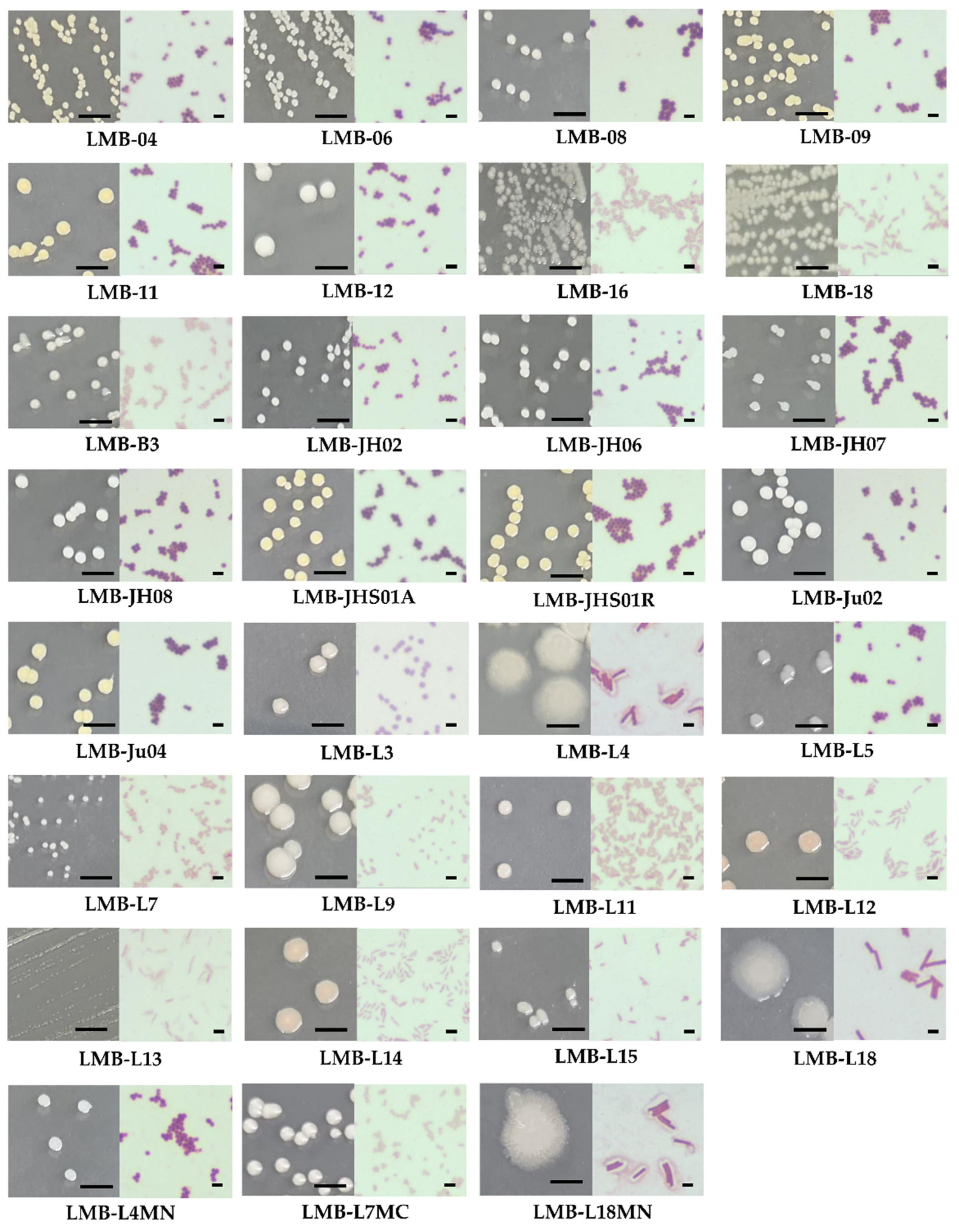
Figure 1. Morphological appearance of colonies grown on nutrient agar for 24 h (left panels) and corresponding Gram-stained micrographs of bacterial cells (right panels) isolated from anchovy gut. Scale bars: 0.5 cm (colonies); 2 µm (micrographs).
Applsci 15 10930 g001
Figure 2. Screening of lipolytic bacteria isolated from anchovy gut. (A) Representative images of bacterial isolates showing lipolytic activity on Tributyrin Agar, Tween 80 Agar, and Rhodamine B Agar plates. (1–4): Bacterial isolates (1: LMB-Ju02; 2: LMB-Ju04; 3: LMB-JHS01A; 4: LMB-JHS01R); (C+): Positive control (Staphylococcus aureus ATCC 25923); (C−): Negative control (Escherichia coli ATCC 25922). Red arrows indicate (i) clear halos of tributyrin hydrolysis, (ii) calcium salt precipitation halos from Tween 80 hydrolysis, and (iii) fluorescence under UV light due to rhodamine B–olive oil hydrolysis, respectively. (B) Summary table of lipolytic activity results for all 31 bacterial isolates tested on the three media. Symbols indicate relative enzymatic activity estimated by visual inspection: (−) no activity; (+) weak; (++) moderate; (+++) strong.
Applsci 15 10930 g002
Figure 3. Representative maximum likelihood phylogenetic tree based on the 16S rRNA gene sequences of lipase-producing bacterial isolates LMB-B3, LMB-06, LMB-L7, LMB-08, LMB-L9, LMB-L11, LMB-12, LMB-JH06, LMB-JH07, and LMB-Ju02. Numbers next to the branches indicate the percentage of bootstrap replicates supporting each node. The scale bar represents the number of substitutions per site. The 16S rRNA gene of Bacteroides fragilis NCTC 9343 was used as an outgroup.
Applsci 15 10930 g003
Figure 4. Lipolytic activity (U/L) of crude secretomes from preselected bacterial strains cultured for 20 and 40 h in production medium A. Enzymatic activity was determined at 37 °C and pH 7.0 using pNPL as the substrate. Values represent the mean of three independent replicates (n = 3); error bars indicate standard deviations (SD). Statistical significance of lipolytic activities between isolates grown for 20 h or 40 h is indicated by lowercase letters (a, b, c, d, e, f, g). All comparisons were analyzed using two-way ANOVA (strain x hours) followed by Tukey’s HSD post hoc test (p < 0.05).
Applsci 15 10930 g004
Figure 5. Lipolytic activity and biomass formation of Staphylococcus strains LMB-06, LMB-Ju02, LMB-JH06, and LMB-JH07 cultured for 20 h in two different production media (A and B). (A) Lipolytic activity (U/L) of crude enzyme determined at 37 °C and pH 7.0 using pNPL as the substrate. (B) Biomass (g) measured after 20 h of cultivation. Values represent the mean of three independent replicates (n = 3); error bars indicate standard deviations (SD). Statistical significance of lipolytic activities (A) and biomass formation (B) between isolates grown in Medium A or Medium B is indicated by lowercase letters (a, b, c, d). All comparisons were analyzed using two-way ANOVA (strain x medium) followed by Tukey’s HSD post hoc test (p < 0.05).
Applsci 15 10930 g005
Figure 6. Effect of pH and temperature on the lipase activity and stability of secretomes from S. ureilyticus strains LMB-06 (A,B) and LMB-Ju02 (C,D), cultured for 20 h in production Medium B. Relative activity (%) was calculated by considering the maximum lipolytic activity as 100%; Residual activity (%) was determined after preincubation of the enzymatic extract at different temperatures or pH values, using the initial activity as 100%. Enzymatic activity was assayed at pH 7.0 using pNPL as the substrate. Filled circles (●) represent the effect of pH or temperature on enzyme activity; unfilled symbols represent the effect on enzyme stability after 1 (), 4 () and 18 h () of incubation. Values represent the mean of three independent replicates (n = 3); error bars indicate standard deviations (SD).
Applsci 15 10930 g006
Figure 7. Effect of metal ions (A), organic solvents (B) and other compounds (C) on lipase activity and stability of crude enzymes secreted by S. ureilyticus strains LMB-06 and LMB-Ju02 cultured for 20 h in production Medium B. Relative activity (%) was calculated considering the maximum lipolytic activity as 100%; Residual activity (%) was calculated after pre-incubating the enzymatic extract in the presence of different organic solvents considering the initial activity as 100%. Enzymatic activity was determined at pH 7.0 and optimal temperature using pNPL as the substrate. Values represent the mean of three independent replicates (n = 3); error bars indicate standard deviations (SD). Statistical significance of the relative or residual lipolytic activity, with respect to the control group (secretomes without additives or without preincubation with solvents, respectively) is indicated by asterisks (*). All comparisons were analyzed using one-way ANOVA followed by Dunnett’s post hoc test (p < 0.05).
Applsci 15 10930 g007
Figure 8. Hydrolysis (%) of different oils by lipases from the secretome of S. ureilyticus strains LMB-06 and LMB-Ju02. A commercial lipase (CL) was used as positive control. Triglyceride hydrolysis (%) was determined after 18 h of incubation with 0.1 U of enzymes. Values represent the mean of three independent replicates (n = 3); error bars indicate standard deviations (SD). Statistical significance of hydrolysis (%) between different oils treated with the secretomes or the positive control is indicated by lowercase letters (a, b, c, d, e, f). All comparisons were analyzed using two-way ANOVA followed by Tukey’s HSD post hoc test (p < 0.05).
Applsci 15 10930 g008
Table 1. Identities of preselected lipase-producing bacterial isolates.
Bacterial IsolateIdentityAccession N°
LMB-L7Serratia sp. PQ967986
LMB-B3Serratia sp. PQ967987
LMB-L9Aeromonas sp. PQ967984
LMB-L11Aeromonas sp. PQ967985
LMB-12Staphylococcus haemolyticus PQ967991
LMB-Ju02Staphylococcus ureilyticus PQ967989
LMB-06Staphylococcus ureilyticus PQ967988
LMB-08Staphylococcus hominis subsp. novobiosepticus PQ967990
LMB-JH06Staphylococcus epidermidis PQ967992
LMB-JH07Staphylococcus warneri PQ967993
Table 2. Productivity parameters of lipases secreted by Staphylococcus strains LMB-06, LMB-Ju02, LMB-JH06 and LMB-JH07 after 20 h of cultivation in two different production media (A and B).
Strain Medium AMedium B
SA aYE/X bΓX cΓV dSA aYE/X bΓX cΓV d
S. ureilyticus LMB-Ju02 238.0 ± 15.514.9 ± 2.37.5 × 10−1 ± 1.2 × 10−11.3 ± 5 × 10−2166.4 ± 38.313.5 ± 1.26.7 × 10−1 ± 9 × 10−22.4 ± 3.1 × 10⁻¹
S. ureilyticus LMB-06457.8 ± 122.79.2 ± 3.14.6 × 10−1 ± 1.5 × 10−11.1 ± 1.9 × 10−1159.0 ± 26.09.5 ± 0.34.7 × 10−1 ± 8 × 10−22.3 ± 2.7 × 10⁻¹
S. epidermidis LMB-JH06231.2 ± 35.74.3 ± 1.62.2 ×10−1 ± 8.1 × 10−20.6 ± 4.6 × 10−239.0 ± 15.82.9 ± 0.41.5 × 10−1 ± 8.3 × 10−20.5 ± 2.1 × 10⁻¹
S. warneri LMB-JH07556.4 ± 179.15.8 ± 1.02.9 × 10−1 ± 4.9 × 10−20.9 ± 1.4 × 10−145.9 ± 5.33.4 ± 0.51.7 × 10−1 ± 2.3 × 10−20.6 ± 7.2 × 10⁻²
(a) Specific activity (SA): lipase units per gram of secreted protein (U·g−1). (b) Specific yield (YE/X): lipase units per gram of biomass produced (U·g−1). (c) Specific productivity (ΓX): lipase units per gram of biomass per hour (U·g−1·h−1). (d) Volumetric productivity (Γv): lipase units per litre of culture per hour (U·L−1·h−1). Enzymatic activity was determined at 37 °C and pH 7.0 using pNPL as the substrate. Data are presented as mean ± standard deviation (SD) from three independent replicates (n = 3).
Table 3. Density, acid value and saponification value of selected oils.
Oil TypeDensity (g/mL)Acid Value
(mgKOH/g)
%FFASaponification Value
(mgKOH/g)
Olive0.8770.4490.244184.026
Sunflower0.8920.4490.193232.713
Palm0.8730.5240.203257.618
Anchovy0.8532.3801.457229.130
Disclaimer/Publisher’s Note: The statements, opinions and data contained in all publications are solely those of the individual author(s) and contributor(s) and not of MDPI and/or the editor(s). MDPI and/or the editor(s) disclaim responsibility for any injury to people or property resulting from any ideas, methods, instructions or products referred to in the content.